Alterations in Erythrocyte and Platelet Characteristics Are Poor Indicators of Metastasis in Dogs with Carcinoma or Sarcoma: A Preliminary Study

Simple Summary
Abstract
1. Introduction
2. Material and Methods
2.1. Experimental Design
2.2. Experimental Procedure
2.3. Post-Mortem Procedure and Sample Collection
2.4. Statistical Analysis
3. Results
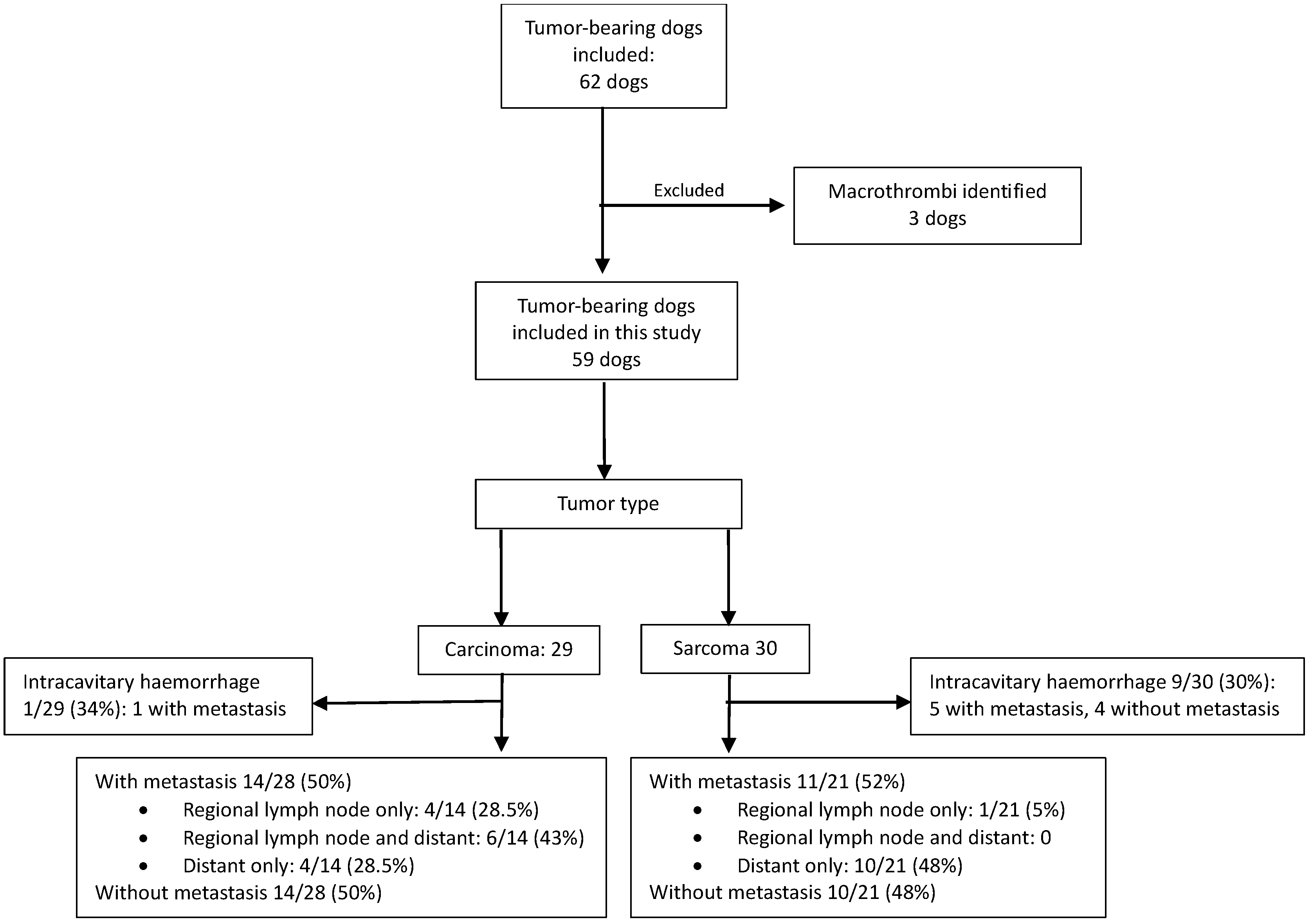
3.1. Study Population
3.2. Histopathological Evidence of Inflammation Associated with Tumors and Inflammation Unrelated to Tumors
3.3. Erythrocyte and Platelet Characteristics Compared Between Tumor-Bearing Dogs Without Intracavitary Hemorrhage (n = 49) and Healthy Unaffected Dogs (n = 20)
3.4. Erythrocyte and Platelet Characteristics Compared Between Tumor-Bearing Dogs with Intracavitary Hemorrhage (n = 10) and Healthy Unaffected Dogs (n = 20)
3.5. Erythrocyte and Platelet Characteristics Compared in Tumor-Bearing Dogs, Without Intracavitary Hemorrhage, with Sarcoma (n = 21) and with Carcinoma (n = 28)
3.6. Erythrocyte and Platelet Characteristics in Tumor-Bearing Dogs Without Intracavitary Hemorrhage, with (n = 25) and Without (n = 24) Metastasis
4. Discussion
5. Conclusions
Author Contributions
Funding
Institutional Review Board Statement
Informed Consent Statement
Data Availability Statement
Conflicts of Interest
Abbreviations
| ARC | Absolute reticulocyte count |
| CBC | Complete blood count |
| CHr | Reticulocyte hemoglobin content |
| CRP | C-reactive protein |
| HCT | Hematocrit |
| HGB | Hemoglobin |
| IL-1 | Interleukin 1 |
| IL-1α | Interleukin 1 alpha |
| IL-2 | Interleukin 2 |
| IL-6 | Interleukin 6 |
| MCHC | Mean cell hemoglobin concentration |
| MCV | Mean cell volume |
| MCVr | Reticulocyte mean corpuscular volume |
| MPC | Mean platelet component concentration |
| MPM | Mean platelet mass |
| MPV | Mean platelet volume |
| PCT | Plateletcrit |
| PCDW | Platelet component distribution width |
| PDW | Platelet volume distribution width |
| PLT | Platelet concentration |
| PMDW | Platelet dry mass distribution width |
| PTHR1 | Parathyroid hormone receptor 1 |
| RBC | Red blood cell count/concentration |
| RET% | Reticulocyte percentage |
| RDW | Erythrocyte distribution width |
| SAA | Serum amyloid A |
| TNF-α | Tumor necrosis factor alpha |
Appendix A

| Median Age in Years (IQR) | Sex | Breeds | |
|---|---|---|---|
| Tumor-bearing Dogs | 11 (9.0–13.6) | FI: 20 FS: 16 MI: 9 MN:14 | Mixed breed (8), German shepherd dog (7), Jack Russel terrier (7), Boerboel (6), dachshund (5), Labrador retriever (5), cocker spaniel (2), fox terrier (2), American pitbull terrier (2), pug (2), Staffordshire bull terrier (2), and one each of beagle, border collie, bullmastiff, rottweiler, Scottish terrier, bull terrier, Pekingese, Pomeranian, standard poodle, Great Dane, Irish terrier |
| Healthy unaffected dogs | 10.25 years (9.3–11.9) | FS: 9 MN: 9 MI: 2 | Mixed breed (8), Jack Russel terrier (4), dachshund (2) and one each of basset hound, German shepherd dog, bull terrier, Staffordshire bull terrier, miniature French poodle, Pekingese |
| Primary Tumor | Secondary Tumor | Tertiary/Quaternary Tumor or Concurrent Inflammation Identified |
|---|---|---|
| Sarcomas | ||
| Subcutaneous soft tissue sarcoma | Focal hepatic cystadenoma | |
| Splenic hemangiosarcoma | Round cell tumor of the face | Multifocal, moderate, acute to subacute renal tubular and medullary necrosis |
| Right atrial hemangiosarcoma | Focal hepatocellular adenoma | Focal adrenal cortical nodular hyperplasia |
| Splenic & right atrial hemangiosarcoma | Pheochromocytoma | |
| Hepatic hemangiosarcoma | Adrenal cortical carcinoma | |
| Hepatic hemangiosarcoma | Focal chronic-active pyogranulomatous aortic lesion with intralesional Spirocerca lupi | |
| Cutaneous hemangiosarcoma | Pulmonary bronchoalveolar carcinoma | Pheochromocytoma Moderate chronic lympho-histiocytic interstitial nephritis Bacterial cystitis |
| Cutaneous hemangiosarcoma | Early splenic stromal sarcoma | Renal adenomas Mild granulomatous cholangiohepatitis |
| Subcutaneous hemangiosarcoma | Thyroid follicular carcinoma | |
| Mammary osteosarcoma | Mammary mixed-type carcinoma | Multifocal adrenal cortical adenomas |
| Mammary osteosarcoma | Bacterial cystitis | |
| Maxillary osteosarcoma | Compact thyroid carcinoma of the ascending aorta | |
| Mandibular osteosarcoma | Pulmonary squamous cell carcinoma (primary organ not identified) | |
| Splenic stromal sarcoma | Simple mammary intraductal papillary carcinoma | |
| Muscle soft tissue sarcoma | Subcutaneous hemangioma | Bacterial prostatic abscess & cystitis |
| Cutaneous soft tissue sarcoma | Suspected indolent lymphoma in part of the duodenal lymph node | |
| Carcinomas | ||
| Complex mammary carcinoma | Simple tubulopapillary mammary carcinoma | Bronchoalveolar adenoma Adrenal cortical adenoma |
| Complex mammary carcinoma | Metastatic melanoma (metastasis to inguinal ln and liver), source unknown | Hepatocellular adenoma |
| Complex mammary carcinoma | Oesophageal osteosarcoma (S. lupi) | Bacterial cystitis |
| Simple tubular mammary carcinoma | Subcutaneous soft tissue sarcoma (hindlimb) | |
| Simple tubular mammary carcinoma | Complex mammary carcinoma | Subcutaneous mast cell tumor Subcutaneous hemangioma Benign fibrolipoma Bacterial cystitis |
| Mixed mammary carcinoma | Compact thyroid carcinoma | Adrenal cortical carcinoma |
| Mixed mammary carcinoma | Cutaneous melanoma (metastatic to inguinal lymph node) | Bacterial cystitis |
| Anaplastic mammary carcinoma | Complex mammary carcinoma | |
| Simple tubulopapillary mammary carcinoma | Focal scirrhous pulmonary carcinoma | Severe chronic, multifocal to extensive, renal interstitial fibrosis & lymphoplasmacytic nephritis Bacterial cystitis |
| Simple tubular mammary carcinoma | Benign oesophageal Spirocerca lupi nodule | |
| Mixed-type carcinoma & simple tubulopapillary mammary carcinoma | Spirocerca lupi migratory track: severe mural inflammation of the aorta | |
| Mammary ductal carcinoma | Pheochromocytoma | Mild chronic interstitial lymphocytic and fibrosing nephritis |
| Sinonasal transitional carcinoma | Benign oesophageal Spirocerca lupi nodule | |
| Squamous cell carcinoma | Multifocal granulomatous nephritis | |
| Squamous cell carcinoma | Chronic interstitial nephritis and a chronic membranous glomerulopathy | |
| Solid scirrhous prostatic carcinoma | Focal, subacute, mild aspiration pneumonia | |
References
- Bray, F.; Laversanne, M.; Sung, H.; Ferlay, J.; Siegel, R.L.; Soerjomataram, I.; Jemal, A. Global cancer statistics 2022: GLOBOCAN estimates of incidence and mortality worldwide for 36 cancers in 185 countries. CA Cancer J. Clin. 2024, 74, 229–263. [Google Scholar] [CrossRef] [Scilit]
- Qin, Y.; Wang, P.; Huang, Z.; Huang, G.; Tang, J.; Guo, Y.; Huang, P.; Lai, Z.; Lin, F. The value of red cell distribution width in patients with ovarian cancer. Medicine 2017, 96, e6752. [Google Scholar] [CrossRef] [Scilit] [PubMed]
- Voutsadakis, I.A. Thrombocytosis as a prognostic marker in gastrointestinal cancers. World J. Gastrointest. Oncol. 2014, 6, 34–40. [Google Scholar] [CrossRef] [Scilit]
- Fu, S.; Liu, L.; Zhang, X.; Liu, Z.P.; Wang, R.T. Platelet indices in laryngeal cancer. Cancer Biomark. 2018, 21, 675–680. [Google Scholar] [CrossRef] [Scilit] [PubMed]
- Al-Khan, A.A.; Nimmo, J.S.; Tayebi, M.; Ryan, S.D.; Simcock, J.O.; Tarzi, R.; Kuntz, C.A.; Saad, E.S.; Day, M.J.; Richardson, S.J.; et al. Parathyroid hormone receptor 1 (PTHR1) is a prognostic indicator in canine osteosarcoma. Sci. Rep. 2020, 10, 1564. [Google Scholar] [CrossRef] [Scilit]
- Nishimori, T.; Hanazono, K.; Matsuda, K.; Kawamura, Y.; Kadosawa, T.; Endo, Y.; Uchide, T. Prognostic role of ΔNp63 expression in canine transitional cell carcinoma of the urinary bladder. Open Vet. J. 2021, 11, 700–706. [Google Scholar] [CrossRef] [Scilit] [PubMed]
- Masyr, A.R.; Rendahl, A.K.; Winter, A.L.; Borgatti, A.; Modiano, J.F. Retrospective evaluation of thrombocytopenia and tumor stage as prognostic indicators in dogs with splenic hemangiosarcoma. J. Am. Vet. Med. Assoc. 2021, 258, 630–637. [Google Scholar] [CrossRef] [Scilit]
- Aleksandrowicz, K.; Polityńska, B.; Kruszewska, J.; Radziwon, P.; Wojtukiewicz, A.M.; Honn, K.V.; Wojtukiewicz, M.Z. Thrombocytosis in cancer patients: When more is not better-in fact, the opposite. Cancer Metastasis Rev. 2026, 45, 1. [Google Scholar] [CrossRef] [Scilit]
- Ge, W.; Xie, J.; Chang, L. Elevated red blood cell distribution width predicts poor prognosis in patients with oral squamous cell carcinoma. Cancer Manag. Res. 2018, 10, 3611–3618. [Google Scholar] [CrossRef] [Scilit]
- Xie, X.; Zeng, X.; Cao, S.; Hu, X.; Shi, Q.; Li, D.; Zhou, S.; Gu, P.; Zhang, Z. Elevated pretreatment platelet distribution width and platelet count predict poor prognosis in nasopharyngeal carcinoma. Oncotarget 2017, 8, 106089–106097. [Google Scholar] [CrossRef] [Scilit]
- Del Giudice, M.; Gangestad, S.W. Rethinking IL-6 and CRP: Why they are more than inflammatory biomarkers, and why it matters. Brain Behav. Immun. 2018, 70, 61–75. [Google Scholar] [CrossRef] [Scilit]
- Zhang, Y.; Zhang, J.; Sheng, H.; Li, H.; Wang, R. Acute phase reactant serum amyloid A in inflammation and other diseases. Adv. Clin. Chem. 2019, 90, 25–80. [Google Scholar]
- Weiss, G.; Ganz, T.; Goodnough, L.T. Anemia of inflammation. Blood 2019, 133, 40–50. [Google Scholar] [CrossRef] [Scilit]
- Woolcock, A.D.; Keenan, A.; Cheung, C.; Christian, J.A.; Moore, G.E. Thrombocytosis in 715 Dogs (2011–2015). J. Vet. Intern. Med. 2017, 31, 1691–1699. [Google Scholar] [CrossRef] [Scilit] [PubMed]
- Kociba, G.J. Erythrocytes. Vet. Clin. N. Am. Small Anim. Pract. 1989, 19, 627–635. [Google Scholar] [CrossRef] [Scilit] [PubMed]
- Pazzi, P.; Fosgate, G.T.; Rixon, A.; Hanekom, J.; Kristensen, A.T.; Goddard, A. A prospective evaluation of the prevalence of thromboemboli and associated hemostatic dysfunction in dogs with carcinoma or sarcoma. J. Vet. Intern. Med. 2023, 37, 1848–1863. [Google Scholar] [CrossRef] [Scilit]
- Weiss, D.J. Uniform evaluation and semiquantitative reporting of hematologic data in veterinary laboratories. Vet. Clin. Pathol. 1984, 13, 27–31. [Google Scholar] [CrossRef] [Scilit]
- Hillström, A.; Hagman, R.; Tvedten, H.; Kjelgaard-Hansen, M. Validation of a commercially available automated canine-specific immunoturbidimetric method for measuring canine C-reactive protein. Vet. Clin. Pathol. 2014, 43, 235–243. [Google Scholar] [CrossRef] [Scilit] [PubMed]
- Christensen, M.; Jacobsen, S.; Ichiyanagi, T.; Kjelgaard-Hansen, M. Evaluation of an automated assay based on monoclonal anti-human serum amyloid A (SAA) antibodies for measurement of canine, feline, and equine SAA. Vet. J. 2012, 194, 332–337. [Google Scholar] [CrossRef] [Scilit]
- Bauer, N.; Moritz, A. Characterisation of changes in the haemostasis system in dogs with thrombosis. J. Small Anim. Pract. 2013, 54, 129–136. [Google Scholar] [CrossRef] [Scilit]
- Childress, M.O. Hematologic abnormalities in the small animal cancer patient. Vet. Clin. N. Am. Small Anim. Pract. 2012, 42, 123–155. [Google Scholar] [CrossRef] [Scilit]
- Rašková, M.; Lacina, L.; Kejík, Z.; Venhauerová, A.; Skaličková, M.; Kolář, M.; Jakubek, M.; Rosel, D.; Smetana, K.; Brábek, J. The Role of IL-6 in Cancer Cell Invasiveness and Metastasis-Overview and Therapeutic Opportunities. Cells 2022, 11, 3698. [Google Scholar] [CrossRef] [Scilit]
- Rébé, C.; Ghiringhelli, F. Interleukin-1β and Cancer. Cancers 2020, 12, 1791. [Google Scholar] [CrossRef] [Scilit]
- Mihara, M.; Ohsugi, Y.; Kishimoto, T. Tocilizumab, a humanized anti-interleukin-6 receptor antibody, for treatment of rheumatoid arthritis. Open Access Rheumatol. 2011, 3, 19–29. [Google Scholar] [CrossRef] [Scilit]
- Luan, Y.Y.; Yao, Y.M. The Clinical Significance and Potential Role of C-Reactive Protein in Chronic Inflammatory and Neurodegenerative Diseases. Front. Immunol. 2018, 9, 1302. [Google Scholar] [CrossRef] [Scilit]
- Bessman, J.D. Reticulocytes. In Clinical Methods: The History, Physical, and Laboratory Examinations, 3rd ed.; Butterworth Publishers, Reed Publishing: Boston, MA, USA, 1990. [Google Scholar]
- Moritz, A.; Fickenscher, Y.; Meyer, K.; Failing, K.; Weiss, D.J. Canine and feline hematology reference values for the ADVIA 120 hematology system. Vet. Clin. Pathol. 2004, 33, 32–38. [Google Scholar] [CrossRef] [Scilit] [PubMed]
- Aapro, M.; Österborg, A.; Gascón, P.; Ludwig, H.; Beguin, Y. Prevalence and management of cancer-related anaemia, iron deficiency and the specific role of i.v. iron. Ann. Oncol. 2012, 23, 1954–1962. [Google Scholar] [CrossRef] [Scilit] [PubMed]
- Radakovich, L.B.; Santangelo, K.S.; Olver, C.S. Reticulocyte hemoglobin content does not differentiate true from functional iron deficiency in dogs. Vet. Clin. Pathol. 2015, 44, 511–518. [Google Scholar] [CrossRef] [Scilit] [PubMed]
- Steinberg, J.D.; Olver, C.S. Hematologic and biochemical abnormalities indicating iron deficiency are associated with decreased reticulocyte hemoglobin content (CHr) and reticulocyte volume (rMCV) in dogs. Vet. Clin. Pathol. 2005, 34, 23–27. [Google Scholar] [CrossRef] [Scilit]
- Schaefer, D.M.; Stokol, T. Retrospective study of reticulocyte indices as indicators of iron-restricted erythropoiesis in dogs with immune-mediated hemolytic anemia. J. Vet. Diagn. Investig. 2016, 28, 304–308. [Google Scholar] [CrossRef] [Scilit]
- Wollmann, M.; Gerzson, B.M.; Schwert, V.; Figuera, R.W.; Ritzel, G.d.O. Reticulocyte maturity indices in iron deficiency anemia. Rev. Bras. Hematol. Hemoter. 2014, 36, 25–28. [Google Scholar] [CrossRef] [Scilit]
- Warry, E.; Bohn, A.; Emanuelli, M.; Thamm, D.; Lana, S. Disease distribution in canine patients with acanthocytosis: 123 cases. Vet. Clin. Pathol. 2013, 42, 465–470. [Google Scholar] [CrossRef] [Scilit] [PubMed]
- Hammer, A.S.; Couto, C.G.; Swardson, C.; Getzy, D. Hemostatic abnormalities in dogs with hemangiosarcoma. J. Vet. Intern. Med. 1991, 5, 11–14. [Google Scholar] [CrossRef] [Scilit]
- Rebar, A.H.; Hahn, F.F.; Halliwell, W.H.; DeNicola, D.B.; Benjamin, S.A. Microangiopathic hemolytic anemia associated with radiation-induced hemangiosarcomas. Vet. Pathol. 1980, 17, 443–454. [Google Scholar] [CrossRef] [Scilit]
- Hirsch, V.M.; Jacobsen, J.; Mills, J.H. A retrospective study of canine hemangiosarcoma and its association with acanthocytosis. Can. Vet. J. 1981, 22, 152–155. [Google Scholar] [PubMed]
- Lee, S.Y.; Ju, M.K.; Jeon, H.M.; Jeong, E.K.; Lee, Y.J.; Kim, C.H.; Park, H.G.; Han, S.I.; Kang, H.S. Regulation of Tumor Progression by Programmed Necrosis. Oxid. Med. Cell. Longev. 2018, 2018, 3537471. [Google Scholar] [CrossRef] [Scilit]
- Cheney, A.; Woolcock, A.D.; Mukhopadhyay, A.; Knapp, D.; Moore, G.E. Interleukin-6 and thrombopoietin concentrations in dogs with carcinoma with and without thrombocytosis. J. Vet. Intern. Med. 2022, 36, 227–233. [Google Scholar] [CrossRef] [Scilit] [PubMed]
- Brissot, E.; Troadec, M.; Loréal, O.; Brissot, P. Iron and platelets: A subtle, under-recognized relationship. Am. J. Hematol. 2021, 96, 1008–1016. [Google Scholar] [CrossRef] [Scilit]
- Jimenez, K.; Leitner, F.; Leitner, A.; Scharbert, G.; Schwabl, P.; Kramer, A.M.; Krnjic, A.; Friske, J.; Helbich, T.; Evstatiev, R.; et al. Iron deficiency-induced thrombocytosis increases thrombotic tendency in rats. Haematologica 2021, 106, 782–794. [Google Scholar] [CrossRef] [Scilit]
- Handtke, S.; Thiele, T. Large and small platelets-(When) do they differ? J. Thromb. Haemost. 2020, 18, 1256–1267. [Google Scholar] [CrossRef] [Scilit]
- Braun, A.; Anders, H.J.; Gudermann, T.; Mammadova-Bach, E. Platelet-Cancer Interplay: Molecular Mechanisms and New Therapeutic Avenues. Front. Oncol. 2021, 11, 665534. [Google Scholar] [CrossRef] [Scilit] [PubMed]
- Burstein, S.A. Effects of interleukin 6 on megakaryocytes and on canine platelet function. Stem Cells 1994, 12, 386–393. [Google Scholar] [CrossRef] [Scilit]
- Nishimura, S.; Nagasaki, M.; Kunishima, S.; Sawaguchi, A.; Sakata, A.; Sakaguchi, H.; Ohmori, T.; Manabe, I.; Italiano, J.E., Jr.; Ryu, T.; et al. IL-1α induces thrombopoiesis through megakaryocyte rupture in response to acute platelet needs. J. Cell Biol. 2015, 209, 453–466. [Google Scholar] [CrossRef] [Scilit] [PubMed]
- Queiroga, F.P.; Marques, A.M.; Gregório, H.; Petrucci, G.N. Canine Splenic Hemangiosarcoma: Biological Behavior, Clinical Challenges and Therapeutic Limitations. Animals 2026, 16, 778. [Google Scholar] [CrossRef] [Scilit]
- Hernández Salas, A.; Páez Bonelo, A.M.; Moscoso Gama, J.M.; Méndez Hurtado, W.A. Review of canine hemangiosarcoma: An aggressive and lethal neoplasm. Vet. México OA 2023, 10. [Google Scholar] [CrossRef] [Scilit]
- Yazawa, D.; Shimada, M.; Kanno, N.; Suzuki, S.; Yogo, T.; Harada, Y.; Hara, Y. Three cases of dogs with osteosarcoma of the forelimb treated with liquid nitrogen for limb-sparing surgery using autologous bone. J. Vet. Med. Sci. 2024, 86, 700–707. [Google Scholar] [CrossRef] [Scilit] [PubMed]
- Souza, C.H.d.M.; Toledo-Piza, E.; Amorin, R.; Barboza, A.; Tobias, K.M. Inflammatory mammary carcinoma in 12 dogs: Clinical features, cyclooxygenase-2 expression, and response to piroxicam treatment. Can. Vet. J. 2009, 50, 506–510. [Google Scholar]
- Dennis, M.M.; McSporran, K.D.; Bacon, N.J.; Schulman, F.Y.; Foster, R.A.; Powers, B.E. Prognostic factors for cutaneous and subcutaneous soft tissue sarcomas in dogs. Vet. Pathol. 2011, 48, 73–84. [Google Scholar] [CrossRef] [Scilit]
- Dos Santos, A.; Lamego, É.C.; Eisenhardt, L.M.; de Vargas, I.; Flores, M.M.; Fighera, R.A.; Kommers, G.D. Prevalence and anatomopathological characterization of cutaneous squamous cell carcinomas with regional and distant metastases in dogs and cats: 20 cases (1985–2020). Vet. Comp. Oncol. 2023, 21, 291–301. [Google Scholar] [CrossRef] [Scilit]
- Withers, S.S.; Skorupski, K.A.; York, D.; Choi, J.W.; Woolard, K.D.; Laufer-Amorim, R.; Sparger, E.E.; Rodriguez, C.O.; McSorley, S.J.; Monjazeb, A.M.; et al. Association of macrophage and lymphocyte infiltration with outcome in canine osteosarcoma. Vet. Comp. Oncol. 2019, 17, 49–60. [Google Scholar] [CrossRef] [Scilit]
- Kirpensteijn, J.; Kik, M.; Rutteman, G.R.; Teske, E. Prognostic significance of a new histologic grading system for canine osteosarcoma. Vet. Pathol. 2002, 39, 240–246. [Google Scholar] [CrossRef] [Scilit] [PubMed]
- Hayburn, R.; Chung, D.; Gupta, A.; Sills, S.; Wu, J.D.; Ambrus, A.; Jing, W.; Ng, J.; Penaloza-MacMaster, P.; Selmek, A.; et al. The Immune Landscape of Canine Soft Tissue Sarcomas as a Model for Human Soft Tissue Sarcomas. Cancers 2025, 17, 3860. [Google Scholar] [CrossRef] [Scilit] [PubMed]
- Estrela-Lima, A.; Araújo, M.S.; Costa-Neto, J.M.; Teixeira-Carvalho, A.; Barrouin-Melo, S.M.; Cardoso, S.V.; A Martins-Filho, O.; Serakides, R.; Cassali, G.D. Immunophenotypic features of tumor infiltrating lymphocytes from mammary carcinomas in female dogs associated with prognostic factors and survival rates. BMC Cancer 2010, 10, 256. [Google Scholar] [CrossRef] [Scilit]
- Monteiro, L.N.; Dos Reis, D.C.; Salgado, B.S.; Cassali, G.D. Clinical significance and prognostic role of tumor-associated macrophages infiltration according to histologic location in canine mammary carcinomas. Res. Vet. Sci. 2021, 135, 329–334. [Google Scholar] [CrossRef] [Scilit]
- Flecher, M.C.; Balabram, D.; Salles, Y.A.; Souza, F.R.; Estrela-Lima, A.; Nakagaki, K.Y.; Cassali, G.D. Evaluation of immunophenotype and inflammation in canine mammary neoplasms with solid arrangement. J. Comp. Pathol. 2024, 213, 1–9. [Google Scholar] [CrossRef] [Scilit]
- Yokota, S.; Kaji, K.; Yonezawa, T.; Momoi, Y.; Maeda, S. CD204+ tumor-associated macrophages are associated with clinical outcome in canine pulmonary adenocarcinoma and transitional cell carcinoma. Vet. J. 2023, 296–297, 105992. [Google Scholar] [CrossRef] [Scilit] [PubMed]
- Avallone, G.; Rasotto, R.; Chambers, J.K.; Miller, A.D.; Behling-Kelly, E.; Monti, P.; Berlato, D.; Valenti, P.; Roccabianca, P. Review of Histological Grading Systems in Veterinary Medicine. Vet. Pathol. 2021, 58, 809–828. [Google Scholar] [CrossRef] [Scilit]
- Marcinowska, A.; Horta, R.D.S.; Queiroga, F.; Giuliano, A. Canine lung carcinoma-A descriptive review. Front. Vet. Sci. 2024, 11, 1464659. [Google Scholar] [CrossRef] [Scilit]
- Polak, K.Z.; Schaffer, P.; Donaghy, D.; Zenk, M.C.; Olver, C.S. Iron, hepcidin, and microcytosis in canine hepatocellular carcinoma. Vet. Clin. Pathol. 2022, 51, 208–215. [Google Scholar] [CrossRef] [Scilit]
- Shiga, A.; Shirota, K.; Nomura, Y. Immunohistochemical and ultrastructural studies on the sinusoidal lining cells of canine hepatocellular carcinoma. J. Vet. Med. Sci. 1996, 58, 909–914. [Google Scholar] [CrossRef] [Scilit]
- Olivia, M.; Patania, J.M.C.; Donald, M. Canine Hepatocellular Pathology Quick Reference Guide (PQRG): Veterinary Cancer Guidelines and Protocols; 2024 Hepatocellular QRG 1.0. Available online: https://vcgp.org/documents/2025/08/canine-hepatocellular-pqrg.pdf/ (accessed on 30 January 2026).
- Chikazawa, S.; Hori, Y.; Hoshi, F.; Kanai, K.; Ito, N.; Higuchi, S. Hyperferritinemia in dogs with splenic hemangiosarcoma. J. Vet. Med. Sci. 2013, 75, 1515–1518. [Google Scholar] [CrossRef] [Scilit]
- De Vico, G.; Martano, M.; Maiolino, P.; Carella, F.; Leonardi, L. Expression of transferrin receptor-1 (TFR-1) in canine osteosarcomas. Vet. Med. Sci. 2020, 6, 272–276. [Google Scholar] [CrossRef] [Scilit]
- Power, K.; Leandri, R.; Federico, G.; De Vico, G.; Leonardi, L. Ferritinophagy: A possible new iron-related metabolic target in canine osteoblastic osteosarcoma. Front. Vet. Sci. 2025, 12, 1546872. [Google Scholar] [CrossRef] [Scilit]
- Kazmierski, K.J.; Ogilvie, G.K.; Fettman, M.J.; Lana, S.E.; Walton, J.A.; Hansen, R.A.; Richardson, K.L.; Hamar, D.W.; Bedwell, C.L.; Andrews, G.; et al. Serum zinc, chromium, and iron concentrations in dogs with lymphoma and osteosarcoma. J. Vet. Intern. Med. 2001, 15, 585–588. [Google Scholar] [CrossRef]
- Caro, J.T.; Marín, L.M.; Iazbik, M.C.; Zaldivar-López, S.; Borghese, H.; Couto, C.G. Markers of iron metabolism in retired racing Greyhounds with and without osteosarcoma. Vet. Clin. Pathol. 2013, 42, 360–363. [Google Scholar] [CrossRef] [Scilit]
- Miller, A.G.; Morley, P.S.; Rao, S.; Avery, A.C.; Lana, S.E.; Olver, C.S. Anemia is associated with decreased survival time in dogs with lymphoma. J. Vet. Intern. Med. 2009, 23, 116–122. [Google Scholar] [CrossRef]
- Lallo, M.A.; Ferrarias, T.M.; Stravino, A.; Rodriguez, J.F.; Zucare, R.L. Hematologic abnormalities in dogs bearing mammary tumors. Rev. Bras. Ciênc. Vet. 2016, 23, 3–8. [Google Scholar] [CrossRef] [Scilit]
- Yang, N.Y.; Zheng, H.H.; Yu, C.; Ye, Y.; Xie, G.H. Diagnosis of Canine Tumours and the Value of Combined Detection of VEGF, P53, SF and NLRP3 for the Early Diagnosis of Canine Mammary Carcinoma. Animals 2024, 14, 1272. [Google Scholar] [CrossRef] [Scilit] [PubMed]
- Bulla, S.C.; Badial, P.R.; Bulla, C. Canine Cancer Cells Activate Platelets via the Platelet P2Y12 Receptor. J. Comp. Pathol. 2022, 192, 41–49. [Google Scholar] [CrossRef] [Scilit]
- Makielski, K.M.; Fox, L.E.; Johannes, C.M.; Rendahl, A.K.; Schulte, A.J.; Kim, J.H.; Husbands, B.D.; Walz, J.Z.; Henson, M.S.; Modiano, J.F.; et al. Evaluation of coated platelets, a subset of highly procoagulant platelets, in healthy dogs and dogs with neoplasia. Am. J. Vet. Res. 2022, 83, 1–9. [Google Scholar] [CrossRef] [Scilit] [PubMed]
- Lawrence, J.; Chang, Y.M.; Szladovits, B.; Davison, L.J.; Garden, O.A. Breed-specific hematological phenotypes in the dog: A natural resource for the genetic dissection of hematological parameters in a mammalian species. PLoS ONE 2013, 8, e81288. [Google Scholar] [CrossRef] [Scilit]
- Schneider, L.; Mischke, R. Platelet variables in healthy dogs: Reference intervals and influence of age, breed and sex. Comp. Clin. Pathol. 2016, 25, 1097–1106. [Google Scholar] [CrossRef] [Scilit]

| Primary Tumor | Metastatic Site of Primary Tumor |
|---|---|
| Sarcoma (30) | |
| Subcutaneous soft tissue sarcoma | Lung |
| Subcutaneous soft tissue sarcoma | N/A |
| Subcutaneous soft tissue sarcoma | N/A |
| Splenic hemangiosarcoma | Right atrium, liver |
| Splenic hemangiosarcoma * | Lymph node, liver, kidney, omentum |
| Splenic hemangiosarcoma * | N/A |
| Right atrial hemangiosarcoma * | Spleen, lungs |
| Right atrial hemangiosarcoma * | N/A |
| Splenic & right atrial hemangiosarcoma * | Liver, omentum |
| Splenic & right atrial hemangiosarcoma * | N/A |
| Hepatic hemangiosarcoma * | Omentum |
| Hepatic hemangiosarcoma * | Diaphragm, lungs |
| Cutaneous hemangiosarcoma | N/A |
| Cutaneous hemangiosarcoma | N/A |
| Subcutaneous hemangiosarcoma | Lungs |
| Muscle hemangiosarcoma | Liver, right atrium, lungs |
| Mammary osteosarcoma | Lymph node |
| Mammary osteosarcoma | Lungs |
| Oesophageal osteosarcoma | Aorta, liver, lungs |
| Oesophageal osteosarcoma | Kidney, liver, lung, caudal vena cava |
| Rib chondroblastic osteosarcoma | Lung |
| Maxillary osteosarcoma | N/A |
| Mandibular osteosarcoma | N/A |
| Mixed peri-orbital anaplastic sarcoma | Liver, pancreas, omentum, cardiac muscle, dermis/subcutis & muscle |
| Splenic stromal sarcoma | Liver |
| Muscle soft tissue sarcoma | N/A |
| Mixed mammary sarcoma | N/A |
| Hepatic spindle cell sarcoma * | N/A |
| Cutaneous soft tissue sarcoma | N/A |
| Mandibular soft tissue sarcoma | N/A |
| Carcinoma (29) | |
| Complex mammary carcinoma | Lymph node |
| Complex mammary carcinoma | Lymph node |
| Complex mammary carcinoma | N/A |
| Complex mammary carcinoma | N/A |
| Simple tubular mammary carcinoma | Lymph node |
| Simple tubular mammary carcinoma | Lungs |
| Simple tubular mammary carcinoma | N/A |
| Mixed mammary carcinoma | Lymph node |
| Mixed mammary carcinoma | Lymph node, lungs |
| Anaplastic mammary carcinoma | Lymph node, lungs |
| Simple solid mammary carcinoma | N/A |
| Simple tubulopapillary mammary carcinoma | N/A |
| Mammary ductal carcinoma | N/A |
| Mixed type carcinoma & simple tubulopapillary mammary carcinoma | N/A |
| Spindle cell mammary carcinoma | N/A |
| Cutaneous squamous cell carcinoma | N/A |
| Cutaneous squamous cell carcinoma | N/A |
| Hepatocellular carcinoma * | Liver |
| Hepatocellular carcinoma | N/A |
| Apocrine gland adenocarcinoma (anal sac) | Lymph node, spleen, adrenal gland, lungs |
| Carcinoma (papillary & solid)—source unknown | Lungs |
| Tubular mesothelioma | Lungs |
| Anaplastic cholangiocellular carcinoma | Lymph node, liver, peri-pancreatic connective tissue, bronchial lnn., lungs |
| Thyroid microfollicular carcinoma | Rib |
| Urothelial carcinoma | Lymph node, rib, gastric and tracheobronchial lymph node |
| Pulmonary carcinomatosis | Lymph node, Adrenal gland, liver, kidney |
| Pulmonary carcinoma (tubulopapillary type) | N/A |
| Sinonasal transitional carcinoma | N/A |
| Solid scirrhous prostatic carcinoma | N/A |
| Variable | Tumor-Bearing Dogs Without Intracavitary Hemorrhage * (n = 49) | Healthy Unaffected Dogs * (n = 20) | p Value † | BH Corrected p Value | CRP rs to Variable (p Value; BH Corrected p Value) | SAA rs to Variable (p Value; BH Corrected p Value) |
|---|---|---|---|---|---|---|
| Complete blood count | ||||||
| Erythrocytes | ||||||
| Hemoglobin (g/L) | 132 (100, 148) | 175 (167, 183) | <0.001 | 0.003 | −0.45 (0.001; 0.009) | −0.42 (0.003; 0.025) |
| Red blood cell count (×1012/L) | 5.73 (4.74, 6.75) | 7.93 (7.16, 8.49) | <0.001 | 0.003 | −0.41 (0.003; 0.012) | −0.38 (0.007; 0.030) |
| Hematocrit (L/L) | 0.39 (0.31, 0.44) | 0.53 (0.50, 0.55) | <0.001 | 0.003 | −0.44 (0.002; 0.011) | −0.40 (0.005; 0.028) |
| Mean cell volume (fL) | 67.85 (65.8, 70.1) | 68.6 (66.2, 71.8) | 0.40 | 0.43 | −0.11 (0.46; 0.49) | −0.09 (0.53; 0.56) |
| Mean cell hemoglobin concentration (g/dL) | 33.0 (32.6, 33.7) | 33.4 (32.8, 33.5) | 0.32 | 0.38 | - | - |
| Red cell distribution width (fL) | 13.8 (13.1, 15.4) | 13.6 (13.05, 13.85) | 0.151 | 0.22 | 0.27 (0.06; 0.13) | 0.30 (0.039; 0.11) |
| Reticulocyte percentage (%) | 1.8 (1.1, 2.8) | 0.95 (0.63, 1.08) | <0.001 | 0.003 | 0.3 (0.038; 0.09) | 0.31 (0.033; 0.11) |
| Absolute reticulocyte count (×109/L) | 94.8 (66.9, 159) | 67.6 (48.4, 84.6) | 0.02 | 0.04 | 0.15 (0.31; 0.40) | 0.17 (0.25; 0.43) |
| Reticulocyte hemoglobin content (pg/cell) | 24.7 (23.1, 25.4) | 26.0 (24.8, 26.5) | 0.01 | 0.024 | −0.42 (0.001; 0.009) | −0.48 (<0.001; 0.017) |
| Reticulocyte mean cell volume (fL) | 87.15 (83.1, 90.1) | 88 (85.5, 92.3) | 0.34 | 0.38 | −0.25 (0.09; 0.17) | −0.24 (0.10; 0.19) |
| Erythrocyte morphology | ||||||
| Anisocytosis | 2 (1, 2) | 1 (1, 1) | 0.001 | 0.003 | - | - |
| Polychromasia | 1 (0, 1) | 0 (0, 0) | 0.011 | 0.024 | - | - |
| Codocytes | 0 (0, 1) | 0 (0, 0) | 0.029 | 0.051 | - | - |
| Macrocytosis | 1 (0, 1) | 0 (0, 1) | 0.052 | 0.08 | - | - |
| Platelets | ||||||
| Platelet concentration (×109/L) | 400 (288, 586) | 268 (228, 299) | <0.001 | 0.003 | 0.09 (0.56; 0.56) | 0.09 (0.55; 0.55) |
| Mean platelet volume (fL) | 12.3 (10.5, 13) | 10.9 (10.4, 12.1) | 0.22 | 0.30 | 0.32 (0.026; 0.07) | 0.24 (0.09, 0.19) |
| Platelet volume distribution width (%) | 64.0 (60.5, 67.9) | 57.2 (52.2, 62.3) | <0.01 | 0.024 | 0.27 (0.12; 0.20) | 0.13 (0.38; 0.50) |
| Plateletcrit (%) | 0.47 (0.33, 0.61) | 0.3 (0.25, 0.37) | <0.01 | 0.024 | 0.17 (0.23; 0.36) | 0.15 (0.29; 0.43) |
| Mean platelet component (g/dL) | 21.2 (19.9, 22.3) | 21.1 (20.1, 21.6) | 0.86 | 0.89 | −0.13 (0.35; 0.40) | −0.15 (0.31; 0.44) |
| Platelet component distribution width (g/dL) | 6.5 (5.5, 7.2) | 6 (4.4, 6.85) | 0.055 | 0.08 | −0.15 (0.31; 0.40) | −0.11 (0.46; 0.53) |
| Mean platelet mass (pg) | 2.12 (1.97, 2.35) | 2.12 (2.03, 2.315) | 0.91 | 0.91 | 0.16 (0.28; 0.40) | 0.13 (0.39; 0.49) |
| Platelet dry mass distribution width (pg) | 0.88 (0.79, 1.0) | 0.8 (0.76, 0.87) | 0.052 | 0.08 | 0.34 (0.018; 0.06) | 0.29 (0.046; 0.11) |
| Platelet Aggregation | 0 (0, 1) | 1 (0, 2) | 0.28 | 0.35 | - | - |
| Acute phase proteins | ||||||
| C-reactive protein (mg/L) | 65.0 (42.8, 142) | <10 (<10, <10) | <0.001 | 0.003 | - | - |
| Serum amyloid A (mg/L) | 25.9 (9.67, 122) | <2 (<2, <2) | <0.001 | 0.003 | - | - |
| Variable | Tumor-Bearing Dogs with Intracavitary Hemorrhage * (n = 10) | Healthy Unaffected Dogs * (n = 20) | p Value † | BH Corrected p Value |
|---|---|---|---|---|
| Complete blood count | ||||
| Erythrocytes | ||||
| Hemoglobin (g/L) | 61.5 (49, 104) | 175 (169, 183) | <0.001 | 0.002 |
| Red blood cell count (×1012/L) | 2.58 (2.29, 4.29) | 7.93 (7.16, 8.49) | <0.001 | 0.002 |
| Hematocrit (%) | 0.20 (0.17, 0.31) | 0.53 (0.50, 0.55) | <0.01 | 0.015 |
| Mean cell volume (fL) | 73.3 (68.1, 77.4) | 68.6 (66.2, 71.8) | 0.14 | 0.16 |
| Mean cell hemoglobin concentration (g/dL) | 32.4 (29.1, 33.1) | 33.4 (32.8, 33.5) | 0.036 | 0.044 |
| Red cell distribution width (fL) | 17.4 (15.5, 19.4) | 13.6 (13.0, 13.8) | <0.001 | 0.002 |
| Reticulocyte percentage (%) | 10.6 (7.4, 21.1) | 0.95 (0.65, 1.08) | <0.001 | 0.002 |
| Absolute reticulocyte count (×109/L) | 289 (205, 447) | 67.6 (48.4, 84.6) | <0.001 | 0.002 |
| Reticulocyte hemoglobin content (pg/cell) | 24.6 (23.7, 26.1) | 26 (24.8, 26.5) | 0.33 | 0.35 |
| Reticulocyte mean cell volume (fL) | 93.0 (86.3, 101) | 88 (85.5, 92.3) | 0.09 | 0.11 |
| Erythrocyte morphology | ||||
| Anisocytosis | 2.5 (2, 3) | 1 (1, 1) | <0.001 | 0.002 |
| Polychromasia | 1 (1, 2) | 0 (0, 0) | <0.001 | 0.002 |
| Codocytes | 0 (0, 1) | 0 (0, 0) | 0.008 | 0.013 |
| Acanthocytes | 0 (0, 1) | 0 (0, 0) | 0.031 | 0.04 |
| Hypochromasia | 0.5 (0, 1) | 0 (0, 0) | 0.002 | 0.004 |
| Keratocytes | 0 (0, 1) | 0 (0, 0) | 0.03 | 0.04 |
| Leptocytes | 0 (0, 1) | 0 (0, 0) | 0.008 | 0.013 |
| Macrocytosis | 1 (1, 1) | 0 (0, 1) | 0.008 | 0.013 |
| Howell-Jolly bodies | 1 (1, 1) | 0 (0, 0) | <0.001 | 0.002 |
| Schistocytes | 0 (0, 1) | 0 (0, 0) | 0.03 | 0.04 |
| Platelets | ||||
| Platelet concentration (×109/L) | 131 (49, 190) | 268 (228, 299) | <0.001 | 0.002 |
| Mean platelet volume (fL) | 17.9 (14.2, 19.5) | 10.9 (10.4, 12.1) | <0.001 | 0.002 |
| Platelet volume distribution width (%) | 68.4 (66.4, 70.4) | 57.2 (52.2, 62.3) | <0.001 | 0.002 |
| Plateletcrit (%) | 0.19 (0.09, 0.27) | 0.3 (0.25, 0.37) | 0.008 | 0.013 |
| Mean platelet component (g/dL) | 21.0 (20.2, 22.1) | 21.1 (20.1, 21.6) | 0.89 | 0.89 |
| Platelet component distribution width (g/dL) | 6.5 (6, 6.8) | 6 (4.4, 6.8) | 0.20 | 0.22 |
| Mean platelet mass (pg) | 2.89 (2.46, 3.07) | 2.12 (2.03, 2.31) | <0.001 | 0.002 |
| Platelet dry mass distribution width (pg) | 1.29 (1.23, 1.46) | 0.8 (0.76, 0.87) | <0.001 | 0.002 |
| Platelet Aggregation | 0 (0, 3) | 1 (0, 2) | 0.38 | 0.59 |
| Platelet morphology | ||||
| Giant Platelets | 1 (1, 1) | 0 (0, 1) | 0.02 | 0.03 |
| Acute phase proteins | ||||
| C-reactive protein (mg/L) | 46.2 (35.0, 64.0) | <10 (<10, <10) | <0.001 | 0.002 |
| Serum amyloid A (mg/L) | 31.4 (20.6, 46.0) | <2 (<2, <2) | <0.001 | 0.002 |
| Variable | Dogs with Carcinoma * (n = 28) | Dogs with Sarcoma * (n = 21) | p Value † |
|---|---|---|---|
| Complete blood count | |||
| Erythrocytes | |||
| Hemoglobin (g/L) | 12.8 (9.90, 15.2) | 14.3 (9.90, 14.8) | 0.86 |
| Red blood cell count (×1012/L) | 5.50 (4.70, 6.77) | 6.07 (4.60, 6.40) | 0.89 |
| Hematocrit (%) | 0.39 (0.31, 0.45) | 0.43 (0.30, 0.44) | 0.88 |
| Mean cell volume (fL) | 67.9 (66.8, 70.1) | 67.6 (65.5, 70.2) | 0.59 |
| Mean cell hemoglobin concentration (g/dL) | 32.3 (32.3, 33.8) | 32.8 (32.6, 33.4) | 0.27 |
| Red cell distribution width (fL) | 14.0 (13.3, 15.6) | 13.7 (13.0, 15.2) | 0.40 |
| Reticulocyte percentage (%) | 1.80 (1.15, 2.30) | 1.90 (1.20, 3.95) | 0.65 |
| Absolute reticulocyte count (×109/L) | 89.2 (70.8, 140) | 102 (65.3, 193) | 0.83 |
| Reticulocyte hemoglobin content (pg/cell) | 24.9 (22.9, 26.0) | 24.4 (23.6, 25.1) | 0.93 |
| Reticulocyte mean cell volume (fL) | 88.4 (83.7, 92.2) | 85.0 (82.1, 89.0) | 0.07 |
| Platelets | |||
| Platelet concentration (×109/L) | 416 (282, 630) | 355 (289, 471) | 0.41 |
| Mean platelet volume (fL) | 12.5 (10.5, 13.4) | 11.8 (10.8, 12.9) | 0.63 |
| Platelet volume distribution width (%) | 66.3 (61.4, 70.0) | 62.1 (60.3, 66.0) | 0.07 |
| Plateletcrit (%) | 0.54 (0.37, 0.64) | 0.41 (0.32, 0.52) | 0.19 |
| Mean platelet component (g/dL) | 21.0 (20.1, 22.3) | 21.2 (18.9, 22.4) | 0.87 |
| Platelet component distribution width (g/dL) | 6.7 (5.8, 6.7) | 6.3 (5.4, 7.0) | 0.45 |
| Mean platelet mass (pg) | 2.13 (1.97, 2.32) | 2.06 (1.94, 2.39) | 0.61 |
| Platelet dry mass distribution width (pg) | 0.88 (0.78, 1.08) | 0.86 (0.80, 0.98) | 0.98 |
| Platelet Aggregation | 0 (0, 1) | 1 (0, 1) | 0.704 |
| Acute phase proteins | |||
| C-reactive protein (mg/L) | 51.5 (32.4, 107) | 97.5 (52.5, 197) | 0.12 |
| Serum amyloid A (mg/L) | 22.7 (6.21, 101) | 49.3 (11.4, 206) | 0.45 |
| Variable | Tumor-Bearing Dogs with Metastasis * (n = 25) | Tumor-Bearing Dogs Without Metastasis * (n = 24) | p Value † | BH Corrected p Value | CRP rs to Variable, with Metastasis (p Value; BH Corrected p Value) | SAA rs to Variable, with Metastasis (p Value; BH Corrected p Value) | CRP rs to Variable, Without Metastasis (p Value; BH Corrected p Value) | SAA rs to Variable, Without Metastasis (p Value; BH Corrected p Value) |
|---|---|---|---|---|---|---|---|---|
| Complete blood count | ||||||||
| Erythrocytes | ||||||||
| Hemoglobin (g/L) | 127 (99, 157) | 132 (105, 146) | 0.73 | 0.84 | −0.46 (0.020; 0.11) | −0.443 (0.027; 0.22) | −0.35 (0.09; 0.38) | −0.47 (0.021; 0.18) |
| Red blood cell count (×1012/L) | 5.87 (4.74, 6.95) | 5.51 (4.57, 6.37) | 0.50 | 0.84 | −0.43 (0.032; 0.11) | −0.36 (0.08; 0.22) | −0.29 (0.17; 0.47) | 0.42 (0.039; 0.16) |
| Hematocrit (%) | 0.4 (0.3, 0.46) | 0.39 (0.32, 0.43) | 0.76 | 0.84 | −0.47 (0.019; 0.11) | −0.39 (0.052; 0.22) | −0.32 (0.13; 0.44) | −0.45 (0.028; 0.16) |
| Mean cell volume (fL) | 66.8 (63.8, 68.1) | 68.4 (67, 70.5) | 0.005 | 0.08 | −0.13 (0.53; 0.76) | −0.20 (0.34; 0.45) | −0.03 (0.90; 0.96) | 0.06 (0.76; 0.92) |
| Mean cell hemoglobin concentration (g/dL) | 33 (32.4, 33.7) | 32.9 (32.5, 33.5) | 0.72 | 0.84 | - | - | - | - |
| Red cell distribution width (fL) | 14.2 (13.3, 15.7) | 13.5 (12.7, 15.2) | 0.17 | 0.84 | 0.28 (0.18; 0.27) | 0.36 (0.07; 0.22) | 0.17 (0.42; 0.79) | 0.26 (0.22; 0.41) |
| Reticulocyte percentage (%) | 2 (1.4, 3.6) | 1.8 (1.1, 2.5) | 0.26 | 0.84 | 0.43 (0.033; 0.11) | 0.44 (0.026; 0.22) | 0.08 (0.70; 1.0) | 0.21 (0.33; 0.56) |
| Absolute reticulocyte count (×109/L) | 121 (70.4, 193) | 87.2 (65.2, 110) | 0.11 | 0.77 | 0.280 (0.17; 0.27) | 0.33 (0.11; 0.22) | −0.08 (0.69; 0.79) | 0.02 (0.92; 0.92) |
| Reticulocyte hemoglobin content (pg/cell) | 25 (23.1, 25.4) | 24.3 (23.2, 25.6) | 0.66 | 0.84 | −0.38 (0.06; 0.15) | −0.33 (0.11; 0.22) | −0.59 (0.003, 0.051) | −0.70 (<0.001; 0.017) |
| Reticulocyte mean cell volume (fL) | 87.3 (83.6, 89.8) | 87 (81.4, 91) | 0.72 | 0.84 | −0.07 (0.74; 0.85) | −0.02 (0.91; 0.96) | −0.43 (0.05; 0.43) | −0.44 (0.047; 0.16) |
| Platelets | ||||||||
| Platelet concentration (×109/L) | 382 (302, 586) | 404 (255, 589) | 0.71 | 0.84 | −0.02 (0.92; 0.92) | −0.04 (0.84; 0.95) | 0.2 (0.36; 0.76) | 0.28 (0.18; 0.38) |
| Mean platelet volume (fL) | 12.3 (10.5, 13.9) | 12.1 (10.7, 12.9) | 0.67 | 0.84 | 0.42 (0.038; 0.11) | 0.32 (0.12; 0.22) | 0.11 (0.61; 0.79) | 0.04 (0.83; 0.92) |
| Platelet volume distribution width (%) | 63.3 (60.4, 70.9) | 64.5 (61.8, 67.5) | 0.91 | 0.91 | 0.33 (0.11; 0.23) | 0.22 (0.30; 0.45) | 0.14 (0.51; 0.79) | −0.02 (0.92; 0.92) |
| Plateletcrit (%) | 0.48 (0.36, 0.61) | 0.43 (0.32, 0.59) | 0.55 | 0.84 | 0.07 (0.75; 0.85) | −0.01 (0.96; 0.96) | 0.24 (0.25; 0.61) | 0.36 (0.08; 0.23) |
| Mean platelet component (g/dL) | 21.2 (19.9, 22.4) | 20.8 (19.9, 22.2) | 0.72 | 0.84 | −0.06 (0.77; 0.85) | −0.20 (0.33; 0.45) | −0160 (0.46; 0.79) | −0.16 (0.44; 0.68) |
| Platelet component distribution width (g/dL) | 6.5 (5.9, 7.4) | 6.6 (5.5, 7.1) | 0.88 | 0.91 | 0.05 (0.80; 0.85 | 0.14 (0.506; 0.61) | −0.4 (0.05; 0.30) | −0.33 (0.12; 0.28) |
| Mean platelet mass (pg) | 2.15 (1.97, 2.35) | 2.12 (1.96, 2.31) | 0.60 | 0.84 | 0.29 (0.16; 0.27) | 0.21 (0.31; 0.45) | 0.01 (0.96; 0.96) | −0.05 (0.81; 0.92) |
| Platelet dry mass distribution width (pg) | 0.9 (0.8, 1.07) | 0.88 (0.76, 0.95) | 0.34 | 0.84 | 0.48 (0.014, 0.11) | 0.41 (0.040; 0.22) | 0.14 (0.51; 0.72) | 0.05 (0.80; 0.92) |
| Platelet morphology | ||||||||
| Giant Platelets | 0 (0, 1) | 1 (1, 1) | 0.008 | 0.08 | - | - | - | - |
| Acute phase proteins | ||||||||
| C-reactive protein (mg/L) | 78.7 (42.8, 160) | 58.0 (31.6, 111) | 0.64 | 0.84 | - | - | - | - |
| Serum amyloid A (mg/L) | 38.9 (11.4, 122) | 22.6 (9.17, 161) | 0.62 | 0.84 | - | - | - | - |
Disclaimer/Publisher’s Note: The statements, opinions and data contained in all publications are solely those of the individual author(s) and contributor(s) and not of MDPI and/or the editor(s). MDPI and/or the editor(s) disclaim responsibility for any injury to people or property resulting from any ideas, methods, instructions or products referred to in the content. |
© 2026 by the authors. Licensee MDPI, Basel, Switzerland. This article is an open access article distributed under the terms and conditions of the Creative Commons Attribution (CC BY) license.
Share and Cite
Mulder, A.A.; Goddard, A.; Pazzi, P. Alterations in Erythrocyte and Platelet Characteristics Are Poor Indicators of Metastasis in Dogs with Carcinoma or Sarcoma: A Preliminary Study. Vet. Sci. 2026, 13, 465. https://doi.org/10.3390/vetsci13050465
Mulder AA, Goddard A, Pazzi P. Alterations in Erythrocyte and Platelet Characteristics Are Poor Indicators of Metastasis in Dogs with Carcinoma or Sarcoma: A Preliminary Study. Veterinary Sciences. 2026; 13(5):465. https://doi.org/10.3390/vetsci13050465
Chicago/Turabian StyleMulder, Adriana A., Amelia Goddard, and Paolo Pazzi. 2026. "Alterations in Erythrocyte and Platelet Characteristics Are Poor Indicators of Metastasis in Dogs with Carcinoma or Sarcoma: A Preliminary Study" Veterinary Sciences 13, no. 5: 465. https://doi.org/10.3390/vetsci13050465
APA StyleMulder, A. A., Goddard, A., & Pazzi, P. (2026). Alterations in Erythrocyte and Platelet Characteristics Are Poor Indicators of Metastasis in Dogs with Carcinoma or Sarcoma: A Preliminary Study. Veterinary Sciences, 13(5), 465. https://doi.org/10.3390/vetsci13050465