


Simple Summary
West Nile virus (WNV) is a mosquito-borne pathogen that affects both humans and horses, with the latter being particularly susceptible to neurological disease. Because horses share environmental exposure with humans, they can serve as early indicators of local virus circulation within a One Health framework. Diagnosis of WNV infection in horses is challenging due to the short and low-level viraemia, which limits the sensitivity of molecular tests. As a result, diagnosis relies mainly on serological methods, although these are affected by cross-reactivity with related flaviviruses and vaccination. This review summarises current diagnostic approaches and surveillance strategies in horses, highlighting their practical limitations. It also outlines key challenges and future priorities, including the need for improved diagnostic tools, standardised protocols and better integration of animal, vector and environmental data to strengthen early detection and outbreak preparedness.
Abstract
West Nile virus (WNV) is a mosquito-borne flavivirus of growing importance for both human and equine health in Europe. Horses are highly susceptible to neurological disease and, because they share ecological exposure with humans, they represent valuable sentinels for detecting local viral circulation within a One Health framework. However, diagnosis of WNV infection in equines is complicated by the short and low-level viraemia, which limits the sensitivity of molecular assays, and by serological cross-reactivity with related flaviviruses and the confounding effects of vaccination. In this narrative review, we summarise the current diagnostic tools for WNV in horses, including direct detection methods (RT-qPCR, virus isolation, antigen detection) and indirect serological approaches (IgM and IgG ELISA, virus neutralisation tests), and discuss their practical performance and constraints in clinical and surveillance settings. We further examine equine surveillance systems, passive clinical reporting, active serosurveys and sentinel cohorts, and their integration with vector, avian and environmental monitoring. Key challenges include methodological heterogeneity, limited access to confirmatory testing and variable cross-sector data sharing. Finally, we outline future directions, highlighting the need for harmonised laboratory protocols, innovative field-deployable diagnostics, genomic surveillance and integrated, multi-source monitoring systems to strengthen early warning capacity and improve preparedness for WNV outbreaks in equine populations.
Keywords:
West Nile virus; horses; diagnosis; serology; surveillance; One Health; Europe; sentinel animals 1. Introduction
West Nile virus (WNV) is an arthropod-borne zoonosis of increasing relevance for both human and veterinary public health [1]. The virus, a member of the family Flaviviridae and genus Orthoflavivirus, circulates primarily in an enzootic cycle involving avian reservoir hosts and Culex mosquitoes [2]. The spillover to incidental hosts such as humans and equines occurs when viral amplification increases, although these hosts typically develop viraemia insufficient for sustaining onward transmission [3].
Equines are particularly vulnerable to WNV and may develop severe neurological disease, with case fatality rates surpassing 25–30% in some outbreaks. Because horses share ecological niches with humans and respond immunologically in a measurable way, they are regarded as valuable sentinel species [4]. The detection of seroconversion or clinical disease in equines often mirrors ongoing regional viral circulation and supports early warning frameworks within a One Health context [5].
Over the past decade, the epidemiology of WNV in Europe has shifted markedly. Lineage 2 has become widely established across Central and Eastern Europe, while climate change, migratory bird dynamics, vector adaptation and environmental modifications have contributed to an increased frequency, intensity and geographic expansion of outbreaks [6]. Recent European reports document widespread human and equine cases and highlight a growing need for harmonised monitoring [7].
Despite their epidemiological importance, equine surveillance systems remain fragmented across Europe. Variations in diagnostic capacity, laboratory methodology, reporting systems and sampling approaches result in inconsistent datasets and reduced comparability between countries [8,9]. Diagnostic challenges further complicate surveillance, e.g., equine viraemia is typically low and short-lived, limiting RT-QPCR sensitivity, whereas serology is influenced by cross-reactivity with related flaviviruses, prior exposure and vaccination [10,11,12].
In this context, a critical synthesis of current diagnostic tools, surveillance strategies and operational challenges is essential. This review aims to (i) evaluate available diagnostic methods for WNV infection in equines; (ii) summarise existing surveillance approaches; (iii) discuss technical and epidemiological constraints affecting diagnostic performance and data interpretation; and (iv) identify future priorities for improving WNV monitoring within a One Health framework.
2. Epidemiology and Biological Background
WNV is a mosquito-borne flavivirus belonging to the family Flaviviridae, genus Orthoflavivirus, and a member of the Japanese encephalitis serocomplex. The virus circulates primarily in an enzootic cycle between avian reservoir hosts, especially migratory and resident passerines, and ornithophilic mosquitoes, predominantly Culex spp. [13,14]. Birds develop high viraemia that supports viral amplification, while incidental hosts such as equines and humans usually develop insufficient viraemia to contribute to transmission, classifying them as “dead-end” hosts [15].
2.1. Viral Lineages and Genetic Evolution
Several genetic lineages of WNV have been described, but lineages 1 and 2 are the most epidemiologically relevant in Europe. Historically, lineage 1 was associated with major outbreaks reported in the early 2000s, whereas lineage 2 has become predominant in many parts of Central and Eastern Europe in recent years [6,16,17]. In southern Europe, the co-circulation of lineages 1 and 2 has also been documented, indicating continued viral movement along migratory bird routes and ongoing local evolution [18,19].
2.2. Transmission Dynamics and Ecological Drivers
Transmission intensity is shaped by interactions between vectors, avian reservoirs and environmental conditions [20]. In Europe, Culex pipiens, Culex modestus and Culex perexiguus are considered the principal vectors involved in enzootic and spillover transmission [20,21,22,23]. Environmental determinants, including temperature, precipitation, humidity, presence of wetlands, irrigated agriculture and urban water sources, directly influence mosquito abundance and feeding behaviour [24]. Warmer summers and mild autumns reduce the extrinsic incubation period of the virus and extend the seasonal transmission window, thereby amplifying outbreak risk [25,26].
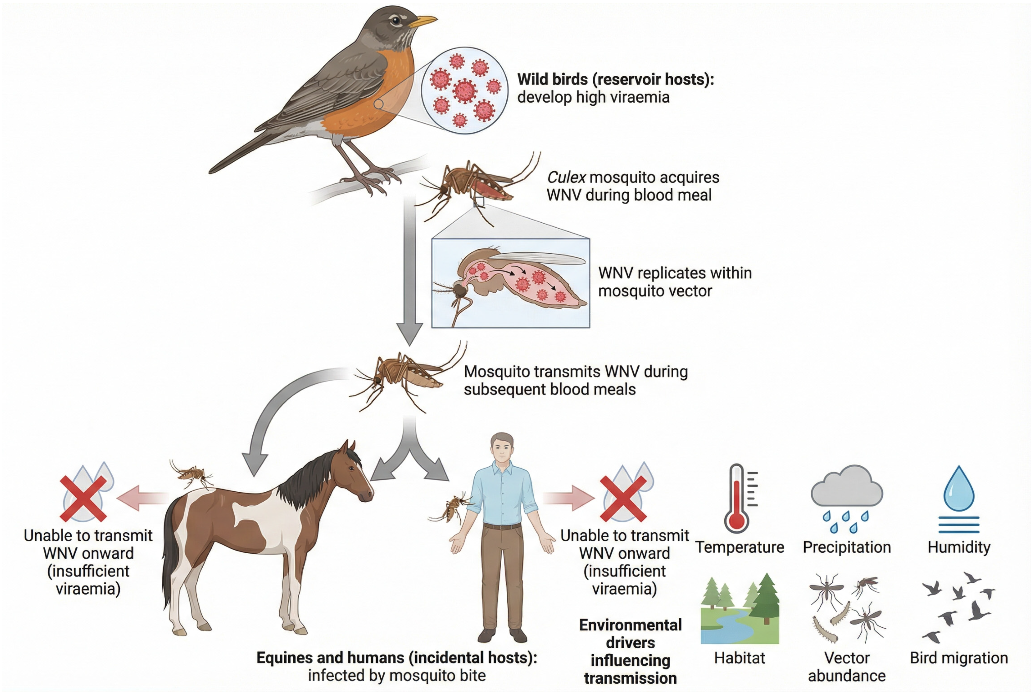
Migratory birds play a critical role in the long-distance movement of WNV across regions [19]. Europe lies along major flyways that connect Africa, the Middle East and Eurasia, allowing repeated viral introductions, lineage mixing and geographic expansion of both established and emerging strains [27,28,29,30]. The classical WNV transmission cycle involving avian hosts, mosquito vectors and incidental equine and human hosts is summarised in Figure 1.
Figure 1.
The transmission cycle of WNV involving avian hosts, mosquito vectors and incidental equine and human hosts. This schematic illustrates the classical transmission cycle of WNV. Birds serve as amplifying hosts, maintaining high levels of viraemia capable of infecting mosquito vectors, primarily Culex spp. Infected mosquitoes subsequently transmit the virus during blood feeding. Equines and humans become infected as incidental dead-end hosts, as their viraemia is insufficient to support further transmission. Environmental factors such as temperature, precipitation and habitat characteristics influence vector abundance and viral amplification.
2.3. Equines as Sentinel and Incidental Hosts
Although most equine infections remain subclinical, clinically affected horses may develop severe neurological disease, including ataxia, cranial nerve deficits, paresis, recumbency and, in some outbreaks, mortality rates of 23–43% [31]. Because horses share environments with humans and are highly exposed to mosquito vectors, equine infections frequently coincide with or precede human cases. These characteristics underpin their use as sentinel species for detecting viral circulation within a One Health surveillance context [32].
Seroprevalence studies in Europe show substantial geographic variation. Investigations in high-risk regions of Central and Eastern Europe have documented equine seroprevalence ranging between approximately 4% and 15% in unvaccinated populations, with higher values in wetland- and river-associated ecosystems [11,31]. Such findings underscore the importance of ecological context when interpreting equine serological data.
2.4. Current European Situation (2021–2025)
During the past decade, Europe has experienced increased frequency, intensity and geographic spread of WNV activity. Several countries in southern, central and eastern Europe report annual outbreaks in humans, horses and birds, with occasional incursions into previously unaffected regions [11,31,33]. Climatic trends, vector adaptation to peri-urban environments, ecological changes and variable surveillance capacity all contribute to the heterogeneous distribution of cases across the continent [20,34,35].
In addition, the circulation of related flaviviruses, particularly Usutu virus, complicates clinical and serological interpretation, further highlighting the need for harmonised diagnostic strategies and integrated surveillance systems [33,36].
3. Diagnostic Methods in Equines
Diagnostic evaluation of WNV infection in horses relies on a combination of direct and indirect methods, each presenting specific strengths and limitations when applied to equine cases [37]. Because viraemia in horses is typically short-lived and of low magnitude, direct viral detection is often challenging [38]. Consequently, serological assays constitute the backbone of equine WNV diagnosis, although their interpretation requires careful consideration of timing, vaccination status and potential cross-reactivity with other flaviviruses [39,40].
3.1. Direct Detection Methods
3.1.1. Molecular Assays (RT-qPCR/Real-Time RT-qPCR)
Reverse transcription polymerase chain reaction (RT-qPCR) is the preferred method for detecting WNV RNA in whole blood, serum, cerebrospinal fluid (CSF) or post-mortem central nervous system (CNS) tissues [41]. Real-time RT-qPCR platforms provide improved sensitivity, specificity and the ability to differentiate between WNV lineages [42].
However, in equines, viraemia is transient and often falls below the limit of detection at the time neurological signs appear [43]. As a result, negative RT-qPCR results do not reliably exclude infection [44,45]. External quality assessment studies have shown strong performance for lineage-specific assays, whereas broad-range flavivirus RT-qPCRs tend to be less sensitive in low-titre samples [46,47,48].
3.1.2. Virus Isolation and Antigen Detection
Virus isolation in cell culture and antigen detection via immunohistochemistry (IHC) are definitive diagnostic tools but are infrequently used in routine equine diagnostics. Virus isolation requires biosafety level 3 (BSL-3) facilities, significant time and high-quality samples, making it impractical for clinical decision making [9]. IHC performed on CNS tissues from fatal cases can provide confirmation even when viral RNA is degraded, but its use is necessarily limited to post-mortem scenarios [49].
3.2. Indirect Detection Methods (Serology)
Because direct detection frequently fails in clinical equine cases, serology is essential for identifying recent or past exposure to WNV [8].
3.2.1. IgM Capture ELISA
The detection of WNV-specific IgM antibodies in serum or CSF remains the most reliable indicator of recent infection. IgM antibodies appear shortly after infection and typically persist for several weeks. IgM ELISAs demonstrate good specificity but variable sensitivity, particularly in samples with low antibody titres or when collection is delayed. Despite these limitations, IgM ELISA is the primary diagnostic tool for clinically suspected acute cases. Most IgM ELISA assays are based on the detection of antibodies directed against the viral envelope (E) protein, which represents the main immunodominant antigen in flavivirus infections [50,51].
3.2.2. IgG ELISA and Competitive ELISA Platforms
IgG-based assays, including competitive or epitope-blocking ELISAs, are widely used in equine serosurveys and for assessing cumulative exposure. These tests are highly sensitive and suitable for large-scale screening [52]. However, IgG assays cannot distinguish between recent and past infection, nor can they differentiate natural infection from vaccine-induced immunity [53]. Their interpretation is further complicated by cross-reactivity with antigenically related flaviviruses such as Usutu virus, which co-circulates in several European regions [52,53]. IgG-based assays, including competitive or epitope-blocking ELISAs, are widely used in equine serosurveys and for assessing cumulative exposure. These assays typically target epitopes of the viral envelope (E) protein, although some newer platforms incorporate NS1-based antigens to improve specificity and reduce cross-reactivity [40,41,42,43,44,45,46,47,48,49,50,51,52,53].
3.2.3. Virus Neutralisation Test (VNT/PRNT)
Neutralisation assays remain the gold standard for confirming WNV-specific antibodies due to their high specificity and ability to resolve serological cross-reactivity. VNT/PRNT assays are particularly important for validating IgG ELISA findings in areas where multiple flaviviruses are present [54]. Their limitations include labour intensity, the requirement for high-containment facilities, longer turnaround times and restricted availability in routine diagnostic laboratories.
3.3. Interpretation of Diagnostic Results
Accurate interpretation requires integrating laboratory findings with clinical presentation, epidemiological context and sampling timing. A positive IgM ELISA or detectable viral RNA strongly supports recent infection [55]. Conversely, the presence of IgG alone, especially in a clinically healthy horse, typically reflects historical exposure or vaccination rather than active disease. In vaccinated populations, serological interpretation must be approached cautiously due to the inability of many assays to differentiate immune responses [56].
3.4. Proposed Diagnostic Workflow
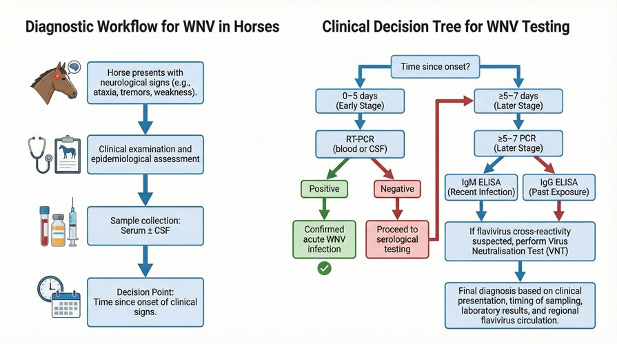
A practical diagnostic approach begins with early sampling of serum and, when feasible, CSF in horses presenting with compatible neurological signs [57]. During early infection, simultaneous RT-QPCR and IgM ELISA testing is recommended. Positive IgM or PCR results indicate probable acute WNV infection [58]. When both tests are negative but clinical suspicion remains, IgG ELISA followed by VNT confirmation can clarify exposure history [57]. For seroepidemiological studies, IgG ELISA is the preferred screening tool, with a subset of positive samples subjected to VNT to ensure specificity [50]. Based on these considerations, a practical diagnostic workflow is proposed (Table 1; Figure 2).
Table 1.
Diagnostic methods for WNV infection in equines: principles, sample types, advantages and limitations.
Figure 2.
Proposed diagnostic workflow for WNV infection in equines. This flow diagram outlines a stepwise diagnostic approach for suspected WNV infection in horses. Following the identification of compatible neurological signs, serum and CSF should be collected. Early-stage infection warrants RT-qPCR testing, whereas IgM ELISA serves as the primary indicator of recent infection. IgG ELISA supports assessment of past exposure, and virus neutralisation testing provides confirmatory specificity where cross-reactivity is suspected. Diagnostic interpretation must integrate clinical context, timing of sampling and regional flavivirus circulation.
4. Surveillance Systems in Equines
Surveillance of WNV in equine populations represents a key component of integrated monitoring frameworks aimed at detecting viral circulation, assessing regional risk and supporting early warning systems for both veterinary and public health authorities [69]. Because horses are highly susceptible to WNV and share ecological exposure with humans, data derived from equine surveillance can provide valuable indicators of local transmission intensity and the emergence of new hotspots [32].
Equine surveillance typically relies on three complementary approaches: passive clinical surveillance, active serological monitoring and sentinel-based surveillance.
Passive clinical surveillance remains the foundational strategy in most countries. It relies on the reporting of horses presenting with neurological signs compatible with WNV infection, followed by targeted diagnostic testing. This method is cost-effective and directly linked to clinically relevant disease; however, its sensitivity is limited by under-recognition, inconsistent reporting practices and the variable awareness of owners or veterinarians. As a result, many subclinical or mild cases remain undetected, leading to an underestimation of the true level of viral circulation [70,71].
In contrast, active serological surveillance aims to detect past exposure by screening healthy equine populations for the presence of IgG antibodies. Cross-sectional surveys can quantify seroprevalence across ecological zones, while repeated sampling enables the identification of temporal trends in exposure risk [11,12]. This approach is particularly useful for detecting silent circulation and identifying areas where viral amplification is occurring in the absence of clinical disease. Nevertheless, it requires structured sampling designs, adequate laboratory capacity and sustained financial resources, and results must be interpreted in the context of regional vaccination practices and potential cross-reactivity with other flaviviruses [30,65].
Sentinel equine surveillance, based on periodic sampling of unvaccinated cohorts, provides an enhanced capacity for early detection of viral introduction or resurgence. Seroconversion in sentinel horses often precedes the occurrence of clinical cases, making this approach a valuable early warning tool in high-risk areas such as wetlands, irrigated agricultural zones or regions with high mosquito density. However, successful implementation requires consistent sampling intervals, careful selection of sentinel groups and reliable access to diagnostic assays capable of distinguishing recent infection [32].
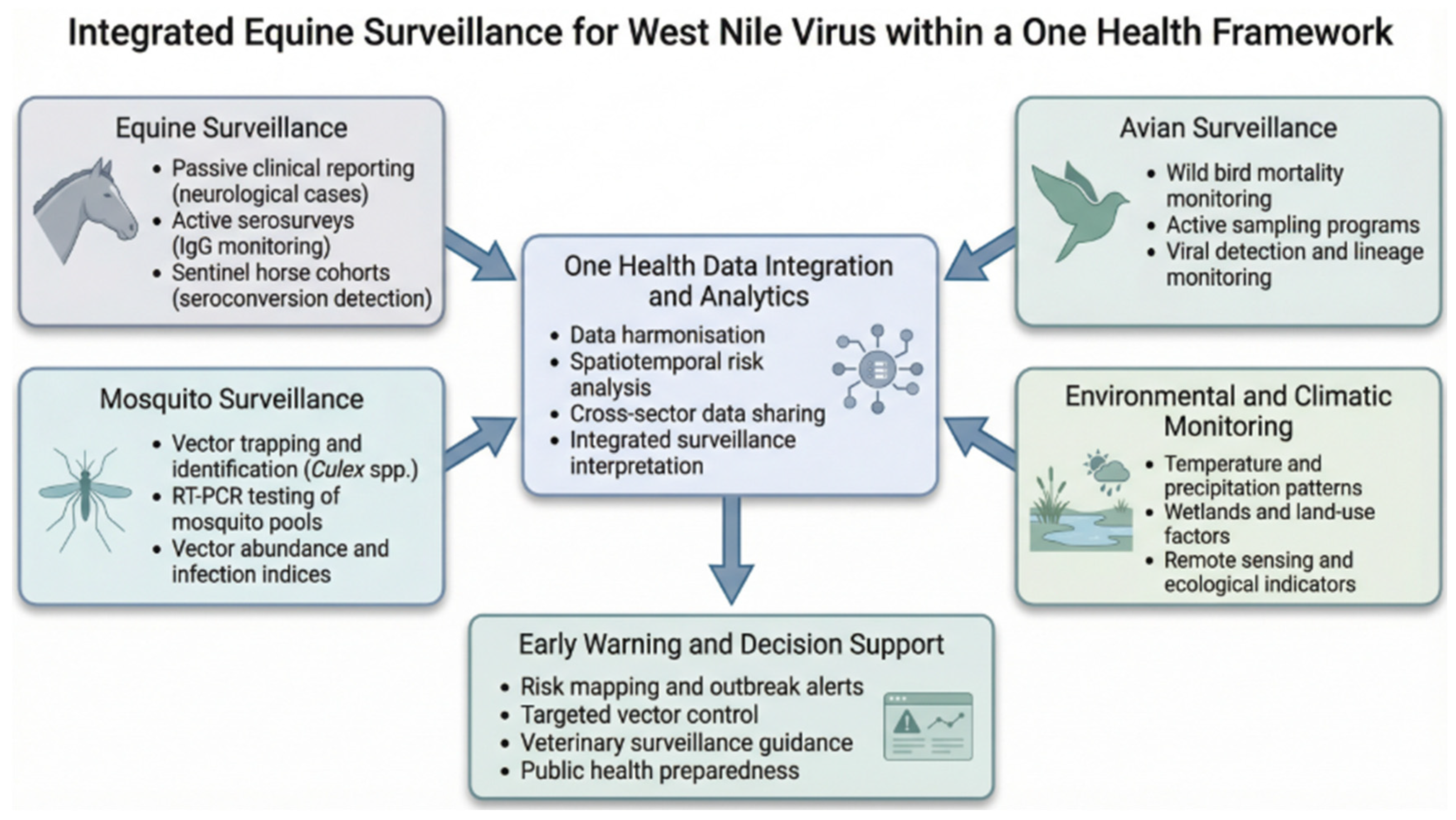
To maximise epidemiological insight, equine surveillance should be integrated within broader One Health systems that incorporate mosquito monitoring, avian surveillance and environmental data [72,73]. Linking equine seroconversion with entomological indices, climatic indicators and bird migration patterns generates a more comprehensive understanding of local transmission dynamics [74,75]. When combined with spatial modelling and risk forecasting, integrated datasets can support targeted vector control measures, guide veterinary alert systems and improve preparedness for human health authorities [76,77].
Despite these advantages, equine surveillance across Europe remains heterogeneous [9]. Differences in assay selection, diagnostic interpretation, sampling frequency and reporting channels limit cross-national comparability and hinder the development of harmonised early warning frameworks. Strengthening laboratory coordination, establishing standardised protocols and improving data exchange between veterinary and public health sectors are essential steps for enhancing surveillance effectiveness and supporting accurate risk assessment [78]. The main surveillance approaches for WNV in equines, including their characteristics, strengths and limitations, are summarised in Table 2, while the main components of equine WNV surveillance within an integrated One Health framework are illustrated in Figure 3.
Table 2.
Surveillance approaches for WNV in equines: characteristics, strengths and limitations.
Figure 3.
Integrated equine surveillance components within a One Health framework. This figure presents the major elements of equine WNV surveillance within a One Health system. Passive clinical surveillance detects neurological cases in horses, while active serological monitoring reveals silent transmission. Sentinel equine cohorts provide early warning of viral amplification. Integration of equine data with mosquito surveillance, avian monitoring and environmental indicators enhances risk assessment, supports predictive modelling and enables timely public health response.
5. Challenges in Diagnosis and Surveillance
Despite notable advances in understanding WNV epidemiology and equine infection dynamics, several biological, methodological and operational challenges continue to limit the accuracy of diagnostic testing and the effectiveness of surveillance systems [10]. These constraints vary across regions and directly influence the timely detection of viral circulation, the interpretation of serological and molecular data, and the comparability of results across laboratories [9,10].
A major diagnostic limitation stems from the intrinsic characteristics of WNV infection in horses. Equines typically develop a short and low-level viraemia that frequently falls below the analytical sensitivity of nucleic acid amplification tests [8]. As a result, RT-qPCR performed on blood or CSF often yields negative results even in clinically compatible cases, particularly when sampling occurs after the early phase of infection [86]. Consequently, molecular assays cannot be relied upon as standalone diagnostic tools in equine medicine and must be interpreted in conjunction with serology and clinical findings [86].
Serological testing, although essential, introduces additional interpretative complexities [8]. IgM capture ELISAs are considered the most informative for detecting recent infection [86]; however, the magnitude and duration of IgM responses vary among individuals, and titres may decline rapidly, leading to false-negative results when sampling is delayed [38,87]. IgG ELISAs provide valuable information for seroprevalence studies but cannot distinguish between recent and past infection, nor can they differentiate natural exposure from vaccine-induced immunity in regions where vaccination is implemented [88]. Cross-reactivity with other flaviviruses, especially Usutu virus, further complicates serological interpretation and underscores the need for confirmatory testing using virus neutralisation tests (VNTs), which remain resource-intensive and are not widely available in all veterinary laboratories [50,88,89].
Beyond methodological constraints, significant heterogeneity exists in laboratory capacity, assay selection, cut-off values and confirmatory protocols across countries [9]. This variability affects the comparability of results and limits the generation of harmonised epidemiological indicators at the national or regional scale [10]. Differences in sample handling procedures, test validation and the availability of reference laboratories also contribute to inconsistent diagnostic performance and reduced confidence in surveillance outputs [8].
Operational challenges further hinder surveillance efficiency [89]. Passive clinical surveillance relies on the recognition and reporting of neurological signs by owners and veterinarians, yet mild, atypical or early cases may go unnoticed or remain untested [11,90]. Active serological surveillance and sentinel horse programmes offer greater sensitivity for detecting silent circulation but require sustained funding, structured sampling strategies, logistical coordination and long-term institutional commitment [91]. In resource-limited regions, such programmes may be implemented intermittently or discontinued, resulting in spatial and temporal gaps in surveillance data [91].
Environmental variability adds additional complexity [20]. Rapid fluctuations in climatic conditions, vector abundance and avian reservoir dynamics can lead to abrupt changes in WNV transmission intensity [35]. Surveillance systems that lack sufficient temporal resolution, geographic coverage or cross-sector integration may fail to detect these shifts in real time [36]. Moreover, the absence of standardised thresholds for triggering public health or veterinary interventions complicates decision making and reduces the operational value of equine-derived indicators [92].
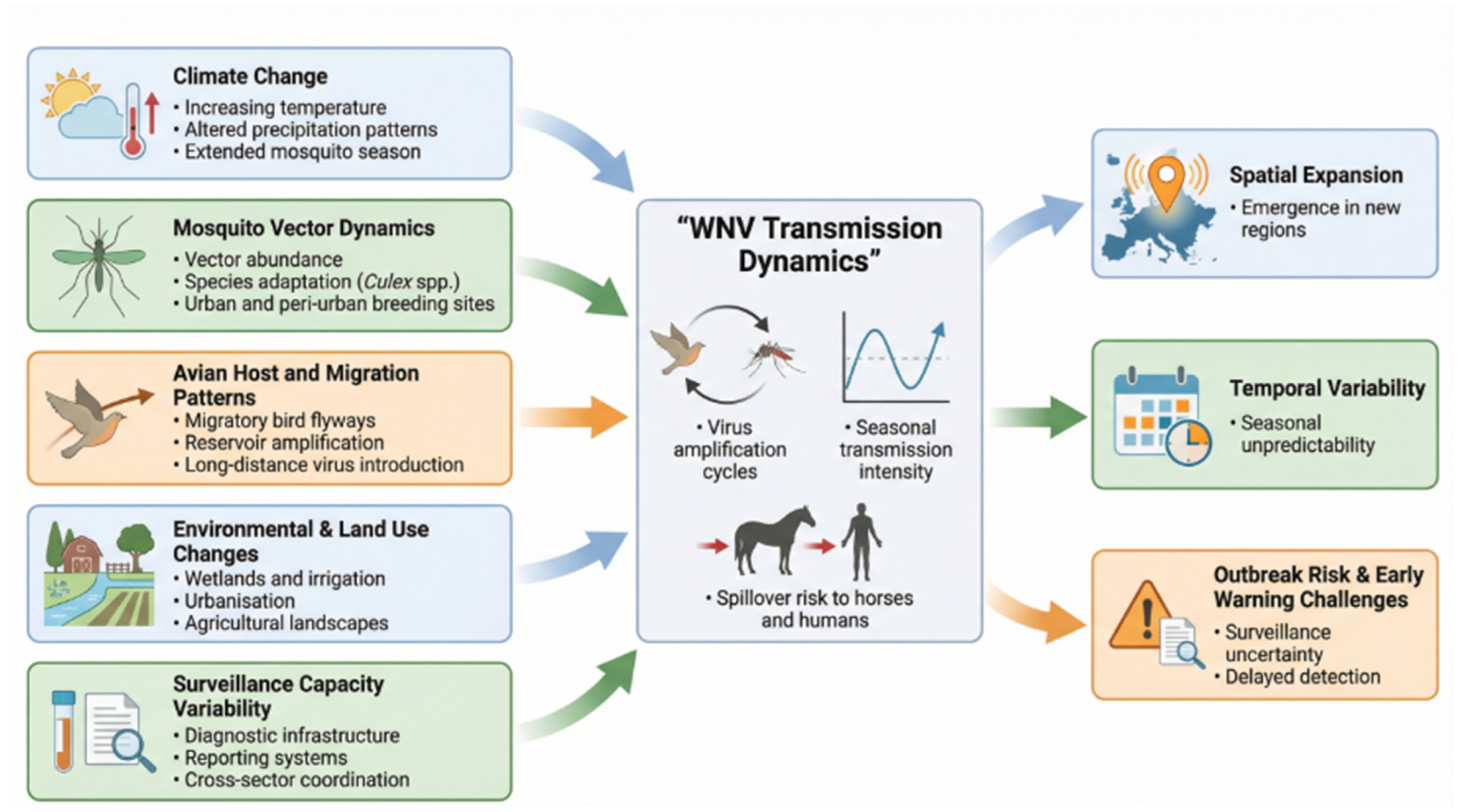
Overall, these challenges highlight the need for improved diagnostic standardisation, enhanced laboratory coordination, strengthened One Health data integration and sustained investment in targeted surveillance [93]. Addressing these limitations is essential for accurately characterising WNV transmission patterns, improving early warning capabilities and reducing the disease burden in equine populations [36]. The main diagnostic challenges and their implications for equine surveillance are summarised in Table 3, while the key ecological, climatic and operational determinants shaping WNV epidemiology are illustrated in Figure 4.
Table 3.
Key diagnostic challenges and implications for surveillance in equines.
Figure 4.
Key ecological, climatic and operational determinants shaping WNV epidemiology in Europe. This conceptual map summarises major determinants influencing WNV transmission dynamics in Europe, including climate change, mosquito vector adaptation, bird migration, urbanisation and variability in surveillance capacity. These interacting factors contribute to the spatial expansion, temporal variability and unpredictability of WNV outbreaks and must be considered when designing surveillance strategies and interpreting equine diagnostic data.
6. Future Directions
Future advancements in the diagnosis and surveillance of WNV infection in equines will rely on the integration of novel technologies, improved diagnostic standardisation and the strengthening of One Health-oriented monitoring frameworks. Several priority areas emerge from the current knowledge gaps [8,73].
The enhancement of diagnostic tools remains essential. Although molecular assays show high analytical specificity, their clinical sensitivity in equines is limited by the short and low-level viraemia characteristic of infection [37,97]. Future progress should focus on developing rapid, field-deployable molecular platforms, such as portable RT-qPCR units, CRISPR-based detection systems or loop-mediated isothermal amplification (LAMP), that maintain high sensitivity even in low-titre samples [98]. In parallel, serological tools require refinement to overcome issues of cross-reactivity with related flaviviruses and to better differentiate recent infection from past exposure or vaccine-induced antibodies [8,85]. Novel ELISA formats, improved antigen targets and well-characterised reference panels are needed to support assay harmonisation [8].
The standardisation of laboratory methodologies across regions will be equally important. Significant variability currently exists in ELISA cut-off values, confirmatory testing strategies and access to virus neutralisation tests (VNTs) [8]. Collaborative validation studies, harmonised protocols and shared quality assurance programmes would enhance diagnostic comparability and improve the interpretation of equine surveillance data at national and international levels [8].
From a surveillance perspective, the expansion of integrated, multi-source monitoring systems represents a major opportunity. Facility-level environmental risk scoring can provide a rapid, low-cost proxy for mosquito-favourable conditions and may complement entomological and serological surveillance to improve prioritisation and early warning capacity, particularly in resource-limited settings [99]. Linking equine serological data with mosquito surveillance, avian testing, climatic indicators and ecological modelling can substantially improve early warning capacity [73]. Advances in remote sensing, machine learning and climate-based prediction models could support the identification of high-risk areas, optimise sampling strategies and guide timely vector control interventions [76]. In this framework, equine sentinel cohorts, particularly in ecologically sensitive or high-transmission regions, can provide sensitive and cost-effective indicators of viral amplification when coupled with regular sampling and robust diagnostic support [74,80].
Additionally, genomic surveillance is expected to play an increasingly prominent role. High-throughput sequencing of WNV-positive samples, even at low coverage, can elucidate viral lineage dynamics, track geographic spread and detect emerging mutations associated with virulence, host adaptation or immune evasion [100]. Incorporating equine-derived genomic data into broader European or international phylogenetic networks would enhance situational awareness and support coordinated public health and veterinary responses [101].
Finally, long-term improvement requires structured investment in training, infrastructure and intersectoral collaboration [102]. Strengthening communication between veterinary laboratories, public health agencies, wildlife surveillance programmes and environmental monitoring systems is essential for operationalising One Health strategies [103]. Improved funding mechanisms, cross-border data sharing and coordinated policy frameworks will further support sustained and effective surveillance.
Collectively, these future directions highlight the need for innovative diagnostic methods, harmonised laboratory practices and integrated surveillance approaches tailored to evolving ecological and epidemiological contexts. Implementing these priorities will be critical for enhancing preparedness, reducing disease impact in equine populations and refining the early detection of WNV circulation at the human–animal–environment interface.
7. Conclusions
WNV infection remains a significant and evolving threat to equine health, with implications extending beyond veterinary medicine into the wider One Health domain. Horses, although incidental hosts, develop severe neurological disease more frequently than humans and therefore provide an early and sensitive indication of viral circulation in the environment. Their clinical and serological responses offer a practical means to detect changes in transmission intensity, making equine surveillance an essential component of integrated monitoring systems.
Despite notable advances in understanding WNV epidemiology in Europe, major gaps persist in both diagnostic practice and surveillance implementation. The short and low-level viraemia characteristic of equine infection limits the usefulness of molecular assays in clinical cases, necessitating a continued reliance on serological tests. However, cross-reactivity with other flaviviruses, variability in IgM kinetics and difficulties in distinguishing natural infection from vaccine-derived immunity complicate serological interpretation. These constraints highlight the need for standardised diagnostic algorithms that combine clinical presentation with carefully selected laboratory methods.
Surveillance systems have expanded in recent years, but substantial heterogeneity remains across Europe. Differences in laboratory capacity, sampling frequency, case definitions and reporting structures impede comparability and weaken early warning capabilities. Integrating equine data with mosquito surveillance, avian monitoring and environmental indicators represents a critical step toward more accurate risk assessment and predictive capability. Harmonised procedures and improved data-sharing mechanisms are essential for building an efficient, coordinated surveillance infrastructure.
Looking forward, investment in diagnostic innovation, including rapid point-of-care tools, improved serological assays and lineage-specific molecular tests, will be important for strengthening field-level detection. Likewise, adaptive surveillance frameworks that incorporate modelling, remote sensing and climate-based forecasting will be needed to address the increasingly dynamic nature of WNV transmission under changing ecological conditions.
Overall, enhancing diagnostic performance, standardising surveillance protocols and embedding equine data more systematically into One Health systems will be crucial for improving outbreak detection, response efficiency and preparedness for future WNV transmission seasons.
Author Contributions
Conceptualization, P.N. and V.H.; methodology, P.N., L.S., V.I. and H.I.; validation, P.N., L.S., H.I. and V.H.; formal analysis, P.N. and A.G.; investigation, P.N., V.I., A.G., A.C., B.F., V.C. and H.I.; resources, V.H. and I.I.; data curation, P.N. and C.H.M.; writing—original draft preparation, P.N.; writing—review and editing, L.S., V.H., B.N., I.I. and H.I.; visualization, P.N. and V.C.; supervision, V.H.; project administration, P.N.; funding acquisition, V.H. All authors have read and agreed to the published version of the manuscript.
Funding
This research received no external funding.
Institutional Review Board Statement
Not applicable. This article is a narrative review and does not involve original experimental or clinical studies.
Informed Consent Statement
Not applicable.
Data Availability Statement
No new data were created or analysed in this study.
Acknowledgments
The authors would like to thank the Doctoral School “Veterinary Medicine”, University of Life Sciences “King Mihai I” from Timişoara (Calea Aradului 119, 300645 Timişoara, Romania), for the academic support and resources provided throughout the development of this research.
Conflicts of Interest
The authors declare no conflicts of interest.
Abbreviations
The following abbreviations are used in this manuscript:
| WNV | West Nile virus |
| CSF | Cerebrospinal fluid |
| CNS | Central nervous system |
| ELISA | Enzyme-linked immunosorbent assay |
| IgM | Immunoglobulin M |
| IgG | Immunoglobulin G |
| PRNT | Plaque reduction neutralisation test |
| BSL | Biosafety level |
| ECDC | European Centre for Disease Prevention and Control |
| EFSA | European Food Safety Authority |
References
- Schwarz, E.R.; Long, M.T. Comparison of West Nile Virus Disease in Humans and Horses: Exploiting Similarities for Enhancing Syndromic Surveillance. Viruses 2023, 15, 1230. [Google Scholar] [CrossRef] [Scilit]
- Wang, Y.; Calzolari, M.; Calvi, G.; Cox, V.; Angelini, P.; Dottori, M.; Wint, W.; Jahn, S.; Marini, G.; Dorigatti, I. Association of avian biodiversity and West Nile Virus circulation in Culex mosquitoes in Emilia-Romagna, Italy. PLoS Negl. Trop. Dis. 2026, 20, e0014076. [Google Scholar] [CrossRef] [Scilit]
- Chancey, C.; Grinev, A.; Volkova, E.; Rios, M. The Global Ecology and Epidemiology of West Nile Virus. BioMed Res. Int. 2015, 2015, 376230. [Google Scholar] [CrossRef] [Scilit]
- Fehér, O.; Fehérvári, P.; Tolnai, C.; Forgách, P.; Malik, P.; Jerzsele, Á.; Wagenhoffer, Z.; Szenci, O.; Korbacska-Kutasi, O. Epidemiology and Clinical Manifestation of West Nile Virus Infections of Equines in Hungary, 2007–2020. Viruses 2022, 14, 2551. [Google Scholar] [CrossRef] [Scilit] [PubMed]
- Angelini, P.; Tamba, M.; Finarelli, A.; Bellini, R.; Albieri, A.; Bonilauri, P.; Cavrini, F.; Dottori, M.; Gaibani, P.; Martini, E.; et al. West Nile virus circulation in Emilia-Romagna, Italy: The integrated surveillance system 2009. Eurosurveillance 2010, 15, 16. [Google Scholar] [CrossRef] [Scilit]
- Koch, R.; Erazo, D.; Folly, A.; Johnson, N.; Dellicour, S.; Grubaugh, N.; Vogels, C. Genomic epidemiology of West Nile virus in Europe. One Health 2024, 18, 100664. [Google Scholar] [CrossRef] [Scilit] [PubMed]
- Abbas, I.; Ahmed, F.; Muqaddas, H.; Alberti, A.; Varcasia, A.; Sedda, L. Epidemiology and surveillance of West Nile virus in the Mediterranean Basin during 2010–2023: A systematic review. Curr. Res. Parasitol. Vector-Borne Dis. 2025, 7, 100277. [Google Scholar] [CrossRef] [Scilit] [PubMed]
- Beck, C.; Lowenski, S.; Durand, B.; Bahuon, C.; Zientara, S.; Lecollinet, S. Improved reliability of serological tools for the diagnosis of West Nile fever in horses within Europe. PLoS Negl. Trop. Dis. 2017, 11, e0005936. [Google Scholar] [CrossRef] [Scilit]
- Pérez-Ramírez, E.; Cano-Gómez, C.; Llorente, F.; Vodica, A.; Veljović, L.; Toklikishvilli, N.; Sherifi, K.; Sghaier, S.; Omani, A.; Kustura, A.; et al. Evaluation of West Nile Virus Diagnostic Capacities in Veterinary Laboratories of the Mediterranean and Black Sea Regions. Pathogens 2020, 9, 1038. [Google Scholar] [CrossRef] [Scilit]
- Lustig, Y.; Sofer, D.; Bucris, E.D.; Mendelson, E. Surveillance and Diagnosis of West Nile Virus in the Face of Flavivirus Cross-Reactivity. Front. Microbiol. 2018, 9, 2421. [Google Scholar] [CrossRef] [Scilit]
- Nistor, P.; Stanga, L.; Chirila, A.; Iorgoni, V.; Gligor, A.; Cireșan, A.; Popa, I.; Florea, B.; Imre, M.; Cocioba, V.; et al. Seroprevalence and Passive Clinical Surveillance of West Nile Virus in Horses from Ecological High-Risk Areas in Western Romania: Exploratory Findings from a Cross-Sectional Study. Microorganisms 2025, 13, 1910. [Google Scholar] [CrossRef] [Scilit]
- Folly, A.J.; Waller, E.S.L.; McCracken, F.; McElhinney, L.M.; Roberts, H.; Johnson, N. Equine seroprevalence of West Nile virus antibodies in the UK in 2019. Parasites Vectors 2020, 13, 596. [Google Scholar] [CrossRef] [Scilit]
- Lu, Z.; Fu, S.; Cao, L.; Tang, C.; Zhang, S.; Li, Z.; Tusong, M.; Yao, X.; Zhang, H.; Wang, P.; et al. Human Infection with West Nile Virus, Xinjiang, China, 2011. Emerg. Infect. Dis. 2014, 20, 1421–1423. [Google Scholar] [CrossRef] [Scilit]
- Bruno, L.; Nappo, M.A.; Frontoso, R.; Perrotta, M.G.; Di Lecce, R.; Guarnieri, C.; Ferrari, L.; Corradi, A. West Nile Virus (WNV): One-Health and Eco-Health Global Risks. Vet. Sci. 2025, 12, 288. [Google Scholar] [CrossRef] [Scilit]
- Fiacre, L.; Pagès, N.; Albina, E.; Richardson, J.; Lecollinet, S.; Gonzalez, G. Molecular Determinants of West Nile Virus Virulence and Pathogenesis in Vertebrate and Invertebrate Hosts. Int. J. Mol. Sci. 2020, 21, 9117. [Google Scholar] [CrossRef] [Scilit]
- Zehender, G.; Veo, C.; Ebranati, E.; Carta, V.; Rovida, F.; Percivalle, E.; Moreno, A.; Lelli, D.; Calzolari, M.; Lavazza, A.; et al. Reconstructing the recent West Nile virus lineage 2 epidemic in Europe and Italy using discrete and continuous phylogeography. PLoS ONE 2017, 12, e0179679. [Google Scholar] [CrossRef] [Scilit]
- Aguilera-Sepúlveda, P.; Napp, S.; Llorente, F.; Solano-Manrique, C.; Molina-López, R.; Obón, E.; Solé, A.; Jiménez-Clavero, M.Á.; Fernández-Pinero, J.; Busquets, N. West Nile Virus Lineage 2 Spreads Westwards in Europe and Overwinters in North-Eastern Spain (2017–2020). Viruses 2022, 14, 569. [Google Scholar] [CrossRef] [Scilit] [PubMed]
- Patzina-Mehling, C.; Kopp, A.; Rauhöft, L.; Șuleșco, T.; Jones, T.C.; Drosten, C.; Sauer, F.G.; Lühken, R.; Junglen, S. Genomic surveillance indicates high site-specific heterogeneity of West Nile virus in mosquitoes in rural regions of Germany across seasons. One Health 2025, 21, 101179. [Google Scholar] [CrossRef] [Scilit] [PubMed]
- García-Carrasco, J.M.; Muñoz, A.R.; Olivero, J.; Figuerola, J.; Fa, J.E.; Real, R. Gone (and spread) with the birds: Can chorotype analysis highlight the spread of West Nile virus within the Afro-Palaearctic flyway? One Health 2023, 17, 100585. [Google Scholar] [CrossRef] [Scilit]
- Giesen, C.; Herrador, Z.; Fernández-Martínez, B.; Figuerola, J.; Gangoso, L.; Vázquez, A.; Gómez-Barroso, D. A systematic review of environmental factors related to WNV circulation in European and Mediterranean countries. One Health 2023, 16, 100478. [Google Scholar] [CrossRef] [Scilit] [PubMed]
- Ferraguti, M.; Heesterbeek, H.; La Puente, M.; Jiménez-Clavero, M.; Vázquez, A.; Ruíz, S.; Llorente, F.; Roiz, D.; Vernooij, H.; Soriguer, R.; et al. The role of different Culex mosquito species in the transmission of West Nile virus and avian malaria parasites in Mediterranean areas. Transbound. Emerg. Dis. 2020, 68, 920–930. [Google Scholar] [CrossRef] [Scilit] [PubMed]
- Jansen, S.; Heitmann, A.; Lühken, R.; Leggewie, M.; Helms, M.; Badusche, M.; Rossini, G.; Schmidt-Chanasit, J.; Tannich, E. Culex torrentium: A Potent Vector for the Transmission of West Nile Virus in Central Europe. Viruses 2019, 11, 492. [Google Scholar] [CrossRef] [Scilit]
- Soto, A.; De Coninck, L.; Devlies, A.; Van De Wiele, C.; Rosas, A.; Wang, L.; Matthijnssens, J.; Delang, L. Belgian Culex pipiens pipiens are competent vectors for West Nile virus while Culex modestus are competent vectors for Usutu virus. PLoS Negl. Trop. Dis. 2023, 17, e0011649. [Google Scholar] [CrossRef] [Scilit]
- Baril, C.; Pilling, B.; Mikkelsen, M.; Sparrow, J.; Duncan, C.; Koloski, C.; LaZerte, S.; Cassone, B. The influence of weather on the population dynamics of common mosquito vector species in the Canadian Prairies. Parasites Vectors 2023, 16, 153. [Google Scholar] [CrossRef] [Scilit]
- Marcantonio, M.; Rizzoli, A.; Metz, M.; Rosà, R.; Marini, G.; Chadwick, E.; Neteler, M. Identifying the Environmental Conditions Favouring West Nile Virus Outbreaks in Europe. PLoS ONE 2015, 10, e0121158. [Google Scholar] [CrossRef] [Scilit]
- Paz, S.; Semenza, J. Environmental Drivers of West Nile Fever Epidemiology in Europe and Western Asia—A Review. Int. J. Environ. Res. Public Health 2013, 10, 3543–3562. [Google Scholar] [CrossRef] [Scilit]
- Mancuso, E.; Cecere, J.G.; Iapaolo, F.; Di Gennaro, A.; Sacchi, M.; Savini, G.; Spina, F.; Monaco, F. West Nile and Usutu Virus Introduction via Migratory Birds: A Retrospective Analysis in Italy. Viruses 2022, 14, 416. [Google Scholar] [CrossRef] [Scilit]
- Srihi, H.; Chatti, N.; Ben Mhadheb, M.; Gharbi, J.; Abid, N. Phylodynamic and phylogeographic analysis of the complete genome of the West Nile virus lineage 2 (WNV-2) in the Mediterranean basin. BMC Ecol. Evol. 2021, 21, 183. [Google Scholar] [CrossRef] [Scilit] [PubMed]
- Mencattelli, G.; Ndione, M.; Silverj, A.; Diagne, M.; Curini, V.; Teodori, L.; Di Domenico, M.; Mbaye, R.; Leone, A.; Marcacci, M.; et al. Spatial and temporal dynamics of West Nile virus between Africa and Europe. Nat. Commun. 2023, 14, 6440. [Google Scholar] [CrossRef] [Scilit]
- Lu, L.; Zhang, F.; Oude Munnink, B.B.; Munger, E.; Sikkema, R.S.; Pappa, S.; Tsioka, K.; Sinigaglia, A.; Molin, E.D.; Shih, B.B.; et al. West Nile virus spread in Europe: Phylogeographic pattern analysis and key drivers. PLoS Pathog. 2024, 20, e1011880. [Google Scholar] [CrossRef] [PubMed]
- Ganzenberg, S.; Sieg, M.; Ziegler, U.; Pfeffer, M.; Vahlenkamp, T.W.; Hörügel, U.; Groschup, M.H.; Lohmann, K.L. Seroprevalence and Risk Factors for Equine West Nile Virus Infections in Eastern Germany, 2020. Viruses 2022, 14, 1191. [Google Scholar] [CrossRef] [Scilit] [PubMed]
- García-Carrasco, J.M.; Muñoz, A.R.; Olivero, J.; Segura, M.; García-Bocanegra, I.; Real, R. West Nile virus in the Iberian Peninsula: Using equine cases to identify high-risk areas for humans. Eurosurveillance 2023, 28, 2200844. [Google Scholar] [CrossRef] [Scilit] [PubMed]
- Simonin, Y. Circulation of West Nile Virus and Usutu Virus in Europe: Overview and Challenges. Viruses 2024, 16, 599. [Google Scholar] [CrossRef] [Scilit]
- Watts, M.J.; Sarto i Monteys, V.; Mortyn, P.G.; Kotsila, P. The rise of West Nile Virus in Southern and Southeastern Europe: A spatial-temporal analysis investigating the combined effects of climate, land use and economic changes. One Health 2021, 13, 100315. [Google Scholar] [CrossRef] [Scilit]
- Taheri, S.; González, M.A.; Ruiz-López, M.J.; Soriguer, R.; Figuerola, J. Patterns of West Nile virus vector co-occurrence and spatial overlap with human cases across Europe. One Health 2025, 20, 101041. [Google Scholar] [CrossRef] [Scilit]
- Serres, K.; Erazo, D.; Despréaux, G.; Vincenti-González, M.; Van Bortel, W.; Arsevska, E.; Dellicour, S. Integrating indicator-based and event-based surveillance data for risk mapping of West Nile virus, Europe, 2006 to 2021. Eurosurveillance 2024, 29, 2400084. [Google Scholar] [CrossRef] [Scilit]
- Kleiboeker, S.; Loiacono, C.; Rottinghaus, A.; Pue, H.; Johnson, G. Diagnosis of West Nile Virus Infection in Horses. J. Vet. Diagn. Invest. 2004, 16, 10–12. [Google Scholar] [CrossRef] [Scilit]
- Tolnai, C.H.; Forgách, P.; Marosi, A.; Fehér, O.; Paszerbovics, B.; Tenk, M.; Wagenhoffer, Z.; Kutasi, O. Long-Term Humoral Immune Response After West Nile Virus Convalescence in Horses in a Geographic Area of Multiple Orthoflavivirus Co-Circulation. J. Vet. Intern. Med. 2025, 39, e70176. [Google Scholar] [CrossRef] [Scilit]
- Kemmerly, S.A. Diagnosis and treatment of west nile infections. Ochsner J. 2003, 5, 16–17. [Google Scholar] [PubMed]
- Paré, J.; Moore, A. West Nile virus in horses—What do you need to know to diagnose the disease? Can. Vet. J. 2018, 59, 1119–1120. [Google Scholar]
- Marino, A.; Vitale, E.; Maniaci, A.; La Via, L.; Moscatt, V.; Spampinato, S.; Senia, P.; Venanzi Rullo, E.; Restivo, V.; Cacopardo, B.; et al. West Nile Virus: Insights into Microbiology, Epidemiology, and Clinical Burden. Acta Microbiol. Hell. 2025, 70, 44. [Google Scholar] [CrossRef] [Scilit]
- Lai, J.; Tessarolo, C.; Ercole, E.; Gallo, M.; Lo Faro, M.; Palmitessa, C.; Carta, V.; Ferrari, A.; Favole, A.; Begovoeva, M.; et al. β-Actin as an Endogenous Control Gene in Real-Time PCR for Detection of West Nile and Usutu Virus in Mosquitoes. Microorganisms 2025, 13, 2518. [Google Scholar] [CrossRef] [Scilit] [PubMed]
- Naveed, A.; Eertink, L.G.; Wang, D.; Li, F. Lessons Learned from West Nile Virus Infection: Vaccinations in Equines and Their Implications for One Health Approaches. Viruses 2024, 16, 781. [Google Scholar] [CrossRef] [Scilit]
- Madere, F.S.; Andrade da Silva, A.V.; Okeze, E.; Tilley, E.; Grinev, A.; Konduru, K.; García, M.; Rios, M. Flavivirus infections and diagnostic challenges for dengue, West Nile and Zika Viruses. NPJ Viruses 2025, 3, 36. [Google Scholar] [CrossRef] [Scilit]
- Patel, P.; Landt, O.; Kaiser, M.; Faye, O.; Koppe, T.; Lass, U.; Sall, A.A.; Niedrig, M. Development of one-step quantitative reverse transcription PCR for the rapid detection of flaviviruses. Virol. J. 2013, 10, 58. [Google Scholar] [CrossRef] [Scilit]
- Pennick, K.E.; McKnight, C.A.; Patterson, J.S.; Latimer, K.S.; Maes, R.K.; Wise, A.G.; Kiupel, M. Diagnostic sensitivity and specificity of in situ hybridization and immunohistochemistry for Eastern equine encephalitis virus and West Nile virus in formalin-fixed, paraffin-embedded brain tissue of horses. J. Vet. Diagn. Invest. 2012, 24, 333–338. [Google Scholar] [CrossRef] [Scilit]
- Vina-Rodriguez, A.; Sachse, K.; Ziegler, U.; Chaintoutis, S.; Keller, M.; Groschup, M.; Eiden, M. A Novel Pan-Flavivirus Detection and Identification Assay Based on RT-qPCR and Microarray. BioMed Res. Int. 2017, 2017, 4248756. [Google Scholar] [CrossRef] [Scilit]
- Scaramozzino, N.; Crance, J.; Jouan, A.; Debriel, D.; Stoll, F.; Garín, D. Comparison of Flavivirus Universal Primer Pairs and Development of a Rapid, Highly Sensitive Heminested Reverse Transcription-PCR Assay for Detection of Flaviviruses Targeted to a Conserved Region of the NS5 Gene Sequences. J. Clin. Microbiol. 2001, 39, 1922–1927. [Google Scholar] [CrossRef] [Scilit]
- Moureau, G.; Temmam, S.; Gonzalez, J.; Charrel, R.; Grard, G.; Lamballerie, X. A Real-Time RT-PCR Method for the Universal Detection and Identification of Flaviviruses. Vector-Borne Zoonotic Dis. 2007, 7, 467–478. [Google Scholar] [CrossRef] [Scilit]
- Girl, P.; Euringer, K.; Coroian, M.; Mihalca, A.D.; Borde, J.P.; Dobler, G. Comparison of Five Serological Methods for the Detection of West Nile Virus Antibodies. Viruses 2024, 16, 788. [Google Scholar] [CrossRef] [Scilit] [PubMed]
- Gómez-Vicente, E.; Garcia, R.; Calatrava, E.; Olivares Duran, M.J.; Gutiérrez-Bautista, J.F.; Rodriguez-Granger, J.; Cobo, F.; Navarro Mari, J.M.; Sampedro-Martinez, A. Comparative evaluation of chemiluminescent immunoassay and enzyme-linked immunosorbent assays for the diagnosis of West Nile virus infections. APMIS 2022, 130, 215–220. [Google Scholar] [CrossRef] [Scilit] [PubMed]
- Vista, F.E.S.; Tantengco, O.A.G.; Dispo, M.D.; Opiso, D.M.S.; Badua, C.L.D.C.; Gerardo, J.P.Z.; Perez, J.R.M.; Baldo, K.A.T.; Chao, D.-Y.; Dalmacio, L.M.M. Trends in ELISA-Based Flavivirus IgG Serosurveys: A Systematic Review. Trop. Med. Infect. Dis. 2023, 8, 224. [Google Scholar] [CrossRef] [Scilit] [PubMed]
- Lustig, Y.; Indenbaum, V.; Koren, R.; Katz-Likvornik, S.; Halpern, O.; Mendelson, E. The diagnostic utility of Immunoglobulin G (IgG) avidity in distinguishing between past and acute infection of West Nile Virus (WNV). J. Clin. Microbiol. 2025, 63, e0095225. [Google Scholar] [CrossRef] [Scilit]
- Chan, K.R.; Ismail, A.A.; Thergarajan, G.; Raju, C.S.; Yam, H.C.; Rishya, M.; Sekaran, S.D. Serological cross-reactivity among common flaviviruses. Front. Cell. Infect. Microbiol. 2022, 12, 975398. [Google Scholar] [CrossRef] [Scilit]
- Odigie, A.E.; Stufano, A.; Schino, V.; Zarea, A.A.K.; Ndiana, L.A.; Mrenoshki, D.; Ugochukwu, I.C.I.; Lovreglio, P.; Greco, G.; Pratelli, A.; et al. West Nile Virus Infection in Occupational Settings—A Systematic Review. Pathogens 2024, 13, 157. [Google Scholar] [CrossRef] [Scilit]
- Approaches to epidemiologic surveillance, diagnosis and prevention of West Nile fever in the Kyiv region, April 2025. One Health J. 2025, 3, 5–31. [CrossRef] [Scilit]
- West Nile Virus in Horses—An Update. Available online: https://laboklin.com/en/west-nile-virus-in-horses-an-update/ (accessed on 12 November 2025).
- West Nile Virus Diagnostic Guidance for Veterinarians. Available online: https://tvmdl.tamu.edu/education-library/west-nile-virus-diagnostic-guidance-for-veterinarians/ (accessed on 19 November 2025).
- Vilibic-Cavlek, T.; Bogdanic, M.; Savic, V.; Hruskar, Z.; Barbic, L.; Stevanovic, V.; Antolasic, L.; Milasincic, L.; Sabadi, D.; Miletic, G.; et al. Diagnosis of West Nile virus infections: Evaluation of different laboratory methods. World J. Virol. 2024, 13, 95986. [Google Scholar] [CrossRef] [Scilit]
- Yazici, Z.; Albayrak, H.; Ozan, E.; Gumusova, S. The first investigation of west nile virus in horses using real time rt-PCR in middle black sea region in Turkey. J. Arthropod Borne Dis. 2012, 6, 151–155. [Google Scholar] [PubMed]
- Lanciotti, R.S.; Kerst, A.J.; Nasci, R.S.; Godsey, M.S.; Mitchell, C.J.; Savage, H.M.; Komar, N.; Panella, N.A.; Allen, B.C.; Volpe, K.E.; et al. Rapid detection of west nile virus from human clinical specimens, field-collected mosquitoes, and avian samples by a TaqMan reverse transcriptase-PCR assay. J. Clin. Microbiol. 2000, 38, 4066–4071. [Google Scholar] [CrossRef] [Scilit]
- WOAH, West Nile Fever. Available online: https://www.woah.org/fileadmin/Home/eng/Health_standards/tahm/3.01.25_WEST_NILE.pdf (accessed on 22 November 2025).
- CDC. Available online: https://www.cdc.gov/west-nile-virus/php/surveillance-and-control-guidelines/index.html (accessed on 12 December 2025).
- Williams, J.H.; van Niekerk, S.; Human, S.; van Wilpe, E.; Venter, M. Pathology of fatal lineage 1 and 2 West Nile virus infections in horses in South Africa. J. S. Afr. Vet. Assoc. 2014, 85, 1105. [Google Scholar] [CrossRef] [Scilit]
- Selim, A.; Megahed, A.; Kandeel, S.; Alouffi, A.; Almutairi, M. West Nile virus seroprevalence and associated risk factors among horses in Egypt. Sci. Rep. 2021, 11, 20932. [Google Scholar] [CrossRef] [Scilit]
- Ben-Mostafa, K.K.; Savini, G.; Di Gennaro, A.; Teodori, L.; Leone, A.; Monaco, F.; Alaoqib, M.M.A.; Rayes, A.A.; Dayhum, A.; Eldaghayes, I. Evidence of West Nile Virus Circulation in Horses and Dogs in Libya. Pathogens 2024, 13, 41. [Google Scholar] [CrossRef] [Scilit]
- Cabre, O.; Grandadam, M.; Marié, J.L.; Gravier, P.; Prangé, A.; Santinelli, Y.; Rous, V.; Bourry, O.; Durand, J.P.; Tolou, H.; et al. West Nile Virus in horses, sub-Saharan Africa. Emerg. Infect. Dis. 2006, 12, 1958–1960. [Google Scholar] [CrossRef] [Scilit] [PubMed]
- Alibekova, D.A.; Barakbayev, K.B.; Omarova, Z.D.; Rystaeva, R.A.; Sultankulova, K.T.; Burashev, Y.D.; Argimbayeva, T.U.; Tulendibayev, A.B.; Aubakir, N.A.; Yermekbay, T.T.; et al. Serological Investigations on West Nile Virus in Horses in Kazakhstan. Microorganisms 2025, 13, 2541. [Google Scholar] [CrossRef] [Scilit]
- Costa, E.A.; Bayeux, J.J.M.; Silva, A.S.G.; de Queiroz, G.A.; Santos, B.S.; Álvares da Silva, S.; Rocha, M.N.; Rehfeld, I.S.; de Souza Franklin, L.F.; Valle, L.B.; et al. Epidemiological surveillance of West Nile virus in the world and Brazil: Relevance of equine surveillance in the context of “One Health”. Braz. J. Vet. Res. Anim. Sci. 2020, 56, e164335. Available online: https://revistas.usp.br/bjvras/article/view/164335 (accessed on 17 December 2025). [CrossRef] [Scilit]
- Faverjon, C.; Vial, F.; Andersson, M.G.; Lecollinet, S.; Leblond, A. Early detection of West Nile virus in France: Quantitative assessment of syndromic surveillance system using nervous signs in horses. Epidemiol. Infect. 2017, 145, 1044–1057. [Google Scholar] [CrossRef] [Scilit]
- Streng, K.; Hakze-van der Honing, R.W.; Graham, H.; van Oort, S.; de Best, P.A.; Abourashed, A.; van der Poel, W.H.M. Orthoflavivirus surveillance in the Netherlands: Insights from a serosurvey in horses & dogs and a questionnaire among horse owners. Zoonoses Public Health 2024, 71, 900–910. [Google Scholar] [CrossRef] [Scilit]
- Gobbo, F.; Chiarello, G.; Sgubin, S.; Toniolo, F.; Gradoni, F.; Danca, L.I.; Carlin, S.; Capello, K.; De Conti, G.; Bortolami, A.; et al. Integrated One Health Surveillance of West Nile Virus and Usutu Virus in the Veneto Region, Northeastern Italy, from 2022 to 2023. Pathogens 2025, 14, 227. [Google Scholar] [CrossRef] [Scilit]
- Busquets, N.; Gardela, J.; José-Cunilleras, E.; Solé, A.; Salvador, M.J.; Obón, E.; Molina-López, R.; Aranda, C.; Montalvo, T.; Corbella, I.; et al. West Nile virus transmission in the Metropolitan Area of Barcelona (Spain): A One-Health surveillance approach. One Health 2025, 21, 101150. [Google Scholar] [CrossRef] [Scilit] [PubMed]
- Magallanes, S.; Llorente, F.; Ruiz-López, M.J.; Martínez-de la Puente, J.; Soriguer, R.; Calderon, J.; Jímenez-Clavero, M.Á.; Aguilera-Sepúlveda, P.; Figuerola, J. Long-term serological surveillance for West Nile and Usutu virus in horses in south-West Spain. One Health 2023, 17, 100578. [Google Scholar] [CrossRef] [Scilit]
- Chevalier, N.; Migné, C.V.; Mariteragi-Helle, T.; Dumarest, M.; De Mas, M.; Chevrier, M.; Queré, E.; Marcillaud-Pitel, C.; Lupo, C.; Bigeard, C.; et al. Seroprevalence of West Nile, Usutu and tick-borne encephalitis viruses in equids from south-western France in 2023. Vet. Res. 2025, 56, 91. [Google Scholar] [CrossRef] [Scilit] [PubMed]
- Farooq, Z.; Rocklöv, J.; Wallin, J.; Abiri, N.; Sewe, M.O.; Sjödin, H.; Semenza, J.C. Artificial intelligence to predict West Nile virus outbreaks with eco-climatic drivers. Lancet Reg. Health Eur. 2022, 17, 100370. [Google Scholar] [CrossRef] [Scilit] [PubMed]
- Farooq, Z.; Sjödin, H.; Semenza, J.C.; Tozan, Y.; Sewe, M.O.; Wallin, J.; Rocklöv, J. European projections of West Nile virus transmission under climate change scenarios. One Health 2023, 16, 100509. [Google Scholar] [CrossRef] [Scilit]
- Gossner, C.M.; Marrama, L.; Carson, M.; Allerberger, F.; Calistri, P.; Dilaveris, D.; Lecollinet, S.; Morgan, D.; Nowotny, N.; Paty, M.C.; et al. West Nile virus surveillance in Europe: Moving towards an integrated animal-human-vector approach. Eurosurveillance 2017, 22, 30526. [Google Scholar] [CrossRef] [Scilit]
- Chevalier, V.; Lecollinet, S.; Durand, B. West Nile virus in Europe: A comparison of surveillance system designs in a changing epidemiological context. Vector Borne Zoonotic Dis. 2011, 11, 1085–1091. [Google Scholar] [CrossRef] [Scilit]
- Gothe, L.M.R.; Ganzenberg, S.; Ziegler, U.; Obiegala, A.; Lohmann, K.L.; Sieg, M.; Vahlenkamp, T.W.; Groschup, M.H.; Hörügel, U.; Pfeffer, M. Horses as Sentinels for the Circulation of Flaviviruses in Eastern–Central Germany. Viruses 2023, 15, 1108. [Google Scholar] [CrossRef] [Scilit]
- Petrović, T.; Šekler, M.; Petrić, D.; Vidanović, D.; Debeljak, Z.; Lazić, G.; Lupulović, D.; Kavran, M.; Samojlović, M.; Ignjatović Ćupina, A.; et al. Intensive West Nile Virus Circulation in Serbia in 2018—Results of Integrated Surveillance Program. Pathogens 2021, 10, 1294. [Google Scholar] [CrossRef] [Scilit]
- Dente, M.G.; Riccardo, F.; Nacca, G.; Ranghiasci, A.; Escadafal, C.; Gaayeb, L.; Jiménez-Clavero, M.A.; Manuguerra, J.-C.; Picard, M.; Fernández-Pinero, J.; et al. Strengthening Preparedness for Arbovirus Infections in Mediterranean and Black Sea Countries: A Conceptual Framework to Assess Integrated Surveillance in the Context of the One Health Strategy. Int. J. Environ. Res. Public Health 2018, 15, 489. [Google Scholar] [CrossRef] [Scilit]
- Piero, F.D. The Diagnosis of West Nile Virus Infection in Horses. Vet. Pathol. 2016, 53, 863. [Google Scholar] [CrossRef] [Scilit]
- Badr, C.; Handous, M.; Nsiri, J.; ElBehi, I.; Arbi, M.; Maaroufi, A.; Bennour, M.A.; Ben Osman, R.; Dachraoui, K.; Abbes, M.; et al. First report of West Nile virus infections in horses in Tunisia from 2018 to 2023. Virol. J. 2025, 22, 318. [Google Scholar] [CrossRef] [Scilit] [PubMed]
- Patroca da Silva, S.; Barbosa de Almeida Medeiros, D.; Ribeiro Cruz, A.C.; Marques França, A.F.; Tardelli Diniz Nunes, B.; Sueli Guerreiro Rodrigues, D.; Vieira Pinto da Silva, E.; Luiz Almada, G.; Medeiros Neves Casseb, L.; Antônio Correia Rodrigues da Cunha, M.; et al. Co-infection of Peruvian horse sickness virus and West Nile virus associated with neurological diseases in horses from Brazil. Heliyon 2022, 8, e12097. [Google Scholar] [CrossRef] [Scilit]
- Bergmann, F.; Trachsel, D.S.; Stoeckle, S.D.; Bernis Sierra, J.; Lübke, S.; Groschup, M.H.; Gehlen, H.; Ziegler, U. Seroepidemiological Survey of West Nile Virus Infections in Horses from Berlin/Brandenburg and North Rhine-Westphalia, Germany. Viruses 2022, 14, 243. [Google Scholar] [CrossRef] [Scilit] [PubMed]
- Joó, K.; Bakonyi, T.; Szenci, O.; Sárdi, S.; Ferenczi, E.; Barna, M.; Malik, P.; Hubalek, Z.; Fehér, O.; Kutasi, O. Comparison of assays for the detection of West Nile virus antibodies in equine serum after natural infection or vaccination. Vet. Immunol. Immunopathol. 2017, 183, 1–6. [Google Scholar] [CrossRef] [Scilit]
- Schwarzer, A.; Ziegler, U.; Fertey, J.; Kreuz, M.; Vahlenkamp, T.W.; Groschup, M.H.; Ulbert, S. Serological differentiation of West Nile, Usutu, and tick-borne encephalitis virus antibodies in birds and horses using mutant E protein ELISAs. Sci. Rep. 2025, 15, 28752. [Google Scholar] [CrossRef] [Scilit]
- Marchino, M.; Paternoster, G.; Favretto, A.R.; Balduzzi, G.; Berezowski, J.; Tomassone, L.; Working Groups for WNV surveillance of Emilia-Romagna, Lombardy and Piedmont Regions. Process evaluation of integrated West Nile virus surveillance in northern Italy: An example of a One Health approach in public health policy. Eval. Program Plann. 2021, 89, 101991. [Google Scholar] [CrossRef] [Scilit]
- Nistor, P.; Stânga, L.; Chirilă, A.; Iorgoni, V.; Cocioba, V.; Cojocaru, R.G.; Gligor, A.; Cireșan, A.; Florea, B.; Iorgoni, H.; et al. Gaps Between Awareness and Prevention of West Nile Virus Among Horse Owners in an Endemic Country: A Cross-Sectional Study from Romania. Vet. Sci. 2026, 13, 239. [Google Scholar] [CrossRef] [Scilit] [PubMed]
- Saegerman, C.; Alba-Casals, A.; García-Bocanegra, I.; Dal Pozzo, F.; van Galen, G. Clinical Sentinel Surveillance of Equine West Nile Fever, Spain. Transbound. Emerg. Dis. 2016, 63, 184–193. [Google Scholar] [CrossRef] [Scilit]
- Young, J.J.; Coulombier, D.; Domanović, D.; European Union West Nile fever working group; Zeller, H.; Gossner, C.M. One Health approach for West Nile virus surveillance in the European Union: Relevance of equine data for blood safety. Eurosurveillance 2019, 24, 1800349. [Google Scholar] [CrossRef] [Scilit]
- Brandolini, M.; Mistral De Pascali, A.; Zaghi, I.; Dirani, G.; Zannoli, S.; Ingletto, L.; Lavazza, A.; Lelli, D.; Dottori, M.; Calzolari, M.; et al. Advancing West Nile virus monitoring through whole genome sequencing: Insights from a One Health genomic surveillance study in Romagna (Italy). One Health 2024, 19, 100937. [Google Scholar] [CrossRef] [Scilit]
- Bunning, M.L.; Bowen, R.A.; Cropp, C.B.; Sullivan, K.G.; Davis, B.S.; Komar, N.; Godsey, M.S.; Baker, D.; Hettler, D.L.; Holmes, D.A.; et al. Experimental infection of horses with West Nile virus. Emerg. Infect. Dis. 2002, 8, 380–386. [Google Scholar] [CrossRef] [Scilit] [PubMed]
- Khatibzadeh, S.M.; Gold, C.B.; Keggan, A.E.; Perkins, G.A.; Glaser, A.L.; Dubovi, E.J.; Wagner, B. West Nile virus-specific immunoglobulin isotype responses in vaccinated and infected horses. Am. J. Vet. Res. 2015, 76, 92–100. [Google Scholar] [CrossRef] [Scilit] [PubMed]
- Rebollo, B.; Sarraseca, J.; Lecollinet, S.; Abouchoaib, N.; Alonso, J.; García-Bocanegra, I.; Sanz, A.J.; Venteo, Á.; Jiménez-Clavero, M.A. Monitoring Anti-NS1 Antibodies in West Nile Virus-Infected and Vaccinated Horses. Biomed Res. Int. 2018, 2018, 8309816. [Google Scholar] [CrossRef] [Scilit]
- Suntrarachun, S.; Akesowan, S.; Khunsup, S. Multiplex SYBR Green real-time RT-PCR assays for the improved detection of viral pathogens in horses: Equine infectious anemia, West Nile and Japanese encephalitis viruses. Thai J. Vet. Med. 2022, 52, 657–667. Available online: https://he01.tci-thaijo.org/index.php/tjvm/article/view/260209 (accessed on 27 January 2026). [CrossRef] [Scilit]
- Varghese, J.; De Silva, I.; Millar, D.S. Latest Advances in Arbovirus Diagnostics. Microorganisms 2023, 11, 1159. [Google Scholar] [CrossRef] [Scilit]
- Nistor, P.; Stanga, L.; Chirila, A.; Iorgoni, V.; Gligor, A.; Ciresan, A.; Florea, B.; Bota, C.; Cocioba, V.; Popa, I.; et al. Mosquito Exposure Risks in Equine Facilities: An Environmental–Managerial Assessment in Western Romania. Microorganisms 2025, 13, 2637. [Google Scholar] [CrossRef] [Scilit] [PubMed]
- Tóth, G.; Petersen, M.; Chevenet, F.; Sikora, M.; Tomazatos, A.; Bialonski, A.; Baum, H.; Horváth, B.; Siriyasatien, P.; Heitmann, A.; et al. Blood donors as sentinels for genomic surveillance of West Nile virus in Germany using a sensitive amplicon-based sequencing approach. J. Infect. 2025, 91, 106647. [Google Scholar] [CrossRef] [Scilit]
- Kolodziejek, J.; Jungbauer, C.; Aberle, S.W.; Allerberger, F.; Bagó, Z.; Camp, J.V.; Dimmel, K.; de Heus, P.; Kolodziejek, M.; Schiefer, P.; et al. Integrated analysis of human-animal-vector surveillance: West Nile virus infections in Austria, 2015–2016. Emerg. Microbes Infect. 2018, 7, 25. [Google Scholar] [CrossRef] [Scilit] [PubMed]
- Ghai, R.R.; Wallace, R.M.; Kile, J.C.; Shoemaker, T.R.; Vieira, A.R.; Negron, M.E.; Shadomy, S.V.; Sinclair, J.R.; Goryoka, G.W.; Salyer, S.J.; et al. A generalizable one health framework for the control of zoonotic diseases. Sci. Rep. 2022, 12, 8588. [Google Scholar] [CrossRef] [Scilit]
- Gonzalez, G.; Migné, C.V.; Duvignaud, A.; Martin-Latil, S.; Bigeard, C.; Touzet, T.; Fontaine, A.; Zientara, S.; de Lamballerie, X.; Malvy, D. Paradigm Shift Toward “One Health” Monitoring of Culex-Borne Arbovirus Circulation in France: The 2022 Inaugural Spotlight on West Nile and Usutu Viruses in Nouvelle-Aquitaine. Open Forum Infect. Dis. 2025, 12, ofaf243. [Google Scholar] [CrossRef] [Scilit]
Disclaimer/Publisher’s Note: The statements, opinions and data contained in all publications are solely those of the individual author(s) and contributor(s) and not of MDPI and/or the editor(s). MDPI and/or the editor(s) disclaim responsibility for any injury to people or property resulting from any ideas, methods, instructions or products referred to in the content. |
© 2026 by the authors. Licensee MDPI, Basel, Switzerland. This article is an open access article distributed under the terms and conditions of the Creative Commons Attribution (CC BY) license.