


Simple Summary
Studies describing the CT features of canine primary appendicular bone neoplasia are rare in the published veterinary literature. The aim of this study is to describe CT features of appendicular bone tumours with a diagnosis of primary bone neoplasia confirmed by cytological and/or histopathologic findings. Permeative osteolysis was the most common pattern, while periosteal reaction characteristics varied according to the osteolytic pattern. Findings consistent with joint involvement were identified in nearly 15% of cases. Sighthounds were significantly overrepresented and most tumours affected the thoracic limb, particularly the humerus. This descriptive study does not identify any imaging findings to differentiate osteosarcoma from other primary-malignant bone neoplasms. Therefore, cytological or histopathological confirmation remains essential.
Abstract
This is a retrospective, observational study with the aim of describing the Computed Tomographic (CT) features of primary-appendicular bone tumours seen in dogs attending several UK referral hospitals. Inclusion criteria for this study were dogs with a definitive cytologic/histologic diagnosis of appendicular bone tumour in which CT was performed. CT findings were reviewed to assess tumour location, attenuation characteristics, presence of fracture, periosteal reaction and osteolytic pattern, soft tissue, and joint changes and presence of pulmonary nodules. Fifty-four dogs met the inclusion criteria. Forty-nine had osteosarcoma (OSA) and two had haemangiosarcoma, with single cases of round-cell tumour, chondrosarcoma, and other sarcoma. Sighthounds were significantly overrepresented (19/54). The majority of bone tumours were within the thoracic limb (38/54), with 20/54 arising from the humerus. Pathological fracture was present in thirteen dogs, twelve with OSA, and one with haemangiosarcoma. Five out of these thirteen affected the femur. Permeative osteolysis was identified in 45/54 cases. Moth-eaten osteolysis was present in 11/54 and associated with mixed osteolytic/osteoblastic tumours (p < 0.001; Fisher’s exact test). Periosteal reaction tended to be mild in cases with osteolytic tumours (23/27; p < 0.001; Cochran–Armitage test) and severe in mixed osteolytic/osteoblastic tumour (17/20; p < 0.001; Cochran–Armitage test). Sunburst periosteal reaction was significantly associated with absence of pathological fracture (p < 0.05; Fisher’s exact test). Features compatible with direct signs of joint involvement were present in 8/54 dogs with a higher prevalence within the pelvic limb. Pulmonary nodules compatible with metastasic spread to the lungs were seen in 17 dogs). Many of the CT features described for canine appendicular osteosarcoma are in keeping with published radiographic studies. Cytological or histopathological confirmation is still required to confirm osteosarcoma and distinguish it from other less common primary-bone neoplasia.
1. Introduction
Osteosarcoma (OSA) is the most commonly reported primary bone neoplasia in dogs, cats, and humans, accounting for 85% of malignancies originating in the canine skeleton [1,2]. The appendicular skeleton is more commonly involved (75%) than the axial skeleton (24%) or soft tissues (1%) [1,3,4,5].
Non-osteosarcoma primary bone neoplasms account for 2–15% of canine bone malignancies [1]. These tumours include chondrosarcomas, fibrosarcomas, haemangiosarcomas, lymphomas, and plasma-cell tumours.
The second most frequent canine primary bone tumour is chondrosarcoma, representing 5–10%, commonly affecting flat bones with a slow growth and lower metastatic rates than OSA [6]. Fibrosarcoma and chondrosarcoma are less common primary bone tumours in cats and dogs [1,4,7].
Haemangiosarcoma is a rare tumour with a stated prevalence of 2–3% of all bone tumours and a high metastatic rate [1,8,9].
The clinical signs in dogs with primary appendicular bone tumour are variable; a progressive intermittent lameness is most frequently observed, accompanied by a soft to moderately firm soft-tissue swelling, which may be variably painful at the primary location. Acute lameness and associated pathological fractures are sometimes reported [1,4,10,11,12].
Most common radiographic appendicular bone neoplasia features are osteolytic, osteoproductive, or most frequently mixed, exhibiting a combination of osteolytic and osteoproductive patterns. Appendicular OSA presents most commonly as a monostotic lesion and seldomly crosses the articular cartilage but may invade the joints, peri-articular soft tissues, and extend to adjacent bones as the tumour grows/expands [1,4,7,12,13]. A spiculated sunburst periosteal reaction is a usual radiographic feature of OSA but is not always present. Codman’s triangle periosteal reaction is frequently observed. These radiographic changes are, in addition to an indistinct or long transition zone within the lesion, features of aggressive bone lesions [1,7,14,15].
Radiography is the imaging modality most commonly used in primary-care veterinary clinics for initial imaging evaluation when appendicular bone disease is suspected [13,16].
Computed tomography (CT) is developing a lead role in the diagnosis of early stages of primary bone neoplasia, where the lytic changes in cortical and cancellous bone may be imperceptible using conventional radiography. CT availability in first opinion clinics is increasing each year, as scanners become more affordable and more transportable.
CT is considered a good method of assessing the cortex, bone-matrix architecture, and to determine the extent of the bone tumour [17].
CT may be of value in differentiating benign from malignant osteolytic lesions and as a prognostic risk indicator of pathological fractures in patients receiving palliative radiation [13,18,19,20,21,22].
A recent study demonstrated the positive role of advanced imaging modalities such as CT and Magnetic Resonance Imaging (MRI) in evaluating the extent of appendicular sarcoma [23].
To the best of our knowledge, studies describing the CT features of canine primary appendicular bone neoplasia are very rare in the published veterinary literature. Considering all the available literature, a definitive diagnosis of appendicular primary bone neoplasia still requires histopathology/cytopathology [13].
The difference in prognosis for dogs with diverse bone tumours presents a challenge for owners and clinicians when deciding whether to perform invasive procedures that could cause secondary complications, as for example a fracture occurrence after biopsy. Cytology of samples harvested from fine-needle aspiration through a cortical defect visualised by diagnostic imaging is routinely a less invasive means of obtaining a diagnosis. A consensus statement and guidelines for the diagnosis, clinical staging, and treatment of osteosarcoma of the appendicular skeleton in dogs has recently been published [13].
The objective of the current study was to describe the CT imaging features of appendicular tumours in dogs with a cytological and/or histopathologic diagnosis of primary-bone neoplasia. Improved understanding of these imaging features may enhance CT interpretation.
2. Materials and Methods
This is a retrospective, multi-centre, observational, and descriptive study based on a review of the electronic medical records and CT images from dogs diagnosed with appendicular primary bone neoplasia at seven United Kingdom-based referral institutions between 2016 and 2025.
CT in all cases was performed with the sole purpose of enhancing patient welfare by facilitating diagnosis. The Royal College Veterinary Surgeons (RCVS) Ethics Review Panel granted approval of methodology described for this retrospective study’s data collection.
The inclusion criteria for this study were dogs with a cytologic/histologic diagnosis of primary appendicular bone tumour where a CT imaging study was performed.
2.1. Data Collection and Analysis
The co-authors of each participating referral hospital selected the cases based on the availability of the CT studies and confirmation of the pathological diagnosis to determine case eligibility for inclusion in the study.
Subsequently, the CT images were anonymised and initially analysed by a board-certified veterinary radiologist and a third-year ECVDI resident who were unaware of the patients’ details and of the final diagnosis, using a picture archiving and communication system (PACS) and an image-processing software application (OsiriX MD 14.2.2; DICOM viewer, Pixmeo, Geneva, Switzerland) for displaying digital imaging and communications in medicine (DICOM) images. In cases of disagreement, the images were re-evaluated. If discrepancies persisted, a final decision was reached by consensus following a joint assessment.
Both readers were aware that all dogs included in the retrospective study had a positive diagnosis of primary bone neoplasia; nevertheless, they were reporting without the knowledge of the final histopathologic/cytologic diagnosis for each dog. The qualitative and quantitative CT features were assessed at the pre- and, where available, post- intravenous iodinated contrast-medium administration studies. All semiquantitative classifications (mild, mild to moderate, moderate and marked) used in this study were based on subjective visual assessment.
Clinical and diagnostic information collected for each patient included: breed, sex, age, weight, clinical presentation/signalment, neoplasia location, cytologic/histologic diagnosis, treatment and, when available, initial case management following diagnosis.
Several qualitative imaging features were evaluated: location (proximal/middle/distal for long bones; cranial/middle/caudal), fracture (presence/absence), bone-tumour classification (osteolytic/osteoblastic/mixed pattern), osteolysis pattern (permeative, moth-eaten, geographic), type of transitional zone (short/long), cortical involvement/lysis (presence/absence), and cancellous bone lysis (presence/absence).
The presence/absence and degree (mild/mild-moderate/moderate/marked) of the periosteal reaction was also assessed in addition to the appearance (continuous/interrupted) and its diverse types (smooth and solid, thick-rough and solid, lamellar, brush-border, palisading, spiculated perpendicular, spiculated sunburst, and amorphous).
A soft tissue mass-like lesion was defined as a well-defined, circumferential, soft tissue component surrounding the primary bone lesion. Diffuse soft tissue swelling was defined as a generalised increase in the volume of the soft tissues surrounding the primary bone lesion. Both were assessed as present or absent.
With reference to the tumour growth, it was described as intra-compartmental or extra-compartmental depending on whether the tumour was strictly contained within (intra-compartmental) or had broken through (extra-compartmental) its original anatomical compartment.
The tumour was considered to have direct imaging features compatible with joint involvement if intra-articular bone disruption or tissue compatible with intra-synovial tumour was present. The criteria for recognition of joint involvement were based on human MRI studies and had not been validated for CT or corroborated by histopathological findings.
When the tumour involved the epiphyseal and/or metaphyseal region the following joint imaging features were assessed: joint effusion (presence/absence) and grading (mild/moderate/marked), synovial thickening (presence/absence), synovial contrast enhancement (presence/absence).
Other imaging features assessed were the presence/absence of bone sclerosis, bone-marrow changes (homogeneous/heterogeneous), contrast enhancement classification (presence/absence; homogeneous/heterogeneous), and grade of contrast enhancement (mild/moderate/marked).
The attenuation of the tumour centre was evaluated by measuring its Hounsfield units (HU). The mean HU values were obtained from both the pre- and post-contrast series using an oval region of interest tool (ROI) measuring 30 mm2, positioned at the geometric centre of the bone lesion. The HU of an equivalent area in the contralateral healthy bone was also recorded.
When a thoracic CT was acquired, this was assessed for the presence or absence of pulmonary nodules. Pulmonary nodules were classified as presumed metastatic lesions based on their CT appearance and the presence of a confirmed primary malignant bone neoplasm. The pulmonary nodule evaluation assessed the number (more than one), size (over 3 mm), and presence of soft tissue attenuation. Histopathological confirmation of pulmonary metastases was not available.
2.2. Computed Tomography
Multiple different CT units (Siemens Somatom go.All, 64-slices, Siemens Healthcare GmbH, Shanghai, China; Siemens Healthineers 16-slices, Siemens Healthcare GmbH, Shanghai, China; Toshiba Aquilion Lightning 16-slices, Toshiba Medical Systems Corporation, Otawara, Japan; GE Medical Systems, Revolution EVO, 64-slices, GE Healthcare Japan Corporation, Tokyo, Japan; GE Medical Systems, LightSpeed VCT, 64-slices, GE Healthcare Japan Corporation, Tokyo, Japan; MX 16-slice, Philips and Neusoft Medical Systems (PNMS), Philips and Neusoft Medical Systems Co., Ltd., Shenyang, China) were used from the different institutions. Helical CTs were performed and the images reconstructed using bone and soft tissue convolution kernels (Table A1), enabling assessment of images from all the different CT units in 0.6–1 mm slices with sufficient spatial resolution for the descriptive purposes of the study.
In the cases where contrast medium was administered, an intravenous solution of iodinated contrast medium Iohexol 300 mgI/mL (Omnipaque®, GE Healthcare Limited, Chalfont St Giles, UK); Ioversol 300 or 320 mgI/mL (Optiray®, Guerbet, France) at a dose of 1.5–2 mL/kg, into the saphenous or cephalic veins. CTs were performed between 60 and 80 s post-contrast administration.
2.3. Statistical Analysis
The presence of association patterns within the dataset was assessed using chi-square and Fisher’s exact test for categorical data. The Cochran–Armitage test was used for ordered categorical data while the Wilcoxon signed-rank test was used to compare continuous variables. Statistical software (R version 4.6.1, 2025, R Core Team, Vienna, Austria) was used to conduct the study.
Given the exploratory nature of the study and the absence of pre-defined hypotheses, the statistical analysis was designed to identify potentially relevant associations among the variables investigated. No adjustments were made for multiple comparisons; therefore, all p-values are reported as unadjusted.
3. Results
3.1. Study Population
A total of 174 medical records of cases with appendicular bone tumour and imaging studies were reviewed. Cases with a final diagnosis of benign appendicular tumour or without CT imaging studies were excluded (116 cases). Four dogs were excluded due to the primary appendicular bone lesion not being included in the CT study. Fifty-four dogs met the inclusion criteria (Appendix Figure A1). Seven (13%) were crossbreed dogs; other breeds included: three (6%) Dogue de Bordeaux, three (6%) German Shepherd, three (6%) Labrador retriever, three (6%) Bullmastiff, six (11%) Lurcher, and twelve (22%) Greyhound. In addition, there were two (4%) each of Golden Retriever, Rhodesian Ridgeback, Saint Bernard, and one (2%) each of Alaskan Malamute, Belgian Shepherd, English Bulldog, Cocker Spaniel, Scottish Deerhound, Dobermann, Giant Schnauzer, Irish Setter, Labradoodle, Louisiana Catahoula Leopard, Rottweiler, and Samoyed. Sighthounds accounted for 35% (19/54) of the dogs in this study.
The predominant population was male 31/54 (57.4%), of which eight were entire. Twenty-three out of 54 dogs (42.6%) were females, of which five were entire.
Most dogs were older than 7 years of age (p < 0.001; chi-square test; average age: 8.26 years; 95% confidence interval: 6.85–9.67 years). The youngest affected patient was 10 months-old. Most dogs were large with bodyweights over 25 kg (p < 0.001; chi-square test; average weight: 33.18 kg; 95% confidence interval: 30.05–36.31 kg). The clinical presentation was progressive/chronic lameness in 40/54 (74.1%) and acute lameness in 14/54 (25.9%) patients.
3.2. Diagnosis
Definitive diagnoses were obtained by histopathology in 35 dogs (n = 54; 64.8%) and cytopathology in 19 dogs (n = 54; 35.2%).
Of the 54 patients, 49 (90.7%) were diagnosed with osteosarcoma (OSA), two patients (3.7%) with haemangiosarcoma, one patient (1.9%) with chondrosarcoma, one patient (1.9%) with round-cell tumour, and one patient (1.9%) with an undifferentiated sarcoma. A total of five dogs with diagnoses other than OSA were combined into a “non-OSA group” to facilitate comparison.
All patients from non-OSA group (chondrosarcoma, haemangiosarcoma, round-cell tumour, non-specific sarcoma) had their final diagnoses determined by histopathology.
3.3. CT Findings
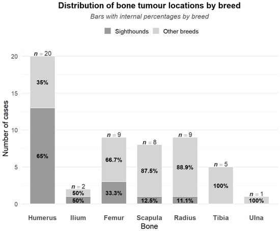
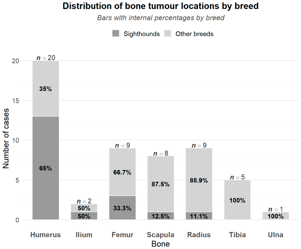
The most frequent location of the tumour was the humerus, with 20/54 (37%) cases. This was followed by the radius and femur, each with nine (16.7%) cases. Eight (14.8%) arose from the scapula, five (9.3%) from the tibia, two (3.7%) from the ilium and one (1.8%) from the ulna. Of the tumours arising from the humeri, 19/20 were diagnosed with OSA, the proximal humerus representing the most frequent location for OSA tumours (p < 0.001; chi-square test). OSA of the humerus was more prevalent in sighthound breeds (p < 0.01, Fisher’s exact test).
Non-OSA group locations were the humerus (one chondrosarcoma), femur (one haemangiosarcoma), radius (one non-specific sarcoma), and scapula (one haemangiosarcoma, one round-cell).
The proximal third of the bone was affected in 31/54 (57.4%) of the dogs; the distal third in 14/54 (25.9%), the middle third in 7/54 (13.0%), and cranial pelvis in 2/54 (3.7%). The majority of tumours, 38/54 (70.4%), involved the thoracic limb, including 4/5 non-OSA group and 34 OSA (34/49, 69.4%) (Figure 1).
Figure 1.
Distribution of the location of primary appendicular bone neoplasia in sighthounds and other breeds.
Permeative osteolysis was seen in 41/54 (75.9%) dogs, 38/41 (92.7%) with OSA and 3/41 (7.3%) non-OSA (two haemangiosarcoma and one non-specific sarcoma). Moth-eaten osteolysis was demonstrated in 7/54 (11.1%) (five OSA, one chondrosarcoma, and one round-cell tumour), followed by 4/54 (7.4%) with a mixed moth-eaten and permeative osteolytic pattern (OSA). Osteolysis was not identified in 2/54 (3.7%) dogs (two OSA). Permeative osteolytic patterns (45/54) were significantly (p < 0.001; Fisher’s exact test) associated with cortical osteolysis and osteolytic patterns while moth-eaten (11/54) changes were associated with mixed osteolytic/osteoblastic patterns (p < 0.001; Fisher’s exact test).
Almost all dogs, 49/54 (90.7%) had cortical lysis (46 OSA, two haemangiosarcoma, and one non-specific sarcoma) and 51/54 (94.4%) had cancellous bone lysis (47 OSA, two hemangiosarcoma, one chondrosarcoma, and one non-specific sarcoma).
A total of 31 dogs (57.4%) demonstrated an osteolytic pattern, (29 OSA and two haemangiosarcoma), 2/54 (3.7%) dogs had an osteoblastic pattern (one OSA, one round-cell), 20/54 (37.0%) dogs had a mixture of osteolytic and osteoblastic patterns (18 OSA, one non-specific sarcoma, and one chondrosarcoma), and one dog with OSA (1.9%) did not present any pattern. Of the OSA patients 29/49 (59.2%) exhibited an osteolytic pattern, 1/49 (2%) osteoblastic, and 18/49 (36.7%) a mixed osteolytic and osteoblastic pattern. From the non-OSA group, an osteolytic pattern was seen in the 2/5 cases, both haemangiosarcoma, an osteoblastic pattern In the round-cell neoplasm, and a mixed pattern in both the non-specific sarcoma and the chondrosarcoma case.
Periosteal reaction was frequently mild in cases with osteolytic tumours (23/27; p < 0.001; Cochran–Armitage test) and severe (moderate or marked grading) in mixed osteolytic/osteoblastic tumours (17/19; p < 0.001; Cochran–Armitage test).
A total of 13/54 (24.1%) dogs demonstrated pathological fractures; 12 with OSA and one with haemangiosarcoma. Four dogs presented with a fracture in the humerus (three proximal, one mid diaphysis), five in the femur (two proximal, one mid diaphysis, two distal), three in the radius (one proximal, two distal) and one in the proximal tibia (Table 1). CT examples of the periosteal reaction can be found in Appendix A (Figure A2 and Figure A3).
Table 1.
Tumour location and presence/absence of a pathological fracture identified by CT.
A periosteal reaction was present in 49/54 and absent in 5/54 dogs. Only dogs with OSA exhibited marked periosteal reactions (7/49, 14.3%). In the OSA group, there were also 23 dogs with mild and 14 dogs with moderate periosteal reactions. In the non-OSA group (n = 5), four had a mild periosteal reaction (one haemangiosarcoma, one chondrosarcoma, one round-cell tumour, and one non-specific sarcoma) and one had a moderate periosteal reaction (haemangiosarcoma). All five dogs without periosteal reactions had OSA.
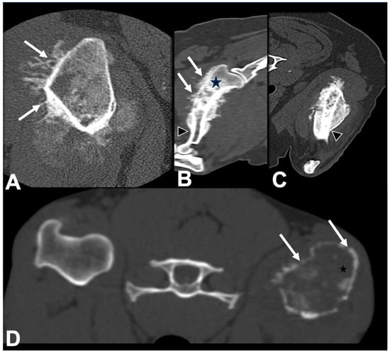
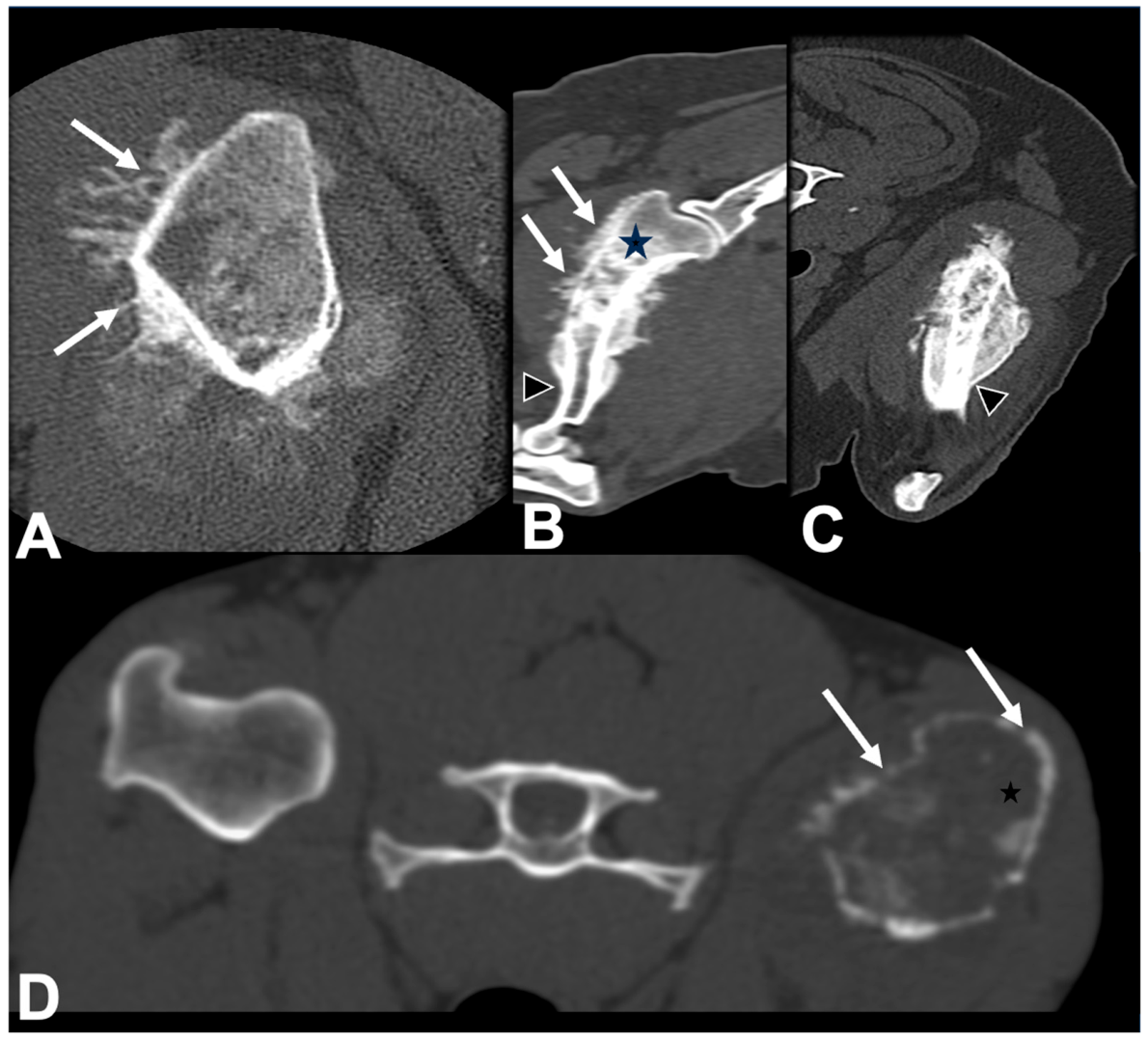
Periosteal reaction was most often amorphous, occurring in 26/54 (48.1%) dogs, 23 with OSA, and three of the non-OSA group (one non-specific sarcoma, two hemangiosarcoma). Sixteen out of 54 (29.6%) dogs demonstrated a brush-border periosteal reaction of which 14/54 were OSA, one chondrosarcoma, and one round-cell tumour. Twelve out of 54 (22.2%) dogs (11 with OSA; one with chondrosarcoma) had a spiculated sunburst periosteal reaction (Figure 2) and 12/54 (22.2%) dogs had a rough and solid irregular periosteal reaction (11 with OSA and one with non-specific sarcoma). Spiculated sunburst periosteal reaction was significantly associated with absence of pathological fracture (p < 0.05, Fisher’s exact test).
Figure 2.
CT images (bone window) of the right humerus (A) (transverse plane) left humerus (B,C) (Multiplanar reconstruction—MPR in dorsal and sagittal planes) and left humerus (D) (transverse plane) of three dogs with osteosarcoma. Image (A) demonstrates the spiculated perpendicular and spiculated sunburst periosteal reaction (arrows); image (B,C) show mixed amorphous and spiculated perpendicular periosteal reaction (arrows), sclerosis of the cancellous bone (star), and Codman’s triangle (arrow head); image (D) displays permeative osteolysis (star) and cortical osteolysis (arrows).
Other types of periosteal reaction were less commonly seen, with 4/54 (7.4%) dogs demonstrating palisading and 5/54 (9.2%) spiculated perpendicular periosteal reactions, all of which had OSA.
Only two dogs out of 49 with OSA (4.1%) demonstrated a lamellated periosteal reaction while none of the non-OSA group did. A smooth and solid periosteal reaction was only seen in 1/49 (2%), a dog with OSA.
A total of 15/54 (27.8%) dogs with appendicular bone tumour had a Codman’s triangle; 12 OSA dogs and 3/5 non-OSA (one round-cell tumour, one non-specific sarcoma, one haemangiosarcoma).
Bone sclerosis was present in 25/54 (46.3%) dogs, where 24 had OSA and one chondrosarcoma. Sclerosis was not seen in 4/5 non-OSA cases (Table 2).
Table 2.
Prevalence of periosteal reaction, osteolysis, sclerosis, and Codman’s triangle in chondrosarcoma, haemangiosarcoma, OSA, round-cell tumour, and non-specific sarcoma.
Bone-marrow changes were identified in 52/54 cases (96.3%), including all 49 (n = 49, 100%) OSA, one chondrosarcoma, one haemangiosarcoma, and one non-specific sarcoma. Changes were heterogeneous in 41 cases and homogeneous in 11 cases (all OSA). No bone-marrow changes were observed in one dog with haemangiosarcoma and in one dog with round-cell tumour.
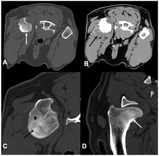
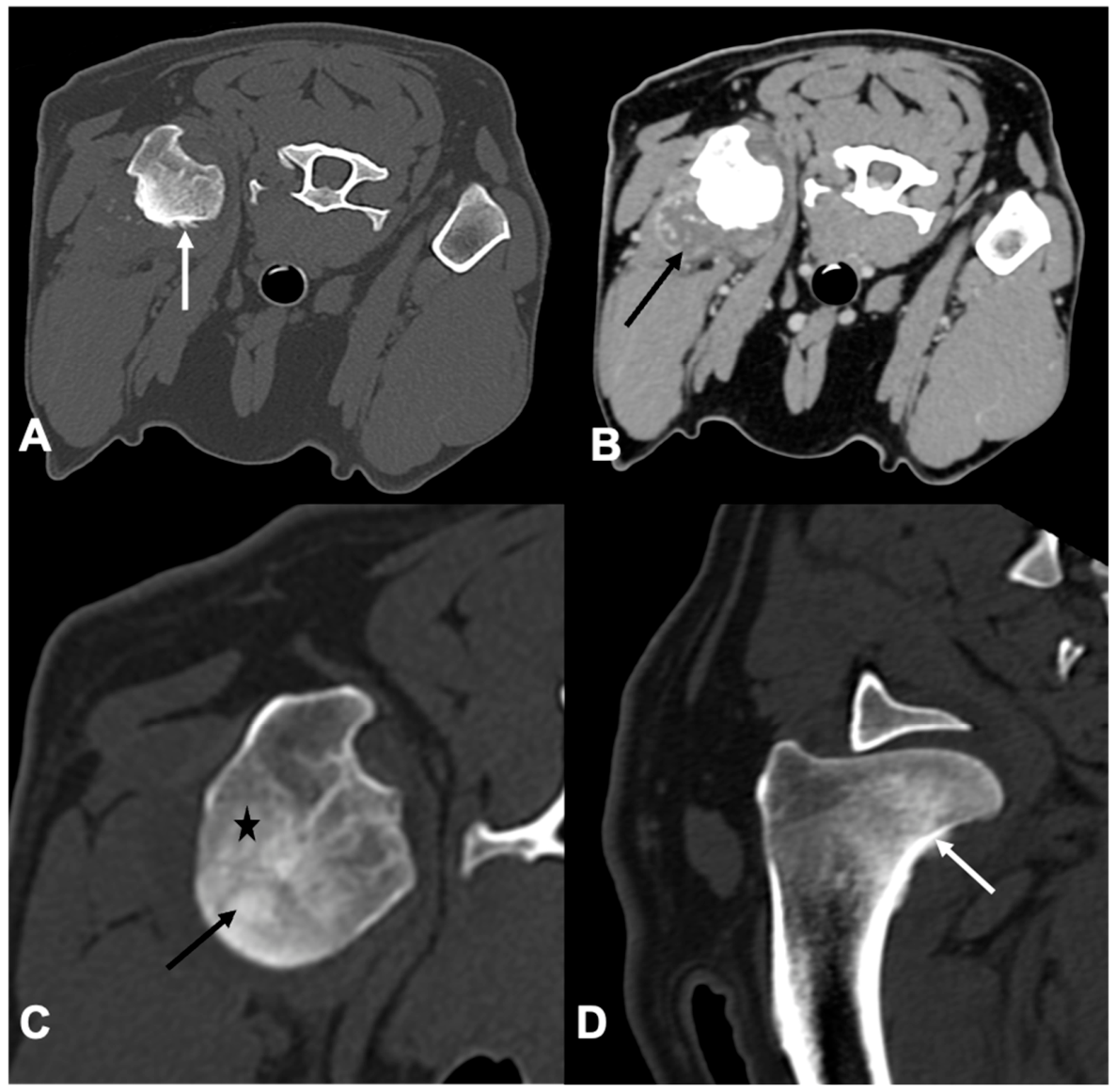
The tumour growth had an extra-compartmental component in 33/54 (61.1%) dogs of which 29 (n = 49, 59.2%) were OSA and four were non-OSA (one non-specific sarcoma, two haemangiosarcoma, one chondrosarcoma). Twenty-one out of 54 (38.9%) remained intra-compartmental, of which 20 (n = 49, 40.8%) were OSA and one non-OSA (round-cell) (Figure 3).
Figure 3.
CT images of the right humerus of a 10-month-old Irish setter diagnosed with a chondrosarcoma (A,C,D), post-contrast bone window (B), post-contrast soft tissue window in a transverse plane (A–C), and multiplanar reconstruction—(MPR) in a sagittal plane. The images display a moth-eaten osteolysis ((C), star) and sclerosis ((C), arrow) of the epiphysis and metaphysis of the right humerus associated with a heterogeneously contrast enhancing soft tissue mass-like lesion ((B), arrow) and focal brush-border ((D), white arrow) and spiculated sunburst periosteal reaction ((A), white arrow).
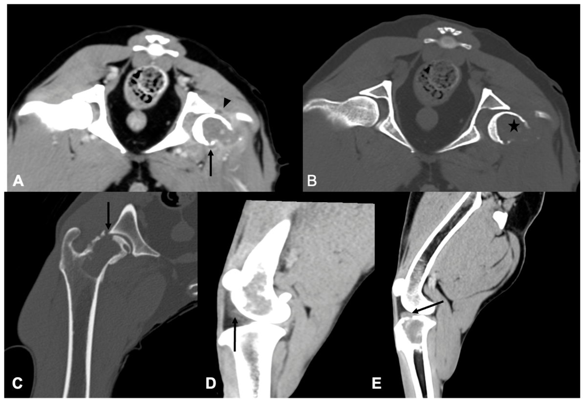
Indirect non-specific signs of joint involvement were seen in 11/54 dogs, of which eight were in conjunction with direct signs, nine dogs demonstrating irregular synovial thickening, and all 11 dogs having a joint effusion (one mild, five moderate, and five marked). Out of these 11, heterogeneous enhancement of the synovium was seen in all dogs (eight of them) receiving contrast medium (Figure 4).
Figure 4.
CT images of the left coxo-femoral joint (A,B) (transverse plane in soft tissue and bone windows), right coxo-femoral joint (C) (MPR dorsal plane in bone window), left femur (D) (MPR sagittal plane in post-contrast soft tissue window), and left stifle (E) (MPR sagittal plane post-contrast in soft tissue window) of four dogs with osteosarcoma. These images show some of the direct and indirect non-specific features compatible with joint involvement. Images (A,B) demonstrate a moderate synovial thickening (arrow head in (A)), mild joint effusion (arrow in (A)) and marked bone osteolysis (star in (B)); image (C) displays intra-articular osteolysis of the femoral head (arrow); image (D) displays joint effusion (arrow) with neoplasia of the distal femur; and image (E) displays findings consistent with invasion of tumour tissue into the joint (arrow).
Of the 11 dogs with indirect, non-specific joint changes, 10 had OSA and one chondrosarcoma. The pelvic limb was more often affected (five stifles, two coxo-femoral joints). The shoulder joint was affected in four dogs. In nine dogs, the tumour was associated with an extra-articular soft tissue mass-like lesion (Table 3).
Table 3.
Prevalence of type of tumour growth, joint involvement, and the presence of a soft tissue mass-like lesion in chondrosarcoma, haemangiosarcoma, OSA, round-cell tumour and non-specific sarcoma.
Direct signs of joint involvement were present in eight dogs (14.8% of the total cases; all with OSA), all of them demonstrating intra-articular bone disruption/lysis, and three of them showing findings compatible with intra-synovial tumour tissue invasion.
Diffuse soft tissue swelling was present in 13/54 (24.1%) dogs (12 OSA and one haemangiosarcoma).
Soft tissue mass-like lesion was identified in 31/54 (57.4%) dogs, including 28 dogs with OSA and three from the non-OSA group (two haemangiosarcoma and one chondrosarcoma).
Fourteen out of 54 dogs (all with OSA) did not receive intravenous contrast medium. The bulk of cases that received contrast medium (40/54) demonstrated contrast enhancement 38/40 (95%), 34 of which were OSA and four non-OSA (one chondrosarcoma, two haemangiosarcoma, one non-specific sarcoma). Mild contrast enhancement was most common, present in 29/40 (72.5%), of which 26 were diagnosed with OSA and three non-OSA neoplasia (one chondrosarcoma, one haemangiosarcoma, one non-specific sarcoma). There were eight cases with moderate contrast enhancement, seven of which were OSA and one was haemangiosarcoma. Only one dog with OSA exhibited marked contrast enhancement. The two cases that did not contrast enhance were diagnosed with round-cell tumour and OSA.
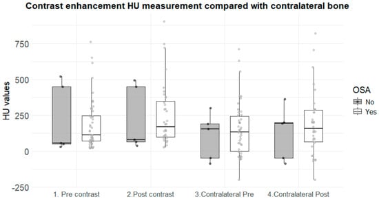
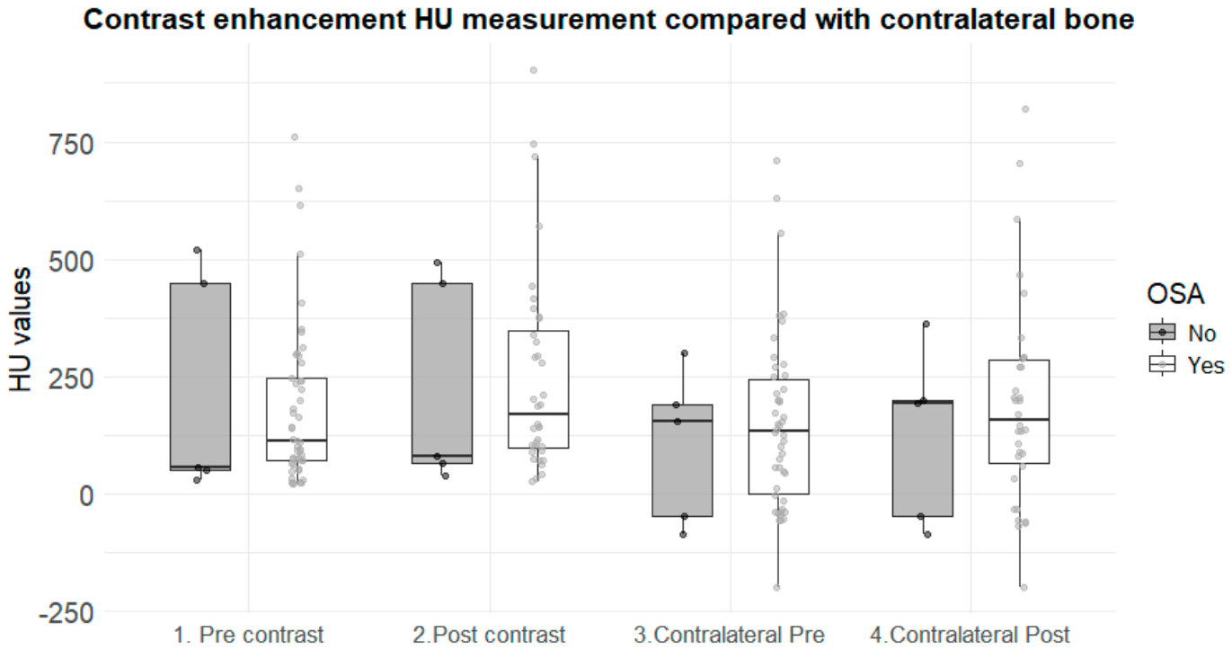
The attenuation of the central tumour lesions was increased by an average of +39 HU (median +27 HU, interquartile range 51 HU) in the post-contrast studies compared with an average of +20 HU (median +6 HU, interquartile range 33 HU) in the same bone location of the contralateral healthy bones. Paired rank analysis confirmed that contrast enhancement was significantly higher in the tumour lesions than in the healthy contralateral bone (p < 0.05; Wilcoxon signed-rank test). When present, most contrast enhancement was heterogeneous in nature (Figure 5).
Figure 5.
Boxplots illustrating the distribution of Hounsfield unit (HU) values in the osteosarcoma (OSA; white boxes) and non-OSA (grey boxes) groups, and the differences in pre- and post-contrast attenuation between affected bones and their contralateral healthy counterparts. Groups 1 and 2 represent affected limbs before and after contrast medium administration, respectively, whereas Groups 3 and 4 represent the corresponding contralateral non-affected limbs before and after contrast medium administration. The increase in HU values following contrast medium administration was significantly greater in affected limbs (+39 HU) than in contralateral non-affected limbs (+20 HU) (p < 0.05, Wilcoxon signed-rank test).
Thoracic CT examinations were available for 51/54 cases. Nodules presumed to represent lung metastases were seen in 17/51 (33.3%) of the cases, 15 of which were OSA (15/49, 30.6%), the remaining cases being a haemangiosarcoma and a round-cell tumour.
3.4. Case Management
Palliative treatment with anti-inflammatories and analgesics was the predominant management approach (19/54, 35.2%), followed by immediate euthanasia (14/54, 25.9%) or a combination of amputation and chemotherapy (12/54, 22.2%). Amputation alone was performed in 5/54 cases (9.3%), while limb-sparing surgery with chemotherapy, chemotherapy alone, and limb-sparing surgery alone were performed in 2/54 (3.7%), 1/54 (1.9%), and 1/54 (1.9%) cases, respectively.
From the OSA group, 18 dogs received palliative treatment, 14 dogs were euthanised after initial diagnosis, 11 dogs were treated with a combination of amputation and chemotherapy, two dogs received amputation, two dogs received limb-sparing treatment with chemotherapy, one dog received limb sparing surgery only, and one received chemotherapy only.
The most common treatment of the non-OSA group was amputation (3/5 being one each of chondrosarcoma, haemangiosarcoma, and non-specific sarcoma). A combination of amputation and chemotherapy was used in one case of haemangiosarcoma whilst the single patient with round-cell neoplasia received palliative treatment.
4. Discussion
This study represents a comprehensive description of CT characteristics for appendicular bone tumours in dogs. Most dogs within our study population were diagnosed with OSA (49/54, 90.7%) in agreement with its predominance in primary bone neoplasia in dogs [1,14].
In keeping with previous literature, our findings showed that the most frequent location of appendicular bone OSA was the proximal humerus (p < 0.001; chi-square test), and it was more prevalent in sighthound breeds (p < 0.01; Fisher’s exact test). Most dogs in our study weighed more than 25 kg (p < 0.001; chi-square test), supporting the previously reported predominance of OSA in large and giant-breed dogs. Similarly, most dogs were older than 7 years (p < 0.001; chi-square test). Previous studies have also reported a higher prevalence of OSA in male dogs. In our study, although males were numerically overrepresented, this difference was not statistically significant [1,3,7,11,12,24,25].
As observed with other imaging modalities in dogs with appendicular bone neoplasia, osteolysis, periosteal reactions, soft tissue mass-like lesion, and Codman’s triangle were frequently present in our patients [7,13,16].
Spiculated sunburst periosteal reaction was significantly associated with absence of pathological fracture (p < 0.05; Fisher’s exact test); however, this association must be interpreted with caution as it could be influenced by factors such as anatomical location or mechanical loading among others.
A study published in 2018 describing appendicular hemangiosarcoma in 41 dogs reported low numbers of cases presenting with periosteal reaction in long bones (14/41, 34.1%) and high numbers with lysis (37/41, 90.2%) [8].
In our study, 13/54 (24%) dogs presented with pathological fractures, most commonly affecting the femur and proximal long bones. In a study by Rubin et al. (2015) [26] a higher percentage (38%) of pathological fractures was reported, which in accordance with our results, most commonly affected the femur. In human medicine, the incidence rate of pathological fractures in appendicular bone neoplasia varies from 5 to 10% [2,27]. In our study, pathological fractures involved the tibia in one case out of 13 fractures (7.7%) and the radius in three cases out of 13 fractures (23.1%). This finding contrasts with previous reports, in which radial pathological fractures were described less frequently [26,28].
Sighthound breeds were significantly overrepresented in our cases (p < 0.001; chi-square test), accounting for 35% (19/54; 12 Greyhounds, six Lurcher, one Scottish Deerhound) of the dogs considering they represent less than 5% of the UK census population [29,30]. These findings are consistent with former research studies that stated that canine predisposition to OSA was greatest among large and giant breeds including Scottish Deerhounds, Leonbergers, Great Danes, Rottweilers, Irish Wolfhounds, St. Bernards and Greyhounds [10,11,24,25].
Our study found a higher incidence of pathological fractures in sighthounds (six out of 19, 31.6%) in comparison with the other-breeds group (seven out of 35, 20%); however, this difference did not reach statistical significance. Differences in bone loading related to conformation and behaviour and the higher incidence of sighthounds in the study could account for this finding [25].
In the assessment for metastatic disease, thoracic CT demonstrated pulmonary nodules presumed to represent metastases in 17 out of 51 dogs (33%) (15 with OSA, one with haemangiosarcoma, and one with round cell tumour). This represents a higher proportion of presumptive metastases at initial clinical presentation than previously reported, with prior studies documenting detectable pulmonary metastasis in 15% and 26% of dogs determined by radiographic and CT studies respectively [1,14,31,32,33]. The lower percentage recognised in radiographic studies is consistent with its lower sensitivity in the detection of pulmonary nodules in dogs compared to CT [34].
Joint invasion in canine appendicular bone OSA is considered rare, as these tumours generally do not to extend across joint spaces [7,12,13,14]. It is hypothesised that the articular cartilage and growth plates function as a natural barrier to tumour invasion [1,7,14]. Despite this, exceptions have been described, particularly in cases involving the femoral head where it invades the acetabulum via round ligament [12,13]. One case report describes a dog diagnosed with extra-skeletal OSA within the elbow synovium [35].
In contrast, 8/54 (14.8%) dogs in this study displayed features compatible with tumour joint involvement. All eight dogs demonstrated intra-articular bone disruption and three dogs showed additional findings compatible with intra-synovial tumour invasion. Non-specific features such as joint effusion was also seen in 11 dogs, with heterogeneous synovial enhancement in all cases where contrast medium was administered. An associated soft tissue mass-like lesion was identified in nine cases.
Human medicine studies report that OSA joint involvement occurs with a prevalence between 19 and 24% [36,37,38]. MRI is considered the imaging modality of choice for local staging of primary bone tumours [17,37,38,39,40]. Direct and indirect MRI findings of joint involvement are routinely assessed in combination to determine if there is neoplastic joint involvement [36,38,41,42]. Pre-operative MRI is used extensively in human patients with appendicular neoplasia delineating the tumour extent precisely to determine whether limb-preserving surgery is feasible. Amputation with extra-articular resection is more likely to be performed if joint invasion is detected [41].
Tamsel et al. 2025 stated that MRI intra-articular bone destruction and capsular invasion had a sensitivity of 84% and 92%, respectively, in diagnosing joint invasion in human patients [36]. Joint effusion alone has a limited predictive value for intra-articular tumour invasion [38].
Evaluation of joint involvement in our study is very limited. The indirect imaging features of synovial thickening, joint effusion, and enhancement of the synovium provide limited information alone and could be associated with other benign pathologies, hence histopathology would be still required for diagnosing tumour joint invasion.
A recently published study evaluating primary appendicular tumours in 20 dogs with MRI describes imaging findings similar to those previously documented in human patients; however, joint assessment was not explored [23].
MRI is a modality that might be used in veterinary medicine for further tumour characterisation and recognition of joint involvement which may be of benefit in determining appropriate level of amputation and feasibility of limb sparing surgery. Limb-sparing surgery is performed uncommonly, with case selection and complications described in the recently published census and guidelines for the treatments of canine appendicular osteosarcoma [13], but it is increasingly reported in veterinary medicine [43,44,45]. Further research involving larger and more diverse populations may be of value in veterinary medicine in order to standardise the CT and MRI imaging features for accurate assessment of tumour tissue extension, joint invasion, and staging.
There are several limitations in the design of this study. The number of cases with appendicular bone tumours other than OSA (non-OSA group) is low with only 1–2 cases per tumour type. Consequently, although descriptions of these cases are offered in this article there is a high risk of individual bias if extrapolated. There is a selection bias inherent to the study population, as all cases originated from referral hospitals. Additional limitations include the inherent subjectivity of certain grading scales used to assess imaging features, potential age-related confounding in the interpretation of presumed pulmonary metastases, and the lack of formal assessment of interobserver agreement. Statistical analysis reports unadjusted p-values that should be considered with caution; further studies are warranted to validate the associations observed and confirm any hypotheses generated by the patterns observed in this study.
An important limitation of this study was that definitive pathology reports confirming joint invasion were not performed, and the direct signs used to describe joint involvement were extrapolated from human MRI studies using the features that were likely to be detected with CT. Imaging features could not be correlated with histopathological margins. Furthermore, the fact that the final diagnosis of 19 cases was based on cytological examination rather than histopathology may have limited the ability to achieve more precise tumour classification and histological subtyping.
Given the retrospective design of this study involving participation of multiple hospitals over a prolonged inclusion period (nine years), CT imaging acquisition and contrast-medium administration protocols were not standardised. These could affect the post-contrast attenuation measurements. Another limitation of the study is that a sizeable proportion of cases (14/54) did not receive contrast medium during CT acquisition limiting the image interpretation.
5. Conclusions
This study describes the CT imaging features of primary appendicular bone tumours in dogs, including the description of findings compatible with joint involvement [7,12,13]. Further investigation of the direct and indirect features of joint invasion by primary bone tumours may be warranted using CT and MRI with histopathological correlation to define and standardise characterisation of findings.
The evaluation of CT features may have the potential to provide valuable information to facilitate early diagnosis, support treatment planning, and guide the selection of the most appropriate therapeutic approach.
Author Contributions
Conceptualisation, M.V., E.M., and B.A.J.G.; methodology, M.V., E.M., and B.A.J.G.; validation, M.V., E.M., and B.A.J.G.; formal analysis, M.V. and J.H.-G.; investigation, M.V., E.M., and B.A.J.G.; resources, M.V., I.A., M.T., H.R., A.K., L.M., A.R.R.F., B.A.J.G.; data curation, M.V., J.H.-G., E.M., and B.A.J.G.; writing—original draft preparation, M.V., E.M., and B.A.J.G.; writing—review and editing, M.V., J.H.-G., I.A., M.T., H.R., A.K., L.M., A.R.R.F., E.M., and B.A.J.G.; visualisation, M.V., J.H.-G., I.A., M.T., H.R., A.K., L.M., A.R.R.F., E.M., and B.A.J.G.; supervision, M.V., E.M., and B.A.J.G.; project administration, M.V., E.M., and B.A.J.G.;. All authors have read and agreed to the published version of the manuscript.
Funding
This research received no external funding.
Institutional Review Board Statement
The animal study protocol was approved by Ethics Review Panel Board of The Royal College Veterinary Surgeons (RCVS) (2025-133-Villuendas/12 May 2026).
Informed Consent Statement
Written informed consent has been obtained from the patients’ owners.
Data Availability Statement
The data presented in this study are available on request from the corresponding author due to ethical restrictions.
Conflicts of Interest
M.V., E.M., and B.A.J.G. are employed by Veterinary Specialists Scotland; I.A. is employed by Davies Veterinary Specialists; A.R.R.F. is employed by Dick White Referrals; L.M. is employed by Paragon Veterinary Referrals; M.T. is employed by North Downs Specialist Referrals; H.R. is employed by Eastcott Veterinary Referrals; and A.K. is employed by Northwest Veterinary Specialists. All the authors declare that the research was conducted in the absence of any commercial or financial relationships that could be construed as a potential conflict of interest.
Abbreviations
The following abbreviations are used in this manuscript:
| CT | Computed Tomography |
| DFOV | Displayed Field of View |
| DICOM | Digital Imaging and Communications in Medicine |
| ECVDI | European College of Veterinary Diagnostic Imaging |
| HU | Hounsfield Units |
| Kvp | Kilovoltage Peak |
| mAS | Milliampere Per Second |
| MRI | Magnetic Resonance Imaging |
| OSA | Osteosarcoma |
| PACS | Picture Archiving and Communication System |
| RCVS | Royal College Veterinary Surgeons |
| ROI | Region of Interest |
Appendix A
Figure A1.
Flowchart representing of the case-selection process of this study.
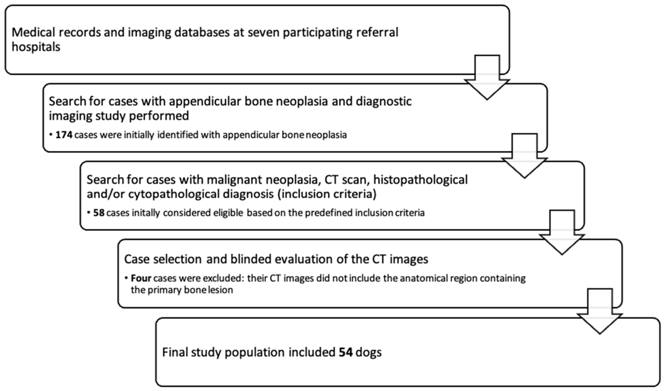
Figure A2.
Examples of different types of periosteal reaction: (A) Spiculated sunburst periosteal reaction (white arrow in (A)): CT bone window in transverse plane of the proximal humerus of a dog with moderate spiculated sunburst periosteal reaction. (B) Spiculated perpendicular periosteal reaction (white arrow in (B)): CT bone window in transverse plane of the proximal humerus of a dog. (C) Brush-border peristoteral reaction (white arrow in (C)): CT bone window in transverse plane of a scapula.
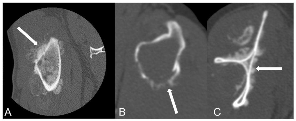
Figure A3.
Other examples of different types of periosteal reaction. (A) Smooth and solid periosteal reaction (white arrow in (A)): CT bone window in sagittal plane multiplanar reconstruction of the proximal humerus of a dog with mild smooth and solid periosteal reaction. (B) Lamellar periosteal reaction (white arrow in (B)): CT bone window in transverse plane of the distal radius of a dog with moderate/marked lamellated periosteal reaction. (C) Palisading periosteal reaction (white arrow in (C)): CT bone window in dorsal-plane multiplanar reconstruction of a distal radius with moderate/marked lamellated periosteal reaction. (D) Mild morphous (white arrow in (D)) and palisading (black arrow in (D)) periosteal reaction: CT bone window, transverse plane of a proximal tibia of a dog with an implant causing mild artefact.
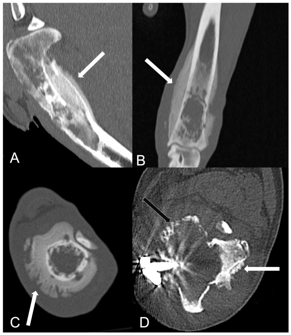
Table A1.
Computed tomography (CT) technical parameters for 54 dogs included in the study.
References
- Ehrhart, N.P.; Christensen, N.I.; Fan, T.M. Tumors of the Skeletal System. In Withrow and MacEwen’s Small Animal Clinical Oncology; Vail, D., Thamm, D.H., Liptak, J.M., Eds.; Elsevier: St. Louis, MO, USA, 2020; pp. 524–564. [Google Scholar]
- Cole, S.; Gianferante, D.M.; Zhu, B.; Mirabello, L. Osteosarcoma: A Surveillance, Epidemiology, and End Results Program-based Analysis from 1975 to 2017. Cancer 2022, 128, 2107–2118. [Google Scholar] [CrossRef] [Scilit] [PubMed]
- Simpson, S.; Dunning, M.D.; de Brot, S.; Grau-Roma, L.; Mongan, N.P.; Rutland, C.S. Comparative Review of Human and Canine Osteosarcoma: Morphology, Epidemiology, Prognosis, Treatment and Genetics. Acta Vet. Scand. 2017, 59, 71. [Google Scholar] [CrossRef] [Scilit] [PubMed]
- Alexander, J.W.; Patton, C.S. Primary Tumors of the Skeletal System. Vet. Clin. N. Am. Small Anim. Pract. 1983, 13, 181–195. [Google Scholar] [CrossRef] [Scilit] [PubMed]
- Morello, E.; Martano, M.; Buracco, P. Biology, Diagnosis and Treatment of Canine Appendicular Osteosarcoma: Similarities and Differences with Human Osteosarcoma. Vet. J. 2011, 189, 268–277. [Google Scholar] [CrossRef] [Scilit] [PubMed]
- Popovitch, C.A.; Weinstein, M.J.; Goldschmidt, M.H.; Shofer, F.S. Chondrosarcoma: A Retrospective Study of 97 Dogs (1987–1990). J. Am. Anim. Hosp. Assoc. 1994, 30, 81–85. [Google Scholar]
- Thrall, D.E. Principles of Radiographic Interpretation of the Appendicular Skeleton and Radiographic Features of Bone Tumors. In Thrall’s Textbook of Veterinary Diagnostic Radiology; Seiler, G., Thrall, D.E., Eds.; Elsevier: St. Louis, MO, USA, 2024; pp. 348–364. [Google Scholar]
- Giuffrida, M.A.; Kamstock, D.A.; Selmic, L.E.; Pass, W.; Szivek, A.; Mison, M.B.; Boston, S.E.; Fox, L.E.; Robat, C.; Grimes, J.A.; et al. Primary Appendicular Hemangiosarcoma and Telangiectatic Osteosarcoma in 70 Dogs: A Veterinary Society of Surgical Oncology Retrospective Study. Vet. Surg. 2018, 47, 774–783. [Google Scholar] [CrossRef] [Scilit] [PubMed]
- Bingel, S.A.; Brodey, R.S.; Allen, H.L.; Riser, W.H. Haemangiosarcoma of Bone in the Dog. J. Small Anim. Pract. 1974, 15, 303–322. [Google Scholar] [CrossRef] [Scilit] [PubMed]
- O’Neill, D.G.; Edmunds, G.L.; Urquhart-Gilmore, J.; Church, D.B.; Rutherford, L.; Smalley, M.J.; Brodbelt, D.C. Dog Breeds and Conformations Predisposed to Osteosarcoma in the UK: A VetCompass Study. Canine Med. Genet. 2023, 10, 8. [Google Scholar] [CrossRef] [Scilit] [PubMed]
- Egenvall, A.; Nødtvedt, A.; von Euler, H. Bone Tumours in a Population of 400 000 Insured Swedish Dogs up to 10 y of Age: Incidence and Survival. Can. J. Vet. Res. 2007, 71, 292–299. [Google Scholar] [PubMed]
- Gibbs, C.; Denny, H.R.; Kelly, D.F. The Radiological Features of Osteosarcoma of the Appendicular Skeleton in Dogs: A Review of 74 Cases. J. Small Anim. Pract. 1984, 25, 177–192. [Google Scholar] [CrossRef] [Scilit]
- Polton, G.; Borrego, J.F.; Clemente-Vicario, F.; Clifford, C.A.; Jagielski, D.; Kessler, M.; Kobayashi, T.; Lanore, D.; Queiroga, F.L.; Rodrigues, L.; et al. Osteosarcoma of the Appendicular Skeleton in Dogs: Consensus and Guidelines. Front. Vet. Sci. 2025, 12, 1633593. [Google Scholar] [CrossRef] [Scilit] [PubMed]
- Dernell, W.S. Tumours of the Skeletal System. In BSAVA Manual of Canine and Feline Oncology; Dobson, J.M., Lascelles, B.D., Eds.; British Small Animal Veterinary Association: Gloucester, UK, 2016; pp. 159–177. [Google Scholar]
- McAllister, H.; Tobin, E. Long Bones—Mature. In BSAVA Manual of Canine and Feline Musculoskeletal Imaging; Kirberger, R.M., McEvoy, F.J., Eds.; BSAVA: Gloucester, UK, 2016; pp. 108–132. [Google Scholar]
- Vanel, M.; Blond, L.; Vanel, D. Imaging of Primary Bone Tumors in Veterinary Medicine: Which Differences? Eur. J. Radiol. 2013, 82, 2129–2139. [Google Scholar] [CrossRef] [Scilit] [PubMed]
- Cè, M.; Cellina, M.; Ueanukul, T.; Carrafiello, G.; Manatrakul, R.; Tangkittithaworn, P.; Jaovisidha, S.; Fuangfa, P.; Resnick, D. Multimodal Imaging of Osteosarcoma: From First Diagnosis to Radiomics. Cancers 2025, 17, 599. [Google Scholar] [CrossRef] [Scilit] [PubMed]
- Martin, T.W.; LaRue, S.M.; Griffin, L. CT Characteristics and Proposed Scoring Scheme Are Predictive of Pathologic Fracture in Dogs with Appendicular Osteosarcoma Treated with Stereotactic Body Radiation Therapy. Vet. Radiol. Ultrasound. 2022, 63, 82–90. [Google Scholar] [CrossRef] [Scilit] [PubMed]
- Garcia, T.C.; Steffey, M.A.; Zwingenberger, A.L.; Daniel, L.; Stover, S.M. CT-Derived Indices of Canine Osteosarcoma-Affected Antebrachial Strength. Vet. Surg. 2017, 46, 549–558. [Google Scholar] [CrossRef] [Scilit] [PubMed]
- Steffey, M.A.; Garcia, T.C.; Gorney, A.; Stover, S.M.; Zwingenberger, A.L. Computed Tomography-Derived Structural Analysis for the Likelihood of Pathologic Fracture in Canine Antebrachial Osteosarcoma. Vet. Radiol. Ultrasound. 2024, 65, 603–612. [Google Scholar] [CrossRef] [Scilit] [PubMed]
- Karnik, K.S.; Samii, V.F.; Weisbrode, S.E.; London, C.A.; Green, E.M. Accuracy of Computed Tomography in Determining Lesion Size in Canine Appendicular Osteosarcoma. Vet. Radiol. Ultrasound. 2012, 53, 273–279. [Google Scholar] [CrossRef] [Scilit] [PubMed]
- Davis, G.J.; Kapatkin, A.S.; Craig, L.E.; Heins, G.S.; Wortman, J.A. Comparison of Radiography, Computed Tomography, and Magnetic Resonance Imaging for Evaluation of Appendicular Osteosarcoma in Dogs. J. Am. Vet. Med. Assoc. 2002, 220, 1171–1176. [Google Scholar] [CrossRef] [Scilit] [PubMed]
- Sapet, J.; Gatel, L.; Floch, F.; Vanel, M. Magnetic Resonance Imaging Features of Primary Appendicular Bone Sarcomas in 20 Dogs. J. Small Anim. Pract. 2026, 64, 343–352. [Google Scholar] [CrossRef] [Scilit] [PubMed]
- Rosenberger, J.A.; Pablo, N.V.; Crawford, C.P. Prevalence of and Intrinsic Risk Factors for Appendicular Osteosarcoma in Dogs: 179 Cases (1996–2005). J. Am. Vet. Med. Assoc. 2007, 231, 1076–1080. [Google Scholar] [CrossRef] [Scilit] [PubMed]
- Ru, G.; Terracini, B.; Glicrman, L.T. Host Related Risk Factors for Canine Osteosarcoma. Vet. J. 1998, 156, 31–39. [Google Scholar] [CrossRef] [Scilit] [PubMed]
- Rubin, J.A.; Suran, J.N.; Brown, D.C.; Agnello, K.A. Factors Associated with Pathological Fractures in Dogs with Appendicular Primary Bone Neoplasia: 84 Cases (2007–2013). J. Am. Vet. Med. Assoc. 2015, 247, 917–923. [Google Scholar] [CrossRef] [Scilit] [PubMed]
- Vermesan, D.; Vermesan, H.; Dragulescu, S.I.; Bera, I.; Di Giovanni, A.; Sabatini, R.; Santacroce, L.; Bottalico, L.; Flace, P.; Cagiano, R. Secondary Pathologic Fractures in Osteosarcoma: Prognosis and Evolution. Eur. Rev. Med. Pharmacol. Sci. 2009, 13, 71–76. [Google Scholar] [PubMed]
- Norquest, C.J.; Maitz, C.A.; Keys, D.A.; Moore, M.; Bryan, J.N.; Ehling, T.J.; Lattimer, J.C.; Flesner, B.K. Fracture Rate and Time to Fracture in Dogs with Appendicular Osteosarcoma Receiving Finely Fractionated Compared to Coarsely Fractionated Radiation Therapy: A Single Institution Study. Vet. Med. Sci. 2022, 8, 1013–1024. [Google Scholar] [CrossRef] [Scilit] [PubMed]
- O’Neill, D.G.; McMillan, K.M.; Church, D.B.; Brodbelt, D.C. Dog Breeds and Conformations in the UK in 2019: VetCompass Canine Demography and Some Consequent Welfare Implications. PLoS ONE 2023, 18, e0288081. [Google Scholar] [CrossRef] [Scilit] [PubMed]
- McMillan, K.M.; Harrison, X.A.; Wong, D.C.; Upjohn, M.M.; Christley, R.M.; Casey, R.A. Estimation of the Size, Density, and Demographic Distribution of the UK Pet Dog Population in 2019. Sci. Rep. 2024, 14, 31746. [Google Scholar] [CrossRef] [Scilit] [PubMed]
- Dobson, J.M.; Samuel, S.; Milstein, H.; Rogers, K.; Wood, J.L.N. Canine Neoplasia in the UK: Estimates of Incidence Rates from a Population of Insured Dogs. J. Small Anim. Pract. 2002, 43, 240–246. [Google Scholar] [CrossRef] [Scilit] [PubMed]
- Hillers, K.R.; Dernell, W.S.; Lafferty, M.H.; Withrow, S.J.; Lana, S.E. Incidence and Prognostic importance of Lymph Node Metastases in dogs with Appendicular Osteosarcoma: 228 Cases (1986–2003). J. Am. Vet. Med. Assoc. 2005, 226, 1364–1367. [Google Scholar] [CrossRef] [Scilit] [PubMed]
- Lamb, C.R.; Whitlock, J.; Foster-Yeow, A.T.L. Prevalence of Pulmonary Nodules in Dogs with Malignant Neoplasia as Determined by CT. Vet. Radiol. Ultrasound 2019, 60, 300–305. [Google Scholar] [CrossRef] [Scilit] [PubMed]
- Armbrust, L.J.; Biller, D.S.; Bamford, A.; Chun, R.; Garrett, L.D.; Sanderson, M.W. Comparison of Three-View Thoracic Radiography and Computed Tomography for Detection of Pulmonary Nodules in Dogs with Neoplasia. J. Am. Vet. Med. Assoc. 2012, 240, 1088–1094. [Google Scholar] [CrossRef] [Scilit] [PubMed]
- Thamm, D.H.; Mauldin, E.A.; Edinger, D.T.; Lustgarten, C. Primary Osteosarcoma of the Synovium in a Dog. J. Am. Anim. Hosp. Assoc. 2000, 36, 326–357. [Google Scholar] [CrossRef] [Scilit] [PubMed]
- Tamsel, İ.; Kaya, H.; Aghamirzayev, O.; Dimdork, O.; Doğanavşargil, B.; Argin, M.; Sabah, D. Diagnosis of Knee Joint Invasion in Patients with Osteosarcoma: The Value of Direct and Indirect MRI Findings. Turk. J. Med. Sci. 2025, 55, 622–631. [Google Scholar] [CrossRef] [Scilit] [PubMed]
- Bodden, J.; Neumann, J.; Rasper, M.; Fingerle, A.A.; Knebel, C.; Von Eisenhart-Rothe, R.; Specht, K.; Mogler, C.; Bollwein, C.; Schwaiger, B.J.; et al. Diagnosis of Joint Invasion in Patients with Malignant Bone Tumors: Value and Reproducibility of Direct and Indirect Signs on MR Imaging. Eur. Radiol. 2022, 32, 4738–4748. [Google Scholar] [CrossRef] [Scilit] [PubMed]
- Schima, W.; Amann, G.; Stiglbauer, R.; Windhager, R.; Kramer, J.; Nicolakis, M.; Farres, M.T.; Imhof, H. Preoperative Staging of Osteosarcoma: Efficacy of MR Imaging in Detecting Joint Involvement. AJR Am. J. Roentgenol. 1994, 163, 1171–1175. [Google Scholar] [CrossRef] [Scilit] [PubMed]
- Bestic, J.M.; Wessell, D.E.; Beaman, F.D.; Cassidy, R.C.; Czuczman, G.J.; Demertzis, J.L.; Lenchik, L.; Motamedi, K.; Pierce, J.L.; Sharma, A.; et al. ACR Appropriateness Criteria® Primary Bone Tumors. J. Am. Coll. Radiol. 2020, 17, S226–S238. [Google Scholar] [CrossRef] [Scilit] [PubMed]
- Bloem, J.L.; Taminiau, A.H.; Eulderink, F.; Hermans, J.; Pauwels, E.K. Radiologic Staging of Primary Bone Sarcoma: MR Imaging, Scintigraphy, Angiography, and CT Correlated with Pathologic Examination. Radiology 1988, 169, 805–810. [Google Scholar] [CrossRef] [Scilit] [PubMed]
- Quan, G.M.Y.; Slavin, J.L.; Schlicht, S.M.; Smith, P.J.; Powell, G.J.; Choong, P.F.M. Osteosarcoma near Joints: Assessment and Implications. J. Surg. Oncol. 2005, 91, 159–166. [Google Scholar] [CrossRef] [Scilit] [PubMed]
- Ozaki, T.; Putzke, M.; Bürger, H.; Gosheger, G.; Winkelmann, W.; Lindner, N. Infiltration of Sarcomas into the Hip Joint Comparison of CT, MRI and Histologic Findings in 67 Cases. Acta Orthop. Scand. 2002, 73, 220–226. [Google Scholar] [CrossRef] [Scilit] [PubMed]
- Griffin, M.A.; Martin, T.W.; Thamm, D.H.; Worley, D.R. Partial Ulnar Ostectomy, Stereotactic Body Radiation Therapy, and Palliative Radiation Therapy as Local Limb Sparing Treatment Modalities for Ulnar Tumors in Dogs. Front. Vet. Sci. 2023, 10, 1172139. [Google Scholar] [CrossRef] [Scilit] [PubMed]
- Hodge, S.C.; Degner, D.; Walshaw, R.; Teunissen, B. Vascularized Ulnar Bone Grafts for Limb-Sparing Surgery for the Treatment of Distal Radial Osteosarcoma. J. Am. Anim. Hosp. Assoc. 2011, 47, 98–111. [Google Scholar] [CrossRef] [Scilit] [PubMed]
- Larue, S.M.; Withrow, S.J.; Powers, B.E.; Wrigley, R.H.; Gillette, E.L.; Schwarz, P.D.; Straw, R.C.; Richter, S.L. Limb-Sparing Treatment for Osteosarcoma in Dogs. J. Am. Vet. Med. Assoc. 1989, 195, 1734–1744. [Google Scholar] [CrossRef] [Scilit]

Disclaimer/Publisher’s Note: The statements, opinions and data contained in all publications are solely those of the individual author(s) and contributor(s) and not of MDPI and/or the editor(s). MDPI and/or the editor(s) disclaim responsibility for any injury to people or property resulting from any ideas, methods, instructions or products referred to in the content. |
© 2026 by the authors. Licensee MDPI, Basel, Switzerland. This article is an open access article distributed under the terms and conditions of the Creative Commons Attribution (CC BY) license.