


Simple Summary
Poor fetal liver development easily leads to the death of newborn piglets in the early neonatal period due to insufficient blood glucose supply, poor cold resistance and milk fat digestion. However, there is no literature reporting the effect of supplementing all-trans retinoic acid (ATRA) to gestating sows on the liver morphological structure and function of newborn piglets. The results of this study showed that maternal ATRA administration had effects on the percentage of bile duct epithelial cells, bile acid components and hepatic gene expression of neonatal piglets. Among the treatments, the diet supplemented with 4 mg of ATRA per kilogram of diet for gestating sows was the most beneficial to the development of porcine fetal liver.
Abstract
(1) Background: Improving the survival rate of neonatal piglets by boosting their hepatic function and morphology is a key priority for swine producers. This study was designed to evaluate the effects of maternal supplementation with ATRA on the liver of neonatal piglets. (2) Methods: Fifteen pregnant sows were randomly assigned to five ATRA treatment groups, including ATRA-0 (0 mg ATRA/kg diet), ATRA-4 (4 mg ATRA/kg diet), ATRA-8 (8 mg ATRA/kg diet), ATRA-16 (16 mg ATRA/kg diet) and ATRA-32 (32 mg ATRA/kg diet); each group had three sows. ATRA were administered to sows from day 12 to day 95 of gestation. Prior to suckling, one male and one female neonatal piglet were chosen from each litter for the collection of liver and bile samples, which were then subjected to analysis by histology, UHPLC-MS/MS, and RNA-seq. (3) Results: Compared with piglets in the ATRA-0 group, those from the ATRA-4 treatment exhibited an increased (p < 0.05) percentage of bile duct epithelial cells, upregulated (p < 0.05) expression of IGF1, PPP1R3E, FOXO1, PCK1, ACOX3 and ABCG1, downregulated (p < 0.05) expression of PPARA, IL1R2 and IL1RAP, higher (p < 0.05) isolithocholic acid concentration and lower (p < 0.05) ursodeoxycholic acid-3-sulfate concentration in the liver. (4) Conclusions: Supplementing sow with 4 mg ATRA/kg diet during gestation can promote hepatic development and function in neonatal piglets. This beneficial effect is achieved by increasing the percentage of bile duct epithelial cells, upregulating the expression of genes related to glycogen synthesis, gluconeogenesis and fatty acid β-oxidation, and elevating some secondary bile acid concentrations.
1. Introduction
The liver carries out core functions across fetal development and adult life, covering hematopoiesis, protein synthesis, carbohydrate and lipid metabolism, detoxification, pathogen clearance, immune-inflammatory response regulation, and bile acid production [1,2,3,4,5,6]. The growth and development of fetal porcine liver are modulated by multiple factors, with maternal nutrition being a particularly critical one.
Vitamin A is generally recognized as an essential nutrient that supports normal physiological functions in animals. ATRA is one of its active metabolites, and ATRA often acts as the key morphogen and signaling molecule that is indispensable for embryonic and fetal development [6,7,8]. ATRA has anti-inflammatory and antioxidant activities and is involved in cellular development, growth and differentiation [9]. Strictly controlled ATRA concentration gradients during gestation are necessary for normal organogenesis and other developmental processes [6], because excessive ATRA supplementation can also exert embryotoxic or teratogenic effects [10,11]. Existing studies have confirmed that ATRA decreases hepatocyte fatty acid uptake and hepatic lipid droplet formation in mouse liver [12], reduces body weight and suppresses adiposity in mice fed a standard chow diet [13], and ameliorates obesity-related metabolic dysfunctions, including hepatic steatosis, insulin resistance and excessive adipogenesis in high-fat diet-fed mice [12,14]. Overall, current evidence shows that ATRA supplementation improves liver function.
The number of piglets weaned per sow per year (PSY) serves as a key metric for evaluating the reproductive performance of sows, and it shows a positive correlation with the liver health and function of neonatal piglets. In large-scale intensive pig farming, liver-related diseases are very common in nursing piglets, with clinical symptoms including diarrhea, growth retardation and increased mortality, all of which are closely associated with abnormal fetal liver development during gestation [15,16]. The liver of suckling piglets undertakes multiple core physiological processes that directly determine postnatal growth, development and overall health. As suckling piglets mainly obtain nutrients from breast milk or commercial milk replacers, all nutrients, such as carbohydrates, proteins and fats, require processing and conversion in the liver. Specifically, the liver converts lactose from breast milk into glycogen for storage to maintain stable blood glucose in piglets; bile secreted by the liver emulsifies milk fat to facilitate its digestion, boosts the absorption of fat and fat-soluble vitamins, and supports organ development and energy supply for growing piglets [17]. In addition, the liver of suckling piglets still retains partial hematopoietic function to meet the needs of rapid growth and development.
In our previous study, administering ATRA to sows at dietary doses of 0, 4, 8, 16, and 32 mg/kg from gestational day 12 through day 95 produced divergent effects on the lungs of neonatal piglets [18]; however, it remains unclear whether these same ATRA doses also influence fetal liver development. Therefore, the present study aimed to examine the effects of maternal ATRA supplementation at those dietary levels on liver development in neonatal piglets.
2. Materials and Methods
2.1. Sows Treatment and Feeding Management
Fifteen healthy Landrace × Large White crossbred sows, all artificially inseminated and in their second parity with an initial body weight of approximately 210 kg, were randomly assigned to five ATRA (Sigma-Aldrich Trading Co., Ltd., Shanghai, China, purity ≥ 98.0%) treatment groups: the ATRA-0 group (0 mg ATRA per kilogram of diet), ATRA-4 group (4 mg ATRA per kilogram of diet), ATRA-8 group (8 mg ATRA per kilogram of diet), ATRA-16 group (16 mg ATRA per kilogram of diet), and ATRA-32 group (32 mg ATRA per kilogram of diet). Each group contained 3 replicates, with 1 sow per replicate. From day 12 to day 95 after insemination, sows in each treatment group were fed the corresponding ATRA-supplemented basal diet twice daily at 7:30 and 17:00. During day 0 to day 11 and from day 96 to farrowing, all sows in the five groups had ad libitum access to the basal diet without ATRA supplementation; The same animals and treatment groups were used in previously published work [18]. The basal diet was formulated to contain 63.4% corn, 18.0% wheat bran, 6.4% beet pulp, 8.4% soybean meal, 0.5% soybean oil, 1.1% calcium carbonate, 0.6% calcium monophosphate, 0.5% sodium chloride, 0.1% magnesium sulfate, and 1.0% premix. Its nutrient composition was as follows: digestible energy 13.10 MJ/kg, crude protein 12.51%, ether extract 3.72%, crude fiber 5.26%, calcium 0.78%, total phosphorus 0.64%, lysine 0.55%, and methionine plus cysteine 0.39% [18].
2.2. Liver Sampling of Neonatal Piglets
For each treatment group, the litter size and birth body weight of alive neonatal piglets were recorded as follows: the ATRA-0 group had 11.33 ± 0.67 alive piglets per litter and 1.35 ± 0.20 kg per piglet; the ATRA-4 group had 11.00 ± 1.00 alive piglets per litter and 1.46 ± 0.14 kg per piglet; the ATRA-8 group had 10.33 ± 1.20 alive piglets per litter and 1.48 ± 0.22 kg per piglet; the ATRA-16 group had 11.00 ± 1.52 alive piglets per litter and 1.56 ± 0.08 kg per piglet; and the ATRA-32 group had 10.33 ± 0.50 alive piglets per litter and 1.56 ± 0.20 kg per piglet. Before suckling, we selected two neonatal piglets (one male and one female) of similar average body weight from each litter. Following pentobarbital sodium injection at a dose of 100 mg/kg body weight, the piglets were euthanized and bled, and experimental procedures were approved by the Animal Ethics Committee of Jiangxi Agricultural University (approval number: JXAULL-2024-10-01). Liver samples were collected from the same region of the left lateral hepatic lobe; part of the samples were fixed in 4% paraformaldehyde solution, and the rest were snap-frozen in liquid nitrogen before being transferred to a −80 °C freezer for storage.
2.3. Bile Collection from Gallbladder
Using an aseptic syringe, we collected all bile from the gallbladder of each piglet into an EB tube. After snap-freezing the samples in liquid nitrogen, they were stored in a −80 °C freezer.
2.4. Oil Red O Staining
Frozen tissue samples were embedded in optimal cutting temperature (OCT) compound, which was purchased from Sakura Finetek Japan Co., Ltd. based in Tokyo, Japan. Next, the embedded samples were sliced into 10 μm-thick sections using a HM525 NX U cryostat microtome produced by Thermo Scientific from the Waltham, MA, USA, before being processed with oil red O staining. After the staining step was completed, the slides were scanned by a NanoZoomer S360 Digital Slide Scanner (model C13220-01, manufactured by HAMAMATSU PHOTONICS in Hamamatsu, Japan) to obtain digital images. We then quantified the area of lipid droplets via Image J FIJI (v 1.8.0), an open-source image processing software developed by NIH located in Bethesda, MD, USA. This software was also used to calculate the integrated density (IntDen) of lipid droplet regions across all sections. Finally, the relative area of lipid droplets was obtained by calculating the ratio of the average IntDen of the treatment group to that of the control group.
2.5. Sirius Red Staining
Paraformaldehyde-fixed liver tissue specimens were embedded in paraffin, cut into 4-μm-thick sections, and mounted onto glass slides. To proceed with staining, the slides were first baked in a 60 °C oven for 2 h, then dewaxed in xylene, rehydrated via a gradient ethanol series, and rinsed with distilled water. After preprocessing, the tissue sections were stained sequentially with celestine blue solution (Merck KGaA, Darmstadt, Germany) and Picro-Sirius Red solution (Hubei BIOSSCI Biotech Co., Ltd. Wuhan, China). Finally, the stained sections were dehydrated with absolute ethanol, cleared in xylene, and mounted with neutral balsam from Wuhan Tongsheng Technology Development Co., Ltd (Wuhang, China).
2.6. Immunofluorescence Staining with CD31
After dewaxing, liver tissue sections were processed via serial gradient ethanol dehydration, followed by antigen retrieval in EDTA buffer (pH 9.0) and blocking with 10% donkey serum. The slides were then incubated overnight at 4 °C with the primary antibody targeting CD31 (Abcam Co., Ltd., Massachusetts, USA, ab182981, 1:1000 dilution). After washing with phosphate-buffered saline (PBS, pH 7.4), the samples were incubated with the secondary antibody Alexa Fluor®594 donkey anti-rabbit IgG (H+L) (Thermo Fisher Scientific Inc., Delaware, USA, A21207, 1:600 dilution). At the last step, the stained slides were scanned with a Pannoramic MIDI scanner (3D HISTECH Ltd., Budapest, Hungary), and images of the target regions were acquired using Case Viewer software (v 2.4) from the same manufacturer (3D HISTECH Ltd., Budapest, Hungary).
2.7. Immunohistochemical Staining with CK19
Deparaffinization was performed by treating slides with environmentally friendly dewaxing solution, followed by sequential dehydration in a graded ethanol series and washing with TBS buffer. Following antigen retrieval in EDTA buffer (pH 9.0), 10% goat serum was applied to the slides and incubated for 30 min at room temperature. The sections were then incubated overnight at 4 °C with the primary antibody targeting CK19 (PTG, 60187-1-Ig, 1:500 dilution). After washing with TBST, slides were incubated with horseradish peroxidase-conjugated goat anti-mouse IgG secondary antibody (Abcam, ab205719, 1:2000 dilution) for 45 min at 37 °C. Immunodetection was performed using DAB substrate, and sections were counterstained with hematoxylin. After staining, slides were fully dehydrated in absolute ethanol, air-dried, and observed under a light microscopy Olympus CX31 (Olympus Corporation, Tokyo, Japan). Six sections (1 section/piglet) were randomly selected from each treatment group for blind measurement.
2.8. Analysis of Bile Acids Composition in Gallbladder Based on Targeted Metabolomics
To start the processing, every bile specimen was separately homogenized via complete vortexing in an 8:2 (volume/volume) mixture of acetonitrile and methanol, which was pre-mixed with internal standards. Following homogenization, the mixture was subjected to 10 min of sonication, then incubated at −20 °C for 60 min, and finally centrifuged at 12,000 rpm and 4 °C for an additional 10 min. The resulting supernatant was harvested, lyophilized, reconstituted, and centrifuged again at 12,000 rpm for 10 min.
Bile acid quantification was carried out at Novogene Co., Ltd. (Beijing, China) using an ultra-high performance liquid chromatography-tandem mass spectrometry (UHPLC-MS/MS) platform (model ExionLC™ AD UHPLC-QTRAP 6500+, manufactured by AB SCIEX Corp., Boston, MA, USA). Chromatographic separation was conducted on a Waters ACQUITY UPLC BEH C18 column (2.1 mm × 100 mm, 1.7 μm) with a column temperature maintained at 50 °C and a mobile phase flow rate set to 0.35 mL/min. The mobile phase system comprised solvent A (ultrapure water with 0.1% formic acid and 5 mM ammonium acetate) and solvent B (acetonitrile), and separation followed this gradient elution procedure: 5% B held for 0.5 min, a linear increase from 5% B to 30% B over 1.5 min, 30% B to 37% B over 4 min, 37% B to 38% B over 5 min, 38% B to 39% B over 5.5 min, 39% B to 42% B over 6 min, 42% B to 43% B over 6.5 min, 43% B to 50% B over 9.5 min, 50% B to 60% B over 11 min, 60% B to 95% B over 12 min, 95% B to 5% B over 13.1 min, and finally 5% B held for 15 min. Mass spectrometry detection was operated in negative electrospray ionization multiple reaction monitoring (MRM) mode with the following parameters: ion spray voltage −4500 V, curtain gas 30 psi, ion source temperature 550 °C, and both ion source gas 1 and gas 2 adjusted to 60 psi. Data acquisition, peak selection and calibration were processed using Analyst 1.6.3 and MultiQuant 3.0.2 (Sciex, Darmstadt, Germany). Differential bile acids between comparison groups were screened with the thresholds of FC > 1.0 and p < 0.05.
2.9. RNA-Seq Analysis
Total RNA isolation from liver tissue samples was performed using the TRNzol Universal Reagent kit, which was obtained from TianGen Biotechnology based in Beijing, China. The RNA-Seq sequencing libraries were constructed using the Fast RNA-seq Lib Prep Kit V2 supplied by ABclonal from Wuhan, China. RNA sequencing was conducted on the Illumina NovaSeq 6000 platform located in San Diego, CA, USA by Novogene Co., Ltd. in Beijing, China. Raw sequencing reads underwent quality filtering and adapter trimming using fastp (version 0.19.7), and the obtained clean reads were then mapped to the reference genome via the HISAT2 algorithm. The quantification of gene expression levels was calculated as Fragments Per Kilobase of transcript per Million mapped fragments (FPKM) through Cufflinks. Differentially expressed genes (DEGs) were identified using the DESeq2 package, and statistical significance was set at the thresholds of p < 0.05 and |log2 fold change (logFC)| > 2. To explore the potential biological functions of the detected DEGs, and Kyoto Encyclopedia of Genes and Genomes (KEGG) pathway enrichment analysis were carried out [18].
2.10. Statistical Analysis
Piglets were used as the experimental unit for the analyses, and all statistical analyses were conducted with the R software package (version 3.6.1, developed by R Foundation for Statistical Computing based in Vienna, Austria). To compare the results of fresh weight, Oil red O staining, Sirius red staining, CD31 staining and CK19 staining, we applied a standard one-way analysis of variance (ANOVA) with Bonferroni post-hoc testing. A t-test was conducted to examine the statistical differences between groups in the results of bile acid composition. The data is shown in diverse formats, and a p-value less than 0.05 was defined as the cut-off for statistical significance.
3. Results
3.1. Effects of Maternal ATRA Supplementation During Pregnancy on Hepatic Morphological Architecture of Neonatal Piglets
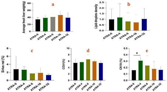
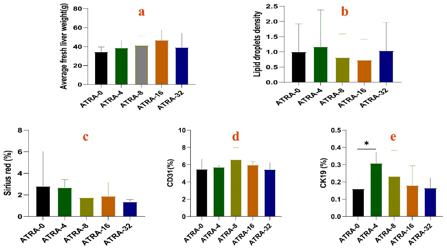
As shown in Figure 1, maternal ATRA supplementation did not significantly decrease hepatic fat accumulation (Figure 1b) or lower the fibrosis index (Figure 1c) in neonatal piglet livers (p > 0.05), but it did produce a downward trend for both hepatic fat deposition and fibrosis. In addition, ATRA supplementation for pregnant sows increased the hepatic fresh weight (Figure 1a), blood capillary density (Figure 1d) and percentage of bile duct epithelial cells (Figure 1e). Notably, compared with the 0 mg ATRA/kg diet treatment group, feeding pregnant sows a diet containing 4 mg ATRA/kg diet significantly increased the percentage of bile duct epithelial cells in the livers of neonatal piglets (p < 0.05).
Figure 1.
Comparison of hepatic architectures among different ATRA treatment groups. (a) comparison of hepatic fresh weight. (b) comparison of hepatic fat deposition. (c) comparison of hepatic fibrosis. (d) comparison of hepatic blood capillaries. (e) comparison of hepatic bile ducts. “*” means p < 0.05.
3.2. Effects of Maternal ATRA Supplementation During Gestation on Bile Acid Composition in Gallbladder of Neonatal Piglets
The concentrations of bile acids with significant differences between groups are shown in Table 1. The contents of chenodeoxycholic acid, cholic acid, and glycochenodeoxycholic acid in the bile of neonatal piglets in the ATRA-4 group were 1.63, 1.15, and 3.90 times those in the ATRA-0 group, respectively. In the ATRA-8 group, the contents of taurochenodeoxycholic acid and taurocholic acid in the bile of neonatal piglets were 5.09 and 1.49 times those in the ATRA-0 group, respectively. The content of taurochenodeoxycholic acid in bile of neonatal piglets in the ATRA-16 group was 1.13 times that of the ATRA-0 group. In the ATRA-32 group, the contents of cholic acid and taurocholic acid in the bile of neonatal piglets were 1.23 and 1.16 times those in the ATRA-0 group, respectively. The content of isolithocholic acid in the gallbladder of neonatal piglets in the ATRA-4 group was significantly higher than that in the ATRA-0 group (p < 0.01), while 3β-glycocholic acid and ursodeoxycholic acid 3-sulfate were significantly lower than those in the ATRA-0 group, respectively (p < 0.05). In the ATRA-16 group, the contents of glycohyocholic acid, 23-norcholic acid, 3β-glycocholic acid, glycoursodeoxycholic acid 3 sulfate, taurolithocholic acid-3-sulfate, and taurodeoxycholic acid-3-sulfate in the gallbladder of neonatal piglets were all significantly lower than those in the ATRA-0 group (p < 0.05). In the ATRA-32 group, the contents of 3β-glycocholic acid, ursodeoxycholic acid 3-sulfate, and taurodeoxycholic acid-3-sulfate in the gallbladder of neonatal piglets were all significantly lower than those in the ATRA-0 group (p < 0.05). The bile acid composition details in the gallbladder of neonatal piglets across the different ATRA groups are available for download from the Supplementary Materials.
Table 1.
Bile acids with significant difference in gallbladder of neonatal piglets between groups.
3.3. Effects of Maternal ATRA Supplementation During Gestation on Hepatic Gene Expression and KEGG Pathways in Neonatal Piglets
Table 2 shows that the differentially expressed genes identified in the liver of neonatal piglets, when comparing the ATRA-4 group and the ATRA-0 group, are significantly enriched in 12 KEGG pathways. These pathways can be categorized into five functional groups: signal transduction pathways (PPAR signaling pathway; inflammatory mediator regulation of TRP channels; NOD-like receptor signaling pathway; TNF signaling pathway), immune and inflammation pathways (viral protein interaction with cytokine and cytokine receptor; cytokine-cytokine receptor interaction), metabolism pathways (glycosaminoglycan biosynthesis—heparan sulfate/heparin; insulin resistance), human diseases pathways (alcoholic liver disease; fluid shear stress and atherosclerosis; lipid and atherosclerosis), and processing pathways (protein processing in endoplasmic reticulum). Among these KEGG pathways, the number of genes with significantly downregulated expression exceeds the number of genes with significantly upregulated expression.
Table 2.
Identification of differentially expressed genes and enrichment of KEGG pathways in hepatic tissues of neonatal piglets between ATRA-4 and ATRA-0 groups.
As shown in Table 3, the differentially expressed genes detected in the liver of neonatal piglets between ATRA-8 ang ATRA-0 groups are significantly enriched in 17 KEGG pathways. These pathways can be classified into five functional categories as follows: metabolism pathways (retinol metabolism; nicotinate and nicotinamide metabolism; biosynthesis of unsaturated fatty acids; fatty acid metabolism; pyrimidine metabolism; pantothenate and CoA biosynthesis; drug metabolism—other enzymes; ascorbate and aldarate metabolism; fatty acid degradation), signal transduction pathways (PPAR signaling pathway; TNF signaling pathway; inflammatory mediator regulation of TRP channels), endocrine and hormone pathways (steroid hormone biosynthesis; melanogenesis; insulin resistance), substance absorption and transport pathways (mineral absorption), and human disease pathways (alcoholic liver disease).
Table 3.
Identification of differentially expressed genes and enrichment of KEGG pathways in hepatic tissues of neonatal piglets between ATRA-8 and ATRA-0 groups.
As illustrated in Table 4, the differentially expressed genes identified in the liver of neonatal piglets between ATRA-16 and ATRA-0 groups show significant enrichment in 12 KEGG pathways. These pathways can be categorized into four functional groups as outlined below: disease and infection pathways (viral carcinogenesis; yersinia infection; salmonella infection; Epstein–Barr virus infection), signal transduction pathways (mTOR signaling pathway; HIF-1 signaling pathway; MAPK signaling pathway), metabolic pathways (endocrine and other factor-regulated calcium reabsorption; biosynthesis of nucleotide sugars; amino sugar and nucleotide sugar metabolism; terpenoid backbone biosynthesis), and cellular process pathways (mitophagy—animal).
Table 4.
Identification of differentially expressed genes and enrichment of KEGG pathways in hepatic tissues of neonatal piglets between ATRA-16 and ATRA-0 groups.
As shown in Table 5, the differentially expressed genes identified in the liver of neonatal piglets between ATRA-32 and ATRA-0 group are significantly enriched across 12 KEGG pathways, which can be divided into five functional categories as follows: metabolism pathways (glycosaminoglycan biosynthesis-heparan sulfate/heparin; aminoacyl-tRNA biosynthesis), genetic information processing pathways (ubiquitin mediated proteolysis), environmental information processing pathways (AMPK signaling pathway; p53 signaling pathway; cell cycle, growth hormone synthesis, secretion and action), cellular processes pathways (cellular senescence; fanconi anemia pathway), human diseases pathways (fluid shear stress and atherosclerosis; proteoglycans in cancer; human cytomegalovirus infection; hepatitis C; proteoglycans in cancer).
Table 5.
Identification of differentially expressed genes and enrichment of KEGG pathways in hepatic tissues of neonatal piglets between ATRA-32 and ATRA-0 groups.
4. Discussion
The liver of newborn piglets plays critical roles in the growth and survival of lactating piglets. Previous studies have shown that imbalanced maternal nutrition can interfere with the developmental process of the fetal liver, resulting in decreased liver fresh weight, reduced vascular density, and fewer bile ducts in the neonate’s liver [19,20,21,22,23]. The findings of this study demonstrate that supplementing ATRA to gestating sows tends to increase the fresh weight, microvascular density and bile duct epithelial cells in the liver of neonatal piglets. Changes in hepatic fresh weight and density of blood and bile duct are tightly regulated by gene expression levels. It has been confirmed that insulin-like growth factor 1 (IGF1) is involved in the development of multiple organs [24], and in the liver specifically, IGF1 is known to regulate cell cycle progression, cell proliferation and hepatocyte differentiation [25]. For example, Dai et al. (2024) reported that prenatal prednisone exposure (PPE) significantly inhibited cell proliferation and hepatocyte differentiation in fetal mouse liver by downregulating the expression of IGF1, proliferating cell nuclear antigen (PCNA) and hepatocyte nuclear factor 4 alpha (HNF4A), while promoting cholangiocyte differentiation by upregulating the expression of cytokeratin 19 (CK19) [26]. Another study by Kobayashi et al. (2022) also found that maternal dexamethasone exposure during late gestation promoted fetal rat hepatocyte differentiation and maturation by increasing HNF4A expression [27]. Our study shows that maternal ATRA administration from day post-conception (dpc) 12 to dpc 95 induced dose-dependent changes in gene expression in the livers of neonatal piglets: compared with the ATRA-0 group, IGF1 expression was higher in the ATRA-4 and ATRA-16 groups; PCNA expression was upregulated and HNF4A expression was downregulated in the ATRA-32 group; and CK19 expression was increased in the ATRA-4 group. All these gene expression changes were associated with increased liver fresh weight and bile duct count.
Immediately after birth, newborns first depend on hepatic glycogen stores for glucose supply, before switching to gluconeogenesis to maintain blood glucose homeostasis—a process critical for neonatal survival [28]. Existing research shows that protein phosphatase 1 regulatory subunit 3E (PPP1R3E) promotes hepatic glycogen synthesis to maintain blood glucose homeostasis, while forkhead box protein O1 (FOXO1) and phosphoenolpyruvate carboxykinase 1 (PCK1) contribute to gluconeogenesis [26,29]. Our study found that the expression levels of PPP1R3E, FOXO1 and PCK1 were significantly higher in the livers of neonatal piglets from the ATRA-4 and ATRA-8 groups compared with the ATRA-0 group. This indicates that these doses of ATRA enhance glucose homeostasis capacity, which may improve neonatal thermoregulation and postnatal survival, consistent with a previous report that prenatal prednisone exposure (PPE) upregulates the gluconeogenic genes PCK1 and G6PC to enhance hepatic glucose production in the fetal liver [26].
Hepatic lipid oxidation serves as a key supplementary pathway for energy provision via glycogen. Insufficient hepatic fat deposition is a common pathological condition in neonatal piglets that impairs key hepatic physiological functions including gluconeogenesis and bile secretion, directly threatening piglet survival. In addition, neonatal piglets are inherently characterized by low hepatic glycogen storage and limited glucose and lipid metabolism capacity, which easily induce clinical symptoms such as hypoglycemia, hypothermia, reduced disease resistance and diarrhea. Ultimately, these issues increase piglet mortality and reduce the survival rate of suckling piglets. Although sow milk is high in fat, neonatal piglets are still prone to death in the early postnatal period [30] due to their insufficient ability to digest and utilize milk fat. Studies have shown that neonatal Meishan piglets have relatively higher liver triglyceride levels and stronger milk fat digestion capacity, which can effectively meet their demands for blood glucose and body temperature maintenance, thus improving the survival rate of Meishan suckling piglets [17]. Therefore, improving hepatic lipid and glycogen reserves and liver function in neonatal piglets is a key direction for solving the problem of nutritional regulation in gestating sows, and our experiment showed that adding 4 mg/kg ATRA to the diet of gestating sows tends increase the liver fat content of neonatal piglets.
In neonatal liver, genes related to fatty acid uptake, β-oxidation, and triglyceride metabolism are abundantly expressed, including peroxisome proliferator-activated receptor alpha (PPARA), retinoid X receptor alpha (RXRA), liver-type carnitine palmitoyltransferase 1 (CPT1A), fatty acid synthase (FAS), acyl-CoA oxidase 3 (ACOX3), solute carrier family 27 member 5 (SLC27A5), and lipin 1 (LPIN1) [31,32,33,34,35]. PPARA deficiency drives hepatic fat accumulation [32,33,34]. Previous studies in adult mice demonstrated that 4 consecutive days of subcutaneous ATRA injection at 10 mg/kg or 100 mg/kg body weight upregulated the expression of PPARA, RXRA, and CPT1A, but downregulated FAS expression, which ultimately reduced overall body weight and adiposity [36,37]. In contrast, both 7 consecutive days of intraperitoneal Pregnenolone 16α-carbonitrile (PCN) injection in mice and prenatal prednisone exposure (PPE) significantly downregulated hepatic PPARA expression [26,38]. Our study reveals that supplementing ATRA at 4 mg/kg to the diet of pregnant sows reduced PPARA expression but increased ACOX3 expression in the liver of neonatal piglets. ACOX3 is a core enzyme for fatty acid β-oxidation, and its expression was downregulated in the liver of rats with nonalcoholic steatohepatitis induced by a high-energy, high-fat diet [34]. We further found that adding ATRA at 8 mg/kg or 32 mg/kg to the diet of gestating sows significantly upregulated CPT1B (the rate-limiting enzyme for long-chain fatty acid β-oxidation) expression in neonatal piglet liver. This result contradicts a previous report showing that ATRA inhibited CPT1B expression to reduce β-oxidation and promote lipid accumulation in human HepaRG cells [39]. This discrepancy may stem from tissue- or developmental stage-specific regulatory effects of ATRA on lipid metabolism. Consistent with a previous report that newborn Rongchang piglets exhibited high hepatic expression of SLC27A5, LPIN1, and ACOX3 to meet the nutritional requirements for survival supported by colostrum [39], our study found that maternal dietary ATRA supplementation regulated the expression of these genes in neonatal piglet liver in a dose-dependent manner: 4 mg ATRA/kg diet upregulated hepatic ACOX3 expression, 8 mg ATRA/kg diet downregulated hepatic SLC27A5 expression, and 16 mg ATRA/kg diet increased hepatic LPIN1 expression. Collectively, these findings indicate that neonatal piglets from sows fed diets supplemented with 4 mg/kg or 16 mg/kg ATRA had a greater capacity to generate energy via fatty acid metabolism after digesting milk fat.
Digesting milk fat requires bile participation, and insufficient bile excretion frequently causes steatorrhea in suckling piglets. Bile acids are the essential components of bile, including primary and secondary bile acids, and play a crucial role in fat emulsification [40]. Primary bile acids, which are primarily composed of cholic acid (CA) and chenodeoxycholic acid (CDCA), are metabolized by gut microbes into secondary bile acids such as deoxycholic acid (DCA) and lithocholic acid (LCA) [41]. Studies have shown that CA rather than CDCA promotes cholesterol absorption [42], so a reduced CA/CDCA ratio may lead to decreased cholesterol absorption. ATRA significantly reduced the levels of most bile acids except CDCA, taurochenodeoxycholic acid (TCDCA) and 3α,6α,7α-trihydroxy-5β-cholan-24-oic acid (THDCA) [43], as well as the CA/CDCA ratio [44]. The present study found that maternal ATRA supplementation did not significantly change the concentrations of most primary and secondary bile acids in the liver of neonatal piglets, with only eight bile acids showing significantly altered levels: isolithocholic acid, 3β-glycocholic acid, ursodeoxycholic acid-3-sulfate, taurodeoxycholic acid-3-sulfate, 23-norcholic acid, glycohyocholic acid, glycoursodeoxycholic acid-3-sulfate, and taurolithocholic acid-3-sulfate. Previous reports have demonstrated that isolithocholic acid can inhibit the growth of Clostridium difficile, a pathogen that frequently causes diarrhea in suckling piglets [45] and effectively suppress intestinal inflammation, improve intestinal barrier function [46,47], and enhance mucosal antiviral immunity [48]. In contrast, ursodeoxycholic acid-3-sulfate can protect the liver and intestine by strengthening the mucus barrier and attenuating inflammatory responses [49,50]. In our experiment, neonatal piglets from ATRA-4 group had significantly higher gallbladder concentration of isolithocholic acid than piglets from other ATRA groups, indicating that these piglets have healthier intestinal and hepatic conditions.
Bile acids are synthesized from cholesterol catabolism in the liver via the classical pathway that involves cytochrome P450 7A1 (CYP7A1) and cytochrome P450 8B1 (CYP8B1), and the acidic pathway that includes cytochrome P450 27A1 (CYP27A1) and cytochrome P450 7B1 (CYP7B1) [51]. CYP7A1 can catalyze the conversion of cholesterol to 7α-hydroxycholesterol for bile production [52]. A previous study reported that when wild-type C57/BL mice were fed a diet containing 150 mg ATRA/kg body weight for 7 days, the expressions of CYP7A1 and CYP8B1, as well as hepatic HNF4A levels, were significantly reduced [44]. Inhibition of CYP7A1 and CYP8B1 ultimately reduces bile acid synthesis and impairs lipid absorption [39,48]. As CYP8B1 is a critical gene for CA synthesis [53], loss of functional CYP8B1 leaves CYP7A1 only able to synthesize CDCA, so changes in CYP8B1 expression directly alter the CA/CDCA ratio [54]. Our results showed that maternal supplementation with ATRA at 4 mg/kg in the diet of gestating sows significantly reduced CYP7A1 expression in the liver of neonatal piglets. However, we also observed that CDCA concentration in the bile of piglets from the ATRA-4 group was higher than that in the ATRA-0 group. This discrepancy may be explained by the significant upregulation of ATP-binding cassette subfamily G member 1 (ABCG1) expression in the liver tissue of neonatal piglets in ATRA-4 group. As ABCG1 is an important reverse cholesterol transporter, increased ABCG1 expression promotes cholesterol transport to facilitate bile acid synthesis and reduces hepatic cholesterol accumulation and excessive fat deposition [55,56,57,58].
This study investigated the effects of ATRA administration to pregnant sows on the hepatic fresh weight, morphology, bile acid production, and gene expression in newborn piglets; however, the findings have the following limitations: 1) treatments were applied at the sow level whereas analyses were conducted at the piglet level, and therefore the findings should be interpreted with appropriate caution and confirmed in future studies with larger numbers of sows and statistical approaches that account for litter effects; 2) no RT-qPCR results were provided for differentially expressed genes; and 3) no data were provided regarding indicators linked to the liver functional outcomes of neonatal piglets, including glucose homeostasis, thermoregulation, and survival.
5. Conclusions
Treatment of pregnant sows with ATRA can affect bile acid secretion and gene expression of the neonate’s liver. Compared with the 0 mg ATRA/kg diet supplement, a maternal 4 mg ATRA/kg diet supplement increases the percentage of bile duct epithelial cells, upregulates the expression of genes related to glycogen synthesis, gluconeogenesis, and fatty acid β-oxidation, and elevates isolithocholic acid concentration in the liver of neonatal piglets.
Supplementary Materials
The following supporting information can be downloaded at: https://www.mdpi.com/article/10.3390/vetsci13111248/s1, File S1: Bile acids data.
Author Contributions
Conceptualization, Y.H.; experiments, C.W. and H.Z.; data analysis, C.W. and H.Z.; writing—original draft preparation, C.W. and Y.H.; writing—review and editing, Y.H.; project administration, W.L. and Y.H.; funding acquisition, Y.H. All authors have read and agreed to the published version of the manuscript.
Funding
This research was funded by the National Natural Science Foundation of China, grant number 32360850.
Institutional Review Board Statement
The animal study protocol was approved by the Animal Ethics Committee of Jiangxi Agricultural University (JXAULL-2024-10-01) on 8 October 2024.
Informed Consent Statement
Informed consent was obtained from all subjects involved in the study.
Data Availability Statement
The data presented in this study are openly available in NCBI Sequence Read Archive at https://www.ncbi.nlm.nih.gov/bioproject/PRJNA1234339/ (accessed on 11 March 2025), reference number PRJNA1234339.
Conflicts of Interest
The authors declare no conflicts of interest.
References
- Schmelzer, E. Hepatic progenitors of the fetal liver: Interactions with hematopoietic stem cells. Differentiation 2019, 106, 9–14. [Google Scholar] [CrossRef] [Scilit] [PubMed]
- Almeida, J.I.; Tenreiro, M.F.; Martinez-Santamaria, L.; Guerrero-Aspizua, S.; Gisbert, J.P.; Alves, P.M.; Serra, M.; Baptista, P.M. Hallmarks of the human intestinal microbiome on liver maturation and function. J. Hepatol. 2022, 76, 694–725. [Google Scholar] [CrossRef] [Scilit] [PubMed]
- Kostallari, E.; Schwabe, R.F.; Guillot, A. Inflammation and immunity in liver homeostasis and disease: A nexus of hepatocytes, nonparenchymal cells and immune cells. Cell. Mol. Immunol. 2025, 22, 1205–1225. [Google Scholar] [CrossRef] [Scilit] [PubMed]
- Xiao, Y.; Ma, Y.; Gan, C.; Huang, Z.; Gao, J. cGAS-STING signaling pathway in MASLD and liver fibrosis. J. Mol. Med. 2026, 104, 29. [Google Scholar] [CrossRef] [Scilit] [PubMed]
- Southey, B.R.; Gomez, A.N.; Sunderland, G.R.; Riggins, C.W.; Villamil, M.B.; Rodriguez-Zas, S.L. Metabolite profiling of the effect of prenatal stimuli across postnatal treatments in the liver. Mol. Cell. Endocrinol. 2026, 615, 112744. [Google Scholar] [CrossRef] [Scilit] [PubMed]
- Rhinn, M.; Dollé, P. Retinoic acid signalling during development. Development 2012, 139, 843–858. [Google Scholar] [CrossRef] [Scilit] [PubMed]
- Mason, J.B.; Sanders, D.; Greiner, T.; Shrimpton, R.; Yukich, J. Vitamin A deficiency: Policy implications of estimates of trends and mortality in children. Lancet Glob. Health 2016, 4, e21. [Google Scholar] [CrossRef] [Scilit] [PubMed]
- Topletz, A.R.; Zhong, G.; Isoherranen, N. Scaling in vitro activity of CYP3A7 suggests human fetal livers do not clear retinoic acid entering from maternal circulation. Sci. Rep. 2019, 9, 4620. [Google Scholar] [CrossRef] [Scilit] [PubMed]
- Zarei, L.; Farhad, N.; Abbasi, A. All-Trans Retinoic Acid (atRA) effectively improves liver steatosis in a rabbit model of high fat induced liver steatosis. Arch. Physiol. Biochem. 2022, 128, 1010–1015. [Google Scholar] [CrossRef] [Scilit] [PubMed]
- Samrani, L.M.; Pennings, J.L.; Hallmark, N.; Bars, R.; Tinwell, H.; Pallardy, M.; Piersma, A.H. Dynamic regulation of gene expression and morphogenesis in the zebrafish embryo test after exposure to all-trans retinoic acid. Reprod. Toxicol. 2023, 115, 8–16. [Google Scholar] [CrossRef] [Scilit] [PubMed]
- Phan, A.; Sokolova, A.; Hilscherova, K. An adverse outcome pathway approach linking retinoid signaling disruption to teratogenicity and population-level outcomes. Aquat. Toxicol. 2024, 277, 107143. [Google Scholar] [CrossRef] [Scilit] [PubMed]
- Cassim Bawa, F.N.; Xu, Y.; Gopoju, R.; Plonski, N.M.; Shiyab, A.; Hu, S.; Chen, S.; Zhu, Y.; Jadhav, K.; Kasumov, T.; et al. Hepatic retinoic acid receptor alpha mediates all-trans retinoic acid’s effect on diet-induced hepatosteatosis. Hepatol. Commun. 2022, 6, 2665–2675. [Google Scholar] [CrossRef] [Scilit] [PubMed]
- Mercader, J.; Ribot, J.; Murano, I.; Felipe, F.; Cinti, S.; Bonet, M.L.; Palou, A. Remodeling of white adipose tissue after retinoic acid administration in mice. Endocrinology 2006, 147, 5325–5332. [Google Scholar] [CrossRef] [Scilit] [PubMed]
- Yang, D.; Vuckovic, M.G.; Smullin, C.P.; Kim, M.; Lo, C.P.; Devericks, E.; Yoo, H.S.; Tintcheva, M.; Deng, Y.; Napoli, J.L. Modest decreases in endogenous all-trans-retinoic acid produced by a mouse Rdh10 heterozygote provoke major abnormalities in adipogenesis and lipid metabolism. Diabetes 2018, 67, 662–673. [Google Scholar] [CrossRef] [Scilit] [PubMed]
- Jain, A.K.; Stoll, B.; Burrin, D.G.; Holst, J.J.; Moore, D.D. Enteral bile acid treatment improves parenteral nutrition-related liver disease and intestinal mucosal atrophy in neonatal pigs. Am. J. Physiol. Gastrointest. Liver Physiol. 2012, 302, G218–G224. [Google Scholar] [CrossRef] [Scilit] [PubMed]
- Gao, T.; Liang, H.; Li, M.; Zhou, R.; Wang, L.; Li, R.; Huang, L.; Zhuo, Y.; Xu, S.; Lin, Y.; et al. Taurolithocholic acid alleviates hepatic glycolipid dysregulation in low-birth-weight piglets by activating HNF4α and FXR. J. Anim. Sci. Biotechnol. 2026, 17, 154. [Google Scholar] [CrossRef] [Scilit] [PubMed]
- Fainberg, H.P.; Bodley, K.; Bacardit, J.; Li, D.; Wessely, F.; Mongan, N.P.; Symonds, M.E.; Clarke, L.; Mostyn, A. Reduced neonatal mortality in Meishan piglets: A role for hepatic fatty acids? PLoS ONE 2012, 7, e49101. [Google Scholar] [CrossRef] [Scilit] [PubMed]
- Xiao, X.H.; Zhou, H.M.; He, D.H.; Wei, P.T.; Zhu, Y.T.; Sun, W.C.; Hao, S.B.; Wu, H.D.; Lu, W.; He, Y.Y. Effect of maternal administration with all-trans retinoic acid on lungs of neonatal pigs. Vet. Sci. 2025, 12, 1132. [Google Scholar] [CrossRef] [Scilit] [PubMed]
- Ramadan, W.S.; Alshiraihi, I.; Al-karim, S. Effect of maternal low protein diet during pregnancy on the fetal liver of rats. Ann. Anat. 2013, 195, 68–76. [Google Scholar] [CrossRef] [Scilit] [PubMed]
- Choi, W.; Kim, J.; Ko, J.W.; Choi, A.; Kwon, Y.H. Effects of maternal branched-chain amino acid and alanine supplementation on growth and biomarkers of protein metabolism in dams fed a low-protein diet and their offspring. Amino Acids 2022, 54, 977–988. [Google Scholar] [CrossRef] [Scilit] [PubMed]
- Yu, E.A.; Chapnick, M.A.; Stein, A.D. The potential physiologic mechanisms by which early-life nutrition may influence Type 2 diabetes pathophysiology in humans. Annu. Rev. Nutr. 2026, 46, 505–531. [Google Scholar] [CrossRef] [Scilit] [PubMed]
- Hu, D.; Wu, X.; Hou, M.; Song, P.; Chen, Q.; Ni, Y. Maternal exposure to high level of ursodeoxycholic acid during pregnancy promotes hepatic lipid deposition in the offspring by reducing the methylation level of Fasn gene. Biochem. Pharmacol. 2025, 241, 117156. [Google Scholar] [CrossRef] [Scilit] [PubMed]
- Nash, M.J.; Dobrinskikh, E.; Al-Juboori, S.I.; Janssen, R.C.; Fernandes, J.; Argabright, A.; D’Alessandro, A.; Kirigiti, M.A.; Kievit, P.; Aagaard, K.M.; et al. Maternal western diet programmes bile acid dysregulation and hepatic fibrosis in fetal and Juvenile Macaques. Liver Int. 2025, 45, e16236. [Google Scholar] [CrossRef] [Scilit] [PubMed]
- Baker, J.; Liu, J.P.; Robertson, E.J.; Efstratiadis, A. Role of insulin-like growth factors in embryonic and postnatal growth. Cell 1993, 75, 73–82. [Google Scholar] [CrossRef] [Scilit]
- Liu, K.; Wang, G.; Li, L.; Chen, G.; Gong, X.; Zhang, Q.; Wang, H. GR-C/EBPa-IGF1 axis mediated azithromycin-induced liver developmental toxicity in fetal mice. Biochem. Pharmacol. 2020, 180, 114130. [Google Scholar] [CrossRef] [Scilit] [PubMed]
- Dai, Y.; Peng, Y.; Lu, Z.; Mao, T.; Chen, K.; Lu, X.; Liu, K.; Zhou, X.; Hu, W.; Wang, H. Prenatal prednisone exposure impacts liver development and function in fetal mice and its characteristics. Toxicol. Sci. 2024, 199, 63–80. [Google Scholar] [CrossRef] [Scilit] [PubMed]
- Kobayashi, T.; Takeba, Y.; Ohta, Y.; Ootaki, M.; Kida, K.; Watanabe, M.; Iiri, T.; Matsumoto, N. Prenatal glucocorticoid administration accelerates the maturation of fetal rat hepatocytes. Mol. Biol. Rep. 2022, 49, 5831–5842. [Google Scholar] [CrossRef] [Scilit] [PubMed]
- Lanoue, L.; Liu, X.J.; Koski, K.G. Postnatal profiles of glycogenolysis and gluconeogenesis are modified in rat pups by maternal dietary glucose restriction. J. Nutr. 1999, 129, 820–827. [Google Scholar] [CrossRef] [Scilit] [PubMed]
- Li, X.; Dong, X.; Zhao, S.; Ma, Y.; Jin, S.; Zhou, C.; Lan, Y.; Luo, S.; Ni, B.; Wei, H.; et al. Adaptation of Diqing Tibetan pigs to hypoxic and cold environments through extramedullary hematopoiesis and uncoupled thermogenesis in the liver. BMC Biol. 2025, 23, 181. [Google Scholar] [CrossRef] [Scilit] [PubMed]
- Newcomb, M.D.; Harmon, D.L.; Nelssen, J.L.; Thulin, A.J.; Allee, G.L. Effect of energy source fed to sows during late gestation on neonatal blood metabolite homeostasis, energy stores and composition. J. Anim. Sci. 1991, 69, 230–236. [Google Scholar] [CrossRef] [Scilit] [PubMed]
- Mirza, A.Z.; Althagafi, I.I.; Shamshad, H. Role of PPAR receptor in different diseases and their ligands: Physiological importance and clinical implications. Eur. J. Med. Chem. 2019, 166, 502–513. [Google Scholar] [CrossRef] [Scilit] [PubMed]
- Chen, L.; Li, J.; Yuan, R.; Wang, Y.; Zhang, J.; Lin, Y.; Wang, L.; Zhu, X.; Zhu, W.; Bai, J.; et al. Dynamic 3D genome reorganization during development and metabolic stress of the porcine liver. Cell Discov. 2022, 8, 56. [Google Scholar] [CrossRef] [Scilit] [PubMed]
- Jiao, P.; Xu, Y.; Lu, H.; Xu, Y.; Fan, C.; Liu, S.; Zhuo, Z.; Mao, S.; Cheng, J.; Liu, W.; et al. Malnutrition during gestation induces lipid metabolic disorders in maternal and fetal livers and eventually affects fetal development. Anim. Adv. 2025, 2, e033. [Google Scholar] [CrossRef] [Scilit]
- Moreto, F.; Ferron, A.J.T.; Francisqueti-Ferron, F.V.; D’Amato, A.; Garcia, J.L.; Costa, M.R.; Silva, C.C.V.A.; Altomare, A.; Correa, C.R.; Aldini, G.; et al. Differentially expressed proteins obtained by label-free quantitative proteomic analysis reveal affected biological processes and functions in Western diet-induced steatohepatitis. J. Biochem. Mol. Toxicol. 2021, 35, 1–11. [Google Scholar] [CrossRef] [Scilit] [PubMed]
- Wang, Y.; Jin, H.; Tong, X.; Yu, H.; Li, X.; Zeng, B. DNA methylation of postnatal liver development in pigs. Genes 2024, 15, 1067. [Google Scholar] [CrossRef] [Scilit] [PubMed]
- Amengual, J.; Ribot, J.; Bonet, M.L.; Palou, A. Retinoic acid treatment enhances lipid oxidation and inhibits lipid biosynthesis capacities in the liver of mice. Cell. Physiol. Biochem. 2010, 25, 657–666. [Google Scholar] [CrossRef] [Scilit] [PubMed]
- Maguire, M.; Larsen, M.C.; Vezina, C.M.; Quadro, L.; Kim, Y.K.; Tanumihardjo, S.A.; Jefcoate, C.R. Cyp1b1 directs Srebp-mediated cholesterol and retinoid synthesis in perinatal liver; Association with retinoic acid activity during fetal development. PLoS ONE 2020, 15, e0228436. [Google Scholar] [CrossRef] [Scilit] [PubMed]
- Yan, L.; Yan, Y.; Yang, K.; Chang, Q.; Zhang, L. Metabolomics reveals dysregulated all-trans retinoic acid and polyunsaturated fatty acid metabolism contribute to PXR-induced hepatic steatosis in mice. Toxicol. Lett. 2024, 398, 150–160. [Google Scholar] [CrossRef] [Scilit] [PubMed]
- Wang, S.; Link, F.; Munker, S.; Wang, W.; Feng, R.; Liebe, R.; Li, Y.; Yao, Y.; Liu, H.; Shao, C.; et al. Retinoic acid generates a beneficial microenvironment for liver progenitor cell activation in acute liver failure. Hepatol. Commun. 2024, 8, e0483. [Google Scholar] [CrossRef] [Scilit] [PubMed]
- Dockray, G.J. Cholecystokinin. Curr. Opin. Endocrinol. Diabetes Obes. 2012, 19, 8–12. [Google Scholar] [CrossRef] [Scilit] [PubMed]
- Tang, B.; Tang, L.; Li, S.; Liu, S.; He, J.; Li, P.; Wang, S.; Yang, M.; Zhang, L.; Lei, Y.; et al. Gut microbiota alters host bile acid metabolism to contribute to intrahepatic cholestasis of pregnancy. Nat. Commun. 2023, 14, 1305. [Google Scholar] [CrossRef] [Scilit] [PubMed]
- Reynier, M.O.; Montet, J.C.; Gerolami, A.; Marteau, C.; Crotte, C.; Montet, A.M.; Mathieu, S. Comparative effects ofcholic, chenodeoxycholic, and ursodeoxycholic acids on micellar solubilization and intestinal absorption of cholesterol. J. Lipid Res. 1981, 22, 467–473. [Google Scholar] [CrossRef] [Scilit]
- Yang, F.; He, Y.; Liu, H.X.; Tsuei, J.; Jiang, X.; Yang, L.; Wang, Z.T.; Wan, Y.J. All-trans retinoic acid regulates hepatic bile acid homeostasis. Biochem. Pharmacol. 2014, 91, 483–489. [Google Scholar] [CrossRef] [Scilit] [PubMed]
- He, Y.; Gong, L.; Fang, Y.; Zhan, Q.; Liu, H.X.; Lu, Y.; Guo, G.L.; Lehman-McKeeman, L.; Fang, J.; Wan, Y.J. The role of retinoic acid in hepatic lipid homeostasis defined by genomic binding and transcriptome profiling. BMC Genom. 2013, 14, 575. [Google Scholar] [CrossRef] [Scilit] [PubMed]
- Aguirre, A.M.; Sorg, J.A. Gut associated metabolites and their roles in Clostridioides difficile pathogenesis. Gut Microbes 2022, 14, 2094672. [Google Scholar] [CrossRef] [Scilit] [PubMed]
- Paik, D.; Yao, L.; Zhang, Y.; Bae, S.; D’Agostino, G.D.; Zhang, M.; Kim, E.; Franzosa, E.A.; Avila-Pacheco, J.; Bisanz, J.E.; et al. Human gut bacteria produce TH17-modulating bile acid metabolites. Nature 2022, 603, 907–912. [Google Scholar] [CrossRef] [Scilit] [PubMed]
- Zhang, P.; Xu, S.; Li, N.; Yu, C. Faecalibacterium prausnitzii ameliorates DSS-induced colitis via modulating bile acid metabolism and regulating FXR signaling. Dig. Dis. Sci. 2025, 70, 3323–3339. [Google Scholar] [CrossRef] [Scilit] [PubMed]
- Iliev, I.D.; Blander, J.M.; Collins, N.; Guo, C.J.; Longman, R.S.; Sonnenberg, G.F.; Zeng, M.Y.; Artis, D. Microbiota-mediated mechanisms of mucosal immunity across the lifespan. Nat. Immunol. 2025, 26, 1645–1659. [Google Scholar] [CrossRef] [Scilit] [PubMed]
- Wang, Q.; Xu, K.; Cai, X.; Wang, C.; Cao, Y.; Xiao, J. Rosmarinic acid restores colonic mucus secretion in colitis mice by regulating gut microbiota-derived metabolites and the activation of inflammasomes. J. Agric. Food Chem. 2023, 71, 4571–4585. [Google Scholar] [CrossRef] [Scilit] [PubMed]
- Yu, X.; Zhu, M.; Wang, K.; Yuan, B.; Lu, Y.; Li, S.; Hu, F. 10-Hydroxy-2 (E)-decenoic acid attenuates metabolic dysfunction-associated steatotic liver disease via modulation of the gut microbiota-bile acid-FXR axis. Food Biosci. 2026, 78, 108532. [Google Scholar] [CrossRef] [Scilit]
- Chiang, J.Y.L. Bile acids: Regulation of synthesis. J. Lipid Res. 2009, 50, 1955–1966. [Google Scholar] [CrossRef] [Scilit] [PubMed]
- Chiang, J.Y.L.; Ferrell, J.M. Up to date on cholesterol 7 alpha-hydroxylase (CYP7A1) in bile acid synthesis. Liver Res. 2020, 4, 47–63. [Google Scholar] [CrossRef] [Scilit] [PubMed]
- Vlahcevic, Z.R.; Eggertsen, G.; Bjorkhem, I.; Hylemon, P.B.; Redford, K.; Pandak, W.M. Regulation of sterol 12alpha-hydroxylase and cholic acid biosynthesis in the rat. Gastroenterology 2000, 118, 599–607. [Google Scholar] [CrossRef] [Scilit] [PubMed]
- Pandak, W.M.; Bohdan, P.; Franklund, C.; Mallonee, D.H.; Eggertsen, G.; Björkhem, I.; Gil, G.; Vlahcevic, Z.R.; Hylemon, P.B. Expression of sterol 12alpha-hydroxylase alters bile acid pool composition in primary rat hepatocytes and in vivo. Gastroenterology 2001, 120, 1801–1809. [Google Scholar] [CrossRef] [Scilit] [PubMed]
- Chambers, K.F.; Day, P.E.; Aboufarrag, H.T.; Kroon, P.A. Polyphenol effects on cholesterol metabolism via bile acid biosynthesis, CYP7A1: A review. Nutrients 2019, 11, 2588. [Google Scholar] [CrossRef] [Scilit] [PubMed]
- Kotlyarov, S.; Kotlyarova, A. Biological functions and clinical significance of the ABCG1 transporter. Biology 2025, 14, 8. [Google Scholar] [CrossRef] [Scilit] [PubMed]
- Yin, Y.; Li, D.; Liu, F.; Wang, X.; Cui, Y.; Li, S.; Li, X. The ameliorating effects of apple polyphenol extract on high-fat-diet-induced hepatic steatosis are SIRT1-dependent: Evidence from hepatic-specific SIRT1 heterozygous mutant C57BL/6 mice. J. Agric. Food Chem. 2022, 70, 5579–5594. [Google Scholar] [CrossRef] [Scilit] [PubMed]
- Hoebinger, C.; Rajcic, D.; Silva, B.; Hendrikx, T. Chronic-binge ethanol feeding aggravates systemic dyslipidemia in Ldlr-/-mice, thereby accelerating hepatic fibrosis. Front. Endocrinol. 2023, 14, 1148827. [Google Scholar] [CrossRef] [Scilit] [PubMed]

Disclaimer/Publisher’s Note: The statements, opinions and data contained in all publications are solely those of the individual author(s) and contributor(s) and not of MDPI and/or the editor(s). MDPI and/or the editor(s) disclaim responsibility for any injury to people or property resulting from any ideas, methods, instructions or products referred to in the content. |
© 2026 by the authors. Licensee MDPI, Basel, Switzerland. This article is an open access article distributed under the terms and conditions of the Creative Commons Attribution (CC BY) license.