


Simple Summary
Alpacas and llamas are important livestock species in the Peruvian Andes, but little is known about the diversity of mammalian orthoreovirus circulating in these animals. This study investigated the presence of this virus and identified its genetic types in 224 fecal samples collected from alpacas and llamas in Cusco, Peru. We detected the virus in 59 animals (26.3%) and identified three genetic types. Type MRV1 was the most frequent, while types MRV2 and MRV3/4 were also detected. Notably, the detection of type 2 may represent the first report of this virus type in South America. Genetic comparisons also revealed close relationships between viruses found in South American camelids and viruses previously identified in bats. Although these similarities do not establish direct transmission between species, they highlight the need for further investigation into the circulation of these viruses in different animal hosts. These findings expand our knowledge of viruses in alpacas and llamas and provide a basis for future research into their diversity, evolution, and potential routes of circulation. This information may contribute to improved animal health surveillance and a better understanding of the viruses circulating among wildlife and livestock in the Peruvian Andes.
Abstract
Mammalian orthoreovirus (MRV) has been detected in South American camelids (SACs) in the Peruvian highlands. However, limited studies and the lack of suitable genotyping tools have constrained understanding of MRV genetic diversity in these animals. This study aimed to detect MRV and identify its genotypes in alpacas and llamas from rural communities and a research institute in Cusco, Peru. We analyzed 224 fecal samples from alpacas (n = 212) and llamas (n = 12) by RT-PCR. Positive samples were genotyped using nested and semi-nested RT-PCR assays targeting MRV1, MRV2, and MRV3–4 through amplification of the S1 gene. Selected amplicons were sequenced for confirmation and phylogenetic analysis. Overall, 59/224 animals (26.3%) tested positive, including 53/212 alpacas (25.0%) and 6/12 llamas (50.0%). Of the 59 MRV-positive animals, 38 (64.4%) were successfully genotyped, revealing MRV1 (n = 28), MRV2 (n = 3), and MRV3–4 (n = 1) monoinfections, as well as six coinfections: MRV1 + MRV2 (n = 4) and MRV1 + MRV3–4 (n = 2). S1 sequence analysis showed that MRV1 sequences were closely related to strains reported in SACs from Peru and in bats from China and Slovenia. The alpaca MRV2 sequence was related to MRV2 strains reported in bats and other mammals, including a lion. Sequencing of the MRV3/4 amplicon was unsuccessful. This is the first report of MRV2 in South America and provides molecular evidence of multiple MRV genotypes circulating in Peruvian SACs. Our findings suggest evolutionary links between MRVs detected in SACs and bat-associated lineages.
1. Introduction
Mammalian orthoreovirus (MRV), recently reclassified as Orthoreovirus mammalis (family Spinareoviridae, order Reovirales), is a non-enveloped virus with a 10-segmented double-stranded RNA (dsRNA) genome encoding structural and non-structural proteins [1]. Its icosahedral structure and conserved use of the JAM-A receptor facilitate infection across a wide range of mammalian hosts [2]. Based on the S1 segment, which encodes the σ1 attachment protein, MRVs are classified into four serotypes: MRV1 (Lang), MRV2 (Jones), MRV3 (Dearing), and MRV4 (Ndelle) [3,4]. Their segmented genome favors reassortment, promoting adaptability and host switching [5]. While MRV infections are often subclinical, they have been linked to gastrointestinal, respiratory, and neurological disease in humans and animals [6,7,8], underscoring their zoonotic potential and the need for surveillance.
South American camelids (SACs), including alpacas (Vicugna pacos) and llamas (Lama glama), are a strategic resource for Andean highland communities, providing fiber, meat, and hides, as well as serving a central role in food security and household economy [9,10]. Approximately 80% of the alpaca population in Peru is managed by smallholders under mixed-production systems characterized by low technification, limited veterinary care, and exposure to harsh environmental conditions [11]. These constraints predispose animals to infectious diseases, especially neonatal enteric and respiratory syndromes [9,12]. Identified agents include Clostridium perfringens, Escherichia coli, Salmonella spp., Pasteurella multocida, Giardia spp., Eimeria spp., Cryptosporidium spp., Respiratory syncytial virus, Parainfluenza virus type 3, Rotavirus alphagastroenteritidis (formerly known as Rotavirus species A; RVA), and Coronavirus [13,14,15,16,17,18,19,20]. These pathogens contribute significantly to morbidity, mortality, and economic loss in SAC production systems.
Despite the importance of SACs and their close contact with humans and wildlife in the Andean highlands, knowledge about MRV circulation in these animals is limited. Previous studies in Peru have identified MRV in neonatal alpacas with diarrhea [16], but information on prevalence, genotypic diversity, and potential for interspecies transmission remains scarce. Considering the role of bats and other wildlife as reservoirs [21,22,23] and the demonstrated capacity of MRV for genetic reassortment [7,24,25], the absence of epidemiological monitoring in high-altitude ecosystems represents a critical gap.
The present study aimed to investigate MRV detection and genotype diversity in alpacas and llamas from Cusco, Peru, using conserved-gene RT-PCR, genotype-specific nested or semi-nested PCR, sequence confirmation, and phylogenetic analysis. By combining previously investigated samples with additional archived specimens from different production systems and clinical contexts, this study provides an opportunity to characterize MRV circulation and genotype diversity in South American camelids.
2. Materials and Methods
2.1. Sampling
This retrospective molecular characterization study was based on 224 archived fecal samples from alpacas and llamas available in the sample bank of the Laboratory of Veterinary Virology and Immunology, Faculty of Veterinary Medicine, Universidad Nacional Mayor de San Marcos (FMV-UNMSM), Lima, Peru. Of these, 68 alpaca samples and 12 llama samples collected in 2015 had been previously analyzed for MRV detection and reported by Castilla et al. (2021) [16]. Forty of these samples, previously positive by RT-PCR targeting a conserved 181-bp fragment of the L1 gene, tested positive again upon retesting. These archived samples were included for further genotype characterization; repeated RT-PCR positivity was not interpreted as evidence of viral persistence or viability.
Samples collected in 2015 originated from the rural communities of Silly, Quisini, and Pataccalasaya in the province of Canchis, Cusco, Peru (approximately 3800–4700 m.a.s.l.) during the birthing season and were obtained in the context of diarrhea outbreaks. Additional samples were collected at the Tropical and Altitude Veterinary Research Institute (IVITA-UNMSM) in 2021 and 2022, representing a semitechnified production system with improved sanitary management. The 2021 sampling included neonatal alpacas aged 1–6 weeks with and without diarrhea, whereas the 2022 sampling included adult alpacas aged 12–16 months without diarrhea at the time of sampling.
Fecal samples were collected directly from the rectum and stored at −80 °C until processing at the Laboratory of Veterinary Virology and Immunology, FMV-UNMSM, Lima, Peru. Clinical status was assessed at the time of sampling and did not allow us to establish whether animals without diarrhea had never been ill, were incubating an infection, or were recovering from a previous episode.
2.2. Sample Preparation and RNA Extraction
The extraction of viral RNA samples required an initial clarification process. Fecal suspensions were prepared at 10% in sterile PBS at a pH of 7.4 and then centrifuged at 2500× g for 5 min. The supernatant was filtered through a 0.22 μm pore size syringe filter. Genomic RNA was then extracted from the supernatants using the Nucleic Acid Extraction Reagent kit (Lifotronic Technology Co., Ltd., Shenzhen, China) in combination with the Auto-Pure32A automated extractor (Nucleic Acid Purification System, ALLSHENG, Hangzhou, China), according to the manufacturer’s recommendations. Samples were stored at −80 °C until processing.
2.3. Reverse Transcription and Detection of MRV
The extracted RNA was used to obtain cDNA by Reverse Transcription (RT) using the GoScriptTM reverse transcription system (Promega, Madison, WI, USA). For a final volume of 10 µL of cDNA, 5 μL of the RNA was denatured at 97 °C for 5 min and then combined with 5 µL of the reaction mixture. The reaction mixture contained the following components: 0.5 μL of nuclease-free water; 2 μL of Transcriptase Buffer (5X); 1.2 μL of MgCl2 (25 mM); 0.4 μL of primer reverse (MRV-RM to 20 µM); 0.5 μL of dNTPs pool (10 mM); and 0.4 μL of enzyme GoScriptTM Reverse. Briefly, the viral RNA was subjected to one reverse-transcription cycle consisting of 5 min at 25 °C followed by 45 min at 42 °C, and one step of 2 min at 95 °C followed by PCR cycles as described below.
The cDNA obtained was subjected to PCR using MRV-FM and MRV-RM primers (Table 1). The protocol described by Castilla et al. [16] was used for the amplification of a portion of the L1 gene (181 bp) that encodes the protein RNA-dependent RNA polymerase (RdRp), which is conserved in all MRV types (MRV1–MRV4). The PCR reaction was performed with a final volume of 25 µL containing 6.5 μL of DNase-RNase-free water (Promega Corporation, Madison, WI, USA), 12.5 µL of Green GoTaq® G2 master mix (2x) (Promega Corporation, Madison, WI, USA), 0.5 μL of each primer (20 μM), and 5 µL of cDNA template were added to the reaction mix. Each PCR included a negative control containing water in place of cDNA and a positive control (5 µL of strain MRV1/SA44/Alpaca, GenBank: MN200216). The PCR was performed in a Bio-Rad T100 PCR thermal cycler (Bio-Rad Laboratories, Hercules, CA, USA) under the following conditions: 3 min at 95 °C; followed by 40 cycles of PCR, each consisting of 40 s at 94 °C, 1 min at 45 °C, and 1 min at 72 °C; and the final extension step for 5 min at 72 °C.
Table 1.
Primers used for detection and genotyping of Mammalian orthoreovirus (MRV).
Then, L1 gene PCR products were loaded into a 1.5% (w/v) agarose gel, stained with ethidium bromide, and visualized under UV light. A 100 bp DNA ladder (New England Biolabs, Ipswich, MA, USA) was used to determine molecular size by electrophoresis.
2.4. Statistical Analysis
The data were processed and analyzed using STATA version 16.1. The 95% confidence intervals (CIs) for MRV detection frequencies were calculated using the Wilson score method for binomial proportions (default setting in the software). All estimates were expressed as percentages.
2.5. Genotyping of MRV by Nested and Semi-Nested RT-PCR
Positive samples of MRV were subjected to RT-PCR and nested or semi-nested PCR using specific primers designed for each of the four genotypes (MRV1, MRV2 and MRV3–MRV4) reported (Table 1). Reverse transcription polymerase chain reaction (RT-PCR) and nested PCR were performed using the GoScript™ reverse transcription system and GoTaq® Green Master Mix (Promega, Madison, WI, USA), respectively. The genomic RNAs were subject to one cycle of reverse transcription (5 min at 25 °C followed by 45 min at 42 °C) and one step of 2 min at 95 °C.
For MRV genotyping, the first round of RT-PCR amplified the S1 gene encoding the sigma-1 (σ1) protein using primers and protocols described by Castilla et al. [16] (Table 1). Five microliters of the first-round amplicons were then used in genotype-specific nested or semi-nested PCR assays developed in this study to generate products of 722 bp for MRV1, 1060 bp for MRV2, and 680 bp for MRV3–4 (Table 1). These assays were used as a rapid molecular genotyping system. The MRV3–4 assay does not distinguish MRV3 from MRV4; the detected product is therefore reported as MRV3–4 in assay-based results, while the epidemiological interpretation of the finding as MRV3 is discussed separately because sequencing confirmation was unsuccessful. The PCR conditions were as follows: 5 min at 95 °C; followed by 40 cycles of PCR, each consisting of 40 s at 94 °C, 1 min at 50 °C, and 2 min 20 s at 72 °C; and the final extension step for 10 min at 72 °C, except for the MRV2 genotype with an annealing temperature of 40 °C.
To validate the PCR assays, positive controls were used for each of the studied genotypes. One control was from an isolated strain MRV1/SA44/Alpaca (GenBank: MN200219.1), and the remaining genotypes were of two synthetic genes (synthetic minigenome).
All primers and synthetic minigenomes were synthesized by IDT (Integrated DNA Technologies, Coralville, IA, USA). Reactions were performed in a Bio-Rad T100 PCR thermal cycler (Bio-Rad Laboratories, Hercules, CA, USA). The PCR products were separated by 1.5% (w/v) agarose gel electrophoresis, stained with ethidium bromide, and visualized under UV light. A 100-bp DNA ladder (New England Biolabs, USA) was used to determine molecular size.
2.6. Sequencing and Phylogenetic Analysis
Selected L1 and S1 amplicons were sequenced by Macrogen Inc. (Seoul, Republic of Korea). Sequences were edited using EditSeq, MegAlign, and SeqMan in the DNASTAR Lasergene software package (Version 7.0, DNASTAR, Madison, WI, USA). Phylogenetic analyses were performed with MEGA12 using the maximum-likelihood method and the General Time Reversible model with gamma-distributed rate variation and a proportion of invariant sites (GTR+G+I), with 1000 bootstrap pseudoreplicates [26,27]. L1 sequences were used to provide molecular confirmation of MRV detection, whereas S1 sequences were used to confirm genotype assignments obtained by the rapid genotype-specific PCR system and to investigate phylogenetic relationships. Sequences generated in this study were compared with representative MRV sequences obtained from GenBank (https://www.ncbi.nlm.nih.gov/nucleotide/) (accessed on 10 September 2026). The sequences generated in this study were deposited under accession numbers PV797027–PV797038 and PZ565362–PZ565365.
3. Results
3.1. Detection and Genotyping of MRV by RT-PCR and Nested PCR
MRV was detected in 59 of 224 fecal samples (26.3%; 95% CI: 21.00–32.47), including 53 of 212 alpacas samples (25.0%; 95% CI: 19.65–31.24) and 6 of 12 llama samples (50.0%; 95% CI: 25.38–74.62). The dataset included samples from rural communities collected in 2015 and from IVITA-UNMSM collected in 2021 and 2022. The detailed distribution by locality, host species, sampling year, and genotype is presented in Table 2. The 2015 samples were collected in the context of diarrhea outbreaks in rural communities, whereas the 2021–2022 samples represented a production system with improved sanitary management and included animals with and without diarrhea at sampling. Differences among groups should therefore be interpreted in light of their distinct sampling frameworks rather than as direct estimates of population prevalence under equivalent conditions. L1 RT-PCR targeting the conserved 181-bp region was used for MRV detection, and selected amplicons were sequenced to confirm the molecular diagnosis.
Table 2.
Detection and genotyping of mammalian orthoreovirus (MRV) in alpacas (Vicugna pacos) and llamas (Lama glama) from the Cusco region, Peru.
Of the 59 MRV-positive samples, 38 (64.4%) were assigned to one or more genotypes by nested or semi-nested PCR targeting S1. MRV1 was predominant as a monoinfection (n = 28), followed by MRV2 (n = 3) and one MRV3–4 assay-positive result. Although the assay could not distinguish between MRV3 and MRV4, this finding was epidemiologically interpreted as MRV3 based on the available evidence regarding the host distribution of these genotypes; however, this interpretation could not be genetically confirmed because sequencing was unsuccessful. six samples showed coinfections, including MRV1 + MRV2 (n = 4) and MRV1 + MRV3–4 (n = 2). Twenty-one MRV-positive samples (35.6%) could not be assigned a genotype. The genotype distribution by host species and locality is presented in Table 2, and individual assignments are detailed in Supplementary Table S1. Notably, sample ALP222/2015 from Silly was identified as an MRV1/MRV2 coinfection by nested RT-PCR, and both genotype-specific S1 products were subsequently recovered and sequenced, providing molecular confirmation of this coinfection.
3.2. Sequencing and Phylogenetic Analysis
Eight L1 RT-PCR products (181 bp) were selected for sequencing based on amplification quality and sample representation. Six yielded sequences (5 alpacas and 1 llama) of adequate quality, confirming MRV detection. The L1 sequences showed 100% and 98.3% nucleotide identity with previously reported camelid and guinea pig Peruvian sequences (GenBank MN200216–MN200218, PX458891), supporting the specificity of the conserved L1 RT-PCR assay Supplementary Table S2. Because L1 is highly conserved among MRV genotypes, these sequences were interpreted as diagnostic confirmation rather than as the basis for genotype assignment.
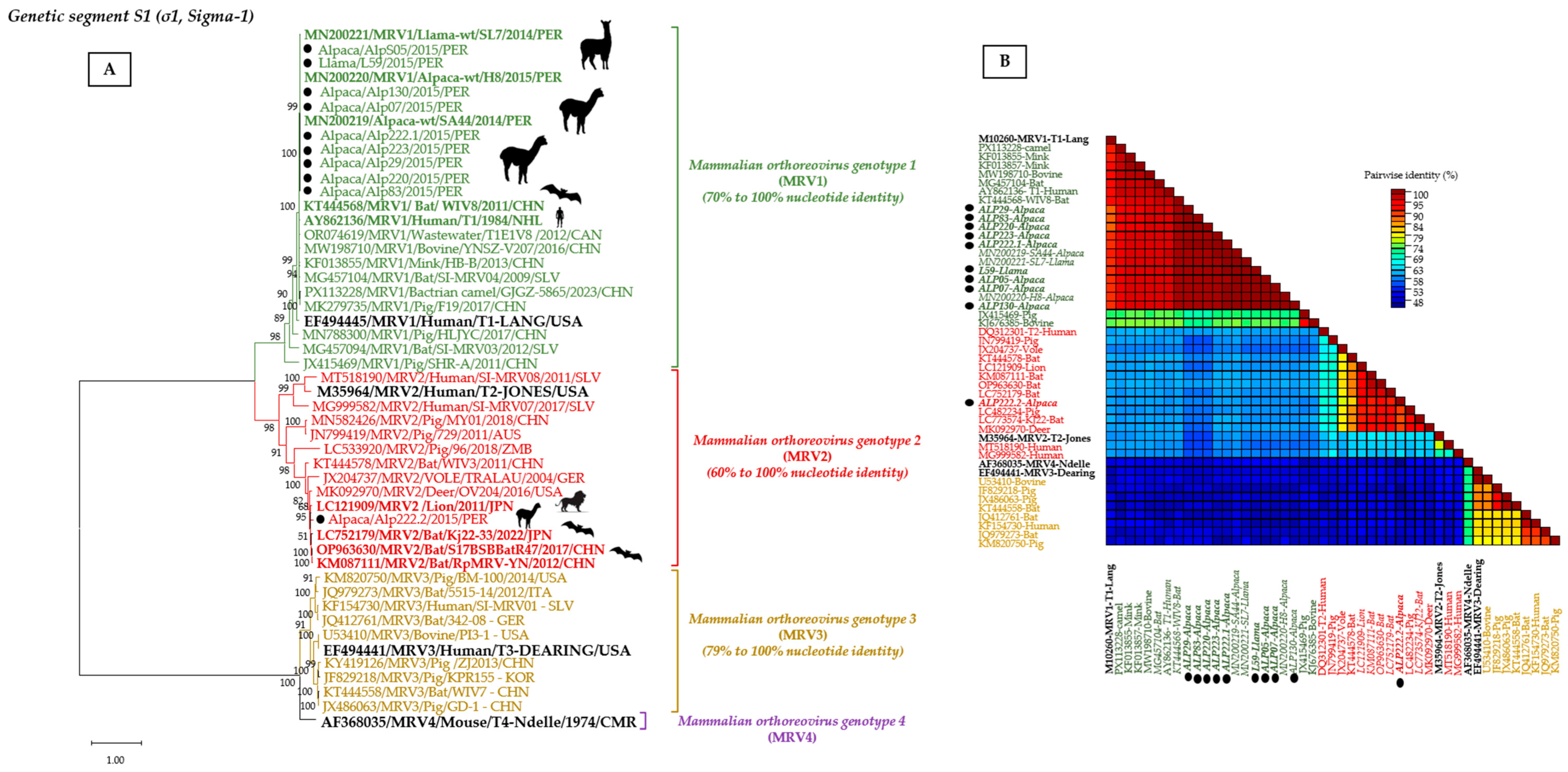
Fifteen S1 products were selected for sequencing to evaluate the genotype-specific nested/semi-nested PCR system and investigate phylogenetic relationships. High-quality sequences were obtained from nine MRV1 products and one of MRV2, importantly, from both genotype-specific products from sample ALP222/2015, which was identified as an MRV1/MRV2 coinfection by nested RT-PCR. ALP222.1 yielded an MRV1 S1 sequence (GenBank PZ565363, 1322 bp), whereas ALP222.2 yielded an MRV2 S1 sequence (GenBank PZ565364, 947 bp). Additional MRV1 S1 sequences ranged from 722 to 1322 bp. The S1 sequences clustered with their corresponding genotype reference strains, supporting the genotype assignments generated by the genotype-specific PCR assays and providing the basis for phylogenetic analysis (Figure 1; Supplementary Table S2).
Figure 1.
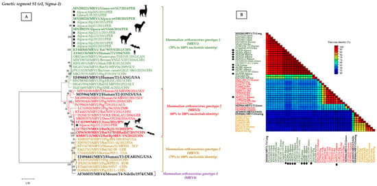
(A) Phylogenetic tree based on nucleotide sequences obtained from the Sigma-1 gene (1322 bp, 1115 bp, 947 bp, and 722 bp) of MRV strains detected in alpacas and llamas in this study. Sequences shown in green, red, and mustard represent MRV1, MRV2, and MRV3 strains, respectively. The analysis included 49 nucleotide sequences: 39 reference sequences retrieved from GenBank and identified by their accession numbers, and 10 sequences obtained in this study. The phylogenetic tree was inferred using the Maximum Likelihood method under the GTR+G+I substitution model. Node support was assessed using 1000 bootstrap replicates, and bootstrap values >75% are shown at the nodes. The scale bar represents nucleotide substitutions per site. (B) Based on the phylogenetic tree shown in panel (A) and the genetic distance heat map of the 49 analyzed sequences, the black circle highlights the alpaca and llama sequences obtained in this study. The pairwise genetic distance heat map was generated using SDTv1.3 software. Reference strains of MRV1, MRV2, MRV3, and MRV4 are shown in black.
Phylogenetic distance analysis of the 10 nucleotide sequences obtained in this study revealed that nine sequences identified as MRV1 shared 99–100% nucleotide identity. Comparison with GenBank reference sequences showed that these nine sequences clustered closely with MRV1 strains, exhibiting high nucleotide identity with strains from Peruvian South American camelids (99.8–100%) and bat-derived strains (96.7–99.3%) from China (Figure 1, Table S3). At the amino acid level, our strains showed 99.7–100% sequence identity with previously reported strains from Peruvian alpacas and llamas (MN200219–MN200221), followed by bat-derived strains from China (98.5%; KT444568), mink strains from China (97.6%; KF013855), and bat-derived strains from Slovenia (97.3%; MG457104) (Figure 1, Table S3).
Specifically, the ALP222.2 sequence, classified as MRV2 by the rapid genotyping assay, exhibited 58.6–59.6% nucleotide (nt) identity with the nine MRV1 sequences obtained in this study and the MRV1 reference strains included in the analysis (Table S3). In contrast, nt identity with the thirteen MRV2 reference sequences ranged from 60.1% to 93.0% (Figure 1; Table S4). Among the MRV2 strains currently available in GenBank, ALP222.2 showed the highest nucleotide identity with a strain detected in a lion in Japan (LC121909; 93.03%), followed by two bat strains from China (OP963630; 92.0% and KM087111; 91.6%). (Figure 1; Tables S2 and S4). Furthermore, BLAST searches against the NCBI database (https://blast.ncbi.nlm.nih.gov/Blast.cgi, accessed on 16 November 2026) revealed that the lion-derived MRV2 strains showed its highest sequence similarities to MRV2 strains previously reported in bats from different regions of Asia and Europe.
The sequence dataset generated in this study includes selected L1 and S1 sequences deposited in GenBank under accession numbers PV797027–PV797038 and PZ565362–PZ565365. The individual sequence lengths, genotype assignments, and accession numbers are listed in Supplementary Table S2.
4. Discussion
Among the zoonotic viruses affecting alpacas and llamas in Peru, coronaviruses (CoVs) and rotaviruses (RVs) have been reported at prevalences ranging from 26.8% to 87.5% and from 20.3% to 47.5%, respectively [1,16,17,20,28,29]. These findings highlight the diversity of RNA viruses circulating in South American camelids (SACs) and their potential for interspecies transmission to sheep and shepherd children in the Cusco region [1,17,28]. Castilla et al. [16] provided the first evidence of mammalian orthoreovirus (MRV) in domestic SACs in Peru, reporting a 50% positivity rate, with 83.7% of positive cases involving coinfections with RVA and CoV.
This study investigated MRV detection and genotyping in 224 fecal samples from alpacas and llamas collected in Cusco, Peru, under contrasting husbandry and epidemiological conditions (Table S1). MRV was detected in 59 samples (26.3%), of which 38 (64.4%) were successfully genotyped. Detection frequencies in samples collected at IVITA-UNMSM in 2021–2022 (10.9–18.6%) were lower than those previously reported by Castilla et al. (2021) [16] in samples from three rural communities in the same region (33.3–66.7%; Table 2). These differences should be interpreted descriptively because the samples came from different sampling periods and husbandry contexts. In addition, MRV was detected again in archived samples previously identified as positive by Castilla et al. (2021) [16]. This supports the reproducibility of molecular detection in these specimens but does not establish viral persistence or viability.
Interpretation of the detection frequencies requires careful consideration of the sampling design. Samples collected in Silly, Quisini, and Pataccalasaya in 2015 came from rural communities experiencing diarrhea outbreaks and therefore represent a clinically enriched sampling context. In contrast, samples collected at IVITA-UNMSM in 2021 and 2022 were obtained under improved sanitary management and included animals with and without clinical signs; the 2022 group consisted of adults without diarrhea at sampling. Consequently, differences among groups should not be interpreted as direct comparisons of population prevalence because the sampling frames, clinical context, age structure, and management conditions differed.
MRV was detected in animals with and without diarrhea at the time of sampling. However, the absence of diarrhea at sampling does not establish that an animal had never experienced diarrhea or was free of an ongoing infection. In a cross-sectional study, an apparently non-diarrheic animal could be in the incubation phase of an infection, recovering from a previous clinical episode, or shedding viral RNA after clinical resolution [30,31]. The present study design therefore cannot determine whether MRV detection was temporally associated with the onset or resolution of diarrhea, nor can it establish a causal relationship between MRV detection and diarrhea [32]. Longitudinal studies involving repeated clinical assessments and virological sampling would be needed to distinguish these possibilities.
Comparable MRV detection frequencies have been reported in wild artiodactyls, including wild boars (45.3%) and deer (40.6%) in Sondrio Province, as well as wild boars (15.4%) in Parma Province, northern Italy [33]. The frequencies reported in Sondrio were similar to those observed in alpacas and llamas from Silly (66.7% in alpacas and 50.0% in llamas), Quisini (34.8%), and Pataccalasaya (33.3%) (Table 2). These similarities may reflect shared ecological conditions, including mountainous landscapes, wetlands, and exposure to livestock and wildlife. In contrast, a study of intensively managed domestic artiodactyls (sheep and goats) in China reported an MRV detection frequency of 8.2% (35/429) using qRT-PCR [34], comparable to the frequencies observed at IVITA-UNMSM (10.9–18.6%). These comparisons suggest that MRV detection frequencies may be associated with production systems. Nevertheless, differences in sampling context, study populations, and diagnostic methods must also be considered. The lower frequencies observed under intensive or semi-intensive management are consistent with the possibility that production systems influence MRV circulation, although MRV was still detected at IVITA-UNMSM despite improved sanitary management. Furthermore, Quispe et al. [35] reported MRV in 23.53% of guinea pigs from small-scale family farms in Cusco, extending the evidence of MRV detection to another domestic species raised in the same region and broadening the regional perspective on MRV occurrence across animal populations and production systems.
Sequencing of the L1 amplicons provided molecular confirmation of MRV detection by RT-PCR. The L1 sequences showed 98.3–100% nucleotide identity with previously reported guinea pig and Peruvian camelid strains (Table S2). Phylogenetic analysis also showed close relationships with MRV strains detected in other hosts, including a bat (OQ789607/MRV2; 98.3%), a deer (MK092964/MRV2; 97.8%), and a mink (KT224504/MRV3; 97.2%). Of the 31 sequences analyzed, 30 clustered within the same clade, irrespective of genotype, with nucleotide identities ranging from 78.3% to 100%. These findings support the utility of the conserved 181-bp L1 target for MRV detection across the four genotypes (figure not shown). The only sequence outside this clade was the RVA outgroup, which showed 43.6–46.9% nucleotide identity with MRV sequences.
Of the 59 MRV-positive samples, 38 were successfully genotyped by nested and semi-nested PCR, revealing MRV1, MRV2, and MRV3–4, as well as several coinfections. MRV1 was the most frequent genotype, detected in 34 animals (28 monoinfections and six coinfections), and was distributed across all study sites (Table 2). This genotype has been reported not only in SACs but also in a wide range of species, including calves, cats, shrews, minks, pigs, and several bat genera (including Eptesicus, Myotis, and Rhinolophus) [1,24,36,37,38,39,40,41,42]. MRV1 has also been detected in wastewater and in humans with meningitis or diarrhea [6,43,44].
MRV2 was detected by genotype-specific semi-nested RT-PCR in seven animals: three monoinfections in Silly (two llamas and one alpaca) and four coinfections, three in alpacas from IVITA-UNMSM and one in an alpaca from Silly (Table 2). However, S1 sequencing confirmation was obtained for only one of these animals (ALP222.2). This genotype has previously been reported in infants with diverse clinical manifestations, piglets with diarrhea, white-tailed deer with disease, bats, voles, cats, lions, and wastewater samples [9,24,43,45,46,47,48]. The S1 sequence obtained in this study provides the first sequence-based evidence of MRV2 in South America and the first molecular characterization of this genotype in South American camelids.
MRV3–4 was detected in three alpacas: one monoinfection at IVITA-UNMSM and two coinfections with MRV1, one in Pataccalasaya and one at IVITA-UNMSM (Table 2). The MRV3–4-specific PCR assay yielded a 680-bp amplicon in samples assigned to this genotype group. Because the assay targets an S1 region shared by MRV3 and MRV4 and sequencing confirmation was unsuccessful, the molecular results are reported as MRV3–4. Nevertheless, the epidemiological context favors interpreting these findings as MRV3 [5]. MRV4 has been reported only once, in laboratory mice, and only one strain, Ndelle reovirus, has been described to date [2,4,5]. By contrast, MRV3 is among the most frequently isolated and widely distributed genotypes in mammals, including horses, cats, bats, shrews, wild boars, cattle, civets, piglets, deer, wild goats, sheep, dogs, Alpine chamois, and humans with diarrhea [6,24,49,50,51,52,53,54]. If interpreted as MRV3, this would represent the second report of this genotype in South America, following its detection in a child in Brazil [6]. This interpretation is epidemiological rather than sequence-confirmed and should be understood accordingly.
The clearest molecular evidence of coinfection in this study came from sample ALP222/2015. Nested RT-PCR detected MRV1 and MRV2 in the same sample, and sequencing independently recovered an MRV1 S1 sequence (ALP222.1/PZ565363) and an MRV2 S1 sequence (ALP222.2/PZ565364) (Table S2). This agreement between PCR and sequence data strengthens confidence in the genotype-specific assays and demonstrates that mixed MRV infections can be detected and molecularly resolved in field samples. Coinfections involving different MRV genotypes have been reported in mammals, including calves, bats, pigs, deer, and humans [6,24,39,47,48]. Because the MRV genome is segmented, coinfection of a host cell by different strains can lead to reassortment of genome segments. Although reassortment generally occurs between viruses of the same species, it can also occur between different species within the same taxonomic family [24,39,55,56]. Complete-genome analyses have documented this process in MRV. In China, an MRV2 strain isolated from pigs was identified as a novel chimeric strain, likely resulting from coinfections involving human, porcine, goat, bat, and mink MRVs [48]. A porcine MRV3 strain from the same study showed reassortment involving genome segments closely related to those of human, bovine, and porcine MRV3 strains previously reported in Italy in 2015–2016 [57]. Similarly, an MRV2 strain from white-tailed deer in the United States contained genome segments highly similar to those of MRV3 strains circulating in deer from the same region [47]. These examples illustrate how coinfections can provide opportunities for genetic exchange among MRV strains.
In the present study, coinfections involving different MRV genotypes, together with the distribution and predominance of MRV1 across all study sites, suggest that multiple genotypes circulate in the local camelid population. This pattern is consistent with ongoing MRV circulation and raises the possibility that MRV1 is better adapted to these host populations than the other genotypes detected. However, the duration of circulation and the relative degree of adaptation cannot be determined from the present data alone.
Twenty-one of the 59 MRV-positive samples (35.6%) could not be genotyped using the nested/semi-nested PCR system. Several factors may have contributed to this outcome, including low viral RNA concentrations, sequence variation at primer-binding sites, inhibitors carried over from fecal material, and RNA degradation associated with prolonged sample storage. Failure to assign a genotype should not be interpreted as evidence of infection with an unknown genotype. These findings illustrate both the utility and the limitations of rapid, genotype-specific PCR, particularly given the limited availability of S1 sequences representing different MRV genotypes and host species. This limited sequence representation may constrain the assessment of genetic diversity and the design of primers capable of detecting genetically divergent strains across hosts. Coinfections involving different MRV genotypes may also complicate Sanger sequencing if multiple related templates are co-amplified, potentially producing overlapping chromatographic signals. This possibility is particularly relevant to the MRV2 and MRV3–4 amplicons obtained from coinfected samples, which yielded chromatograms with substantial background noise that prevented reliable sequence recovery. Future studies could complement this approach with amplicon-based next-generation sequencing (NGS) to characterize variation in targeted regions, or with metagenomic sequencing to investigate viral sequences beyond those targeted by genotype-specific primers. These approaches could improve the characterization of samples that remain untyped.
Phylogenetic analysis of the S1 sequences supported the genotype assignments obtained by nested/semi-nested PCR. Our MRV1 sequences (1322; 1115; and 722 bp) clustered closely with one another and with previously reported camelid strains (MN200219–21) [16], showing 99.8–100% nucleotide identity and 99.7–100% amino acid identity (Figure 1, Table S3). They were also closely related to bat-derived strains from China (KT444568; 99.3% nucleotide and 98.6% amino acid identity) and Slovenia (MG457104; 96.7% nucleotide and 97.5% amino acid identity) (Figure 1, Table S3). These relationships are notable in light of previous findings demonstrating the evolutionary diversity of bat-associated MRVs. The Chinese bat strain WIV8 illustrates this complexity: phylogenetic analyses of its S1, M1, and L1 genome segments revealed distinct evolutionary origins, consistent with multiple reassortment events involving MRVs associated with different mammalian hosts. These analyses also revealed close genetic relationships between bat-derived MRVs, and strains identified in mink, pigs, and humans in China [24]. In addition, our BLAST analysis identified an MRV1 strain isolated from a sentinel bovine in China during surveillance for bat-associated viruses (MW198710.1) as the closest match to the Slovenian bat-derived strain SI-MRV04. Together, these findings highlight the evolutionary diversity of MRVs associated with bats and the genetic relationships observed between bat-derived strains and MRVs detected in other mammalian hosts.
The MRV2 sequence obtained from ALP222.2 showed 93% nucleotide identity with the MRV2 strain detected in a lion in Japan (LC121909) and 91.6–92% identity with two strains detected in bats from different regions of China (KM087111 and OP963630, respectively) (Figure 1, Table S4). Although LC121909 was detected in a lion, phylogenetic analysis showed that it clusters closely with MRV2 strains of bat origin (Figure 1). Similarly, the two Chinese bat strains clustered within a relatively restricted group of bat-associated MRV2 sequences, with nucleotide identities ranging from 85% to 98% among the sequences compared (Table S4). Together, these findings extend the observed phylogenetic relationships between camelid MRV sequences and bat-associated lineages beyond MRV1 to MRV2.
Bats are recognized as reservoirs of numerous RNA viruses and can serve as sources of infection for animals and humans. Their ecological characteristics, including long-distance movements and interactions with diverse host species across extensive geographic areas, may facilitate viral dissemination. Coinfection can also promote genetic reassortment in viruses with segmented genomes [39,58,59,60]. Previous studies have reported coronavirus and MRV strains in camelids that are phylogenetically related to bat-associated viruses. During a lethal outbreak of acute pneumonia in alpacas, coronavirus strains related to BatCoV-HKU-10 (OP963607) and Megaderma bat coronavirus (MZ293749) were identified; these viruses belonged to the genus Alphacoronavirus and an unclassified genus, respectively [20]. Similarly, analysis of the S1 segment (σ1 gene; 1015 bp) of the first MRV isolates reported in SACs by Castilla et al. [16] revealed monophyletic clustering with two bat-derived MRV1 strains. These previous findings provide context for interpreting the relationships observed between camelid MRV sequences and bat-associated lineages in the present study.
The close phylogenetic relationships between some camelid MRV sequences and bat-associated strains are epidemiologically noteworthy, particularly in high-Andean environments where wildlife and livestock may share habitats. Phylogenetic proximity alone, however, does not demonstrate direct transmission from bats to camelids. Rather, these relationships are compatible with a possible contribution of wildlife to MRV circulation and provide a rationale for considering the local ecological context.
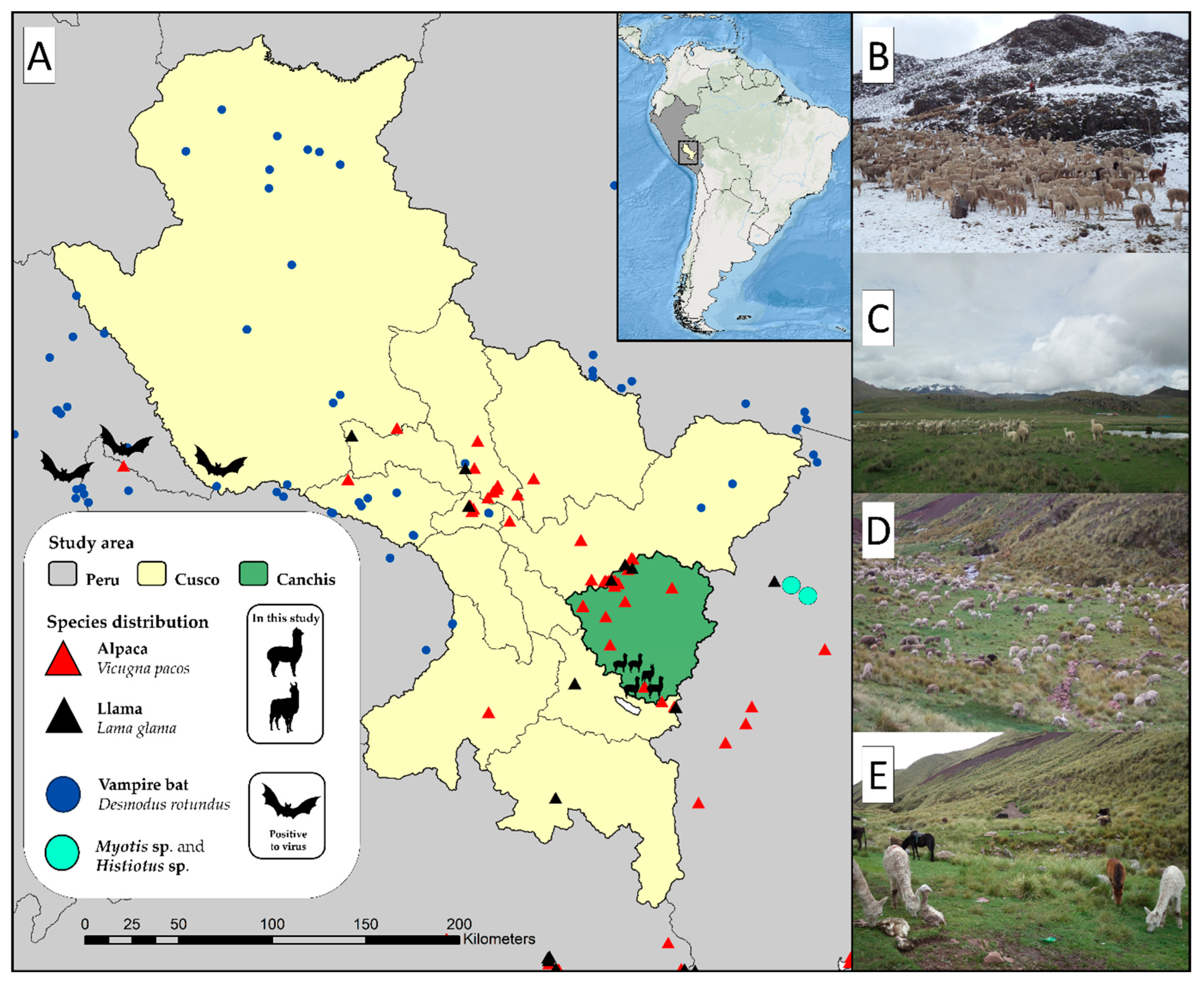
To place these findings in their local ecological context, we reviewed reports of bat species occurring near the four sampling sites. Several species belonging to the genera Histiotus and Myotis have been reported at altitudes ranging from 3500 to 5120 m above sea level [61,62] (Figure 2). These bats feed primarily on insects and fruits and, while ecologically relevant as seed dispersers, may also disseminate diverse pathogens [63]. Indirect exposure of alpacas and llamas in rural communities of Canchis could occur through contamination of water sources, pastures, or fruits with bat feces. Bergner et al. [64,65,66] conducted the first metagenomic survey of common vampire bats (Desmodus rotundus) in Peru, analyzing fecal and oropharyngeal swab samples collected between 2015 and 2016 from individuals captured in caves and trees in Cusco, Ayacucho, and Amazonas. Virome analysis revealed 58 viral families, 17 of which include viruses capable of infecting mammals, including reoviruses and coronaviruses of zoonotic importance. These findings demonstrate the diversity of viruses detected in Peruvian bats and provide additional regional context for investigating possible wildlife contributions to viral circulation.
Figure 2.
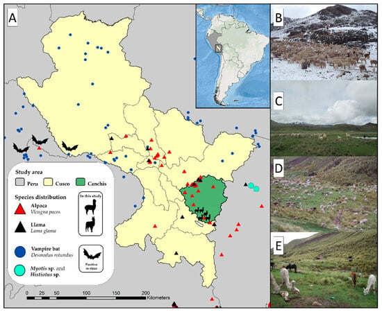
Geographic distribution of alpaca (red triangles) and llama (black triangles) breeding areas in Canchis Province, Cusco, Peru, in relation to reported occurrences of the common vampire bat Desmodus rotundus (blue circles) and insectivorous bats of the genera Histiotus and Myotis (light-blue circles) near the study sites [61,62]. Bat symbols indicate locations where virome studies have been conducted, and viral agents have been detected in bats [64,65,66]. Records of Histiotus and Myotis indicate reported occurrences of these genera in Peru, although virome studies for these bats have not been conducted in the country. (A) Location of the study area in Peru, highlighting Canchis Province (green); (B) Pataccalasaya; (C) Quisini; (D) Silly; and (E) additional sampling sites.
From a One Health perspective, the coexistence of SACs, other domestic animals, wildlife, and people in Andean production systems creates opportunities for viral exchange. The present findings support continued MRV surveillance in camelids and their shared environments, particularly because multiple genotypes and coinfections were detected. However, this study does not establish zoonotic transmission or identify a specific wildlife source. Integrated surveillance involving livestock, wildlife, environmental samples, and longitudinal clinical sampling will be needed to clarify potential transmission pathways.
5. Conclusions
This study expands the molecular evidence for MRV circulation and genetic diversity in SACs. MRV was detected in 59 of 224 samples, and 38 positive samples were genotyped. MRV1 predominated, while MRV2, MRV3–4 assay-positive results interpreted epidemiologically as MRV3, and mixed infections were also identified. The genotype-specific S1 nested/semi-nested PCR assays provided a rapid approach for characterizing positive samples, complemented by sequence confirmation and phylogenetic analysis. To our knowledge, the detection of MRV2 represents the first report of this genotype in South America. The phylogenetic relationships between camelid MRV1/MRV2 sequences and bat-associated lineages suggest possible evolutionary links but do not demonstrate direct transmission. Further genomic and epidemiological studies are warranted to clarify MRV diversity and circulation in South American camelids.
Supplementary Materials
The following supporting information can be downloaded at https://www.mdpi.com/article/10.3390/vetsci13111245/s1, Table S1: Genotyping results of mammalian orthoreovirus (MRV) in alpacas (Vicugna pacos) and llamas (Lama glama) by nested and semi-nested RT-PCR; Table S2: General information on the strains successfully sequenced in the study; Table S3: Nucleotide and amino acid sequence identities of the longest Sigma-1 gene sequences obtained in this study (1322 bp, 1115 bp, and 947 bp), based on multiple sequence alignments with the Lang (MRV1) prototype strain and the other phylogenetically related MRV1 strains; Table S4: Nucleotide sequence identities determined by multiple sequence alignment between the ALP222.2 sequence (947 bp), the prototype Jones strain (MRV2), and other phylogenetically related MRV2 strains.
Author Contributions
Conceptualization, M.R., A.M., and N.S.; methodology, S.Y., A.Q., M.L., N.O., E.A., W.V., D.P., and M.R.; writing—original draft preparation, M.R., D.A.N.-M., and C.L.; writing—review and editing, M.R., C.L., D.A.N.-M., and N.S.; funding acquisition, M.R. and A.M. All authors have read and agreed to the published version of the manuscript.
Funding
This study was supported by the Programa Nacional de Investigación y Estudios Avanzados (PROCIENCIA)–CONCYTEC en el marco de la convocatoria de “Proyecto de Investigación Básica 2024-01” (grant number PE501088213-2024) y “Tesis de Pregrado y Postgrado en Ciencia, Tecnología e Innovación Tecnológica” (grant number PE501085142-2023).
Institutional Review Board Statement
The procedures and techniques performed on alpacas and llamas used in the present study were approved by the Animal Research Ethics Committee of the Faculty of Veterinary Medicine, National University of San Marcos (FMV-UNMSM) and by the Tropical and Altitude Veterinary Research Institute (IVITA), protocol No. 2009-0001. Samples were taken following the animal handling protocol authorized by the Ethics and Animal Welfare Committee of FMV-UNMSM.
Informed Consent Statement
Oral informed consent was obtained from the animal owners in the rural communities.
Data Availability Statement
The original contributions presented in this study are included in the article/Supplementary Material. Further inquiries can be directed to the corresponding author.
Acknowledgments
We thank Miguel Ángel Villareal Balboa for his technical assistance and Elena Yudith Carrasco Charquero for her role as administrative coordinator of the project (Grant No. PE501088213-2024). This work is dedicated to the memory of Lina Mercedes Montes Collave, four years after your passing. I will always love you, Mother. Special thanks to my brother, Mirko, and my nephews, Gael and Alessia
Conflicts of Interest
The authors declare no conflicts of interest. None of the authors of this paper have a financial or personal relationship with other people or organizations that could inappropriately influence the content of the paper. The funders had no role in the design of the study; in the collection, analysis, or interpretation of data; in the writing of the manuscript, or in the decision to publish the results.
References
- International Committee on Taxonomy of Viruses (ICTV) Genus_Orthoreovirus_ICTV. Available online: https://ictv.global/report/chapter/spinareoviridae/spinareoviridae/orthoreovirus (accessed on 26 September 2025).
- Barton, E.S.; Forrest, J.C.; Connolly, J.L.; Chappell, J.D.; Liu, Y.; Schnell, F.J.; Nusrat, A.; Parkos, C.A.; Dermody, T.S. Junction Adhesion Molecule Is a Receptor for Reovirus. Cell 2001, 104, 441–451. [Google Scholar] [CrossRef] [Scilit] [PubMed]
- Matthijnssens, J.; Attoui, H.; Bányai, K.; Brussaard, C.P.D.; Danthi, P.; del Vas, M.; Dermody, T.S.; Duncan, R.; Fāng, Q.; Johne, R.; et al. ICTV Virus Taxonomy Profile: Spinareoviridae 2022. J. General. Virol. 2022, 103, 001781. [Google Scholar] [CrossRef] [Scilit] [PubMed]
- Attoui, H.; Biagini, P.; Stirling, J.; Mertens, P.P.C.; Cantaloube, J.-F.; Meyer, A.; de Micco, P.; de Lamballerie, X. Sequence Characterization of Ndelle Virus Genome Segments 1, 5, 7, 8, and 10: Evidence for Reassignment to the Genus Orthoreovirus, Family Reoviridae. Biochem. Biophys. Res. Commun. 2001, 287, 583–588. [Google Scholar] [CrossRef] [Scilit] [PubMed]
- Rosa, U.A.; Ribeiro, G.d.O.; Villanova, F.; Luchs, A.; Milagres, F.A.d.P.; Komninakis, S.V.; Tahmasebi, R.; Lobato, M.C.A.B.S.; Brustulin, R.; das Chagas, R.T.; et al. First Identification of Mammalian Orthoreovirus Type 3 by Gut Virome Analysis in Diarrheic Child in Brazil. Sci. Rep. 2019, 9, 18599. [Google Scholar] [CrossRef] [Scilit] [PubMed]
- Clarke, P.; Richardson-Burns, S.M.; DeBiasi, R.L.; Tyler, K.L. Mechanisms of Apoptosis during Reovirus Infection. Curr. Top. Microbiol. Immunol. 2005, 289, 1–24. [Google Scholar] [CrossRef] [Scilit] [PubMed]
- Ouattara, L.A.; Barin, F.; Barthez, M.A.; Bonnaud, B.; Roingeard, P.; Goudeau, A.; Castelnau, P.; Vernet, G.; Paranhos-Baccalà, G.; Komurian-Pradel, F. Novel Human Reovirus Isolated from Children with Acute Necrotizing Encephalopathy. Emerg. Infect. Dis. 2011, 17, 1436–1444. [Google Scholar] [CrossRef] [Scilit] [PubMed]
- Yamamoto, S.P.; Motooka, D.; Egawa, K.; Kaida, A.; Hirai, Y.; Kubo, H.; Motomura, K.; Nakamura, S.; Iritani, N. Novel Human Reovirus Isolated from Children and Its Long-Term Circulation with Reassortments. Sci. Rep. 2020, 10, 963. [Google Scholar] [CrossRef] [Scilit] [PubMed]
- FAO (Food and Agriculture Organization of the United Nations). Situación Actual de los Camelidos Sudamericanos en el Perú; Proyecto de Cooperación Técnica en Apoyo a la Crianza y Aprovechamiento de los Camelidos Sudamericanos en la Region Andino; Organización de las Naciones Unidas para la Agricultura y Alimentación: Lima, Peru, 2005. [Google Scholar]
- MINAGRI (Ministerio de Desarrollo Agrario y Riego/Peru). Situacion de las Actividades de Crianza y Producción de Camelidos Sudamericanos en el Peru; Ministerio de Desarrollo Agrario y Riego: Lima, Peru, 2020. [Google Scholar]
- Raul, I.; Chau, H. Agricultura Familiar y Desarrollo Alpaquero en el Sur del Perú; Soluciones Prácticas: Miraflores, Lima, 2017; ISBN 978-612-4134-42-5. [Google Scholar]
- Martín Espada, M.; Pinto Jiménez, C.; Cid Vázquez, M. Camélidos Sudamericanos: Estado Sanitario de Sus Crías. Rev. Complut. Cienc. Vet. 2010, 4, 37–50. Available online: https://www.produccion-animal.com.ar/sanidad_intoxicaciones_metabolicos/enfermedades_camelidos/22-crias.pdf (accessed on 16 November 2026).
- Wellington López, P.; Marycris Chamorro, L.; Antonio, B. Rapid Detection of Rotavirus and Coronavirus in Alpaca Crias (Vicugna Pacos) with Diarrhea in the Cusco Region, Peru. Rev. Investig. Vet. Perú 2011, 22, 407–411. [Google Scholar] [CrossRef] [Scilit]
- Rosadio, A.R.; Maturrano, H.L.; Pérez, J.D.; Luna, E.L. El Complejo Entérico Neonatal en Alpacas Andinas. Rev. Investig. Vet. Perú 2012, 23, 261–271. [Google Scholar] [CrossRef] [Scilit]
- Luna, L.; Brandão, P.E.; Maturrano, L.; Rosadio, R.; Silva, F.D.F.; Soares, R.M.; Gregori, F. Betacoronavirus 1 in Alpacas (Vicugna Pacos) in the High Peruvian Andes. Small Rumin. Res. 2015, 133, 7–9. [Google Scholar] [CrossRef] [Scilit] [PubMed]
- Castilla, D.; Escobar, V.; Ynga, S.; Llanco, L.; Manchego, A.; Lázaro, C.; Navarro, D.; Santos, N.; Rojas, M. Enteric Viral Infections among Domesticated South American Camelids: First Detection of Mammalian Orthoreovirus in Camelids. Animals 2021, 11, 1455. [Google Scholar] [CrossRef] [Scilit] [PubMed]
- Rojas, M.; Manchego, A.; Rocha, C.B.; Fornells, L.A.; Silva, R.C.; Mendes, G.S.; Dias, H.G.; Sandoval, N.; Pezo, D.; Santos, N. Outbreak of Diarrhea among Preweaning Alpacas (Vicugna Pacos) in the Southern Peruvian Highland. J. Infect. Dev. Ctries. 2016, 10, 269–274. [Google Scholar] [CrossRef] [Scilit]
- Rosadio, R.; Cirilo, E.; Manchego, A.; Rivera, H. Respiratory Syncytial and Parainfluenza Type 3 Viruses Coexisting with Pasteurella Multocida and Mannheimia Hemolytica in Acute Pneumonias of Neonatal Alpacas. Small Rumin. Res. 2011, 97, 110–116. [Google Scholar] [CrossRef] [Scilit]
- Cirilo, C.E.; Manchego, S.A.; Rivera, H.H.; Rosadio, A.R. Coexistencia de Virus y Bacterias en Neumonías Agudas en Alpacas Neonatas. Rev. Investig. Vet. Perú 2012, 23, 317–335. [Google Scholar] [CrossRef] [Scilit]
- Llanco, L.; Retamozo, K.; Oviedo, N.; Manchego, A.; Lázaro, C.; Navarro-Mamani, D.A.; Santos, N.; Rojas, M. Co-Circulation of Multiple Coronavirus Genera and Subgenera during an Epizootic of Lethal Respiratory Disease in Newborn Alpacas (Vicugna Pacos) in Peru: First Report of Bat-like Coronaviruses in Alpacas. Animals 2023, 13, 2983. [Google Scholar] [CrossRef] [Scilit] [PubMed]
- Wang, L.; Zheng, B.; Shen, Z.; Nath, N.D.; Li, Y.; Walsh, T.; Li, Y.; Mitchell, W.J.; He, D.; Lee, J.; et al. Isolation and Characterization of Mammalian Orthoreovirus from Bats in the United States. J. Med. Virol. 2023, 95, e28492. [Google Scholar] [CrossRef] [Scilit] [PubMed]
- Thalmann, C.M.; Cummins, D.M.; Yu, M.; Lunt, R.; Pritchard, L.I.; Hansson, E.; Crameri, S.; Hyatt, A.; Wang, L.F. Broome Virus, a New Fusogenic Orthoreovirus Species Isolated from an Australian Fruit Bat. Virology 2010, 402, 26–40. [Google Scholar] [CrossRef] [Scilit] [PubMed]
- Ichikawa, A.; Katayama, M.; Lai, H.; Wataru, S.; Takenaka-Uema, A.; Horimoto, T.; Murakami, S. Isolation and Genetic Characterization of a Mammalian Orthoreovirus from Vespertilio Sinensis in Japan. Arch. Virol. 2023, 168, 165. [Google Scholar] [CrossRef] [Scilit] [PubMed]
- Yang, X.-L.; Tan, B.; Wang, B.; Li, W.; Wang, N.; Luo, C.-M.; Wang, M.-N.; Zhang, W.; Li, B.; Peng, C.; et al. Isolation and Identification of Bat Viruses Closely Related to Human, Porcine and Mink Orthoreoviruses. J. General. Virol. 2015, 96, 3525–3531. [Google Scholar] [CrossRef] [Scilit] [PubMed]
- Wang, L.; Fu, S.; Cao, L.; Lei, W.; Cao, Y.; Song, J.; Tang, Q.; Zhang, H.; Feng, Y.; Yang, W.; et al. Isolation and Identification of a Natural Reassortant Mammalian Orthoreovirus from Least Horseshoe Bat in China. PLoS ONE 2015, 10, e0118598. [Google Scholar] [CrossRef] [Scilit]
- Kumar, S.; Stecher, G.; Suleski, M.; Sanderford, M.; Sharma, S.; Tamura, K. MEGA12: Molecular Evolutionary Genetic Analysis Version 12 for Adaptive and Green Computing. Mol. Biol. Evol. 2024, 41, msae263. [Google Scholar] [CrossRef] [Scilit] [PubMed]
- Nei, M.; Kumar, S. Molecular Evolution and Phylogenetics; Oxford University Press: New York, NY, USA, 2000; ISBN 9780195135848. [Google Scholar]
- Rojas, M.; Dias, H.G.; Gonçalves, J.L.S.; Manchego, A.; Rosadio, R.; Pezo, D.; Santos, N. Genetic Diversity and Zoonotic Potential of Rotavirus A Strains in the Southern Andean Highlands, Peru. Transbound. Emerg. Dis. 2019, 66, tbed.13207. [Google Scholar] [CrossRef] [Scilit] [PubMed]
- Rocha, C.B.; Fornells, L.A.A.M.G.; Rojas, M.; Libetal, M.; Manchego, A.; Pezo, D.; Santos, N. Molecular Epidemiology of Coronavirus in Faeces of Brazilian Calves and Peruvian Camelid Herds. J. Infect. Dev. Ctries. 2018, 12, 037–042. [Google Scholar] [CrossRef] [Scilit] [PubMed]
- Griffin, D.E. Why Does Viral RNA Sometimes Persist after Recovery from Acute Infections? PLoS Biol. 2022, 20, e3001687. [Google Scholar] [CrossRef] [Scilit] [PubMed]
- Puhach, O.; Meyer, B.; Eckerle, I. SARS-CoV-2 Viral Load and Shedding Kinetics. Nat. Rev. Microbiol. 2023, 21, 147–161. [Google Scholar] [CrossRef] [Scilit] [PubMed]
- Savitz, D.A.; Wellenius, G.A. Can Cross-Sectional Studies Contribute to Causal Inference? It Depends. Am. J. Epidemiol. 2023, 192, 514–516. [Google Scholar] [CrossRef] [Scilit] [PubMed]
- Arnaboldi, S.; Righi, F.; Filipello, V.; Trogu, T.; Lelli, D.; Bianchi, A.; Bonardi, S.; Pavoni, E.; Bertasi, B.; Lavazza, A. Mammalian Orthoreovirus (Mrv) Is Widespread in Wild Ungulates of Northern Italy. Viruses 2021, 13, 238. [Google Scholar] [CrossRef] [Scilit] [PubMed]
- Mao, L.; Li, X.; Cai, X.; Li, W.; Li, J.; Yang, S.; Zhai, J.; Suolang, S.; Li, B. First Specific Detection of Mammalian Orthoreovirus from Goats Using TaqMan Real-Time RT-PCR Technology. Vet. Sci. 2024, 11, 141. [Google Scholar] [CrossRef] [Scilit] [PubMed]
- Quispe, A.E.; Vera, R.; Quiñones, J.; Angulo-Tisoc, J.; Lázaro, C.; Manchego, A.; Lostaunau, M.; Valdez, E.; Rojas, M.; Navarro-Mamani, D.A. Molecular Detection of Zoonotic RNA Viruses in Guinea Pigs (Cavia Porcellus) from Small-Scale Family Farming in the Region of Cusco, Peru. One Health 2026, 22, 101335. [Google Scholar] [CrossRef] [Scilit] [PubMed]
- Lian, H.; Liu, Y.; Zhang, S.; Zhang, F.; Hu, R. Novel Orthoreovirus from Mink, China, 2011. Emerg. Infect. Dis. 2013, 19, 1985–1988. [Google Scholar] [CrossRef] [Scilit] [PubMed]
- Anbalagan, S.; Spaans, T.; Hause, B.M. Genome Sequence of the Novel Reassortant Mammalian Orthoreovirus Strain MRV00304/13, Isolated from a Calf with Diarrhea from the United States. Genome Announc. 2014, 2, 13–14. [Google Scholar] [CrossRef] [Scilit] [PubMed]
- Lelli, D.; Moreno, A.; Lavazza, A.; Bresaola, M.; Canelli, E.; Boniotti, M.B.; Cordioli, P. Identification of Mammalian Orthoreovirus Type 3 in Italian Bats. Zoonoses Public Health 2013, 60, 84–92. [Google Scholar] [CrossRef] [Scilit] [PubMed]
- Lelli, D.; Moreno, A.; Steyer, A.; Naglic, T.; Chiapponi, C.; Prosperi, A.; Faccin, F.; Sozzi, E.; Lavazza, A. Detection and Characterization of a Novel Reassortant Mammalian Orthoreovirus in Bats in Europe. Viruses 2015, 7, 5844–5854. [Google Scholar] [CrossRef] [Scilit] [PubMed]
- Lelli, D.; Beato, M.S.; Cavicchio, L.; Lavazza, A.; Chiapponi, C.; Leopardi, S.; Baioni, L.; De Benedictis, P.; Moreno, A. First Identification of Mammalian Orthoreovirus Type 3 in Diarrheic Pigs in Europe. Virol. J. 2016, 13, 139. [Google Scholar] [CrossRef] [Scilit] [PubMed]
- Naglič, T.; Rihtarič, D.; Hostnik, P.; Toplak, N.; Koren, S.; Kuhar, U.; Jamnikar-Ciglenečki, U.; Kutnjak, D.; Steyer, A. Identification of Novel Reassortant Mammalian Orthoreoviruses from Bats in Slovenia. BMC Vet. Res. 2018, 14, 264. [Google Scholar] [CrossRef] [Scilit] [PubMed]
- Li, X.; Sun, X.; Lu, C.; Kuang, D.; Han, Y.; Wang, W.; Tong, P.; Li, N.; Zhou, J.; Dai, J. Isolation and Identification of Two New Strains of Mammalian Orthoreovirus from Chinese Tree Shrews. Arch. Virol. 2020, 165, 1541–1550. [Google Scholar] [CrossRef] [Scilit] [PubMed]
- Kitamura, K.; Takagi, H.; Oka, T.; Kataoka, M.; Ueki, Y.; Sakagami, A. Intertypic Reassortment of Mammalian Orthoreovirus Identified in Wastewater in Japan. Sci. Rep. 2021, 11, 12583. [Google Scholar] [CrossRef] [Scilit] [PubMed]
- Hugo Johansson, P.J.; Sveger, T.; Ahlfors, K.; Ekstrand, J.; Svensson, L. Reovirus Type 1 Associated with Meningitis. Scand. J. Infect. Dis. 1996, 28, 117–120. [Google Scholar] [CrossRef] [Scilit] [PubMed]
- Lim, M.C.Y.; Wang, Y.F.; Huang, S.W.; Yang, J.Y.; Wang, J.R. High Incidence of Mammalian Orthoreovirus Identified by Environmental Surveillance in Taiwan. PLoS ONE 2015, 10, e0142745. [Google Scholar] [CrossRef] [Scilit] [PubMed]
- Fehér, E.; Kemenesi, G.; Oldal, M.; Kurucz, K.; Kugler, R.; Farkas, S.L.; Marton, S.; Horváth, G.; Bányai, K.; Jakab, F. Isolation and Complete Genome Characterization of Novel Reassortant Orthoreovirus from Common Vole (Microtus Arvalis). Virus Genes 2017, 53, 307–311. [Google Scholar] [CrossRef] [Scilit] [PubMed]
- Ahasan, M.S.; Subramaniam, K.; Sayler, K.A.; Loeb, J.C.; Popov, V.L.; Lednicky, J.A.; Wisely, S.M.; Campos Krauer, J.M.; Waltzek, T.B. Molecular Characterization of a Novel Reassortment Mammalian Orthoreovirus Type 2 Isolated from a Florida White-Tailed Deer Fawn. Virus Res. 2019, 270, 197642. [Google Scholar] [CrossRef] [Scilit] [PubMed]
- Luo, Y.; Fei, L.; Yue, H.; Li, S.; Ma, H.; Tang, C. Prevalence and Genomic Characteristics of a Novel Reassortment Mammalian Orthoreovirus Type 2 in Diarrhea Piglets in Sichuan, China. Infect. Genet. Evol. 2020, 85, 104420. [Google Scholar] [CrossRef] [Scilit] [PubMed]
- Kokubu, T.; Takahashi, T.; Takamura, K.; Yasuda, H.; Hiramatsu, K.; Nakai, M. Isolation of Reovirus Type 3 from Dogs with Diarrhea. J. Vet. Med. Sci. 1993, 55, 453–454. [Google Scholar] [CrossRef] [Scilit] [PubMed]
- Conner, M.; Kalica, A.; Kita, J.; Quick, S.; Schiff, E.; Joubert, J.; Gillespie, J. Isolation and Characteristics of an Equine Reovirus Type 3 and an Antibody Prevalence Survey to Reoviruses in Horses Located in New York State. Vet. Microbiol. 1984, 9, 15–25. [Google Scholar] [CrossRef] [Scilit] [PubMed]
- Besozzi, M.; Lauzi, S.; Lelli, D.; Lavazza, A.; Chiapponi, C.; Pisoni, G.; Viganò, R.; Lanfranchi, P.; Luzzago, C. Host Range of Mammalian Orthoreovirus Type 3 Widening to Alpine Chamois. Vet. Microbiol. 2019, 230, 72–77. [Google Scholar] [CrossRef] [Scilit] [PubMed]
- Zhang, W.; Kataoka, M.; Doan, Y.H.; Oi, T.; Furuya, T.; Oba, M.; Mizutani, T.; Oka, T.; Li, T.C.; Nagai, M. Isolation and Characterization of Mammalian Orthoreovirus Type 3 from a Fecal Sample from a Wild Boar in Japan. Arch. Virol. 2021, 166, 1671–1680. [Google Scholar] [CrossRef] [Scilit] [PubMed]
- Li, Z.; Shao, Y.; Liu, C.; Liu, D.; Guo, D.; Qiu, Z.; Tian, J.; Zhang, X.; Liu, S.; Qu, L. Isolation and Pathogenicity of the Mammalian Orthoreovirus MPC/04 from Masked Civet Cats. Infect. Genet. Evol. 2015, 36, 55–61. [Google Scholar] [CrossRef] [Scilit] [PubMed]
- Narayanappa, A.T.; Sooryanarain, H.; Deventhiran, J.; Cao, D.; Venkatachalam, B.A.; Kambiranda, D.; LeRoith, T.; Heffron, C.L.; Lindstrom, N.; Hall, K.; et al. A Novel Pathogenic Mammalian Orthoreovirus from Diarrheic Pigs and Swine Blood Meal in the United States. mBio 2015, 6, e00593-15. [Google Scholar] [CrossRef] [Scilit] [PubMed]
- Després, G.D.; Lemay, G. Emerging Reoviruses: The next Pandemic? Virologie 2023, 27, 50–62. [Google Scholar] [CrossRef] [Scilit] [PubMed]
- Oba, M.; Shimotori, M.; Teshima, N.; Yokota, T.; Takemae, H.; Sakaguchi, S.; Mizuno, S.; Ishida, H.; Murakami, H.; Mizutani, T.; et al. Identification of Multiple Inter- and Intra-Genotype Reassortment Mammalian Orthoreoviruses from Japanese Black Cattle in a Beef Cattle Farm. Sci. Rep. 2024, 14, 19887. [Google Scholar] [CrossRef] [Scilit] [PubMed]
- Luo, Y.; Wang, Y.; Tang, W.; Wang, C.; Liu, H.; Wang, X.; Xie, J.; Wang, J.; Ouyang, K.; Chen, Y.; et al. Isolation and Identification of a Novel Porcine-Related Recombinant Mammalian Orthoreovirus Type 3 Strain from Cattle in Guangxi Province, China. Front. Microbiol. 2024, 15, 1419691. [Google Scholar] [CrossRef] [Scilit] [PubMed]
- Calisher, C.H.; Childs, J.E.; Field, H.E.; Holmes, K.V.; Schountz, T. Bats: Important Reservoir Hosts of Emerging Viruses. Clin. Microbiol. Rev. 2006, 19, 531–545. [Google Scholar] [CrossRef] [Scilit] [PubMed]
- Hanadhita, D.; Satyaningtijas, A.S.; Priyono, S.A. Bats as a Viral Reservoir: A Short Review of Their Ecological Characters and Immune System. In Proceedings of the 1st International Conference in One Health (ICOH 2017); Atlantis Press: Paris, France, 2018. [Google Scholar]
- Qin, P.; Li, H.; Wang, J.W.; Wang, B.; Xie, R.H.; Xu, H.; Zhao, L.Y.; Li, L.; Pan, Y.; Song, Y.; et al. Genetic and Pathogenic Characterization of a Novel Reassortant Mammalian Orthoreovirus 3 (MRV3) from a Diarrheic Piglet and Seroepidemiological Survey of MRV3 in Diarrheic Pigs from East China. Vet. Microbiol. 2017, 208, 126–136. [Google Scholar] [CrossRef] [Scilit] [PubMed]
- Medina, C.E.; Medina, Y.K.; Pino, K.; Pari, A.; Zeballos, H.; López, E. New Records of Bats from Peru. Rev. Peru. Biol. 2023, 30, e24867. [Google Scholar] [CrossRef] [Scilit]
- Ruelas-Cabana, C.; Pino, A.; Huisa, D.; Llerena-Reátegui, G. Murciélagos de Alto Vuelo: Registros a mayor altura de quirópteros en el departamento de Puno, Perú. In Proceedings of the Mastozoología 2018—IV Congreso Latinoamericano y VIII Congreso Boliviano, La Paz, Bolivia, 10–13 July 2018. [Google Scholar]
- Ramírez-Fráncel, L.A.; García-Herrera, L.V.; Losada-Prado, S.; Reinoso-Flórez, G.; Sánchez-Hernández, A.; Estrada-Villegas, S.; Lim, B.K.; Guevara, G. Bats and Their Vital Ecosystem Services: A Global Review. Integr. Zool. 2022, 17, 2–23. [Google Scholar] [CrossRef] [Scilit] [PubMed]
- Bergner, L.M.; Orton, R.J.; da Silva Filipe, A.; Shaw, A.E.; Becker, D.J.; Tello, C.; Biek, R.; Streicker, D.G. Using Noninvasive Metagenomics to Characterize Viral Communities from Wildlife. Mol. Ecol. Resour. 2019, 19, 128–143. [Google Scholar] [CrossRef] [Scilit] [PubMed]
- Bergner, L.M.; Mollentze, N.; Orton, R.J.; Tello, C.; Broos, A.; Biek, R.; Streicker, D.G. Characterizing and Evaluating the Zoonotic Potential of Novel Viruses Discovered in Vampire Bats. Viruses 2021, 13, 252. [Google Scholar] [CrossRef] [Scilit] [PubMed]
- Bergner, L.M.; Orton, R.J.; Streicker, D.G. Complete Genome Sequence of an Alphacoronavirus from Common Vampire Bats in Peru. Microbiol. Resour. Announc. 2020, 9, e00742-20. [Google Scholar] [CrossRef] [Scilit] [PubMed]

Disclaimer/Publisher’s Note: The statements, opinions and data contained in all publications are solely those of the individual author(s) and contributor(s) and not of MDPI and/or the editor(s). MDPI and/or the editor(s) disclaim responsibility for any injury to people or property resulting from any ideas, methods, instructions or products referred to in the content. |
© 2026 by the authors. Licensee MDPI, Basel, Switzerland. This article is an open access article distributed under the terms and conditions of the Creative Commons Attribution (CC BY) license.