


Simple Summary
Feeding high-concentrate rations is necessary for fattening yaks in cold alpine areas; however, it disturbs the segment-specific gut microecology and triggers metabolic disorders, immune activation and oxidative stress. In this 140-day study, 120 male Pamir yaks supplemented with Lactobacillus fermentum exhibited remodeled small intestine–hindgut microbial communities and rebalanced regional short-chain fatty acid distribution and metabolic profiles. It raised the richness and diversity of the small-intestine microbiota and enriched fiber-degrading and short-chain fatty acid-producing bacteria while alleviating excessive immune oxidative stress and restoring intestinal metabolic homeostasis. The research outcomes reveal that Lactobacillus fermentum can improve the gut health and systemic metabolic status of yaks fed on high-concentrate diets. It provides a scientific basis for using probiotics to maintain intestinal homeostasis and support the intensive fattening of plateau Pamir yaks.
Abstract
The high-concentration fattening practices commonly adopted in modern yak production disrupt the gut microecological balance and intestinal homeostasis, leading to gastrointestinal metabolic disorders. However, the molecular mechanisms through which the intestinal microbiota and the corresponding metabolites mediate Lactobacillus fermentum-driven segment-specific gut homeostasis across different gut segments under concentrate-feeding conditions remain unclear. In our study, we utilized 120 male Pamir yaks to establish a concentrate-based feeding model, conducting a 140-day feeding trial that included a 20-day adaptation period. We employed phenotypic indicators, including blood biochemistry, immune–antioxidant profiles, and segmental short-chain fatty acids (SCFAs), along with multi-omics analyses (16S rDNA microbial sequencing and LC-MS-based non-targeted metabolomics of various gut segments), to investigate the regulatory effects of a Lactobacillus fermentum intervention on the gut microbiota structure and metabolic signatures in yaks. The results indicated that high-concentrate feeding changed serum protein and lipid indices, raised circulating immunoglobulins and cytokines, lowered antioxidant enzyme activity (p < 0.05), and led to abnormal SCFA accumulation as well as dysbiosis in the microbial communities of the small intestine and hindgut in yaks (Kruskal–Wallis H test, p < 0.05). Lactobacillus fermentum supplementation remodeled segment-specific gut-microbial communities, resulting in the increased richness and diversity of the small-intestine (duodenum and ileum) microbiota (Shannon and Ace indices, p < 0.05), as well as an enhanced relative abundance of fiber-degrading and SCFA-producing bacteria (p < 0.05). Furthermore, supplementation with Lactobacillus fermentum was accompanied by shifts in the regional distribution of SCFAs and distinct small intestine–hindgut metabolomic patterns across gut segments. These multi-omics changes significantly improved the serum nutritional status, alleviated excessive immune–oxidative stress (p < 0.05), and restored intestinal metabolic homeostasis under high-concentrate feeding conditions. Collectively, these findings illustrate that Lactobacillus fermentum exerts region-dependent beneficial effects on yak gut health, particularly in response to concentrate-intensive feeding (PERMANOVA, p < 0.001). Moreover, the segment-specific differential gut microbes and metabolites identified can serve as candidate microbial and metabolic signatures for evaluating probiotic targeted feeding strategies for plateau yaks.
1. Introduction
The evolution of modern yak farming practices has led to high-concentrate fattening becoming the leading method of raising these animals. This strategy significantly boosts daily weight gain and improves slaughter efficiency [1]. However, it also increases the likelihood of gastrointestinal issues [2]. Prolonged feeding of high-concentrate diets disrupts the natural rumination rhythm in yaks, primarily due to changes in the composition of the gut microbiota and the imbalance of their metabolic outputs [3]. Research indicates that substituting crude fiber with large amounts of starchy grains causes notable shifts in the ruminal microbiome [4]. In particular, there is a significant reduction in fiber-degrading bacteria, while those that ferment starch increase rapidly. This imbalance alters the SCFA profile, particularly lowering the ratio of acetic to propionic acid, which can lead to the greater lysis of Gram-negative bacteria and increased release of lipopolysaccharides (LPS) [5,6]. The movement of LPS through the rumen epithelium into the bloodstream can trigger various stress responses, including oxidative stress, immune activation, and inflammation [7]. The fields of microbiomics and metabolomics are being increasingly utilized to investigate animal gastrointestinal health, focusing on issues like hepatic lipid accumulation, fecal microbiota transplantation, and ruminal acidosis [8]. Numerous studies have shown that high-concentrate diets lead to gastrointestinal dysbiosis, which is closely linked to ruminal acidosis, metabolic disturbances, and changes in intestinal microbiota [9]. An analysis of the microbiota–gut–organ relationship resulting from high-concentrate feeding reveals a decrease in the populations of Faecalibacterium and Roseburia in the ruminant gut, along with a reduction in the expression of intestinal tight junction proteins, ultimately compromising intestinal barrier integrity [10].
Numerous essential bacterial genera, such as Prevotella and Ruminococcus, have been discovered in the gastrointestinal systems of yaks. These microorganisms are crucial in breaking down fiber, fermenting starch, synthesizing vitamins, and regulating the immune system through the fermentation of dietary carbohydrates. Lactobacillus fermentum, which are beneficial probiotics that naturally inhabit the gastrointestinal tracts of these animals, generate lactic acid and SCFAs through carbohydrate fermentation [11]. This process helps to lower the pH in the gut, inhibit the growth of pathogens, and enhance the integrity of the intestinal barrier, thus supporting the overall gut health and immune function of the host. Research indicates that Lactobacillus fermentum can provide acid-buffering and competitive exclusion benefits in high-concentrate diets by utilizing glycolytic pathways, lactate dehydrogenase isoenzymes, and the ability to produce bacteriocins [12], which helps to alleviate lactic acid buildup in the gastrointestinal tract and decrease the likelihood of acidosis [13]. These bacteria inhabit various parts of the rumen and intestines in ruminants, but their population levels are significantly affected by factors such as diet, management practices, and the physiological condition of the host. Previous studies have demonstrated that adding Lactobacillus fermentum externally can enhance daily weight gain, improve feed efficiency, and optimize ruminal fermentation in ruminants while also lowering the occurrence of diarrhea [14,15]. Nonetheless, most research has focused on the role of Lactobacillus fermentum in the rumen, leaving the specific regulatory effects of Lactobacillus on microbial community dynamics and metabolomic profiles in different intestinal regions largely unexplored. In particular, thorough investigation is needed regarding the influence of Lactobacillus fermentum and their metabolites on the metabolic health of the host through the alteration of microecological networks in various intestinal segments under high-concentrate fattening conditions.
The gut in mammals functions as a complex cellular ecosystem that plays a vital role in the absorption of nutrients and electrolytes, the secretion of endocrine hormones, interactions with beneficial microbiota, and defense against intestinal pathogens, all of which are essential for maintaining energy metabolism and homeostasis in the host [16]. A key feature of the gut is its spatial and temporal diversity, which influences both local and systemic physiological processes [17]. Each villus in the gut is lined with epithelial cells, where enterocytes develop microvilli on their apical surface to optimize nutrient absorption from both dietary sources and metabolites produced by the microbiome. Previous research has indicated that introducing external substances into various gut segments significantly affects local and systemic responses [18]. However, studies examining the relationship between external nutrients, commensal microbiota, and metabolic pathways often overlook the unique contributions of nutrients from different intestinal areas [19]. For probiotics, the anatomical site of strain persistence and metabolic activity dictates substrate utilization patterns and determines which microbial metabolites are generated locally and subsequently absorbed by the host. Prior investigations of Lactobacillus fermentum in yaks have focused predominantly on the rumen, with only one study extending to the jejunum. Notably, no study has yet conducted a within-animal, segment-resolved comparison of the small intestine (duodenum, jejunum, ileum) and hindgut (cecum, colon, rectum) under high-concentrate feeding conditions, which is a context that is highly relevant to intensive yak fattening practices. In this investigation, male Pamir yaks were utilized to create a concentrate-based rearing model with Lactobacillus fermentum intervention from 1 November 2023 to 18 March 2024. During this timeframe, yaks were randomly assigned to two groups, the high-energy diet group (LEG) and the high-energy diet supplemented with Lactobacillus fermentum group (LLG). Additionally, a normal-energy diet group (CKG) and an extremely high-energy diet group (HEG) were included as comparative cohorts in a meticulously controlled experimental setup. A comprehensive analysis was conducted, focusing on blood biochemical markers, SCFAs from different gut tissue segments, 16S rDNA sequencing of the microbiome, and non-targeted metabolomics using LC-MS, to investigate the regulatory impacts of Lactobacillus fermentum intervention on gut microbiota structure and function as well as metabolite profiles in yaks during high-concentrate fattening.
2. Materials and Methods
2.1. Ethics Statement
The Animal Care Committee at Qilu Normal University in China granted approval for all procedures (approval number 20230361-109). The process of animal euthanasia adhered to the relevant national guidelines concerning the slaughter and quarantine of experimental animals.
2.2. Animal Models and Experimental Design
The research took place at the Junmahong Livestock Farm located in the Taxkorgan Tajik Autonomous County of Xinjiang, China. A total of 120 male Pamir yaks, aged between 4 and 5 years and weighing between 250 and 280 kg, were kept in covered shelters for a period of intensive feeding after grazing on the native pastures of the Pamir Plateau. The study, which lasted 140 days from 1 November 2023 to 18 March 2024, included a 20-day acclimatization phase where the amount of concentrate was gradually increased, followed by 120 days dedicated to data gathering. The diets were designed based on the anticipated grazing intake in accordance with the Chinese Beef Cattle Raising Standard (NY/T 815-2004). The concentrates were produced by Teamgene (Shandong) Agricultural Technology Co., Ltd. (Zibo, China) and were mixed thoroughly with roughage (wheat straw) in the specified proportions and nutrient levels outlined in Table 1.
Table 1.
The feed ingredients and nutrient composition of yak diets containing CKG (0.82 MJ/kg NEG), LEG (2.12 MJ/kg NEG), and HEG (2.69 MJ/kg NEG).
Following this, yaks were categorized based on their body weights and assigned to four different treatment groups using the SAS 9.4 random sampling method (with six sheds for each treatment and five yaks per shed, treating each shed as the experimental unit). For terminal sampling, at the end of the 120-day feeding period, only one yak was randomly selected for humane euthanasia from each shed, such that the six slaughtered animals per treatment were each derived from an independent shed replicate (one representative animal per shed). This terminal sampling design ensured that each slaughtered animal corresponded to one shed-level biological replicate for postmortem measurements. The LEG group was given a high-energy diet for yaks (NEG 2.12 MJ/kg; 5 kg of mixed feed per yak daily). The LLG group received the same diet of LEG that included 0.02% Lactobacillus fermentum powder for the duration of the study. The HEG group was provided with an extremely high-energy diet for yaks (NEG 2.69 MJ/kg; 5 kg per yak each day), while the CKG group was fed a normal-energy diet typically used during the nutrient-scarce cold season (NEG 0.82 MJ/kg; 5 kg per yak daily). The commercial product utilized contained Lactobacillus fermentum NCIMB 30169 (1 × 10⁹ CFU/g; Wei’er Bioengineering Co., Ltd., Beijing, China). The actual daily viable bacterial dose (CFU/head/day) administered to each yak was also 1 × 10⁹.
2.3. Sample Collection and Measurement
From each group, six yaks were randomly chosen for humane euthanasia using a captive bolt pistol aimed at the forehead to induce immediate unconsciousness, followed by swift carotid exsanguination to confirm death. Gut tissues from various segments were promptly collected. The contents of these segments were gathered and divided into three 2 mL centrifuge tubes and then stored at −80 °C for subsequent 16S rDNA gene sequencing and SCFA analysis. After thawing to room temperature, a 100 mg sample was placed in a 1.5 mL centrifuge tube, and 0.2 mL of a 25% metaphosphoric acid solution containing the internal standard (2-ethylbutyric acid) was added. A second centrifugation step was performed at 12,000 r/min for 15 min at 4 °C. The SCFAs were measured using gas chromatography (GC-2014, Shimadzu Corporation, Kyoto, Japan).
A total of 10 mL of jugular blood was drawn into heparin-coated vacutainer tubes and subjected to centrifugation at 2500 rpm for 15 min. The resulting plasma samples were then preserved at −20 °C. Various analyses, including blood urea nitrogen (BUN), total protein (TP), albumin (Alb), alanine aminotransferase (ALT), aspartate aminotransferase (AST), triglycerides (TG), cholesterol (CHOL), and glucose (GLU), were performed using a multi-parameter blood analyzer at Qilu Normal University’s laboratory. Additionally, the levels of immunoglobulin A (IgA), immunoglobulin G (IgG), immunoglobulin M (IgM), interferon gamma (IFN-ɣ), interleukin-2 (IL-2), interleukin-4 (IL-4), total superoxide dismutase (T-SOD), glutathione peroxidase (GSH-Px), total antioxidant capacity (T-AOC), and malondialdehyde (MDA) were quantified colorimetrically with an enzyme-labeled instrument (Varioskan LUX, Thermo Fisher Scientific, Waltham, MA, USA) using commercially available TaKaRa kits in the same laboratory.
DNA from microbial sources was isolated from intestinal samples and subsequently analyzed for 16S rDNA gene sequencing by Majorbio Co., Ltd. in Shanghai, China. The integrity and concentration of the DNA were assessed using 1% agarose gel electrophoresis. The V3–V4 region was amplified using the primers 5′-CCTACGGGNGGCWGCAG-3′ and 5′-GGACTACHVGGGTATCTAAT-3′, with sequencing performed on an Illumina NovaSeq/HiSeq X Ten platform. The PCR protocol included an initial denaturation step at 95 °C for 2 min, followed by 35 cycles of denaturation at 95 °C for 30 s, annealing at 56 °C for 30 s, extension at 72 °C for 30 s, and a final extension at 72 °C for 5 min. The 16S rDNA sequences were analyzed using R version 3.1.2, QIIME version 1.9.1 [20], and USEARCH v11.0.667 (Edgar, 2013) [21]. Sequences were grouped into operational taxonomic units (OTUs) at a 97% similarity threshold and classified taxonomically using the RDP Classifier version 2.2. Alpha diversity metrics were assessed through rank abundance and rarefaction curves, along with the Shannon, Chao1, Simpson, and ACE indices. PCoA analyses utilized weighted UniFrac distances, and functional predictions were derived using PICRUSt [22]. Comparisons of taxa were conducted with Wilcoxon rank-sum tests, with the significance threshold set at an FDR-adjusted p < 0.05.
Metabolomics assessments were conducted by Majorbio Co., Ltd. (Shanghai, China). Digesta for metabolomic profiling was taken from the mixed tissue samples from the small intestine (duodenum, jejunum, and ileum) and the mixed tissue samples from the cecum, colon, and rectum, with each sample per segment and animal, which gave 12 samples per treatment (6 animals × 2 segments). Samples from the same animal were treated as repeated measurements rather than as independent animal-level replicates. The metabolic characteristics of the gut segment contents were analyzed using an Agilent (Santa Clara, CA, USA) 1290 Infinity LC system paired with an AB SCIEX Triple TOF 6600 System. For chromatographic separation, an ACQUITY UPLC BEH Amide column with a particle size of 1.7 µm and dimensions of 2.1 × 100 mm was employed in both positive and negative ionization modes. Mobile phase A was a blend of 25 mM ammonium acetate and ammonium hydroxide in water, while mobile phase B was composed of acetonitrile. The UPLC-Q-TOF/MS data were converted to mzXML format using ProteoWizard MSConvert and analyzed in XCMS with centWave (Δm/z 25 ppm; peak width 10–60 s), with retention time correction via obiwarp (profStep = 1) and the alignment parameters set to minfrac = 0.5, bw = 5, and mzwid = 0.025. Following normalization and integration through support vector regression, the data were evaluated using MetaboAnalyst 4.0 [23]. The log-transformed and Pareto-scaled data were subjected to 3D-PCA and OPLS-DA analyses, identifying features with VIP > 1 and a Student’s t-test p < 0.05 as significant.
2.4. Statistical Analysis
Data were first screened for outliers with box plots and checked for normality with the Shapiro–Wilk test. Results are reported as mean ± SEM. Two data structures were analyzed separately, because the experimental unit differed between them.
For variables measured once per animal (serum biochemical, immune, and antioxidant indices), 30 yaks per treatment were sampled and five yaks shared each shed. These data were analyzed with a linear mixed model in which the treatment was the fixed effect and the shed was a random intercept (PROC MIXED, SAS 9.4), which accounted for animals housed within the same shed. Denominator degrees of freedom were estimated by the Kenward–Roger approximation.
For variables measured in several segments of the same animal (SCFA concentrations, alpha diversity indices, and metabolite abundances), six yaks per treatment were sampled at slaughter, one from each shed, so that each slaughtered animal represented one shed-level replicate. These data were analyzed with a linear mixed model in which the segment, treatment, and their interaction were fixed effects and the animal (nested within shed) was a random intercept; when the segment × treatment interaction was significant, treatments were compared within each segment (n = 6 animals per treatment and segment).
Where model residuals deviated from normality, variables were compared with the Kruskal–Wallis H test followed by Dunn’s test. Multiple comparisons were adjusted for by controlling the false discovery rate. PERMANOVA was performed on Bray–Curtis dissimilarity matrices with 999 permutations to test differences in overall microbial composition among treatments and between intestinal regions. Spearman correlation analysis was used to examine associations among microbiota, metabolites, and serum indices, and results are interpreted as associations only. Significance was set at p < 0.05, and p < 0.10 was considered to indicate a trend.
3. Results
3.1. Serum Biochemistry, Immune and Antioxidant Indices, and Segment-Specific SCFA Profiles
The hematological, physiological, and biochemical parameters of yaks across different treatment groups are summarized in Table 2. The LEG group demonstrated a notable reduction in serum albumin (ALB) and total protein (TP) levels when compared to the CKG group (p < 0.05), alongside a significant rise in alkaline phosphatase (ALP), aspartate aminotransferase (AST), and triglycerides (TG) (p < 0.05). No significant variations were noted in alanine aminotransferase (ALT), urea (UREA), and cholesterol (CHOL) (p > 0.05). In the HEG group, there was a significant increase in AST levels (p < 0.05), while the TP, CHOL, and UREA levels saw significant decreases (p < 0.05). These findings imply that diets high in concentrates may hinder protein metabolism and elevate the metabolic burden on the body, with increased energy levels potentially disrupting both protein and lipid metabolism, thus heightening the risk of damage to gut barrier function. When compared to the LEG group, the LLG group exhibited significantly higher levels of ALB, TP, ALP, and ALT (p < 0.05) and a significant reduction in UREA levels (p < 0.05), with no significant differences in AST, glucose (GLU), CHOL, and TG (p > 0.05). This suggests that Lactobacillus fermentum intervention can boost serum protein levels and enhance the nutritional and metabolic status related to protein in the body.
Table 2.
Hematological, physiological, and biochemical parameters.
The serum immune-associated and antioxidant parameters of yaks across different treatment groups are summarized in Table 3. The LEG group showed a notable increase in IgA, IgM, IFN-γ, IL-2, and IL-4 levels (p < 0.05) when compared to the CKG group, while the T-SOD, GSH-Px, and T-AOC levels were significantly lower (p < 0.05), and the MDA levels were significantly higher (p < 0.05). In the HEG group, antioxidant enzyme activity (T-SOD, GSH-Px, T-AOC) was significantly suppressed relative to CKG (p < 0.05), indicating impaired systemic antioxidant defense under high-energy feeding. Notably, the serum MDA concentration did not follow this trend and was comparable between HEG and CKG. Compared with the LEG group, the LLG group exhibited higher serum IgA, IgG, and IFN-γ concentrations (p < 0.05), whereas the activity of T-SOD, GSH-Px, and T-AOC was lower in the LLG group (p < 0.05). These data show that the probiotic was accompanied by higher circulating immunoglobulins and IFN-γ, whereas the suppression of antioxidant enzyme activity produced by the high-concentrate diet persisted rather than being reversed.
Table 3.
Serum immune-associated and antioxidant parameters.
Table 4 presents the segment-specific SCFA profiles in yaks subjected to high-concentrate feeding, especially the targeted modulatory effects of Lactobacillus fermentum. In the duodenum, LEG had higher acetate, propionate, butyrate, and isovalerate concentrations than CKG (p < 0.05), and HEG showed the highest acetate concentration of all groups. Relative to LEG, LLG had higher acetate, propionate, isobutyrate, butyrate, isohexanoate, and hexanoate (p < 0.05) and a higher acetate/propionate (A/P) ratio. In the jejunum, the pattern was reversed: acetate, propionate, isobutyrate, butyrate, and isovalerate were lower in LLG than in LEG (p < 0.05), whereas the A/P ratio was higher. In the ileum, LLG showed markedly higher concentrations of all measured SCFAs than LEG (p < 0.05), together with a lower A/P ratio. In the cecum, LLG had slightly lower acetate, propionate, butyrate, and isovalerate (p < 0.05) and a higher A/P ratio. In the colon, acetate and propionate were slightly higher in LLG than in LEG, whereas butyrate was lower (p < 0.05). In the rectum, acetate, propionate, butyrate, isovalerate, and valerate were higher in LLG (p < 0.05), and the A/P ratio was lower. Considered across segments, supplementation did not lower SCFA concentrations uniformly. Relative to LEG, the SCFA load was higher in the duodenum, ileum, and rectum and lower in the jejunum and cecum, and the regional A/P ratio shifted in a segment-dependent manner. High-concentrate feeding raised acetate, propionate, butyrate, and isovalerate in the cecum but lowered most analytes in the colon and rectum, so over-accumulation was confined to the cecum rather than to the hindgut as a whole. Within every group, the concentrations in the three small-intestinal segments were lower than in the hindgut (p < 0.05), consistent with the absorptive role of the small intestine.
Table 4.
Segment-specific SCFA profiles when feeding yak diets containing CKG (0.82 MJ/kg NEG), LEG (2.12 MJ/kg NEG), LLG (2.12 MJ/kg NEG + 0.02% Lactobacillus fermentum), and HEG (2.69 MJ/kg NEG).
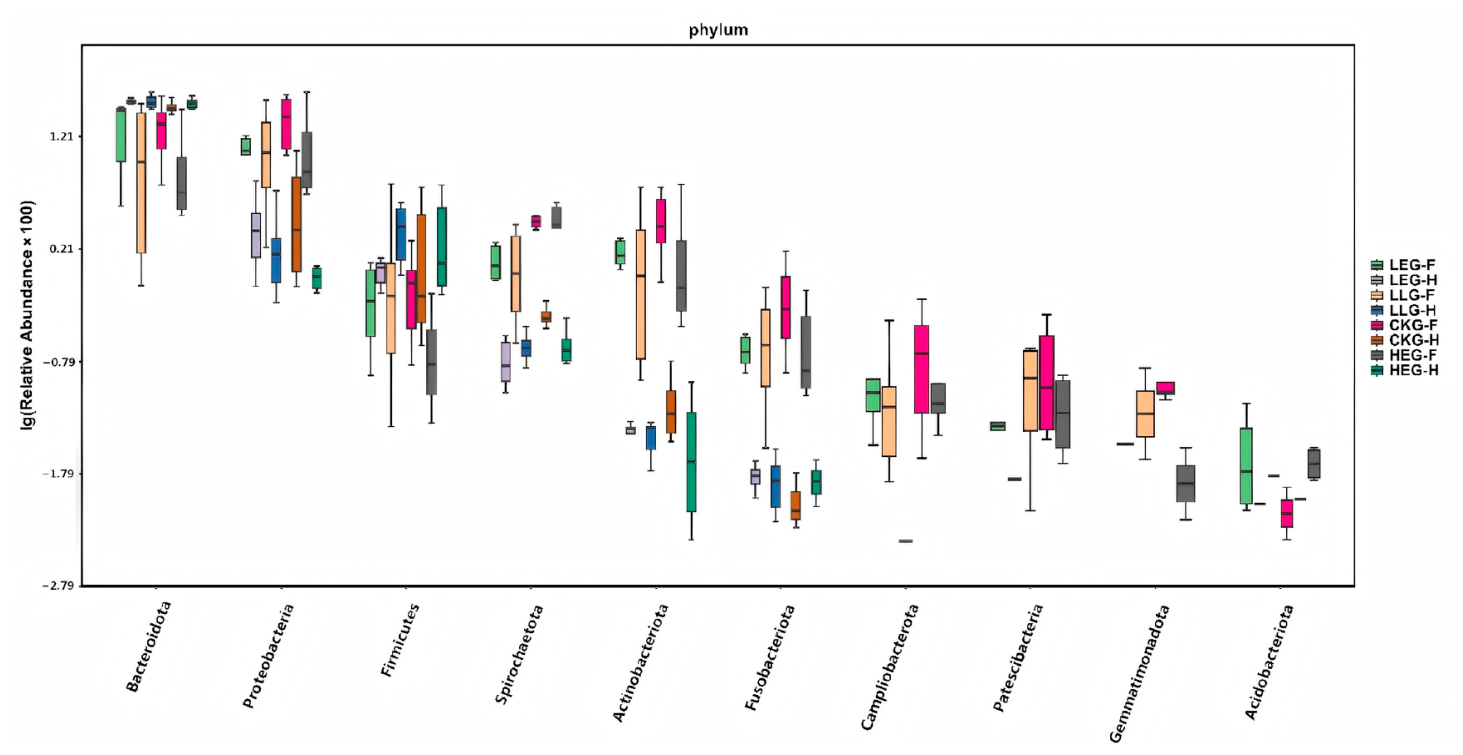
Figure 1.
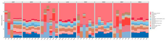
Segment-specific values of major bacterial taxa at the phylum level when feeding yak diets containing CKG (0.82 MJ/kg NEG), LEG (2.12 MJ/kg NEG), LLG (2.12 MJ/kg NEG+0.02% Lactobacillus fermentum), and HEG (2.69 MJ/kg NEG).
3.2. Lactobacillus fermentum Intervention Is Linked to Compositional Gut Microbiota Changes
To assess how Lactobacillus fermentum influences the restoration of gut structure and function in yaks consuming high-concentrate diets, 16S rDNA sequencing was performed. The analysis of the microbial communities identified the duodenum, jejunum, and ileum as part of the small intestine, while the cecum, colon, and rectum were identified as components of the hindgut. A total of 28,621 high-quality sequences were extracted from 144 gut samples (6 segments × 6 animals × 4 treatments), with 21,979 clean reads retained and organized into 2050 groups, representing 1 kingdom, 23 phyla, 54 classes, 129 orders, 215 families, 468 genera, and 1160 species. The small intestine was mainly dominated by Bacteroidota, Firmicutes, and Proteobacteria, while the hindgut was largely composed of Firmicutes and Bacteroidota (Figure 1). Feeding with high-concentrate diets notably raised the relative abundance of Proteobacteria in the small intestine and Firmicutes in the hindgut, while the Bacteroidetes levels decreased, yet they remained above the CKG thresholds (Kruskal–Wallis H test, p < 0.05). The introduction of Lactobacillus fermentum significantly boosted the levels of Firmicutes and Bacteroidota in the small intestine, as well as Bacteroidota and the Firmicutes/Bacteroidota (F/B) ratio in the hindgut (p < 0.05). Overall, these changes suggest that Lactobacillus fermentum promotes the production of SCFAs and enhances fiber breakdown, thus improving the energy efficiency of yaks on fibrous diets.
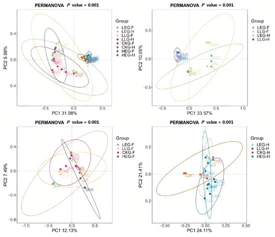
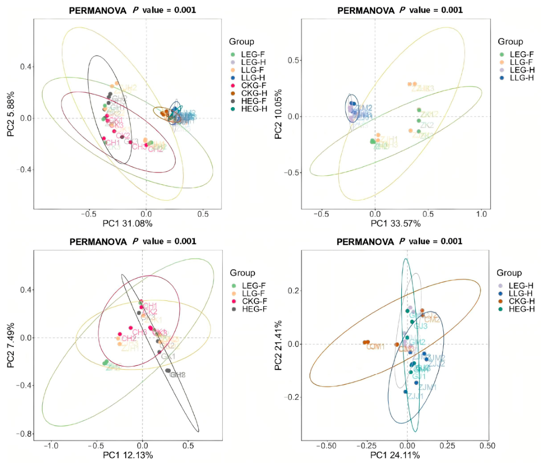
The alpha diversity analysis revealed that the Lactobacillus fermentum intervention group (LLG) showed significant increases in the Shannon index (9.12 ± 0.08 versus 7.99 ± 0.42 in the LEG group), the Ace index (1316.29 ± 8.09 compared to 902.58 ± 138.18), and the Chao1 index (1323.75 ± 7.74 vs. 908.90 ± 140.62) within the duodenum (p < 0.05, Table 5). Consistently with the Ace index, the Chao1 estimator also indicated significantly higher species richness in the ilea of LLG animals (1092.46 ± 25.27) relative to the LEG group (502.73 ± 26.17, p < 0.05). The Simpson index in the cecum was recorded at 0.9948 ± 0.0004, indicating that the hindgut’s overall diversity exceeded that of the small intestine. In the rectum, the LLG group demonstrated the highest Shannon index (8.81 ± 0.12), ACE value (937 ± 58), and Chao1 index (939.31 ± 56.79), although these differences did not reach statistical significance. Coverage values were all above 0.999, confirming the sufficient sequencing depth. These results imply that a Lactobacillus fermentum intervention can modify the microbial community structure by increasing the species richness in critical areas like the duodenum and ileum. The high-diversity microecological advantage, shaped by the unique environment and complex metabolic activity in the hindgut, supports the optimization of high-concentrate feeding strategies. Furthermore, high-energy diets had a notable impact on the microbial community structure. In the small intestine (duodenum, jejunum, and ileum), the β-diversity revealed distinct separation among the CKG, HEG, and LEG groups in response to different dietary energy levels, while the LLG group with the Lactobacillus intervention exhibited a clustered pattern that differed from that in the LEG group. The principal coordinate analysis (PCoA) illustrated distinct spatial distributions of the microbial communities across different intestinal segments (Figure 2). In the small intestine, the first principal component (PC1, 31.08%) and the second principal component (PC2, 5.88%) explained the majority of the variance. For the hindgut, PC1 (33.57%) and PC2 (10.05%) also showed significant explanatory power. The small intestine data mirrored the overall segmental characteristics (PERMANOVA p = 0.001). Samples within each group were tightly clustered, while clear independent clusters emerged between groups across different energy treatments and the Lactobacillus fermentum intervention. The most significant differences in microbial community structure were observed between the small intestine (PC1 12.13%/PC2 7.49%) and the hindgut (PC1 24.11%/PC2 21.41%), with the PERMANOVA analysis confirming p = 0.001 for all comparisons.
Table 5.
The alpha diversity of bacterial communities when feeding yak diets containing CKG (0.82 MJ/kg NEG), LEG (2.12 MJ/kg NEG), LLG (2.12 MJ/kg NEG + 0.02% Lactobacillus), and HEG (2.69 MJ/kg NEG).
Figure 2.
The PCoA profiles of gut-bacterial communities across intestinal segments when feeding yak diets containing CKG (0.82 MJ/kg NEG), LEG (2.12 MJ/kg NEG), LLG (2.12 MJ/kg NEG+0.02% Lactobacillus fermentum), and HEG (2.69 MJ/kg NEG).
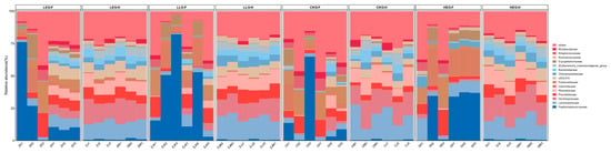
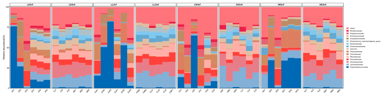
Given that the yak serum biochemical parameters and segment-specific SCFA profiles are known to modify the composition of the microbiota during dietary energy-mediated processes, we investigated the evolution of the gut microbiota composition under the influence of Lactobacillus fermentum throughout this process (Figure 3). In the small intestine, the primary bacterial families identified were Peptostreptococcaceae, Clostridiaceae, and Erysipelotrichaceae, together with oral-derived or facultative anaerobic taxa such as Pasteurellaceae, Streptococcaceae, and Prevotellaceae. Meanwhile, the hindgut was largely dominated by strictly anaerobic, fiber-degrading taxa including Oscillospiraceae, Lachnospiraceae, Rikenellaceae, and UCG-010. Significant differences were noted among dietary treatments in the small intestine, where the CKG group showed the highest presence of oral-derived or facultative anaerobic species, with Pasteurellaceae (0.123), Prevotellaceae (0.081), and Streptococcaceae (0.056) ranking among the top families, whereas the HEG group exhibited a pronounced shift towards clostridial dominance, led by Clostridiaceae (0.151) and Peptostreptococcaceae (0.241), and the LEG group displayed an intermediate profile characterized by Peptostreptococcaceae (0.222). In contrast, the hindgut was primarily inhabited by strictly anaerobic, fiber-degrading bacteria across all groups, including Oscillospiraceae (0.139), Lachnospiraceae (0.173), Rikenellaceae (0.106), UCG-010 (0.065), Bacteroidaceae (0.054), and Christensenellaceae (0.059), with relatively minor inter-group variation. Furthermore, the LLG group displayed the most notable abundance of clostridial taxa in the small intestine, such as Peptostreptococcaceae (0.360) and Clostridiaceae (0.092), accompanied by the lowest levels of oral-derived families (Pasteurellaceae, 0.068 and Streptococcaceae, 0.024) among all treatments, suggesting that the Lactobacillus fermentum intervention further strengthened the energy-associated enrichment of clostridial populations in the small intestine.
Figure 3.
The segment-specific distribution of dominant bacterial families when feeding yak diets containing CKG (0.82 MJ/kg NEG), LEG (2.12 MJ/kg NEG), LLG (2.12 MJ/kg NEG + 0.02% Lactobacillus fermentum), and HEG (2.69 MJ/kg NEG).
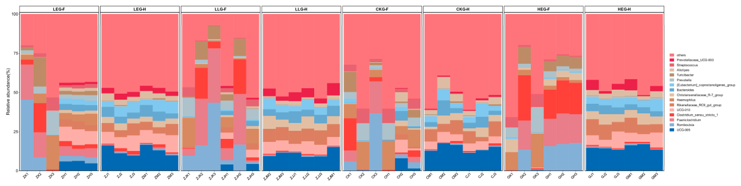
A genus-level species abundance clustering analysis was conducted among the CKG, HEG, LEG, and LLG groups (Figure 4). The results indicated that both dietary energy and the Lactobacillus fermentum intervention induced region-specific changes in differential bacterial genera across the small intestine and hindgut. In the small intestine, the HEG group was enriched in clostridial taxa including Clostridium_sensu_stricto_1, Paeniclostridium, and Romboutsia (p < 0.05). The LEG group showed intermediate abundances of these clostridial genera, while the CKG control group harbored higher relative abundances of facultative anaerobic genera such as Haemophilus and Streptococcus (p < 0.05). In the hindgut, core fibrolytic and SCFA-producing genera (UCG-005, UCG-010, Rikenellaceae_RC9_gut_group, Christensenellaceae_R-7_group, Bacteroides, Alistipes) dominated across CKG, LEG, and HEG (p < 0.05), with relatively moderate inter-group variation. For the comparison between LEG and LLG in the small intestine, the abundances of LLG group clostridial taxa, including Paeniclostridium, Romboutsia, Clostridium_sensu_stricto_1, and Turicibacter, were markedly higher than those in the LEG group (p < 0.05). Conversely, the abundances of potential opportunistic or pro-inflammatory genera, such as Haemophilus, Streptococcus, and Prevotella, were significantly reduced (p < 0.05). In the hindgut, the LLG group demonstrated markedly upregulated abundances of fiber-degrading and SCFA-producing taxa, including Rikenellaceae_RC9_gut_group, Christensenellaceae_R-7_group, Prevotellaceae_UCG-003, and Bacteroides, whereas genera including UCG-005, UCG-010, Alistipes, and [Eubacterium]_coprostanoligenes_group were significantly downregulated (p < 0.05). These findings suggest that dietary energy and Lactobacillus fermentum supplementation jointly exert pronounced effects in terms of shifts in the relative abundance of these dominant genera, with Lactobacillus fermentum intervention as a critical factor in shaping the genus-level microbial community structure across various gut segments.
Figure 4.
Genus-level abundance clustering of differential bacterial taxa across small intestine–hindgut segments when feeding yak diets containing CKG (0.82 MJ/kg NEG), LEG (2.12 MJ/kg NEG), LLG (2.12 MJ/kg NEG+0.02% Lactobacillus fermentum), and HEG (2.69 MJ/kg NEG).
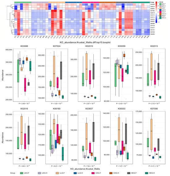
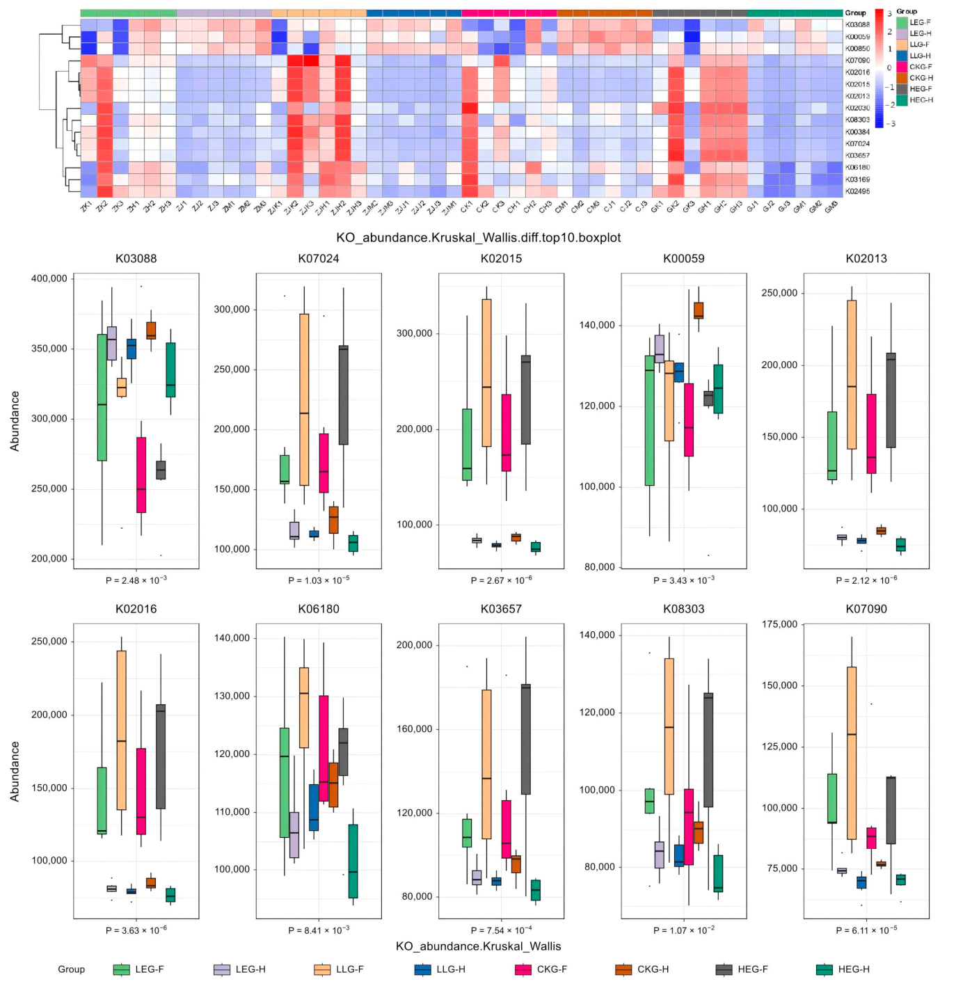
Based on the KEGG functional annotation of KO profiles (Figure 5), we analyzed the microbial functional gene profiles in different intestinal segments among the four groups. The top ten differential KO functions in each intestinal segment were predominantly involved in diverse metabolic pathways, including carbohydrate metabolism, amino acid metabolism, cofactor and vitamin metabolism, and energy metabolism. Additionally, genes related to translation, membrane transport, replication and repair, nucleotide metabolism, and polysaccharide biosynthesis and metabolism were also identified. The small intestine (duodenum, jejunum, and ileum) exhibited higher enrichment of pathways associated with cellular communities (prokaryotes), while the hindgut (cecum, colon, and rectum) was enriched in signal transduction pathways. From an energy perspective, the core functional gene composition of the gut microbiota across diets of different energy levels (CKG, LEG, HEG) remained largely conserved across intestinal segments, with notable differences in the distribution of signal transduction and cellular community (prokaryotes) pathways. When comparing the LEG and LLG groups in terms of the Lactobacillus fermentum intervention, the intervention did not alter the core functional framework of the gut microbiota but modulated the enrichment patterns of signal transduction and various metabolic pathways (Kruskal–Wallis tests, p < 0.01). These results suggest that the functional differences among microbial communities in different intestinal segments of yaks primarily focus on these differential KO terms, participating in carbohydrate, amino acid, energy, polysaccharide, cofactor, and vitamin metabolism.
Figure 5.
KEGG KO profile heatmap and boxplots of differential microbial functional genes across intestinal segments when feeding yak diets containing CKG (0.82 MJ/kg NEG), LEG (2.12 MJ/kg NEG), LLG (2.12 MJ/kg NEG+0.02% Lactobacillus fermentum), and HEG (2.69 MJ/kg NEG).
3.3. Demonstration of Metabolomic Alterations Across Different Lactobacillus fermentum Supplementation Strategies and Identification of Segment-Specific Signature Metabolites
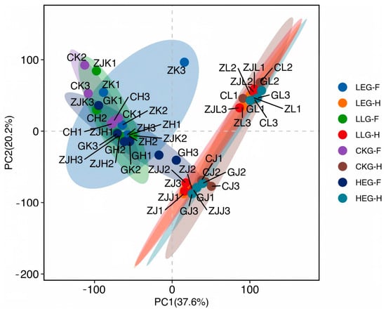
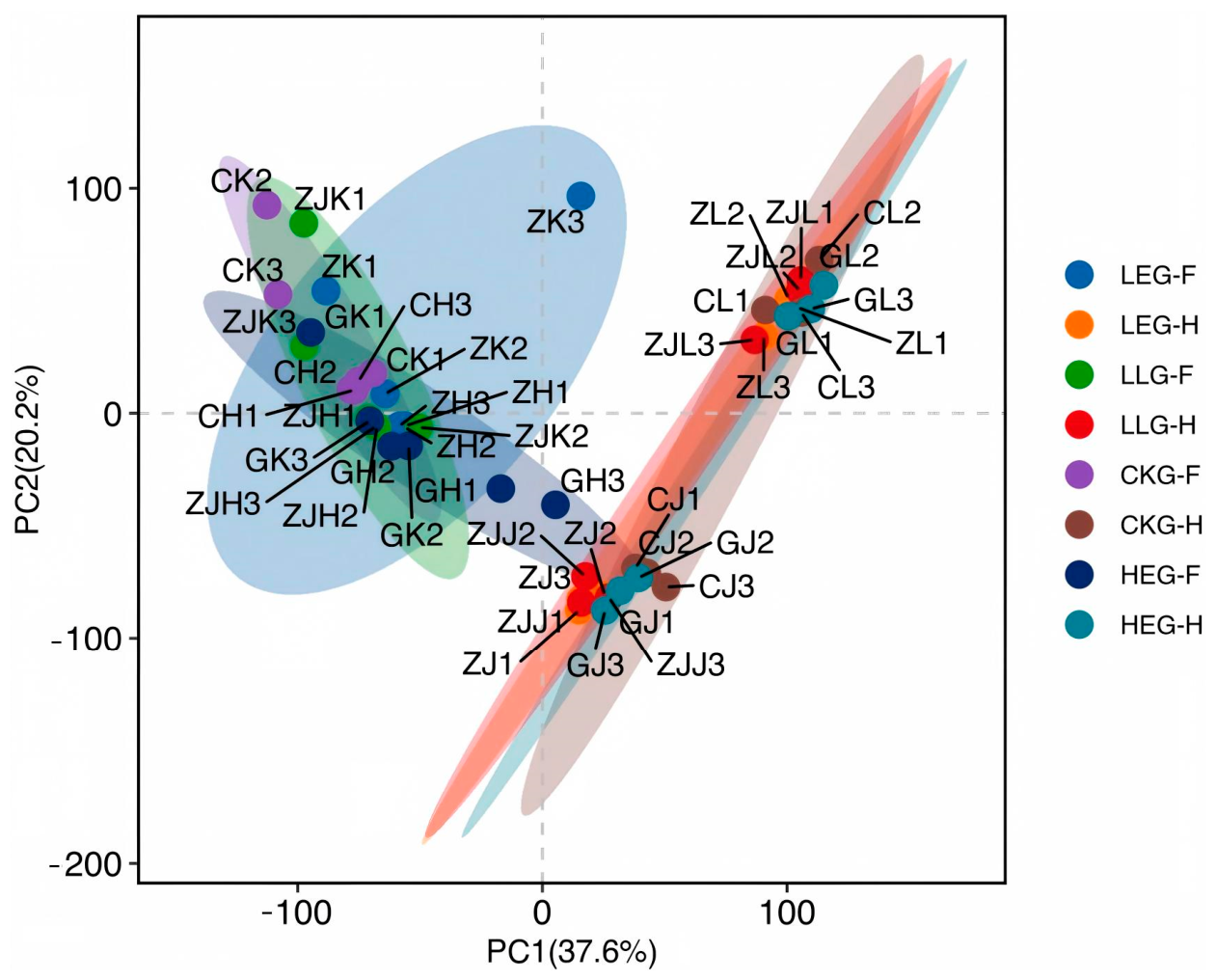
To investigate the regulatory effects of dietary energy and Lactobacillus fermentum intervention on the gut-microbial community structure, we utilized the principal coordinate analysis (PCoA) multivariate statistical approach to assess the variations in gut microbiota in the small intestine and hindgut (Figure 6). The PCoA model successfully revealed distinct clusters of microbial communities under different treatments, with noticeable separation between the small intestine and hindgut. To further clarify the effects of dietary energy and Lactobacillus fermentum intervention on microbial community differences between the small intestine and hindgut in yaks, we conducted separate analyses for each gut region, which led to the identification of clear spatial segregation among microbial assemblages. Most of these microbial communities, with varying distributions, were shaped by two primary factors (the gut segment and Lactobacillus fermentum). In the small intestine, yaks receiving diets of different energy levels (LEG, HEG, and CKG) exhibited distinguishable microbial clusters, and the LLG group showed divergent microbiota profiles compared with those in the LEG group. Conversely, in the hindgut, yaks from the LEG, HEG, CKG, and LLG groups clustered within the hindgut area, while the Lactobacillus fermentum intervention still altered the microbiota composition when compared to the LEG group. These findings suggest the involvement of marked gut segment-specific and energy-dependent microbial assembly, and Lactobacillus fermentum intervention can modulate the community structure of the gut microbiota in both the small intestine and hindgut in yaks.
Figure 6.
PCoA showing spatial segregation of small intestine and hindgut bacterial communities when feeding yak diets containing CKG (0.82 MJ/kg NEG), LEG (2.12 MJ/kg NEG), LLG (2.12 MJ/kg NEG + 0.02% Lactobacillus fermentum), and HEG (2.69 MJ/kg NEG). Note. The grey dotted lines indicate the zero positions of the PC1 and PC2 axes.
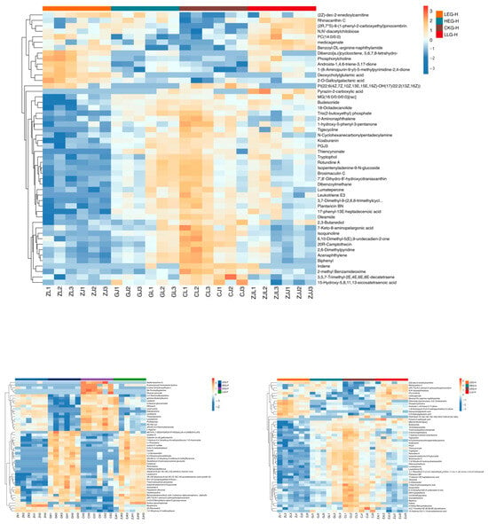
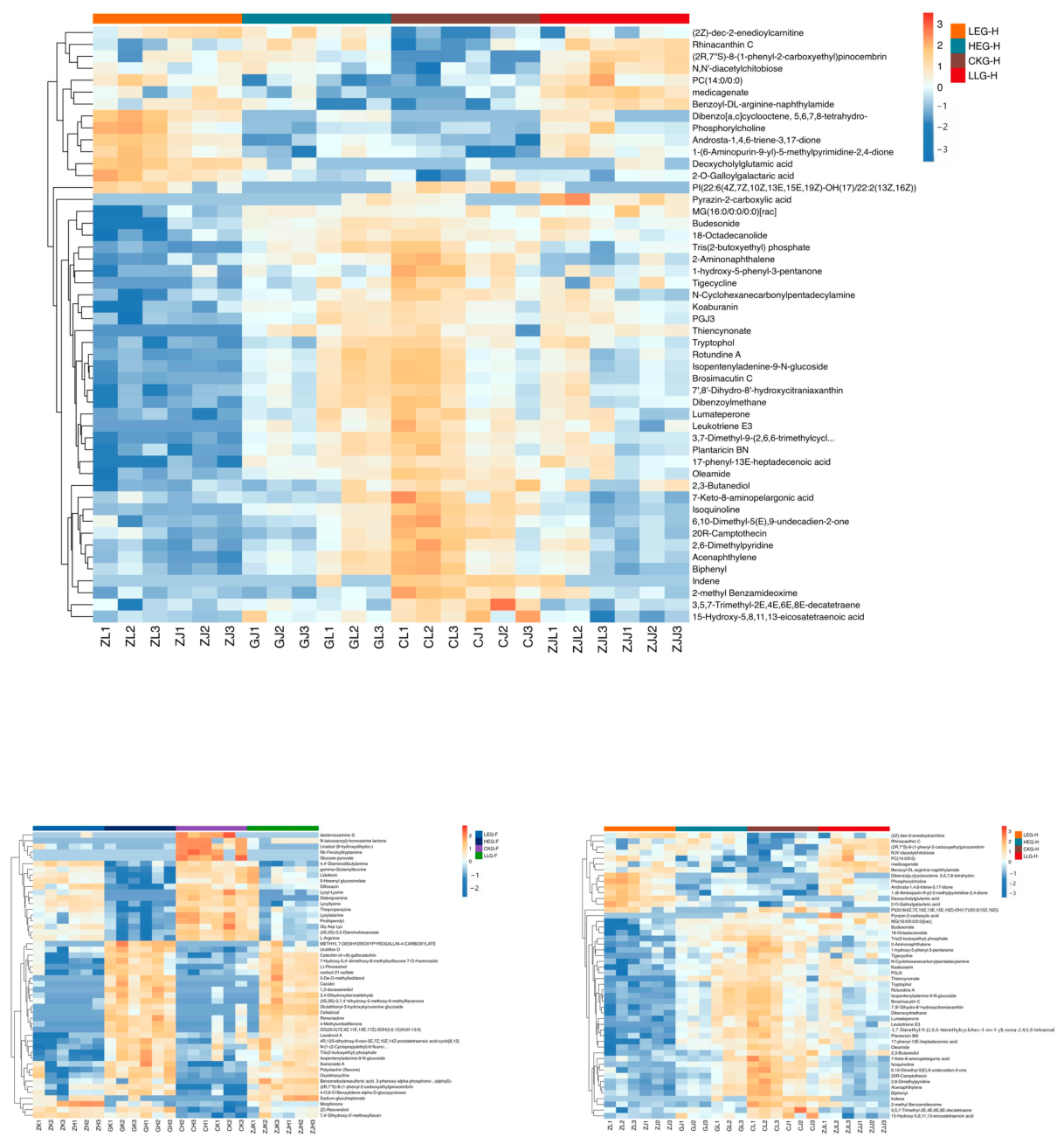
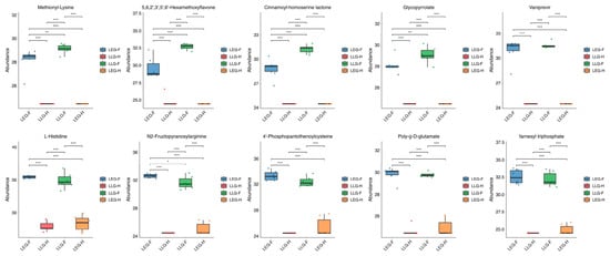
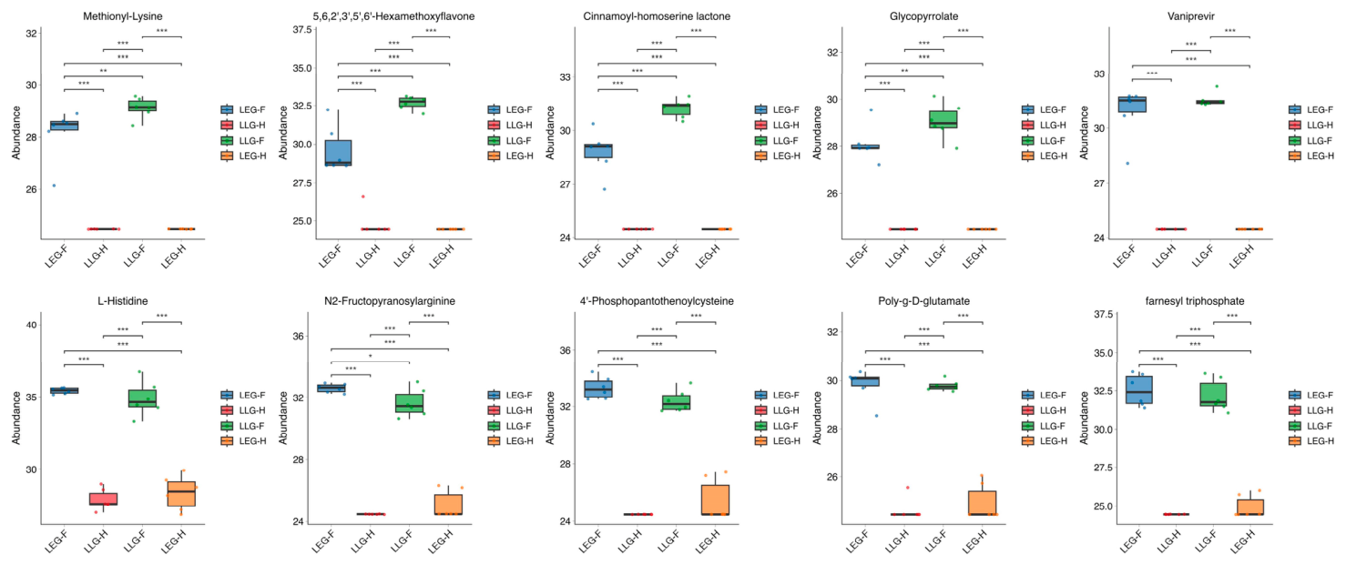
A hierarchical cluster analysis (HCA) combined with heatmap visualization was conducted to examine the variations in the small intestine and hindgut metabolomes of yaks across different dietary energy levels and Lactobacillus fermentum (Figure 7). The HCA showed that the shifts in dietary energy and the Lactobacillus fermentum intervention significantly altered the small intestine and hindgut metabolomes (p < 0.05), with clear intergroup differences highlighted in the heatmap. An analysis of the metabolic pathways revealed several metabolites linked to amino acid, lipid, and flavonoid metabolism in both gut regions; in particular, the abundance of multiple bioactive metabolites was significantly reshaped in the LLG group (p < 0.05), suggesting that the Lactobacillus fermentum intervention helped to restore the balance of gut metabolism disrupted by the variable dietary energy supply. The gut metabolite profiles were distinctly modulated by the dietary energy levels among the LEG, HEG, and CKG groups. Boxplots of metabolites were created, leading to the identification of candidate microbial and metabolic signatures (Figure 8). A total of 20 differential metabolites were found through pairwise comparisons, including various compounds in the small intestine (cinnamoyl-homoserine lactone, 5,6,2′,3′,5′,6′-hexamethoxyflavone, glycopyrrolate, vaniprevir, and methylonyl-lysine) and hindgut (N2-fructopyranosylarginine, 4′-phosphopantothenoylcysteine, poly-g-D-glutamate, farnesyltriphosphate, and L-histidine). Overall, these results indicate that intervention with Lactobacillus fermentum modifies the gut-metabolic profile in yaks, underscoring the enhanced efficacy of LLG compared to LEG in mitigating gut-metabolic issues under high-concentrate feeding conditions.
Figure 7.
HCA heatmap of intestinal metabolomes in small intestine and hindgut when feeding yak diets containing CKG (0.82 MJ/kg NEG), LEG (2.12 MJ/kg NEG), LLG (2.12 MJ/kg NEG+0.02% Lactobacillus fermentum), and HEG (2.69 MJ/kg NEG).
Figure 8.
Boxplot visualization of 20 representative differential intestinal metabolites screened by pairwise comparisons when feeding yak diets containing CKG (0.82 MJ/kg NEG), LEG (2.12 MJ/kg NEG), LLG (2.12 MJ/kg NEG + 0.02% Lactobacillus fermentum), and HEG (2.69 MJ/kg NEG). Note. Asterisks indicate statistical significance: * p < 0.05, ** p < 0.01, *** p < 0.001.
3.4. Multi-Omics Signature Integration Research for High-Concentrate Diets and Lactobacillus fermentum Intervention Treatment
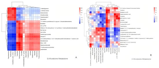
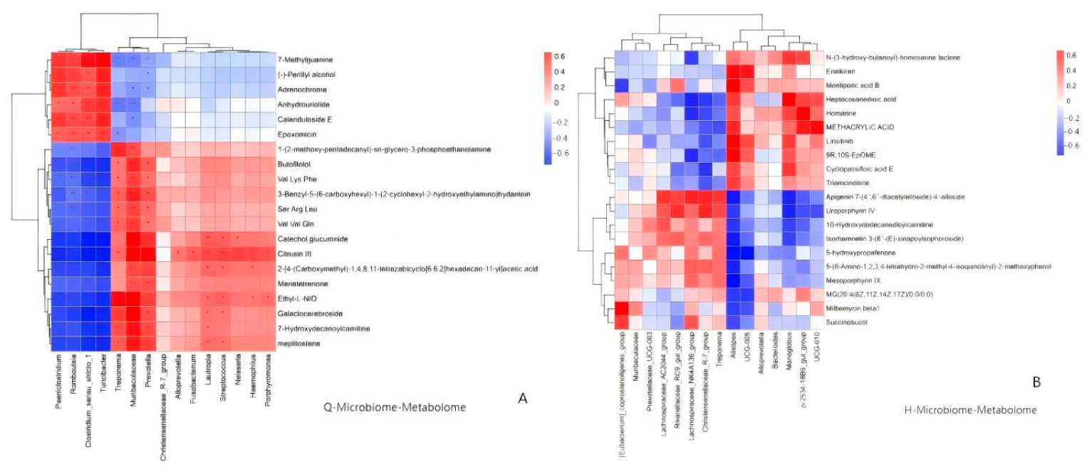
Spearman correlation coefficients were utilized to construct a correlation network aimed at assessing the functional relationships between the microbiota and metabolites (VIP > 2, p < 0.05) (Figure 9). Notable correlations were identified across these multi-omics variables. In the small intestine, the target metabolites, such as 4,4′-diaminodibutylamine, cinnamoyl-homoserine lactone, and zardaverine, exhibited positive correlations with Romboutsia, Paeniclostridium, Clostridium_sensu_stricto_1, and Streptococcus, while showing negative correlations with flavonoid compounds and other differential metabolites. Conversely, gut microbiota including Prevotella, UCG-005, Rikenellaceae_RC9_gut_group, and Christensenellaceae_R-7_group displayed an inverse correlation pattern with these metabolites. In the hindgut, the target microbiota, such as UCG-010, Alistipes, Alloprevotella, Rikenellaceae_RC9_gut_group, Christensenellaceae_R-7_group, and Bacteroides, were positively associated with (2Z)-dec-2-enedioylcarnitine, rhinacanthin C, medicagenate, and other metabolites, while showing negative correlations with polycyclic alcohols and lipid compounds.
Figure 9.
Spearman correlation heatmap illustrating associations between gut microbiota and differential metabolites in small intestine and hindgut when feeding yak diets containing CKG (0.82 MJ/kg NEG), LEG (2.12 MJ/kg NEG), LLG (2.12 MJ/kg NEG+0.02% Lactobacillus fermentum), and HEG (2.69 MJ/kg NEG). Note. (A) is the Spearman correlation heatmap illustrating associations between the gut microbiota and differential metabolites in the small intestine; (B) is the Spearman correlation heatmap illustrating associations between the gut microbiota and differential metabolites in the hindgut.
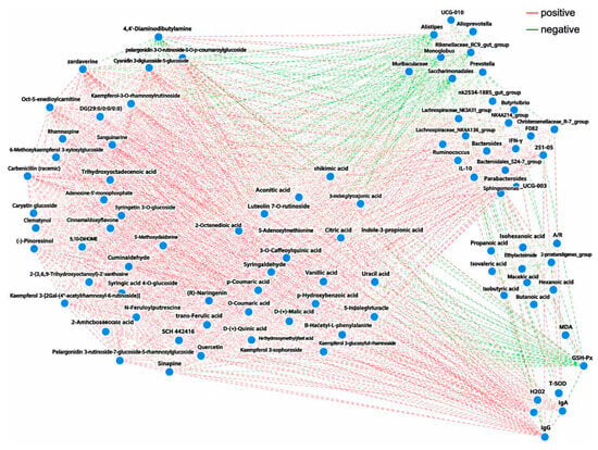
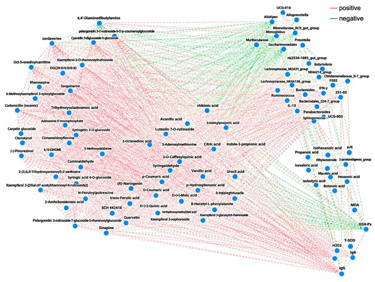
Comprehensive analyses integrating omics and phenotypic statistics were conducted to investigate the overall metabolic impacts of Lactobacillus fermentum supplementation in yaks. The findings indicated that these bacteria altered the gut microecology and transformed the metabolic profiles of the host guts (Figure 10). In contrast to the LEG control group, the LLG group showed significant metabolic enhancements, particularly with elevated levels of SCFAs (acetate, propionate, butyrate) in LLG; these SCFAs were closely linked to lipid–glucose metabolism and immune responses. Propionate, acting as a gluconeogenic precursor, improved blood glucose regulation, while butyrate fortified intestinal barrier function and boosted the systemic antioxidant capacity (T-AOC, SOD), collectively promoting a healthy lipid metabolism phenotype. Additionally, harmful microbial metabolites such as LPS, phenol, and p-cresol were significantly diminished in LLG, with these substances showing negative correlations with serum LPS, pro-inflammatory cytokines (TNF-α, IL-6), and liver enzyme activity (ALT, AST). Multi-omics correlation networks revealed associative relationships whereby probiotic-altered bacterial taxa were correlated with the diminished abundance of potential opportunistic microbes and lower levels of detrimental metabolites, which were further correlated with improved serum inflammatory antioxidant and hepatic enzyme profiles under high-concentrate feeding. The metabolic profiles of branched-chain amino acids (leucine, valine) and essential amino acids in both the gut and blood shifted towards equilibrium in LLG, demonstrating positive associations with serum total protein (TP) and albumin (ALB). Lactobacillus fermentum enhanced the absorption and utilization of gut amino acids and optimized overall nitrogen metabolism, channeling more amino acids into protein synthesis rather than breakdown to support yak growth. Furthermore, the ratio of primary bile acids (CA, CDCA) to secondary bile acids (DCA, LCA) was modified in LLG and linked to serum triglyceride (TG) and cholesterol (CHOL) levels. Lactobacillus fermentum influenced the activity of bile acid hydrolase in the gut microbiota and regulated the enterohepatic circulation of bile acids, improving the emulsification of fats and the digestibility of dietary lipids. Overall, the addition of Lactobacillus fermentum provides beneficial effects through a multi-faceted microbiota–metabolite–host interaction rather than a singular pathway. By enhancing beneficial metabolites like SCFAs and diminishing harmful compounds such as LPS and amines, Lactobacillus fermentum collaboratively stabilized energy metabolism, reduced hepatic oxidative stress and inflammation, strengthened gut barrier integrity, and promoted amino acid utilization and protein synthesis. These results underscore the effectiveness of LLG in alleviating metabolic disorders induced by high-concentrate diets and in preserving the health of yaks.
Figure 10.
Spearman-based multi-omics correlation network integrating serum biochemical indicators, SCFAs, gut microbiota, and intestinal metabolites when feeding yak diets containing CKG (0.82 MJ/kg NEG), LEG (2.12 MJ/kg NEG), LLG (2.12 MJ/kg NEG+0.02% Lactobacillus fermentum), and HEG (2.69 MJ/kg NEG).
4. Discussion
The gut microbiome is increasingly acknowledged as a vital influencer of host health and phenotypic diversity, with its composition showing considerable variation among different host species and sections of the gastrointestinal tract [24]. For instance, in ruminant animals, the combined presence of Firmicutes and Bacteroidetes constituted up to 80% of the gut microbiota, although their proportions differed across various intestinal regions [25]. In contrast, the microbial communities in the ileum, cecum, and colon in monogastric animals were more similar, with these two phyla representing only 50–60% of the overall microbiota [26,27]. Such variations underscore the evolutionary and physiological distinctiveness of ruminants, whose specialized multi-chambered digestive systems offer fundamentally different efficiencies in feed digestion and metabolic processes compared to non-ruminant mammals. The Pamir yak, a unique domestic ruminant adapted to extreme plateau conditions, illustrates these intricate gastrointestinal adaptations, with each part of its digestive system showing specialized functional changes that are closely associated with the composition and metabolic functions of its resident microbial community [28]. This research revealed that higher dietary energy levels negatively impacted systemic and gut homeostasis in yaks, as indicated by lower serum albumin and total protein levels, increased immune activation markers, reduced antioxidant capacity, and significant changes in SCFAs and microbial community structure. Importantly, the addition of dietary Lactobacillus fermentum partially mitigated several adverse effects, particularly by restoring serum albumin and total protein levels, lowering urea concentrations and circulating immunoglobulin/cytokine markers, and boosting the activity of T-SOD, GSH-Px, T-AOC, and MDA. However, it should be noted that serum MDA, as a lipid peroxidation end product, may not perfectly reflect the whole-body oxidative status in yak high-concentrate feeding models, as its circulating concentration can be modulated by metabolite translocation from intestinal segments and in vivo clearance rates. The higher serum immunoglobulin concentrations in LLG reflect enhanced humoral immune competence rather than pathological inflammatory over activation, which is consistent with decreased pro-inflammatory cytokine levels. Additionally, probiotic supplementation enhanced the microbial diversity in the duodenum and ileum, leading to the significant restructuring of microbial taxa and metabolites. Notably, the responses of the SCFA, microbiome, and metabolic profiles were not consistent throughout the gut. Instead, they displayed segment-specific variations, suggesting that the effects of the probiotics were spatially distinct. Overall, these results demonstrate that dietary energy levels have a widespread negative influence on blood biochemistry, antioxidant, and immune parameters, as well as gut SCFA and microbial profiles in ruminants, while Lactobacillus fermentum intervention can selectively modify the microbiome and metabolic environment along the intestinal tract, thereby aiding in the preservation of intestinal homeostasis.
In animals, the gut is home to a rich and stable community of microorganisms that play a crucial role in maintaining optimal pH levels, which in turn supports effective feed digestion and various microbial metabolic activities [29,30]. Lactobacillus supplementation can significantly alter the structure of an imbalanced gut microbiota caused by a high-concentrate diet, facilitating the breakdown of complex food elements like dietary fiber and influencing host metabolism through interactions with bile acids, lipids, and amino acids [31]. Microbial profiles reveal that the location within the intestine is a key factor in how diet and Lactobacillus fermentum affect the microbiota [28]. In the small intestine, Firmicutes, Bacteroidota, and Proteobacteria are predominant, while, in the hindgut, Firmicutes and Bacteroidota are more prevalent, consistent with earlier studies on yaks [32]. Notably, the significant recovery of the Shannon, ACE, and Chao1 indices in the small intestine in LLG compared to the LEG group is remarkable. At the genus level, Prevotella is a relatively metabolically active microbial community that includes Treponema, NK4A214_group, Rikenellaceae_RC9_gut_group, Christensenellaceae_R-7_group, Prevotellaceae_UCG-003, and various Lachnospiraceae lineages across different segments. Many of these genera are linked to the breakdown of complex plant polysaccharides in ruminants and the metabolism of SCFAs like acetate, propionate, and butyrate. Their increased presence in the small intestine and hindgut corresponds with observed shifts in carbohydrate metabolism and SCFA distribution. Studies on yak rumen fermentation have similarly shown that changes in concentrate levels affect the abundance of taxa such as Prevotella, Christensenellaceae_R-7_group, NK4A214_group, and other fermentation-related groups [33]. Moreover, Lactobacillus supplementation can reshape the rumen microbiome and impact fatty acid metabolism. Conversely, the hindgut displayed a unique pattern, with increases in Paeniclostridium, Clostridium_sensu_stricto_1, Romboutsia, Haemophilus, Prevotella, Streptococcus, and Neisseria. This observation challenges the simplistic classification of genera as strictly beneficial or harmful. Prevotella copri (P. copri), a well-studied species within the Prevotella genus, was first isolated and validated from the human gut [34]. Emerging evidence suggests that P. copri is significant in the dynamics of the ruminant gut microbiome, particularly in relation to dietary adaptation, fiber metabolism, and host–microbe metabolic interactions. Research also indicates that P. copri is crucial for breaking down pectin and hemicellulose in the rumen of cattle and sheep, indirectly affecting methane emissions through its interactions with methanogenic archaea [35]. In our research, the prevalence of Prevotella copri rose under high-concentrate feeding, potentially linked to the regulation of rapid starch fermentation and lactic acid metabolism. Additionally, studies suggest that the metabolic byproducts of this bacterium can influence host gut epithelial barrier function and local immune responses.
The role of the gut microbiota and their metabolites in influencing host health and characteristics is increasingly acknowledged. Notably, there are marked differences in the intestinal microbiota of monogastric animals compared to ruminants [36]. In our study, we categorized the duodenum, jejunum, and ileum as the small intestine, while the cecum, colon, and rectum were identified as the hindgut, based on their microbial profiles. Our results reveal that gut microorganisms can generate a range of metabolites, such as fatty acids, bile acids, polyamines, indoles, and vitamins, either through their metabolic processes or in conjunction with the host, all of which play a role in host health. The metabolomic analysis complements our taxonomic observations; the results indicate region-dependent effects of Lactobacillus fermentum that likely reflect fundamental differences in the niche environment of the yak small intestine and hindgut, including the resident microbial background, fermentable substrate supply, digesta retention time, and local redox conditions. Such compartmental features may constrain the persistence and metabolic activity of the supplemented strain, leading to divergent shifts in SCFAs and metabolomic signatures across gut segments. Specifically, following the supplementation of Lactobacillus fermentum, there was an increase in only one metabolite in the small intestine, while 49 others decreased; in contrast, the hindgut showed a more balanced distribution of increases and decreases. This imbalance implies that the probiotic had its most substantial impact before the digesta reached the distal gut, while the hindgut experienced the broader rebalancing of competing metabolic pathways [37]. The enhancement of fatty acid, carbohydrate, and amino acid metabolism corresponds with the data on SCFAs and the improvement in serum protein levels. These predicted pathways suggest a potential capacity for enhanced SCFA synthesis, which is further supported by our measured digesta SCFA and metabolomic data. Overall, these results suggest a preliminary mechanism connecting the microbiota, metabolites, and host interactions. Lactobacillus fermentum may initially alter substrate use and ecological dynamics in the proximal intestine, resulting in increased community diversity at sensitive sites and a shift in the balance between saccharolytic and proteolytic fermentation. These alterations could decrease the excessive supply of fermentable substrates to the hindgut, redirect hindgut fermentation towards propionate production, and modify microbial lipid and amino acid metabolism. The resulting metabolite environment is linked to improved antioxidant defenses, decreased immune activation, and better serum protein levels [38,39]. This model is consistent with recent research on yaks, which showed that Lactobacillus fermentum enhanced the jejunal structure and modified the jejunal microbiota, SCFAs, and metabolomic pathways under high-energy feeding conditions.
5. Conclusions
In summary, dietary energy levels substantially modulated serum biochemical and immune–antioxidant indices, segment-specific SCFA patterns, and small intestine–hindgut microbiota–metabolome profiles in Pamir yaks. High-energy feeding exacerbated immune–oxidative stress, disrupted nutrient metabolism, and induced gut-microbial dysbiosis. Supplementation with Lactobacillus fermentum under high-energy feeding was associated with shifts in gut-microbial diversity, the relative abundance of fibre-degrading and SCFA-producing taxa, digesta SCFA profiles and host serum immune–antioxidant phenotypes in yaks. The Lactobacillus fermentum intervention reshaped microbiota–metabolite–host interactions without altering the core microbial function, ameliorating concentrate-triggered gut-metabolic disturbances and improving the host’s systemic status. These segment-specific microbial and metabolic signatures provide candidate microbial and metabolic signatures for probiotic-based fattening regimens for plateau yaks.
Author Contributions
Conceptualization, J.J. and Q.C.; methodology, M.A.; software, Q.C.; validation, H.R., Q.L. and X.F.; formal analysis, X.F.; investigation, Q.L.; resources, J.J. and H.R.; data curation, Q.C.; writing—original draft preparation, H.R. and J.J.; writing—review and editing, M.A.; visualization, H.R.; supervision, Q.C.; project administration, Q.L.; funding acquisition, X.F. All authors have read and agreed to the published version of the manuscript.
Funding
This research was funded by the Natural Science Foundation of Shandong Province, grant number ZR2023MC164, and the Youth Innovation Team Project Program of Shandong Provincial Education Department, grant number 2022KJ137. The APC was funded by the Youth Innovation Team Project Program of Shandong Provincial Education Department, grant number 2022KJ137.
Institutional Review Board Statement
The animal study protocol was approved by the Animal Care Committee at Qilu Normal University, China (20230361-109, date 6 March 2023). Furthermore, approval for the slaughtering of the animals was obtained in accordance with the National Administration of Experimental Animal Slaughtering and Quarantine Regulations.
Informed Consent Statement
Not applicable.
Data Availability Statement
The datasets presented in this study can be found in online repositories. All raw sequence data were deposited in the NCBI Sequence Read Archive database under accession number PRJNA1233467.
Conflicts of Interest
The authors declare no conflicts of interest.
References
- Ma, Z.; Liu, H.; Degen, A.A.; Zhang, L.; Mi, J.; Zhou, J. Effects of fattening strategies on carcass traits, meat quality, fatty acid composition, and oxidative stability of longissimus muscle in yaks (Bos grunniens). Food Chem. X 2026, 33, 103540. [Google Scholar] [CrossRef] [Scilit] [PubMed]
- Hu, K.; Wu, Q.; Chen, T.; Wang, J.; Zhu, Z.; Shen, Y.; Wang, C.; Wei, X. Grazing practice affects the growth performance, meat quality and nutritional composition, and fecal microbiota of fattening yaks. Anim. Biosci. 2025, 39, 250052. [Google Scholar] [CrossRef] [Scilit] [PubMed]
- Ren, H.; Chen, Q.; Aikelaimuc, M.; Qin, L.; Zhang, G.; Liu, L.; Jia, J. Lactobacillus Modulates the Rumen Microbiota and Transcriptome to Enhance Nutrient Digestion in Yaks Fed High-Concentrate Diets During the Cold Season. Animals 2026, 16, 2644. [Google Scholar] [CrossRef] [Scilit] [PubMed]
- Humer, E.; Aditya, S.; Kaltenegger, A.; Klevenhusen, F.; Petri, R.M.; Zebeli, Q. Graded substitution of grains with bakery by-products modulates ruminal fermentation, nutrient degradation, and microbial community composition in vitro. J. Dairy Sci. 2018, 101, 3085–3098. [Google Scholar] [CrossRef] [Scilit] [PubMed]
- Modasia, A.A.; Spiller, R.C.; Warren, F.J. Role of fermentation-resistant and non-fermentable fibers in gastrointestinal health: Mechanisms, benefits, and challenges. Crit. Rev. Food. Sci. 2026, 66, 3821–3837. [Google Scholar] [CrossRef] [Scilit] [PubMed]
- Gao, P.; Pang, S.; Tang, Y.; Wang, Q.; Li, Q.; Zhang, W.; Nie, C.; Niu, J.; Lian, K. Alterations in serum immune parameters, cytokines, intestinal permeability, fecal microbiota, and short-chain fatty acids in healthy and diarrheic suckling calves. Agriculture 2025, 15, 2289. [Google Scholar] [CrossRef] [Scilit]
- Chirivi, M.; Contreras, G.A. Endotoxin-induced alterations of adipose tissue function: A pathway to bovine metabolic stress. J. Anim. Sci. Biotechnol. 2024, 15, 53. [Google Scholar] [CrossRef] [Scilit] [PubMed]
- Urga; Wang, X.; Wei, H.; Zhao, G. Mechanisms and applications of gastrointestinal microbiota–metabolite interactions in ruminants: A review. Microorganisms 2025, 13, 2880. [Google Scholar] [CrossRef] [Scilit] [PubMed]
- Zhang, L.; Xia, Z.; Fu, J.; Yang, Y. Role of the rumen epithelium and associated changes under high-concentrate diets. Int. J. Mol. Sci. 2025, 26, 2573. [Google Scholar] [CrossRef] [Scilit] [PubMed]
- Rivera-Chacon, R.; Pacífico, C.; Ricci, S.; Petri, R.M.; Reisinger, N.; Zebeli, Q.; Castillo-Lopez, E. Prolonged feeding of high-concentrate diet remodels the hindgut microbiome and modulates nutrient degradation in the rumen and the total gastrointestinal tract of cows. J. Dairy Sci. 2024, 107, 9235–9250. [Google Scholar] [CrossRef] [Scilit] [PubMed]
- Kahouli, I.; Malhotra, M.; Tomaro-Duchesneau, C.; Rodes, L.S.; Alaoui-Jamali, M.A.; Prakash, S. Identification of lactobacillus fermentum strains with potential against colorectal cancer by characterizing short chain fatty acids production, anti-proliferative activity and survival in an intestinal fluid: In vitro analysis. J. Bioanal. Biomed. 2015, 7, 104–113. [Google Scholar]
- Wang, C.; Zhang, Q.; Wei, C.; Jin, Y. Ganoderma lucidum dietary fiber and lactobacillus fermentum CECT5716 alleviate constipation via regulation of gut barrier and microbiota. Int. J. Biol. Macromol. 2025, 333, 149010. [Google Scholar] [CrossRef] [Scilit] [PubMed]
- Castro-López, C.; Romero-Luna, H.E.; García, H.S.; Vallejo-Cordoba, B.; González-Córdova, A.F.; Hernández-Mendoza, A. Key stress response mechanisms of probiotics during their journey through the digestive system: A review. Probiotics Antimicrob. Proteins 2023, 15, 1250–1270. [Google Scholar] [CrossRef] [Scilit] [PubMed]
- Jia, J.; Bao, P.; Li, N.; Kong, S.; Chu, M.; Chen, Q.; Yan, P. Lactobacillus supplementation modulates rumen microbiota and metabolism in yaks under fattening feeding conditions: A comprehensive multi-omics analysis. Animals 2025, 15, 1681. [Google Scholar] [CrossRef] [Scilit] [PubMed]
- Alawneh, J.I.; Ramay, H.; Olchowy, T.; Allavena, R.; Soust, M.; Jassim, R.A. Effect of a lactobacilli-based direct-fed microbial product on gut microbiota and gastrointestinal morphological changes. Animals 2024, 14, 693. [Google Scholar] [CrossRef] [Scilit] [PubMed]
- Zhang, M.; Han, R.; Zhang, A.; Xu, C.; Liu, J.; Li, M.; Zhang, N.; Pang, X.; Wang, S. Difference Analysis of Blood Biochemistry, Slaughter Performance and Gastrointestinal Microbiota in Small-Tailed Han Sheep of Different Sexes. Animals 2026, 16, 1332. [Google Scholar] [CrossRef] [Scilit] [PubMed]
- Yang, K.; Li, G.; Li, Q.; Wang, W.; Zhao, X.; Shao, N.; Qiu, H.; Liu, J.; Xu, L.; Zhao, J. Distribution of gut microbiota across intestinal segments and their impact on human physiological and pathological processes. Cell Biosci. 2025, 15, 47. [Google Scholar] [CrossRef] [Scilit] [PubMed]
- Touhara, K.K.; Rossen, N.D.; Deng, F.; Castro, J.; Harrington, A.M.; Chu, T.; Garcia-Caraballo, S.; Brizuela, M.; O’Donnell, T.; Xu, J.; et al. Topological segregation of stress sensors along the gut crypt-villus axis. Nature 2025, 640, 732–742. [Google Scholar] [CrossRef] [Scilit] [PubMed]
- Zhang, J.; Tian, R.; Liu, J.; Yuan, J.; Zhang, S.; Chi, Z.; Yu, W.; Yu, Q.; Wang, Z.; Chen, S.; et al. A two-front nutrient supply environment fuels small intestinal physiology through differential regulation of nutrient absorption and host defense. Cell 2024, 187, 6251–6271.e20. [Google Scholar] [CrossRef] [Scilit] [PubMed]
- Caporaso, J.G.; Kuczynski, J.; Stombaugh, J.; Bittinger, K.; Bushman, F.D.; Costello, E.K.; Fierer, N.; Peña, A.G.; Goodrich, J.K.; Gordon, J.I.; et al. QIIME allows analysis of high-throughput community sequencing data. Nat. Methods 2010, 7, 335–336. [Google Scholar] [CrossRef] [Scilit] [PubMed]
- Edgar, R.C. UPARSE: Highly accurate OTU sequences from microbial amplicon reads. Nat. Methods 2013, 10, 996–998. [Google Scholar] [CrossRef] [Scilit] [PubMed]
- Langille, M.G.; Zaneveld, J.; Caporaso, J.G.; McDonald, D.; Knights, D.; Reyes, J.A.; Clemente, J.C.; Burkepile, D.E.; Vega Thurber, R.L.; Knight, R.; et al. Predictive functional profiling of microbial communities using 16S rRNA marker gene sequences. Nat. Biotechnol. 2013, 31, 814–821. [Google Scholar] [CrossRef] [Scilit] [PubMed]
- Chong, J.; Soufan, O.; Li, C.; Caraus, I.; Li, S.; Bourque, G.; Wishart, D.S.; Xia, J. MetaboAnalyst 4.0: Towards more transparent and integrative metabolomics analysis. Nucleic Acids Res. 2018, 46, W486–W494. [Google Scholar] [CrossRef] [Scilit] [PubMed]
- Wei, J.; Wei, L.; Ullah, A.; Geng, M.; Zhang, X.; Wang, C.; Khan, M.Z.; Wang, C.; Zhang, Z. Metagenomic applications to herbivore gut microbiomes: A comprehensive review of microbial diversity and host interactions. Animals 2025, 15, 2938. [Google Scholar] [CrossRef] [Scilit] [PubMed]
- Forcina, G.; Pérez-Pardal, L.; Carvalheira, J.; Beja-Pereira, A. Gut microbiome studies in livestock: Achievements, challenges, and perspectives. Animals 2022, 12, 3375. [Google Scholar] [CrossRef] [Scilit] [PubMed]
- Xiao, Y.; Xiang, Y.; Zhou, W.; Chen, J.; Li, K.; Yang, H. Microbial community mapping in intestinal tract of broiler chicken. Poult. Sci. 2017, 96, 1387–1393. [Google Scholar] [CrossRef] [Scilit] [PubMed]
- McEwan, N.R.; Nava-Morales, G.M.; Morrison, P.K.; Reis de Souza, T.; Escobar-García, K.; Bautista-Marín, S.E.; Mariscal-Ladín, G.; Silva-Jarquín, J.C.; Gomez-Soto, J.G.; Olvera-Ramírez, A.M. Using Gut Microbiota and Gut Metabolites as Biomarkers for Diseases in Monogastric Farm Animals. Animals 2026, 16, 2247. [Google Scholar] [CrossRef] [Scilit] [PubMed]
- Jia, J.; Zuo, E.; Li, N.; Kong, S.; Bao, P.; Chen, Q.; Yan, P. Overexpression of miRNA29a gene inhibits proliferation and promotes apoptosis of jejunal epithelial cells in yak. Anim. Biotechnol. 2024, 35, 2391520. [Google Scholar] [CrossRef] [Scilit] [PubMed]
- Rawal, S.; Kaur, H.; Bhathan, S.; Mittal, D.; Kaur, G.; Ali, S.A. Ruminant gut microbiota: Interplay, implications, and innovations for sustainable livestock production. In Sustainable Agriculture Reviews: Animal Biotechnology for Livestock Production 4; Springer Nature: Cham, Switzerland, 2024; pp. 205–228. [Google Scholar]
- Zhou, X.; Shen, X. Probiotics modulate the ruminal microbiome and metabolite availability to enhance rumen barrier function and growth performance in goats fed a high-concentrate diet. Probiotics Antimicrob. Proteins 2026, 18, 1983–2000. [Google Scholar] [CrossRef] [Scilit] [PubMed]
- Yu, Q.; Huang, C.; Bao, P.; Li, N.; Zhang, M.; Wang, T.; Ma, C.; Deng, J.; Cao, X.; Jia, J.; et al. Lactobacillus fermentum supplementation modulates jejunal microbiota, metabolome, and morphology in yaks under high-energy feeding. Anim. Microbiome 2026, 8, 17. [Google Scholar] [CrossRef] [Scilit] [PubMed]
- Zhang, L.; Jiang, X.; Li, A.; Waqas, M.; Gao, X.; Li, K.; Xie, G.; Zhang, J.; Mehmood, K.; Zhao, S.; et al. Characterization of the microbial community structure in intestinal segments of yak (Bos grunniens). Anaerobe 2020, 61, 102115. [Google Scholar] [CrossRef] [Scilit] [PubMed]
- Ma, J.; Zhu, Y.; Wang, Z.; Yu, X.; Hu, R.; Wang, X.; Cao, G.; Zou, H.; Shah, A.M.; Peng, Q.; et al. Comparing the bacterial community in the gastrointestinal tracts between growth-retarded and normal yaks on the Qinghai-Tibetan Plateau. Front. Microbiol. 2020, 11, 600516. [Google Scholar] [CrossRef] [Scilit] [PubMed]
- Pang, K.; Dai, D.; Yang, Y.; Wang, X.; Liu, S.; Huang, W.; Xue, B.; Chai, S.; Wang, S. Effects of high concentrate rations on ruminal fermentation and microbiota of yaks. Front. Microbiol. 2022, 13, 957152. [Google Scholar] [CrossRef] [Scilit] [PubMed]
- Wang, D.D.; Nguyen, L.H.; Li, Y.; Yan, Y.; Ma, W.; Rinott, E.; Ivey, K.L.; Shai, I.; Willett, W.C.; Hu, F.B.; et al. The gut microbiome modulates the protective association between a Mediterranean diet and cardiometabolic disease risk. Nat. Med. 2021, 27, 333–343. [Google Scholar] [CrossRef] [Scilit] [PubMed]
- Betancur-Murillo, C.L.; Aguilar-Marín, S.B.; Jovel, J. Prevotella: A key player in ruminal metabolism. Microorganisms 2022, 11, 1. [Google Scholar] [CrossRef] [Scilit] [PubMed]
- Tardiolo, G.; La Fauci, D.; Riggio, V.; Daghio, M.; Di Salvo, E.; Zumbo, A.; Sutera, A.M. Gut microbiota of ruminants and monogastric livestock: An overview. Animals 2025, 15, 758. [Google Scholar] [CrossRef] [Scilit] [PubMed]
- O’Donnell, M.M.; Harris, H.M.; Ross, R.P.; O’Toole, P.W. Core fecal microbiota of domesticated herbivorous ruminant, hindgut fermenters, and monogastric animals. Microbiologyopen 2017, 6, e00509. [Google Scholar] [CrossRef] [Scilit] [PubMed]
- Zhen, Y.; Xi, Z.; Nasr, S.M.; He, F.; Han, M.; Yin, J.; Ge, L.; Chen, Y.; Wang, Y.; Wei, W.; et al. Multi-omics reveals the impact of exogenous short-chain fatty acid infusion on rumen homeostasis: Insights into crosstalk between the microbiome and the epithelium in a goat model. Microbiol. Spectr. 2023, 11, e05343-22. [Google Scholar] [CrossRef] [Scilit] [PubMed]

Disclaimer/Publisher’s Note: The statements, opinions and data contained in all publications are solely those of the individual author(s) and contributor(s) and not of MDPI and/or the editor(s). MDPI and/or the editor(s) disclaim responsibility for any injury to people or property resulting from any ideas, methods, instructions or products referred to in the content. |
© 2026 by the authors. Licensee MDPI, Basel, Switzerland. This article is an open access article distributed under the terms and conditions of the Creative Commons Attribution (CC BY) license.