


Simple Summary
Domestic cats are increasingly popular pets in Chinese households, and share close contact with humans. However, they can carry tick-borne bacteria such as Anaplasma, some of which can cause disease in people. In this study, we tested blood samples from 229 domestic cats in Zhengzhou, China, to check for Anaplasma spp. infection. We found that 20.5% of the cats were infected with at least one type of Anaplasma. Three species were detected: Anaplasma ovis (17.5%), Anaplasma capra (5.2%), and Anaplasma platys (3.1%). Some cats had mixed infections. This is the first time A. capra has been found in cats anywhere in the world. Because cats and humans live in such close proximity, we recommend implementing regular veterinary ectoparasite control programs and strengthening tick and flea control measures to reduce the risk of disease transmission. Our findings highlight the need for greater awareness of these infections in pet cats and their potential impact on public health.
Abstract
Anaplasma spp. are tick-borne zoonotic pathogens, but their molecular detection frequency and clinical sinificance in domestic cats in urban China remain largely unknown. To address this gap, we investigated Anaplasma infection in cats from Zhengzhou, Henan Province. A total of 229 feline blood samples collected from four veterinary hospitals in Zhengzhou were screened for Anaplasma spp. using nested PCR, and positive products were subjected to phylogenetic analysis. The overall Anaplasma molecular detection frequency was 20.5% (47/229). Specifically, A. ovis, A. capra, and A. platys were detected in 17.5% (40/229), 5.2% (12/229) and 3.1% (7/229) of samples, respectively, with co-infections accounting for 5.2% (12/229). PCR positivity was significantly higher in cats diagnosed with influenza and gastrointestinal disorders (p = 0.041 and p = 0.013, respectively), and in those not dewormed within the preceding 3 months (p < 0.001). This study provides the first molecular evidence of A. capra in domestic cats. Partial gltA and msp4 sequences (MW520362 and MW520360) from feline-derived A. capra, together with one A. ovis msp4 sequence (MW520355), shared 100% nucleotide identity with previously published zoonotic isolates from human, animal, and tick sources, suggesting a potential public health risk; however, full-length genome data are needed to further assess their zoonotic capacity. Dual-species co-infections of Anaplasma were also observed among the sampled cats. We recommend implementing regular veterinary-supervised ectoparasite control programs to reduce the risk of disease transmission.
1. Introduction
Cats have become increasingly popular as ideal companions among urban young adults, due to their lower space requirements and no need for daily walks. Consequently, an increasing number of cats have become members of Chinese households, closely interacting with humans and sharing living spaces. However, cats can be infected with Anaplasma species, including Anaplasma phagocytophilum and Anaplasma ovis [1,2,3]. Notably, cats infected with Anaplasma spp. are often asymptomatic or present with nonspecific fever, posing a hidden risk of zoonotic transmission through vectors such as ticks [2,3]. Consequently, a “One Health” approach should be adopted for the prevention and control of feline anaplasmosis [4].
Anaplasma species are zoonotic pathogens transmitted by ticks, which holds significant veterinary and public health importance. Currently, seven common species have been identified based on 16S rRNA and groEL gene analysis, including A. phagocytophilum, Anaplasma bovis, A. ovis, Anaplasma marginale, Anaplasma platys, Anaplasma centrale, and Anaplasma capra [5]. To date, A. phagocytophilum, A. ovis, and A. capra have been identified as pathogenic species associated with human anaplasmosis, and A. phagocytophilum being the primary etiological agent [1,6,7]. A. ovis was once detected in a patient in Cyprus presenting with an 11-day fever following a tick bite, accompanied by clinical manifestations including thrombocytopenia and moderate anemia [8]. In 2014, A. capra was identified in blood samples from 28 patients with a history of tick exposure in Northeast China [1]. Nowadays, A. capra is regarded as a newly emerging human pathogen [7,9]. Recent reports of A. bovis or A. bovis-like infections in humans from China and the United States suggest this pathogen poses a potential zoonotic risk [10,11,12,13]. For decades, A. platys was considered exclusively a canine pathogen; however, recent reports have documented its presence in cats, humans, and both domestic and wild ruminants [7,14,15,16,17,18,19,20].
It is worth noting that the above potential zoonotic agents such as A. phagocytophilum, A. ovis, A. platys and A. bovis has been identified in domestic and wild cats [2,3,14,21]. However, to our knowledge, there have been no worldwide reports of A. capra infection in cats to date. In China, limited studies have investigated vector-borne zoonotic pathogens in cats from the Qinghai–Tibetan Plateau, Tianjin, and Hong Kong SAR [3,22,23]. These studies have only detected Anaplasma-specific IgG or IgM in stray cats from the Tibetan Plateau and A. ovis in companion cats from Tianjin [3,22]. The provincial capital of Henan, Zhengzhou, serves as a major central city in central China and a key national comprehensive transportation hub. Its population of over 13 million provides a substantial base that supports a significant number of domestic cat breeders. To investigate the molecular detection frequency, clinical manifestations, and risk factors of Anaplasma species infection in domestic cats from Zhengzhou, Henan Province, we conducted this study.
2. Materials and Methods
2.1. Sample Collection
A total of 229 feline blood samples were collected from four veterinary hospitals in Zhengzhou City during 2017–2018 and again in 2025. During the sampling periods, samples were obtained from cats that required blood collection for physical examination or disease diagnosis, provided that the owner gave verbal consent and a sufficient volume of EDTA-anticoagulated whole blood was available for DNA extraction. This approach was adopted to avoid causing direct harm to the animals, as no blood was collected specifically for research purposes. Samples with insufficient blood volume or severe hemolysis were excluded from further molecular testing. Patient medical record numbers, cat names, and owner names were recorded to prevent duplicate sampling. After obtaining verbal consent from all owners, licensed veterinarians collected blood samples via venipuncture of the forelimb or hindlimb veins using sterile EDTA tubes. Samples were stored at −20 °C until DNA extraction. Clinical data, including age, sex, and clinical manifestations, were obtained from medical records, owner-reported histories, and veterinary clinical diagnoses.
2.2. DNA Preparation and Pathogen Detection
All samples used for DNA extraction were blood samples stored for no more than one month. Genomic DNA was extracted from 200 μL aliquots of EDTA-treated blood using the Blood DNA Kit (Omega, Norcross, GA, USA) according to the manufacturer’s instructions, and DNA concentration and quality were assessed using a NanoDrop-2000 spectrophotometer (Thermo Fisher Scientific, Waltham, MA, USA). Samples with a minimum DNA concentration of 20 ng/µL were considered suitable for subsequent PCR analysis. The extracted DNA was stored at −20 °C until further use. DNA samples were identified for Anaplasma species using PCR amplification. A. phagocytophilum and A. bovis were identified based on 16S rRNA gene using the EE1/EE2 primer pair for the first PCR round, the SSAP2f/SSAP2r and AB1f/AB1r primer pairs for the second PCR round, respectively [24]. A. capra was detected by amplification of the citrate synthase gene (gltA), heat shock protein gene (groEL), and major surface protein 4 gene (msp4) using primers acagltf1/acagltr1 and acagltf2/acagltr2, groELf/groELr, msp4f/msp4r, respectively, and each DNA sample was tested for the presence of all three A. capra-specific genetic markers, with successful amplification of at least one target gene considered indicative of a positive infection [1,25]. The primer pairs of AMOF/AMOR and MSP4F/MSP4R were used to amplify the msp4 gene of A. ovis [26]. The primer pairs 8F/1448R and PLATYS/EHR16SR targeting the 16S rRNA gene were used to screen A. platys [27]. All the primer sequences and PCR conditions are shown in Table 1. The PCR amplification systems used in this study were prepared as follows. The first PCR reaction was set up in a 25 μL volume comprising 4 µL dNTP mixture (2.5 mmol L−1), 2.5 μL 10× LA PCR Buffer (Mg2+ Plus), 0.5 μL each of forward and reverse primers (10 pmol each), 1.25 U LA Taq DNA Polymerase (5 UμL−1) (TaKaRa, Dalian, Liaoning, China), 1 μL DNA template, and 16.25 µL distilled water. The second amplification reaction was carried out in a 25 μL volume comprising 2.0 μL PCR products, 0.75 U Taq DNA polymerase (5 UμL−1) (TaKaRa, Dalian, Liaoning, China), 2.5 μL 10× PCR buffer (Mg2+ Plus), 2.0 μL dNTPs (2.5 mmol L−1), 0.5 μL primers (10 pmol each), and 17.35 μL distilled water.
Table 1.
Primer sequences and PCR conditions for the identification of Anaplasma species.
All PCR amplifications were performed employing an ABI 2720 thermal cycler (Applied Biosystems, Foster City, CA, USA). To ensure the reliability of the results, three parallel PCR amplifications were performed for each gene locus of every DNA sample, using nuclease-free water as a negative control. Positive controls consisted of confirmed A. phagocytophilum, A. bovis, A. capra, A. ovis, and A. platys DNA samples, which had been previously sequenced and maintained in the laboratory. The final PCR products (6 μL) were separated by 1.0% agarose gel electrophoresis and detected by ethidium bromide fluorescence under UV illumination.
2.3. Sequencing and Phylogenetic Analysis
PCR-positive products were subjected to bidirectional sequencing by a commercial company (TSINGKE, Beijing, China) on an ABI 3730 XL DNA Sequencer (Applied Biosystems, Foster City, CA, USA). Sequences were identified and analyzed using Chromas Lite v2.66 (https://technelysium.com.au/wp/chromas/) (accessed on 1 November 2026) and BLASTN (BLAST+ v2.17.0+) (https://blast.ncbi.nlm.nih.gov/Blast.cgi?PROGRAM=blastn&PAGE_TYPE=BlastSearch&LINK_LOC=blasthome) (accessed on 1 November 2026). Multiple sequence alignments of the 16S rRNA, msp4, groEL, and gltA genes were performed using CLUSTAL W v2.0.10 (https://www.ebi.ac.uk/Tools/msa/clustalo/) (accessed on 1 November 2026). Nucleotide sequences were analyzed using BLAST tool (BLAST+ v2.17.0+) (http://www.ncbi.nlm.nih.gov/BLAST) (accessed on 1 November 2026) to determine similarity with known sequences in GenBank. When constructing the phylogenetic tree, the final sequence length used corresponded essentially to the length of the sequence submitted to NCBI for each gene locus. To make the genetic distances in the tree more closely reflect reality, intermediate gaps were not deleted. For outgroup selection, we generally chose species sequences with a relatively long evolutionary distance. Phylogenetic reconstruction was performed using the neighbor-joining algorithm with Kimura 2-parameter distance modeling in MEGA 12.0 (MEGA Software, State College, PA, USA) to determine the relationships among Anaplasma spp. The confidence values for each branch of the resulting tree were confirmed by bootstrap analysis with 1000 replicates.
2.4. Statistical Analysis
Categorical comparisons of Anaplasma spp. PCR positivity across variables including sampling site, sampling period, breed, age group, season, gender, clinical signs, and vermifuge-usage history were performed using Pearson’s chi-square test. Fisher’s exact test was adopted for 2 × 2 contingency tables when one or more cells had expected counts less than 5. Records marked as “Unknown” were excluded from each corresponding statistical comparison. The 95% confidence intervals for PCR-positivity proportions were calculated using the Clopper-Pearson exact binomial method. All statistical analyses were performed in SPSS 24.0 (IBM Corp., Armonk, NY, USA). p < 0.05 was considered statistically significant.
2.5. Nucleotide Sequence Accession Numbers
The GenBank accession numbers for msp4 gene sequences obtained in this study were as follows: MW520355 to MW520359 for A. ovis, MW520360 for A. capra. The gltA gene sequences of A. capra were assigned accession numbers MW520361 and MW520362. The groEL gene sequence of A. capra was assigned accession numbers MW520363. The partial 16S rRNA gene sequences of A. platys obtained in this study was PV834200.
3. Results
3.1. Infection of Anaplasma Species in Domestic Cats
A total of 229 blood samples from domestic cats were analyzed in this study, and 47 were positive for at least one Anaplasma, with an overall PCR positivity of 20.5% (47/229, 95% CI: 15.29–25.76). A total of three Anaplasma species including A. ovis, A. platys and A. capra were screened, with PCR positivity of 17.5% (40/229, 95% CI: 12.55–22.38), 3.1% (7/229, 95% CI: 0.83–5.29) and 5.2% (12/229, 95% CI: 2.35–8.13), respectively. Significant differences were observed between the different Anaplasma species. (Table 2).
Table 2.
Total detections by different Anaplasma species, single-species infections, co-infections and total positive cats in domestic cats (n = 229).
In this study, blood samples were collected from domestic cats during two sampling periods: 2017–2018 and 2025, and the molecular detection frequency of Anaplasma spp. were 20.9% (27/129, 95% CI: 13.91–27.95) and 20.0% (20/100, 95% CI: 12.16–27.84), respectively. No statistically significant difference was observed between the two periods (p = 0.863).
Regarding PCR positivity across sampling sites, all four sites tested positive for at least one Anaplasma species, with positivity rates ranging from 4% to 33.3% and marked differences among sites (p = 0.001) (Table 3). Additionally, further analysis of PCR positivity across breeds, seasons, and genders, revealed no statistically significant differences (p = 0.055, 0.077 and 0.117, respectively). For different age groups, the PCR positivity was highest in cats aged 1–5 years (34.2%, 95% CI: 19.13–49.29) and lowest in those over 5 years (15.6%, 95% CI: 3.04–28.21) (p = 0.164) (Table 3). Cats diagnosed with influenza or gastroenteritis had a higher PCR positivity than those without these diseases (p = 0.041 and p = 0.013, respectively; Table 3). Likewise, the PCR positivity among cats that had not received vermifuge in the past three months was significantly higher than that among cats that underwent regular deworming (p < 0.001; Table 3).
Table 3.
Univariable of risk factors associated with Anaplasma spp. in cats.
As shown in Table 2, co-infection with A. ovis, A. platys, and A. capra was not detected in any of the cats. Mixed infections were only found between A. ovis and A. capra (3.5%, 8/229) and between A. ovis and A. platys (1.7%, 4/229), resulting in an overall mixed PCR positivity of 5.2% (12/229).
3.2. Phylogenetic Analysis
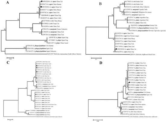
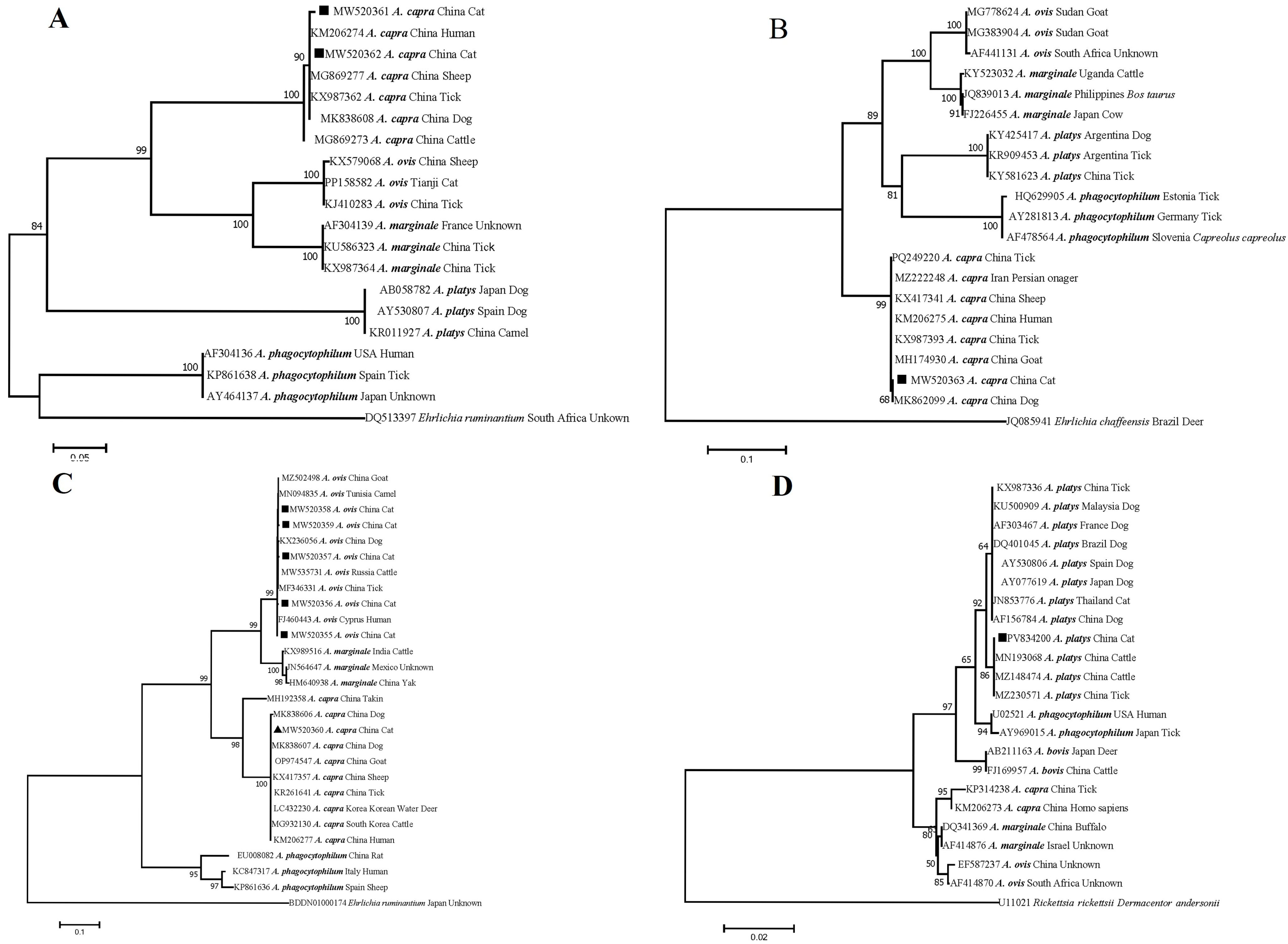
Regarding A. capra, a total of 12 positive samples were identified based on the amplification of at least one target gene, including 5 positive for gltA, 11 for msp4, and 1 for groEL. Sequence analysis of gltA revealed two distinct sequence types: one (comprising three isolate sequences, MW520362) exhibited 100% identity with A. capra isolates from China, which have been previously detected in humans (KM206274), sheep (MG869277), dogs (MK838608), and ticks (KX987362) (Figure 1 and Figure S1). The other type (represented by two isolate sequences, MW520361) displayed 99.7% identity with human-derived A. capra isolates (KM206274) (Figure S1). Phylogenetic analysis based on gltA placed both sequence types within a monophyletic clade of A. capra, supporting their close affinity to this species while distinguishing them from other established Anaplasma species (Figure 1). All msp4 gene sequences of A. capra obtained in this study (MW520360) exhibited 100% identity. The results showed that these sequences shared 100% identity with those isolated from humans (KM206277), dogs (MK838607), goats (OP974547), sheep (KX417357), and ticks (KR261641), but only 79.4% similarity with the sequence isolated from takin (MH192358) (Figure S1). Correspondingly, in the phylogenetic tree, the variants from cats, dogs, humans, goats, sheep, and ticks clustered together on the same evolutionary branch, which was distinct from that of the takin isolate. All these sequences formed a clade corresponding to A. capra, clearly separate from other Anaplasma species (Figure 1). For the groEL gene, only one sequence was acquired in this study. It shared 100% identity with the sequence from a dog in China (MK862099), and 99.9% identity with sequences obtained from humans (KM206275), Persian onager (MZ222248), sheep (KX417341), and ticks (KX987393, PQ249220) (Figure S1). Phylogenetic analysis based on groEL sequences revealed that the isolate clustered within the A. capra clade but was distinct from other Anaplasma species (Figure 1).
Figure 1.
Phylogenetic analyses of A. capra based on gltA (A), groEL (B), and msp4 (C); of A. ovis based on msp4 (C); and of A. platys based on 16S rRNA (D). Phylogenetic trees were constructed using the neighbor-joining method with the Kimura 2-parameter model and 1000 bootstrap replicates. Numbers at branch nodes indicate the percentage of bootstrap replicates that reproduced the topology of each clade (only values > 50% are shown). Sequences generated in this study are indicated by black squares and triangles. Sample labels consist of GenBank accession numbers followed by the corresponding Anaplasma species names. Ehrlichia ruminantium, Ehrlichia chaffeensis, and Rickettsia rickettsii were used as outgroups.
Regarding the msp4 gene of A. ovis, five distinct sequences (MW520355–MW520359) were obtained from cats in the present study. Among these, only one sequence (MW520355) showed 100% similarity with a human-derived isolate from Cyprus (FJ460443). The sequence MW520356 differed by a single SNP (C→T at position 366) compared to the human reference (FJ460443). Sequences MW520357 (C→A at position 286 and C→T at position 470) and MW520358 (C→T at positions 366 and 470) each contained two SNPs relative to FJ460443. Another sequence, MW520359, exhibited three SNPs (C→T at 366, C→T at 470, and C→T at 537). With the exception of MW520355, the other four sequences shared 96.9–99.8% similarity with reported sequences of A. ovis from humans (FJ460443), goats (MZ502498), camels (MN094835), cattle (MW535731), dogs (KX236056), and ticks (MF346331) (Figure S1). Phylogenetic analysis revealed that the A. ovis sequences obtained in this study formed a well-defined branch, distinct from other established Anaplasma species (Figure 1).
Based on 16S rRNA gene sequence analysis, all A. platys sequences obtained in this study were identical (PV834200) and shared 100% identity with cattle (MZ148474) and tick (MZ230571) isolates from China, clustering with them on the same phylogenetic branch (Figure 1).
4. Discussion
Currently, dogs and cats remain the most popular companion animals in China. According to 2024 statistics, 64.3% of first-time pet owners chose cats, while 57% opted for dogs (2025 China Pet Industry White Paperps, https://pdf.dfcfw.com/pdf/H3_AP202512111798269901_1.pdf) (accessed on 1 November 2026). Furthermore, from a “One Health” perspective, pet cats not only act as direct sentinels for human infections due to their close proximity but may also facilitate zoonotic transmission via intense contact and a shared living environment [4,28]. Anaplasma, a significant zoonotic pathogen, is transmitted through vectors. While there have been documented cases of this pathogen in dogs across China, reports of its occurrence in cats remain scarce [3,23,29,30,31,32,33]. To our knowledge, this study documents Anaplasma spp. infection in domestic pet cats in Henan and provides molecular detection frequency of A. capra, A. ovis and A. platys in cats from four veterinary hospitals in Zhengzhou City. The overall molecular detection frequency of Anaplasma spp. was 20.5% (47/229, 95% CI: 15.29–25.76), indicating a considerable exposure risk in this region. Notably, species-specific PCR positivity varied substantially: A. ovis was the most prevalent (17.5%, 40/229), followed by A. capra (5.2%, 12/229) and A. platys (3.1%, 7/229), with statistically significant differences among them.
A. capra was first isolated from asymptomatic goats and subsequently found in patients with a history of tick bites in northern China [1,9,34]. Subsequently, A. capra has been detected worldwide across a broad spectrum of hosts, including domestic animals (e.g., goats, sheep, cattle, yaks, and dogs), wildlife (e.g., takins, muntjacs, water deer, musk deer, onagers, serows, and brown hares), and various tick species (e.g., I. persulcatus, H. longicornis, H. qinghaiensis, D. abaensis, D. nuttalli, and R. microplus) [32,34,35,36,37,38,39,40,41]. However, there have been no reports of A. capra infection in cats to date. In this study, A. capra was detected in 12 of 229 (5.2%) domestic cat samples, suggesting that cats are susceptible to A. capra; however, their epidemiological role remains unknown.
A. ovis is considered a tick-borne pathogen of small ruminants and causes ovine anaplasmosis [42]. The pathogen was first reported in sheep in 1912, and subsequently described in sheep, goats and some wild ruminants, cattle and dogs throughout the world [3,19,29,42,43,44,45]. Recently, an A. ovis variant was detected in a patient, indicating the zoonotic potential of this agent [8,46]. Furthermore, A. ovis had been detected in some vectors such as ticks (e.g., D. nuttalli, D. silvarum, H. longicornis and H. qinghaiensis), Melophagus ovinus and Lipoptena cervi [46,47,48]. A. ovis was first detected in a clinically healthy cat in Tianjin of China in 2021, with the PCR positivity 0.6%, which is markedly lower than PCR positivity observed in the present study (17.5%). Differences in the study population and sample size may be potential factors underlying this contrast.
A. platys was primarily considered a canine pathogen that causes a decrease in platelet count in infected dogs [49]. Nowadays, this pathogen has been identified in a variety of animals, such as cattle, goats, sheep, camels, foxes, wild boars, and red deer [50,51,52,53]. It was not until 2013 that the first DNA sequence-confirmed human case of A. platys infection was reported, involving a veterinarian [17]. Subsequently, the pathogen was detected in a doctor near Chicago, Illinois, as well as in the doctor’s daughter and the family dog—all three of which shared completely identical genetic sequences [16]. Furthermore, A. platys was detected in 2014 in two women from Venezuela with a history of peridomestic tick exposure [15]. From then on, A. platys has been regarded as a potential zoonotic pathogen [50]. To date, sporadic cases of A. platys infection in cats have been documented in North America, Brazil, and Cyprus [54,55,56,57]. Subsequently, the identification of both A. platys-like and A. platys strains has been successively reported in feline populations across Italy, Chile, Turkey, and Saudi Arabia [21,58,59,60]. In this context, the present study provides, to our knowledge, the first evidence of A. platys infection in domestic cats in China, demonstrating an overall PCR positivity of 3.1% (7/229). This PCR positivity is comparatively higher than that reported in Saudi Arabia (2.27%) yet lower than the rates observed in free-roaming domestic cats from Chile (6.6%, 2/30) and household cats from Turkey (30.5%) [21,59,60]. These interregional discrepancies are likely attributable to variations in sampling time, geographic location, rearing practices, and sample size.
Since Anaplasma species are transmitted by arthropod vectors, which can carry multiple pathogens, mixed infections are expected to be frequent. However, in our study, no cat was found to be co-infected with all three agents (A. ovis, A. platys, and A. capra); rather, we observed 12 cases of dual infection, yielding an overall mixed-PCR positivity of 5.2% (12/229). This figure is marginally higher than the co-infection rate previously reported in stray dogs in Anhui, China [30]. These findings underscore that, in the context of feline anaplasmosis diagnosis and control, the clinical manifestations resulting from mixed infections deserve due attention, as they may inform targeted prevention strategies.
Sequence analysis of gltA and msp4 sequences of A. capra from our feline samples (MW520362, MW520360) showed 100% identical to isolates from humans, sheep, dogs, goats, and ticks, while the A. ovis msp4 sequence (MW520355) matched a human-derived isolate from Cyprus [1,8,32,35,61,62,63]. These partial-marker similarities raise public-health vigilance; nevertheless, complete genome comparisons are required to definitively evaluate whether these feline strains are genetically indistinguishable from human-origin zoonotic isolates.
5. Conclusions
In conclusion, this study provides the first molecular evidence documenting A. capra infection in domestic cats from China. Three Anaplasma species, namely A. ovis, A. capra, and A. platys, were detected in feline blood samples by nested PCR, with dual-species co-infections observed among sampled cats. Partial gene sequences of feline-origin A. capra and A. ovis shared high nucleotide identity with previously reported zoonotic isolates from humans, animals and ticks. Nevertheless, the epidemiological role of domestic cats, their reservoir competence for these Anaplasma pathogens, and the actual risk of pathogen transmission from cats to humans remain unknown. Further investigations including full-genome sequencing, experimental infection studies and field-based epidemiological surveillance are required to address these open questions. These findings highlight the necessity to conduct routine molecular surveillance of Anaplasma pathogens in companion domestic cats in urban China.
Supplementary Materials
The following supporting information can be downloaded at https://www.mdpi.com/article/10.3390/vetsci13111229/s1, Figure S1. Homology analyses of Anaplasma species based on various genetic markers. (A–C) A. capra based on gltA, msp4, and groEL sequences, respectively; (D) A. platys based on 16S rRNA sequences; (E) A. ovis based on msp4 sequences.
Author Contributions
Conceptualization, K.S. and C.N.; resources, K.S. and C.D.; methodology, K.S. and L.W.; investigation, K.S., Z.Z. and H.L.; visualization, K.S., L.Z. and W.W.; formal analysis, W.W., J.C. and P.P.; supervision, C.N.; writing—original draft, K.S.; writing—review and editing, K.S., C.N. and W.W.; project administration, K.S. and C.N.; funding acquisition, K.S., P.P., J.C. and L.Z. All authors have read and agreed to the published version of the manuscript.
Funding
This research was supported by the Doctoral Research Start-up Fund of Xinxiang University (1366020227), the Science and Technology Planning Project of Henan Province (252102310412 and 262102111012), and the Promotion Funding for Excellent Young Backbone Teachers of Henan Province (2024GGJS142). The funders played no role in the study design, data collection or interpretation, or the decision to submit the work for publication.
Institutional Review Board Statement
Ethical approval for this study was granted by the Independent Ethics Committee of Henan Agricultural University on 26 March 2017 (Reference No. IEC-HENAU-20170326-428-08). Before sample collection, each cat owner was informed of the purpose of the study, the sampling procedure, and the intended use of the data. Verbal informed consent was obtained from each owner, and all animals were enrolled with the owner’s prior approval.
Informed Consent Statement
Informed consent was obtained from all animal owners involved in the study.
Data Availability Statement
The original contributions presented in this study are included in the article/supplementary material. Further inquiries can be directed to the corresponding author.
Acknowledgments
We are grateful to the staff at the veterinary hospitals for their assistance with sample collection.
Conflicts of Interest
The authors declare no conflict of interest.
References
- Li, H.; Zheng, Y.C.; Ma, L.; Jia, N.; Jiang, B.G.; Jiang, R.R.; Huo, Q.B.; Wang, Y.W.; Liu, H.B.; Chu, Y.L.; et al. Human infection with a novel tick-borne Anaplasma species in China: A surveillance study. Lancet Infect. Dis. 2015, 15, 663–670. [Google Scholar] [CrossRef] [Scilit] [PubMed]
- Schäfer, I.; Kohn, B. Anaplasma phagocytophilum infection in cats: A literature review to raise clinical awareness. J. Feline Med. Surg. 2020, 22, 428–441. [Google Scholar] [CrossRef] [Scilit] [PubMed]
- Jian, R.; Xue, J.; Xu, Z.Y.; Chen, S.S.; Wang, F.N.; Du, L.; Xie, G.C.; Guo, W.P. Genetic diversity of vector-borne zoonotic pathogens in companion dogs and cats, Tianjin, China. Front. Vet. Sci. 2024, 11, 1373178. [Google Scholar] [CrossRef] [Scilit] [PubMed]
- Little, S.E. Future challenges for parasitology: Vector control and one health in the Americas. Vet. Parasitol. 2013, 195, 249–255. [Google Scholar] [CrossRef] [Scilit] [PubMed]
- Akwongo, C.J.; Byaruhanga, C. Epidemiology of Anaplasma species amongst cattle in Africa from 1970 to 2022: A systematic review and meta-analysis. Prev. Vet. Med. 2024, 228, 106214. [Google Scholar] [CrossRef] [Scilit] [PubMed]
- Madison-Antenucci, S.; Kramer, L.D.; Gebhardt, L.L.; Kauffman, E. Emerging Tick-Borne Diseases. Clin. Microbiol. Rev. 2020, 33, e00083-18. [Google Scholar] [CrossRef] [Scilit] [PubMed]
- Rar, V.; Tkachev, S.; Tikunova, N. Genetic diversity of Anaplasma bacteria: Twenty years later. Infect. Genet. Evol. 2021, 91, 104833. [Google Scholar] [CrossRef] [Scilit] [PubMed]
- Chochlakis, D.; Ioannou, I.; Tselentis, Y.; Psaroulaki, A. Human anaplasmosis and Anaplasma ovis variant. Emerg. Infect. Dis. 2010, 16, 1031–1032. [Google Scholar] [CrossRef] [Scilit] [PubMed]
- Altay, K.; Erol, U.; Sahin, O.F. Anaplasma capra: A new emerging tick-borne zoonotic pathogen. Vet. Res. Commun. 2024, 48, 1329–1340. [Google Scholar] [CrossRef] [Scilit] [PubMed]
- Lu, M.; Li, F.; Liao, Y.; Shen, J.J.; Xu, J.M.; Chen, Y.Z.; Li, J.H.; Holmes, E.C.; Zhang, Y.Z. Epidemiology and Diversity of Rickettsiales Bacteria in Humans and Animals in Jiangsu and Jiangxi provinces, China. Sci. Rep. 2019, 9, 13176. [Google Scholar] [CrossRef] [Scilit] [PubMed]
- Lu, M.; Chen, Q.; Qin, X.; Lyu, Y.; Teng, Z.; Li, K.; Yu, L.; Jin, X.; Chang, H.; Wang, W.; et al. Anaplasma bovis Infection in Fever and Thrombocytopenia Patients—Anhui Province, China, 2021. China CDC Wkly. 2022, 4, 249–253. [Google Scholar] [CrossRef] [Scilit] [PubMed]
- Karpathy, S.E.; Kingry, L.; Pritt, B.S.; Berry, J.C.; Chilton, N.B.; Dergousoff, S.J.; Cortinas, R.; Sheldon, S.W.; Oatman, S.; Anacker, M.; et al. Anaplasma bovis-Like Infections in Humans, United States, 2015–2017. Emerg. Infect. Dis. 2023, 29, 1904–1907. [Google Scholar] [CrossRef] [Scilit] [PubMed]
- Han, X.Y.; Du, L.F.; Lin, Z.T.; Li, C.; Xiong, T.; Zhu, W.J.; Ye, R.Z.; Wang, N.; Wang, Y.F.; Gao, W.Y.; et al. Genomic characters of Anaplasma bovis and genetic diversity in China. Emerg. Microbes. Infect. 2024, 13, 2323153. [Google Scholar] [CrossRef] [Scilit] [PubMed]
- Chan, P.K.; Hawley, J.R.; Lappin, M.R. Evaluation of the role of Babesia species and Cytauxzoon felis in feline anemia cases in Colorado, USA. JFMS Open Rep. 2021, 7, 20551169211024967. [Google Scholar] [CrossRef] [Scilit] [PubMed]
- Arraga-Alvarado, C.M.; Qurollo, B.A.; Parra, O.C.; Berrueta, M.A.; Hegarty, B.C.; Breitschwerdt, E.B. Case report: Molecular evidence of Anaplasma platys infection in two women from Venezuela. Am. J. Trop. Med. Hyg. 2014, 91, 1161–1165. [Google Scholar] [CrossRef] [Scilit] [PubMed]
- Breitschwerdt, E.B.; Hegarty, B.C.; Qurollo, B.A.; Saito, T.B.; Maggi, R.G.; Blanton, L.S.; Bouyer, D.H. Intravascular persistence of Anaplasma platys, Ehrlichia chaffeensis, and Ehrlichia ewingii DNA in the blood of a dog and two family members. Parasites Vectors 2014, 7, 298. [Google Scholar] [CrossRef] [Scilit] [PubMed]
- Maggi, R.G.; Mascarelli, P.E.; Havenga, L.N.; Naidoo, V.; Breitschwerdt, E.B. Co-infection with Anaplasma platys, Bartonella henselae and Candidatus Mycoplasma haematoparvum in a veterinarian. Parasites Vectors 2013, 6, 103. [Google Scholar] [CrossRef] [Scilit] [PubMed]
- Li, Y.; Yang, J.; Chen, Z.; Qin, G.; Li, Y.; Li, Q.; Liu, J.; Liu, Z.; Guan, G.; Yin, H.; et al. Anaplasma infection of Bactrian camels (Camelus bactrianus) and ticks in Xinjiang, China. Parasites Vectors 2015, 8, 313. [Google Scholar] [CrossRef] [Scilit] [PubMed]
- Li, Y.; Chen, Z.; Liu, Z.; Liu, J.; Yang, J.; Li, Q.; Li, Y.; Luo, J.; Yin, H. Molecular Survey of Anaplasma and Ehrlichia of Red Deer and Sika Deer in Gansu, China in 2013. Transbound. Emerg. Dis. 2016, 63, e228–e236. [Google Scholar] [CrossRef] [Scilit] [PubMed]
- Dahmani, M.; Davoust, B.; Benterki, M.S.; Fenollar, F.; Raoult, D.; Mediannikov, O. Development of a new PCR-based assay to detect Anaplasmataceae and the first report of Anaplasma phagocytophilum and Anaplasma platys in cattle from Algeria. Comp. Immunol. Microbiol. Infect. Dis. 2015, 39, 39–45. [Google Scholar] [CrossRef] [Scilit] [PubMed]
- Sacristán, I.; Sieg, M.; Acuña, F.; Aguilar, E.; García, S.; López, M.J.; Cevidanes, A.; Hidalgo-Hermoso, E.; Cabello, J.; Vahlenkamp, T.W.; et al. Molecular and serological survey of carnivore pathogens in free-roaming domestic cats of rural communities in southern Chile. J. Vet. Med. Sci. 2019, 81, 1740–1748. [Google Scholar] [CrossRef] [Scilit] [PubMed]
- Zhang, J.; Ma, H.; Ai, J.; Qi, T.; Kang, M.; Li, J.; Sun, Y. Serological Analysis of IgG and IgM Antibodies against Anaplasma spp. in Various Animal Species of the Qinghai-Tibetan Plateau. Animals 2022, 12, 2723. [Google Scholar] [CrossRef] [Scilit] [PubMed]
- Hussain, S.; Hussain, A.; Aziz, M.U.; Song, B.; Zeb, J.; Hasib, F.M.Y.; Almendros, A.; Cabezas-Cruz, A.; George, D.; Sparagano, O. First molecular confirmation of multiple zoonotic vector-borne diseases in pet dogs and cats of Hong Kong SAR. Ticks Tick Borne Dis. 2023, 14, 102191. [Google Scholar] [CrossRef] [Scilit] [PubMed]
- Barlough, J.E.; Madigan, J.E.; DeRock, E.; Bigornia, L. Nested polymerase chain reaction for detection of Ehrlichia equi genomic DNA in horses and ticks (Ixodes pacificus). Vet. Parasitol. 1996, 63, 319–329. [Google Scholar] [CrossRef] [Scilit] [PubMed]
- Yang, J.; Liu, Z.; Niu, Q.; Liu, J.; Han, R.; Guan, G.; Hassan, M.A.; Liu, G.; Luo, J.; Yin, H. A novel zoonotic Anaplasma species is prevalent in small ruminants: Potential public health implications. Parasites Vectors 2017, 10, 264. [Google Scholar] [CrossRef] [Scilit] [PubMed]
- Wang, G.Q. Development of Nested-PCR and Real-Time Fluorescence PCR Method for Diagnosing Anaplasmosis. Master’s Thesis, Huazhong Agricultural University, Wuhan, China, 2007. [Google Scholar]
- Martin, A.R.; Brown, G.K.; Dunstan, R.H.; Roberts, T.K. Anaplasma platys: An improved PCR for its detection in dogs. Exp. Parasitol. 2005, 109, 176–180. [Google Scholar] [CrossRef] [Scilit] [PubMed]
- Skotarczak, B. The role of companion animals in the environmental circulation of tick-borne bacterial pathogens. Ann. Agric. Environ. Med. 2018, 25, 473–480. [Google Scholar] [CrossRef] [Scilit] [PubMed]
- Cui, Y.; Yan, Y.; Wang, X.; Cao, S.; Zhang, Y.; Jian, F.; Zhang, L.; Wang, R.; Shi, K.; Ning, C. First molecular evidence of mixed infections of Anaplasma species in dogs in Henan, China. Ticks Tick Borne Dis. 2017, 8, 283–289. [Google Scholar] [CrossRef] [Scilit] [PubMed]
- Yang, B.; Ye, C.; Sun, E.; Wen, Y.; Qian, D.; Sun, H. First molecular evidence of Anaplasma spp. co-infection in stray dogs from Anhui, China. Acta Trop. 2020, 206, 105453. [Google Scholar] [CrossRef] [Scilit] [PubMed]
- Zhang, L.; Liu, H.; Xu, B.; Lu, Q.; Li, L.; Chang, L.; Zhang, X.; Fan, D.; Li, G.; Jin, Y.; et al. Anaplasma phagocytophilum infection in domestic animals in ten provinces/cities of China. Am. J. Trop. Med. Hyg. 2012, 87, 185–189. [Google Scholar] [CrossRef] [Scilit] [PubMed]
- Shi, K.; Li, J.; Yan, Y.; Chen, Q.; Wang, K.; Zhou, Y.; Li, D.; Chen, Y.; Yu, F.; Peng, Y.; et al. Dogs as New Hosts for the Emerging Zoonotic Pathogen Anaplasma capra in China. Front. Cell. Infect. Microbiol. 2019, 9, 394. [Google Scholar] [CrossRef] [Scilit] [PubMed]
- Wang, S.; He, J.; Zhang, L. Serological investigation of vector-borne disease in dogs from rural areas of China. Asian Pac. J. Trop. Biomed. 2012, 2, 102–103. [Google Scholar] [CrossRef] [Scilit] [PubMed]
- Lin, Z.T.; Du, L.F.; Zhang, M.Z.; Han, X.Y.; Wang, B.H.; Meng, J.; Yu, F.X.; Zhou, X.Q.; Wang, N.; Li, C.; et al. Genomic Characteristics of Emerging Intraerythrocytic Anaplasma capra and High Prevalence in Goats, China. Emerg. Infect. Dis. 2023, 29, 1780–1788. [Google Scholar] [CrossRef] [Scilit] [PubMed]
- Razzaq, M.A.; Imran, M.; Atif, F.A.; Abbas, R.Z.; Alvi, M.A.; Swelum, A.A.; Sindhu, Z.U.; Khan, M.K.; Sabir Mughal, M.A.; Khan, A.; et al. Molecular surveillance based on anaplasmosis in domestic small ruminants: First report on zoonotic Anaplasma capra and phylogenetic insights from Faisalabad, Pakistan. PLoS ONE 2024, 19, e0305412. [Google Scholar] [CrossRef] [Scilit] [PubMed]
- Seo, M.G.; Ouh, I.O.; Lee, H.; Geraldino, P.J.L.; Rhee, M.H.; Kwon, O.D.; Kwak, D. Differential identification of Anaplasma in cattle and potential of cattle to serve as reservoirs of Anaplasma capra, an emerging tick-borne zoonotic pathogen. Vet. Microbiol. 2018, 226, 15–22. [Google Scholar] [CrossRef] [Scilit] [PubMed]
- Wang, Y.; Zhang, Q.; Han, S.; Li, Y.; Wang, B.; Yuan, G.; Zhang, P.; Yang, Z.; Zhang, H.; Sun, Y.; et al. Ehrlichia chaffeensis and Four Anaplasma Species With Veterinary and Public Health Significance Identified in Tibetan Sheep (Ovis aries) and Yaks (Bos grunniens) in Qinghai, China. Front. Vet. Sci. 2021, 8, 727166. [Google Scholar] [CrossRef] [Scilit] [PubMed]
- Yang, J.; Liu, Z.; Niu, Q.; Mukhtar, M.U.; Guan, G.; Liu, G.; Luo, J.; Yin, H. A novel genotype of “Anaplasma capra” in wildlife and its phylogenetic relationship with the human genotypes. Emerg. Microbes. Infect. 2018, 7, 210. [Google Scholar] [CrossRef] [Scilit] [PubMed]
- Staji, H.; Yousefi, M.; Hamedani, M.A.; Tamai, I.A.; Khaligh, S.G. Genetic characterization and phylogenetic of Anaplasma capra in Persian onagers (Equus hemionus onager). Vet. Microbiol. 2021, 261, 109199. [Google Scholar] [CrossRef] [Scilit] [PubMed]
- Rocchigiani, G.; Ebani, V.V.; Nardoni, S.; Bertelloni, F.; Bascherini, A.; Leoni, A.; Mancianti, F.; Poli, A. Molecular survey on the occurrence of arthropod-borne pathogens in wild brown hares (Lepus europaeus) from Central Italy. Infect. Genet. Evol. 2018, 59, 142–147. [Google Scholar] [CrossRef] [Scilit] [PubMed]
- Amer, S.; Kim, S.; Yun, Y.; Na, K.J. Novel variants of the newly emerged Anaplasma capra from Korean water deer (Hydropotes inermis argyropus) in South Korea. Parasites Vectors 2019, 12, 365. [Google Scholar] [CrossRef] [Scilit] [PubMed]
- Han, R.; Yang, J.; Liu, Z.; Gao, S.; Niu, Q.; Hassan, M.A.; Luo, J.; Yin, H. Characterization of Anaplasma ovis strains using the major surface protein 1a repeat sequences. Parasites Vectors 2017, 10, 447. [Google Scholar] [CrossRef] [Scilit] [PubMed]
- Renneker, S.; Abdo, J.; Salih, D.E.; Karagenç, T.; Bilgiç, H.; Torina, A.; Oliva, A.G.; Campos, J.; Kullmann, B.; Ahmed, J.; et al. Can Anaplasma ovis in small ruminants be neglected any longer? Transbound. Emerg. Dis. 2013, 60, 105–112. [Google Scholar] [CrossRef] [Scilit] [PubMed]
- Li, Y.; Chen, Z.; Liu, Z.; Liu, J.; Yang, J.; Li, Q.; Li, Y.; Ren, Q.; Niu, Q.; Guan, G.; et al. First report of Theileria and Anaplasma in the Mongolian gazelle, Procapra gutturosa. Parasites Vectors 2014, 7, 614. [Google Scholar] [CrossRef] [Scilit] [PubMed]
- Hornok, S.; Micsutka, A.; Fernández de Mera, I.G.; Meli, M.L.; Gönczi, E.; Tánczos, B.; Mangold, A.J.; Farkas, R.; Lutz, H.; Hofmann-Lehmann, R.; et al. Fatal bovine anaplasmosis in a herd with new genotypes of Anaplasma marginale, Anaplasma ovis and concurrent haemoplasmosis. Res. Vet. Sci. 2012, 92, 30–35. [Google Scholar] [CrossRef] [Scilit] [PubMed]
- Zhao, L.; He, B.; Li, K.R.; Li, F.; Zhang, L.Y.; Li, X.Q.; Liu, Y.H. First report of Anaplasma ovis in pupal and adult Melophagus ovinus (sheep ked) collected in South Xinjiang, China. Parasites Vectors 2018, 11, 258. [Google Scholar] [CrossRef] [Scilit] [PubMed]
- Zhu, W.J.; Ye, R.Z.; Tian, D.; Wang, N.; Gao, W.Y.; Wang, B.H.; Lin, Z.T.; Liu, Y.T.; Wang, Y.F.; Zhu, D.Y.; et al. The first direct detection of spotted fever group Rickettsia spp. diversity in ticks from Ningxia, northwestern China. PLoS Negl. Trop. Dis. 2025, 19, e0012729. [Google Scholar] [CrossRef] [Scilit] [PubMed]
- Halos, L.; Jamal, T.; Maillard, R.; Girard, B.; Guillot, J.; Chomel, B.; Vayssier-Taussat, M.; Boulouis, H.J. Role of Hippoboscidae flies as potential vectors of Bartonella spp. infecting wild and domestic ruminants. Appl. Environ. Microbiol. 2004, 70, 6302–6305. [Google Scholar] [CrossRef] [Scilit] [PubMed]
- Mnisi, Z.T.H.; Makgabo, S.M.; Byaruhanga, C. Anaplasma platys beyond canines: A systematic review of host range, phylogenetic relatedness, and knowledge gaps in Africa. Parasites Vectors 2026, 19, 128. [Google Scholar] [CrossRef] [Scilit] [PubMed]
- Ben Said, M.; Belkahia, H.; El Mabrouk, N.; Saidani, M.; Alberti, A.; Zobba, R.; Cherif, A.; Mahjoub, T.; Bouattour, A.; Messadi, L. Anaplasma platys-like strains in ruminants from Tunisia. Infect. Genet. Evol. 2017, 49, 226–233. [Google Scholar] [CrossRef] [Scilit] [PubMed]
- Alanazi, A.D.; Nguyen, V.L.; Alyousif, M.S.; Manoj, R.R.S.; Alouffi, A.S.; Donato, R.; Sazmand, A.; Mendoza-Roldan, J.A.; Dantas-Torres, F.; Otranto, D. Ticks and associated pathogens in camels (Camelus dromedarius) from Riyadh Province, Saudi Arabia. Parasites Vectors 2020, 13, 110. [Google Scholar] [CrossRef] [Scilit] [PubMed]
- Cardoso, L.; Gilad, M.; Cortes, H.C.; Nachum-Biala, Y.; Lopes, A.P.; Vila-Viçosa, M.J.; Simões, M.; Rodrigues, P.A.; Baneth, G. First report of Anaplasma platys infection in red foxes (Vulpes vulpes) and molecular detection of Ehrlichia canis and Leishmania infantum in foxes from Portugal. Parasites Vectors 2015, 8, 144. [Google Scholar] [CrossRef] [Scilit] [PubMed]
- Pereira, A.; Parreira, R.; Nunes, M.; Casadinho, A.; Vieira, M.L.; Campino, L.; Maia, C. Molecular detection of tick-borne bacteria and protozoa in cervids and wild boars from Portugal. Parasites Vectors 2016, 9, 251. [Google Scholar] [CrossRef] [Scilit] [PubMed]
- Hegarty, B.C.; Qurollo, B.A.; Thomas, B.; Park, K.; Chandrashekar, R.; Beall, M.J.; Thatcher, B.; Breitschwerdt, E.B. Serological and molecular analysis of feline vector-borne anaplasmosis and ehrlichiosis using species-specific peptides and PCR. Parasites Vectors 2015, 8, 320. [Google Scholar] [CrossRef] [Scilit] [PubMed]
- Qurollo, B.A.; Balakrishnan, N.; Cannon, C.Z.; Maggi, R.G.; Breitschwerdt, E.B. Co-infection with Anaplasma platys, Bartonella henselae, Bartonella koehlerae and ‘Candidatus Mycoplasma haemominutum’ in a cat diagnosed with splenic plasmacytosis and multiple myeloma. J. Feline. Med. Surg. 2014, 16, 713–720. [Google Scholar] [CrossRef] [Scilit] [PubMed]
- Lima, M.L.; Soares, P.T.; Ramos, C.A.; Araújo, F.R.; Ramos, R.A.; Souza, I.I.F.; Faustino, M.A.; Alves, L.C. Molecular detection of Anaplasma platys in a naturally-infected cat in Brazil. Braz. J. Microbiol. 2010, 41, 381–385. [Google Scholar] [CrossRef] [Scilit]
- Attipa, C.; Papasouliotis, K.; Solano-Gallego, L.; Baneth, G.; Nachum-Biala, Y.; Sarvani, E.; Knowles, T.G.; Mengi, S.; Morris, D.; Helps, C.; et al. Prevalence study and risk factor analysis of selected bacterial, protozoal and viral, including vector-borne, pathogens in cats from Cyprus. Parasites Vectors 2017, 10, 130. [Google Scholar] [CrossRef] [Scilit] [PubMed]
- Zobba, R.; Anfossi, A.G.; Visco, S.; Sotgiu, F.; Dedola, C.; Pinna Parpaglia, M.L.; Battilani, M.; Pittau, M.; Alberti, A. Cell tropism and molecular epidemiology of Anaplasma platys-like strains in cats. Ticks Tick Borne Dis. 2015, 6, 272–280. [Google Scholar] [CrossRef] [Scilit] [PubMed]
- Muz, M.N.; Erat, S.; Mumcuoglu, K.Y. Protozoan and Microbial Pathogens of House Cats in the Province of Tekirdag in Western Turkey. Pathogens 2021, 10, 1114. [Google Scholar] [CrossRef] [Scilit] [PubMed]
- Alanazi, A.D.; Alouffi, A.S.; Alyousif, M.S.; Alshahrani, M.Y.; Abdullah, H.H.A.M.; Abdel-Shafy, S.; Calvani, N.E.D.; Ansari-Lari, M.; Sazmand, A.; Otranto, D. Molecular Survey of Vector-Borne Pathogens of Dogs and Cats in Two Regions of Saudi Arabia. Pathogens 2020, 10, 25. [Google Scholar] [CrossRef] [Scilit] [PubMed]
- Guo, W.P.; Huang, B.; Zhao, Q.; Xu, G.; Liu, B.; Wang, Y.H.; Zhou, E.M. Human-pathogenic Anaplasma spp., and Rickettsia spp. in animals in Xi’an, China. PLoS Negl. Trop. Dis. 2018, 12, e0006916. [Google Scholar] [CrossRef] [Scilit] [PubMed]
- Lu, M.; Tian, J.H.; Yu, B.; Guo, W.P.; Holmes, E.C.; Zhang, Y.Z. Extensive diversity of rickettsiales bacteria in ticks from Wuhan, China. Ticks Tick Borne Dis. 2017, 8, 574–580. [Google Scholar] [CrossRef] [Scilit] [PubMed]
- Lu, M.; Meng, C.; Li, Y.; Zhou, G.; Wang, L.; Xu, X.; Li, N.; Ji, Y.; Tian, J.; Wang, W.; et al. Rickettsia sp. and Anaplasma spp. in Haemaphysalis longicornis from Shandong province of China, with evidence of a novel species “Candidatus Anaplasma Shandongensis”. Ticks Tick Borne Dis. 2023, 14, 102082. [Google Scholar] [CrossRef] [Scilit] [PubMed]

Disclaimer/Publisher’s Note: The statements, opinions and data contained in all publications are solely those of the individual author(s) and contributor(s) and not of MDPI and/or the editor(s). MDPI and/or the editor(s) disclaim responsibility for any injury to people or property resulting from any ideas, methods, instructions or products referred to in the content. |
© 2026 by the authors. Licensee MDPI, Basel, Switzerland. This article is an open access article distributed under the terms and conditions of the Creative Commons Attribution (CC BY) license.