


Simple Summary
Transarterial embolization is an interventional procedure used to treat liver tumors in dogs by blocking the blood supply to the tumor. This report describes an 8-year-old dog that developed a liver abscess after this procedure, resulting in septic peritonitis, a severe bacterial infection of the abdominal cavity, which required surgery. The dog then developed several life-threatening complications, including a blood clot obstructing the aorta, the main artery supplying the hind limbs, which caused neurological deficits, as well as kidney injury and coagulation disorders. With intensive care, including antibiotics and clot-dissolving treatment, the dog gradually recovered and remained stable seven months later. This case may help veterinarians recognize and treat these rare but serious complications and provides useful information for owners considering this type of treatment for their dog.
Abstract
This report details the case of an 8-year-old neutered male Shiba Inu who developed septic peritonitis following transarterial embolization of a hepatic tumor. After surgical management of the hepatic abscess and peritonitis, the dog experienced several severe complications, including aortic thrombosis with pelvic limb neurological deficits, acute kidney injury, coagulopathy, and marked hypoalbuminemia. Intensive care included hemodynamic and surgical stabilization, antimicrobial therapy, analgesia, nutritional support, and a combination of antithrombotic and thrombolytic treatments. The clinical course was characterized by progressive stabilization of sepsis and the hepatic lesion, partial restoration of aortic blood flow, neurological improvement, and gradual recovery of renal function. Seven months after discharge, the dog remained clinically stable, with resolution of the hepatic lesion and persistent stability of the aortic thrombus on ultrasonography. This case demonstrates that life-threatening aortic thrombosis and septic complications following hepatic embolization can be successfully managed with timely and multimodal therapy, including thrombolysis.
1. Introduction
Sepsis is defined as life-threatening organ dysfunction caused by a dysregulated host response to infection and is associated with high mortality [1,2]. Acute inflammation activates the coagulation system through the widespread expression of tissue factor, leading to excessive thrombin generation, platelet activation, and fibrin formation within the microvasculature. At the same time, natural anticoagulant pathways are impaired, and fibrinolysis is suppressed. This results in a prothrombotic state (hypercoagulability), progressing toward uncontrolled, systemic clotting activation with massive thrombin formation and fibrin deposition in the microcirculation [3,4,5,6]. However, thrombus formation rarely results from hypercoagulability alone and is classically explained by Virchow’s triad, which comprises endothelial injury, alterations in blood flow, and hypercoagulability [7].
In humans, sepsis-induced coagulopathy is most often associated with venous thromboembolic disease, including deep vein thrombosis and pulmonary embolism [7]. Aortic thrombosis (Ath) is a rare condition in dogs defined as local (mural or occlusive) thrombus formation in the aortic lumen, most often in the distal abdominal aorta and aorto-iliac region, due to a regional prothrombotic environment [8,9]. Thrombi can partially or totally occlude blood flow to the pelvic limbs, leading to pain, weakness, or paralysis. Ath is rare, accounting for roughly 0.0005% of hospital admissions in one dog case series [6]. It is often associated with a hypercoagulable state typically secondary to protein-losing nephropathy, neoplasia, endocrine disease, exogenous corticosteroids, and infections, including sepsis [6,9].
Veterinary consensus guidelines support the use of antithrombotic prophylaxis in high-risk critically ill dogs and cats with sepsis, particularly when multiple risk factors are present [10]. In contrast, thrombolytic therapy in small animals remains promising but poorly defined [11]. Current evidence is largely limited to experimental studies and small case series, with most clinical data derived from feline aortic thromboembolism (FATE) rather than canine Ath, providing insufficient data to confirm improvement in patient-centered outcomes and preventing the establishment of evidence-based dosing protocols [12]. As a result, clinical use relies on individualized risk–benefit assessment, given the significant risks of hemorrhage and reperfusion injury and the wide variability in published protocols, highlighting the need for further prospective studies and standardization [11,13].
This case report describes the use of systemic thrombolysis with alteplase, guided by viscoelastic testing, in a critically ill dog that developed acute Ath following septic peritonitis caused by rupture of a hepatic abscess secondary to hepatic tumor embolization.
2. Case Description
An 8-year-old neutered male Shiba Inu was presented to the ICU of a veterinary hospital for acute onset of lethargy.
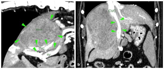
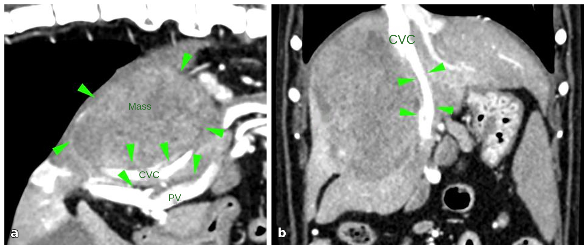
Five months earlier, a hepatic mass had been diagnosed with abdominal ultrasound and computed tomography. Contrast-enhanced imaging identified a large (8 × 8 cm) heterogeneous mass in the right lateral liver lobe with prominent arterial vascularization. The mass caused a marked regional mass effect with displacement and compression of the caudal vena cava and portal vein, without evidence of vascular invasion (Figure 1). Given the mass’s position relative to major vasculature and its size, an endovascular approach was elected. Arterial embolization was performed via the right femoral artery, targeting the phrenic branch of the right phrenicoabdominal artery with 400-micron microspheres, followed by embolization of the two hepatic arterial branches supplying the mass with 250-micron microspheres. Final angiographic control confirmed complete embolization of all tumor-feeding arteries. At that time, serum biochemistry showed a marked increase in hepatic enzymes (ALT 3703 U/L [reference interval:10-125U/L]; ALKP 1504 U/L [reference interval:23-212U/L]). Furthermore, 1 month post-operatively, abdominal ultrasound showed an evolution of the hepatic mass into a larger cavitary lesion with richly corpuscular content (compatible with necrosis and intralesional hemorrhage), without Doppler activity within the lesion, and relatively discrete perilesional steatitis. Secondary to the vascular access during surgery, signs of vasculitis and thrombosis of the right femoral and external iliac arteries were observed (to the level of the aortic trifurcation with an absence of pulse).
Figure 1.
Abdominal CTA ((a) sagittal plane/(b) dorsal plane) showing a large (approximately 8 × 8 cm) heterogeneous mass within the right lateral liver lobe (green arrows, (a)). The lesion shows heterogeneous contrast enhancement with prominent arterial vascularization. Marked regional mass effect is present, with displacement and compression of caudal vena cava (CVC) and portal vein (PV), without evidence of intraluminal vascular invasion (green arrows, (b)).
Two days after the previous ultrasound, the dog experienced acute clinical deterioration characterized by repeated vomiting, abdominal pain, and generalized weakness. Upon admission, the patient was non-ambulatory, exhibited pale mucous membranes, was tachycardic (HR 204 bpm), hypotensive (Doppler blood pressure 50 mmHg), hypothermic (T: 36.7 °C), and had cold extremities with weak pulses. Abdominal palpation elicited marked pain and revealed abdominal distension with a positive fluid wave sign.
Point-of-care ultrasound (POCUS) identified a substantial volume of abdominal effusion. Analysis of the effusion demonstrated an exudate (specific gravity 1040, total protein 50 g/L) consistent with septic peritonitis, evidenced by the presence of intra- and extracellular bacteria with phagocytosis. The effusion lactate concentration (12.6 mmol/L) was twice that in blood (6 mmol/L; reference interval: 0.50–2.50 mmol/L), and effusion glucose was decreased (1.4 mmol/L) compared to blood glucose (2.8 mmol/L; reference interval: 4.11–7.94 mmol/L). Comprehensive bloodwork (Catalyst and ProCyte, IDEXX, Saint-Denis, France) revealed marked thrombocytopenia confirmed by manual count (53 K/µL; reference interval: 148–484 K/μL), increased creatinine (164.56 µmol/L; reference interval: 44–159 µmol/L), elevated ALT (459 U/L; reference interval: 10–125 U/L), elevated lactate (6 mmol/L), and hypoglycemia (glucose 2.75 mmol/L).
Medical stabilization involved fluid resuscitation with isotonic crystalloid boluses (Ringer lactate, total volume 35 mL/kg), a single glucose bolus (0.3 g/kg), and a hypertonic saline bolus (NaCl 7.5% at 3 mL/kg), resulting in restoration of adequate perfusion parameters (blood pressure: 120 mmHg; lactate: 1.6 mmol/L). Analgesia was achieved using a fentanyl bolus (Fentadon, Dechra, Montigny-le-Bretonneux, France; 2 µg/kg IV) followed by a constant-rate infusion (CRI) at 2 µg/kg/min, and a ketamine bolus (Ketamidor, Axience SAS, Pantin, France; 1 mg/kg IV) followed by a CRI at 10 µg/kg/min.
To control the septic source, broad-spectrum antibiotic therapy with ampicillin–sulbactam (Unacim, Haupt Pharma, Borgo San Michele, Italy; 30 mg/kg IV) was promptly initiated, followed by surgical intervention. Surgical exploration confirmed an abscessed hepatic mass, as previously visualized on ultrasonography, yielding purulent material upon aspiration. The abscess was drained and debrided via open laparotomy. Both the abscess cavity and abdominal cavity were thoroughly irrigated with warm sterile saline in accordance with standard surgical protocols [13]. A Blake drain was placed within the abscess cavity, and abdominal closure was performed using routine layered techniques, with each anatomical layer apposed individually using appropriate suture material.
During hospitalization, medical treatment was directed by the attending clinician and comprised analgesic, antimicrobial, and gastrointestinal support. Analgesia was provided with a fentanyl CRI (2 µg/kg/h IV), later transitioned to buprenorphine (Bupaq, Virbac, Carros, France; 0.02 mg/kg IV, TID) based on pain assessment. Gastrointestinal management included prokinetic and antiemetic therapy with metoclopramide (Emeprid, Ceva, Libourne, France; CRI 2–6 mg/kg/day or 0.5 mg/kg IV, TID) and maropitant (Prevomax, Dechra, Montigny-le-Bretonneux, France; 1 mg/kg IV, SID). Broad-spectrum antibiotic coverage was maintained with ampicillin–sulbactam (30 mg/kg IV, TID).
Following surgical drainage and debridement of the hepatic abscess, postoperative evolution was initially favorable. Abdominal comfort improved rapidly following surgical intervention, with concurrent normalization of lactate concentrations and stabilization of vital parameters (HR 120 bpm; T° 38 °C; BP 120 mmHg). Drain production progressively decreased, and cytological analysis of the fluid repeatedly showed inflammatory cells and erythrocytes, with no bacteria.
Aerobic culture of the hepatic abscess yielded Enterococcus faecium. Antimicrobial susceptibility testing confirmed sensitivity to ampicillin, supporting the appropriateness of the empiric regimen. Histopathological analysis of the abscess wall revealed a cavitary lesion containing necrotic and suppurative material, surrounded by vascularized fibrous tissue and inflammatory infiltrates. The remaining hepatic tissue exhibited both partially preserved architecture and solid regions composed of atypical hepatocytes consistent with hepatocellular carcinoma.
However, the subsequent clinical course was complicated by the development of major Ath, acute kidney injury, catheter-associated thrombophlebitis, and significant hypoalbuminemia, each necessitating targeted management strategies. These complications are addressed in detail in the following sections.
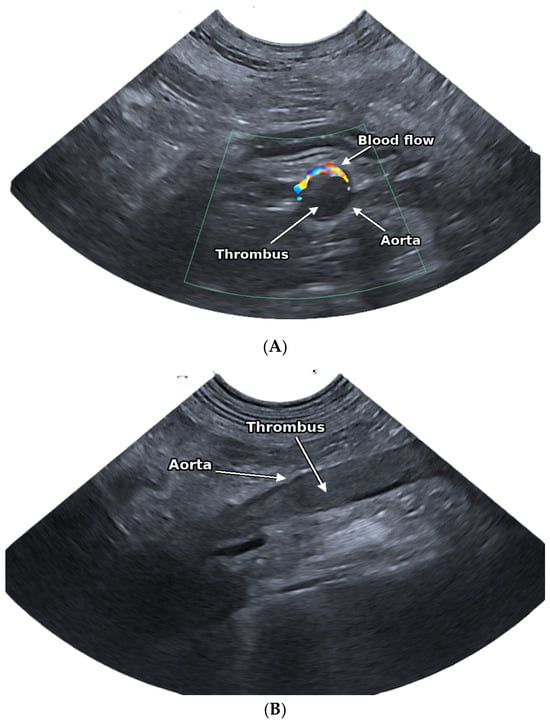
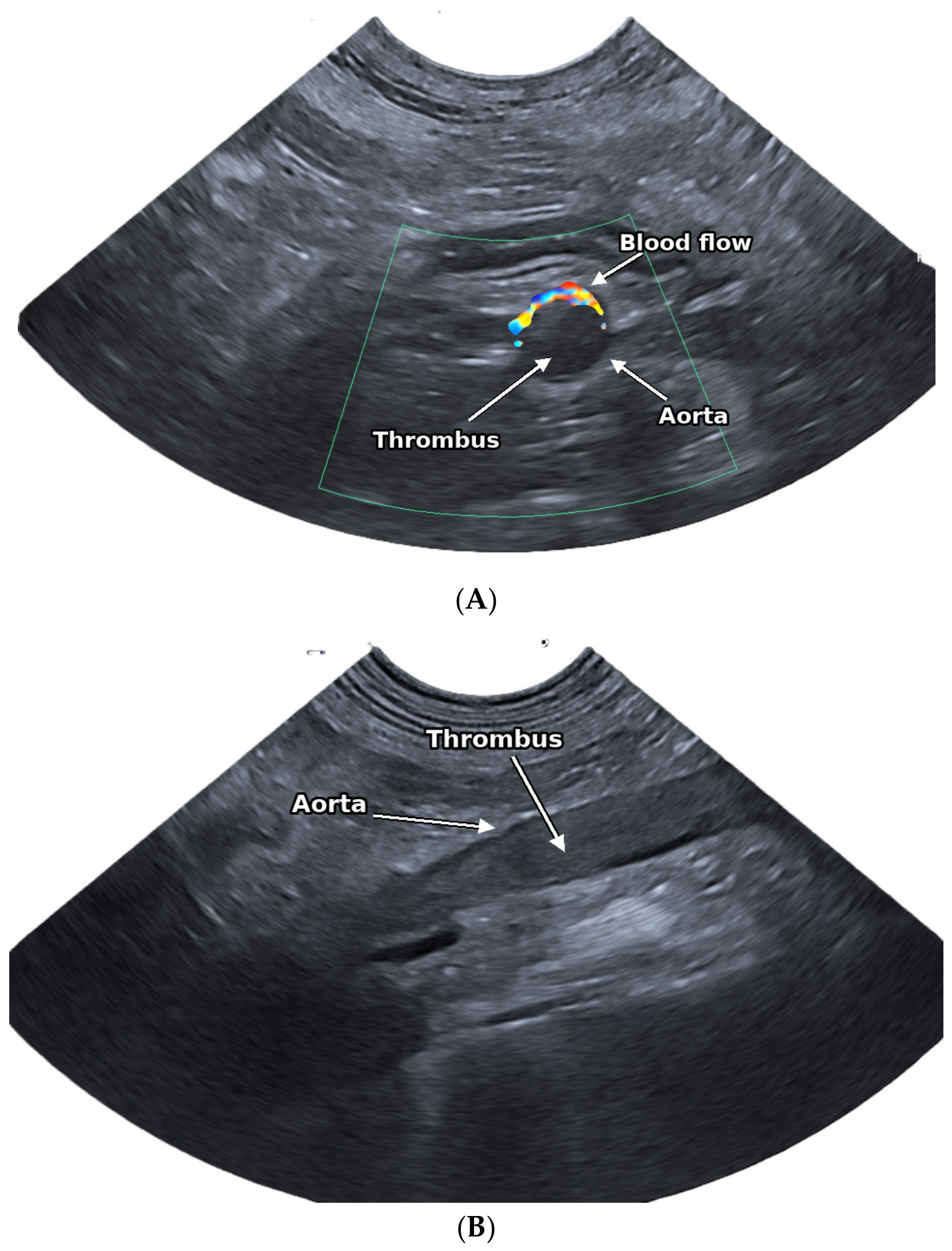
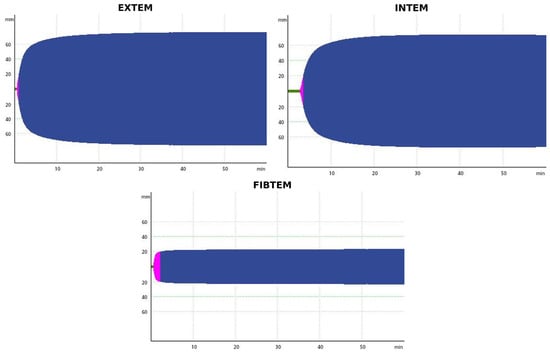
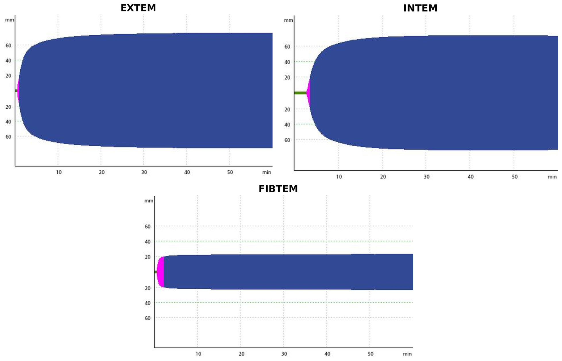
A major Ath, resulting in greater than 80% luminal obstruction and extending from the cranial mesenteric artery to the iliac bifurcation with reduced renal artery blood flow, was identified by abdominal ultrasound (Aplio a550, Canon Medical Systems, Otawara, Japan) on day 2 (Figure 2). This was associated with acute pelvic limb paralysis, absence of femoral pulses, and lower glucose concentration in the pelvic limb compared to the thoracic limb. Rotational thromboelastometry (ROTEM sigma, Werfen, Le Pré-Saint-Gervais, France), performed prior to alteplase administration, revealed a hypercoagulable state, with markedly increased clot firmness in EXTEM, INTEM, and FIBTEM (elevated A10, MCF, and G), and a pronounced increase in FIBTEM MCF (22 mm; reference interval 1.09–6.91 mm), indicating enhanced fibrin contribution to clot strength. The EXTEM clotting time was within the reference interval, while the INTEM clotting time was prolonged (203 s; reference interval 98.85–151.15 s). No evidence of hyperfibrinolysis was observed (Figure 3 and Table 1). Reference intervals were based on previously published values for ROTEM Sigma in healthy dogs [14]. Medical thrombolysis was performed using alteplase (Actilyse, Boehringer Ingelheim, Paris, France) at a dose of 1.2 mg/kg, administered as a constant-rate infusion over 12 h, without a loading bolus. The lyophilized powder was reconstituted with the sterile water for injection provided by the manufacturer to a concentration of 1 mg/mL and administered without further dilution. The total volume (16.56 mL, i.e., 1.38 mL/h) was delivered using a syringe pump (BeneFusion eSP, Mindray, Shenzhen, China) with a 20 mL syringe connected to an extension line with a priming volume of 1.5 mL. At the end of the infusion, the line was flushed with 0.9% NaCl to deliver the residual volume. Antithrombotic therapy with clopidogrel (Plavix, Sanofi Winthrop Industrie, Gentilly, France; 2 mg/kg PO, SID) and rivaroxaban (Xarelto, Bayer HealthCare, Lille, France; 1 mg/kg PO, BID) was initiated 6 h after completion of the alteplase infusion. In the hours following thrombolysis, femoral pulses became palpable and progressive recovery of pelvic limb motor function was observed, with the dog regaining ambulatory status. Serial ultrasonographic examinations demonstrated partial improvement in luminal obstruction (50–60%), restoration of arterial flow in the left iliac artery, and improvement in renal blood flow. No clinically significant bleeding was detected during or after the alteplase infusion, as assessed by serial monitoring of abdominal drain output, abdominal POCUS every 4 h, and blood and drain fluid packed cell volume every 6 h.
Figure 2.
Abdominal ultrasonographic images of the aortic thrombus at diagnosis. (A) Longitudinal B-mode view of the abdominal aorta showing an echogenic thrombus occupying almost the entire aortic lumen. (B) Transverse colour Doppler view of the abdominal aorta showing the thrombus filling the lumen, with residual blood flow limited to the periphery of the vessel.
Figure 3.
Rotational thromboelastometry (ROTEM) tracings (EXTEM, INTEM and FIBTEM) obtained from the patient. Tracings demonstrate increased clot firmness, with elevated amplitudes (A10) and maximum clot firmness (MCF) in EXTEM, INTEM, and FIBTEM, consistent with a hypercoagulable state. FIBTEM tracing highlights an increased fibrinogen contribution to clot strength. No evidence of hyperfibrinolysis was observed (normal maximum lysis (ML) and lysis at 60 min (LI60) values).
Table 1.
ROTEM results and reference intervals. Reference intervals (robust) were derived from previously published ROTEM Sigma values in healthy dogs [14]. Negative lower limits for ML were set to 0%. Values in bold are outside the reference range.
The dog developed severe azotemia on day 2, with serum creatinine rising from 419.9 µmol/L to 919 µmol/L (reference interval: 44–159 µmol/L) and a urine output of 0.3 mL/kg/h, consistent with IRIS acute kidney injury (AKI) grade V (oliguric) [15]. The AKI was suspected to result from renal hypoperfusion and ischemia secondary to the Ath, as supported by reduced renal arterial flow on Doppler ultrasonography. Due to persistent oliguria, a furosemide stress test was performed at the end of the alteplase infusion, consisting of an IV bolus of furosemide (Lasilix, Sanofi Winthrop Industrie, Gentilly, France; 1 mg/kg) adapted from published protocols [16,17], followed by a CRI at 2 mg/kg/h. In accordance with our institutional protocol, the CRI was maintained until urine output reached at least 1 mL/kg/h. After 12 h, urine output reached 1 mL/kg/h, allowing discontinuation of the CRI. This increase in urine output coincided with the return of bilateral femoral pulses and restoration of renal arterial flow on Doppler ultrasonography. Urine output subsequently increased after furosemide discontinuation, reaching a polyuric phase (5.6 mL/kg/h) on day 3, before normalizing by day 5. Serum creatinine gradually decreased (288 µmol/L on day 14), and follow-up Doppler ultrasonography demonstrated improved renal perfusion. Arterial blood pressure remained within reference intervals throughout hospitalization, and urinalysis was unremarkable. By day 14, the dog was classified as having IRIS AKI grade III.
Marked hypoalbuminemia developed on day 2, with serum albumin decreasing to 15 g/L (reference interval: 23–40 g/L). Total parenteral nutrition was initiated via a central venous catheter using a commercially prepared bag (SmofKabiven, Fresenius Kabi, Sèvres, France) containing glucose, amino acids, lipids, and electrolytes at 100% of the resting energy requirement (RER). Simultaneously, enteral nutrition was progressively increased from 10% to 100% of the RER. Serum albumin concentration increased to 20 g/L by day 5, permitting discontinuation of total parenteral nutrition, and further increased to 22 g/L at discharge.
During hospitalization, the patient developed thrombophlebitis of the right jugular vein at the central venous catheterization site, resulting in cranial vena cava syndrome manifested by facial edema. Catheter removal and appropriate medical therapy led to gradual and complete resolution of clinical signs.
The patient was discharged after 15 days of hospitalization in a clinically stable state. At discharge, the dog was alert, ambulatory, and had regained a spontaneous appetite, meeting 100% of his resting energy requirement (RER). Renal function had improved, with a creatinine concentration of 236 µmol/L recorded on day 15. Discharge medications included amoxicillin–clavulanate, maropitant, rivaroxaban, and clopidogrel.
Seven months after discharge, the patient remained clinically stable. Serum creatinine was 206 µmol/L, and follow-up abdominal ultrasonography demonstrated complete resolution of the hepatic lesion with persistence of a stable aortic thrombus.
3. Discussion
In human medicine, surgical resection and percutaneous ablation (radiofrequency or microwave) remain the primary, potentially curative strategies for early-stage hepatocellular carcinoma, while transarterial chemoembolization (TACE) is considered the standard locoregional therapy for intermediate-stage tumors not amenable to resection or ablation [18]. Transcatheter arterial embolization (TAE), which does not employ a chemotherapeutic agent as in the present case, is based on a similar therapeutic principle but is less commonly used than TACE. The procedure consists of selective catheterization of the tumor’s arterial supply followed by embolic material injection, inducing ischemic necrosis. Although this is generally regarded as a safe and minimally invasive intervention, hepatic abscess formation is a recognized, albeit rare, complication associated with post-embolization necrosis and secondary bacterial infection [19,20,21,22,23,24,25]. In humans, reported incidences of hepatic abscess after embolization range from 0.2% to 0.54% in recent large series and meta-analyses, though earlier studies reported rates as high as 4% [19,20,21,22,23,24,25]. In veterinary medicine, surgical resection is likewise the first-line therapy for resectable tumors, and TAE or TACE is considered for dogs with non-resectable disease; however, data on complications remain scarce and heterogeneous. A prospective study assessing drug-eluting bead TACE in dogs noted major complications in 3 of 27 procedures (11%), including hepatic abscess formation, whereas a retrospective analysis of 14 dogs treated with TAE reported no cases of abscesses [26,27]. A more recent retrospective series of 18 dogs with massive hepatocellular carcinoma treated with TAE confirmed its potential utility in selected cases, with complications primarily limited to mild, self-resolving clinical signs (diarrhea, anorexia, mild anemia) and occasional gallbladder infarction [28]. The variability in reported outcomes likely reflects both the small number of published veterinary cases and differences in technique and case selection.
In the current case, delayed clinical deterioration observed one month after embolization, alongside imaging and intraoperative findings, was most consistent with hepatic abscess formation secondary to post-embolization necrosis. Documented risk factors for this complication include large tumor size, biliary tract abnormalities, hepatic metastases, and intra-abdominal hemorrhage [19,21,25,26,29]. In this patient, substantial tumor burden, extensive necrosis, and cavitary transformation likely promoted bacterial colonization and subsequent septic peritonitis. Nonetheless, the possibility of a pre-existing, subclinical infection cannot be definitively excluded. Prophylactic antimicrobial therapy has also been associated with reduced abscess rates in human cohorts [19,21]. In our case, Enterococcus faecium was isolated, which is a recognized pathogen in pyogenic liver abscesses (PLAs), particularly those of biliary origin or following surgical interventions. Literature reports indicate that Enterococcus spp. are associated with elevated mortality, primary treatment failure, and multidrug resistance in PLA patients, underscoring the clinical severity of infections involving this organism [30,31,32]. Post-embolization hepatic abscesses are most frequently attributed to enteric Gram-negative bacteria, especially Escherichia coli and Klebsiella pneumoniae, supporting a biliary or translocation-related infectious mechanism [23,24,29].
A major and unusual complication in this case was the development of extensive ATh, which resulted in pelvic limb paralysis, absent femoral pulses, impaired renal perfusion, and acute kidney injury. The pathogenesis was likely multifactorial and can be interpreted using Virchow’s triad: endothelial injury, alterations in blood flow, and hypercoagulability [7].
First, endothelial injury was probably caused by the previous right femoral artery access. Mechanical disruption of the endothelium and altered local shear stress, resulting from the introduction of arterial sheaths and guide catheters during transfemoral vascular access, may have directly triggered thrombus formation at or near the aortic trifurcation. In this dog, a right femoral and external iliac thrombus was detected two days before the onset of septic peritonitis, yet it had not been observed during earlier follow-up examinations. No antithrombotic therapy was started after the embolization procedure as local complications are uncommon after most endovascular interventions performed at our institution.
Second, this pre-existing thrombus at the catheter insertion site likely altered local blood flow and could have served as a nidus for the subsequent development of the aortic macrothrombus. Therefore, the aortic thrombosis most likely represented the propagation of this initial catheter-related thrombus rather than a purely de novo event, although both mechanisms cannot be entirely distinguished.
Third, a hypercoagulable profile was revealed by ROTEM, which showed increased clot firmness. The isolated prolongation of INTEM CT may indicate early consumption of coagulation factors, as the dog had not received anticoagulant prior to sampling; however, a pre-analytical artifact cannot be excluded [33]. In sepsis, acute inflammation and endothelial activation lead to systemic activation of coagulation through widespread tissue factor expression, resulting in excessive thrombin generation, platelet activation, and fibrin formation. Concurrently, natural anticoagulant pathways such as antithrombin and the protein C system are impaired due to consumption and reduced activity, while fibrinolysis is suppressed as a result of increased levels of plasminogen activator inhibitor type 1 and thrombin-activatable fibrinolysis inhibitor [3,4,5,7]. Rare thrombotic complications—including intracardiac thrombus formation—associated with enterococcal liver abscesses have been reported in human medicine [34], further supporting a role for sepsis-induced hypercoagulability in the present case. Additionally, the underlying hepatic adenocarcinoma may have contributed to an inherent prothrombotic state via the release of tissue factor and inflammatory cytokines [35,36].
Thus, the aortic macrothrombus likely resulted from the combination of pre-existing local endothelial injury and altered blood flow related to vascular access, exacerbated by sepsis- and neoplasia-associated hypercoagulability. Given the life-threatening organ dysfunction associated with the thrombus, the primary therapeutic goal was rapid restoration of perfusion to prevent further ischemic injury.
For this reason, the thrombolytic drug alteplase (a tPA analog) was chosen in combination with prophylactic antithrombotic therapy. Several case reports have described the use of alteplase in selected canine patients, with varying outcomes. In a retrospective study involving 100 dogs with ATh, thrombolytic therapy was given to 12 cases, but no survival benefit was observed compared to other treatment strategies [8]. Importantly, dogs presenting with acute clinical signs were significantly less likely to survive to discharge than those with chronic presentations [8], which is particularly relevant considering the acute onset in the present case. Canine protocols reported in the literature are highly variable, with dosing regimens ranging from 1.0 to 1.5 mg/kg IV administered over 1 to 4 h [37,38,39,40]. Reported outcomes have been inconsistent: some dogs failed to improve or deteriorated after treatment [39], whereas successful recanalization has also been documented [37,38]. In cats, thrombolysis is more extensively described in the setting of FATE secondary to cardiomyopathy, with protocols largely extrapolated from human acute ischemic stroke guidelines. These protocols typically use a total dose of 1 mg/kg, with 10% administered as an initial bolus and the remainder infused over 1 h [41]. Overall, veterinary data remain limited and heterogeneous, and an evidence-based dosing protocol for dogs with ATh has yet to be established.
Human data on the use of alteplase for ATh are limited to isolated case reports, and no standardized protocol has been established. Reported dosing regimens vary widely, highlighting the challenge of balancing thrombus resolution with the risk of hemorrhage. Full-dose systemic alteplase (100 mg over 2 h) has been linked to rapid reperfusion but also to significant hemorrhagic complications during subsequent anticoagulation [42]. Conversely, ultra-low-dose prolonged infusion protocols—for example, 1 mg/h repeated over 75 h—have achieved complete thrombus resolution without bleeding complications in selected patients [43]. In pediatric and neonatal populations, infusion rates of 0.1 to 0.5 mg/kg/h for up to 6 h, with or without an initial bolus, are recommended for limb-threatening thrombosis [44]. Notably, higher infusion rates have been associated with a significantly increased risk of hemorrhage without clear improvement in thrombus resolution, supporting the use of the lowest effective dose whenever possible [45].
In the present case, the dosing regimen employed was more closely aligned with the low-dose prolonged infusion protocols reported in human medicine. This approach was chosen due to the lack of standardized veterinary guidelines and the considerable hemorrhagic risk associated with thrombolytic therapy in a critically ill patient. Thrombolysis resulted in the return of femoral pulses, improvement in pelvic limb function, partial regression of the thrombus on ultrasonography, and enhanced renal perfusion as assessed by Doppler, indicating a positive response in a challenging clinical scenario.
Catheter-directed thrombolysis, possibly combined with mechanical clot disruption, is an alternative to systemic administration and has shown superior angiographic outcomes in human studies of acute limb ischemia [44,45]. However, in veterinary medicine, this technique remains infrequently reported, and the 2022 CURATIVE consensus states that insufficient evidence exists to demonstrate improvement in patient-centered outcomes with intra-arterial thrombolysis in dogs with acute arterial thrombosis [11]. In this case, catheter-directed therapy was not attempted due to the patient’s critical condition and the need for prompt treatment initiation; therefore, systemic alteplase was selected as the most immediately practical option.
To the authors’ knowledge, this is one of the few veterinary reports documenting successful systemic alteplase administration for life-threatening ATh secondary to septic complications following hepatic TAE. Nonetheless, several limitations must be acknowledged. First, as a single retrospective case, causality between transarterial embolization and the subsequent development of hepatic abscessation, septic peritonitis, and systemic thrombotic complications cannot be definitively established. Second, although hepatic abscessation was attributed to post-embolization necrosis, the possibility of an undetected pre-existing infection prior to the initial embolization procedure cannot be excluded. Third, interpreting viscoelastic testing results as evidence of true hypercoagulability requires caution, as the relationship between a hypercoagulable ROTEM profile and clinically relevant hypercoagulability remains debated in veterinary medicine, with no consensus definition based solely on viscoelastic parameters [33]. Additionally, D-dimer, fibrinogen, and antithrombin concentrations were not assessed, which limited the characterization of the coagulation status. Fourth, the favorable response to alteplase was observed in the context of multimodal intensive care and antithrombotic therapy, making it difficult to isolate the effect of thrombolysis alone. Fifth, renal perfusion was assessed qualitatively, without resistive index measurements, and the concurrent use of furosemide precluded a clear distinction between drug-induced and post-ischemic reperfusion diuresis. Furthermore, long-term oncologic outcomes could not be evaluated due to short follow-up, and the progression of residual hepatocellular carcinoma tissue remains unknown. Finally, because interventional hepatic embolization procedures are infrequently reported in veterinary medicine, these findings should be extrapolated to other patients, tumor types, or institutions with caution.
4. Conclusions
This case report illustrates the successful management of severe delayed complications following hepatic transarterial embolization in a dog with hepatocellular carcinoma, including hepatic abscessation with septic peritonitis and aortic thrombosis. Early recognition, prompt surgical source control, intensive supportive care, and a multimodal antithrombotic approach incorporating systemic thrombolysis contributed to the restoration of perfusion and a favorable short-term outcome. Although such complications are rare, clinicians should remain alert to the possibility of delayed septic and thrombotic events after hepatic embolization, as timely diagnosis and aggressive intervention may be critical for patient survival.
Author Contributions
Conceptualization, S.D., M.C. and C.B.; investigation, S.D., M.C., E.G., J.-L.H. and C.B.; data curation, S.D.; writing—original draft preparation, S.D.; writing—review and editing, M.C., E.G., J.-L.H. and C.B.; visualization, S.D.; supervision, M.C. and C.B. All authors have read and agreed to the published version of the manuscript.
Funding
This research received no external funding. The APC was funded by IVC Evidensia Research Fund, grant number RS250002-FR. We wish to thank IVC Evidensia Research Fund for providing funding for the publication of this article.
Institutional Review Board Statement
Ethical review and approval were waived for this study because all diagnostic and therapeutic procedures were performed as part of routine veterinary clinical care.
Informed Consent Statement
Informed consent was obtained from all subjects involved in the study.
Data Availability Statement
The original contributions presented in this study are included in the article. Further inquiries can be directed to the corresponding author.
Acknowledgments
During the preparation of this manuscript, the authors used Claude (Opus 5.5, Anthropic) for the purposes of language editing, proofreading, and translation. The authors have reviewed and edited the output and take full responsibility for the content of this publication.
Conflicts of Interest
The authors declare no conflicts of interest.
Abbreviations
The following abbreviations are used in this manuscript:
| AKI | Acute Kidney Injury |
| ALP | Alkaline Phosphatase |
| ALT | Alanine Aminotransferase |
| ATh | Aortic Thrombosis |
| CRI | Constant-Rate Infusion |
| FATE | Feline Aortic Thromboembolism |
| IRIS | International Renal Interest Society |
| IV | Intravenous |
| PO | Per os |
| POCUS | Point-of-Care Ultrasound |
| RER | Resting Energy Requirement |
| ROTEM | Rotational Thromboelastometry |
References
- Kenney, E.M.; Rozanski, E.A.; Rush, J.E.; deLaforcade-Buress, A.M.; Berg, J.R.; Silverstein, D.C.; Montealegre, C.D.; Jutkowitz, L.A.; Adamantos, S.; Ovbey, D.H.; et al. Association between outcome and organ system dysfunction in dogs with sepsis: 114 cases (2003–2007). J. Am. Vet. Med. Assoc. 2010, 236, 83–87. [Google Scholar] [CrossRef] [Scilit] [PubMed]
- Singer, M.; Deutschman, C.S.; Seymour, C.W.; Shankar-Hari, M.; Annane, D.; Bauer, M.; Bellomo, R.; Bernard, G.R.; Chiche, J.-D.; Coopersmith, C.M.; et al. The Third International Consensus Definitions for Sepsis and Septic Shock (Sepsis-3). JAMA 2016, 315, 801–810. [Google Scholar] [CrossRef] [Scilit] [PubMed] [PubMed Central]
- Hill, E.; Zhu, Y.; Brooks, M.B.; Goggs, R. Dogs with sepsis are more hypercoagulable and have higher fibrinolysis inhibitor activities than dogs with non-septic systemic inflammation. Front. Vet. Sci. 2025, 12, 1559994. [Google Scholar] [CrossRef] [Scilit] [PubMed]
- Hopper, K.; Bateman, S. An updated view of hemostasis: Mechanisms of hemostatic dysfuntion associated with sepsis. J. Vet. Emerg. Crit. Care 2005, 15, 83–91. [Google Scholar] [CrossRef] [Scilit]
- Semeraro, N.; Ammollo, C.; Semeraro, F.; Colucci, M. Coagulopathy of Acute Sepsis. Semin. Thromb. Hemost. 2015, 41, 650–658. [Google Scholar] [CrossRef] [Scilit] [PubMed]
- Lake-Bakaar, G.A.; Johnson, E.G.; Griffiths, L.G. Aortic thrombosis in dogs: 31 cases (2000–2010). J. Am. Vet. Med. Assoc. 2012, 241, 910–915. [Google Scholar] [CrossRef] [Scilit] [PubMed]
- Galli, E.; Maggio, E.; Pomero, F. Venous Thromboembolism in Sepsis: From Bench to Bedside. Biomedicines 2022, 10, 1651. [Google Scholar] [CrossRef] [Scilit] [PubMed]
- Ruehl, M.; Lynch, A.M.; O’Toole, T.E.; Morris, B.; Rush, J.; Couto, C.G.; Hmelo, S.; Sonnenshein, S.; Butler, A.; Guillaumin, J. Outcome and treatments of dogs with aortic thrombosis: 100 cases (1997–2014). J. Vet. Intern. Med. 2020, 34, 1759–1767. [Google Scholar] [CrossRef] [Scilit] [PubMed]
- Williams, T.P.E.; Shaw, S.; Porter, A.; Berkwitt, L. Aortic thrombosis in dogs. J. Vet. Emerg. Crit. Care 2017, 27, 9–22. [Google Scholar] [CrossRef] [Scilit] [PubMed]
- deLaforcade, A.; Bacek, L.; Blais, M.C.; Boyd, C.; Brainard, B.M.; Chan, D.L.; Cortellini, S.; Goggs, R.; Hoareau, G.L.; Koenigshof, A.; et al. 2022 Update of the Consensus on the Rational Use of Antithrombotics and Thrombolytics in Veterinary Critical Care (CURATIVE) Domain 1- Defining populations at risk. J. Vet. Emerg. Crit. Care 2022, 32, 289–314. [Google Scholar] [CrossRef] [Scilit] [PubMed] [PubMed Central]
- Sharp, C.R.; Blais, M.C.; Boyd, C.J.; Brainard, B.M.; Chan, D.L.; de Laforcade, A.; Goggs, R.; Guillaumin, J.; Lynch, A.; Mays, E.; et al. 2022 Update of the Consensus on the Rational Use of Antithrombotics and Thrombolytics in Veterinary Critical Care (CURATIVE) Domain 6: Defining rational use of thrombolytics. J. Vet. Emerg. Crit. Care 2022, 32, 446–470. [Google Scholar] [CrossRef] [Scilit] [PubMed] [PubMed Central]
- Borgeat, K.; Wright, J.; Garrod, O.; Payne, J.R.; Fuentes, V.L. Arterial thromboembolism in 250 cats in general practice: 2004-2012. J. Vet. Intern. Med. 2014, 28, 102–108. [Google Scholar] [CrossRef] [Scilit] [PubMed] [PubMed Central]
- O’Marra, S.K.; Campbell, B.G. Perioperative management of septic peritonitis in small animals: A review. Vet. Surg. 2026, 55, 39–58. [Google Scholar] [CrossRef] [Scilit] [PubMed]
- Jourdan, A.; Noval Montero, J.; Troussier, G.; Anderco, A.; Dania, C.; Moumadah, Y.; Cambournac, M. Determination of canine ROTEM Sigma reference ranges and comparative analysis with ROTEM Delta. Front. Vet. Sci. 2026, 12, 1737842. [Google Scholar] [CrossRef] [Scilit] [PubMed]
- Segev, G.; Cortellini, S.; Foster, J.D.; Francey, T.; Langston, C.; Londoño, L.; Schweighauser, A.; Jepson, R.E. International Renal Interest Society best practice consensus guidelines for the diagnosis and management of acute kidney injury in cats and dogs. Vet. J. 2024, 305, 106068. [Google Scholar] [CrossRef] [Scilit] [PubMed]
- McMahon, B.A.; Chawla, L.S. The furosemide stress test: Current use and future potential. Ren. Fail. 2021, 43, 830–839. [Google Scholar] [CrossRef] [Scilit] [PubMed] [PubMed Central]
- Chawla, L.S.; Davison, D.L.; Brasha-Mitchell, E.; Koyner, J.L.; Arthur, J.M.; Shaw, A.D.; Tumlin, J.A.; Trevino, S.A.; Kimmel, P.L.; Seneff, M.G. Development and standardization of a furosemide stress test to predict the severity of acute kidney injury. Crit. Care 2013, 17, R207. [Google Scholar] [CrossRef] [Scilit] [PubMed]
- Reig, M.; Forner, A.; Rimola, J.; Ferrer-Fàbrega, J.; Burrel, M.; Garcia-Criado, Á.; Kelley, R.K.; Galle, P.R.; Mazzaferro, V.; Salem, R.; et al. BCLC strategy for prognosis prediction and treatment recommendation: The 2022 update. J. Hepatol. 2022, 76, 681–693. [Google Scholar] [CrossRef] [Scilit] [PubMed] [PubMed Central]
- Wang, Y.; Wang, H.; Liu, Z.; Chang, Z. Evolution of transarterial chemoembolization-related liver abscess over time: A systematic review and meta-analysis. Quant. Imaging Med. Surg. 2025, 15, 2707–2721. [Google Scholar] [CrossRef] [Scilit] [PubMed]
- Arslan, M.; Degirmencioglu, S. Liver abscesses after transcatheter arterial embolization. J. Int. Med. Res. 2019, 47, 1124–1130. [Google Scholar] [CrossRef] [Scilit] [PubMed]
- Kim, W.; Clark, T.W.I.; Baum, R.A.; Soulen, M.C. Risk Factors for Liver Abscess Formation after Hepatic Chemoembolization. J. Vasc. Interv. Radiol. 2001, 12, 965–968. [Google Scholar] [CrossRef] [Scilit] [PubMed]
- Li, J.H.; Yao, R.R.; Shen, H.J.; Zhang, L.; Xie, X.Y.; Chen, R.X.; Wang, Y.H.; Ren, Z.G. Clostridium perfringens infection after transarterial chemoembolization for large hepatocellular carcinoma. World J. Gastroenterol. 2015, 21, 4397–4401. [Google Scholar] [CrossRef] [Scilit] [PubMed] [PubMed Central]
- Mezhir, J.J.; Fong, Y.; Fleischer, D.; Seo, S.K.; D’Amico, F.; Petre, E.; Covey, A.M.; Getrajdman, G.I.; Thornton, R.H.; Solomon, S.B.; et al. Pyogenic Abscess after Hepatic Artery Embolization: A Rare but Potentially Lethal Complication. J. Vasc. Interv. Radiol. 2011, 22, 177–182. [Google Scholar] [CrossRef] [Scilit] [PubMed]
- Ong, G.Y.; Changchien, C.S.; Lee, C.M.; Wang, J.H.; Tung, H.D.; Chuah, S.K.; Chiu, K.-W.; Chiou, S.-S.; Cheng, Y.-F.; Lu, S.-N. Liver abscess complicating transcatheter arterial embolization: A rare but serious complication. A retrospective study after 3878 procedures. Eur. J. Gastroenterol. Hepatol. 2004, 16, 737–742. [Google Scholar] [CrossRef] [Scilit] [PubMed]
- Sun, W.; Xu, F.; Li, X.; Li, C.R. A Case Series of Liver Abscess Formation after Transcatheter Arterial Chemoembolization for Hepatic Tumors. Chin. Med. J. 2017, 130, 1314. [Google Scholar] [CrossRef] [Scilit] [PubMed]
- Kawamura, Y.; Itou, H.; Kida, A.; Sunakawa, H.; Suzuki, M.; Kawamura, K. Therapeutic response and prognostic factors of 14 dogs undergoing transcatheter arterial embolization for hepatocellular masses: A retrospective study. J. Vet. Intern. Med. 2023, 37, 1455–1465. [Google Scholar] [CrossRef] [Scilit] [PubMed]
- Rogatko, C.P.; Weisse, C.; Schwarz, T.; Berent, A.C.; Diniz, M.A. Drug-eluting bead chemoembolization for the treatment of nonresectable hepatic carcinoma in dogs: A prospective clinical trial. J. Vet. Intern. Med. 2021, 35, 1487–1495. [Google Scholar] [CrossRef] [Scilit] [PubMed]
- Kadowaki, K.; Nishimura, S.; Toyoda, A.; Yamamoto, R. Transcatheter arterial embolization for massive hepatocellular carcinoma in 18 dogs (2020–2025). Am. J. Vet. Res. 2026, 87, ajvr.26.04.0189. [Google Scholar] [CrossRef] [Scilit] [PubMed]
- Song, S.Y.; Chung, J.W.; Han, J.K.; Lim, H.G.; Koh, Y.H.; Park, J.H.; Lee, H.-S.; Kim, C.Y. Liver Abscess after Transcatheter Oily Chemoembolization for Hepatic Tumors: Incidence, Predisposing Factors, and Clinical Outcome. J. Vasc. Interv. Radiol. 2001, 12, 313–320. [Google Scholar] [CrossRef] [Scilit] [PubMed]
- Große, K.; Ohm, D.; Würstle, S.; Brozat, J.F.; Schmid, R.M.; Trautwein, C.; Stallmach, A.; Bruns, T.; Reuken, P.A. Clinical characteristics and outcome of patients with enterococcal liver abscess. Sci. Rep. 2021, 11, 22265. [Google Scholar] [CrossRef] [Scilit] [PubMed] [PubMed Central]
- Oliosi, E.; Rossi, G.; Nguyen, Y.; Honsel, V.; Bert, F.; Roux, O.; Fantin, B.; Lefort, A. Enterococcal pyogenic liver abscesses: High risk of treatment failure and mortality. Eur. J. Clin. Microbiol. Infect. Dis. 2023, 42, 193–199. [Google Scholar] [CrossRef] [Scilit] [PubMed]
- Summers, N.A.; Gharbin, J.; Friedman-Moraco, R.; Lyon, G.M.; Lutgring, J. Multi-drug-resistant Enterococcus faecium bacteraemia in a liver transplant recipient. JMM Case Rep. 2019, 6, e005172. [Google Scholar] [CrossRef] [Scilit] [PubMed]
- Hennink, I.; Peters, L.; van Geest, G.; Adamik, K.N. Evaluation of a Viscoelastic Coagulation Monitoring System (VCM Vet®) and Its Correlation with Thromboelastometry (ROTEM®) in Diseased and Healthy Dogs. Animals 2023, 13, 405. [Google Scholar] [CrossRef] [Scilit] [PubMed] [PubMed Central]
- Yamamoto, H.; Takamatsu, M.; Hamada, K.; Rikitake, K. A Thrombus in the Right Atrium Associated with Pyogenic Liver Abscess. Kyobu Geka 2024, 77, 330–334. [Google Scholar] [PubMed]
- Neumann, S.; Kaup, F.J.; Scheulen, S. Interleukin-6 (IL-6) serum concentrations in dogs with hepatitis and hepatic tumours compared with those with extra-hepatic inflammation and tumours. Comp. Clin. Pathol. 2012, 21, 539–544. [Google Scholar] [CrossRef] [Scilit] [PubMed]
- Fontes, G.S.; Wavreille, V.A.; Lapsley, J.M.; Cooper, E.S.; Guillaumin, J.; Selmic, L.E. Thromboelastographic results and hypercoagulability in dogs with surgically treated hepatocellular adenoma and carcinoma: A Veterinary Society of Surgical Oncology prospective study. Vet. Comp. Oncol. 2023, 21, 616–622. [Google Scholar] [CrossRef] [Scilit] [PubMed]
- Ahn, J.O.; Park, S.C.; Song, W.J.; Ryu, M.O.; Li, Q.; Oh, H.J.; Chung, J.-Y.; Choi, M.-C.; Youn, H.-Y. Aortic thromboembolism. A different predisposing disease in four dogs: A case report. Vet. Med. 2019, 64, 417–421. [Google Scholar] [CrossRef] [Scilit]
- Winter, R.L.; Sedacca, C.D.; Adams, A.; Orton, E.C. Aortic thrombosis in dogs: Presentation, therapy, and outcome in 26 cases. J. Vet. Cardiol. Off. J. Eur. Soc. Vet. Cardiol. 2012, 14, 333–342. [Google Scholar] [CrossRef] [Scilit] [PubMed]
- Diaz, D.M.; Scansen, B.A.; Lake, Z.; Shropshire, S.; Orton, E.C.; Guillaumin, J. Clinical use of tissue plasminogen activator for systemic thrombolysis in dogs and cats. J. Vet. Cardiol. Off. J. Eur. Soc. Vet. Cardiol. 2022, 41, 154–164. [Google Scholar] [CrossRef] [Scilit] [PubMed]
- Albisinni, R.; Marrazzo, T.; Karruli, A.; Manduca, S.; Nobile, G.; Galdieri, N.; De Feo, M. Successful treatment of aortic arch mural thrombosis with low-dose, ultra-slow-flow thrombolysis: A case report and literature review. Bleeding Thromb. Vasc. Biol. 2022, 1, 44. [Google Scholar] [CrossRef] [Scilit]
- Gallagher, G.; Handler, K.; Ferguson, B. Acute Aortic Occlusion Causing Bilateral Lower Extremity Ischemia That Resolved with tPA Administration. J. Vasc. Dis. 2025, 4, 36. [Google Scholar] [CrossRef] [Scilit]
- Leong, R.; Patel, J.; Samji, N.; Paes, B.A.; Chan, A.K.C.; Petropoulos, J.A.; Bhatt, M.D. Use of thrombolytic agents to treat neonatal thrombosis in clinical practice. Blood Coagul. Fibrinolysis 2022, 33, 193. [Google Scholar] [CrossRef] [Scilit] [PubMed]
- Ansah, D.A.; Patel, K.N.; Montegna, L.; Nicholson, G.T.; Ehrlich, A.C.; Petit, C.J. Tissue Plasminogen Activator Use in Children: Bleeding Complications and Thrombus Resolution. J. Pediatr. 2016, 171, 67.e2–72.e2. [Google Scholar] [CrossRef] [Scilit] [PubMed]
- Clark, A.H.; Levy, B.E.; Castle, J.T.; Fleming, M.; Lee, D.; Tyagi, S.C. Recanalization of aortic and iliocaval occlusions by a new catheter-directed thrombolytic system-Report of two cases. Ann. Vasc. Surg.-Brief Rep. Innov. 2022, 2, 100122. [Google Scholar] [CrossRef] [Scilit]
- Saroukhani, A.; Ravari, H.; Pezeshki Rad, M. Effects of Intravenous and Catheter Directed Thrombolytic Therapy with Recombinant Tissue Plasminogen Activator (Alteplase) in Non-Traumatic Acute Limb Ischemia; A Randomized Double-Blind Clinical Trial. Bull. Emerg. Trauma 2015, 3, 86–92. [Google Scholar] [PubMed] [PubMed Central]

Disclaimer/Publisher’s Note: The statements, opinions and data contained in all publications are solely those of the individual author(s) and contributor(s) and not of MDPI and/or the editor(s). MDPI and/or the editor(s) disclaim responsibility for any injury to people or property resulting from any ideas, methods, instructions or products referred to in the content. |
© 2026 by the authors. Licensee MDPI, Basel, Switzerland. This article is an open access article distributed under the terms and conditions of the Creative Commons Attribution (CC BY) license.