


Simple Summary
Canine osteosarcoma is an aggressive primary bone malignancy associated with poor survival because of its high metastatic potential. Advances in treatment have been limited; therefore, new therapeutic approaches are required. Immunohistochemistry can be used to investigate protein expression and identify potential biomarkers. This study utilised genes that had higher mRNA expression in canine osteosarcoma to investigate whether the protein levels were also high. This study aimed to improve the understanding of canine OSA and identify candidate biomarkers and therapeutic targets.
Abstract
Osteosarcoma (OSA) is a metastatic bone tumour. In dogs, it predominantly affects older animals and large and giant breeds. The cancer has poor survival rates due to most patients developing dissemination to secondary sites prior to an initial diagnosis. Therefore, novel therapeutic approaches are required to help overcome the growth and metastatic progression of the disease and increase survival rates. Immunohistochemical staining and histological scoring (H-scoring) are often used for diagnostic and prognostic purposes in human medicine. Unfortunately, these techniques have not yet been fully established and implemented within the veterinary field, but ongoing investigations and developments of potential biomarkers are expanding. Four proteins (ELOVL2, LIPG, RPL4, and RPSA) whose genes showed higher expression in canine OSA underwent immunohistochemical investigations. Immunohistochemistry (IHC) and consequent H-score analysis were performed to investigate nuclear and/or cytoplasmic protein expression encoded by the four canine overexpressed genes (ELOVL2 (n = 22), LIPG (n = 18), RPL4 (n = 18), and RPSA (n = 19)). Statistical analyses of the nuclear/cytoplasmic staining scores of the whole cohort, as well as sex (male versus female) and anatomical location (axial and soft tissue versus appendicular) differences, were conducted. This study revealed novel findings regarding the subcellular expression of the proteins corresponding to the overexpressed genes of interest (ELOVL2, LIPG, RPL4, and RPSA) in Rottweiler OSA, including the identification of significant sex-associated differences for LIPG nuclear staining. These candidate biomarkers could be utilised in future investigations to validate their potential as diagnostic, prognostic, and treatment avenues for canine OSA.
1. Introduction
Osteosarcoma (OSA) is a highly metastatic bone malignancy with a high incidence, affecting approximately 13.9/100,000 dogs [1]. Risk factors, such as height and weight, can predispose patients to lesion formation [2]. Larger breeds (>25 kg), such as Scottish Deerhounds (3.28%), Leonbergers (1.48%), Great Danes (0.87%), and Rottweilers (0.84%), are most susceptible to tumour development [3,4,5,6]. Chondrodystrophic (short-legged) breeds are at a lower risk of developing OSA compared to non-chondrodystrophic (long-legged) breeds (odds ratio (OR) 0.13) [3]. The risk has been reported to be slightly higher in males than females [7]. Neutered individuals also exhibit a higher risk of OSA formation than those that remain entire [3,7]. In dogs, the distal radius and proximal humerus are common sites for OSA formation—accounting for 24% and 26% of OSA cases, respectively [3]. These tumours are usually located specifically within the highly proliferative metaphyseal regions [5,8,9].
OSA is a disease of mesenchymal origin [10]. The tumours display complex pathogenesis due to their ability to attain a variety of cellular phenotypes, such as chondroblastic, fibroblastic, osteoblastic, and telangiectatic [8,11]. The prognostic outcomes for OSA are poor due to the disease’s high metastatic potential, with the lungs being the most common site, leading to fatalities in approximately 80% of affected dogs [12]. Despite the advancements made in OSA therapeutics, survival rates are low, highlighting the requirement for novel prognostic and diagnostic biomarker identification. Understanding the genetic and proteomic characteristics of OSA is fundamental in biomarker development. The present study utilises previous RNA sequencing data [13], obtained by our group, to identify differentially expressed genes in canine OSA. Four overexpressed genes, namely ELOVL2, LIPG, RPL4, and RPSA, underwent immunohistochemical analysis to investigate protein expression within canine OSA tumour samples [13]. These four proteins were among the top 20 differentially expressed genes in canine OSA, have not been investigated previously in this tumour or species, and have known implications in other cancer types. Immunohistochemistry (IHC) and consequent histological scoring (H-scoring) are techniques that are widely used in human medicine to aid diagnosis, predict prognosis, and establish therapeutic outcomes for disease [14,15]. The use of these techniques within the veterinary field requires the establishment of definitive biomarkers to aid diagnostic, prognostic, and therapeutic decision-making and to develop, or repurpose, therapeutic interventions. The aims of this study were to explore the protein expression of ELOVL2, LIPG, RPL4, and RPSA in canine OSA tissue samples, to investigate and compare the protein expression within nuclear and cytoplasmic subcellular locations, and to investigate differences between protein expression in males and females and in differing anatomical locations.
2. Materials and Methods
2.1. Specimen Preparation and Ethics
The use of canine and control tissues was approved by the University of Nottingham School of Veterinary Medicine and Science ethics committee and AWERB (ethics permission number 1823 160714, 959 130925, and 3832 230502). This work complied with international and national ethical standards, and informed owner consent was provided. OSA samples (n = 23) were excised from Rottweilers (from n = 23 dogs, Table 1) that were euthanised or undergoing amputation surgery for their medical needs, not for research. Each specimen was reviewed by a board-certified veterinary pathologist and confirmed as OSA. All samples were derived from primary OSA tumours, and specimens were classified as axial and soft tissue or appendicular depending on the region of excision. The axial and soft tissue category included specimens from extra-skeletal sites, as subcategorising further would have resulted in sample numbers deemed too small for statistical analysis.
Table 1.
Rottweiler OSA samples (n = 23).
The tissues were fixed and processed to create formalin-fixed paraffin-embedded (FFPE) specimens. The tissues were sectioned at 7 µm using a microtome (Leica, Wetzlar, Germany), with R35 feather blades for hard material (Fisher Scientific, Loughborough, UK), and mounted onto polysilinated slides (Thermofisher, Waltham, MA, USA).
2.2. Immunohistochemistry Antibodies, Protocol and Microscopy
2.2.1. Antibody Selection
Antibodies to detect the corresponding proteins were checked using BLAST (v2.16.0). This sequence alignment software was used to determine the coverage and identity of the antibodies raised against human immunogens compared to canine sequences [16]. The BLAST searches utilised the immunogen sequences provided on the manufacturer’s websites within the product specificity information [17,18,19,20]. Table 2 displays the BLAST result for each antibody.
Table 2.
BLAST search results for ELOVL2, LIPG, RPL4, and RPSA.
2.2.2. Immunohistochemistry
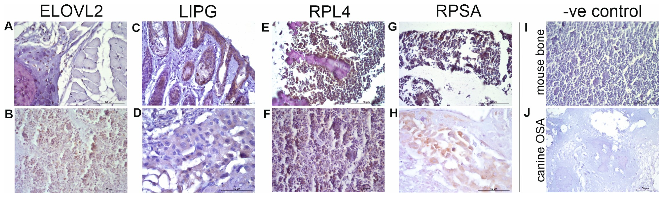
IHC was performed on sectioned Rottweiler OSA FFPE samples (n = 23, not decalcified) using the Leica Novolink Polymer Detection Kit (Leica, Wetzlar, Germany), in accordance with the manufacturer’s protocol. Dilution optimisations concluded a 1:100 dilution for ELOVL2 (20308-1-AP; Proteintech, Manchester, UK) and RPSA (10668-1-AP; Proteintech, UK), a 1:50 dilution for LIPG (PA5-82568; Invitrogen, Paisley, UK), and a 1:150 dilution for RPL4 (PA5-96464; Invitrogen, UK). Positive controls were also performed on mouse tissues (Figure A1), using the ELOVL2 (1:100 dilution), LIPG (1:50 dilution), RPL4 (1:100 and 1:150 dilutions), and RPSA (1:100 dilution) antibodies above, alongside negative controls (no primary antibody) in mouse bone and canine OSA tissue.
2.2.3. Microscopy
Photomicrographs were obtained at 400× magnification (Leica, Wetzlar, Germany) using systematic random sampling techniques, with n = 5 images per section. H-scoring analysis was performed using these photomicrographs to semi-quantitatively assess protein expression within the nucleus and cytoplasm separately in the canine OSA tissue samples (Table 3 and Table 4).
Table 3.
H-scores for ELOVL2, LIPG, RPL4, and RPSA.
Table 4.
Subcellular staining H-scores (nuclear and cytoplasmic) for ELOVL2, LIPG, RPL4, and RPSA.
2.3. Analysis
2.3.1. H-Scoring
H-scoring is a method for immunohistochemical interpretation in human medicine but is not commonly used within the veterinary profession yet. The semi-quantitative assessment of protein expression is performed based on the staining intensity and coverage within tissue sections. Manual scoring was graded as absent, weak, moderate, or strong staining—categorised as 0, 1+, 2+, or 3+, respectively—and cytoplasmic and nuclear staining were categorised separately. The following formula was used to achieve the standard H-score of between 0 and 300: H-score = [1 × (% cells 1+) + 2 × (% cells 2+) + 3 × (% cells 3+)]. All five of the H-scored images were averaged per specimen and per antibody to enable single H-scores for each specimen for further analysis, ensuring coverage of the whole tumour area. Where insufficient cell counts were available, the specimen was excluded as per the inclusion criteria. The analysis for each protein (ELOVL2, LIPG, RPL4, and RPSA) was undertaken by a blinded single scorer, and a second blinded scorer scored a randomised 100% of the samples to ensure concordance (ICC (3,1) two-way mixed effects, single rater, absolute agreement). Across the four proteins, an intraclass correlation coefficient of >0.9 or above was achieved. Values above 0.8 indicate excellent interobserver concordance [21] and confirmed interpretation consistency.
2.3.2. Statistical Analysis
For each protein, the mean cytoplasmic and nuclear H-scores ± standard error of the mean (SEM) and ranges (minimum–maximum scores) were calculated (Table 3). Subcellular staining locations were subcategorised based on the staining intensity (low/moderate/high), calculated using the greatest H-score range (from nuclear or cytoplasmic; for example, ELOVL2 nuclear range = 133 and cytoplasmic range = 128—hence, the range used was 133) for each individual protein/antibody (Table 4), calculated in thirds of the highest range for each protein (lowest third = low, middle third = moderate, highest third = high). ELOVL2 = low ≤ 44, moderate 45–88, high ≥89; LIPG = low ≤ 33, moderate 34–66, high ≥ 67; RPL4 = low ≤ 52, moderate 53–104, high ≥ 105; RPSA = low ≤ 29, moderate 30–58, high ≥ 59. Graphs to demonstrate the staining intensity (scored 0–3), correlation, and anatomical location-/sex-associated differences were generated using GraphPad Prism (GraphPad Prism, version 10.5.0, San Diego, CA, USA) for ELOVL2, LIPG, RPL4, and RPSA (Figure 1, Figure 2, Figure 3 and Figure 4). The correct Shapiro–Wilk Test was used on all data to check for normality prior to t-tests and correlation assessment. Paired t-tests were performed for nuclear versus cytoplasmic scores using GraphPad Prism (GraphPad Prism, version 10.5.0, USA). Pearson’s correlation coefficient was used where data were parametric, and Spearman’s rank correlation coefficient was used where non-parametric data were included. Statistical unpaired t-tests (where data were normally distributed) and Mann–Whitney U tests (MWU; where data were not normally distributed) were performed using GraphPad Prism (GraphPad Prism, version 10.5.0, USA) for anatomical location and sex-associated differences. The sample size differed for each antibody for the sex and anatomical location analysis due to the specimen exclusion criteria, e.g., absent information within pathological reports. The statistical analysis was performed using the mean H-score (nuclear and cytoplasmic) for each specimen, not the results from each individual photomicrograph.
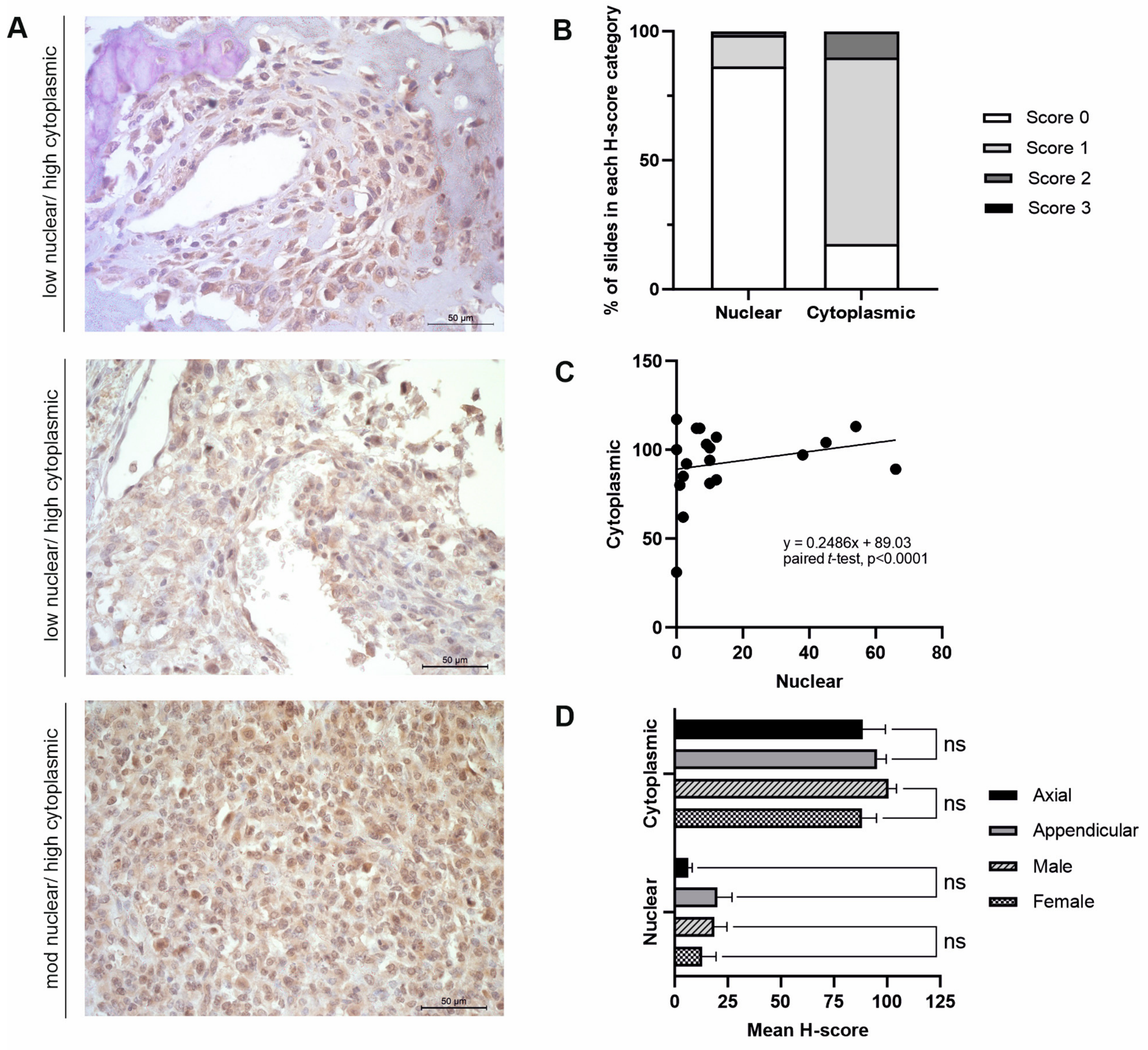
Figure 1.
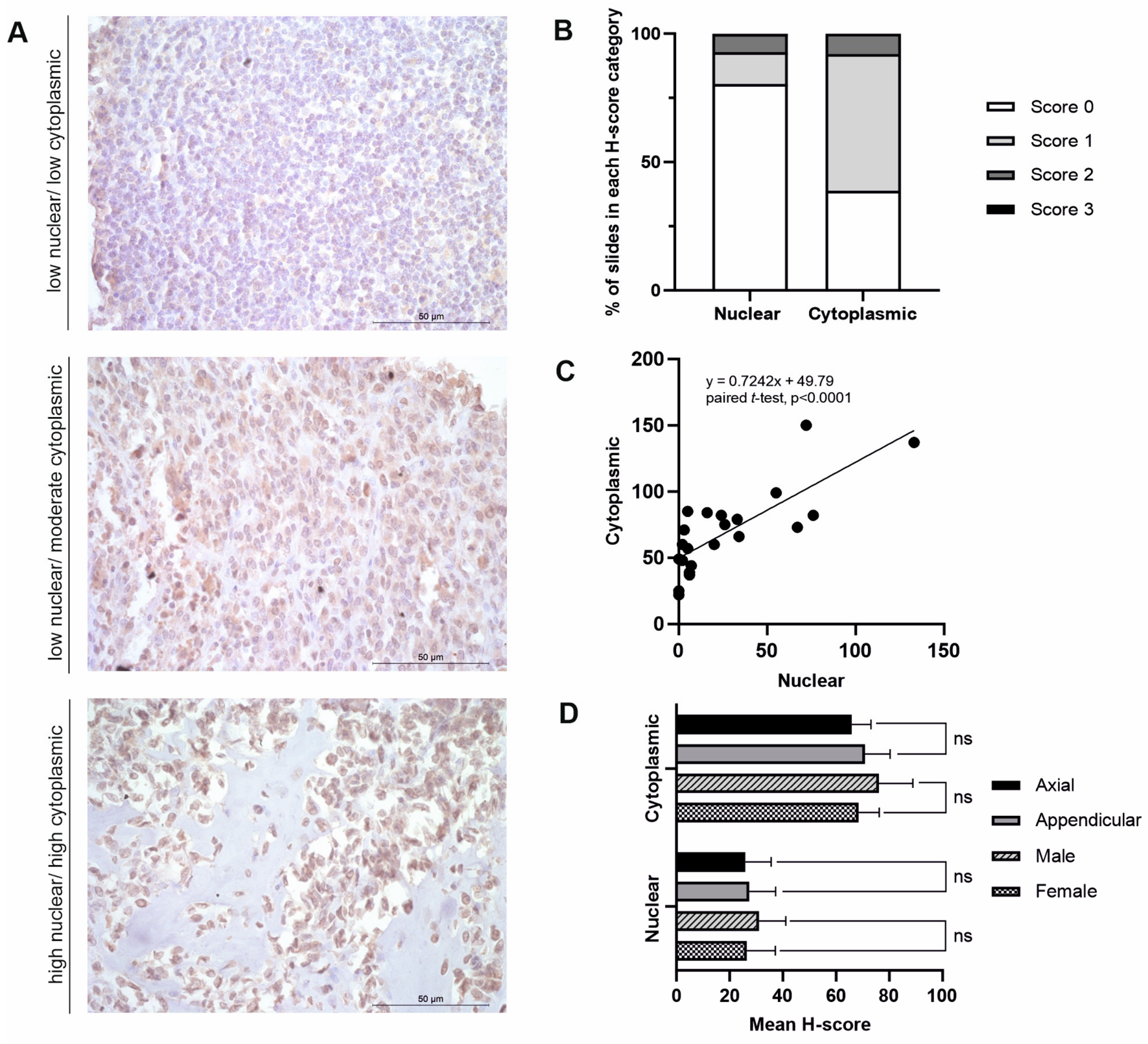
ELOVL2 analysis (n = 22). (A) Photomicrographs depicting positive ELOVL2 expression in canine OSA tissue. (B) Staining intensity scores (0–3). (C) Correlation between nuclear and cytoplasmic H-scores; Spearman’s correlation. Nuclear and cytoplasmic scores also compared using paired t-test; p < 0.0001. (D) Anatomical location and sex differences between cytoplasmic and nuclear H-scores (axial + soft tissue n = 7, appendicular n = 15, male n = 8, female n = 13); comparisons using Mann–Whitney U test (both nuclear and cytoplasmic axial + soft tissue versus appendicular tests and nuclear male versus female) and t-test (male versus female cytoplasmic scores).
Figure 2.
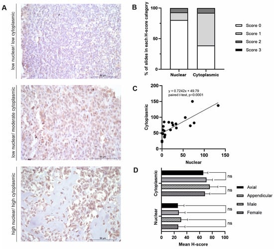
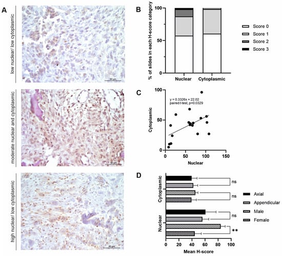
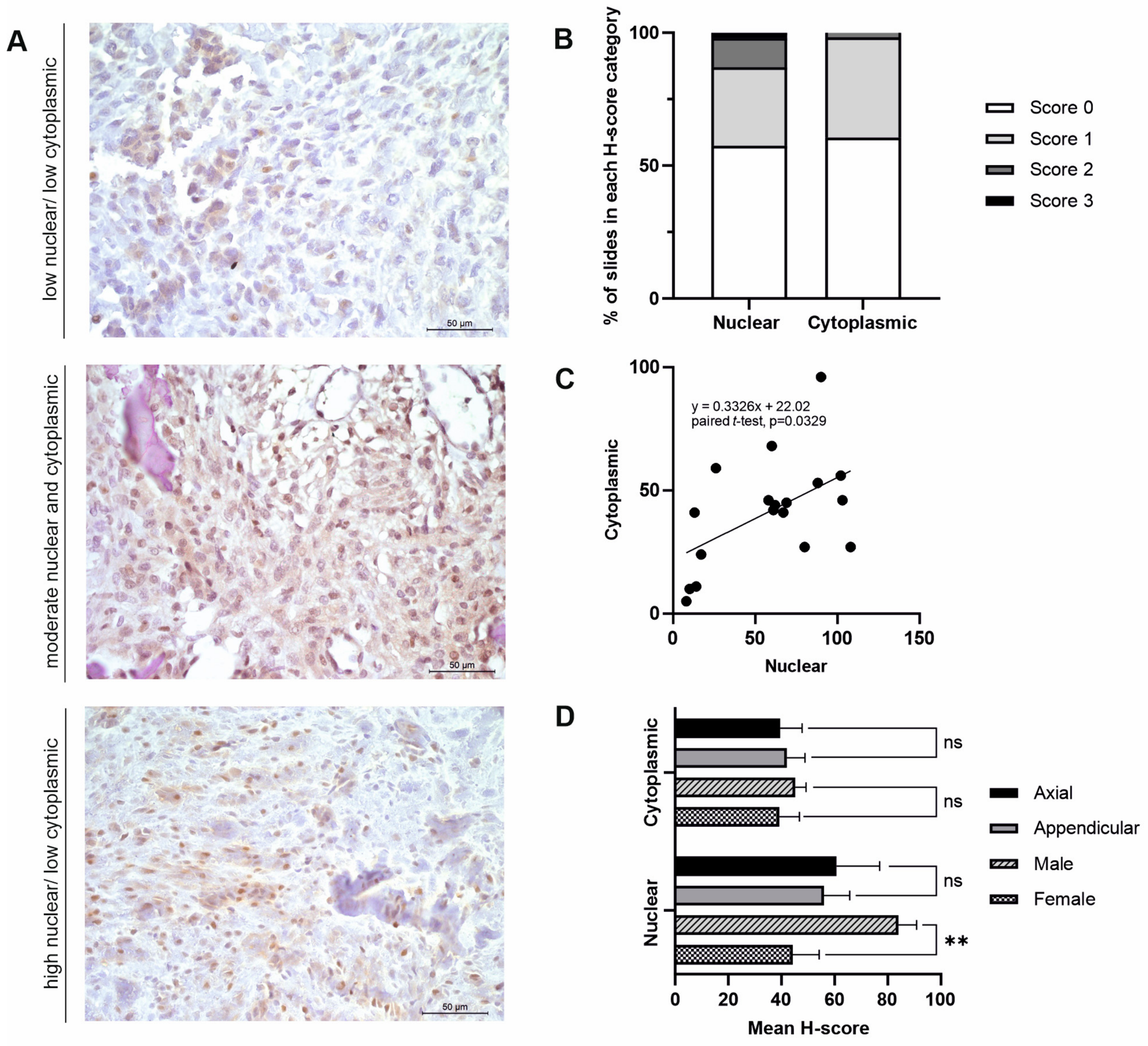
LIPG analysis (n = 18). (A) Photomicrographs depicting positive LIPG expression in canine OSA tissue. (B) Staining intensity score (0–3). (C) Correlation between cytoplasmic and nuclear H-scores; Pearson’s correlation. Nuclear and cytoplasmic scores also compared using paired t-test; p = 0.0329. (D) t-test to identify anatomical location and sex differences between cytoplasmic and nuclear H-scores (axial + soft tissue n = 6, appendicular n = 12, male n = 6, female n = 12); ** represents 0.0046. ns=not statistically significant.
Figure 3.
RPL4 analysis (n = 18). (A) Photomicrographs depicting positive RPL4 expression in canine OSA tissue. (B) Staining intensity score (0–3). (C) Correlation between nuclear and cytoplasmic H-scores; Spearman’s correlation. Nuclear and cytoplasmic scores also compared using paired t-test; p < 0.0001. (D) Anatomical location and sex differences between cytoplasmic and nuclear H-scores (axial + soft tissue n = 7, appendicular n = 11, male n = 6, female n = 11); comparisons using Mann–Whitney U test (both nuclear and cytoplasmic axial + soft tissue versus appendicular tests and nuclear male versus female) and t-test (male versus female cytoplasmic scores).
Figure 4.
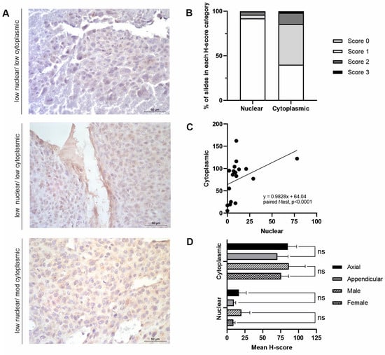
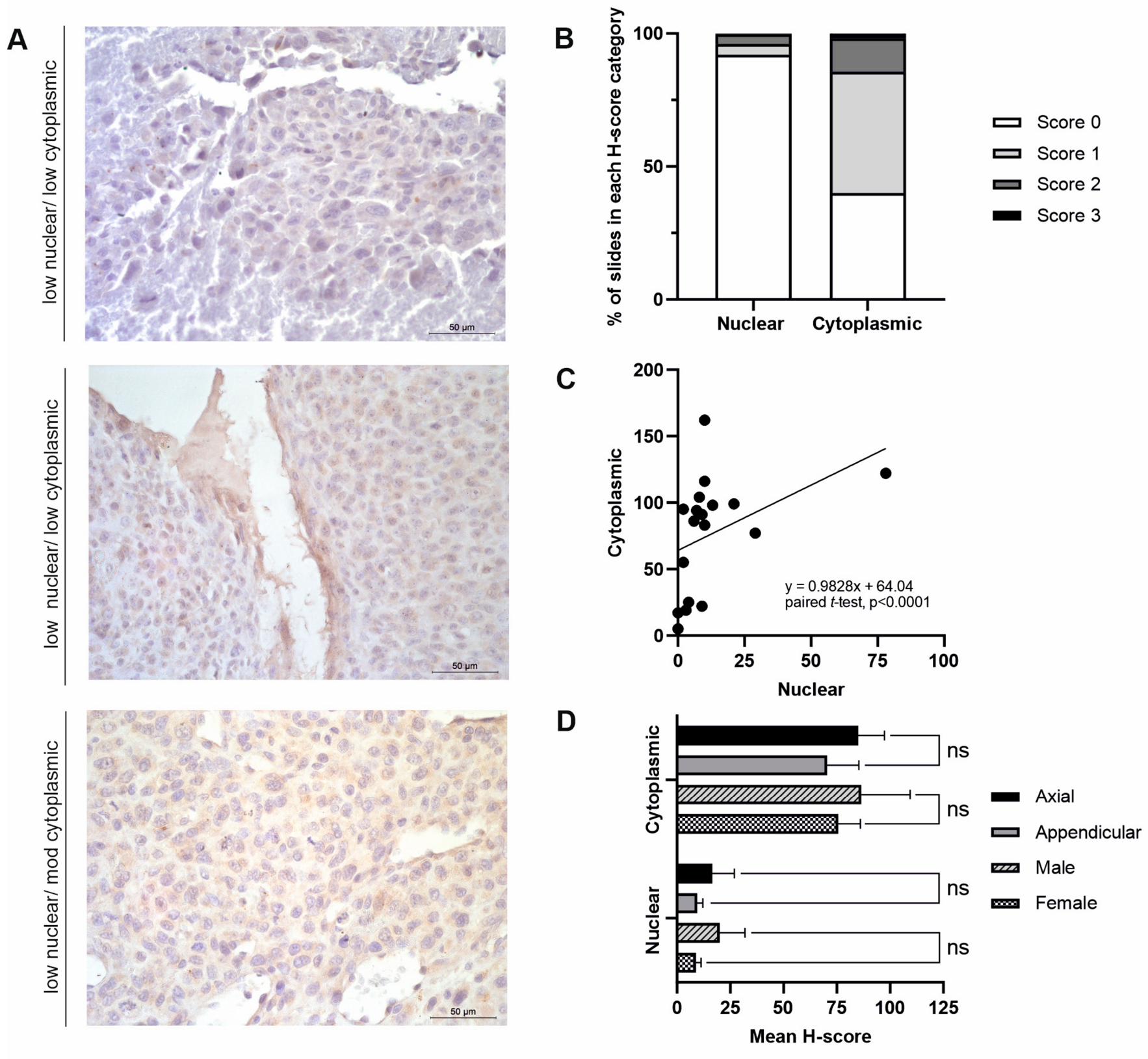
RPSA analysis (n = 19). (A) Photomicrographs depicting positive RPSA expression in canine OSA tissue. (B) Staining intensity score (0–3). (C) Correlation between nuclear and cytoplasmic H-scores; Spearman’s correlation. Nuclear and cytoplasmic scores also compared using paired t-test; p < 0.0001. (D) Anatomical location and sex differences between cytoplasmic and nuclear H-scores (axial + soft tissue n = 7, appendicular n = 12, male n = 7, female n = 12); comparisons using Mann–Whitney U test (nuclear axial + soft tissue versus appendicular and nuclear male versus female) and t-test (cytoplasmic male versus female and cytoplasmic axial + soft tissue versus appendicular scores). ns=not statistically significant.
3. Results
3.1. Protein Intensities and Cellular Locations in OSA Tissues
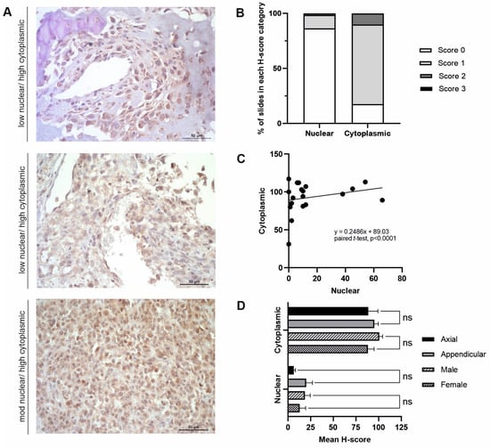
In a previous transcriptomic analysis, the ELOVL2, LIPG, RPL4, and RPSA mRNA levels were significantly higher in canine OSA compared to non-malignant, patient-matched counterparts (log2 fold change 4.23619, p = 0.00035; 4.44654, p = 0.0061; 3.92678, p = 0.00015 and 3.86821, p = 0.00135, respectively) [13]. Hence, immunohistochemical techniques were implemented to reveal their protein expression in canine OSA tissues (n = 23). The final sample sizes varied for each protein as cell counts had to reach the required minimum of n = 200 cells per sample for H-score semi-quantification, with the high-quality visualisation of the staining and no visible tissue damage. Therefore, the final numbers for each antibody were as follows: ELOVL2 (n = 22), LIPG (n = 18), RPL4 (n = 18), and RPSA (n = 19). Table 3 and Table 4 display the overall IHC staining results for the four proteins investigated. Overall, all antibodies displayed positive staining at varying intensities across OSA tissues, as shown in Figure 1A, Figure 2A, Figure 3A and Figure 4A. H-score analysis for each protein of interest was performed, displaying the correlation and H-score staining intensity (scored 0–3), as well as for investigating subcellular staining differences between anatomical locations (axial and soft tissue versus appendicular) and sexes.
3.2. ELOVL2 Expression in Canine OSA
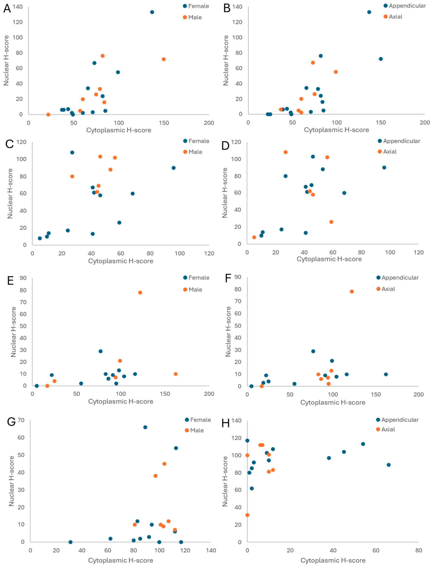
ELOVL2 staining was diffuse across n = 22 canine OSA samples (Figure 1A,B). Cytoplasmic staining was predominantly categorised as ‘moderate’ (14/22), with 5/22 classified as ‘low’ and 3/22 as ‘high’ (Table 4). Nuclear staining was ‘low’ in n = 17 samples, ‘moderate’ in 4/22, and ‘high’ in only 1/22 tissues (Table 4). There was a significant positive correlation between the cytoplasmic and nuclear H-scores (Spearman r = 0.7237, p = 0.0001, 95% confidence interval (0.4240 to 0.8806); Figure 1C). There was a significant difference between the nuclear and cytoplasmic H-scores (Shapiro–Wilk test, W = 0.9660, p = 0.6193; paired t-test, p < 0.0001; Figure 1C). Anatomical location and sex differences were investigated. No significant findings were discovered between H-scores (both nuclear and cytoplasmic) within these groups (MWU for nuclear and t-tests for cytoplasmic, p > 0.05; Figure 1D and Figure A2).
3.3. LIPG Expression in Canine OSA
Overall, LIPG staining was present at varying intensities in n = 18 OSA tissues (Figure 2A,B). Cytoplasmic staining was predominantly ‘low’ and ‘moderate’ (n = 6 and n = 10, respectively), with only two samples displaying ‘high’ cytoplasmic staining (Table 4). Nuclear staining was varied, with 6/18 samples having ‘low’, 4/18 having ‘moderate’, and 8/18 classified as having ‘high’ H-scores (Table 4). There was a significant correlation between the nuclear and cytoplasmic scores (Pearson r = 0.5217, p = 0.0264, 95% confidence interval (0.0724 to 0.7949); Figure 2C). There was a significant difference between the nuclear and cytoplasmic H-scores (Shapiro–Wilk test, W = 0.9752, p = 0.8870; paired t-test, p = 0.0329; Figure 2C). When investigating differences in the nuclear and cytoplasmic H-scores in different sexes, it was found that females had significantly lower nuclear H-scores than males (t-test, p = 0.0046; Figure 2D). In contrast, there were no significant findings among the cytoplasmic H-scores between sexes or among the H-scores in axial and soft-tissue versus appendicular locations (t-test, p > 0.05; Figure 2D and Figure A2).
3.4. RPL4 Expression in Canine OSA
RPL4 staining was diffuse across n = 18 samples (Figure 3A,B). Nuclear staining was predominantly ‘low’ (17/18 samples), with n = 1 tissue showing ‘moderate’ staining (Table 4). Cytoplasmic staining was predominantly ’moderate’ (10/18 tissues), with 5/18 classified as ‘low’ and 3/18 as ‘high’ (Table 4). There was a significant correlation between the nuclear and cytoplasmic H-scores (Spearman r = 0.6442, p = 0.0039, 95% confidence interval (0.2396 to 0.8582); Figure 3C). There was a significant difference between the nuclear and cytoplasmic H-scores (Shapiro–Wilk test, W = 0.9394, p = 0.2833; paired t-test, p < 0.0001; Figure 3C). When comparing the subcellular H-scores in different anatomical locations (axial and soft tissue versus appendicular) and between sexes, there were no significant results (t-test for cytoplasmic male vs. feamle, MWU for other tests, p > 0.05; Figure 3D and Figure A2).
3.5. RPSA Expression in Canine OSA
Overall, RPSA staining was diffuse and present in all n = 19 samples (Figure 4A,B). Cytoplasmic staining was classified as ‘high’ in 18/19 samples and ‘moderate’ in n = 1 (Table 4). Nuclear staining remained ‘low’ in the majority of tissues (15/19), with n = 3 and n = 1 having ‘moderate’ and ‘high’ nuclear staining, respectively (Table 4). There was no significant correlation between the nuclear and cytoplasmic H-scores (Spearman r = 0.2283, p = 0.3472, 95% confidence interval (−0.2379 to 0.6276); Figure 4C). There was a significant difference between the nuclear and cytoplasmic H-scores (Shapiro–Wilk test, W = 0.9515, p = 0.4197; paired t-test, p < 0.0001; Figure 4C). Anatomical location and sex differences were investigated, although no significant associations were seen between the H-scores (both nuclear and cytoplasmic) within these groups (nuclear MWU and cytoplasmic t-test, p > 0.05; Figure 4D and Figure A2).
4. Discussion
4.1. ELOVL2
ELOVL2 is a member of the ELOVL fatty acid elongase family. Although it contributes to several cellular functions, a key role is the elongation of PUFAs, which have roles in inflammation, energy metabolism, and membranes [22,23,24,25]. ELOVL2 is predominantly localised to the endoplasmic reticulum and primarily expressed in the brain, liver, and retina [26]. The role of ELOVL2 has yet to be investigated in OSA, although it has been shown to have multiple functions in cancer progression, which vary between different cancer types [25,27,28,29]. In canine OSA, ELOVL2 mRNA expression was found to be significantly higher in OSA tissue than in normal bone (log2FoldChange: 4.23619, p = 0.00035) [13], and the present study identified positive protein expression within canine OSA.
In renal cell carcinoma (RCC), ELOVL2 has been shown to have tumour-promoting effects [29]. The overexpression of ELOVL2 promotes lipid droplet (LD) accumulation, and intracellular LDs are a hallmark characteristic of clear-cell RCC and contribute to a poor prognosis [29,30,31,32]. ELOVL2 promoted tumour growth via the elongation of PUFAs, inhibiting apoptosis, and ELOVL2-induced LDs were suggested to have protective effects against cellular stress, contributing to RCC progression [29]. SAM-pointed domain-containing ETS transcription factor has been shown to have a pro-oncogenic role in clear-cell RCC, transcriptionally regulating ELOVL2 by binding to the −141 bp site within the ELOVL2 promoter, activating its transcription [33].
In breast cancer, ELOVL2 had a tumour-suppressive effect [25,34]. Decreased ELOVL2 expression correlated with a poorer prognosis, and the ablation of ELOVL2 may promote the migration and colony formation of breast cancer cells [34]. In tamoxifen-resistant MCF-7 breast cancer cells, ELOVL2 was shown to be hypermethylated and consequently downregulated compared to tamoxifen-sensitive patients [25]. ELOVL2 regulates genes from the Akt and ERa signalling pathways, such as Akt-regulated genes (THEM4, BMF, and FTO), which play crucial roles in drug resistance [25]. In xenograft mouse models, Jeong et al. (2021) showed that ELOVL2 could recover tamoxifen sensitivity by up to 70% [25].
In neuroblastoma, high expression of ELOVL2 was associated with favourable survival rates and suppressed the proliferation of cancer cells in vivo [35]. MYCN RNA interference significantly upregulated docosahexaenoic acid (DHA) and ELOVL2 levels [35]. In prostate cancer, ELOVL2 was upregulated in tumours, and individuals with high ELOVL2 expression were suggested to have a better prognosis [28]. Knockdown of ELOVL2 activated the PI3K/Akt signalling pathway via the downregulation of INPP4B and promoted the cellular proliferation, migration, and invasion of prostate cancer cells [28]. Conversely, the upregulation of ELOVL2 in castration-resistant prostate cancer played a critical role in driving enzalutamide resistance via the stabilisation of androgen receptor through inhibiting ubiquitin–proteasome-mediated degradation [27].
The use of the EZH2 (enhancer of zeste homolog 2) inhibitor GSK126 has been investigated in human osteosarcoma cell lines, inducing apoptosis and autophagic cell death, as well as reducing the migration of OSA cells via its inhibitory activity on the FBP1/C-Myc signalling axis [36]. Zhang et al. (2022) identified ELOVL2 as a target gene of EZH2 via H3K27me3 modification [37]. Inhibition of EZH2 using GSK126 in melanoma and hepatocarcinoma cell lines reduced the H3K27me3 level on the ELOVL2 promoter and consequently increased the expression of ELOVL2 [37]. More research is required to identify ELOVL2’s role in the tumorigenesis of OSA, and, given the literature showing both tumour-promoting and tumour-suppressing roles, more work aimed at understanding when and how these conditions are observed is crucial. If this protein/gene does exert a pro-tumour effect or exerts it under particular conditions or in specific tumours, inhibiting ELOVL2 could also enhance the anti-cancer effect of EZH2 inhibitors, which could represent a future direction for clinical trials in canine OSA.
4.2. LIPG
LIPG, also known as lipase G, endothelial type, is a member of the triglyceride lipase family and plays essential roles in lipoprotein metabolism, as well as having an important role as a hydrolase enzyme [38]. LIPG is synthesised as a 55 kDa protein before being glycosylated and secreted in its 68 kDa form [39].
LIPG mediates inflammatory processes through pro-inflammatory cytokines, and increased LIPG expression can be influenced by the higher presence of inflammatory markers such as Sicam, IL-6, sTNFr2, leptin, and hsCRP [38,40,41]. Thus, altered LIPG expression may be the result of a host defence mechanism for acute or chronic inflammation [38]. LIPG’s role in the uptake and then metabolism of plasma lipoproteins has also been identified as a potential precursor for the energy generation required for the rapid proliferation and development of cancer cells, including migration [38,42,43]. Hence, high LIPG expression within tumours is indicative of dysregulated lipid metabolism, potentially associated with neoplasm generation, development, and prognosis [44]. This may also be the case in Rottweiler OSA.
AMPK activation, when caused by severe oxidative stress, has been reported to be a trigger for LIPG overexpression [45]. In breast cancer, the upregulation of LIPG allows cells to adapt to oxidative stress and enables them to utilise lipoproteins as a nutrient source for growth [46]. LIPG was preferentially overexpressed in basal-like triple-negative breast cancer (TNBC) in comparison to other breast cancer subtypes and is required for tumorigenesis and the metastatic progression of the cancer [42]. The DTX3L-ISG15 signalling pathway regulates protein function and stability via ISGylation [42]. LIPG was mechanistically shown to execute its oncogenic function in basal-like TNBC cells via the DTX3L-ISG15 pathway [42]. Lo et al. (2018) also suggested that the protein DTX3L was required for the maintenance of LIPG expression levels in basal-like TNBC cells by inhibiting proteasome-mediated protein degradation [42]. In vitro LIPG knockdown resulted in reduced cell growth, migration, and CSC sphere formation, and the dysfunction of LIPG led to disrupted DTX3L-LIPG-ISG15 signalling, further suggesting LIPG’s role in oncogenesis [42].
In lung adenocarcinoma (LUAD), LIPG expression was higher in neoplastic compared to normal tissues, particularly in patients with lymph node metastasis and those older than 65 years old [47]. High LIPG expression was a predictor of poor survival, although patients with high LIPG expression did respond well to anti-neoplastic agents and had low drug resistance rates [47]. LIPG expression was also positively correlated with the expression levels of multiple immune cell infiltrates within the LUAD microenvironment, i.e., CD4+ T-cells, M1 macrophages, neutrophils, and plasma cells [47].
In canine OSA, LIPG mRNA was significantly overexpressed in tumours compared to non-malignant counterparts (log2FoldChange: 4.44654, p = 0.0061) [13], and positive protein expression was observed in the present study within canine OSA tissues. Some studies have suggested no sex differences in LIPG expression [38,40], but, in lung adenocarcinoma, its expression was higher in women [47]. The present research identified a significant difference between male (higher) and female (lower) nuclear H-scores (t-test; p = 0.0046); however, we note that the relatively low number of males (n = 6) in this particular cohort means that the results should be considered exploratory. The role of LIPG in osteosarcoma has yet to be identified, but potential roles in adapting to oxidative stress and activating AMPK have been suggested [45].
4.3. RPL4
Ribosomal protein L4 (RPL4) is a protein-encoding gene whose product is a component of the 60S ribosomal subunit, located within the cytoplasm [48,49]. RPL4 belongs to the L4E family of ribosomal proteins, responsible for ribosome biogenesis, which is essential for cell growth and proliferation, and the synthesis of proteins [49]. Often, genes that encode ribosomal proteins have multiple processed pseudogenes within the genome, as shown in previous canine RNA sequencing data from Simpson et al. (2020) [13]; the Ensembl ID for the significantly differentially expressed protein-encoding gene is ENSCAFG00000017309 (log2 fold change: 3.92678, p = 0.00015), whereas the Ensembl ID that encodes the non-significant processed pseudogene is ENSCAFG00000011417 (p = 0.38445).
RPL4 is suggested to have a role in ribosome composition and biogenesis initiation and regulation, as well as translation [49]. Tumorigenesis may occur during the activation of ribosome biogenesis, over-translation of proteins, or changes in ribosome production [50,51]. In cancer, RPL4 is involved in cell adhesion, neoplasm development, and metastasis via pathways including proteoglycan, TOR, and AMPK signalling [49]. RPL4 expression also negatively correlates with sensitivity to certain inhibitors, such as dasatinib, rositinib (CO-1686), and erdatinib (JNJ-42756493), implying that RPL4’s differential expression can affect tumour treatment [49]. As well as the high mRNA expression of RPL4 displayed in canine OSA [13], cBioPortal revealed that 6% of human OSA samples/patients displayed genetic alterations in the TARGET GDC 2025 cohort (n = 66), with 3% of these displaying high mRNA [52]. High expression of RPL4 has been shown in various cancers, including colon cancer and oesophageal carcinoma [53,54].
RPL4 is a novel regulator of the MDM2-p53 loop that maintains cellular homeostasis [55], although this has yet to be investigated in canine OSA. Mouse double minute 2 (MDM2) is an oncogene and serves as a negative regulator of p53 [56,57]. RPL4 directly binds to the MDM2 protein within cells, inhibiting the MDM2-mediated ubiquitination and degradation of p53 in cells and preventing the downregulation of p53 tumour-suppressive pathways [55,58]. The overexpression of RPL4 has been shown to promote MDM2-RPL5-RPL11 complex formation within cells, further suppressing MDM2 activity [55]. Conversely, the knockdown of RPL4 in a human osteosarcoma cell line (U2OS) induced p53 levels and activity, as well as inducing p53-dependent cell cycle arrest, potentially disrupting ribosome biogenesis and inducing ribosomal stress [55]. Children who carry the TP53 mutation, associated with Li Fraumeni syndrome, have a high risk of developing many cancers within their lifetime, including OSA [59]. In canine OSA, the frequency of TP53 mutations ranges from 24 to 47% and they are mostly located within highly conserved domains, nearly identical to those present in the disease in humans [60]. Hence, further investigation into RPL4’s role in the regulation of p53 is pivotal for understanding the development of OSA in both species.
4.4. RPSA
RPSA, also known as ribosomal protein SA, 37LRP, or LAMR1, encodes a 37 kDa laminin receptor (LR) in humans, a precursor of the higher-molecular-weight isoform, including the 67 kDa LR [61]. The post-translational modifications for the formation of the higher-molecular-weight species from the 37 kDa LR are not fully understood [61]. RPSA’s functions include cellular migration, invasion, viability, and growth, as well as histone and chromatin binding [62]. RPSA can be expressed in the plasma membrane, nucleus, ribosomes, and cytosol, and its subcellular location is dependent on its function [62]. The RPSA/LAMR1 antibody (10668-1-AP; Proteintech) used in the present study also produced positive cytoplasmic staining in a variety of control mouse tissue samples, such as the heart, brain, liver, lung, and kidney [63]. The Human Protein Atlas [26] reports that RPSA showed cytoplasmic expression in all tissues, with low tissue specificity, which corresponds with the findings from this study. The protein has also been linked to neoplasm metastasis and invasion in various cancer types, such as LUAD, prostate cancer, and fibrosarcoma [64,65,66].
RPSA expression has been shown to be increased in neoplastic tissues compared to non-malignant counterparts [61,67,68]. Knockdown of RPSA, using siRNA, significantly decreased cell viability in breast, oesophageal, pancreatic, neuroblastoma, melanoma, and colorectal carcinoma cells [69,70,71,72]. RPSA has also shown associations with cell adhesion-mediated drug resistance in both in vivo and in vitro models of a glucocorticoid drug-resistant cell line [61]. RPSA stimulates pathways such as FAK phosphorylation and the PI3K/Akt and MEK/ERK1/2 survival pathways [61,73]. RPSA also functions as a cell surface receptor for laminin-1, an extracellular matrix glycoprotein that is implicated in processes such as cell adhesion, differentiation, migration, and growth [61,74]. The binding of laminin to RPSA activates several proteolytic enzymes that degrade the basal lamina, consequently increasing cellular migration and the invasion of tumour cells [75,76]. These would be interesting areas of future research around canine OSA given the findings in our study, as our work does not present any functional studies.
Yang et al. (2014) discovered an osteosarcoma-specific fusion gene, LRP1-SNRNP25, in humans, formed by the fusion of LRP1 exon 8 and SNRNP25 exon 2, which played pivotal roles in neoplastic cell invasion and migration [77,78]. This gene was shown to upregulate 37LRP/RPSA levels, as well as the migration- and invasion-related protein pJNK [77]. RPSA can interact with both pJNK and MMP2, which also have migratory and invasive functions [77]. Immunofluorescence staining revealed the intracellular co-localisation of LRP1-SNRNP25, pJNK, and RPSA proteins in OSA [77]. The use of inhibitors of these, such as SP600125 (pJNK inhibitor) and marimastat (MMP2 inhibitor), was able to suppress the pJNK/37LRP/MMP2 signalling pathway, which promotes the invasion, migration, and metastasis of OSA [77]. In lung adenocarcinoma, it was also found that the use of SP600125 decreased 37LRP expression [79]. In canine OSA, further studies are required to determine whether the LRP1-SNRNP25 fusion gene exists and contributes to tumorigenesis. The Simpson et al. (2020) mRNA sequencing dataset reveals that only RPSA is differentially expressed within the pJNK/37LRP/MMP2 pathway at the mRNA level in canine OSA (log2 fold change: 3.86821, p = 0.00135) [13], although the protein expression is not yet known for the other components of the pJNK/37LRP/MMP2 signalling axis.
4.5. Limitations to the Study and Future Work
The present study investigated the subcellular protein expression of ELOVL2, LIPG, RPL4, and RPSA in canine OSA via immunohistochemistry and semi-quantitative H-scoring, alongside exploring sex-associated and anatomical location differences. Prior to the clinical use of these proteins as biomarkers in canine OSA, the appropriate validation of antibodies and protocols and the standardisation of fixation, staining, and analytical techniques would need to be performed. The present study investigated protein expression in only Rottweiler samples, but the use of tissues from other breeds would provide valuable data for the clinical application of ELOVL2, LIPG, RPL4, and RPSA, as well information relating to the heterogenetic nature of the tumour. Comparing expression levels against a range of non-tumour bone and patient-matched normal tissues, alongside tissues specific to each antibody, could also provide insights into differential expression, including differing expression between anatomical locations, as well as providing validation for the antibodies used, as this study is the first to use them in canine samples. Future studies exploring larger cohorts, as well investigations into different sub-groups, including tumour grade, stage, T substages I and II, invasion status, and metastasis groups, alongside clinical progression and outcome data, would be a valuable next step. Techniques such as Western blotting, mass spectrometry, and ELISA for protein quantification and semi-quantification may provide further validation and antibody specificity or development. The exploration of repurposing drugs presently known to target ELOVL2, LIPG, RPL4, and RPSA, and functional and pathway studies such as those mentioned in this study’s discussion, would offer valuable information to explore treatment opportunities for OSA. More general work into the implementation of H-scoring in veterinary medicine in general is also required as, unlike within human medicine, this technique is not used as standard.
5. Conclusions
This study characterised the subcellular expression of the proteins corresponding to the overexpressed genes of interest (ELOVL2, LIPG, RPL4, and RPSA) in canine OSA tumours. The interpretation and analysis of the IHC conducted in this experiment revealed significant variance in the subcellular expression of ELOVL2, RPL4, and RPSA. They also add to the limited number of presently known potential biomarkers in canine osteosarcoma, indicating that biomarker panels may be a future development [80,81,82,83]. The further identification of biomarkers, such as those in the present study, is required to aid diagnostic and treatment decision-making. Significant sex-associated differences were identified for LIPG nuclear staining, and further investigations into this protein are required to assess whether these differences affect cancer progression in certain groups of animals. Given the biological similarities between human and canine OSA, these findings may warrant investigation in comparative oncology [5,84]. A deeper understanding of protein interactions and their mechanisms within canine OSA is required to explore these potential biomarkers for new diagnostic, prognostic, and treatment opportunities for this cancer type.
Author Contributions
Conceptualisation, J.J.-O., N.P.M. and C.S.R.; methodology, J.J.-O., A.A.A., N.P.M. and C.S.R.; validation, J.J.-O., A.A.A., N.P.M. and C.S.R.; formal analysis, J.J.-O., A.A.A., N.P.M. and C.S.R.; investigation, J.J.-O., A.A.A., I.N., J.L.-R., S.d.B., M.D.D., J.N.J., N.P.M. and C.S.R.; resources, J.J.-O., M.D.D., N.P.M. and C.S.R.; data curation, J.J.-O., N.P.M. and C.S.R.; writing—original draft preparation, J.J.-O., A.A.A., I.N., J.L.-R., S.d.B., M.D.D., J.N.J., N.P.M. and C.S.R.; writing—review and editing, J.J.-O., A.A.A., I.N., J.L.-R., S.d.B., M.D.D., J.N.J., N.P.M. and C.S.R.; visualisation, J.J.-O., A.A.A. and C.S.R.; supervision, S.d.B., M.D.D., J.N.J., N.P.M. and C.S.R.; project administration, N.P.M. and C.S.R.; funding acquisition, M.D.D., N.P.M. and C.S.R. All authors have read and agreed to the published version of the manuscript.
Funding
J.J.-O. was funded by the Geoff Butler PhD Scholarship and the School of Veterinary Medicine and Science, supervised by S.d.B., M.D.D., J.N.J., N.P.M., and C.S.R. The BBSRC doctoral training programme and the School of Veterinary Medicine and Science, University of Nottingham, also funded J.L.-R., supervised by C.S.R. and N.P.M. (BB/M008770/1). We would like to thank the Stanyard, Moody, Thorman, and Kenyon families for their unrestricted gifts, as well as Cancer Research UK (CRUK; #CGCATF-2023/100029) and the School of Veterinary Medicine and Science, University of Nottingham, for funding I.N. (supervised by N.P.M., C.S.R., and J.N.J.).
Institutional Review Board Statement
The study was conducted in accordance with the ARRIVE Guidelines and approved by the University of Nottingham School of Veterinary Medicine and Science CARE ethics committee (ethics permission numbers 1823 160714, 20 July 2016; 959 130925, 12 July 2016; and 3832 230502, 12 May 2023). International and national ethical standards were adhered to. OSA samples were excised from Rottweilers following euthanasia or amputation for reasons unrelated to the present study.
Informed Consent Statement
Informed owner consent was obtained.
Data Availability Statement
The original contributions presented in this study are included in the article. Further inquiries can be directed to the corresponding authors.
Conflicts of Interest
Catrin Rutland is an Editorial Board Member for the MDPI journal Veterinary Sciences but had no role in the peer review or other administrative processes. Otherwise, the authors declare no conflicts of interest. The funders had no role in the design of the study; in the collection, analyses, or interpretation of the data; in the writing of the manuscript; or in the decision to publish the results.
Abbreviations
The following abbreviations are used in this manuscript:
| DHA | Docosahexaenoic acid |
| ELOVL2 | ELOVL fatty acid elongase |
| FFPE | Formalin-fixed paraffin-embedded |
| IHC | Immunohistochemistry |
| LD | Lipid droplet |
| LIPG | Lipase G, endothelial type |
| LR | Laminin receptor |
| LUAD | Lung adenocarcinoma |
| MWU | Mann–Whitney U |
| OR | Odds ratio |
| OSA | Osteosarcoma |
| PUFA | Polyunsaturated fatty acid |
| RCC | Renal cell carcinoma |
| RPL4 | Ribosomal protein L4 |
| RPSA | Ribosomal protein SA (LAMR1) |
| SEM | Standard error of mean |
| TNBC | Triple-negative breast cancer |
Appendix A
Figure A1.
Positive and negative controls in mouse tissues and canine OSA. (A) Mouse bone (ELOVL2, 1:100); (B) mouse spleen (ELOVL2, 1:100); (C) mouse bone (LIPG, 1:50); (D) mouse placenta (LIPG, 1:50); (E) mouse bone (RPL4, 1:150); (F) mouse spleen (RPL4, 1:100); (G) mouse bone (RPSA, 1:100); (H) mouse placenta (RPSA, 1:100); (I) mouse bone (negative control); (J) canine OSA (negative control).
Appendix B
Figure A2.
Nuclear and cytoplasmic H-scores for each specimen based on sex and anatomical location (appendicular vs. axial + soft tissue). (A) ELOVL2 sex, (B) ELOVL2 anatomical location, (C) LIPG sex, (D) LIPG anatomical location, (E) RPL4 sex, (F) RPL4 anatomical location, (G) RPSA sex, (H) RPSA anatomical location.
References
- Rowell, J.L.; McCarthy, D.O.; Alvarez, C.E. Dog models of naturally occurring cancer. Trends Mol. Med. 2011, 17, 380–388. [Google Scholar] [CrossRef] [Scilit] [PubMed]
- Makielski, K.M.; Mills, L.J.; Sarver, A.L.; Henson, M.S.; Spector, L.G.; Naik, S.; Modiano, J.F. Risk Factors for Development of Canine and Human Osteosarcoma: A Comparative Review. Vet. Sci. 2019, 6, 48. [Google Scholar] [CrossRef] [Scilit] [PubMed]
- Edmunds, G.L.; Smalley, M.J.; Beck, S.; Errington, R.J.; Gould, S.; Winter, H.; Brodbelt, D.C.; O’Neill, D.G. Dog breeds and body conformations with predisposition to osteosarcoma in the UK: A case-control study. Canine Med. Genet. 2021, 8, 2. [Google Scholar] [CrossRef] [Scilit] [PubMed]
- Salt, C.; Morris, P.J.; German, A.J.; Wilson, D.; Lund, E.M.; Cole, T.J.; Butterwick, R.F. Growth standard charts for monitoring bodyweight in dogs of different sizes. PLoS ONE 2017, 12, e0182064. [Google Scholar] [CrossRef] [Scilit] [PubMed]
- Simpson, S.; Dunning, M.D.; de Brot, S.; Grau-Roma, L.; Mongan, N.P.; Rutland, C.S. Comparative review of human and canine osteosarcoma: Morphology, epidemiology, prognosis, treatment and genetics. Acta Vet. Scand. 2017, 59, 71. [Google Scholar] [CrossRef] [Scilit] [PubMed]
- Simpson, S.; Rizvanov, A.A.; Jeyapalan, J.N.; de Brot, S.; Rutland, C.S. Canine osteosarcoma in comparative oncology: Molecular mechanisms through to treatment discovery. Front. Vet. Sci. 2022, 9, 2022. [Google Scholar] [CrossRef] [Scilit] [PubMed]
- Ru, G.; Terracini, B.; Glickman, L.T. Host related risk factors for canine osteosarcoma. Vet. J. 1998, 156, 31–39. [Google Scholar] [CrossRef] [Scilit] [PubMed]
- Fan, T.M.; Khanna, C. Comparative Aspects of Osteosarcoma Pathogenesis in Humans and Dogs. Vet. Sci. 2015, 2, 210–230. [Google Scholar] [CrossRef] [Scilit] [PubMed]
- Morello, E.; Martano, M.; Buracco, P. Biology, diagnosis and treatment of canine appendicular osteosarcoma: Similarities and differences with human osteosarcoma. Vet. J. 2011, 189, 268–277. [Google Scholar] [CrossRef] [Scilit] [PubMed]
- Urlić, I.; Jovičić, M.Š.; Ostojić, K.; Ivković, A. Cellular and Genetic Background of Osteosarcoma. Curr. Issues Mol. Biol. 2023, 45, 4344–4358. [Google Scholar] [CrossRef] [Scilit] [PubMed]
- Klein, M.J.; Siegal, G.P. Osteosarcoma: Anatomic and histologic variants. Am. J. Clin. Pathol. 2006, 125, 555–581. [Google Scholar] [CrossRef] [Scilit] [PubMed]
- Szewczyk, M.; Lechowski, R.; Zabielska, K. What do we know about canine osteosarcoma treatment? Review. Vet. Res. Commun. 2015, 39, 61–67. [Google Scholar] [CrossRef] [Scilit] [PubMed]
- Simpson, S.; Dunning, M.; de Brot, S.; Alibhai, A.; Bailey, C.; Woodcock, C.L.; Mestas, M.; Akhtar, S.; Jeyapalan, J.N.; Lothion-Roy, J.; et al. Molecular Characterisation of Canine Osteosarcoma in High Risk Breeds. Cancers 2020, 12, 2405. [Google Scholar] [CrossRef] [Scilit] [PubMed]
- Allott, E.H.; Cohen, S.M.; Geradts, J.; Sun, X.; Khoury, T.; Bshara, W.; Zirpoli, G.R.; Miller, C.R.; Hwang, H.; Thorne, L.B.; et al. Performance of Three-Biomarker Immunohistochemistry for Intrinsic Breast Cancer Subtyping in the AMBER Consortium. Cancer Epidemiol. Biomarkers Prev. 2016, 25, 470–478. [Google Scholar] [CrossRef] [Scilit] [PubMed]
- Duraiyan, J.; Govindarajan, R.; Kaliyappan, K.; Palanisamy, M. Applications of immunohistochemistry. J. Pharm. Bioallied Sci. 2012, 4, S307–S309. [Google Scholar] [CrossRef] [Scilit] [PubMed]
- Altschul, S.F.; Gish, W.; Miller, W.; Myers, E.W.; Lipman, D.J. Basic local alignment search tool. J. Mol. Biol. 1990, 215, 403–410. [Google Scholar] [CrossRef] [PubMed]
- Thermofisher. ELOVL2 Polyclonal Antibody. Available online: https://www.thermofisher.com/antibody/product/ELOVL2-Antibody-Polyclonal/20308-1-AP (accessed on 16 September 2025).
- Thermofisher. LIPG Polyclonal Antibody. Available online: https://www.thermofisher.com/antibody/product/LIPG-Antibody-Polyclonal/PA5-82568 (accessed on 16 September 2025).
- Thermofisher. RPL4 Polyclonal Antibody. Available online: https://www.thermofisher.com/antibody/product/RPL4-Antibody-Polyclonal/PA5-96464 (accessed on 16 September 2025).
- Thermofisher. LAMR1/RPSA Polyclonal Antibody. Available online: https://www.thermofisher.com/antibody/product/LAMR1-RPSA-Antibody-Polyclonal/10668-1-AP (accessed on 16 September 2025).
- Koo, T.K.; Li, M.Y. A Guideline of Selecting and Reporting Intraclass Correlation Coefficients for Reliability Research. J. Chiropr. Med. 2016, 15, 155–163. [Google Scholar] [CrossRef] [Scilit] [PubMed]
- Chen, D.; Chao, D.L.; Rocha, L.; Kolar, M.; Nguyen Huu, V.A.; Krawczyk, M.; Dasyani, M.; Wang, T.; Jafari, M.; Jabari, M.; et al. The lipid elongation enzyme ELOVL2 is a molecular regulator of aging in the retina. Aging Cell 2020, 19, e13100. [Google Scholar] [CrossRef] [Scilit] [PubMed]
- Garagnani, P.; Bacalini, M.G.; Pirazzini, C.; Gori, D.; Giuliani, C.; Mari, D.; Di Blasio, A.M.; Gentilini, D.; Vitale, G.; Collino, S.; et al. Methylation of ELOVL2 gene as a new epigenetic marker of age. Aging Cell 2012, 11, 1132–1134. [Google Scholar] [CrossRef] [Scilit] [PubMed]
- Gregory, M.K.; Gibson, R.A.; Cook-Johnson, R.J.; Cleland, L.G.; James, M.J. Elongase reactions as control points in long-chain polyunsaturated fatty acid synthesis. PLoS ONE 2011, 6, e29662. [Google Scholar] [CrossRef] [Scilit] [PubMed]
- Jeong, D.; Ham, J.; Kim, H.W.; Kim, H.; Ji, H.W.; Yun, S.H.; Park, J.E.; Lee, K.S.; Jo, H.; Han, J.H.; et al. ELOVL2: A novel tumor suppressor attenuating tamoxifen resistance in breast cancer. Am. J. Cancer Res. 2021, 11, 2568–2589. [Google Scholar] [PubMed]
- Uhlen, M.; Oksvold, P.; Fagerberg, L.; Lundberg, E.; Jonasson, K.; Forsberg, M.; Zwahlen, M.; Kampf, C.; Wester, K.; Hober, S.; et al. Towards a knowledge-based Human Protein Atlas. Nat. Biotechnol. 2010, 28, 1248–1250. [Google Scholar] [CrossRef] [Scilit] [PubMed]
- Cen, J.; Guo, J.; Zeng, X.; Song, X.; Ge, S.; Chen, M.; Li, Q.; Yu, Y.; Lv, D.; Zhao, S. ELOVL2 mediated stabilization of AR contributes to enzalutamide resistance in prostate cancer. Front. Cell Dev. Biol. 2025, 13, 1598400. [Google Scholar] [CrossRef] [Scilit] [PubMed]
- Hu, T.; Zhang, H.; Du, Y.; Luo, S.; Yang, X.; Zhang, H.; Feng, J.; Chen, X.; Tu, X.; Wang, C.; et al. ELOVL2 restrains cell proliferation, migration, and invasion of prostate cancer via regulation of the tumor suppressor INPP4B. Cell. Signal. 2022, 96, 110373. [Google Scholar] [CrossRef] [Scilit] [PubMed]
- Tanaka, K.; Kandori, S.; Sakka, S.; Nitta, S.; Tanuma, K.; Shiga, M.; Nagumo, Y.; Negoro, H.; Kojima, T.; Mathis, B.J.; et al. ELOVL2 promotes cancer progression by inhibiting cell apoptosis in renal cell carcinoma. Oncol. Rep. 2022, 47, 1–11. [Google Scholar] [CrossRef] [Scilit] [PubMed]
- Ackerman, D.; Tumanov, S.; Qiu, B.; Michalopoulou, E.; Spata, M.; Azzam, A.; Xie, H.; Simon, M.C.; Kamphorst, J.J. Triglycerides Promote Lipid Homeostasis during Hypoxic Stress by Balancing Fatty Acid Saturation. Cell Rep. 2018, 24, 2596–2605 e2595. [Google Scholar] [CrossRef] [Scilit] [PubMed]
- Qiu, B.; Ackerman, D.; Sanchez, D.J.; Li, B.; Ochocki, J.D.; Grazioli, A.; Bobrovnikova-Marjon, E.; Diehl, J.A.; Keith, B.; Simon, M.C. HIF2alpha-Dependent Lipid Storage Promotes Endoplasmic Reticulum Homeostasis in Clear-Cell Renal Cell Carcinoma. Cancer Discov. 2015, 5, 652–667. [Google Scholar] [CrossRef] [Scilit] [PubMed]
- Xie, H.; Song, J.; Godfrey, J.; Riscal, R.; Skuli, N.; Nissim, I.; Simon, M.C. Glycogen metabolism is dispensable for tumour progression in clear cell renal cell carcinoma. Nat. Metab. 2021, 3, 327–336. [Google Scholar] [CrossRef] [Scilit] [PubMed]
- Zhang, P.; Yu, W.; Luo, H.; Wang, D.; Li, M.; Wang, T.; Guo, Y.; Xiong, W.; Gong, B.; He, Y. SPDEF promotes clear cell renal cell carcinoma progression by transcriptionally activating ELOVL2-mediated lipid metabolic reprogramming. Biochim. Biophys. Acta Mol. Basis Dis. 2026, 1872, 168315. [Google Scholar] [CrossRef] [Scilit] [PubMed]
- Kang, Y.P.; Yoon, J.H.; Long, N.P.; Koo, G.B.; Noh, H.J.; Oh, S.J.; Lee, S.B.; Kim, H.M.; Hong, J.Y.; Lee, W.J.; et al. Spheroid-Induced Epithelial-Mesenchymal Transition Provokes Global Alterations of Breast Cancer Lipidome: A Multi-Layered Omics Analysis. Front. Oncol. 2019, 9, 145. [Google Scholar] [CrossRef] [Scilit] [PubMed]
- Ding, Y.; Yang, J.; Ma, Y.; Yao, T.; Chen, X.; Ge, S.; Wang, L.; Fan, X. MYCN and PRC1 cooperatively repress docosahexaenoic acid synthesis in neuroblastoma via ELOVL2. J. Exp. Clin. Cancer Res. 2019, 38, 498. [Google Scholar] [CrossRef] [Scilit] [PubMed]
- Xiong, X.; Liu, Y.; Du, Y.; Lai, X.; Si, C.; Miao, H. Unveiling the effects of GSK126 on osteosarcoma cells implications for apoptosis, autophagy, and cellular migration. Discov. Oncol. 2025, 16, 245. [Google Scholar] [CrossRef] [Scilit] [PubMed]
- Zhang, T.; Guo, Z.; Huo, X.; Gong, Y.; Li, C.; Huang, J.; Wang, Y.; Feng, H.; Ma, X.; Jiang, C.; et al. Dysregulated lipid metabolism blunts the sensitivity of cancer cells to EZH2 inhibitor. EBioMedicine 2022, 77, 103872. [Google Scholar] [CrossRef] [Scilit] [PubMed]
- Hong, C.; Deng, R.; Wang, P.; Lu, X.; Zhao, X.; Wang, X.; Cai, R.; Lin, J. LIPG: An inflammation and cancer modulator. Cancer Gene Ther. 2021, 28, 27–32. [Google Scholar] [CrossRef] [Scilit] [PubMed]
- Gauster, M.; Hrzenjak, A.; Schick, K.; Frank, S. Endothelial lipase is inactivated upon cleavage by the members of the proprotein convertase family. J. Lipid Res. 2005, 46, 977–987. [Google Scholar] [CrossRef] [Scilit] [PubMed][Green Version]
- Badellino, K.O.; Wolfe, M.L.; Reilly, M.P.; Rader, D.J. Endothelial lipase is increased in vivo by inflammation in humans. Circulation 2008, 117, 678–685. [Google Scholar] [CrossRef] [Scilit] [PubMed]
- Riederer, M.; Lechleitner, M.; Hrzenjak, A.; Koefeler, H.; Desoye, G.; Heinemann, A.; Frank, S. Endothelial lipase (EL) and EL-generated lysophosphatidylcholines promote IL-8 expression in endothelial cells. Atherosclerosis 2011, 214, 338–344. [Google Scholar] [CrossRef] [Scilit] [PubMed]
- Lo, P.K.; Yao, Y.; Lee, J.S.; Zhang, Y.; Huang, W.; Kane, M.A.; Zhou, Q. LIPG signaling promotes tumor initiation and metastasis of human basal-like triple-negative breast cancer. Elife 2018, 7, e31334. [Google Scholar] [CrossRef] [Scilit] [PubMed]
- Tatematsu, S.; Francis, S.A.; Natarajan, P.; Rader, D.J.; Saghatelian, A.; Brown, J.D.; Michel, T.; Plutzky, J. Endothelial lipase is a critical determinant of high-density lipoprotein-stimulated sphingosine 1-phosphate-dependent signaling in vascular endothelium. Arterioscler. Thromb. Vasc. Biol. 2013, 33, 1788–1794. [Google Scholar] [CrossRef] [Scilit] [PubMed]
- Boren, J.; Taskinen, M.R.; Adiels, M. Kinetic studies to investigate lipoprotein metabolism. J. Intern. Med. 2012, 271, 166–173. [Google Scholar] [CrossRef] [Scilit] [PubMed]
- Slebe, F.; Rojo, F.; Vinaixa, M.; Garcia-Rocha, M.; Testoni, G.; Guiu, M.; Planet, E.; Samino, S.; Arenas, E.J.; Beltran, A.; et al. FoxA and LIPG endothelial lipase control the uptake of extracellular lipids for breast cancer growth. Nat. Commun. 2016, 7, 11199. [Google Scholar] [CrossRef] [Scilit] [PubMed]
- Cadenas, C.; Vosbeck, S.; Edlund, K.; Grgas, K.; Madjar, K.; Hellwig, B.; Adawy, A.; Glotzbach, A.; Stewart, J.D.; Lesjak, M.S.; et al. LIPG-promoted lipid storage mediates adaptation to oxidative stress in breast cancer. Int. J. Cancer 2019, 145, 901–915. [Google Scholar] [CrossRef] [Scilit] [PubMed]
- Wang, S.; Chen, Z.; Lv, H.; Wang, C.; Wei, H.; Yu, J. LIPG is a novel prognostic biomarker and correlated with immune infiltrates in lung adenocarcinoma. J. Clin. Lab. Anal. 2023, 37, e24824. [Google Scholar] [CrossRef] [Scilit] [PubMed]
- Nosrati, N.; Kapoor, N.R.; Kumar, V. Combinatorial action of transcription factors orchestrates cell cycle-dependent expression of the ribosomal protein genes and ribosome biogenesis. FEBS J. 2014, 281, 2339–2352. [Google Scholar] [CrossRef] [Scilit] [PubMed]
- Liu, Y.; Li, W.; Zhou, S.; Cui, M.; Zhang, L. Pan-cancer analysis of the prognostic and immunological role of RPL4. Heliyon 2024, 10, e34461. [Google Scholar] [CrossRef] [Scilit] [PubMed]
- Kang, J.; Brajanovski, N.; Chan, K.T.; Xuan, J.; Pearson, R.B.; Sanij, E. Ribosomal proteins and human diseases: Molecular mechanisms and targeted therapy. Signal Transduct. Target. Ther. 2021, 6, 323. [Google Scholar] [CrossRef] [Scilit] [PubMed]
- Pelletier, J.; Thomas, G.; Volarevic, S. Ribosome biogenesis in cancer: New players and therapeutic avenues. Nat. Rev. Cancer 2018, 18, 51–63. [Google Scholar] [CrossRef] [Scilit] [PubMed]
- Cerami, E.; Gao, J.; Dogrusoz, U.; Gross, B.E.; Sumer, S.O.; Aksoy, B.A.; Jacobsen, A.; Byrne, C.J.; Heuer, M.L.; Larsson, E.; et al. The cBio cancer genomics portal: An open platform for exploring multidimensional cancer genomics data. Cancer Discov. 2012, 2, 401–404. [Google Scholar] [CrossRef] [Scilit] [PubMed]
- Uhlen, M.; Fagerberg, L.; Hallstrom, B.M.; Lindskog, C.; Oksvold, P.; Mardinoglu, A.; Sivertsson, A.; Kampf, C.; Sjostedt, E.; Asplund, A.; et al. Proteomics. Tissue-based map of the human proteome. Science 2015, 347, 1260419. [Google Scholar] [CrossRef] [Scilit] [PubMed]
- Thul, P.J.; Akesson, L.; Wiking, M.; Mahdessian, D.; Geladaki, A.; Ait Blal, H.; Alm, T.; Asplund, A.; Bjork, L.; Breckels, L.M.; et al. A subcellular map of the human proteome. Science 2017, 356, eaal3321. [Google Scholar] [CrossRef] [Scilit] [PubMed]
- He, X.; Li, Y.; Dai, M.S.; Sun, X.X. Ribosomal protein L4 is a novel regulator of the MDM2-p53 loop. Oncotarget 2016, 7, 16217–16226. [Google Scholar] [CrossRef] [Scilit] [PubMed]
- Fang, S.; Jensen, J.P.; Ludwig, R.L.; Vousden, K.H.; Weissman, A.M. Mdm2 is a RING finger-dependent ubiquitin protein ligase for itself and p53. J. Biol. Chem. 2000, 275, 8945–8951. [Google Scholar] [CrossRef] [Scilit] [PubMed]
- Honda, R.; Tanaka, H.; Yasuda, H. Oncoprotein MDM2 is a ubiquitin ligase E3 for tumor suppressor p53. FEBS Lett. 1997, 420, 25–27. [Google Scholar] [CrossRef] [Scilit] [PubMed]
- Nag, S.; Qin, J.; Srivenugopal, K.S.; Wang, M.; Zhang, R. The MDM2-p53 pathway revisited. J. Biomed. Res. 2013, 27, 254–271. [Google Scholar] [CrossRef] [Scilit] [PubMed]
- O’Neill, D.G.; Edmunds, G.L.; Urquhart-Gilmore, J.; Church, D.B.; Rutherford, L.; Smalley, M.J.; Brodbelt, D.C. Dog breeds and conformations predisposed to osteosarcoma in the UK: A VetCompass study. Canine Med. Genet. 2023, 10, 8. [Google Scholar] [CrossRef] [Scilit] [PubMed]
- Kirpensteijn, J.; Kik, M.; Teske, E.; Rutteman, G.R. TP53 gene mutations in canine osteosarcoma. Vet. Surg. 2008, 37, 454–460. [Google Scholar] [CrossRef] [Scilit] [PubMed]
- Limone, A.; Maggisano, V.; Sarnataro, D.; Bulotta, S. Emerging roles of the cellular prion protein (PrP(C)) and 37/67 kDa laminin receptor (RPSA) interaction in cancer biology. Cell Mol. Life Sci. 2023, 80, 207. [Google Scholar] [CrossRef] [Scilit] [PubMed]
- DiGiacomo, V.; Meruelo, D. Looking into laminin receptor: Critical discussion regarding the non-integrin 37/67-kDa laminin receptor/RPSA protein. Biol. Rev. Camb. Philos. Soc. 2016, 91, 288–310. [Google Scholar] [CrossRef] [Scilit] [PubMed]
- Liu, M.; Li, N.; Guo, W.; Jia, L.; Jiang, H.; Li, Z.; Wang, J.; Zhang, X.; Zhu, R.; Bao, C.; et al. RPSA distribution and expression in tissues and immune cells of pathogen-infected mice. Microb. Pathog. 2021, 152, 104609. [Google Scholar] [CrossRef] [Scilit] [PubMed]
- Wang, Y.; Dong, H.; Xu, M.; Xin, B.; Niu, W.; Xu, D.; Zhao, P.; Zhang, B.; Li, Z.; Liu, L. 37-kDa laminin receptor precursor promotes lung adenocarcinoma cell invasion and metastasis by epithelial-to-mesenchymal transition. Cancer Gene Ther. 2014, 21, 150–157. [Google Scholar] [CrossRef] [Scilit] [PubMed]
- Annabi, B.; Currie, J.C.; Bouzeghrane, M.; Dulude, H.; Daigneault, L.; Garde, S.; Rabbani, S.A.; Panchal, C.; Wu, J.J.; Beliveau, R. Contribution of the 37-kDa laminin receptor precursor in the anti-metastatic PSP94-derived peptide PCK3145 cell surface binding. Biochem. Biophys. Res. Commun. 2006, 346, 358–366. [Google Scholar] [CrossRef] [Scilit] [PubMed]
- Jaseja, M.; Mergen, L.; Gillette, K.; Forbes, K.; Sehgal, I.; Copie, V. Structure-function studies of the functional and binding epitope of the human 37 kDa laminin receptor precursor protein. J. Pept. Res. 2005, 66, 9–18. [Google Scholar] [CrossRef] [Scilit] [PubMed]
- Jovanovic, K.; Chetty, C.J.; Khumalo, T.; Da Costa Dias, B.; Ferreira, E.; Malindisa, S.T.; Caveney, R.; Letsolo, B.T.; Weiss, S.F. Novel patented therapeutic approaches targeting the 37/67 kDa laminin receptor for treatment of cancer and Alzheimer’s disease. Expert. Opin. Ther. Pat. 2015, 25, 567–582. [Google Scholar] [CrossRef] [Scilit] [PubMed]
- Vania, L.; Morris, G.; Otgaar, T.C.; Bignoux, M.J.; Bernert, M.; Burns, J.; Gabathuse, A.; Singh, E.; Ferreira, E.; Weiss, S.F.T. Patented therapeutic approaches targeting LRP/LR for cancer treatment. Expert. Opin. Ther. Pat. 2019, 29, 987–1009. [Google Scholar] [CrossRef] [Scilit] [PubMed]
- Chetty, C.J.; Ferreira, E.; Jovanovic, K.; Weiss, S.F.T. Knockdown of LRP/LR induces apoptosis in pancreatic cancer and neuroblastoma cells through activation of caspases. Exp. Cell Res. 2017, 360, 264–272. [Google Scholar] [CrossRef] [Scilit] [PubMed]
- Khumalo, T.; Ferreira, E.; Jovanovic, K.; Veale, R.B.; Weiss, S.F. Knockdown of LRP/LR Induces Apoptosis in Breast and Oesophageal Cancer Cells. PLoS ONE 2015, 10, e0139584. [Google Scholar] [CrossRef] [Scilit] [PubMed]
- Rebelo, T.M.; Vania, L.; Ferreira, E.; Weiss, S.F.T. siRNA-Mediated LRP/LR knock-down reduces cellular viability of malignant melanoma cells through the activation of apoptotic caspases. Exp. Cell Res. 2018, 368, 1–12. [Google Scholar] [CrossRef] [Scilit] [PubMed]
- Vania, L.; Morris, G.; Ferreira, E.; Weiss, S.F.T. Knock-down of LRP/LR influences signalling pathways in late-stage colorectal carcinoma cells. BMC Cancer 2021, 21, 392. [Google Scholar] [CrossRef] [Scilit] [PubMed]
- Sun, L.; Liu, L.; Liu, X.; Wang, Y.; Li, M.; Yao, L.; Yang, J.; Ji, G.; Guo, C.; Pan, Y.; et al. MGr1-Ag/37LRP induces cell adhesion-mediated drug resistance through FAK/PI3K and MAPK pathway in gastric cancer. Cancer Sci. 2014, 105, 651–659. [Google Scholar] [CrossRef] [Scilit] [PubMed]
- Domogatskaya, A.; Rodin, S.; Tryggvason, K. Functional diversity of laminins. Annu. Rev. Cell Dev. Biol. 2012, 28, 523–553. [Google Scholar] [CrossRef] [Scilit] [PubMed]
- Ardini, E.; Sporchia, B.; Pollegioni, L.; Modugno, M.; Ghirelli, C.; Castiglioni, F.; Tagliabue, E.; Menard, S. Identification of a novel function for 67-kDa laminin receptor: Increase in laminin degradation rate and release of motility fragments. Cancer Res. 2002, 62, 1321–1325. [Google Scholar] [PubMed]
- Pupa, S.M.; Menard, S.; Forti, S.; Tagliabue, E. New insights into the role of extracellular matrix during tumor onset and progression. J. Cell. Physiol. 2002, 192, 259–267. [Google Scholar] [CrossRef] [Scilit] [PubMed]
- Xing, P.; Liu, H.; Xiao, W.; Zhang, G.; Zhang, C.; Liao, Z.; Li, T.; Yang, J. The fusion gene LRP1-SNRNP25 drives invasion and migration by activating the pJNK/37LRP/MMP2 signaling pathway in osteosarcoma. Cell Death Discov. 2024, 10, 198. [Google Scholar] [CrossRef] [Scilit] [PubMed]
- Yang, J.; Annala, M.; Ji, P.; Wang, G.; Zheng, H.; Codgell, D.; Du, X.; Fang, Z.; Sun, B.; Nykter, M.; et al. Recurrent LRP1-SNRNP25 and KCNMB4-CCND3 fusion genes promote tumor cell motility in human osteosarcoma. J. Hematol. Oncol. 2014, 7, 76. [Google Scholar] [CrossRef] [Scilit] [PubMed]
- Zhou, Y.; Wang, Y.; Zhao, Z.; Wang, Y.; Zhang, N.; Zhang, H.; Liu, L. 37LRP induces invasion in hypoxic lung adenocarcinoma cancer cells A549 through the JNK/ERK/c-Jun signaling cascade. Tumour Biol. 2017, 39, 1010428317701655. [Google Scholar] [CrossRef] [Scilit] [PubMed]
- Brot, S.d.; Cobb, J.; Alibhai, A.A.; Jackson-Oxley, J.; Haque, M.; Patke, R.; Harris, A.E.; Woodcock, C.L.; Lothion-Roy, J.; Varun, D.; et al. Immunohistochemical Investigation into Protein Expression Patterns of FOXO4, IRF8 and LEF1 in Canine Osteosarcoma. Cancers 2024, 16, 1945. [Google Scholar] [CrossRef] [Scilit] [PubMed]
- Jackson-Oxley, J.; Alibhai, A.A.; Guerin, J.; Thompson, R.; Patke, R.; Harris, A.E.; Woodcock, C.L.; Varun, D.; Haque, M.; Modikoane, T.K.; et al. Comparison of Differentially Expressed Genes in Human and Canine Osteosarcoma. Life 2025, 15, 951. [Google Scholar] [CrossRef] [Scilit] [PubMed]
- Jackson-Oxley, J.; Alibhai, A.A.; Thompson, R.; Lothion-Roy, J.; de Brot, S.; Dunning, M.D.; Jeyapalan, J.N.; Mongan, N.P.; Rutland, C.S. Identification and Characterisation of Canine Osteosarcoma Biomarkers and Therapeutic Targets. Cancers 2026, 18, 262. [Google Scholar] [CrossRef] [Scilit] [PubMed]
- Rutland, C.S.; Cockcroft, J.M.; Lothion-Roy, J.; Harris, A.E.; Jeyapalan, J.N.; Simpson, S.; Alibhai, A.; Bailey, C.; Ballard-Reisch, A.C.; Rizvanov, A.A.; et al. Immunohistochemical Characterisation of GLUT1, MMP3 and NRF2 in Osteosarcoma. Front. Vet. Sci. 2021, 8, 2021. [Google Scholar] [CrossRef] [Scilit] [PubMed]
- Sadykova, L.R.; Ntekim, A.I.; Muyangwa-Semenova, M.; Rutland, C.S.; Jeyapalan, J.N.; Blatt, N.; Rizvanov, A.A. Epidemiology and Risk Factors of Osteosarcoma. Cancer Investig. 2020, 38, 259–269. [Google Scholar] [CrossRef] [Scilit] [PubMed]

Disclaimer/Publisher’s Note: The statements, opinions and data contained in all publications are solely those of the individual author(s) and contributor(s) and not of MDPI and/or the editor(s). MDPI and/or the editor(s) disclaim responsibility for any injury to people or property resulting from any ideas, methods, instructions or products referred to in the content. |
© 2026 by the authors. Licensee MDPI, Basel, Switzerland. This article is an open access article distributed under the terms and conditions of the Creative Commons Attribution (CC BY) license.