


Simple Summary
Hospitalisation can be particularly stressful for feral cats because they are not accustomed to close human contact and may also be affected by pain and illness. We followed 20 hospitalised feral cats that received four doses of pregabalin, a medicine used to reduce fear and pain. We assessed facial signs of pain, behaviour, sedation, heart and breathing rates, and cortisol in saliva as a marker of stress before, during, and after treatment. Pain-related and defensive-behaviour scores generally decreased, while sedation increased modestly. Heart rate, breathing rate, and cortisol did not change significantly for the group as a whole. Cortisol showed different patterns according to illness severity, and clinical procedures were associated with short-lived increases. No adverse events were recorded. Because there was no untreated comparison group and the cats also received other necessary treatments, the changes cannot be attributed specifically to pregabalin. Recovery, adaptation to the hospital, repeated handling, and familiarity with the staff may also have contributed. Larger controlled studies are needed to determine whether pregabalin can facilitate the clinical management of hospitalised feral cats and improve their welfare.
Abstract
Hospitalised feral cats may experience stress, pain, and defensive behaviour that complicate care. This prospective, exploratory, uncontrolled study described temporal changes during repeated pregabalin administration. Twenty-five cats were enrolled and 20 completed the study. Pregabalin (10 mg/kg) was given every 12 h for four doses. Heart rate (HR), respiratory rate (RR), Feline Grimace Scale (FGS), Demeanor Scoring System (DSS), and Global Sedation Score (GSS) were assessed from T-Pre to T60; salivary cortisol was measured. Repeated-measures general linear models included 18 complete cases for scores, eight for HR, and 15 for RR; cortisol subgroup analyses were post hoc. Time affected FGS, DSS, and GSS (all p < 0.001). At T36, mean individual changes were −48.3% (95% CI, −65.8% to −30.8%) for FGS and −54.1% (95% CI, −63.7% to −44.5%) for DSS. GSS peaked at T6, indicating mild sedation. No significant temporal effects were detected for HR (p = 0.112), RR (p = 0.259), or overall cortisol (n = 16). At washout (Tw), cats with severe persistent illness had higher cortisol than those without (p < 0.001); procedures were associated with transient increases. No adverse events were recorded. Clinical recovery, acclimatisation, concomitant treatments, rescue analgesia, and repeated handling may have contributed, so these associations do not establish pregabalin efficacy. Controlled studies are required.
1. Introduction
Cats and humans have coexisted for thousands of years; however, domestic cats have retained many ancestral behavioural traits in both free-roaming and indoor environments [1]. Cats are considered feral when they live freely within a territory and have had little or no contact with humans during the socialisation period [2,3]. Feral cats may nevertheless require hospitalisation and direct handling for veterinary care.
Feral cats are particularly susceptible to stress in unfamiliar environments [4]. When unable to escape during handling, they may display defensive behaviour that endangers both the animals and veterinary personnel and may alter physiological variables [5]. Transportation, physical examination, and diagnostic procedures can add further stress [6]. These challenges activate central threat-response mechanisms and the hypothalamic–pituitary–adrenal (HPA) axis [7]. Stress is therefore a complex, multifactorial response to events that threaten an animal’s perceived control [8].
Activation of the HPA axis increases cortisol release [7], making cortisol a useful objective marker of stress. Because blood collection may itself provoke an additional response, salivary cortisol provides a less invasive approach to evaluating stress in cats [9].
Strategies to reduce stress in hospitalised cats include appropriate handling, environmental enrichment, and hiding opportunities [10,11,12]. Cats may also conceal signs of pain, which can further complicate clinical assessment [13]. Pharmacological support may therefore be required, and pregabalin has attracted increasing interest in veterinary medicine [14].
Pregabalin is a structural analogue of gamma-aminobutyric acid (GABA) but does not bind directly to GABA receptors [15]. It binds to the auxiliary alpha-2-delta subunit of presynaptic voltage-gated calcium channels and reduces the release of excitatory neurotransmitters [16].
An oral pregabalin solution permits more precise dosing than solid formulations. Previous studies have evaluated single oral doses of 5–10 mg/kg in cats [17,18]. To the authors’ knowledge, repeated administration of 10 mg/kg has not been studied in hospitalised feral cats.
Pain, defensive behaviour, sedation, neuroendocrine stress, and physiological variables provide complementary rather than interchangeable information. The FGS was selected to quantify pain-related facial expression through observation, which is especially useful when direct handling should be minimised [19]. The DSS was used to characterise approach, defensive behaviour, and tolerance of interaction [20], whereas the GSS assessed sedation and alertness [21]. Salivary cortisol provided a non-invasive measure of adrenocortical activation [9,22]. Heart rate and respiratory rate are routinely assessed, non-specific clinical variables that may complement behavioural and endocrine measures when evaluating an animal’s overall physiological status.
Accordingly, this prospective, exploratory, uncontrolled study aimed to describe temporal changes in pain-related facial expression, demeanour, sedation, physiological variables, and salivary cortisol in hospitalised feral cats receiving repeated oral pregabalin and to assess their tolerance of handling and clinical procedures.
2. Materials and Methods
This was a prospective, exploratory, uncontrolled, single-arm clinical study evaluating temporal changes in pain-related facial expression, demeanour, sedation, physiological variables, and salivary cortisol during and after repeated oral administration of pregabalin in hospitalised feral cats.
2.1. Ethical Approval and Animals
The study was approved by the Comitato Etico per gli Studi Clinici e Zootecnici DIMEPRE-J, University of Bari Aldo Moro, Italy, under protocol number 2001-5 III/13 on 21 December 2023. All cats were enrolled after approval. Written authorisation for enrolment and for the associated veterinary procedures and treatments was obtained from the managers responsible for the respective feline colonies. The study was conducted from March 2024 to September 2025 at the University Veterinary Teaching Hospital of Bari Aldo Moro.
Free-roaming cats identified as ill by colony managers were transported to the hospital in carriers and admitted for clinical evaluation. Their feral status was established from the history provided by the colony managers, including a free-roaming lifestyle and little or no socialisation with humans. Temperament was further assessed within 24 h of admission, before pregabalin administration, using the DSS [20]. Scores of 0, 1–8, 9–18, 19–24, and 25 correspond to friendly and confident, friendly and shy, withdrawn and protective, withdrawn and aggressive, and overtly aggressive behaviour, respectively. A DSS score of at least 9/25 was required for inclusion because 9 is the lower boundary of the withdrawn and protective category. The DSS criterion supported, but did not replace, the history-based classification of feral status.
Cats were also required to be able to eat independently and to have a clinical condition compatible with completion of the protocol. Exclusion criteria included terminal neoplasia, shock or sepsis, altered consciousness or sensory status, neurological or renal disease, inability to eat independently, continuous infusion of sedative or analgesic drugs, surgery scheduled within 24 h of admission, or absence of a definitive diagnosis.
2.2. Study Design
The study rooms housed only feline patients. Light and sound could not be standardised because the cats were receiving clinical care. Cats were housed individually in cages positioned to prevent direct contact. Each cage contained food and water bowls, a litter tray, and a cardboard hiding box. The study period for each cat was four days. Only the study operators interacted directly with the cats, administered medication, and provided food and water during the protocol. Hospitalisation continued after the study when clinically required.
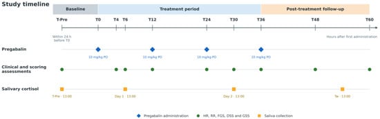
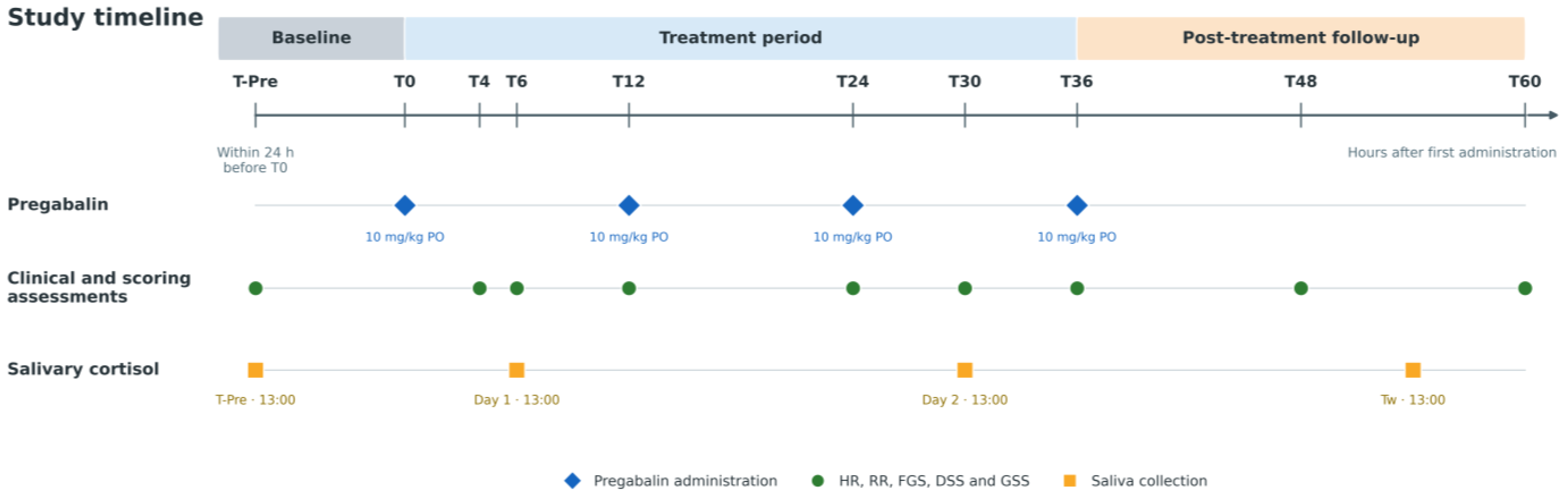
A baseline assessment was performed within 24 h before the first pregabalin administration (T-Pre). Pregabalin oral solution (Bonqat®, Orion Corporation, Orion Pharma, Espoo, Finland) was administered at 10 mg/kg with a small amount of wet or dry food every 12 h for four doses at T0, T12, T24, and T36. HR, RR, FGS, DSS, and GSS were assessed at T-Pre, T4, T6, T12, T24, T30, T36, T48, and T60. T48 and T60 corresponded to 12 and 24 h after the final dose. The study schedule is summarised in Figure 1.
Figure 1.
Study timeline. The baseline evaluation (T-Pre) was performed within 24 h before the first pregabalin administration at T0. Pregabalin (10 mg/kg orally) was administered at T0, T12, T24, and T36. HR, RR, FGS, DSS, and GSS were assessed at T-Pre, T4, T6, T12, T24, T30, T36, T48, and T60. T48 and T60 corresponded to 12 and 24 h after the final dose, respectively. Saliva was collected at 13:00 at T-Pre, on Day 1 and Day 2, and during the washout period (Tw). HR, heart rate; RR, respiratory rate; FGS, Feline Grimace Scale; DSS, Demeanor Scoring System; GSS, Global Sedation Score.
Cats received medical treatment tailored to their underlying conditions; these treatments were not standardised by the study protocol and could include anti-inflammatory drugs. An intravenous catheter was placed in each cat. The FGS assessment preceded any rescue intervention at every scheduled observation. Intravenous methadone (0.1 mg/kg) was administered when the FGS score was at least 4 or rescue analgesia was otherwise clinically indicated. The pre-intervention FGS value was recorded for the corresponding time point, and subsequent study assessments were delayed for at least 4 h. Measurements obtained after rescue analgesia were retained at the adjusted time point, but rescue analgesia was not included as a covariate in the statistical model. All adverse events were recorded.
All procedures performed during the study were documented in the medical records. When additional sedation was required for a clinical procedure, propofol (1–5 mg/kg IV) was administered. Scheduled FGS, DSS, and GSS assessments were postponed until the cat had completely recovered from the sedative effects and were then performed according to the adjusted schedule. Cats requiring general anaesthesia for major procedures or another substantial protocol deviation were excluded and managed according to their clinical needs.
2.3. Assessment Scales
Pain-related facial expression was assessed using the FGS [19]. Five facial action units are each scored from 0 to 2, producing a total score from 0 to 10; higher scores indicate greater expression of pain at the time of observation.
Behaviour was evaluated using the DSS [20], which includes eight items assessed without direct contact and during interaction with the operator. The total score ranges from 0 to 25 and defines five categories: friendly and confident (0), friendly and shy (1–8), withdrawn and protective (9–18), withdrawn and aggressive (19–24), and overtly aggressive (25). Higher scores indicate more defensive or aggressive behaviour, whereas lower scores indicate friendlier and less defensive responses.
Sedation was assessed using the GSS [21], which ranges from −3 to +3. Negative values indicate increasing alertness and positive values indicate increasing sedation. An increase in GSS therefore represents greater sedation, whereas a shift towards more negative values represents greater alertness.
2.4. Salivary Cortisol Analysis
Saliva was collected at 13:00 at T-Pre, on Day 1 and Day 2, and during Tw by gently swabbing the oral cavity for as long as the cat permitted. Samples were centrifuged at 15,000× g for 15 min and stored at −20 °C until analysis.
Cortisol concentration was measured using a high-sensitivity salivary cortisol enzyme immunoassay kit (Salimetrics, State College, PA, USA; code 1-3002), validated for use in humans and animals, including cats [22]. The lower limit of detection was 0.007 μg/dL. Samples were analysed in duplicate according to the manufacturer’s protocol.
Based on information recorded in the medical records, the 16 cats with complete cortisol measurements were retrospectively classified into two clinical subgroups. Classification was based on the primary diagnosis, the overall severity of the clinical condition, the presence of systemic involvement, and the persistence of clinical compromise throughout the study period. Cats showing substantial and persistent clinical compromise were classified as having severe persistent illness, whereas cats with localised or clinically stable conditions without marked persistent systemic compromise were classified as not having severe persistent illness. No validated disease-severity scale or prospectively defined numerical threshold was used for this classification.
2.5. Statistical Analysis
Descriptive data are presented as mean ± standard deviation (SD), together with the 95% confidence interval (CI) of the mean. For FGS and DSS, percentage change from baseline was calculated separately for each cat as [(follow-up value − T-Pre value)/T-Pre value] × 100. Only cats with values at both T-Pre and the corresponding follow-up time point were included in each calculation. Mean individual percentage changes and their 95% CIs are reported. Percentage changes were not calculated for GSS because the scale includes zero and negative values, making relative percentage changes undefined or potentially misleading.
Statistical analyses were performed using IBM SPSS Statistics version 17. Data distribution was assessed using the Kolmogorov–Smirnov test. HR, RR, FGS, DSS, and GSS were analysed separately using repeated-measures general linear models (GLMs), with time as the within-subject factor. The models used complete cases for each outcome; missing values were not imputed. When sphericity was not met, Greenhouse–Geisser correction was applied. After a significant overall time effect, Fisher’s least significant difference pairwise comparisons were performed between each follow-up time point and T-Pre. The T36–T48 and T36–T60 contrasts were also examined for FGS, DSS, and GSS. Assessments performed after complete clinical recovery from propofol or after the required delay following methadone were included at their adjusted time point. Rescue analgesia was not entered as a covariate.
An exploratory post hoc analysis was conducted in the 16 cats with complete cortisol measurements. Temporal changes in cortisol concentrations were evaluated in the overall group and within each retrospectively defined clinical subgroup using repeated-measures GLMs. The comparison between subgroups at Tw was performed using one-way analysis of variance. Cortisol concentrations before, during, and after a clinical procedure were compared in the subset of 10 cats with the required measurements using a repeated-measures GLM. Fisher’s least significant difference test was used for pairwise comparisons after a significant omnibus result. Statistical significance was set at p < 0.05, and exact p values are reported where available.
3. Results
3.1. Study Population and Clinical Characteristics
A total of 25 feral cats were initially enrolled. Five cats were subsequently excluded because of conditions that interfered with the study protocol, including neurological abnormalities, impaired mastication, and mandibular fracture. Therefore, 20 cats, comprising nine females and 11 males, completed the study and were included in the analysis.
The cats presented with heterogeneous clinical conditions. These included chronic gingivostomatitis in three cats, ear disorders in three, pelvic fractures in two, and ocular disorders in two. The remaining cats presented with myiasis, a cutaneous abscess, a tail injury, haematuria, weight loss, respiratory distress, intestinal obstruction, anorexia, or cutaneous wounds, with the latter occurring in two cats. Exact ages could not be reliably determined because of their feral status. Mean body weight was 3.90 ± 0.93 kg, and mean body condition score was 5.00 ± 0.97 on a nine-point scale.
3.2. Physiological Variables
Descriptive values for HR and RR are reported in Table 1. Because of unavailable measurements at some time points, the complete-case repeated-measures analyses included eight cats for HR and 15 cats for RR. No significant effect of time was detected for either HR [F(3.36, 23.50) = 2.18, p = 0.112; Greenhouse–Geisser correction] or RR [F(2.94, 41.13) = 1.39, p = 0.259; Greenhouse–Geisser correction].
Table 1.
Heart rate and respiratory rate at each study time point.
3.3. Pain, Demeanour, and Sedation Scores
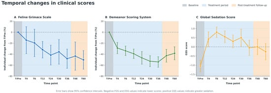
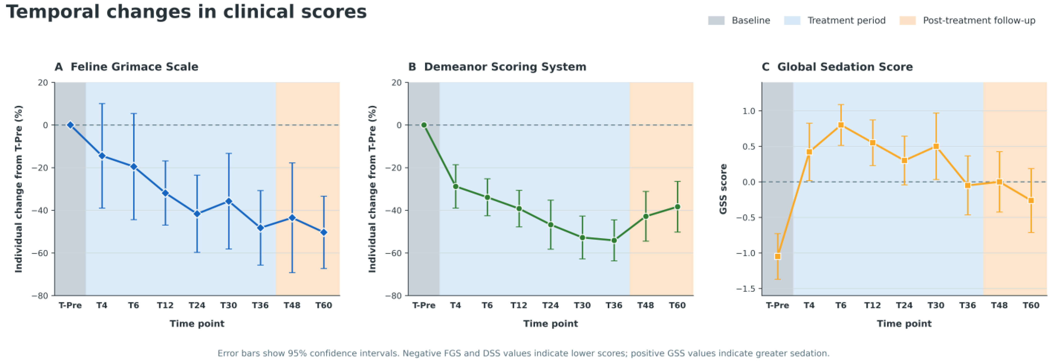
Descriptive values for FGS, DSS, and GSS are reported in Table 2, while mean individual percentage changes in FGS and DSS are presented in Table 3 and Figure 2. The complete-case repeated-measures GLM included 18 cats for each scale.
Table 2.
Feline Grimace Scale, Demeanor Scoring System, and Global Sedation Score at each study time point.
Table 3.
Individual percentage changes from T-Pre in Feline Grimace Scale and Demeanor Scoring System.
Figure 2.
Temporal changes in clinical scores. (A) Feline Grimace Scale (FGS) and (B) Demeanor Scoring System (DSS) are presented as the mean individual percentage change from T-Pre, with 95% confidence intervals. (C) Global Sedation Score (GSS) is presented as the mean score with 95% confidence intervals. Percentage changes were not calculated for GSS because the scale includes zero and negative values, making percentage estimates mathematically inappropriate and potentially misleading. The shaded areas indicate baseline (grey), the treatment period (light blue), and the post-treatment follow-up period (light orange). Negative percentage changes in FGS and DSS indicate lower scores compared with T-Pre. For GSS, positive values indicate greater sedation, whereas negative values indicate greater alertness. The dashed horizontal lines indicate zero change from baseline in panels A and B and a GSS score of zero in panel C.
A significant effect of time was identified for FGS [F(4.32, 73.43) = 8.78, p < 0.001; Greenhouse–Geisser correction]. Compared with T-Pre, FGS was significantly lower at T12, T24, T36, T48, and T60 (all p < 0.001) and at T30 (p = 0.002). Differences at T4 (p = 0.059) and T6 (p = 0.053) did not reach statistical significance. The lowest mean FGS score was recorded at T36, decreasing from 3.25 ± 1.33 (95% CI, 2.63 to 3.87) at T-Pre to 1.40 ± 1.05 (95% CI, 0.91 to 1.89). The paired mean difference at T36 was −2.06 points (95% CI, −2.90 to −1.21), and the mean individual percentage change was −48.3% (95% CI, −65.8% to −30.8%).
DSS also changed significantly over time [F(4.79, 81.46) = 28.60, p < 0.001; Greenhouse–Geisser correction]. Compared with T-Pre, DSS was lower at every follow-up assessment from T4 to T60 (all p < 0.001). The lowest mean DSS score was recorded at T36, decreasing from 15.85 ± 2.68 (95% CI, 14.60 to 17.11) at T-Pre to 7.25 ± 3.32 (95% CI, 5.70 to 8.81). The paired mean difference at T36 was −9.06 points (95% CI, −10.70 to −7.42), and the mean individual percentage change was −54.1% (95% CI, −63.7% to −44.5%).
A significant effect of time was also identified for GSS [F(5.22, 88.73) = 9.57, p < 0.001; Greenhouse–Geisser correction]. Compared with T-Pre, GSS was higher at T4, T6, T12, T24, and T30 (all p < 0.001), at T36 (p = 0.002), at T48 (p < 0.001), and at T60 (p = 0.003), indicating greater sedation relative to baseline. The highest mean GSS was recorded at T6, increasing from −1.05 ± 0.69 (95% CI, −1.37 to −0.73) at T-Pre to 0.80 ± 0.62 (95% CI, 0.51 to 1.09). The paired mean difference at T6 was 1.78 points (95% CI, 1.34 to 2.21). Percentage changes were not calculated for GSS because the scale includes zero and negative values.
In the post-treatment period, DSS increased significantly from T36 to T48 [mean difference, 1.78 points (95% CI, 0.48 to 3.07); p = 0.010] and from T36 to T60 [mean difference, 2.61 points (95% CI, 0.70 to 4.53); p = 0.010]. No corresponding differences between T36 and T48 or T60 were detected for FGS (p = 0.607 and p = 1.000, respectively) or GSS (p = 0.651 and p = 0.528, respectively).
3.4. Rescue Analgesia and Adverse Events
The number of cats receiving rescue analgesia was lower at every post-baseline assessment than at T-Pre, although it varied across follow-up time points. Rescue methadone was administered to 8/20 cats (40%) at T-Pre, 1/20 (5%) at T4, 4/20 (20%) at T6, none at T12, 1/20 (5%) at T24, none at T30, 1/20 (5%) at T36, 3/20 (15%) at T48, and none at T60 (Table 4). No adverse events were recorded during the study.
Table 4.
Cats receiving rescue analgesia at each study time point.
3.5. Salivary Cortisol Concentration
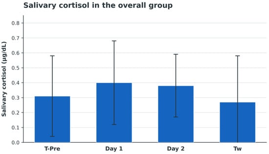
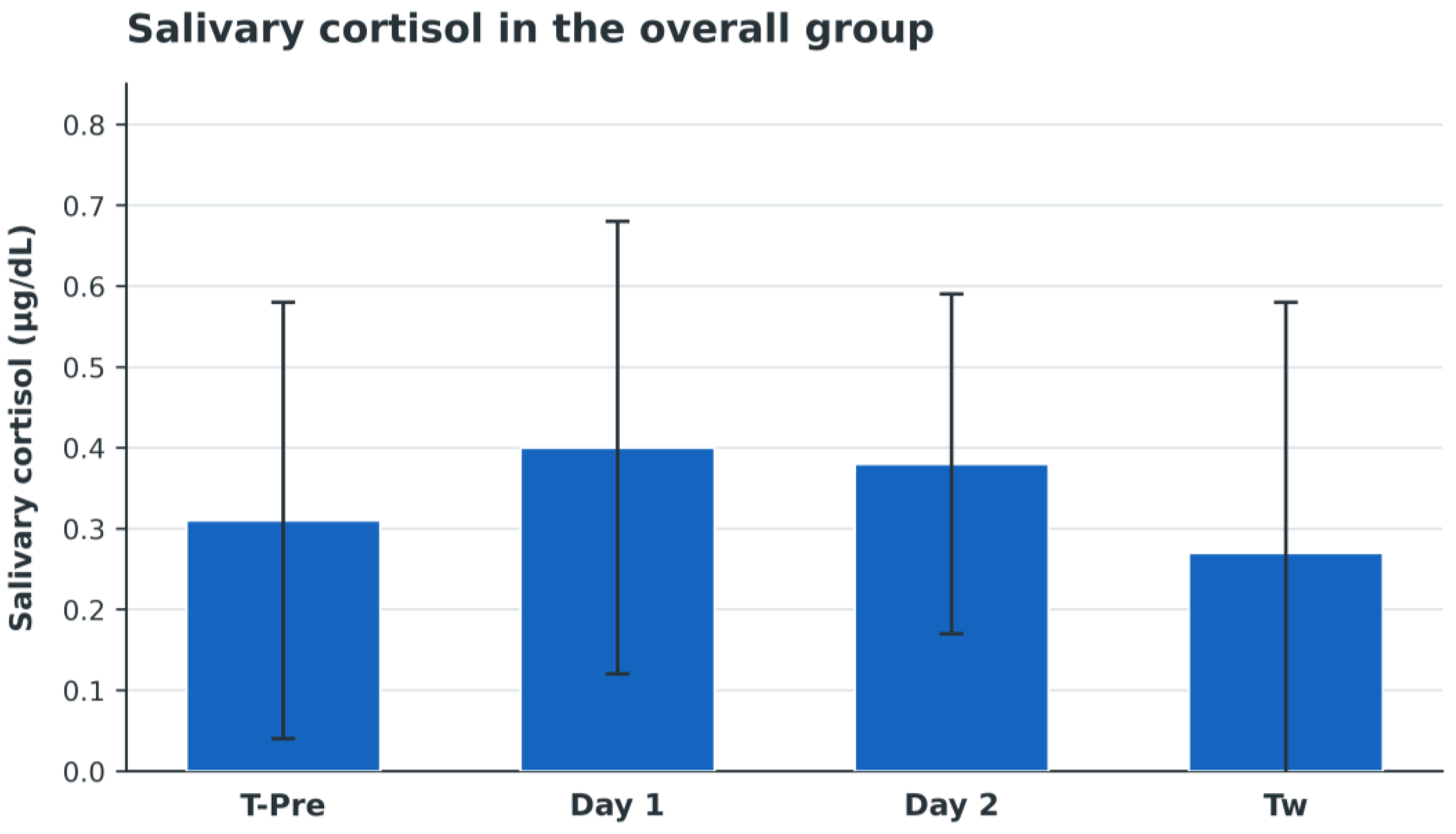
Four cats were excluded from the cortisol analysis because complete saliva samples could not be obtained at all planned sampling times. Cortisol was detectable in all samples from the remaining 16 cats. For the overall group, mean salivary cortisol concentration was 0.31 ± 0.27 μg/dL at T-Pre, 0.40 ± 0.28 μg/dL on Day 1, 0.38 ± 0.21 μg/dL on Day 2, and 0.27 ± 0.31 μg/dL during the washout period (Tw). No significant overall temporal change was detected (p > 0.05; Figure 3).
Figure 3.
Mean ± SD salivary cortisol concentrations in cats with complete measurements (n = 16) at T-Pre, Day 1, Day 2, and Tw. No significant overall temporal change was detected (p > 0.05). SD, standard deviation; Tw, washout period.
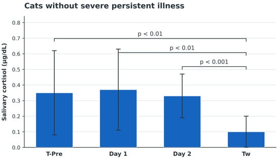
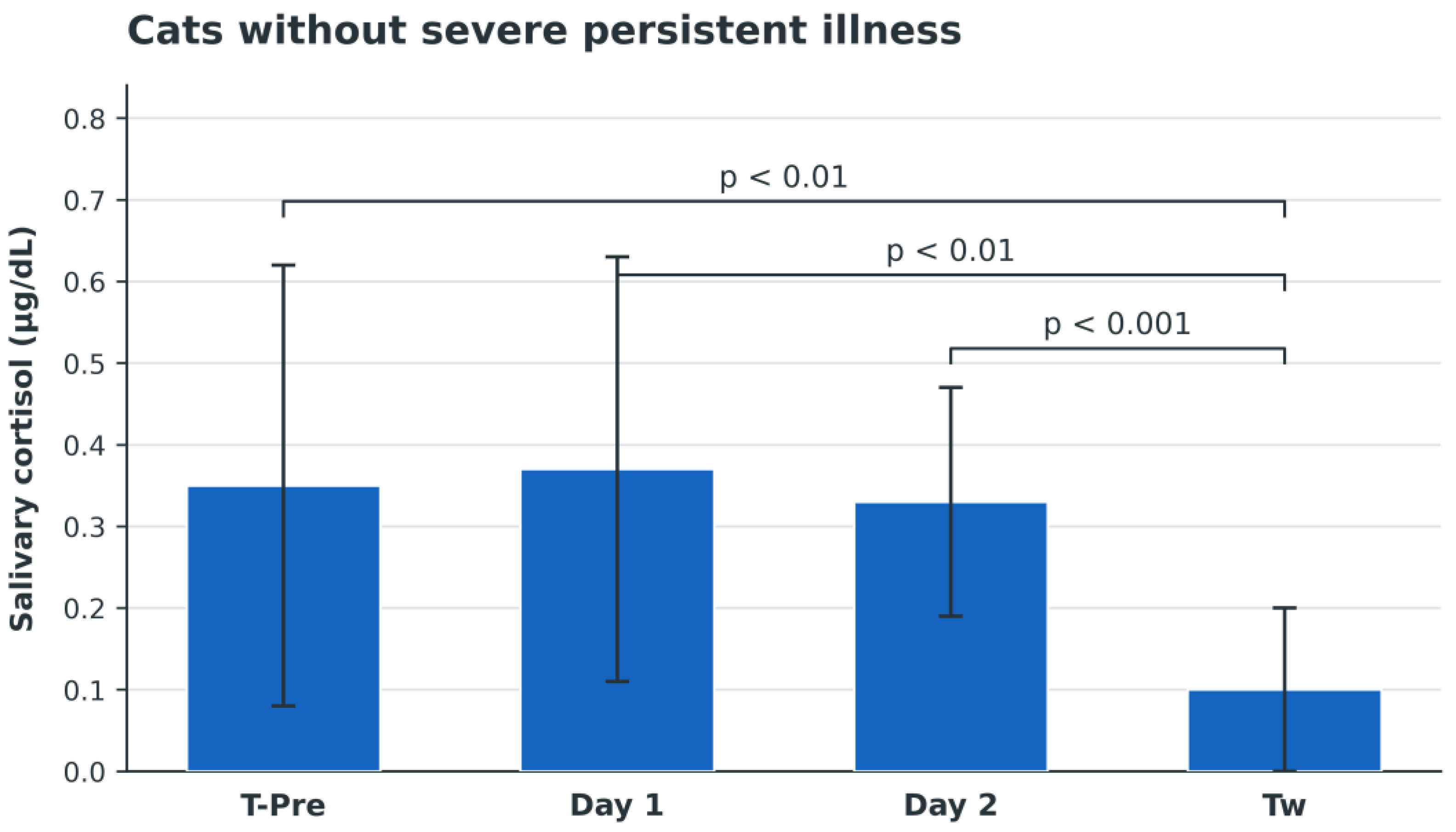
Among the 16 cats with complete cortisol measurements, 10 were retrospectively classified as not having severe persistent illness and six as having severe persistent illness. In cats without severe persistent illness, mean cortisol concentration was 0.35 ± 0.27 μg/dL at T-Pre, 0.37 ± 0.26 μg/dL on Day 1, 0.33 ± 0.14 μg/dL on Day 2, and 0.10 ± 0.10 μg/dL at Tw. Cortisol concentration was lower at Tw than at T-Pre and Day 1 (both p < 0.01) and Day 2 (p < 0.001; Figure 4).
Figure 4.
Mean ± SD salivary cortisol concentrations in cats without severe persistent illness (n = 10). Cortisol was lower at Tw than at T-Pre and Day 1 (both p < 0.01) and Day 2 (p < 0.001). SD, standard deviation; Tw, washout period.
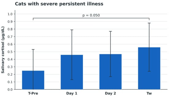
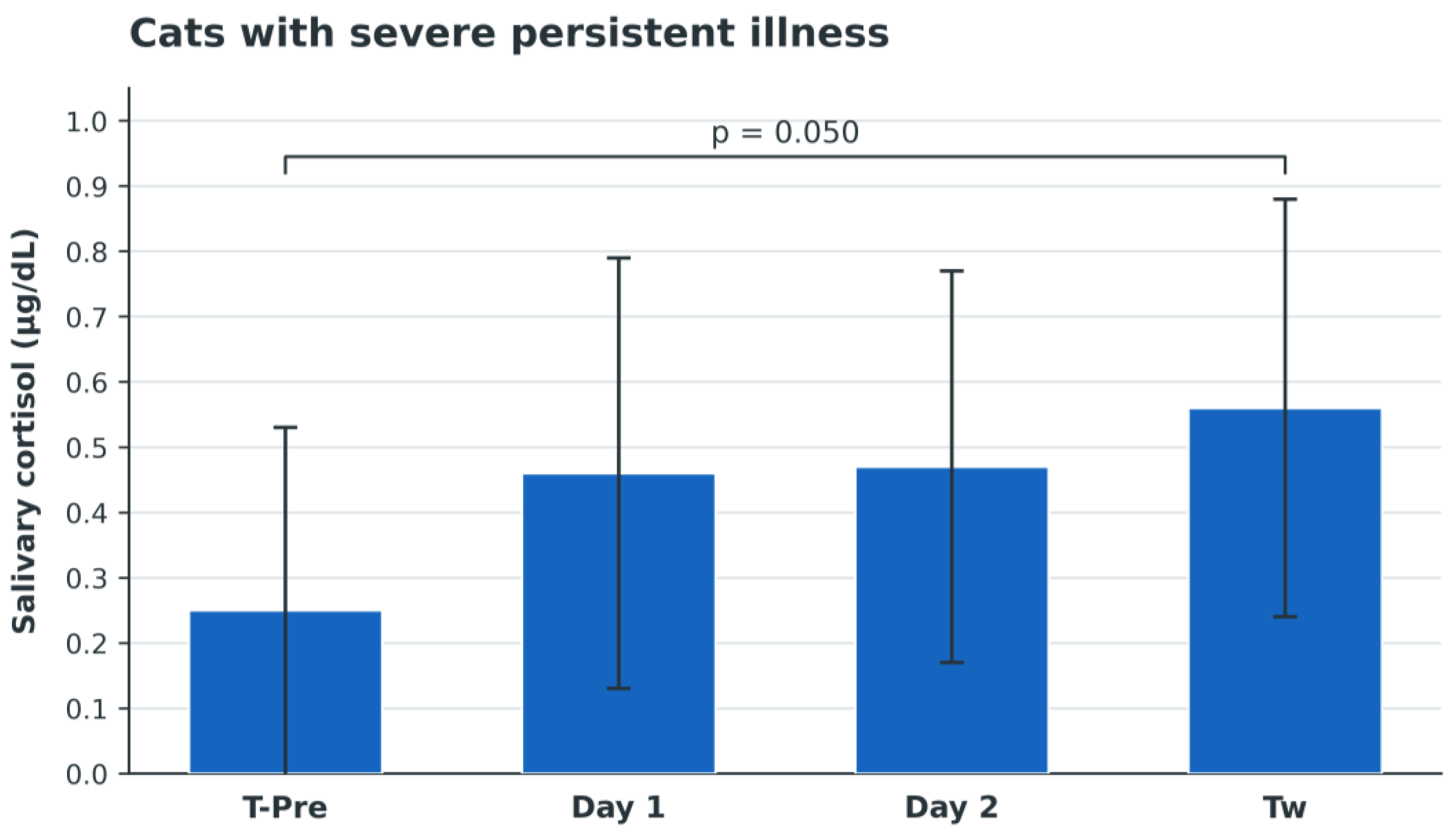
In cats with severe persistent illness, mean cortisol concentration was 0.25 ± 0.28 μg/dL at T-Pre, 0.46 ± 0.33 μg/dL on Day 1, 0.47 ± 0.30 μg/dL on Day 2, and 0.56 ± 0.32 μg/dL at Tw. The comparison between T-Pre and Tw yielded p = 0.050 (Figure 5).
Figure 5.
Mean ± SD salivary cortisol concentrations in cats with severe persistent illness (n = 6). The T-Pre–Tw comparison yielded p = 0.050. SD, standard deviation; Tw, washout period.
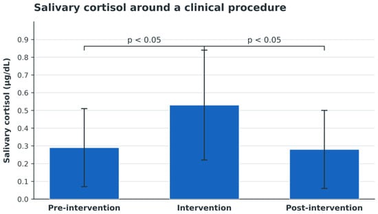
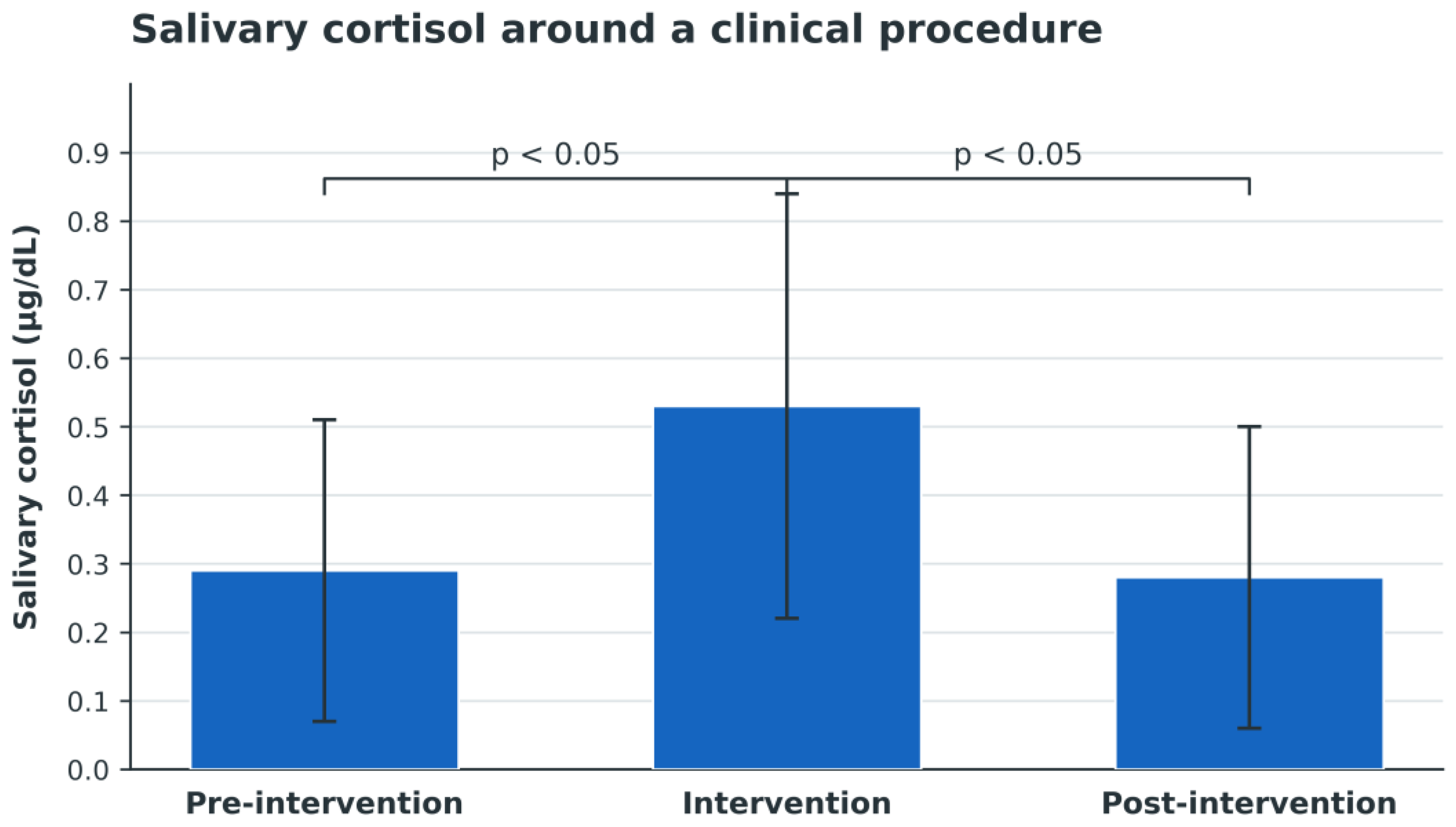
In the subset of 10 cats evaluated in relation to a clinical procedure, including urinary catheterisation or radiographic examination, cortisol concentration increased during the intervention compared with the pre-intervention measurement and subsequently decreased after the intervention (both p < 0.05; Figure 6).
Figure 6.
Mean ± SD salivary cortisol concentrations before, during, and after a clinical procedure (n = 10). Cortisol increased from pre-intervention to intervention and decreased after intervention (both p < 0.05). SD, standard deviation.
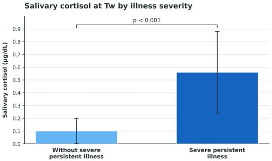
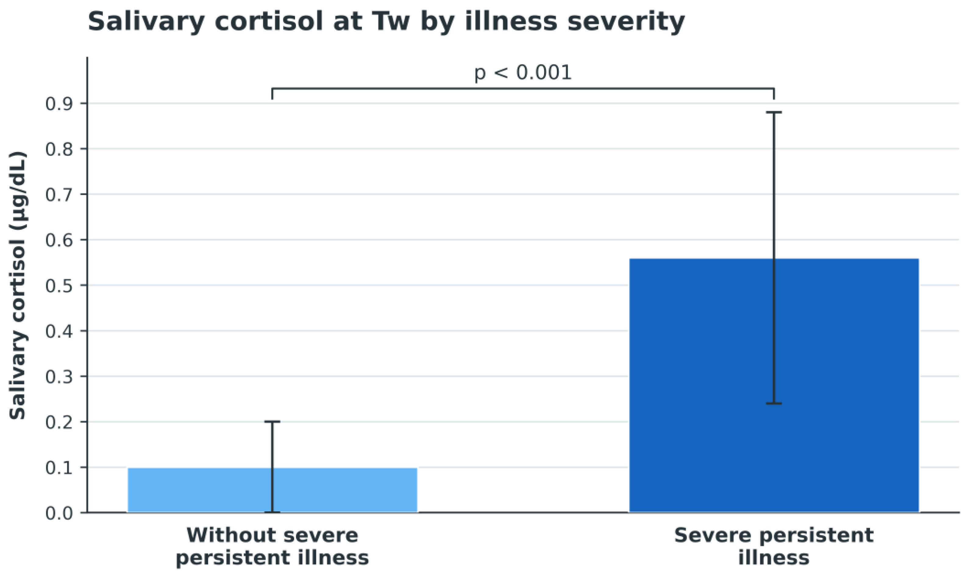
At Tw, cats with severe persistent illness had a higher mean cortisol concentration than cats without severe persistent illness (0.56 ± 0.32 versus 0.10 ± 0.10 μg/dL; p < 0.001; Figure 7).
Figure 7.
Mean ± SD salivary cortisol concentrations at Tw in cats with severe persistent illness (n = 6) and cats without severe persistent illness (n = 10). The groups differed at Tw (p < 0.001). SD, standard deviation; Tw, washout period.
4. Discussion
This prospective, exploratory, uncontrolled study characterised temporal changes in clinical scores, physiological variables, rescue analgesia, and salivary cortisol during and after repeated oral pregabalin administration in hospitalised feral cats. FGS and DSS decreased over time, while GSS increased modestly during treatment. HR, RR, and salivary cortisol in the overall group did not show significant temporal effects. These findings describe associations across the study period and do not establish pregabalin efficacy.
The progressive reductions in DSS and FGS scores observed during hospitalisation are compatible with less defensive behaviour and lower expression of pain at the evaluated time points. Although pregabalin may have contributed to these changes, the absence of a comparator group prevents their attribution to the drug alone. Clinical recovery, concomitant anti-inflammatory and rescue analgesic treatments, repeated handling, habituation to the hospital environment, and increasing familiarity with the operators may also have influenced both scores. These findings should therefore be regarded as temporal associations rather than evidence of a direct causal effect of pregabalin.
FGS and DSS measure different clinical domains. FGS quantifies pain-related facial expression at the time of observation and does not determine whether the underlying condition is acute, persistent, or chronic [19]. DSS describes the cat’s response to approach and handling [20]. Their reductions may therefore reflect different combinations of analgesia, clinical recovery, acclimatisation, and altered interaction with the operators. The significant increase in DSS from T36 to T48 and T60 was not accompanied by corresponding changes in FGS or GSS. Thus, the three scales did not demonstrate a consistent off–on–off pattern or evidence of a cumulative effect at T36.
Rescue methadone use was lower at every post-baseline assessment than at T-Pre, but the number of treated cats varied across time and was not analysed as an independent outcome. Eight cats received rescue analgesia at baseline, so opioid exposure could have influenced subsequent measurements despite the minimum 4 h delay. In addition, complete per-cat schedules for all concomitant anti-inflammatory and other medical treatments were not available for modelling. The FGS results and rescue-analgesia counts should consequently be interpreted together and not as independent evidence of a pregabalin-specific analgesic effect.
GSS shifted from a negative baseline mean to mildly positive values, peaking at T6. This pattern indicates modest sedation rather than a decrease in sedation. Previous studies of single-dose pregabalin in cats have also reported sedation alongside reduced fear or anxiety [17,18]. In the present setting, GSS alone could not distinguish pharmacological sedation from reduced hypervigilance or increasing tolerance of familiar operators. Although assessments were delayed until clinical recovery from propofol, a residual effect on later behavioural or sedation scores cannot be excluded.
Neither HR nor RR showed a significant overall effect of time. The available measurements were incomplete, particularly for HR, and the complete-case analysis was therefore based on only eight cats. These findings do not support a consistent temporal change in these physiological variables, but the limited sample restricts precision. Previous work has examined physiological and echocardiographic variables after oral pregabalin in healthy cats [23]; the present results should not be directly equated with that controlled healthy-cat setting.
No adverse events were recorded. Nevertheless, the study was not powered to establish safety, and its four-day observation period could not identify uncommon or delayed effects. Adverse events during pregabalin treatment have been described in other clinical populations [24], so the absence of observed events in this cohort should be interpreted cautiously.
Salivary cortisol was selected because it permits repeated, minimally invasive sampling and reflects the unbound cortisol fraction at the time of collection [9,22,25,26]. Published values in cats vary across populations and study conditions [27,28], which is consistent with the marked dispersion observed here. No significant temporal effect was detected in the overall group, suggesting that any common pattern was small relative to the heterogeneity between cats.
The procedure-related analysis showed a transient increase in cortisol during clinical intervention, followed by a decrease afterwards. This pattern supports the sensitivity of the measure to short-lived clinical stressors. It also illustrates that procedures, handling, and sampling context could influence cortisol independently of pregabalin or disease progression.
In the exploratory post hoc subgroup analysis, cats without severe persistent illness had lower cortisol at Tw, whereas cats with severe persistent illness had higher values and differed from the other subgroup at Tw. These findings are hypothesis-generating. The classification was retrospective, the subgroup sizes were small, and no validated severity scale or prospectively defined threshold was used. Disease severity, systemic involvement, clinical evolution, concomitant treatment, and procedures may all have contributed. Because only salivary cortisol was measured, the results do not establish broader modulation of HPA-axis activity.
The principal limitations were the absence of an untreated or placebo comparator, the small and clinically heterogeneous sample, incomplete measurements, non-standardised concomitant treatment, rescue analgesia, and possible effects of propofol, repeated handling, hospital acclimatisation, clinical recovery, and operator familiarity. Baseline assessment within 24 h of admission may also have captured peak novelty stress. Behavioural scores are additionally subject to individual variation and observer judgement. Complete individual concomitant-treatment schedules were not available, and rescue analgesia was not modelled as a time-varying covariate. These constraints preclude causal inference and limit generalisability.
Future randomised controlled studies should include an appropriate comparator, larger samples, standardised concomitant treatment and rescue-analgesia protocols, prospectively defined illness-severity criteria, blinded assessment, and prespecified repeated-measures models. Such studies are required to separate a pregabalin effect from recovery and acclimatisation and to determine whether repeated administration has a clinically useful role in hospitalised feral cats.
5. Conclusions
In this prospective, exploratory, uncontrolled cohort, pain-related facial expression, demeanour, and sedation scores changed over time during and after repeated oral pregabalin administration in hospitalised feral cats, whereas no significant temporal changes were detected in HR, RR, or salivary cortisol concentrations in the overall population. Because clinical recovery, acclimatisation, repeated handling, concomitant treatments, rescue analgesia, and clinical procedures may also have influenced the measured outcomes, these findings do not establish a causal effect or the clinical efficacy of pregabalin. The post hoc cortisol subgroup findings should be considered hypothesis-generating. Although no adverse events were recorded, the sample size and follow-up duration preclude definitive conclusions regarding safety. Randomised controlled studies with larger samples, standardised concomitant treatments, and prospectively defined clinical-severity criteria are required to determine the contribution of pregabalin and its potential clinical role in this population.
Author Contributions
Conceptualization, F.S.; methodology, F.S. and C.P.; software, F.S.; validation, M.V.L., C.P., C.B., M.P., F.G., M.A. and F.S.; formal analysis, M.V.L., C.P. and M.A.; investigation, M.V.L., C.P., C.B., M.P., M.A. and F.S.; resources, M.V.L., C.P., C.B., M.P., F.G. and M.A.; data curation, M.V.L., C.P., C.B., M.P., F.G. and M.A.; writing—original draft preparation, M.V.L.; writing—review and editing, C.P., C.B., F.G., M.A. and F.S.; visualization, M.V.L.; supervision, F.S.; project administration, F.S.; funding acquisition, F.S. All authors have read and agreed to the published version of the manuscript.
Funding
Orion Corporation provided financial support for the study and supplied the pregabalin product used.
Institutional Review Board Statement
The study was approved by the Comitato Etico per gli Studi Clinici e Zootecnici DIMEPRE-J, University of Bari Aldo Moro, under protocol number 2001-5 III/13, dated 21 December 2023.
Informed Consent Statement
Written authorisation for enrolment in the study and for the associated veterinary procedures and treatments was obtained from the managers responsible for the respective feline colonies.
Data Availability Statement
The original contributions presented in this study are included in the article. Further inquiries can be directed to the corresponding author.
Acknowledgments
The authors have reviewed and edited the output and take full responsibility for the content of this publication.
Conflicts of Interest
Orion Corporation provided financial support for the study and supplied the pregabalin product used. However, the company had no role in the study design; collection, analysis, or interpretation of the data; preparation or revision of the manuscript; or the decision to submit the results for publication. None of the authors received personal payments, honoraria, consultancy fees, or other personal benefits from Orion.
Abbreviations
The following abbreviations are used in this manuscript:
| CI | Confidence interval |
| DSS | Demeanor Scoring System |
| FGS | Feline Grimace Scale |
| GABA | Gamma-aminobutyric acid |
| GLM | General linear model |
| GSS | Global Sedation Score |
| HPA | Hypothalamic–pituitary–adrenal |
| HR | Heart rate |
| RR | Respiratory rate |
| SD | Standard deviation |
| T-Pre | Baseline assessment performed within 24 h before T0 |
| T4–T60 | Hours after the first pregabalin administration |
| Tw | Washout-period salivary cortisol assessment |
References
- Turner, D.C.; Bateson, P.P.G. The Domestic Cat: The Biology of Its Behaviour; Cambridge University Press: Cambridge, UK; New York, NY, USA, 2000. [Google Scholar]
- Miller, J. The domestic cat: Perspective on the nature and diversity of cats. J. Am. Vet. Med. Assoc. 1996, 208, 498–502. [Google Scholar] [CrossRef] [Scilit]
- Martínez-Byer, S.; Hudson, R.; Bánszegi, O.; Szenczi, P. Effects of early social separation on the behaviour of kittens of the domestic cat. Appl. Anim. Behav. Sci. 2023, 259, 105849. [Google Scholar] [CrossRef] [Scilit]
- Griffin, B.; Hume, K.R. Recognition and management of stress in housed cats. Consult. Feline Intern. Med. 2006, 5, 717–734. [Google Scholar] [CrossRef] [Scilit]
- Turner, D.C. The Mechanics of Social Interactions Between Cats and Their Owners. Front. Vet. Sci. 2021, 8, 650143. [Google Scholar] [CrossRef] [Scilit]
- Mariti, C.; Guerrini, F.; Vallini, V.; Bowen, J.E.; Fatjó, J.; Diverio, S.; Sighieri, C.; Gazzano, A. The perception of cat stress by Italian owners. J. Vet. Behav. 2017, 20, 74–81. [Google Scholar] [CrossRef] [Scilit]
- Kinlein, S.A.; Wilson, C.D.; Karatsoreos, I.N. Dysregulated hypothalamic–pituitary–adrenal axis function contributes to altered endocrine and neurobehavioral responses to acute stress. Front. Psychiatry 2015, 6, 31. [Google Scholar] [CrossRef] [Scilit]
- Buffington, C.A.T.; Bain, M. Stress and Feline Health. Vet. Clin. N. Am. Small Anim. Pract. 2020, 50, 653–662. [Google Scholar] [CrossRef] [Scilit]
- Cook, N.J. Review: Minimally invasive sampling media and the measurement of corticosteroids as biomarkers of stress in animals. Can. J. Anim. Sci. 2012, 92, 227–259. [Google Scholar] [CrossRef] [Scilit]
- Klintip, W.; Jarudecha, T.; Rattanatumhi, K.; Ritchoo, S.; Muikaew, R.; Wangsud, S.; Sussadee, M. First study on stress evaluation and reduction in hospitalized cats after neutering surgery. Vet. World 2022, 15, 2111–2118. [Google Scholar] [CrossRef] [Scilit]
- Rodan, I.; Dowgray, N.; Carney, H.C.; Carozza, E.; Ellis, S.L.; Heath, S.; Niel, L.; Denis, K.S.; Taylor, S. 2022 AAFP/ISFM Cat Friendly Veterinary Interaction Guidelines: Approach and Handling Techniques. J. Feline Med. Surg. 2022, 24, 1093–1132. [Google Scholar] [CrossRef] [Scilit]
- Kry, K.; Casey, R. The effect of hiding enrichment on stress levels and behaviour of domestic cats (Felis sylvestris catus) in a shelter setting and the implications for adoption potential. Anim. Welf. 2007, 16, 375–383. [Google Scholar] [CrossRef] [Scilit]
- Rusbridge, C. Neuropathic pain in cats: Mechanisms and multimodal management. J. Feline Med. Surg. 2024, 26, 1098612X241246518. [Google Scholar] [CrossRef] [Scilit]
- Goodman, C.W.; Brett, A.S. Gabapentin and Pregabalin for Pain—Is Increased Prescribing a Cause for Concern? N. Engl. J. Med. 2017, 377, 411–414. [Google Scholar] [CrossRef] [Scilit]
- Alles, S.R.A.; Cain, S.M.; Snutch, T.P. Pregabalin as a pain therapeutic: Beyond calcium channels. Front. Cell. Neurosci. 2020, 14, 83. [Google Scholar] [CrossRef] [Scilit]
- Micó, J.A.; Prieto, R. Elucidating the mechanism of action of pregabalin: α2δ as a therapeutic target in anxiety. CNS Drugs 2012, 26, 637–648. [Google Scholar]
- Lamminen, T.; Korpivaara, M.; Aspegrén, J.; Palestrini, C.; Overall, K.L. Pregabalin alleviates anxiety and fear in cats during transportation and veterinary visits—A clinical field study. Animals 2023, 13, 371. [Google Scholar] [CrossRef] [Scilit]
- Lamminen, T.; Korpivaara, M.; Suokko, M.; Aspegrén, J.; Palestrini, C.; Overall, K. Efficacy of a single dose of pregabalin on signs of anxiety in cats during transportation—A pilot study. Front. Vet. Sci. 2021, 8, 711816. [Google Scholar] [CrossRef] [Scilit]
- Evangelista, M.C.; Watanabe, R.; Leung, V.S.Y.; Monteiro, B.P.; O’Toole, E.; Pang, D.S.J.; Steagall, P.V. Facial expressions of pain in cats: The development and validation of a Feline Grimace Scale. Sci. Rep. 2019, 9, 19128. [Google Scholar] [CrossRef] [Scilit]
- Zeiler, G.E.; Fosgate, G.T.; Van Vollenhoven, E.; Rioja, E. Assessment of behavioural changes in domestic cats during short-term hospitalisation. J. Feline Med. Surg. 2014, 16, 499–503. [Google Scholar] [CrossRef] [Scilit]
- Pankratz, K.E.; Ferris, K.K.; Griffith, E.H.; Sherman, B.L. Use of single-dose oral gabapentin to attenuate fear responses in cage-trap confined community cats: A double-blind, placebo-controlled field trial. J. Feline Med. Surg. 2018, 20, 535–543. [Google Scholar] [CrossRef] [Scilit]
- Contreras, E.T.; Vanderstichel, R.; Hovenga, C.; Lappin, M.R. Evaluation of hair and nail cortisol concentrations and associations with behavioral, physical, and environmental indicators of chronic stress in cats. J. Vet. Intern. Med. 2021, 35, 2662–2672. [Google Scholar] [CrossRef] [Scilit]
- Li, M.; Wu, Y.; Chen, H.; Xu, X.; Peng, H.; Wei, B.; Zhu, Y.; Yang, Z. Effect of oral administration of pregabalin on physiological and echocardiographic variables in healthy cats. J. Feline Med. Surg. 2024, 26, 1098612X241250245. [Google Scholar] [CrossRef] [Scilit]
- Kanbayashi, Y.; Onishi, K.; Hosokawa, T. Factors predicting adverse events associated with pregabalin administered for neuropathic pain relief. Pain Res. Manag. 2014, 19, e164–e167. [Google Scholar] [CrossRef] [Scilit]
- Hernandez, C.E.; Thierfelder, T.; Svennersten-Sjaunja, K.; Berg, C.; Orihuela, A.; Lidfors, L. Time lag between peak concentrations of plasma and salivary cortisol following a stressful procedure in dairy cattle. Acta Vet. Scand. 2014, 56, 61. [Google Scholar] [CrossRef] [Scilit]
- King, J.A.; Rosal, M.C.; Ma, Y.; Reed, G.; Kelly, T.-A.; Stanek, E.J., III; Ockene, I.S. Sequence and seasonal effects of salivary cortisol. Behav. Med. 2000, 26, 67–73. [Google Scholar] [CrossRef] [Scilit]
- Siegford, J.M.; Walshaw, S.O.; Brunner, P.; Zanella, A.J. Validation of a temperament test for domestic cats. Anthrozoös 2003, 16, 332–351. [Google Scholar] [CrossRef] [Scilit]
- Silva, B.P.L.; Knackfuss, F.B.; Labarthe, N.; Mendes-de-Almeida, F. Effect of a synthetic analogue of the feline facial pheromone on salivary cortisol levels in the domestic cat. Pesqui. Vet. Bras. 2017, 37, 287–290. [Google Scholar] [CrossRef] [Scilit]

Disclaimer/Publisher’s Note: The statements, opinions and data contained in all publications are solely those of the individual author(s) and contributor(s) and not of MDPI and/or the editor(s). MDPI and/or the editor(s) disclaim responsibility for any injury to people or property resulting from any ideas, methods, instructions or products referred to in the content. |
© 2026 by the authors. Licensee MDPI, Basel, Switzerland. This article is an open access article distributed under the terms and conditions of the Creative Commons Attribution (CC BY) license.