


Abstract
Background: Chronic wounds that fail to heal despite debridement and advanced care are an unmet clinical need, particularly in elderly patients and those with peripheral arterial disease, for whom the alternatives are often indefinite maintenance or amputation. The mesenchymal stromal cell (MSC) secretome is pro-reparative in preclinical models, but human evidence is concentrated on shorter-duration diabetic foot ulcers and has usually reported area reduction rather than complete closure. Methods: We describe two patients with long-standing, treatment-refractory wounds treated with a cell-free Wharton’s jelly-derived MSC secretome delivered in a thermoresponsive polymeric carrier that gels at the wound bed, applied every three days alongside standard care. Results: The cases comprise the following: Case 1, a 95-year-old woman with peripheral arterial disease and a two-year ankle ulcer refractory to multiple advanced products; and Case 2, a 69-year-old man with a one-year neuro-ischemic diabetic foot ulcer persisting after amputation and repeated debridement. Both achieved complete closure at approximately 27 and 28 weeks, respectively, from baseline wound areas of approximately 7.5 cm2 (Case 1) and 12 cm2 (Case 2), using approximately 20 mL of preparation each. Closure was sustained at the latest follow-up (27 March 2026, Case 1; 21 June 2026, Case 2). Treatment was well tolerated; Case 2 had an intercurrent infection requiring a brief pause. Conclusions: A two-patient series cannot establish efficacy, but complete closure in wounds this refractory supports controlled evaluation of this cell-free platform, with complete closure as the primary endpoint.
1. Introduction
Chronic wounds are among the most stubbornly unsolved problems in clinical medicine. Defined by their failure to progress through the ordered phases of repair, they persist for months or years, and their principal forms—diabetic foot ulcers, vascular ulcers, and pressure injuries—share a common endpoint despite differing origins: a wound bed locked in a self-sustaining state of inflammation, impaired angiogenesis, and arrested re-epithelialization [1]. The clinical consequences are severe, extending to infection, hospitalization, and limb loss, and the economic burden is measured in tens of billions of dollars annually in the United States alone [2,3]. An aging population with rising rates of diabetes and peripheral arterial disease ensures that this burden continues to grow.
Development of therapeutic intervention has been incremental rather than transformative. Debridement, offloading, moisture balance, and infection control remain the foundation of care, and the advanced modalities layered on top—bioengineered skin substitutes, extracellular matrix scaffolds, topical oxygen, negative-pressure therapy, and growth factor preparations—improve outcomes for some patients but leave a substantial minority unhealed [4]. This refractory group is clinically distinct: for wounds that remain open after every available intervention has been tried, in patients whose age or vascular status makes surgical reconstruction unattractive or impossible, the realistic alternatives become indefinite maintenance or amputation [5].
Mesenchymal stromal cells (MSCs) have long been proposed as a way out of this impasse, and it is now widely accepted that their reparative influence is exerted largely through paracrine signaling rather than engraftment and differentiation [6]. Cells transplanted into a hostile wound bed survive poorly, yet still accelerate healing—an observation that has shifted attention decisively from the cells themselves to what they secrete [7]. The MSC secretome, a complex mixture of growth factors, chemokines, matrix modulators and extracellular vesicles (EVs), reproduces the parent cells’ activity while dispensing with many challenges associated with their handling [8,9]. As a cell-free material, it can, in principle, be characterized, stored, and standardized, and it carries no risk of uncontrolled proliferation or ectopic differentiation; such preparations have accordingly been proposed to reduce the cold-chain infrastructure and specialist handling that have kept living-cell therapies confined to well-resourced centers [8,9]. Among available sources, umbilical cord Wharton’s jelly is particularly attractive: the tissue is obtained non-invasively from routinely discarded material which can be ethically donated, and the cells derived from it are developmentally young, expand robustly, and secrete a factor profile weighted towards angiogenesis and matrix remodeling [10].
The preclinical evidence is now extensive and consistent. Across cell-based assays, three-dimensional skin equivalents, and rodent wound models, MSC-derived secretome and EV fractions, including exosomes, accelerate re-epithelialization, promote angiogenesis, recruit fibroblasts, and hasten resolution of inflammation [11,12,13]. Translation into patients, however, remains at an early stage. The available human evidence, including randomized studies of Wharton’s jelly-derived EVs and early-phase trials of conditioned media, has focused almost exclusively on diabetic foot ulcers, typically of relatively short duration, and has most often reported percentage reduction in wound area rather than complete closure [14,15]. Purified EV fractions have dominated, despite the cost and complexity of their manufacture and evidence that the broader secretome retains the full complement of soluble mediators [16]. Little has been published on the wounds that matter most clinically: those open for a year or more, already refractory to surgery and to multiple advanced products, and in patients with significant arterial disease or at the extremes of age. A further, often neglected obstacle is delivery. A low-viscosity liquid preparation (e.g., culture medium) applied to an exuding, irregular wound bed is quickly lost, and sustained contact between the secretome and the wound surface is a prerequisite for any significant biological effect.
Conventional wound-delivery formats address this only partially. Simple dressings and aqueous or semi-solid vehicles either wash out of an exuding, contoured wound bed or must be reapplied so frequently that sustained exposure is difficult to maintain, while preformed hydrogel sheets conform poorly to irregular cavities and undermined margins [17]. Thermoresponsive (in situ gelling) polymers offer a way around this trade-off: applied cold as a low-viscosity liquid, they flow into and coat an irregular wound surface and then gel at skin and body temperature to form a conformal depot that resists displacement and holds a payload in prolonged contact with the tissue [18]. Poloxamer-based thermoresponsive carriers are among the best-characterized systems of this class and have an established record as biocompatible vehicles for topical and wound applications [18]. Coupling a whole-secretome biologic to such a carrier—the approach whose preclinical development we described previously [11]—is a rational strategy to convert a transiently applied liquid into a retained, wound-contacting reservoir, and provides the design rationale for the platform reported here.
Here, we address these gaps directly. We report two patients—one aged 95 with a post-traumatic ankle ulcer complicated by peripheral arterial disease and open for two years despite a sequence of advanced wound care products, the other aged 69 with a neuro-ischemic diabetic foot ulcer persisting for a year after amputation and repeated debridement—in whom a cell-free Wharton’s jelly-derived MSC secretome, formulated in a thermoresponsive polymeric carrier to maintain contact with the wound bed, was applied as adjunctive compassionate treatment under the care of the treating physician. Both wounds, having failed all prior therapy, went on to close completely during the treatment period, without treatment-related adverse events. We present these two cases as an initial clinical proof-of-concept for this engineered, cell-free platform (a whole-secretome biologic coupled to a thermoresponsive delivery carrier) in wounds that had exhausted the available standard of care.
2. Materials and Methods
2.1. Study Design and Setting
This is a retrospective description of two patients with chronic, non-healing wounds treated with a cell-free mesenchymal stromal cell secretome preparation, delivered in a thermoresponsive polymeric carrier, as individualized compassionate clinical management. Both patients were managed by the treating physician (P.K.), a specialist in podiatric medicine and surgery, at the Foot Clinic and Wound Care service, Vimut-Theptarin Hospital, Bangkok, Thailand. In each case, the secretome preparation was offered as an adjunct to continued standard of care after the wound had failed to progress under conventional treatment; no randomization, control group, or research-specific procedures were involved, and all clinical decisions were made by the treating physician in the patient’s interest. Data were extracted retrospectively from de-identified clinical records and serial clinical photographs. The clinical cases are reported in accordance with the CARE (CAse REport) reporting guidelines [19].
2.2. Secretome Preparation and Quality Control
The material used was a cell-free mesenchymal stromal cell secretome: a concentrated preparation derived from the serum-free conditioned medium of human umbilical cord Wharton’s jelly-derived mesenchymal stromal cells, produced by prolonged, high-density culture. The harvested conditioned medium was fractionated and concentrated 50-fold by ultrafiltration, with only the retentate retained, as previously described [11]. Throughout this manuscript, “secretome” refers to this concentrated retentate preparation; “conditioned medium” refers to the unconcentrated culture supernatant from which it is derived; and “extracellular vesicles (EVs)” denotes the vesicular fraction retained within the secretome, of which exosomes are one subtype. For topical application, the minimally processed and concentrated secretome was incorporated into a poloxamer-based thermoresponsive polymeric carrier—the same class of carrier prepared from the same poloxamer reagent and used in the foundational preclinical work [11]. In aqueous solution, the carrier is a free-flowing liquid at low temperature, permitting mixing, loading and delivery, and undergoes a reversible sol–gel transition as it warms towards skin and body temperature to form a soft in situ gel, thereby retaining the secretome in sustained contact with the wound bed following application; its thermoreversible behavior and formulation have been described and characterized previously [11]. Poloxamer is a well-established, biocompatible polymeric carrier widely used as a pharmaceutical excipient and incorporated into approved topical and wound-care products [18]. The reagent used here was of research (cell-culture) grade rather than a pharmacopeial excipient lot, and the finished secretome–carrier preparation was applied on a compassionate, named-patient basis rather than under a marketing authorization for wound management. The specific carrier composition and concentration are proprietary to the manufacturer and are not disclosed here; the general formulation approach and the concentrations evaluated during preclinical development are reported in the foundational study [11].
The secretome used for both patients was derived from a single master cell bank (one umbilical cord donor). The finished secretome–carrier preparation was supplied as two lots produced to identical materials, protocols and quality-control specifications; each patient received approximately equal quantities from both lots, and the consistent healing trajectory across the treatment period gave no indication of lot-to-lot variation in performance.
The preparation used in these cases was of research grade and was supplied as a finished product by Celligenics Pte Ltd, Singapore; its individual formulation components are proprietary to the manufacturer and are not separately disclosed. All starting materials, culture media, and carrier components were obtained as sterile, certified reagents supplied with certificates of analysis. Sterility of each batch was verified in-house prior to clinical use: test material was incubated in sterile culture medium (DMEM; Gibco, Thermo Fisher Scientific, Waltham, MA, USA) for 72 h and examined microscopically for microbial growth, assessed as the appearance of particulates or turbidity, alongside a negative (medium-only) control. Absence of detectable particulates at 72 h was required for release, and any detectable particulates rendered the batch unsuitable for use. This screening was applied to the secretome, the carrier, and the final combined preparation. No positive growth control was included, and no compendial sterility or bacterial endotoxin testing was performed; endotoxin was not separately assayed, on the basis that all inputs were supplied sterile and no microbial growth was detected at any stage. These limitations are addressed in Section 4.3.
2.3. Treatment Protocol
Following debridement, the secretome–carrier preparation was applied at approximately 3-day intervals, timed to the routine dressing-change schedule and to the expected in situ residence of the gelled carrier, so as to maintain sustained exposure with minimal disturbance of the wound bed. It was dripped from above as a thin covering layer, without the syringe tip contacting the wound. The per-application volume could not be precisely measured but was estimated at up to ~0.8 mL (≈16 drops) on the larger, earlier wounds, falling to ~0.1–0.2 mL (2–4 drops) as the wounds approached closure, and was determined by wound size at the treating physician’s discretion. Approximately 20 mL was used per wound over the full course, supplied in re-sealable 5 mL screw-cap syringes (BD; Becton, Dickinson and Company, Franklin Lakes, NJ, USA). The carrier’s slight viscosity in its liquid state facilitated application relative to an aqueous-conditioned medium, and a non-absorbent secondary dressing was applied over the preparation to protect the applied layer and prolong its retention at the wound surface. Standard wound care continued throughout, comprising periodic debridement, moisture-balancing dressings, application of a barrier preparation to the periwound skin, and offloading or pressure management as clinically appropriate. Treatment continued until complete wound closure. The number of applications and total treatment duration for each patient are given in Section 3 and Table 1.
Table 1.
Patient characteristics, treatment parameters, and outcomes.
2.4. Wound Assessment and Outcomes
Wounds were assessed at each clinical visit by the treating physician. Wound dimensions (length × width × depth, cm) were recorded using a disposable paper wound ruler (unbranded), and wound-bed characteristics (granulation, slough or necrosis, exudate, and epithelialization) were documented and supported by serial clinical photography. The primary outcome was complete wound closure, defined as full re-epithelialization of the wound surface with no residual open area and no drainage, as determined by the treating physician [20]. Given the descriptive nature of a two-patient series, no formal statistical analysis was performed.
2.5. Photographic Documentation and Figure Preparation
Serial clinical photographs were obtained during routine wound care by the treating physician using standard digital photography. Images were acquired without standardized lighting, fixed camera distance, or in-frame scale reference, and are therefore presented as qualitative documentation of wound progression; all quantitative wound dimensions reported in this manuscript were obtained by direct clinical measurement (Section 2.4) rather than from photographs.
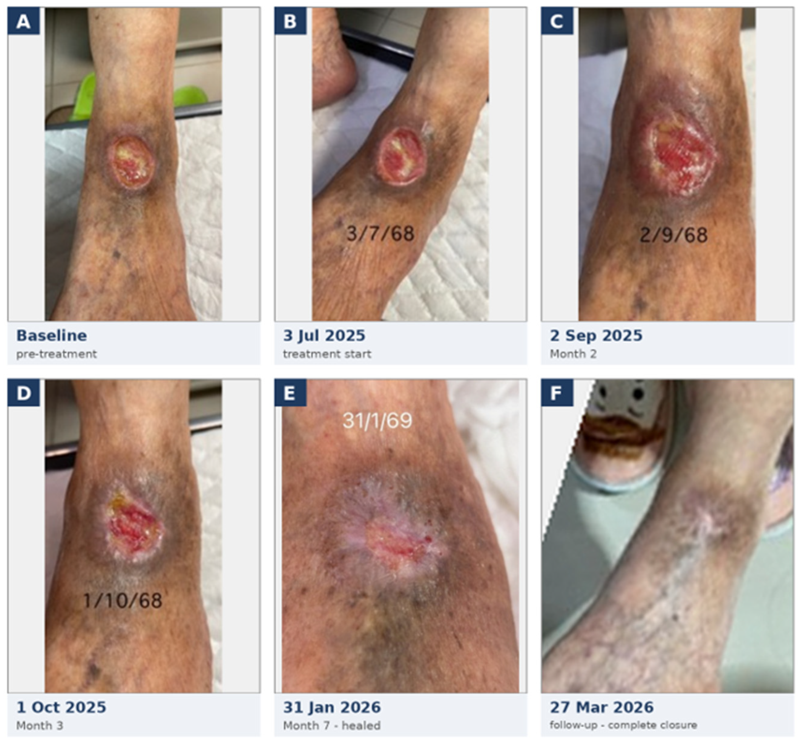
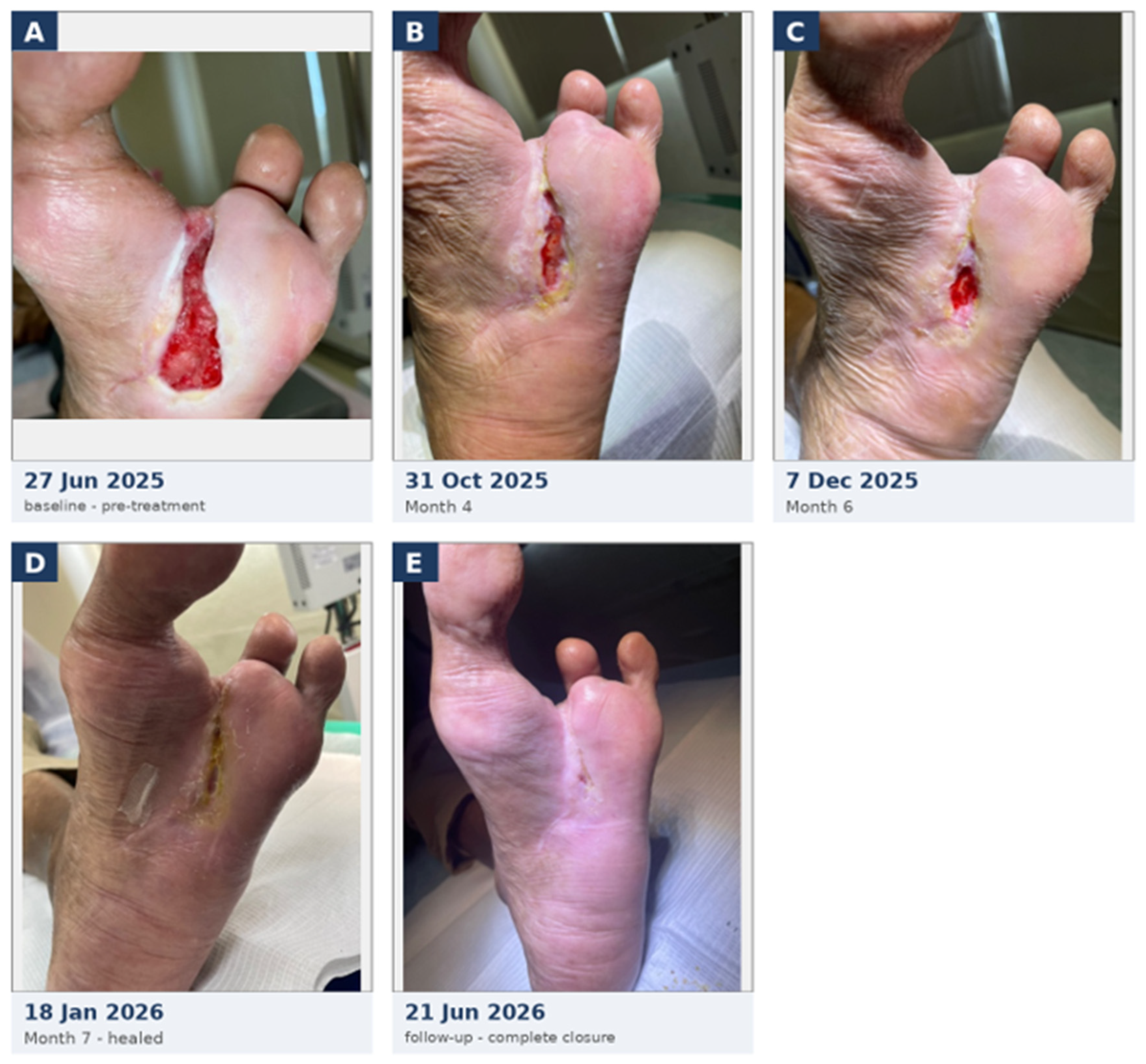
Figure 1 and Figure 2 were assembled from unaltered original photographs. An AI assistant (Claude Opus 4.8; Anthropic, San Francisco, CA, USA; https://www.anthropic.com, accessed on 20 August 2026) was used to support panel assembly, cropping, framing and the addition of panel letters and date labels. In Figure 1, panel F was rotated and cropped to approximate the viewing distance of the other panels. No image content was generated, enhanced, retouched or otherwise modified, and no adjustments were made to color, contrast, or wound appearance. All panel assignments and dates were verified against the source clinical records by the treating physician (P.K.).
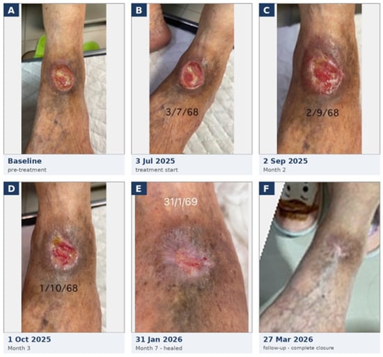
Figure 1.
Progressive healing of a refractory peripheral arterial disease (PAD)-associated ulcer of the left ankle in a 95-year-old female, treated topically with cell-free Wharton’s jelly-derived mesenchymal stromal cell secretome in a thermoresponsive polymeric carrier. (A) Baseline, pre-treatment: chronic full-thickness ulcer with a sloughy, poorly granulating bed. (B) 3 July 2025, at commencement of treatment. (C) 2 September 2025 (~2 months): established granulation with margin contraction. (D) 1 October 2025 (~3 months): marked area reduction with healthy granulation. (E) 31 January 2026: complete closure sustained at follow-up (closure first documented 8 January 2026). (F) 27 March 2026: further follow-up confirming durable, complete closure. Images are de-identified and cropped to the wound; panel F has been reframed and rotated to approximate the viewing distance of the other panels (wound dimensions in Table 1).
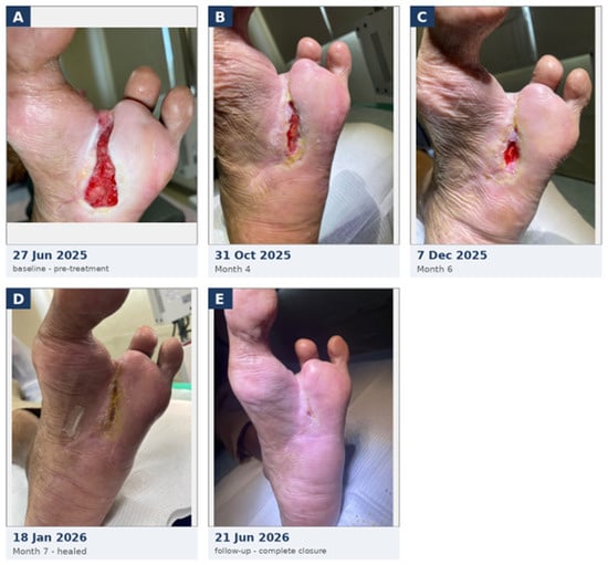
Figure 2.
Progressive healing of a refractory neuro-ischemic diabetic foot ulcer with peripheral arterial disease (PAD) at the plantar forefoot in a 69-year-old male, treated topically with cell-free Wharton’s jelly-derived mesenchymal stromal cell secretome in a thermoresponsive polymeric carrier. The wound had remained refractory for approximately one year despite prior amputation, repeated surgical debridement, intravenous antibiotics and advanced local wound care. (A) 27 June 2025, prior to treatment: open, debrided plantar forefoot wound with a fully granulating base; secretome treatment commenced on 4 July 2025. (B) 31 October 2025 (Month 4): granulation with margin contraction and resolution of the full-thickness undermining. (C) 7 December 2025 (Month 6): substantial area reduction with healthy granulation and circumferential epithelialization. (D) 18 January 2026 (Month 7): complete closure, with thin epithelial cover over the wound surface. (E) 21 June 2026: follow-up confirming durable, complete closure. Images are de-identified and cropped to the wound (wound dimensions in Table 1).
3. Results
3.1. Case 1: Refractory Chronic Ankle Ulcer in a 95-Year-Old Patient with Peripheral Arterial Disease
A 95-year-old woman with a background of peripheral arterial disease, maintained on clopidogrel and low-dose aspirin for secondary prevention of thrombotic events, presented with a chronic ulcer on the anterior aspect of the left ankle. The wound originated in October 2023 after the patient scratched an insect bite, which subsequently became infected, and had remained open for approximately two years. Prior management had included surgical debridement and a succession of advanced wound care modalities—topical oxygen therapy, Granulox (topical hemoglobin spray), a protease-modulating dressing, Endoform (ovine forestomach extracellular-matrix dressing), and Kerecis (omega-3 acellular fish-skin matrix). The wound had initially approached closure but subsequently deteriorated over the preceding seven months despite consistent standard of care, and was therefore considered refractory to available treatments.
The ulcer had reached a maximal recorded size of 3.8 × 4.0 × 0.2 cm. At baseline assessment on 31 May 2025, immediately prior to treatment, it measured 2.5 × 3.0 × 0.2 cm (an approximate wound area of 7.5 cm2, calculated as the length × width product of ruler measurements rather than by planimetry). The wound bed showed a mixed fibrotic and granulating base in an approximate 70:30 ratio, with a rolled wound border and hyperpigmented surrounding skin. There was no probing to bone, no undermining, and no tunneling. Doppler assessment of the left leg demonstrated strong but monophasic waveforms at the dorsalis pedis and posterior tibial arteries, with a biphasic signal at the popliteal artery, consistent with peripheral arterial disease.
Treatment with the secretome preparation began on 3 July 2025 and was applied every 3 days. A total of four syringes (approximately 20 mL) were used over the treatment course. Adjunctive care comprised weekly debridement for the first three months, thereafter reduced to bi-weekly debridement, performed carefully so as to avoid bleeding, together with application of a skin barrier preparation around the wound margin to protect the patient’s fragile periwound skin.
The wound responded progressively. By week 5, it had reduced to 2.0 × 2.0 × 0.1 cm (~4.0 cm2, a ~47% reduction from baseline), with a softer granulating base covering more than 90% of the wound surface and resolution of the rolled border. By week 9, the wound measured 1.3 × 1.8 × 0.1 cm (~2.3 cm2, a ~69% reduction), with near-complete granulation coverage and epithelialization advancing from the 12 to 9 o’clock positions. At week 14, the wound measured 1.2 × 1.5 × 0.1 cm (~1.8 cm2, a ~76% reduction) with a fully granulating base and circumferential epithelialization.
Complete wound closure, with a thin epithelial cover over the entire wound surface, was documented on 8 January 2026. Sustained closure was confirmed at follow-up assessments on 31 January 2026 and 27 March 2026, with no evidence of recurrence or breakdown (Figure 1). Total time to complete closure was approximately 27 weeks. No adverse events, wound infection, or treatment-related complications occurred during the treatment period.
3.2. Case 2: Refractory Neuro-Ischemic Diabetic Foot Ulcer in a 69-Year-Old Patient
A 69-year-old man with type 2 diabetes mellitus complicated by peripheral neuropathy and peripheral arterial disease, maintained on low-dose aspirin, presented with a chronic neuro-ischemic diabetic foot ulcer on the plantar aspect of the forefoot, a weight-bearing location characteristically slow to heal. His diabetes was of more than 20 years’ duration, managed with oral hypoglycemic agents and dietary control. He had severe sensory neuropathy, with absent protective sensation on 10 g monofilament testing and impaired vibration perception on 128 Hz tuning-fork examination bilaterally and characteristically bore weight on the plantar ulcer without pain. The wound had been present since June 2024, approximately one year prior to treatment, and had persisted despite prior partial amputation of the second ray and third toe, repeated surgical debridement, a prolonged course of intravenous antibiotics, and advanced local wound care—initially Granulox applied topically and subsequently a protease-modulating collagen dressing with silver antimicrobial foam (Allevyn Ag)—which had resulted in near-complete granulation of the wound bed but not closure. PolyMem foam, a hydrophilic polyurethane matrix dressing, was subsequently applied to facilitate wound closure; however, complete closure was not achieved at that time.
The ulcer had reached a maximal recorded size of 3.0 × 6.5 × 0.7 cm. At baseline assessment on 27 June 2025, it measured 2.0 × 6.0 × 0.3 cm (an approximate wound area of 12 cm2), with the wound bed fully covered by granulation tissue (100%). Full-thickness undermining, previously extending approximately 2 cm (1 to 3 o’clock), had reduced to approximately 1 cm at baseline.
Treatment with the secretome preparation began on 4 July 2025 and was administered every 3 days, with the frequency reduced to weekly during the final month of treatment. In total, approximately four syringes (20 mL) were used. Adjunctive wound care consisted of bi-weekly debridement, cleansing with polyhexamethylene biguanide (PHMB) solution, intermittent offloading with custom-cut adhesive felt, and application of a non-adherent contact layer (UrgoTul) beneath a foam dressing with a second non-adherent wound-contact layer (Allevyn). During the second month of treatment, the wound became infected (first noted on 21 September 2025), and secretome application was temporarily suspended for approximately four weeks. Wound cultures grew Stenotrophomonas maltophilia and Staphylococcus aureus. Empirical clindamycin plus ciprofloxacin was given for one week, followed by culture-directed levofloxacin, to which both isolates were susceptible. During this interval, the dressing regimen was changed to a daily topical antibiotic ointment. Secretome treatment was resumed on 19 October 2025, following clinical resolution of the infection. The infection was not considered related to the preparation: S. maltophilia and S. aureus are common pathogens of chronic diabetic foot ulcers at weight-bearing sites, the preparation is acellular, no local hypersensitivity or reaction to it was observed at any time, and healing resumed and proceeded to complete closure once the same preparation was reintroduced.
Progressive contraction, granulation, and epithelialization were observed. By Month 4 (31 October 2025), the wound measured 0.5 × 2.3 × 0.1 cm (~1.2 cm2, a ~90% reduction from baseline), with a fully granulating base (100%), resolution of the full-thickness undermining, resolution of the epibole, and epithelialization advancing circumferentially. By Month 6 (7 December 2025), it measured 0.5 × 1.5 × 0.1 cm (~0.8 cm2, a ~94% reduction), with firm central granulation and epithelialization around the entire wound margin.
Complete wound closure, with thin epithelial cover, was documented on 18 January 2026 (Month 7; Figure 2), giving a total time to complete closure of approximately 28 weeks from treatment initiation. Closure was sustained at follow-up on 21 June 2026, approximately five months later, with the healed skin closely resembling the surrounding native skin and only a small residual callus. Aside from the second-month wound infection described above, no adverse events, local reactions, or treatment-related complications occurred.
3.3. Summary of Outcomes
Both wounds achieved complete closure. In both cases, the wounds had been present for one to two years and had failed to heal despite surgical debridement and, in Case 1, multiple advanced wound care products; in Case 2, the wound persisted despite prior amputation and treatment for infection. In Case 2, glycemic control was reasonably well maintained during the treatment period, with HbA1c ranging from 6.7% to 7.1%, and the ankle-brachial index (ABI) was 0.71, for which no additional invasive vascular intervention was required. Over the treatment period, both wounds showed progressive reduction in area and depth, granulation, and circumferential epithelialization, culminating in complete closure, in patients aged 95 and 69 years with significant vascular comorbidity. In Case 2, treatment was interrupted for approximately four weeks by an intercurrent wound infection before healing resumed. Closure was achieved with modest total volumes of the preparation (approximately 20 mL per case). No local reactions to the preparation occurred; the only intercurrent complication was the Case 2 wound infection noted above. Baseline characteristics, treatment parameters, and outcomes are summarized in Table 1.
4. Discussion
Two chronic wounds, each open for a year or more and each having failed the treatments conventionally offered to such patients, closed completely after introduction of a cell-free Wharton’s jelly-derived MSC secretome delivered in a thermoresponsive polymeric carrier. Both had already exhausted advanced wound care—in Case 1, multiple advanced products in a 95-year-old with peripheral arterial disease; in Case 2, amputation, repeated debridement, and intravenous antibiotics in a 69-year-old with diabetes, neuropathy, and arterial disease. In both, complete re-epithelialization was achieved over the course of treatment following introduction of the secretome preparation; in Case 2, an intercurrent second-month infection required a brief interruption before healing resumed to closure.
4.1. Relation to Existing Evidence
The clinical literature on MSC-derived secretome in wound healing, though expanding rapidly, remains narrow in a specific way. Most human studies have enrolled diabetic foot ulcers of relatively short standing, and the primary endpoint has commonly been percentage reduction in wound area over a defined observation window rather than complete closure [14,15]. Percentage area reduction is a defensible surrogate for a trial, but it is not what determines whether a patient keeps a limb. The cases reported here differ in three respects that we believe are clinically meaningful. First, both wounds were of long duration and demonstrably refractory: each had already exhausted the interventions that would ordinarily be offered, which substantially weakens the argument that closure reflected the natural history of an otherwise treatable ulcer. Second, the endpoint achieved was complete closure rather than partial improvement. Third, the patients sit at the difficult end of the clinical spectrum, one of them 95 years old with arterial insufficiency, the other with combined neuropathy and ischemia at a mechanically unfavorable site; precisely the populations underrepresented in existing trials, in which age limits, comorbidity exclusions, and ulcer duration criteria tend to select for wounds more likely to heal. To the best of our knowledge, based on the literature reviewed, complete closure has not previously been reported in chronic wounds of this duration and refractoriness treated with a cell-free MSC secretome.
Our observations also bear on which secretome fraction should be used clinically. Much of the recent translational literature has favored purified EVs or exosomes on the premise that they contain the principal therapeutic cargo. Yet vesicle isolation is costly, technically demanding, and difficult to scale [21], and it discards the soluble fraction of growth factors, cytokines, chemokines, and matrix modulators, which are also biologically active. The material used here was the whole secretome, retaining both the EV fractions as well as the majority of soluble protein factors.
It should be noted that “whole secretome” does not mean unprocessed conditioned medium. The preparation is concentrated by ultrafiltration above a defined molecular-weight cut-off, and only the retentate is retained; the specific cut-off and the rationale for selection are proprietary to the manufacturer and are described in the foundational methods [11]. This step therefore functions as a coarse purification as well as concentration: low-molecular-weight species such as residual medium components, salts and other solutes, degraded peptides and nucleic acid fragments are largely removed with the permeate, while soluble protein factors and all EV classes are retained. Beyond simplifying the composition of the applied material, this passive depletion of small-molecule and degradation-derived species is a desirable feature for a preparation intended for repeated topical application to an open wound bed, in which the barrier to systemic exposure is compromised.
That two wounds of this severity closed under such a preparation is consistent with (though it cannot by itself confirm) a growing body of work suggesting that the whole secretome may be therapeutically sufficient for cutaneous repair, and that the soluble, non-EV fraction contributes to this activity in its own right rather than serving merely as a carrier and stabilizer for EVs [22,23]. Studies in which the EV fraction is selectively depleted from conditioned medium report attenuated but not abolished healing [23], the residual activity implicating the soluble mediators [24], while paracrine soluble factors alone recruit macrophages and endothelial lineage cells and accelerate wound closure [25]. The balance between EV and soluble contributions appears to be tissue-dependent; some injury models favor exosome-enriched fractions [26], but in skin, the available evidence supports retention of both compartments, which the whole-secretome preparation used here achieves. Whether this translates into superior clinical closure relative to a purified fraction remains a hypothesis these two cases cannot establish; a direct head-to-head comparison in a controlled setting would be required to test it.
4.2. Plausible Mechanisms
The biology offers a coherent account of what was observed. Chronic wounds are arrested rather than merely slow: the wound bed is held in a state of persistent M1-type inflammation, with excessive protease activity, degraded matrix, impaired angiogenesis, and fibroblasts and keratinocytes that are themselves functionally exhausted and poorly migratory [1]. A therapy that acts on a single node in this network—for example, supplying one growth factor or debriding necrotic tissue alone—tends to produce only transient improvement because the underlying pathology inhibits further progress. The MSC secretome acts across several of these nodes simultaneously [7]. Reported activities include promotion of endothelial proliferation and vessel formation [27], recruitment and activation of fibroblasts with subsequent collagen deposition [28], stimulation of keratinocyte proliferation and migration [28], extracellular matrix remodeling [7], and polarization of macrophages toward a reparative phenotype [29]. The clinical sequence we observed is consistent with this multi-target action: conversion of a fibrotic wound bed to healthy granulation tissue, resolution of the rolled wound edge in Case 1 (a hallmark of a stalled epidermal front) and subsequent circumferential epithelialization rather than closure from a single margin. None of these mechanisms was directly evaluated in the two patients reported here; they are drawn from prior preclinical and clinical work and are presented to frame the observed healing rather than to evidence a mechanism in these cases.
The polymeric carrier used for delivery merits comment. A secretome applied as a liquid to an exuding wound is rapidly diluted and lost, and biological activity presupposes sustained contact with the wound surface. A thermoresponsive carrier—liquid when cool, semi-solid at body temperature—allows the preparation to conform to an irregular wound bed, including undermined regions (as in Case 2), and to remain in place between dressing changes. Beyond retaining the secretome, the carrier confers the benefits long-recognized for occlusive and semi-occlusive dressings: it provides a physical barrier against contamination and maintains a moist wound environment, which supports keratinocyte migration and proliferation and is associated with more efficient healing than a dry wound bed [30]. Release from poloxamer gels of this type is well established: being physical, non-covalently crosslinked thermogels, they liberate entrapped payload predominantly by erosion and dissolution in an aqueous environment [18], progressively delivering the secretome to the wound bed. This was consistent with the clinical course: at each dressing change, the applied preparation had thinned relative to its appearance on application, without drying out, indicating in situ erosion and local release rather than absorption into the non-absorbent secondary dressing.
It is also plausible that sustained local delivery of the secretome contributes to the quality of the healed tissue; MSC secretome and conditioned medium have been reported to attenuate fibrosis and reduce hypertrophic and keloid scarring in preclinical models, including formulations combining conditioned medium with a hydrogel carrier [31,32,33]. Scar quality was not a formal endpoint in the present cases, and this remains a property observed in other settings rather than one these two cases establish. We therefore regard the delivery format as an integral part of the intervention rather than an inert accessory; the total volumes required to achieve closure were modest, at approximately 20 mL across the full treatment course for each wound.
4.3. Limitations
The constraints on what may be concluded from this report have been considered, and we want to state them plainly here. This is a retrospective series of two patients, without a control group, randomization, or blinding, and no causal inference may be claimed from such a design. Both patients continued to receive standard wound care throughout, including regular debridement and periwound skin protection, and the contribution of the secretome cannot be formally separated from that of the concurrent care, although it is highly relevant that both wounds had failed to heal under standard care, including debridement, for one to two years beforehand. In addition, because the secretome was delivered within a carrier, its biological contribution cannot be distinguished from any independent effect of the carrier itself, which, as an occlusive, moisture-retaining vehicle, may influence healing in its own right. Separating the contributions of the secretome, the carrier, and concurrent standard care would require a controlled design incorporating carrier-alone and secretome-plus-carrier arms. Spontaneous late healing cannot be formally excluded in any individual case; however unlikely the prior clinical course makes it.
Consistent with the compassionate, observational nature of this report, batch-specific characterization of the secretome (protein and extracellular-vesicle content, growth-factor profile, and potency) and in vitro release or histological analyses were not performed here; the composition and bioactivity of this secretome preparation are characterized in the foundational studies [11,34].
Because the preparation was research-grade and was not released against compendial specifications, parameters such as bioburden, endotoxin content and defined potency were not formally tested; residual endotoxin, if present, could in principle influence the local inflammatory response. Future controlled studies should apply predefined release specifications including sterility, endotoxin limits, and a quantitative potency assay to the finished product.
Wound measurement was performed with a disposable ruler at clinical visits rather than by digital image-based measurement of the wound area, and closure was determined by the treating physician’s assessment rather than by assessment from a blinded independent observer; both introduce measurement imprecision [35]. Although HbA1c and ABI were available for Case 2, objective vascular indices and glycemic control data were not consistently recorded across all cases, limiting full characterization of the wound environment. Follow-up after closure was limited but confirmed durability in both cases: Case 1 remained healed at reviews on 31 January and 27 March 2026, and Case 2 at review on 21 June 2026 (approximately five months after closure); durability beyond these points is not established.
Finally, the preparation used was of research grade. Release testing consisted of a 72 h in-house microbiological screen with a negative control; no positive growth control was included, and no compendial sterility or bacterial endotoxin testing was performed. This is an acknowledged limitation of the material as used in these compassionate cases and would require replacement by validated, compendial release testing before any wider or regulated clinical use.
4.4. Implications
Within these constraints, the observations are encouraging. Two wounds that had defeated the available standard of care closed completely, in patients for whom the realistic alternatives were indefinite wound maintenance or, in the second case, further tissue loss. The preparation was well tolerated, with no local reactions in either patient. Case 2’s course was complicated by a wound infection in the second month, a common event in diabetic foot ulcers [5], not considered related to the preparation and which resolved after a brief interruption of treatment, following which healing continued to completion. No other adverse events occurred. This tolerability is consistent with the safety profile expected of an acellular, allogeneic-source material [22,36]. What these cases justify is not a claim of efficacy but a case for proper evaluation: a prospective, controlled study in patients with long-standing, treatment-refractory wounds, using complete closure as the primary endpoint, objective wound-area measurement, and a fully characterized preparation manufactured under validated conditions. The population that stands to gain most is the one currently least studied, this being older patients with vascular disease and wounds that everything else has failed to heal.
A prospective, controlled study is warranted, ideally incorporating comparator arms (standard care alone, carrier alone, and secretome-plus-carrier) with blinded outcome assessment. Complete closure should serve as the primary endpoint, with time-to-closure and planimetric percentage area reduction as secondary measures, supported by standardized serial photography using a fixed-scale marker and defined lighting and distance, and by follow-up of sufficient duration to capture recurrence.
5. Conclusions
Adjunctive treatment with a cell-free Wharton’s jelly-derived mesenchymal stromal cell secretome delivered in a thermoresponsive polymeric carrier resulted in complete healing of two chronic wounds: a two-year ulcer in a 95-year-old woman with peripheral arterial disease and a one-year neuro-ischemic diabetic foot ulcer after amputation and repeated debridement. Both wounds progressed to full re-epithelialization with modest total volumes of the preparation and good overall tolerability, the only intercurrent complication being a wound infection in Case 2 that resolved with a brief interruption of treatment.
A two-patient retrospective series cannot establish efficacy, and we make no such claim. What these cases do provide is a clinically grounded rationale for formal evaluation: they demonstrate that complete closure, rather than partial area reduction, is achievable in wounds that had already defeated the available standard of care, in patients whose age and vascular status place them outside the populations typically enrolled in trials of advanced wound therapies. Beyond the clinical observations, the intervention illustrates an engineered, cell-free delivery format. As a class, cell-free secretome preparations offer potential practical advantages over living-cell therapies, including amenability to storage and standardization and, being cell-free, freedom from the tumorigenicity and ectopic-differentiation risks associated with living-cell therapies [8,9]. The preparation used here was supplied with a manufacturer-defined refrigerated shelf-life established through the supplier’s stability testing. Cell-free secretomes are also, in principle, simpler and more scalable to produce than purified EV or exosome fractions, and here, the secretome was retained at the wound bed by a poloxamer-based thermoresponsive carrier that addresses the retention problem limiting liquid secretome preparations. These observations provide preliminary clinical justification for further controlled investigation of this combination in settings where refractory wounds are managed. Prospective, controlled study in this refractory population, using complete closure as the primary endpoint, objective wound-area measurement, and a preparation manufactured and released under validated conditions, is warranted.
Author Contributions
Conceptualization, J.A.D.; methodology, J.A.D. and A.W.M.; investigation and clinical management, P.K.; data curation, A.W.M. and P.K.; writing—original draft preparation, J.A.D.; writing—review and editing, A.W.M. and P.K.; project administration, J.A.D. All authors have read and agreed to the published version of the manuscript.
Funding
This research received no external funding. The secretome preparation was purchased by Miskawaan Health Group.
Institutional Review Board Statement
Ethics committee review and approval were not required for this report. Both patients were treated within routine clinical care, with all treatment decisions made by the treating physician in each patient’s individual clinical interest, and the decision to report the outcomes was taken retrospectively, after treatment was complete, from a review of the two patients’ de-identified medical records. Under the standard operating procedures of the Central Ethics Committee on Human Research of the Thai Ministry of Public Health (CECOPS-MOPH; Version 2.0) and the Human Research Ethics Committee of the Institute of Chest Disease (1st Edition, 2020), retrospective case reports involving no more than three patients and using de-identified data do not constitute human subjects research and are exempt from ethics committee review; accordingly, no institutional review board or ethics committee approval was sought or required for this report. The study was conducted in accordance with the Declaration of Helsinki.
Informed Consent Statement
Both patients consented to the treatment as part of their routine clinical care, including written informed consent for the publication of their clinical details and images.
Data Availability Statement
The data supporting the findings of this study are contained within the article. De-identified clinical records are available from the corresponding author on reasonable request, subject to patient confidentiality.
Acknowledgments
The authors acknowledge Celligenics Pte Ltd., Singapore, as the source of the polymeric carrier incorporating Wharton’s jelly-derived mesenchymal stromal cell secretome used as the research material in these cases. During the preparation of this manuscript, the authors used an AI assistant (Claude Opus 4.8; Anthropic, San Francisco, CA, USA; https://www.anthropic.com, accessed on 20 August 2026) to assist with the assembly of Figure 1 and Figure 2, specifically the cropping, collation and panel labeling of clinical photographs. The authors also used Claude to help refine the wording of selected sentences for clarity, consistency, and readability, without generating new scientific content or altering the authors’ original ideas, analysis, or conclusions. No generative AI was used to create, enhance, or otherwise alter any text or image content. The authors reviewed and verified all outputs and take full responsibility for the content of this publication.
Conflicts of Interest
J.A.D. is Chief Scientific Officer of Miskawaan Health Group (MHG) and declares a commercial interest in the subject matter of this article. J.A.D. served as a consultant to Celligenics Pte Ltd., the supplier of the secretome preparation, until early 2024; that engagement has ended, and no commercial, data-sharing, or collaborative agreement exists between the authors, their institutions, and the supplier. J.A.D. is a co-author on prior published work relating to this class of material. The finished secretome–carrier product was manufactured and supplied by the commercial provider as a white-label preparation, sterile-sealed in primary packaging within well-sealed, temporary research-grade secondary packaging. MHG applied secondary labeling and packaging, prepared the accompanying instructions for use, and distributed the product to the treating physician without charge. The supplier had no role in patient selection, clinical management, data collection, analysis, or the decision to publish. A.W.M. is an employee of Miskawaan Health Group (MHG), Bangkok, Thailand, and contributed to this work in the course of that employment. P.K. is a podiatric physician and surgeon at Vimut-Theptarin Hospital, Bangkok, Thailand, where the patients were treated.
References
- Pastar, I.; Balukoff, N.C.; Marjanovic, J.; Chen, V.Y.; Stone, R.C.; Tomic-Canic, M. Molecular Pathophysiology of Chronic Wounds: Current State and Future Directions. Cold Spring Harb. Perspect. Biol. 2023, 15, a041243. [Google Scholar] [CrossRef] [Scilit]
- Sen, C.K. Human Wound and Its Burden: Updated 2025 Compendium of Estimates. Adv. Wound Care 2025, 14, 429–438. [Google Scholar] [CrossRef] [Scilit]
- Nussbaum, S.R.; Carter, M.J.; Fife, C.E.; DaVanzo, J.; Haught, R.; Nusgart, M.; Cartwright, D. An Economic Evaluation of the Impact, Cost, and Medicare Policy Implications of Chronic Nonhealing Wounds. Value Health J. Int. Soc. Pharmacoeconomics Outcomes Res. 2018, 21, 27–32. [Google Scholar] [CrossRef] [Scilit]
- Frykberg, R.G.; Banks, J. Challenges in the Treatment of Chronic Wounds. Adv. Wound Care 2015, 4, 560–582. [Google Scholar] [CrossRef] [Scilit]
- Armstrong, D.G.; Boulton, A.J.M.; Bus, S.A. Diabetic Foot Ulcers and Their Recurrence. N. Engl. J. Med. 2017, 376, 2367–2375. [Google Scholar] [CrossRef] [Scilit]
- Liang, X.; Ding, Y.; Zhang, Y.; Tse, H.-F.; Lian, Q. Paracrine Mechanisms of Mesenchymal Stem Cell-Based Therapy: Current Status and Perspectives. Cell Transplant. 2014, 23, 1045–1059. [Google Scholar] [CrossRef] [Scilit]
- Harrell, C.R.; Fellabaum, C.; Jovicic, N.; Djonov, V.; Arsenijevic, N.; Volarevic, V. Molecular Mechanisms Responsible for Therapeutic Potential of Mesenchymal Stem Cell-Derived Secretome. Cells 2019, 8, 467. [Google Scholar] [CrossRef] [Scilit]
- Dangerfield, J.A.; Metzner, C. Collecting Eggs, Not Killing Chickens: Why Stem Cell Secretome and Exosomes Are Redefining Regenerative Medicine for Healthspan Extension. Biomedicines 2026, 14, 854. [Google Scholar] [CrossRef] [Scilit]
- González-González, A.; García-Sánchez, D.; Dotta, M.; Rodríguez-Rey, J.C.; Pérez-Campo, F.M. Mesenchymal Stem Cells Secretome: The Cornerstone of Cell-Free Regenerative Medicine. World J. Stem Cells 2020, 12, 1529–1552. [Google Scholar] [CrossRef] [Scilit]
- Drobiova, H.; Sindhu, S.; Ahmad, R.; Haddad, D.; Al-Mulla, F.; Al Madhoun, A. Wharton’s Jelly Mesenchymal Stem Cells: A Concise Review of Their Secretome and Prospective Clinical Applications. Front. Cell Dev. Biol. 2023, 11, 1211217. [Google Scholar] [CrossRef] [Scilit]
- Chin, J.S.; Tan, M.L.L.; Lim, P.L.K.; Sharma, B.; Yeo, A.; Aw, Y.B.; Ng, Y.Z.; Bonnard, C.; Becker, D.L.; Mok, P. Secretome from Prolonged High-density Human Wharton’s Jelly Stem Cell Culture Accelerates Wound Healing in Both in Vitro and in Vivo Models. Int. Wound J. 2025, 22, e70033. [Google Scholar] [CrossRef] [Scilit]
- Han, Y.; Sun, T.; Han, Y.; Lin, L.; Liu, C.; Liu, J.; Yan, G.; Tao, R. Human Umbilical Cord Mesenchymal Stem Cells Implantation Accelerates Cutaneous Wound Healing in Diabetic Rats via the Wnt Signaling Pathway. Eur. J. Med. Res. 2019, 24, 10. [Google Scholar] [CrossRef] [Scilit]
- Liu, L.; Yu, Y.; Hou, Y.; Chai, J.; Duan, H.; Chu, W.; Zhang, H.; Hu, Q.; Du, J. Human Umbilical Cord Mesenchymal Stem Cells Transplantation Promotes Cutaneous Wound Healing of Severe Burned Rats. PLoS ONE 2014, 9, e88348. [Google Scholar] [CrossRef] [Scilit]
- Kishta, M.S.; Hafez, A.M.; Hydara, T.; Hamed, Z.; Bahr, M.M.; Shamaa, A.A.; Abdallah, A.N. The Transforming Role of Wharton’s Jelly Mesenchymal Stem Cell-Derived Exosomes for Diabetic Foot Ulcer Healing: A Randomized Controlled Clinical Trial. Stem Cell Res. Ther. 2025, 16, 559. [Google Scholar] [CrossRef] [Scilit]
- Tan, S.T.; Aisyah, P.B.; Firmansyah, Y.; Nathasia, N.; Budi, E.; Hendrawan, S. Effectiveness of Secretome from Human Umbilical Cord Mesenchymal Stem Cells in Gel (10% SM-hUCMSC Gel) for Chronic Wounds (Diabetic and Trophic Ulcer)—Phase 2 Clinical Trial. J. Multidiscip. Healthc. 2023, 16, 1763–1777. [Google Scholar] [CrossRef] [Scilit]
- Jafar, H.; Almousa, R.; Alhawari, H.; Shahin, D.; Al Soudi, M.; Abusharieh, E.; Aboalhaija, N.H.; Awidi, A. Human Umbilical Cord Mesenchymal Stromal Cells Derivatives in Treating Diabetic Foot Ulcers: A Phase I/II Safety and Efficacy Trial. Stem Cell Res. Ther. 2025, 16, 657. [Google Scholar] [CrossRef] [Scilit]
- Boateng, J.S.; Matthews, K.H.; Stevens, H.N.E.; Eccleston, G.M. Wound Healing Dressings and Drug Delivery Systems: A Review. J. Pharm. Sci. 2008, 97, 2892–2923. [Google Scholar] [CrossRef] [Scilit]
- Zarrintaj, P.; Ramsey, J.D.; Samadi, A.; Atoufi, Z.; Yazdi, M.K.; Ganjali, M.R.; Amirabad, L.M.; Zangene, E.; Farokhi, M.; Formela, K.; et al. Poloxamer: A Versatile Tri-Block Copolymer for Biomedical Applications. Acta Biomater. 2020, 110, 37–67. [Google Scholar] [CrossRef] [Scilit]
- Gagnier, J.J.; Kienle, G.; Altman, D.G.; Moher, D.; Sox, H.; Riley, D.; CARE Group. The CARE Guidelines: Consensus-Based Clinical Case Report Guideline Development. J. Clin. Epidemiol. 2014, 67, 46–51. [Google Scholar] [CrossRef] [Scilit]
- U.S. Food and Drug Administration. Guidance for Industry: Chronic Cutaneous Ulcer and Burn Wounds—Developing Products for Treatment; U.S. Department of Health and Human Services, Food and Drug Administration (CDER, CBER, CDRH): Rockville, MD, USA, 2006; pp. 258–268.
- Li, Z.; Yan, J.; Li, X.; Chen, H.; Lin, C.; Zhang, Y.; Gao, T.; Zhang, Y.; Shu, Y.; Pan, S.; et al. Advancements in Extracellular Vesicles Biomanufacturing: A Comprehensive Overview of Large-Scale Production and Clinical Research. Front. Bioeng. Biotechnol. 2025, 13, 1487627. [Google Scholar] [CrossRef] [Scilit]
- Vizoso, F.J.; Eiro, N.; Cid, S.; Schneider, J.; Perez-Fernandez, R. Mesenchymal Stem Cell Secretome: Toward Cell-Free Therapeutic Strategies in Regenerative Medicine. Int. J. Mol. Sci. 2017, 18, 1852. [Google Scholar] [CrossRef] [Scilit]
- Lei, R.; da Silva, T.B.; Cui, Z.; Ye, H. Small Extracellular Vesicles from Human Bone Marrow Mesenchymal Stromal Cells Enhance Migration and Regulate Reparative Gene Expression in Dermal Fibroblasts. Sci. Rep. 2025, 15, 19383. [Google Scholar] [CrossRef] [Scilit]
- An, S.; Anwar, K.; Ashraf, M.; Lee, H.; Jung, R.; Koganti, R.; Ghassemi, M.; Djalilian, A.R. Wound-Healing Effects of Mesenchymal Stromal Cell Secretome in the Cornea and the Role of Exosomes. Pharmaceutics 2023, 15, 1486. [Google Scholar] [CrossRef] [Scilit]
- Chen, L.; Tredget, E.E.; Wu, P.Y.G.; Wu, Y. Paracrine Factors of Mesenchymal Stem Cells Recruit Macrophages and Endothelial Lineage Cells and Enhance Wound Healing. PLoS ONE 2008, 3, e1886. [Google Scholar] [CrossRef] [Scilit]
- Damania, A.; Jaiman, D.; Teotia, A.K.; Kumar, A. Mesenchymal Stromal Cell-Derived Exosome-Rich Fractionated Secretome Confers a Hepatoprotective Effect in Liver Injury. Stem Cell Res. Ther. 2018, 9, 31. [Google Scholar] [CrossRef] [Scilit]
- Silveira, B.M.; Ribeiro, T.O.; Freitas, R.S.; Carreira, A.C.O.; Gonçalves, M.S.; Sogayar, M.; Meyer, R.; Birbrair, A.; Fortuna, V. Secretome from Human Adipose-Derived Mesenchymal Stem Cells Promotes Blood Vessel Formation and Pericyte Coverage in Experimental Skin Repair. PLoS ONE 2022, 17, e0277863. [Google Scholar] [CrossRef] [Scilit]
- Ferreira, A.d.F.; Cunha, P.d.S.; Carregal, V.M.; da Silva, P.d.C.; de Miranda, M.C.; Kunrath-Lima, M.; de Melo, M.I.A.; Faraco, C.C.F.; Barbosa, J.L.; Frezard, F.; et al. Extracellular Vesicles from Adipose-Derived Mesenchymal Stem/Stromal Cells Accelerate Migration and Activate AKT Pathway in Human Keratinocytes and Fibroblasts Independently of miR-205 Activity. Stem Cells Int. 2017, 2017, 9841035. [Google Scholar] [CrossRef] [Scilit]
- Ti, D.; Yi, J.; Chen, H.; Hao, H.; Shi, C. The Role of Mesenchymal Stem/Stromal Cells Secretome in Macrophage Polarization: Perspectives on Treating Inflammatory Diseases. Curr. Stem Cell Res. Ther. 2024, 19, 894–905. [Google Scholar] [CrossRef] [Scilit]
- Winter, G.D. Formation of the Scab and the Rate of Epithelization of Superficial Wounds in the Skin of the Young Domestic Pig. Nature 1962, 193, 293–294. [Google Scholar] [CrossRef] [Scilit]
- Hu, C.-H.; Tseng, Y.-W.; Chiou, C.-Y.; Lan, K.-C.; Chou, C.-H.; Tai, C.-S.; Huang, H.-D.; Hu, C.-W.; Liao, K.-H.; Chuang, S.-S.; et al. Bone Marrow Concentrate-Induced Mesenchymal Stem Cell Conditioned Medium Facilitates Wound Healing and Prevents Hypertrophic Scar Formation in a Rabbit Ear Model. Stem Cell Res. Ther. 2019, 10, 275. [Google Scholar] [CrossRef] [Scilit]
- Zhang, C.; Wang, T.; Zhang, L.; Chen, P.; Tang, S.; Chen, A.; Li, M.; Peng, G.; Gao, H.; Weng, H.; et al. Combination of Lyophilized Adipose-Derived Stem Cell Concentrated Conditioned Medium and Polysaccharide Hydrogel in the Inhibition of Hypertrophic Scarring. Stem Cell Res. Ther. 2021, 12, 23. [Google Scholar] [CrossRef] [Scilit]
- Zhong, Y.; Zhang, Y.; Yu, A.; Zhang, Z.; Deng, Z.; Xiong, K.; Wang, Q.; Zhang, J. Therapeutic Role of Exosomes and Conditioned Medium in Keloid and Hypertrophic Scar and Possible Mechanisms. Front. Physiol. 2023, 14, 1247734. [Google Scholar] [CrossRef] [Scilit]
- Aw, Y.B.; Chen, S.; Yeo, A.; Dangerfield, J.A.; Mok, P. Development and Functional Testing of a Novel in Vitro Delayed Scratch Closure Assay. Histochem. Cell Biol. 2024, 162, 245–255. [Google Scholar] [CrossRef] [Scilit]
- Langemo, D.; Anderson, J.; Hanson, D.; Hunter, S.; Thompson, P. Measuring Wound Length, Width, and Area: Which Technique? Adv. Skin Wound Care 2008, 21, 42–45; quiz 45–47. [Google Scholar] [CrossRef] [Scilit]
- Van Delen, M.; Derdelinckx, J.; Wouters, K.; Nelissen, I.; Cools, N. A Systematic Review and Meta-Analysis of Clinical Trials Assessing Safety and Efficacy of Human Extracellular Vesicle-Based Therapy. J. Extracell. Vesicles 2024, 13, e12458. [Google Scholar] [CrossRef] [Scilit]

Disclaimer/Publisher’s Note: The statements, opinions and data contained in all publications are solely those of the individual author(s) and contributor(s) and not of MDPI and/or the editor(s). MDPI and/or the editor(s) disclaim responsibility for any injury to people or property resulting from any ideas, methods, instructions or products referred to in the content. |
© 2026 by the authors. Licensee MDPI, Basel, Switzerland. This article is an open access article distributed under the terms and conditions of the Creative Commons Attribution (CC BY) license.