


Abstract
This study evaluated the physical and mechanical properties of acrylic resins used for interim fixed prostheses, with and without metal reinforcement, before and after aging. A total of 138 samples were divided into three groups: VIPI + Wire (control), VIPI, and Diamond D. Samples were assessed for microhardness, porosity, roughness, and flexural strength. Aging was simulated using 500 thermocycling cycles at 5 and 55 ± 1 °C. Data were analyzed using ANOVA and Tukey’s test. Group Diamond D did not fracture during flexural testing, but it exhibited significantly lower microhardness at both baseline and after aging. Before aging, Group Diamond D had higher roughness than Group VIPI, which exhibited greater porosity. Aging increased the microhardness of Group VIPI and the roughness of Group Diamond D. The percentage of porosity decreased significantly for Groups VIPI + Wire and VIPI, and pore size was reduced in all groups. Based on the results obtained from Diamond D material, this resin does not meet the required properties for the proposed indication for temporary fixed prostheses, whereas VIPI with reinforcement showed superior properties and greater stability after aging.
1. Introduction
To rehabilitate an edentulous patient, a conventional complete denture made of acrylic resin (polymethyl methacrylate) is indicated [1]. Another treatment alternative is the implant-supported fixed complete denture, which offers an excellent strategy for restoring esthetics, function, and mastication. Compared with conventional complete dentures, these prostheses offer greater stability and retention and provide greater satisfaction to wearers, as demonstrated in several clinical studies [2,3,4]. They are usually screwed in place, making them reversible, so they can be removed to retighten or replace screws, substitute fractured components, clean, and assess peri-implant tissues [5,6].
During the rehabilitation process with implant-supported fixed complete dentures, it may be necessary to use conventional dentures between implant placement and installation of the definitive prosthesis. This situation commonly occurs when not all implants can support immediate load after surgery, requiring 3 to 6 months for osseointegration [7]. However, implants with adequate length and initial bone stability can support a provisional implant-supported fixed denture for immediate loading, reestablishing esthetics and function, while allowing osseointegration of the remaining implants. This provides stability and comfort, supporting both esthetic and masticatory functions [8].
Implant-supported fixed provisional complete dentures are transitional prostheses used temporarily before definitive rehabilitation. They must have satisfactory strength and aesthetics and provide proper cleansability [9]. These prostheses are usually fabricated using an acrylic resin structure with artificial teeth. Additionally, they incorporate provisional prosthetic components (such as titanium cylinders) that support the prosthesis. Metallic reinforcements can be added to improve strength and stiffness [10]. Unlike the definitive prosthesis, which generally incorporates a metal bar cast into its internal structure, the metal reinforcement of the provisional prosthesis often uses materials such as thick orthodontic wire [11].
Due to high masticatory distress and accidental damage, fracture is a common complaint among denture wearers [12,13]. Provisional implant-supported fixed dentures have lower resistance to occlusal forces than definitive dentures due to the absence of metal internal reinforcement [14,15]. This reinforcement compensates for the low flexural strength and high fracture risk of PMMA-based prostheses [11,16]. To optimize fracture resistance, manufacturers can incorporate reinforcements such as copolymers or fibers [16]. In the dental market, one brand stands out due to its ultra-impact thermosetting acrylic resin composition. According to the manufacturer, this resin eliminates the need for metal reinforcements and meshes, thanks to its high impact strength and exceptional flexural strength [17,18,19].
During use, but especially during cleaning, the prosthesis is subjected to abrasion forces that may result in scratches and plastic deformation. Lower microhardness indicates a weaker surface, which is more susceptible to damage [20].
Microbial contamination can compromise the integrity of biological tissues (bone and soft tissue) surrounding osseointegrated implants [21]. Therefore, ensuring the prosthesis maintains a smooth surface, thereby reducing the space available for microbial deposits, is crucial for osseointegration. Surface porosity and roughness significantly influence smoothness.
Temperature variation and oral fluid absorption in denture polymers can affect the prosthesis’ physical properties [22]. Thermocycling is commonly used in vitro to simulate temperature changes in the mouth [23]. One of the most widely used techniques in scientific research is thermocycling, which simulates the extreme temperatures to which materials are exposed in the oral environment [24]. According to Carreira et al., materials tend to expand when heated and shrink when cooled. These temperature variations can generate mechanical stresses at the interface between the organic resin matrix and inorganic filler particles due to differences in their thermal expansion coefficients. This can lead to microleakages and fatigue fractures associated with the hydrolytic degradation of the polymer matrix and filler particles [24,25,26]. Aging and temperature variation can therefore influence resin properties [22,23].
Given the various reinforcement techniques available to strengthen PMMA-based resins and the different commercial brands on the market, this study compared the flexural strength, microhardness, roughness, and porosity of two PMMA-based resins. VIPI Cril Plus resin is widely used in dental practice and well-documented in the literature [27,28,29,30], making it the control in this study. The purpose was to determine whether Diamond D resin could be used as the base for a provisional implant-supported fixed full-arch denture without internal metallic reinforcement, as proposed by the manufacturer, while still providing adequate flexural strength and surface smoothness. The null hypothesis was that no differences would be observed between the resin groups, regardless of internal metal reinforcement or aging.
2. Materials and Methods
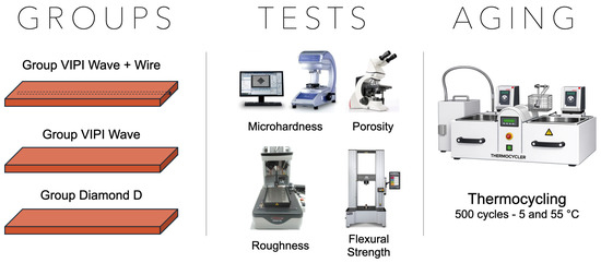
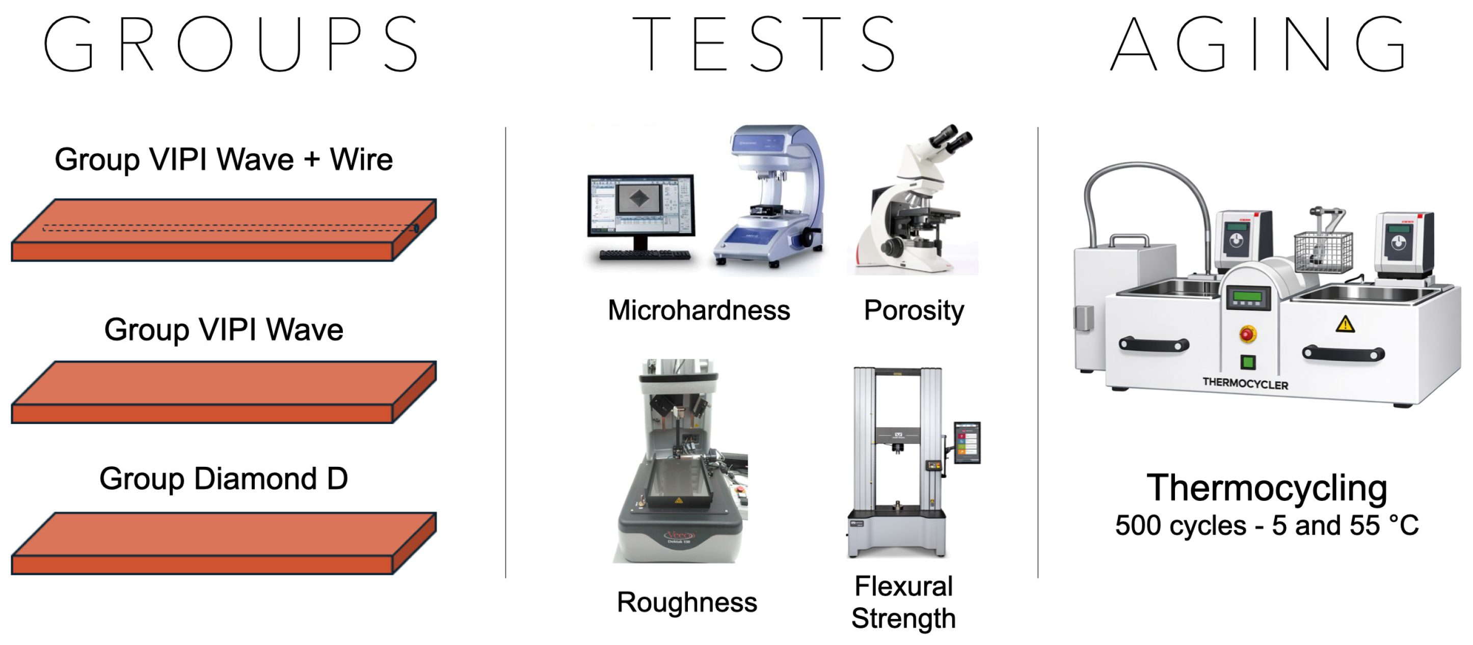
The experimental design of the present study involved the preparation of 138 acrylic resin samples, divided into 3 groups: Group VIPI + Wire (VIPI Cril Plus with metal reinforcement, control), Group VIPI (VIPI Cril Plus without reinforcement), and Group Diamond D (Diamond D without reinforcement). The VIPI + Wire was considered the control group, to replicate the clinical scenario, as these provisional implant-supported fixed full-arch dentures often need a metal reinforcement. The samples were evaluated for microhardness, porosity, roughness, and flexural strength before and after 500 thermocycling cycles (Figure 1). The sample size was determined based on a pilot study performed for roughness and flexural strength tests. A power analysis was conducted with a level of significance of 0.05, 80% power, and a medium effect size (GPower 3.1 software). The power analysis indicated that 8 samples were required for the roughness test and 15 for the flexural strength test. Thus, 10 and 18 samples were used, respectively. For roughness and porosity tests, the same samples were used before and after accelerated aging (n = 10/group). For microhardness and flexural strength analyses, half of the samples were tested before aging (n = 18/group), and the other half after aging (n = 18/group).
Figure 1.
Experimental workflow used in the study.
Samples were prepared according to a previously established protocol using a metal mold with dimensions of 67 mm in length, 12 mm in width, and 3 mm in height, in accordance with ANSI/ADA specification No. 12 [31]. VIPI Wave acrylic resin samples (VIPI® Indústria, Comércio, Exportação e Importação de Produtos Odontológicos, Pirassununga, Brazil) were prepared following the manufacturer’s instructions. For the VIPI Wave samples with orthodontic wires, a channel-shaped cavity was created with a Carbide spherical drill (KG Sorense, Cotia, Brazil) to insert a 0.8 mm metal wire (Morelli Ortodontia, Sorocaba, Brazil). After inserting the wire, additional resin was applied, and the samples were pressed again [32]. Polymerization was carried out in a microwave oven (Electrolux S/A, MEF41-31 L, 1000 Watts, Curitiba, Brazil) with an initial cycle at 20% power for 20 min and a final cycle at 60% power for 5 min [33].
For the Diamond D resin samples, preparation followed the manufacturer’s recommendations. Polymerization was performed in the same microwave at 90% power for 4 min. The flasks were cooled slowly in water for 24 h, after which the samples were removed and stored [34,35].
The specimens were finished with aluminum oxide water sandpaper (Erios Equipamentos LTDA, São Paulo, Brazil) in a decreasing grit sequence (600, 800, 1200, 1500, 4000) under refrigeration, using an Arotec Model APL-4 horizontal polisher (Arotec Indústria e Comércio Ltda, Cotia, Brazil). Polishing was carried out with felt disks (American Burrs, Palhoça, Brazil) and polishing paste (KOTA Indústria e Comércio LTDA, São Paulo, Brazil) [36]. Sample thickness was measured using a digital caliper (500-171-20B, Mitutoyo, Kawasaki, Japan). The samples were stored at room temperature (23 ± 2 °C) in distilled water for 24 h prior to testing [37].
Knoop microhardness was measured using a microhardness tester (Shimadzu, Kyoto, Japan) according to ASTM E384 [38]. Three indentations were made on each sample using a 25 g load for 10 s. For surface porosity evaluation, optical microscopy (DM RVE, Wetzlar, Germany) was used, and pore area fraction was determined from 5 optical micrographs for each group (Leica QWin, Wetzlar, Germany) [39]. Surface roughness was assessed with a profilometer (Dektak d-150; Veeco, Plainview, NY, USA). Ra values were measured with a 500 μm cut-off for 12 s, with three readings taken on each sample and the average calculated. These values were given in Ångströms (Å) and subsequently converted to nanometers (nm) [40].
Flexural strength was analyzed using a 3-point bending test (ISO 20795-1) [41] on a mechanical testing machine (EMIC DL 2000, EMIC Equipamentos e Sistemas de Ensaios Ltda, São José dos Pinhais, Brazil). Samples were placed horizontally on metal support rods 50 mm apart. The test was performed at a constant speed of 0.5 mm/min until fracture. Flexural strength (MPa) was calculated using the formula [42]: Fs = 3Pml/2bh2, where Pm is the maximum load, l is the distance between supports, b is the sample width, and h is its thickness. The specimens were aged in a thermocycler (Convel, São Paulo, Brazil), with immersion in distilled water in alternating 30 s baths at 5 ± 1 °C and 55 ± 1 °C, for a total of 500 cycles, simulating 6 months of clinical use [43,44].
Statistical analysis was performed using SPSS software (version 24.0, SPSS Inc., Chicago, IL, USA). The Kolmogorov–Smirnov test was used to assess normality of the data. Microhardness, roughness, and porosity data (pore size and percentage of pores per area) were analyzed using two-factor repeated measures ANOVA (factors: resin type and period), followed by the Tukey test. Flexural strength data were analyzed using two-factor ANOVA (factors: resin type and period). All analyses were performed at a 5% significance level. Generative artificial intelligence (GenAI) was used in the study design of this paper.
3. Results
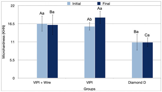
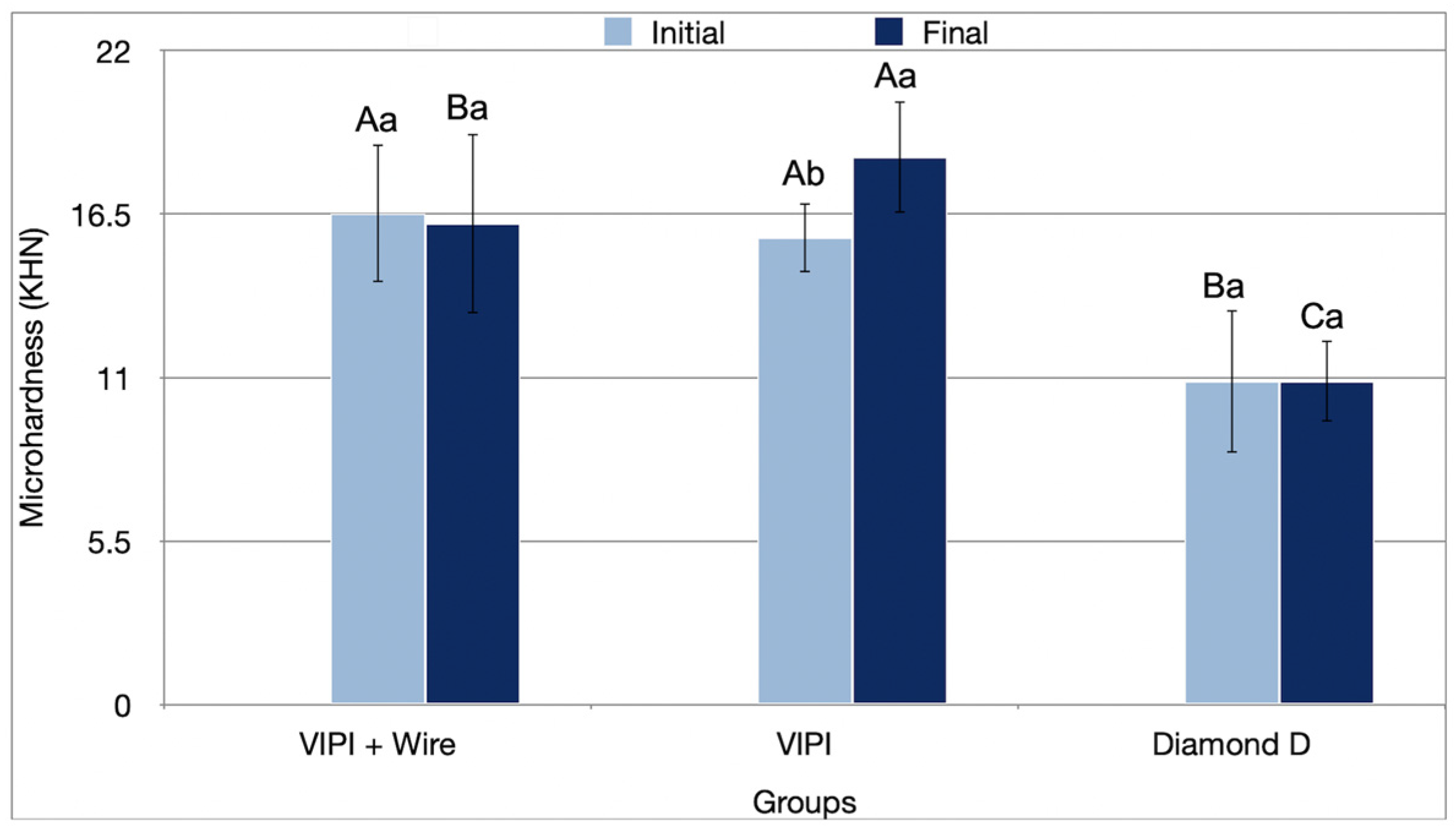
The resin type significantly influenced the microhardness results (p < 0.001) (Table 1). Group Diamond D showed significantly lower microhardness than the other groups in the initial (10.87 KHN, p < 0.001) and final periods (10.88 KHN; p < 0.001). Aging resulted in a significant increase in microhardness for the VIPI Group (15.70 KHN to 18.42 KHN; p ≤ 0.03) (Figure 2).
Table 1.
Two-way repeated measures ANOVA for the microhardness, roughness, percentage of porosity per area, and pore size, and two-way ANOVA for the flexural strength of the resins tested, before and after thermocycling.
Figure 2.
Mean values (standard deviation) of the microhardness (KHN) of the resins tested in the initial and final periods. Different uppercase letters indicate statistical differences for the same period between groups (p < 0.05). Different lowercase letters indicate statistical differences for the same group between periods (p < 0.05).
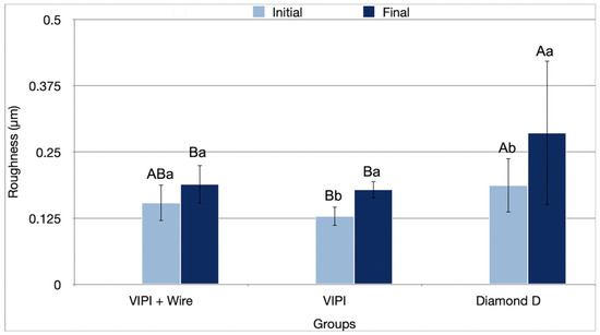
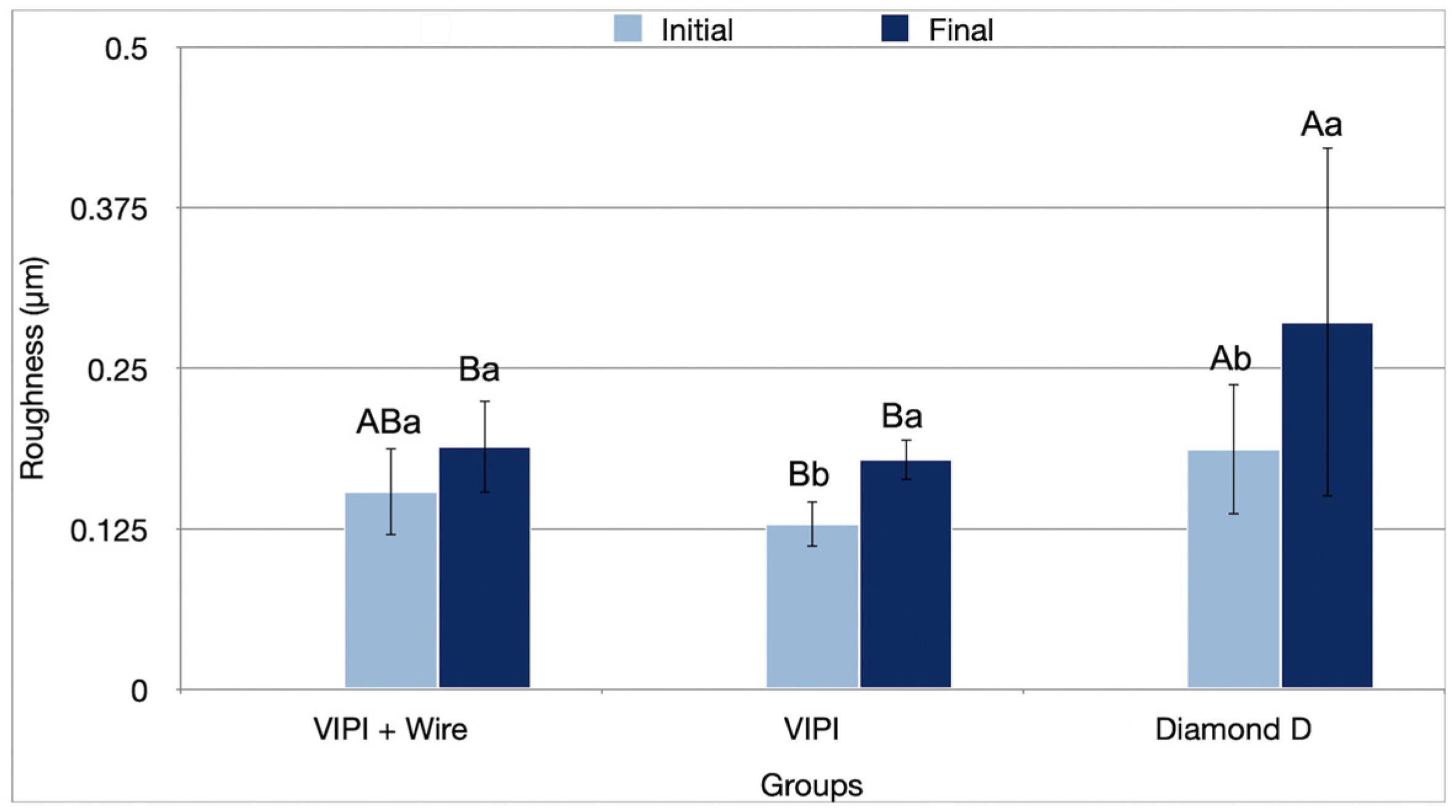
The interaction between resin type and period affected surface roughness (Ra) results (p = 0.038) (Table 1). In the initial period, Group Diamond D showed significantly higher roughness (0.187 µm) than Group VIPI (0.129 µm; p = 0.002). In the final period, Group Diamond D exhibited significantly higher roughness (0.286 µm; p ≤ 0.013) than the other groups. Roughness increased for Groups VIPI (0.129 µm to 0.179 µm; p = 0.008) and Diamond D (0.187 µm to 0.286 µm; p = 0.017) after aging (Figure 3).
Figure 3.
Mean values (standard deviation) of roughness (Ra) (µm) of the resins tested in the initial and final periods. Different uppercase letters indicate statistical differences for the same period between groups (p < 0.05). Different lowercase letters indicate statistical differences for the same group between periods (p < 0.05).
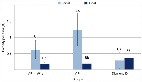
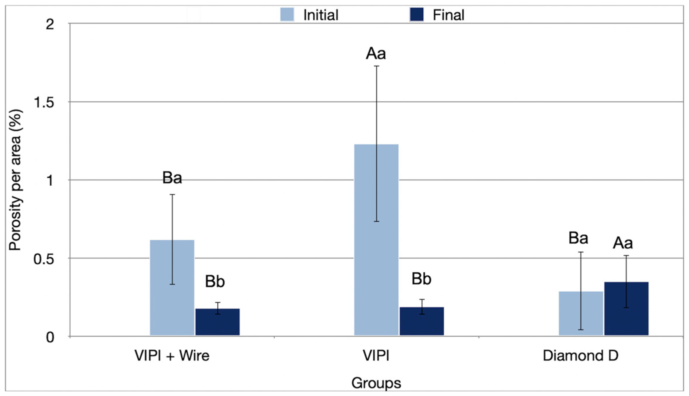
The interaction of resin type and period significantly impacted the porosity percentage (p < 0.001) (Table 1). In the initial period, Group VIPI showed a higher percentage of porosity per area (1.23%; p ≤ 0.001) than the other groups. By the final period, Group Diamond D showed a higher porosity percentage (1.23%; p ≤ 0.001) than the other groups. There was a significant reduction in the percentage of porosity in the final period for VIPI + Wire (0.62% to 0.18%; p = 0.003) and VIPI (1.23% to 0.19%; p ≤ 0.001) (Figure 4).
Figure 4.
Percentage values (standard deviation) of porosity of the resins tested in the initial and final periods. Different uppercase letters indicate statistical differences for the same period between groups (p < 0.05). Different lowercase letters indicate statistical differences for the same group between periods (p < 0.05).
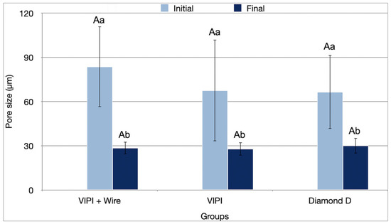
In contrast, the period factor had a significant effect on pore size (p < 0.001) (Table 1), with all groups showing a significant reduction in pore size after aging. Group VIPI + Wire showed a decrease from 83.65 µm to 28.50 µm (p ≤ 0.001), Group VIPI from 67.52 µm to 27.91 µm (p ≤ 0.001), and Group Diamond D from 66.48 µm to 30.05 µm (p ≤ 0.001) (Figure 5).
Figure 5.
Mean pore size values (standard deviation) of the resins tested in the initial and final periods. Different uppercase letters indicate statistical differences for the same period between groups (p < 0.05). Different lowercase letters indicate statistical differences for the same group between periods (p < 0.05).
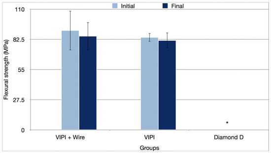
None of the samples from Group Diamond D fractured during the flexural strength test, with deflections greater than 5 mm, which led to the test being canceled for this group. Therefore, the analysis was conducted only for Groups VIPI + Wire and VIPI. Neither the resin type (p = 0.170), nor the period (p = 0.296), nor their interaction (p = 0.737) affected the flexural strength results (Table 1), with the average values shown in Figure 6.
Figure 6.
Mean values (standard deviation) of flexural strength (MPa) of the resins tested in the initial and final periods. * The test was not applicable since none of the samples fractured during testing.
4. Discussion
During the thermally activated polymerization process of acrylic resin, polymer chains form as methyl methacrylate monomers are converted into polymers. However, residual monomer may remain in the polymerized resin as the temperature decreases. The present study standardized the sample size, thickness, shape, and polishing protocol to evaluate the physical–mechanical properties of the acrylic resins used.
Group Diamond D showed lower microhardness and higher roughness than the other groups during the initial and final periods. This result may have been attributed to the polymerization method recommended by the manufacturer and the level of residual monomer remaining in the resin [45]. Diamond D resin polymerization requires a higher temperature and a shorter polymerization time (4 min at 90% power in a 1000 W oven), which may result in more residual monomer. In contrast, VIPI Wave resin undergoes a longer polymerization cycle (20 min at 20% power, followed by 5 min at 60% power), reducing residual monomer and yielding higher microhardness and lower roughness values [46,47]. Additionally, excess heat could have compromised the material’s mechanical properties [48]. Though flexural strength tests for Group Diamond D were not feasible due to the material’s deflection greater than 5 mm, its increased roughness and lower hardness suggest the need for shorter intervals between polishing sessions in clinical practice. Additionally, alternative polymerization protocols should be investigated.
Group VIPI showed an increase in microhardness with aging. Microhardness is influenced by the degree of crosslinking and the amount of residual monomer [47]. Thermocycling may have enhanced polymerization, reducing residual monomers and thus increasing microhardness [49].
Regarding surface roughness, the ideal value for intraoral prostheses is less than 0.2 µm to minimize biofilm retention (bacteria and fungi), color alteration, and patient discomfort [50]. The polishing technique plays a significant role in surface roughness, as the speed at which the sandpaper is rotated and the pressure applied to the samples are difficult to standardize. In clinical practice, the irregular surface of complete dentures complicates polishing, potentially affecting surface roughness [51,52]. Group Diamond D showed higher roughness than the other groups, exceeding the ideal threshold after aging, with a high standard deviation. This result could be due to polymerization-induced contraction, with higher polymerization temperatures leading to greater contraction and surface roughness [53]. Additionally, this may have occurred due to polymerization at higher power in the microwave oven, with a short polymerization time, resulting in a greater number of residual monomers, which contributes to increased surface roughness [54]. It has been shown that conventional fabrication of PMMA results in greater surface roughness, which in turn leads to microcrack formation under cyclic loading, whereas digitally processed materials exhibit more homogeneous polymer structures [55,56]. Additionally, Diamond D resin includes fibers for improved strength [19], which may have contributed to the roughness [57]. After aging, roughness increased for Groups VIPI and Diamond D. This can be attributed to water absorption during thermocycling, which weakens the polymer matrix and increases surface roughness [50,58].
The porosity results were influenced by accelerated aging. Although an increase in porosity and pore size was expected, all groups showed a decrease in pore size, with Groups VIPI + Wire and VIPI also exhibiting reduced porosity percentage after aging. Despite this, porosity remained below 1.23%, which is clinically acceptable, as it is desirable to have less than 11% porosity in acrylic resin pieces [59]. In addition, the American Dental Association specifies that no bubbles or pores should be found when examined without magnification [60]. In this study, no pores or bubbles were visible without magnification. A recent systematic review comparing fabrication methods for interim restorations found that digital processing methods (3D printing and CAD/CAM) reduce internal defects and porosity, thereby directly improving the mechanical performance of these materials [61].
The Group Diamond D exhibited a high standard deviation, especially in the initial period. This may be due to the short polymerization cycle at high power, which results in rapid polymerization but may not have been sufficient to fully reduce residual monomer [48]. The high standard deviation could also result from non-homogeneous heat distribution during rapid heating.
Before aging, Groups VIPI + Wire and VIPI had greater porosity than Group Diamond D. This is likely due to the longer polymerization cycle of the VIPI resin, as microwave polymerization involves agitation of the molecules and potential degradation during polymerization. This results in increased energy and temperature, which can lead to greater porosity in the material after monomer vaporization [59,62]. In addition, VIPI resin contains benzoyl peroxide, which can cause overheating during polymerization and create porosity in the material [63]. The decrease in porosity could be explained by water absorption during aging and the expansion of the sample size [64]. Additionally, the aging process may have removed the superficial layer of the samples, homogenizing the surface [64].
Regarding the relationship between microhardness and flexural strength, acrylic resin used for dental prostheses must have adequate flexural properties to resist masticatory forces and surface hardness to withstand abrasion. Both analyses assess resistance to plastic deformation. However, while flexural strength evaluates the structural flexibility of the material, hardness measures resistance to surface plastic deformation following mechanical indentation [65]. Although Group Diamond D did not fracture during flexural testing, the lower microhardness and higher flexibility may explain its ability to withstand more deflection, making it more ductile than the other groups. The results may be attributed to the polymerization process recommended by the manufacturer, which involves a short cycle with higher microwave power, leading to a higher number of residual monomers, reduced crosslink conversion, and fewer secondary polymerization reactions [66,67]. Although the composition and treatment of the added fibers are not disclosed by the manufacturer, the incorporation of fibers into the polymer matrix, when done optimally, can enhance the flexural strength of the resin [68]. On the other hand, another study evaluating Diamond D resin reported flexural strengths of 84.9 MPa before and 88.3 MPa after fatigue testing, which were statistically similar to the other PMMA materials evaluated in the study [69].
The resin used in provisional implant-supported complete fixed prostheses must be relatively flexible to resist fracture under masticatory forces [68]. However, it is important that these prostheses distribute the forces evenly across the implants, preventing overloading, especially when provisional prostheses are used while waiting for all implants to osseointegrate before proceeding with definitive rehabilitation [70,71]. In this context, a resin with a high deflection modulus may not be able to effectively distribute the forces [68].
According to ISO 20795-1:2013 [41], denture base materials must have a flexural strength of at least 65 MPa. Although no significant differences were found between Groups VIPI and VIPI + Wire, both groups exhibited flexural strength values above this threshold. With aging, the values decreased numerically but remained above the recommended limit. In addition, there was a decrease in porosity and an increase in roughness, likely due to factors mentioned earlier, such as water sorption during thermocycling [64] and a weakening of the polymer matrix [50,58].
The addition of orthodontic wire to VIPI resin did not improve flexural strength. From a clinical standpoint, wire-free provisional prostheses may offer greater ease of production. However, caution should be exercised when extrapolating in vitro results to clinical practice, as the shape and conditions of the tested samples differ from those of actual prostheses.
As a limitation of the study, the samples were not designed to simulate a real prosthesis, which limits the ability to extrapolate the results to clinical conditions. Polydispersity and molecular weight were also not evaluated in the current study. Additionally, future studies are recommended to explore complementary methodologies, such as cross-sectional analysis, atomic force microscopy (AFM), or brushing simulation, to strengthen the findings. Another limitation is related to the technologies used to fabricate the samples, such as digital technologies (3D printing and CAD/CAM), which were not included.
5. Conclusions
Although Group Diamond D did not fracture under flexural testing, it showed lower microhardness, higher roughness, and greater porosity than the other groups after aging. From this in vitro analysis, it is possible to conclude that Diamond D resin does not present adequate properties for use in provisional implant-supported full-arch prostheses. In contrast, VIPI with metallic reinforcement exhibited higher hardness, lower roughness, greater stability after aging, and reduced porosity.
Author Contributions
Conceptualization, E.V.F.d.S. and B.L.T.L.; Methodology, C.L.e.O., M.S.G. and W.J.A.B.C.; Validation, K.F.S., S.J.C.B. and P.F.C.; Formal analysis, E.V.F.d.S., K.F.S., S.J.C.B., P.F.C., N.A.B.-B. and S.B.B.; Investigation, C.L.e.O., M.S.G. and W.J.A.B.C.; Data curation, C.L.e.O., M.S.G., W.J.A.B.C., K.F.S., S.J.C.B. and P.F.C.; Writing—original draft, E.V.F.d.S., C.L.e.O., M.S.G. and W.J.A.B.C.; Writing—review & editing, E.V.F.d.S., P.F.C., N.A.B.-B., S.B.B. and B.L.T.L.; Visualization, K.F.S. and S.J.C.B.; Supervision, E.V.F.d.S. and B.L.T.L.; Project administration, E.V.F.d.S., N.A.B.-B., S.B.B. and B.L.T.L.; Funding acquisition, E.V.F.d.S. All authors have read and agreed to the published version of the manuscript.
Funding
The authors would like to thank the São Paulo Research Foundation (FAPESP, #2022/12824-7) for the scholarship awarded to Carol Ortiz.
Institutional Review Board Statement
Not applicable.
Informed Consent Statement
Not applicable.
Data Availability Statement
The original contributions presented in this study are included in the article. Further inquiries can be directed to the corresponding author.
Acknowledgments
During the preparation of this manuscript, the authors used Copilot (M365 Copilot, Underlying model: GPT-5 (chat)–based, Platform: Cloud-hosted by Microsoft), to help illustrate the study design. The authors have reviewed and edited the output and take full responsibility for the content of this publication.
Conflicts of Interest
The authors declare no conflicts of interest.
References
- Alqutaibi, A.Y.; Baik, A.; Almuzaini, S.A.; Farghal, A.E.; Alnazzawi, A.A.; Borzangy, S.; Aboalrejal, A.N.; AbdElaziz, M.H.; Mahmoud, I.I.; Zafar, M.S. Polymeric denture base materials: A review. Polymers 2023, 15, 3258. [Google Scholar] [CrossRef] [Scilit] [PubMed]
- Oh, S.H.; Kim, Y.; Park, J.Y.; Jung, Y.J.; Kim, S.K.; Park, S.Y. Comparison of fixed implant-supported prostheses, removable implant-supported prostheses, and complete dentures: Patient satisfaction and oral health-related quality of life. Clin. Oral Implants Res. 2014, 27, e31–e37. [Google Scholar] [CrossRef] [Scilit]
- Compagnoni, M.A.; Paleari, A.G.; Rodriguez, L.S.; Giro, G.; Mendoza Marin, D.O.; Pero, A.C. Impact of replacing conventional complete dentures with implant-supported fixed complete dentures. Int. J. Periodontics Restor. Dent. 2014, 34, 833–839. [Google Scholar] [CrossRef] [Scilit]
- Brennan, M.; Houston, F.; O’Sullivan, M.; O’Connell, B. Patient satisfaction and oral health-related quality of life outcomes of implant overdentures and fixed complete dentures. Int. J. Oral Maxillofac. Implants 2010, 25, 791–800. [Google Scholar] [PubMed]
- Martínez-Lage-Azorín, J.F.; Segura-Andrés, G.; Faus-López, J.; Agustín-Panadero, R. Rehabilitation with implant-supported overdentures in total edentulous patients: A review. J. Clin. Exp. Dent. 2013, 5, e267–e272. [Google Scholar] [CrossRef] [Scilit]
- Carvalho, A.; Pellizzer, B. Fundamentos em Implantodontia—Uma Visão Contemporânea, 2nd ed.; Quintessence: São Paulo, Brazil, 2015; pp. 1–300. [Google Scholar]
- Velasco-Ortega, E.; Cracel-Lopes, J.L.; Matos-Garrido, N.; Jiménez-Guerra, A.; Ortiz-Garcia, I.; Moreno-Muñoz, J.; Núñez-Márquez, E.; Rondón-Romero, J.L.; López-López, J.; Monsalve-Guil, L. Immediate functional loading with full-arch fixed implant-retained rehabilitation in periodontal patients: Clinical study. Int. J. Environ. Res. Public Health 2022, 19, 13162. [Google Scholar] [CrossRef] [Scilit]
- Sandoval-Guevara, D.; Grajeda-Núñez, E.; Alarcón-Sánchez, M.A.; Heboyan, A. Immediate functional loading of implant-supported mandibular prostheses with customized UCLA attachments with distal extension: A case report. SAGE Open Med. Case Rep. 2024, 12, 2050313X241269572. [Google Scholar] [CrossRef] [Scilit]
- Al Baker, A.; Habib, S.R.; Al Amri, M.D. Preserving esthetics, occlusion and occlusal vertical dimension in a patient with fixed prostheses seeking dental implant treatment. Saudi Dent. J. 2016, 28, 203–208. [Google Scholar] [CrossRef] [Scilit]
- Bagegni, A.; Abou-Ayash, S.; Rücker, G.; Algarny, A.; Att, W. The influence of prosthetic material on implant and prosthetic survival of implant-supported fixed complete dentures: A systematic review and meta-analysis. J. Prosthodont. Res. 2019, 63, 251–265. [Google Scholar] [CrossRef] [Scilit] [PubMed]
- Fayyad, A. Prosthetic complications in immediately loaded all-on-four mandibular prosthesis using titanium wire reinforcement versus nonreinforced temporary framework: A randomized clinical trial. Egypt. Dent. J. 2022, 68, 885–892. [Google Scholar] [CrossRef] [Scilit]
- Aupaphong, V.; Kraiwattanawong, K.; Thanathornwong, B. Effects of nanocrystal cellulose from bamboo on the flexural strength of acrylic resin: In vitro. Dent. J. 2022, 10, 129. [Google Scholar] [CrossRef] [Scilit]
- Apimanchindakul, C.; Na Nan, P.; Aimjirakul, N. Effect of reinforced self-cured acrylic resin on flexural strength. Int. J. Dent. 2022, 2022, 2698995. [Google Scholar] [CrossRef] [Scilit] [PubMed]
- Godoy, A.; Siegel, S.C. Simplified technique for incorporating a metal mesh into record bases for mandibular implant overdentures. J. Prosthodont. 2015, 24, 661–664. [Google Scholar] [CrossRef] [Scilit]
- Colán Guzmán, P.; Freitas, F.F.; Ferreira, P.M.; Freitas, C.A.; Reis, K.R. Influence of different cantilever extensions and glass or polyaramide reinforcement fibers on fracture strength of implant-supported temporary restorations. J. Appl. Oral Sci. 2008, 16, 111–115. [Google Scholar] [CrossRef] [Scilit]
- Ibrahim, S.; Laftah, S.; Hussain, W. Effect of reinforcing PMMA denture base material by stainless steel 316L wires on flexural strength. J. Appl. Sci. Nanotechnol. 2023, 3, 51–58. [Google Scholar] [CrossRef] [Scilit]
- Dental News. Diamond D—Resina Acrílica Termopolimerizável. Available online: https://dentalnews.com.br/produto/545/ (accessed on 11 February 2025).
- Ellakwa, A.E.; Morsy, M.A.; El-Sheikh, A.M. Effect of aluminum oxide addition on the flexural strength and thermal diffusivity of heat-polymerized acrylic resin. J. Prosthodont. 2008, 17, 439–444. [Google Scholar] [CrossRef] [Scilit]
- Keystone Industries. Diamond D Denture Acrylic. 2019. Available online: https://dental.keystoneindustries.com/wp-content/uploads/2015/11/SS_DiamondD_EN_2019_WEB.pdf (accessed on 11 February 2025).
- Moslehifard, E.; Ghaffari, T.; Abolghasemi, H.; Maleki Dizaj, S. Comparison of conventional pressure-packed and injection molding processing methods for acrylic resin dentures based on microhardness, surface roughness, and water sorption. Int. J. Dent. 2022, 2022, 7069507. [Google Scholar] [CrossRef] [Scilit]
- Dhir, S. Biofilm and dental implant: The microbial link. J. Indian Soc. Periodontol. 2013, 17, 5–11. [Google Scholar] [CrossRef] [Scilit] [PubMed]
- AlQahtani, M.; Haralur, S.B. Influence of different repair acrylic resin and thermocycling on the flexural strength of denture base resin. Medicina 2020, 56, 50. [Google Scholar] [CrossRef] [Scilit]
- Barbosa, D.B.; Barão, V.A.R.; Monteiro, D.R.; Compagnoni, M.A.; Marra, J. Bond strength of denture teeth to acrylic resin: Effect of thermocycling and polymerisation methods. Gerodontology 2008, 25, 237–244. [Google Scholar] [CrossRef] [Scilit]
- Carreira, M.; Antunes, P.V.; Ramalho, A.; Paula, A.; Carrilho, E. Thermocycling effect on mechanical and tribological characterization of two indirect dental restorative materials. J. Braz. Soc. Mech. Sci. Eng. 2017, 39, 1–17. [Google Scholar] [CrossRef] [Scilit]
- Göhring, T.N.; Gallo, L.; Lüthy, H. Effect of water storage, thermocycling, and the incorporation and site of placement of glass fibers on the flexural strength of veneering composite. Dent. Mater. 2005, 21, 761–772. [Google Scholar] [CrossRef] [Scilit]
- Meriç, G.; Ruyter, E. Effect of thermal cycling on composites reinforced with two differently sized silica-glass fibers. Dent. Mater. 2007, 23, 1157–1163. [Google Scholar] [CrossRef] [Scilit]
- Puig, G.; Collares, F.M.; Grazioli, G.; Leitune, V.C.B. Evaluation of microtensile bond strength between CAD/CAM denture resin and acrylic artificial teeth. J. Clin. Dent. Res. 2023, 20, 18–25. [Google Scholar] [CrossRef] [Scilit]
- Consani, R.L.X.; Pucciarelli, M.G.R.; Mesquita, M.F.; Nogueira, M.C.F.; Barão, V.A.R. Polymerization cycles on hardness and surface gloss of denture bases. Int. J. Contemp. Dent. Med. Rev. 2014, 2014, 1–6. [Google Scholar]
- Capla, M.; Sardi, J.C.O.; Pfeifer, C.; Consani, R.L.X. Effect of different simulated microwave disinfection cycles on hardness, roughness and gloss of acrylic resins. Int. J. Dent. Oral Sci. 2017, 4, 575–580. [Google Scholar] [CrossRef] [Scilit]
- Catanoze, I.A.; Cunha, B.G.; Costa, G.Q.; Silva, E.V.F.; Duque, C.; Guiotti, A.M. Antagonistic effect of isolated and commercially available probiotics on the growth of Candida albicans on acrylic resin denture surfaces. J. Prosthet. Dent. 2022, 127, 338–344. [Google Scholar] [CrossRef] [Scilit]
- American Dental Association. Specifications No. 12 for denture base polymers. J. Am. Dent. Assoc. 1975, 90, 39–49. [Google Scholar] [CrossRef] [Scilit]
- Golbidi, F.; Mousavi, T. Transverse strength of repaired denture base material with wire and two auto-polymerized acrylic resins. J. Dent. 2007, 4, 183–187. [Google Scholar]
- VIPI Indústria, Comércio, Exportação e Importação de Produtos Odontológicos Ltda. VIPI Wave Acrylic Resin. Available online: https://vipi.com.br/portal/bulario/ft_vipiwave.pdf (accessed on 11 February 2025).
- Keystone Industries. Diamond D Heat Cure Acrylic Resin. Powder SDS. Available online: https://dental.keystoneindustries.com/wp-content/uploads/2015/11/Diamond-D-Heat-Cure-Powder-SDS-US-20151204.pdf (accessed on 11 February 2025).
- Keystone Industries. Diamond D Heat Cure Acrylic Resin. Liquid SDS. Available online: https://dental.keystoneindustries.com/wp-content/uploads/2015/11/Diamond-D-Heat-Cure-Liquid-SDS-US-20150904.pdf (accessed on 11 February 2025).
- Santos, D.M.; Nagay, B.E.; da Silva, E.V.; Bonatto, L.R.; Sonego, M.V.; Moreno, A.; Rangel, E.C.; da Cruz, N.C.; Goiato, M.C. In vitro analysis of different properties of acrylic resins for ocular prosthesis submitted to accelerated aging with or without photopolymerized glaze. Mater. Sci. Eng. C 2016, 69, 995–1003. [Google Scholar] [CrossRef] [Scilit]
- Sofou, A.; Tsoupi, I.; Emmanouil, J.; Karayannis, M. HPLC determination of residual monomers released from heat-cured acrylic resins. Anal. Bioanal. Chem. 2005, 381, 1336–1346. [Google Scholar] [CrossRef] [Scilit]
- ASTM E384-11e1; Standard Test Method for Knoop and Vickers Hardness of Materials. ASTM International: West Conshohocken, PA, USA, 2012.
- Gonzaga, C.C.; Cesar, P.F.; Miranda, W.G., Jr.; Yoshimura, H.N. Slow crack growth and reliability of dental ceramics. Dent. Mater. 2011, 27, 394–406. [Google Scholar] [CrossRef] [Scilit]
- Gonzaga, C.C.; Cesar, P.F.; Okada, C.Y.; Fredericci, C.; Neto, B.; Yoshimura, H.N. Mechanical properties and porosity of dental glass-ceramics hot-pressed at different temperatures. Mater. Res. 2008, 11, 301–306. [Google Scholar] [CrossRef] [Scilit]
- ISO 20795-1:2013; Dentistry—Base Polymers—Part 1: Denture Base Polymers. ISO: Geneva, Switzerland, 2013.
- Kanie, T.; Arikawa, H.; Fujii, K.; Ban, S. Mechanical properties of reinforced denture base resin: The effect of position and the number of woven glass fibers. Dent. Mater. J. 2002, 21, 261–269. [Google Scholar] [CrossRef] [Scilit]
- ISO TR11405; Dental Materials—Guidance on Testing of Adhesion to Tooth Structure. ISO: Geneva, Switzerland, 1994; pp. 1–15.
- Pinto, J.R.R.; Mesquita, M.F.; Nóbilo, M.A.A.; Henriques, G.E.P. Evaluation of varying amounts of thermal cycling on bond strength and permanent deformation of two resilient denture liners. J. Prosthet. Dent. 2004, 92, 288–293. [Google Scholar] [CrossRef] [Scilit]
- Gad, M.M.; Fouda, S.M.; ArRejaie, A.S.; Al-Thobity, A.M. Comparative effect of different polymerization techniques on the flexural and surface properties of acrylic denture bases. J. Prosthodont. 2019, 28, 458–465. [Google Scholar] [CrossRef] [Scilit] [PubMed]
- Leão, R.S.; de Moraes, S.L.D.; Aquino, K.A.D.S.; Isolan, C.P.; Casado, B.G.D.S.; Montes, M.A.J.R. Effect of pressure, post-pressing time, and polymerization cycle on the degree of conversion of thermoactivated acrylic resin. Int. J. Dent. 2018, 2018, 5743840. [Google Scholar] [CrossRef] [Scilit] [PubMed]
- Schoeffel, A.C.; Bagio, P.; Sakima, V.T.; Soares, S.; Neppelenbroek, K.H.; Urban, V.M. Knoop microhardness of conventional and microwaved denture base acrylic resins. Indian J. Dent. Res. 2019, 30, 927–932. [Google Scholar] [CrossRef] [Scilit] [PubMed]
- Nisar, S.; Moeen, F.; Hasan, U. Effect of varying curing regimes and powder–liquid ratios on the flexural strength and surface porosities of heat cure acrylic: An in-vitro experiment. Int. J. Dent. Sci. Res. 2015, 3, 64–71. [Google Scholar]
- Al-Jmmal, A.Y.; Mohammed, N.Z.; Al-Kateb, H.M. The effect of aging on hardness of heat-cured denture base resin modified with recycled acrylic resin. Clin. Exp. Dent. Res. 2024, 10, e828. [Google Scholar] [CrossRef] [Scilit]
- Moraes Melo Neto, C.L.; Bernardi, B.S.; Dekon, S.F.C.; dos Santos, D.M.; Goiato, M.C. Influence of thermal cycles and disinfection on the roughness, microhardness and color of PETG/TPU and PMMA. Polym. Med. 2023, 53, 19–24. [Google Scholar] [CrossRef] [Scilit]
- Quezada, M.M.; Salgado, H.; Correia, A.; Fernandes, C.; Fonseca, P. Investigation of the effect of the same polishing protocol on the surface roughness of denture base acrylic resins. Biomedicines 2022, 10, 1971. [Google Scholar] [CrossRef] [Scilit]
- Fiore, A.D.; Meneghello, R.; Brun, P.; Rosso, S.; Gattazzo, A.; Stellini, E.; Yilmaz, B. Comparison of the flexural and surface properties of milled, 3D-printed, and heat-polymerized PMMA resins for denture bases: An in vitro study. J. Prosthodont. Res. 2022, 66, 502–508. [Google Scholar] [CrossRef] [Scilit]
- de Gee, A.J.; ten Harkel, E.C.; Davidson, C.L. Measuring procedure for the determination of the three-dimensional shape of dentures. J. Prosthet. Dent. 1979, 42, 149–153. [Google Scholar] [CrossRef] [Scilit]
- Zissis, A.; Yannikakis, S.; Polyzois, G.; Harrison, A. A long-term study on residual monomer release from denture materials. Eur. J. Prosthodont. Restor. Dent. 2008, 16, 81–84. [Google Scholar] [PubMed]
- Abdelmohsen, N.; Bourauel, C.; Elshazly, T.M. Interim Fixed Dental Prostheses Fabrication Techniques and Factors Affecting Their Success: A Narrative Review. Oral 2025, 5, 44. [Google Scholar] [CrossRef] [Scilit]
- Yan, X.; Liu, Y.; Han, C.; Bai, S. Factors affecting the surface quality of 3D printed resin dental restorations in digitally assisted dentistry: A systematic review. J. Prosthet. Dent. 2026, in press. [Google Scholar] [CrossRef] [Scilit] [PubMed]
- Gad, M.M.; Fouda, S.M.; Rahoma, A.; Al-Thobity, A.M. Effect of polymerization technique and glass fiber addition on the surface roughness and hardness of PMMA denture base material. Dent. Mater. J. 2018, 37, 746–753. [Google Scholar] [CrossRef] [Scilit]
- Temizci, T.; Bozoğulları, H.N. Effect of thermal cycling on the flexural strength of 3D-printed, CAD/CAM milled, and heat-polymerized denture base materials. BMC Oral Health 2024, 24, 357. [Google Scholar] [CrossRef] [Scilit]
- Singh, S.; Palaskar, J.N.; Mittal, S. Comparative evaluation of surface porosities in conventional heat-polymerized acrylic resin cured by water bath and microwave energy with microwavable acrylic resin cured by microwave energy. Contemp. Clin. Dent. 2013, 4, 147–151. [Google Scholar] [CrossRef] [Scilit]
- Council on Dental Materials and Devices. Revised American Dental Association Specification No. 12 for denture base polymers. J. Am. Dent. Assoc. 1975, 90, 451–458. [Google Scholar] [CrossRef] [Scilit]
- Sankeshwari, B.; Kanitkar, A.; Pattanshetti, C.; Kanitkar, A. Comparative Analysis of 3D Printing, Computer Aided Design and Milling and Conventional Fabrication Techniques on Fracture Resistance of Interim Restorations: A Systematic Review and Meta-analysis. J. Clin. Diag. Res. 2025, 19, ZC39–ZC44. [Google Scholar] [CrossRef] [Scilit]
- Silva, E.V.F.; Goiato, M.C.; Bitencourt, S.B.; Finer, Y.; Brito, V.G.B.; Takamiya, A.S.; de Oliveira, S.H.P.; Santos, D.M.D. Effect of processing methods on the cytotoxicity of methyl methacrylate-based ocular prostheses: An in vitro study. Toxicol. In Vitro 2021, 76, 105211. [Google Scholar] [CrossRef] [Scilit] [PubMed]
- Silva, S.M.L.M.; Bindo, M.J.F.; Leão, M.P. O uso de energia de microondas para polimerização de resinas acrílicas. DENS 2006, 14, 11–21. [Google Scholar] [CrossRef] [Scilit]
- Alignani, G.M.S.; Pantoja, J.M.C.N.; Takahashi, J.M.F.K.; Vasconcellos, A.A.; Mesquita, M.F.; Miranda, M.E. Effect of thermocycling in a hard denture liner and acrylic resin after different chemical and mechanical polishing. Rev. Gaúch. Odontol. 2015, 63, 397–404. [Google Scholar] [CrossRef] [Scilit]
- Fouda, S.M.; Gad, M.M.; Abualsaud, R.; Ellakany, P.; AlRumaih, H.S.; Khan, S.Q.; Akhtar, S.; al-Qarni, F.D.; Al-Harbi, F.A. Flexural properties and hardness of CAD-CAM denture base materials. J. Prosthodont. 2023, 32, 318–324. [Google Scholar] [CrossRef] [Scilit]
- Azzarri, M.J.; Cortizo, M.S.; Alessandrini, J.L. Effect of curing conditions on the properties of an acrylic denture base resin microwave-polymerized. J. Dent. 2003, 31, 463–468. [Google Scholar] [CrossRef] [Scilit]
- Spartalis, G.K.; Cappelletti, L.K.; Schoeffel, A.C.; Michél, M.D.; Pegoraro, T.A.; Arrais, C.A.; Neppelenbroek, K.H.; Urban, V.M. Effect of conventional water-bath and experimental microwave polymerization cycles on the flexural properties of denture base acrylic resins. Dent. Mater. J. 2015, 34, 623–628. [Google Scholar] [CrossRef] [Scilit]
- Ucar, Y.; Akova, T.; Aysan, I. Mechanical properties of polyamide versus different PMMA denture base materials. J. Prosthodont. 2012, 21, 173–176. [Google Scholar] [CrossRef] [Scilit]
- Diaz-Arnold, A.M.; Vargas, M.A.; Shaull, K.L.; Laffoon, J.E.; Qian, F. Flexural and fatigue strengths of denture base resin. J. Prosthet. Dent. 2008, 100, 47–51. [Google Scholar] [CrossRef] [Scilit] [PubMed]
- Weinberg, L.A. The biomechanics of force distribution in implant-supported prostheses. Int. J. Oral Maxillofac. Implants 1993, 8, 19–31. [Google Scholar]
- Balwani, D.; Hegde, R.; Hegde, C. Effect of inter-implant distance on fracture resistance of implant-supported provisional fixed dental prosthesis. J. Oral Implantol. 2024, 50, 529–536. [Google Scholar] [CrossRef] [Scilit]

Disclaimer/Publisher’s Note: The statements, opinions and data contained in all publications are solely those of the individual author(s) and contributor(s) and not of MDPI and/or the editor(s). MDPI and/or the editor(s) disclaim responsibility for any injury to people or property resulting from any ideas, methods, instructions or products referred to in the content. |
© 2026 by the authors. Licensee MDPI, Basel, Switzerland. This article is an open access article distributed under the terms and conditions of the Creative Commons Attribution (CC BY) license.