


Abstract
Background: The shell technique is a well-recognized method for alveolar ridge augmentation; however, conventional protocols are often technically demanding, requiring substantial autogenous bone harvesting and precise fixation of thin cortical shells, which is a step associated with risks of graft fracture and increased operative time. Objective: The Kamali technique, described in this case report, represents a modification of the conventional shell technique that employs corticocancellous allogeneic bone blocks to provide structural support for graft containment while minimizing reliance on autogenous bone. Methods: This report describes a single patient who underwent horizontal alveolar ridge augmentation using the modified corticocancellous shell (Kamali) technique as part of routine clinical care. Clinical records, surgical documentation, clinical photographs, and preoperative and postoperative cone-beam computed tomography (CBCT) images were reviewed to describe the treatment procedure and clinical and radiographic findings. No additional intervention was performed for research purposes. The corticocancellous allogeneic block was contoured to fit the osseous defect and stabilized using fixation screws. Particulate autogenous bone harvested from an intraoral donor site was combined with a bone substitute and placed within the grafted compartment. Clinical and radiographic healing was assessed at approximately six months. Results/Clinical Observations: The corticocancellous composition of the allogeneic block provided a structural framework for particulate graft containment and space maintenance. The porous architecture of the cancellous portion appeared to facilitate graft integration and vascular ingrowth. Ridge width increased from 2.9 mm preoperatively to 8.5 mm after six months, allowing placement of a 4 × 9.5 mm implant with more than 35 Ncm primary stability. In this case, the technique allowed the use of a reduced volume of autogenous bone and was successfully applied for graft fixation and horizontal ridge augmentation. Conclusions: The Kamali technique represents a potential modification of conventional shell techniques for horizontal alveolar ridge augmentation. However, its clinical efficacy and reproducibility require further investigation through larger clinical studies and controlled trials.
1. Introduction
Tooth loss frequently leads to alveolar ridge resorption, often necessitating bone augmentation to restore adequate ridge dimensions. Autogenous bone grafts are widely regarded as one of the most effective augmentation options, owing to their osteogenic, osteoinductive, and osteoconductive properties. Nevertheless, the designation of autogenous bone as a universal gold standard remains debated, since the ideal graft material is influenced by multiple factors including defect morphology, the location and volume of the augmentation site, remodeling behavior, and resorption rate [1,2,3,4].
Despite their biological advantages, autogenous bone grafts present several limitations. Harvesting autogenous bone requires an additional surgical site, thereby increasing operative time, donor-site morbidity, patient discomfort, and the risk of postoperative complications. Furthermore, the available graft volume may be limited, particularly in patients requiring extensive reconstruction, and autogenous grafts are associated with variable postoperative resorption. Consequently, allogeneic bone grafts have emerged as a reliable alternative for alveolar ridge augmentation. In general, allogeneic grafts provide excellent osteoconductive properties, while their potential osteoinductive capacity has also been reported and appears to depend largely on graft processing methods [5,6,7,8,9,10].
The shell technique introduced by Khoury et al. remains one of the most predictable and well-established approaches for horizontal and combined alveolar ridge augmentation. The technique is based on creating a biologic compartment using cortical plates that are fixed to the recipient site and subsequently filled with particulate bone graft material, thereby providing both mechanical stability and an environment favorable for bone regeneration [11,12,13].
Although the original shell technique has demonstrated excellent clinical outcomes, it presents several technical limitations. Harvesting autogenous cortical plates from intraoral donor sites, such as the mandibular ramus or symphysis, increases operative time, donor-site morbidity, and patient discomfort while providing only a limited volume of graft material. In cases involving extensive alveolar defects, obtaining sufficient autogenous bone may therefore be challenging. To overcome these limitations, allogeneic cortical shells have been introduced as an alternative to autogenous cortical plates. However, fixation of thin cortical shells remains technically demanding. Owing to their limited thickness, these shells may fracture during contouring or screw fixation, and maintaining the desired three-dimensional graft configuration often requires meticulous manipulation and multiple fixation screws [14,15,16,17,18,19].
Despite the increasing adoption of allogeneic shell techniques, an important technical challenge remains the manipulation and stabilization of thin cortical shells during surgery. Thin cortical plates are susceptible to fracture during contouring or screw fixation and frequently require multiple fixation screws to maintain the desired regenerative compartment. These limitations increase the technical sensitivity of the procedure and may prolong operative time, particularly in extensive horizontal ridge defects. To address these challenges, we developed a modified corticocancellous shell technique, herein referred to as the Kamali Technique. Rather than separating the cortical component from the cancellous portion, this modification preserves the corticocancellous architecture of the allogeneic block and incorporates internal cancellous chambers that function as a biologic scaffold for particulate graft containment. This structural modification is designed to provide a corticocancellous framework for graft containment and space maintenance while potentially facilitating adaptation and fixation, reducing reliance on multiple fixation screws, and decreasing the amount of autogenous bone harvesting. These proposed technical benefits have not yet been validated through comparative clinical evaluation. The novelty of the Kamali technique lies in preserving the corticocancellous architecture of the allogeneic block rather than using a thin, isolated cortical shell, as described in previously reported allogeneic shell techniques. Unlike conventional allogeneic shell approaches, this modification retains the cancellous component as an intrinsic spacer and creates additional semicircular recesses within it to accommodate particulate graft material. This configuration combines structural support, graft containment, and space maintenance within a single corticocancellous construct, potentially simplifying fixation and reducing dependence on extensive autogenous bone harvesting. Therefore, the technique represents a structural and procedural modification of existing allogeneic shell approaches rather than merely a substitution of autogenous cortical plates with allogeneic plates. The purpose of this case report describes the clinical application of the modified corticocancellous shell (Kamali) technique in a patient with a severe horizontal alveolar ridge defect treated as part of routine clinical care. The report presents the clinical decision-making process, surgical procedure, and available clinical and radiographic findings during follow-up. The technical characteristics of the procedure are discussed in relation to previously described shell-based augmentation approaches. This report is intended to document the clinical application of the technique in a single patient rather than to evaluate its efficacy, establish comparative advantages, or demonstrate its reproducibility.
2. Materials and Methods
2.1. Study Design and Clinical Data Collection
This manuscript is a single-patient case report based on a description of routine clinical care. It documents the clinical application of the modified corticocancellous shell (Kamali) technique in a patient who underwent horizontal alveolar ridge augmentation for subsequent implant-supported rehabilitation. The surgical intervention and postoperative management were performed as part of the patient’s standard clinical treatment and were not undertaken as part of a prospective research protocol.
Clinical information was obtained from the patient’s medical and dental records, surgical documentation, clinical photographs, and preoperative and postoperative CBCT examinations. The available records were reviewed to describe the patient’s clinical presentation, treatment planning, surgical procedure, postoperative healing, radiographic changes, and subsequent implant placement. The report does not involve a comparison group, statistical analysis, or evaluation of treatment efficacy. No additional clinical procedures or interventions were performed specifically for research purposes.
2.2. Patient Information
This manuscript is presented as a case report describing the surgical procedure and clinical and radiographic outcomes of the modified corticocancellous shell (Kamali) technique in a patient with severe horizontal alveolar ridge deficiency. The patient was a 41-year-old male classified as ASA Physical Status I, indicating a healthy patient without systemic disease. The alveolar ridge defects were located in the posterior maxillary region at the sites of teeth 2–6 and 11–15. A healthy adult patient presented with a severe horizontal alveolar ridge defect in the posterior maxilla following tooth loss. The patient was referred for implant-supported rehabilitation after clinical examination revealed insufficient alveolar ridge width for prosthetically driven implant placement. The patient’s medical history was non-contributory, with no systemic diseases known to affect bone healing. The patient was a non-smoker and reported no previous history of bisphosphonate therapy, radiotherapy to the head and neck region, or uncontrolled metabolic disease. Oral hygiene was considered satisfactory, and no signs of active periodontal or periapical infection were observed at the recipient site. Written informed consent for the surgical intervention was obtained from the patient before any surgical procedure. Written informed consent was also obtained from the patient for publication of the clinical details, clinical photographs, intraoral radiographs, and CBCT images presented in this report.
2.3. Clinical Findings
Clinical examination demonstrated a pronounced horizontal deficiency of the alveolar ridge with inadequate bone volume for implant placement. Soft tissues were healthy without evidence of inflammation, fistula formation, or mucosal pathology. Palpation confirmed a narrow residual ridge extending throughout the planned implant site. The periodontal phenotype at the recipient sites was classified as thick. The width of the remaining keratinized mucosa was approximately 5 mm, measured at the planned augmentation sites. Preoperative cone-beam computed tomography (CBCT) measurements demonstrated a residual alveolar ridge width of 2.9 mm at the crestal level and 4 mm at 3 mm apical to the crest. The vertical ridge height was 15 mm. CBCT assessment demonstrated the anterior D3/posterior D4 and resorption B according to the Lekholm and Zarb classification. The residual vertical bone height between the alveolar crest and the maxillary sinus floor was approximately 2.5 mm at the planned implant sites. Based on the horizontal ridge deficiency and the relatively preserved vertical dimension, the defect was considered consistent with a Siebert Class I defect, characterized by buccolingual ridge deficiency.
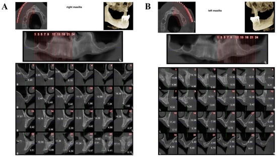
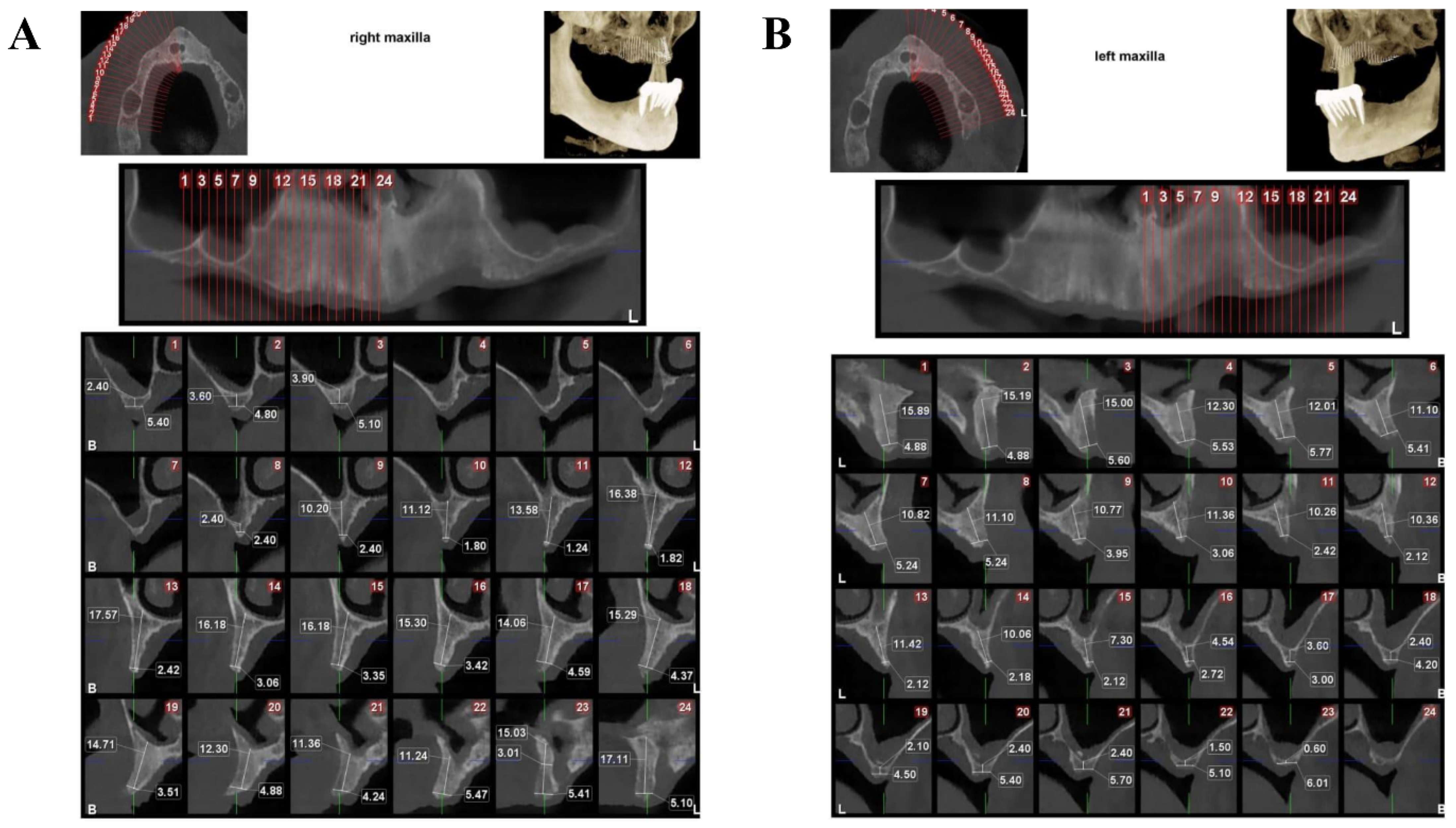
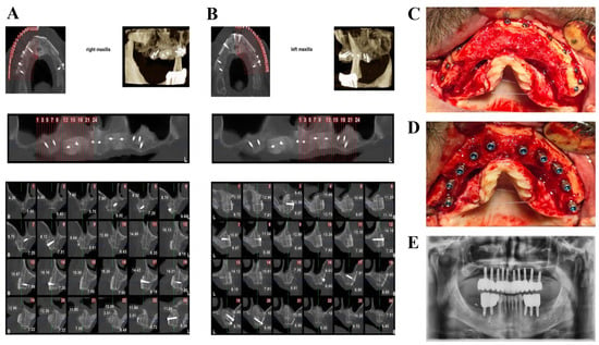
To facilitate radiographic interpretation, representative cross-sectional CBCT images were selected from the preoperative and six-month postoperative examinations at corresponding anatomical locations. The serial cross-sectional images were intended to demonstrate the spatial distribution of the horizontal ridge deficiency before treatment and the dimensional changes following augmentation across the treated maxillary sites. Particular attention was given to the crestal level and the region 3 mm apical to the alveolar crest, corresponding to the anatomical levels used for quantitative ridge-width measurements. This approach was used to provide a visual representation of the anatomical changes associated with the clinical measurements reported in this case.
2.4. Diagnostic Assessment
Diagnosis was established through clinical examination and CBCT analysis. Cross-sectional CBCT images were evaluated to assess residual alveolar ridge width and height, as well as the anatomical relationships with the maxillary sinus, nasal floor, adjacent tooth roots, and neighboring neurovascular structures, where relevant to the surgical site. CBCT examinations were performed using a NewTom GiANO HR imaging system (CEFLA s.c., Imola, Italy), with a field of view (FOV) of 6 × 10 cm, a tube voltage of 90 kVp, a tube current–time product of 40.06 mAs, and a voxel size of 150 μm. These acquisition parameters were used to evaluate the alveolar ridge morphology and relevant anatomical structures for preoperative surgical planning. The primary diagnosis was a severe horizontal alveolar ridge deficiency requiring augmentation prior to implant placement (Figure 1). Alternative treatment options, including guided bone regeneration with particulate biomaterials alone, autogenous block grafting, and conventional autogenous shell technique, were considered during treatment planning. Because of the extent of the horizontal defect and the desire to reduce donor-site morbidity while maintaining graft stability, a modified corticocancellous shell approach was selected. All treatment options, potential risks, expected benefits, and alternative procedures were discussed with the patient before obtaining written informed consent.
Figure 1.
Preoperative CBCT assessment of the horizontal alveolar ridge deficiencies in the right maxilla (A) and left maxilla (B). Representative serial cross-sectional images illustrate the distribution and extent of the residual ridge deficiency across the planned implant sites and provide the anatomical reference for subsequent postoperative comparisons.
Before and after augmentation, horizontal alveolar ridge width was assessed using cross-sectional CBCT images at corresponding anatomical locations. Measurement sites were selected based on reproducible anatomical landmarks and the planned implant positions. Ridge width was measured at the crestal level and at 3 mm apical to the alveolar crest, perpendicular to the longitudinal axis of the alveolar ridge. The same anatomical reference points and measurement levels were used for preoperative and six-month postoperative assessments to ensure consistency and comparability of measurements. The right and left maxillary sites are presented separately in the clinical and radiographic documentation to clearly distinguish the anatomical location and treatment outcomes of each site.
2.5. Surgical Technique (Kamali Technique)
Following local anesthesia, a crestal incision with vertical releasing incisions was performed according to defect morphology, and a full-thickness mucoperiosteal flap was elevated to expose the recipient site. Residual fibrous tissue was removed while preserving the integrity of the recipient cortical bone. No cortical decortication or perforation was performed. The recipient cortical bone was intentionally preserved to maintain its structural integrity and provide a stable foundation for fixation of the corticocancellous allogeneic block. Particulate autogenous bone was harvested from the mandibular ramus using a bonesier, with five or six harvesting passes performed, each at an approximate depth of 3 mm. The harvested bone was collected under sterile conditions. Approximately 5 cc of autogenous particulate bone was obtained, depending on the size of the recipient defect.
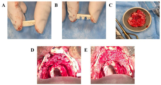
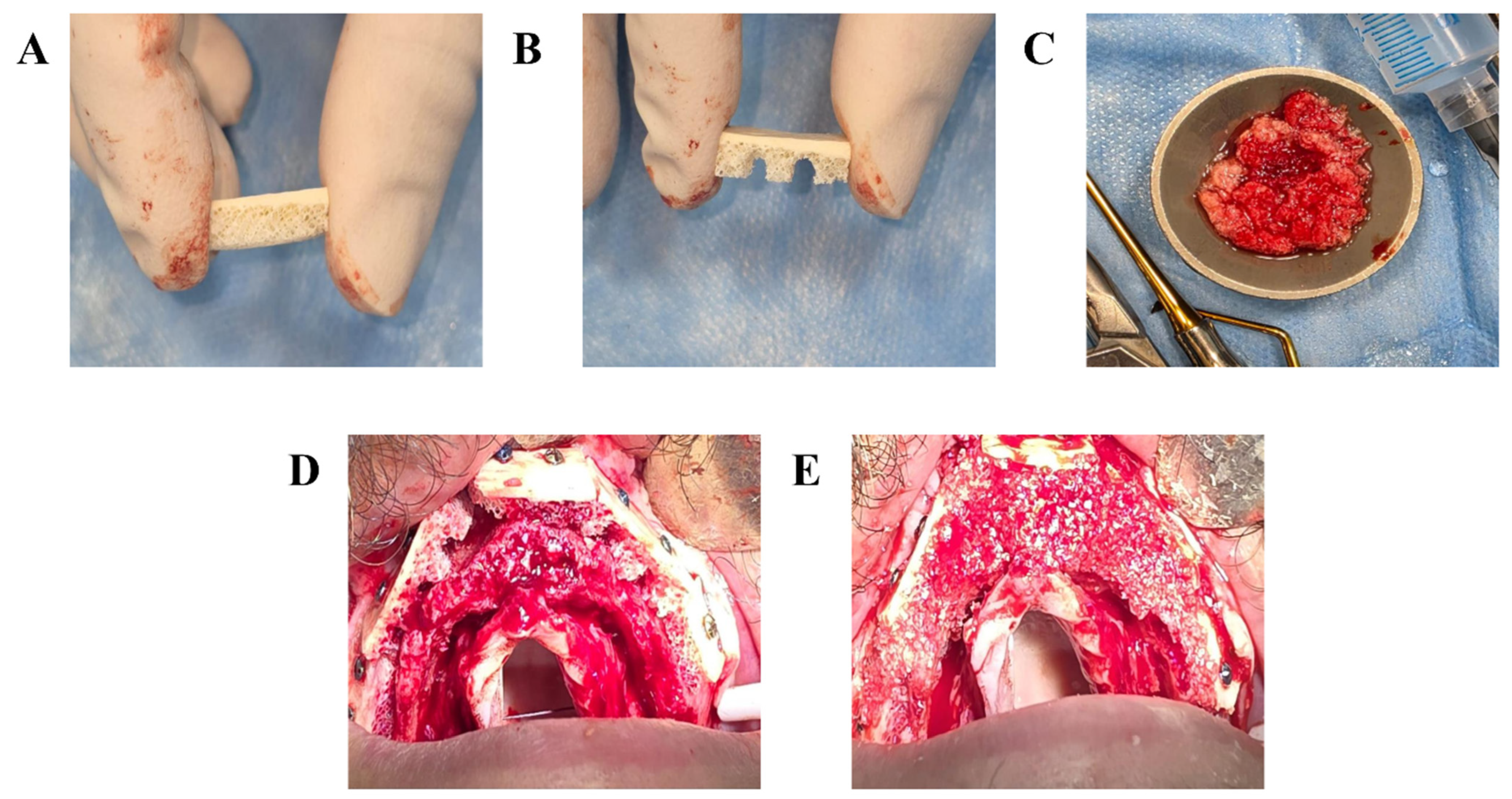
A commercially available mineralized corticocancellous allogeneic bone block (Iranian Tissue Product & Regen Allograft, Tehran, Iran; dimensions: 20 × 10 × 10 mm) was used for reconstruction. The block was trimmed and contoured to match the dimensions and morphology of the recipient alveolar ridge using rotary instruments under copious sterile saline irrigation. During preparation, particular care was taken to preserve the cortical component to maintain structural rigidity and the cancellous component to provide an internal space for particulate graft containment. Subsequently, multiple semicircular recesses were created within the cancellous portion using a rongeur. These recesses increased the available internal volume for particulate graft placement while preserving the structural integrity of the cortical component. The prepared block was then adapted to the recipient site and stabilized using titanium osteosynthesis screws (Imen Ijaz, Tehran, Iran; diameter: 1.6 mm/rarely 2 mm; length: 10 or 12 mm/rarely 14 mm).
Specifically, 5 fixation screws were used at the right maxillary surgical site and 6 fixation screws were used at the left maxillary surgical site. The screws were positioned at residual alveolar bone to ensure adequate fixation of the graft to the recipient bone and maintain the desired regenerative space. Screw placement was performed carefully to avoid graft fracture and interference with the planned implant position. The retained cancellous component functioned as an intrinsic spacer between the cortical surface and recipient bone, facilitating stabilization and reducing the number of fixation screws required. The regenerative compartment was filled with a composite graft consisting of approximately 30–40% autogenous particulate bone mixed with particulate bone substitute material (Iranian Tissue Product & Regen Allograft, Tehran, Iran, Lyophilized Mineralized Bone Allograft Powder, Mineralized Cortico-Cancellous, Size: 500–1000 μm). The graft mixture was carefully condensed within both the internal cancellous chambers and the space beneath the corticocancellous block. Periosteal releasing incisions were performed to achieve passive flap advancement, and tension-free primary closure was obtained using interrupted and mattress sutures (Figure 2).
Figure 2.
Surgical steps of the modified corticocancellous shell (Kamali) technique. (A) Adaptation of the corticocancellous allogeneic block to the recipient site. (B) Preparation of additional semicircular recesses within the cancellous portion of the graft. (C) Autogenous bone harvested and autogenous bone graft. (D) Plates placement. (E) Filling of the space beneath and within the graft with a mixture of particulate autogenous bone and bone substitute material.
2.6. Postoperative Management
The patient received routine postoperative medications according to the institutional protocol, including systemic antibiotics, analgesics, and chlorhexidine mouth rinse. Instructions included maintenance of oral hygiene, avoidance of mechanical trauma to the surgical site, and adherence to a soft diet during the initial healing period. Sutures were removed approximately two weeks after surgery following satisfactory soft tissue healing. Patient were subsequently monitored through periodic clinical examinations until re-entry surgery at approximately six months.
3. Results
The clinical and radiographic findings presented in this report were obtained from the available records of a single patient treated using the modified corticocancellous shell (Kamali) technique as part of routine clinical care. The postoperative course was reviewed based on clinical documentation and radiographic examinations. Healing progressed uneventfully during the documented postoperative period. No early complications, including wound dehiscence, graft exposure, infection, or fixation screw loosening, were documented during clinical follow-up. Soft tissue closure remained stable until re-entry surgery. At the approximately six-month postoperative evaluation, the keratinized mucosa was clinically assessed and measured approximately 5 mm in width. No additional mucogingival procedures were required at the time of implant placement or during the subsequent follow-up period.
Clinical re-entry after approximately six months demonstrated satisfactory incorporation of the corticocancellous allogeneic block into the recipient site with substantial horizontal ridge regeneration. The regenerated ridge exhibited sufficient bone volume for prosthetically driven implant placement (SIC, Switzerland) without requiring additional augmentation procedures. Postoperative CBCT confirmed restoration of horizontal ridge dimensions and continuity between the regenerated bone and the native alveolar ridge. Implant placement was achieved with satisfactory primary stability, followed by successful prosthetic rehabilitation.
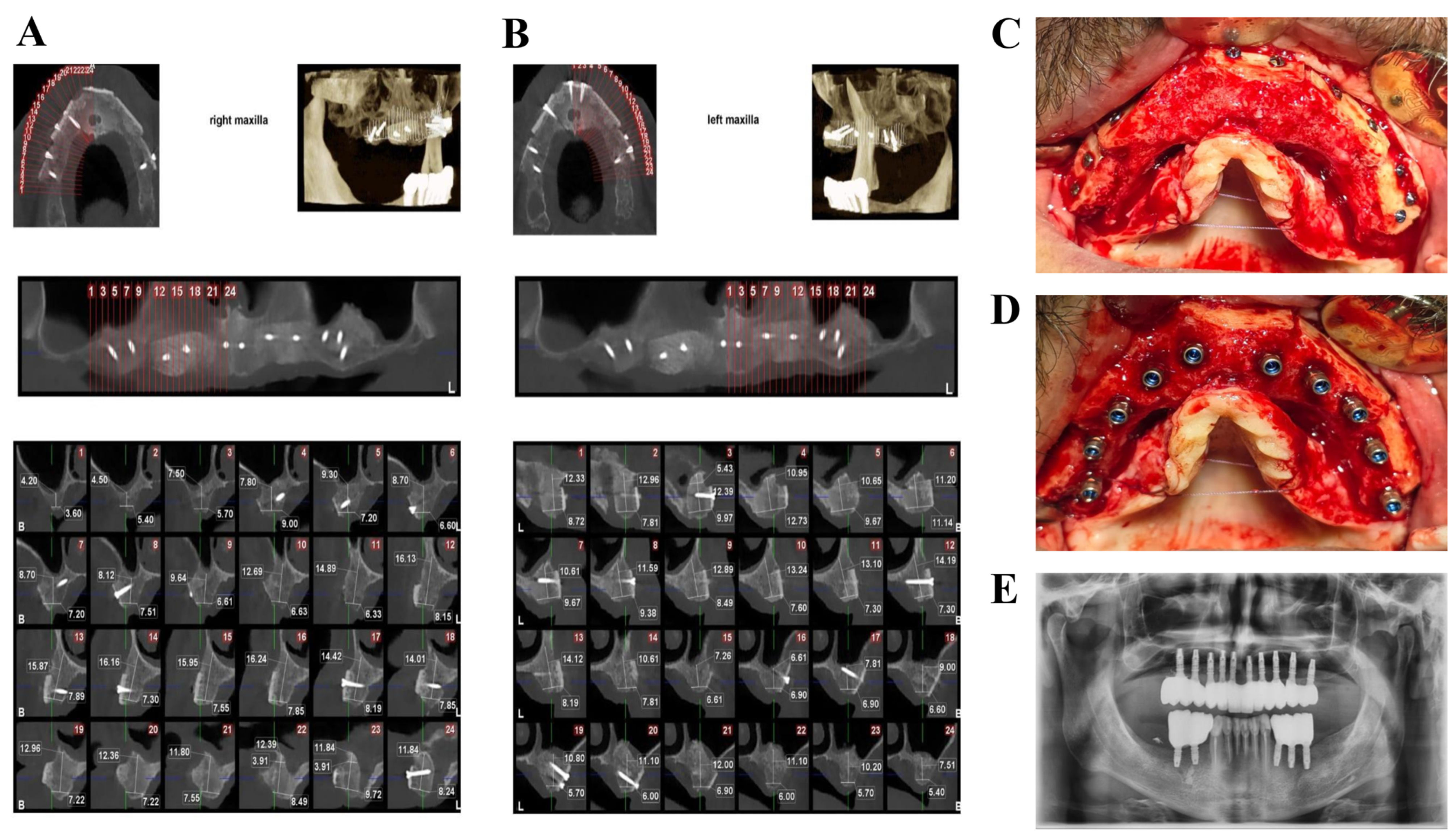
Following the six-month healing period, a total of 5 dental implants were placed in the right maxillary augmented site and 5 dental implants were placed in the left maxillary augmented site. The implants were placed in the regenerated alveolar ridges without requiring additional bone augmentation. The number and distribution of implants corresponded to the planned prosthetic rehabilitation. Following implant placement and prosthetic restoration, the patient was followed clinically and radiographically for 30 months. During this follow-up period, the implant remained clinically stable, with healthy peri-implant soft tissues and no evidence of marginal bone loss or biological or mechanical complications. Follow-up clinical examination demonstrated healthy peri-implant soft tissues without signs of inflammation or graft-related complications. Representative clinical photographs and CBCT images illustrating preoperative defect morphology, surgical augmentation, healed ridge anatomy, and implant placement are presented in Figure 1, Figure 2 and Figure 3.
Figure 3.
Clinical and radiographic outcomes following horizontal alveolar ridge augmentation using the modified corticocancellous shell (Kamali) technique. (A) Postoperative CBCT assessment of the right maxilla and (B) left maxilla, including representative serial cross-sectional images illustrating the regenerated ridge dimensions at the treated sites. (C) Clinical re-entry after six months demonstrating graft incorporation. (D) Implant placement within the regenerated ridge. (E) Post-restorative panoramic radiograph demonstrating the completed implant-supported rehabilitation.
The representative serial cross-sectional CBCT images were included to illustrate the anatomical distribution of the regenerated bone across the treated maxillary sites. These findings include changes in horizontal ridge dimensions, radiographic evidence of graft incorporation, implant placement feasibility, and postoperative healing observations. The measurements are presented descriptively and reflect the clinical course of this individual case. The clinical and radiographic findings documented before and after augmentation in this patient are summarized in Table 1.
Table 1.
Descriptive summary of clinical and radiographic findings before and after horizontal alveolar ridge augmentation in the reported patient.
As summarized in Table 1, the modified corticocancellous shell technique resulted in clinically successful horizontal ridge reconstruction with qualitative CBCT findings demonstrating continuity between the augmented region and adjacent native alveolar bone after six months of healing. The regenerated ridge provided adequate bone volume for prosthetically driven implant placement without additional augmentation procedures. Healing was uneventful, with no postoperative complications such as graft exposure, wound dehiscence, or fixation failure observed during the documented follow-up period. The preservation of the cancellous component may provide additional structural support during graft fixation and increase the internal volume available for particulate graft placement. However, these potential advantages have not been evaluated through direct comparative measurements and should therefore be interpreted as technical observations rather than demonstrated clinical benefits. This modification may be particularly valuable in extensive horizontal alveolar ridge defects where minimizing donor-site morbidity and simplifying graft fixation are important clinical objectives.
4. Discussion
Horizontal alveolar ridge augmentation remains one of the most challenging procedures in implant reconstructive surgery because successful regeneration requires simultaneous space maintenance, mechanical stability, adequate vascularization, and sufficient osteogenic potential. The shell technique introduced by Khoury has become one of the most predictable approaches for reconstructing horizontally deficient ridges because it creates a biologically protected compartment that supports new bone formation. Nevertheless, the procedure remains technically demanding due to harvesting autogenous cortical plates, adaptation of thin shells, and stabilization with multiple fixation screws [11,12,13,20,21,22]. The present report describes a modification of this concept by preserving the corticocancellous architecture of an allogeneic block, thereby combining the structural rigidity of cortical bone with the porous architecture of cancellous bone within a single graft construct. Unlike the conventional shell technique, the corticocancellous configuration acts not only as a barrier defining the regenerative compartment but also as an intrinsic scaffold capable of containing particulate graft material within interconnected cancellous spaces. This design provides an alternative structural configuration for shell-based augmentation while maintaining the biological principles of compartment-based guided bone regeneration. Whether this configuration reduces the technical complexity of graft fixation or improves mechanical performance compared with conventional shell techniques remains to be investigated.
The original Khoury shell technique remains an important reference standard for horizontal ridge augmentation because of its long-term predictability and excellent implant survival rates. However, its successful execution requires harvesting thin cortical plates from intraoral donor sites, including the mandibular ramus or symphysis. Although autogenous cortical bone possesses osteogenic potential, harvesting introduces donor-site morbidity, prolongs operative time, and limits the available graft volume in extensive defects. The Kamali Technique does not replace the biological principles of the Khoury technique; rather, it modifies the structural configuration of the shell component. Instead of harvesting and adapting thin cortical plates, a corticocancellous allogeneic block is adapted while preserving its cancellous architecture. Consequently, the regenerative compartment is maintained by the graft itself rather than relying exclusively on manually positioned cortical plates. The principal distinction between the Kamali technique and previously described allogeneic shell techniques is the preservation and modification of the cancellous component of the allogeneic block. While conventional techniques primarily use thin cortical plates to establish the regenerative compartment, the Kamali technique utilizes the intact corticocancellous structure, with additional internal recesses to facilitate particulate graft containment. This modification incorporates the cancellous component as an intrinsic part of the graft construct and provides an alternative approach to particulate graft containment. Although this configuration may offer practical advantages during graft adaptation and fixation, its effects on mechanical stability and fixation requirements have not been established. Comparative clinical studies are therefore necessary to determine whether these proposed technical characteristics result in measurable clinical benefits.
Another important distinction involves the proportion of autogenous bone required. Conventional shell techniques frequently require larger volumes of particulate autogenous bone harvested from intraoral donor sites. In the present modification, approximately 30–40% of autogenous particulate bone was combined with particulate bone substitute material while maintaining satisfactory compartment filling. Reducing the volume of harvested autogenous bone may decrease donor-site morbidity without eliminating the biological contribution of autogenous graft material.
The clinical effectiveness of shell techniques has been extensively documented in the literature. Kämmerer et al. reported an overall complication rate of 8.1%, including wound dehiscence, shell fracture, and screw exposure. Despite these complications, implant placement was successfully achieved in the majority of cases, confirming the technique’s overall predictability [1]. Similarly, Monje et al. demonstrated mean horizontal bone gains of approximately 5 mm in severely resorbed anterior maxillae using autogenous cortical blocks combined with particulate xenograft. However, they also highlighted important drawbacks of autogenous grafting, including donor-site morbidity and postoperative graft resorption [23].
To overcome these disadvantages, several investigators have evaluated allogeneic cortical shells as an alternative to autogenous bone. In a split-mouth clinical study, Tunkel et al. demonstrated that autogenous and allogeneic shell techniques achieved comparable horizontal and vertical bone gain, similar resorption rates, and successful implant placement in all augmented sites [5]. In a subsequent clinical investigation, the same group reported comparable implant survival rates while significantly reducing operative time with allogeneic shells, supporting their clinical applicability as a less invasive alternative to autogenous grafts [2].
Additional studies have confirmed these findings. Karkar et al. and Doliveux demonstrated that both conventional and CAD/CAM-manufactured allogeneic shells can achieve substantial horizontal ridge augmentation with minimal graft resorption and low complication rates, particularly in the esthetic zone [24,25]. Likewise, Khojasteh et al. evaluated the block-tenting technique using autogenous and allogeneic blocks as shell materials and reported considerable horizontal bone gain in both groups. Nevertheless, a higher graft failure rate was observed with allogeneic blocks, emphasizing the importance of careful patient selection, appropriate graft handling, and meticulous surgical technique [26]. Clinical studies have demonstrated the potential of allogeneic bone grafts in implant site development. However, evidence from socket preservation and immediate implant rehabilitation should be interpreted separately from outcomes of shell-based horizontal ridge augmentation. Therefore, further clinical investigations are required to establish the long-term stability and regenerative performance of allogeneic grafts specifically in shell-based augmentation procedures [14].
Although socket preservation differs from shell-based ridge augmentation, these long-term outcomes further support the biological potential and clinical stability of allogeneic grafts for implant site development. The present modification offers a different structural configuration compared with conventional shell techniques. The preserved cancellous component may provide an additional structural framework within the graft construct and facilitate particulate graft containment and adaptation. This configuration represents a technical modification that may offer an alternative to the use of thin, isolated cortical shells. However, no direct biomechanical testing or comparative clinical assessment was performed to determine whether corticocancellous blocks provide greater mechanical stability, reduce technical sensitivity during fixation, or decrease the risk of graft fracture. Although no graft fracture or fixation-related complication was observed in the present case, this single clinical observation cannot establish a reduction in fracture risk. Therefore, the proposed structural and technical advantages should be considered theoretical or preliminary and require further investigation through controlled comparative studies. The reduced reliance on autogenous bone harvesting represents a practical characteristic of the described protocol, although its impact on donor-site morbidity should also be evaluated in larger clinical studies.
In the present case, the corticocancellous configuration functioned as an intrinsic spacer and provided additional space for particulate graft placement. This structural modification may facilitate graft adaptation and stabilization; however, no comparative assessment of fixation requirements, operative time, graft fracture rates, or procedural costs was performed. Therefore, these observations should be interpreted as technical considerations rather than evidence of clinical or procedural superiority over established shell techniques.
Overall, the present case suggests that the use of corticocancellous allogeneic blocks represents a feasible modification of the shell technique that provides an alternative approach to graft adaptation and particulate graft containment while preserving the biological concept of compartment-based bone regeneration. This approach may represent a valuable alternative for alveolar ridge augmentation before implant placement, particularly in cases requiring extensive reconstruction or limited autogenous bone harvesting.
Several limitations should be acknowledged. First, this manuscript represents a description of routine clinical care in a single patient and should therefore be interpreted as a descriptive case report rather than a clinical investigation designed to evaluate treatment efficacy. The clinical and radiographic findings reflect the individual patient’s treatment course and cannot be generalized to a broader patient population. No comparative group, prospective data collection, quantitative assessment of operative time or treatment costs, or patient-reported outcome measures were available. Although clinical and radiographic follow-up was documented, observations from a single case cannot establish the long-term predictability, efficacy, or reproducibility of the technique. Further clinical investigations involving appropriately designed comparative studies and larger patient populations are needed to determine the potential clinical value and limitations of this modification.
5. Conclusions
This case report describes the clinical application of the modified corticocancellous shell technique (Kamali Technique) in a patient undergoing horizontal alveolar ridge augmentation as part of routine clinical care. In this individual case, satisfactory graft incorporation and horizontal ridge augmentation were observed at the approximately six-month follow-up, allowing subsequent implant placement with adequate primary stability. Preservation of the cancellous component and the creation of internal graft chambers represent the principal technical characteristics of this modification and provide an alternative structural configuration for particulate graft containment. However, these findings represent observations from a single patient and should not be interpreted as evidence of clinical efficacy, superiority, or reproducibility. Further clinical investigations are necessary to evaluate the potential benefits, limitations, and long-term outcomes of this approach.
Author Contributions
Conceptualization: L.K.H., A.G., H.M., F.N., H.T. and N.N.; Methodology: L.K.H., A.G., H.M., F.N., H.T. and N.N.; Investigation: L.K.H., A.G., H.M., F.N., H.T. and N.N.; Data curation: L.K.H., A.G., H.M., F.N. and N.N.; Writing—original draft preparation: L.K.H., A.G., H.M., F.N. and N.N.; Writing—review and editing: L.K.H., A.G., H.M., F.N., H.T. and N.N.; Visualization: L.K.H., A.G., H.M., F.N., H.T. and N.N.; Supervision: A.G. and N.N. All authors have read and agreed to the published version of the manuscript.
Funding
There was no funding provided.
Institutional Review Board Statement
The study was conducted in accordance with the Declaration of Helsinki, and approved by the private Institutional Review Board (IRB) of Kamali Private Clinic (protocol code 1404-01-12/10-35; 12 April 2024 of approval).
Informed Consent Statement
Informed consent was obtained from the patient.
Data Availability Statement
Data are contained within the article.
Acknowledgments
The authors thank their colleagues for their insightful comments.
Conflicts of Interest
The authors declare no conflicts of interest.
References
- Kämmerer, P.W.; Tunkel, J.; Götz, W.; Würdinger, R.; Kloss, F.; Pabst, A. The allogeneic shell technique for alveolar ridge augmentation: A multicenter case series and experiences of more than 300 cases. Int. J. Implant Dent. 2022, 8, 48. [Google Scholar] [CrossRef] [Scilit] [PubMed]
- Tunkel, J.; Hoffmann, F.; Schmelcher, Y.; Kloss-Brandstätter, A.; Kämmerer, P.W. Allogeneic versus autogenous shell technique augmentation procedures: A prospective-observational clinical trial comparing surgical time and complication rates. Int. J. Implant Dent. 2023, 9, 52. [Google Scholar] [CrossRef] [Scilit] [PubMed]
- Bubalo, M.; Dugonjic, S.; Dubovina, D.; Stojanovic, Z.; Gardasevic, M.; Mijatovic, J.; Milovanovic, B.; Stevic, M.; Stepovic, M.; Jeremic, D.; et al. Bone Substitutes in Alveolar Ridge Augmentation: A Narrative Literature Review. J. Funct. Biomater. 2026, 17, 176. [Google Scholar] [CrossRef] [Scilit] [PubMed]
- Dimova, C.; Evrosimovska, B.; Zlatanovska, K.; Zarkova, J. Alveolar Augmentation Using Different Bone Substitutes. In Handbook of Bioceramics and Biocomposites; Antoniac, I.V., Ed.; Springer International Publishing: Cham, Switzerland, 2016; pp. 1159–1199. [Google Scholar]
- Tunkel, J.; de Stavola, L.; Kloss-Brandstätter, A. Alveolar ridge augmentation using the shell technique with allogeneic and autogenous bone plates in a split-mouth design-A retrospective case report from five patients. Clin. Case Rep. 2021, 9, 947–959. [Google Scholar] [CrossRef] [Scilit] [PubMed]
- Wang, H.; Kang, J. Bone grafts and synthetic substitutes in dental applications: A comprehensive review of molecular mechanisms, materials evolution, and clinical perspective. Front. Bioeng. Biotechnol. 2025, 13, 1759864. [Google Scholar] [CrossRef] [Scilit] [PubMed]
- Schmidt, A.H. Autologous bone graft: Is it still the gold standard? Injury 2021, 52, S18–S22. [Google Scholar] [CrossRef] [Scilit] [PubMed]
- Streckbein, P.; Meier, M.; Kähling, C.; Wilbrand, J.-F.; Langguth, T.; Schaaf, H.; Howaldt, H.-P.; Streckbein, R.; Attia, S. Donor-site Morbidity after Retromolar Bone Harvesting Using a Standardised Press Fit Cylinder Protocol. Materials 2019, 12, 3802. [Google Scholar] [CrossRef] [Scilit] [PubMed]
- Haugen, H.J.; Sanz, J.; Perale, G.; Saiz, A.M.; Romandini, M.; Rahmati, M. Bone Grafts: Everything You Need to Know. J. Periodontal Res. 2026, 1–29. [Google Scholar] [CrossRef] [Scilit] [PubMed]
- Starch-Jensen, T.; Ahmad, M.; Bruun, N.H.; Becktor, J.P. Patient’s perception of recovery after maxillary sinus floor augmentation with autogenous bone graft compared with composite grafts: A single-blinded randomized controlled trial. Int. J. Implant Dent. 2021, 7, 99. [Google Scholar] [CrossRef] [Scilit] [PubMed]
- Khoury, F.; Hanser, T. Mandibular bone block harvesting from the retromolar region: A 10-year prospective clinical study. Int. J. Oral Maxillofac. Implant. 2015, 30, 688–697. [Google Scholar] [CrossRef] [Scilit] [PubMed]
- Khoury, F.; Hanser, T. Three-dimensional vertical alveolar ridge augmentation in the posterior maxilla: A 10-year clinical study. Int. J. Oral Maxillofac. Implant. 2019, 34, 471–480. [Google Scholar] [CrossRef] [Scilit] [PubMed]
- Khoury, F.; Khoury, C.H. Mandibular bone block grafts: Diagnosis, instrumentation, harvesting techniques and surgical procedures. In Bone Augmentation in Oral Implantology; Quintessence: Berlin, Germany, 2007; pp. 115–212. [Google Scholar]
- Saveinai, S.; Banodkar, A.; Gaikwad, R.; Jain, H.; Shirsat, S.; Dixit, G.; Kiso, A.; Chidambaram, M.; Metaliya, N. Clinical Efficacy of Allogeneic Bone Plate Shell Technique for Alveolar Ridge Augmentation: A Systematic Review. Cureus 2025, 17, e99228. [Google Scholar] [CrossRef] [Scilit] [PubMed]
- Saleh, H.; Eldibany, R.; Metawie, D. Clinical and radiographic outcomes of horizontal auto-augmentation of the anterior mandible using a single-site surgical technique prior to delayed implant placement: A case series. BMC Oral Health 2026, 26, 1243. [Google Scholar] [CrossRef] [Scilit] [PubMed]
- Salahi, S.; Shaar, M.K.; Pitman, J.; Vervaeke, S.; Cosyn, J.; Younes, F.; De Bruyckere, T. Patient-Reported Outcomes in Intraoral Bone Block Augmentation Compared to GBR Procedures Prior to Implant Placement: A Systematic Review. J. Clin. Med. 2025, 14, 5331. [Google Scholar] [CrossRef] [Scilit] [PubMed]
- Ku, J.K.; Ghim, M.S.; Park, J.H.; Leem, D.H. A ramus cortical bone harvesting technique without bone marrow invasion. J. Korean Assoc. Oral Maxillofac. Surg. 2023, 49, 100–104. [Google Scholar] [CrossRef] [Scilit] [PubMed]
- Palombo, D.; Ríos-Barbero, J.; Conforti, T.; Chiapasco, M. Autologous Bone for Periodontal and Craniofacial Regeneration: Donor Sites, Characteristics and Indications. J. Periodontal Res. 2026, 1–41. [Google Scholar] [CrossRef] [Scilit] [PubMed]
- Reininger, D.; Cobo-Vázquez, C.; Monteserín-Matesanz, M.; López-Quiles, J. Complications in the use of the mandibular body, ramus and symphysis as donor sites in bone graft surgery. A systematic review. Med. Oral Patol. Oral Cir. Bucal 2016, 21, e241–e249. [Google Scholar] [CrossRef] [Scilit] [PubMed]
- Łoś, J.; Szałkowska-Łoś, J.; Pikuła, M.; Adamska, P. An Autologous Tooth-Shell Technique for Ridge Reconstruction and Immediate Implant Placement: A Two-Case Technical Report and Review of the Literature. J. Funct. Biomater. 2026, 17, 402. [Google Scholar] [CrossRef] [Scilit] [PubMed]
- Fu, J.-H.; Choo, H.J.S.; Ong, D.-S.; Kwek, H. Long-term stability of horizontal bone augmentation at implant sites. Periodontol. 2000 2026, 1–28. [Google Scholar] [CrossRef] [Scilit] [PubMed]
- Elboraey, M.O.; Alqutaibi, A.Y.; Aboalrejal, A.N.; Borzangy, S.; Zafar, M.S.; Al-Gabri, R.; Alghauli, M.A.; Ramalingam, S. Regenerative approaches in alveolar bone augmentation for dental implant placement: Techniques, biomaterials, and clinical decision-making: A comprehensive review. J. Dent. 2025, 154, 105612. [Google Scholar] [CrossRef] [Scilit] [PubMed]
- Monje, A.; Monje, F.; Hernández-Alfaro, F.; Gonzalez-García, R.; Suárez-López del Amo, F.; Galindo-Moreno, P.; Montanero-Fernández, J.; Wang, H.-L. Horizontal Bone Augmentation Using Autogenous Block Grafts and Particulate Xenograft in the Severe Atrophic Maxillary Anterior Ridges: A Cone-Beam Computerized Tomography Case Series. J. Oral Implantol. 2015, 41, 366–371. [Google Scholar] [CrossRef] [Scilit] [PubMed]
- Doliveux, R.; Doliveux, S. Guided and Prosthetically Driven Bone Augmentation Using the Shell Technique and Allogeneic Cortical Plate: A Prospective Case Series. Int. J. Oral Maxillofac. Implant. 2024, 39, 263–270. [Google Scholar] [CrossRef] [Scilit] [PubMed]
- Karkar, K.T.; Metwally, S.A.; Mohsen, K.M.A. Anterior Aesthetic Zone Reconstruction with Allogenic Bone Shell and Autogenous Bone Chips—An Evaluative Study. Ann. Maxillofac. Surg. 2023, 13, 13–18. [Google Scholar] [CrossRef] [Scilit] [PubMed]
- Khojasteh, A.; Behnia, H.; Shayesteh, Y.S.; Morad, G.; Alikhasi, M. Localized bone augmentation with cortical bone blocks tented over different particulate bone substitutes: A retrospective study. Int. J. Oral Maxillofac. Implant. 2012, 27, 1481–1493. [Google Scholar]

Disclaimer/Publisher’s Note: The statements, opinions and data contained in all publications are solely those of the individual author(s) and contributor(s) and not of MDPI and/or the editor(s). MDPI and/or the editor(s) disclaim responsibility for any injury to people or property resulting from any ideas, methods, instructions or products referred to in the content. |
© 2026 by the authors. Licensee MDPI, Basel, Switzerland. This article is an open access article distributed under the terms and conditions of the Creative Commons Attribution (CC BY) license.