


Abstract
Objectives: Studies have shown that nanoparticles can impart beneficial properties when integrated into resin composites; however, it remains unclear how this affects shear bond strength. This study aimed to evaluate the effects of composite formulation and zein-coated magnesium oxide nanoparticle (zMgO NP) concentration on shear bond strength (SBS) and failure mode following thermocycling. Methods: A total of 180 extracted human molars were randomly allocated among four composite formulations and three zMgO NP concentrations (0%, 0.5%, and 1%; n = 15). Composite cylinders were bonded to prepared dentin surfaces and thermocycled for 10,000 cycles between 5 °C and 55 °C. SBS was analyzed using two-way ANOVA and post hoc multiple comparisons with Tukey’s test. Failure modes were assessed stereomicroscopically and analyzed using the chi-square test. Results: The SBS of the modified composites in all groups significantly increased, with the 0.5% zMgO NP groups exhibiting the highest SBS. The 0.5% zMgO NPs N-Ceram group had the highest SBS overall. The unmodified Z350 composite exhibited the highest adhesive failure, while the control N-Flow composite demonstrated the highest cohesive failure. The modified conventional and flowable composites showed initially lower adhesive failure, which increased progressively as zMgO NPs increased. The bulk-fill composite initially showed higher adhesive failure, with the 0.5% group demonstrating lower adhesive failure and the 1% group exhibiting increased adhesive failure. Conclusions: Adding zMgO NPs to resin composites improved SBS after thermocycling, with material- and concentration-dependent differences in failure-mode analysis. Further validation studies are needed to verify clinical viability.
1. Introduction
The current landscape of restorative dentistry relies heavily on resin-based composites because of their adaptability, esthetic compatibility with tooth structure, and cost-effectiveness [1]. The long-standing history of resin composite use also involves clinical limitations, particularly polymerization shrinkage and insufficient resistance to functional stresses, which may contribute to marginal breakdown or restoration failure [2,3]. Researchers have explored incorporating nanoscale fillers in resin composites because they can introduce antibacterial activity, enhance mineral deposition, and improve the structural integrity of restorative materials. The addition to preformulated resin composites may also influence physicochemical characteristics relevant to bonding [4,5,6].
Nanoparticle incorporation can alter the effective filler–resin ratio, while nanoparticle concentration and dispersion may affect light transmission, polymerization kinetics, and degree of conversion [7,8]. Related changes in elastic modulus, polymerization shrinkage, wettability, water sorption, and interfacial stress may consequently influence bond performance [9,10,11,12]. Presently, investigations on the scope of nanoparticle use are still emerging [13,14]. This knowledge gap limits their use in dentistry, where material durability and strength are crucial considerations [15].
It is still unclear whether their addition might impact other properties, since their small size and high reactivity can lead to unanticipated interactions [5,16,17]. Nanoparticles tend to cluster to reduce the surface energy of surface atoms that lack neighbors to bind to [18]. This clustering, which typically happens at higher concentrations, can ultimately affect the dental material and impede its mechanical properties [4]. To mitigate this agglomeration, various techniques such as surface modification, use of a silane coupling agent, pH control, and reduction in concentration can be employed to enhance nanoparticle dispersion and stability in hydrophilic environments [19].
Several types of inorganic nanoparticles have been incorporated into resin composites. In one study, the authors assessed the color stability of resin composite modified with ZrO2, TiO2, and SiO2. They discovered that 3% and 7% ZrO2, and only the lower concentrations of TiO2 and SiO2, improved the color stability of the resin composite [20]. Similarly, in another study, the mechanical effects of adding ZrO2, TiO2, and SiO2 to resin composite were investigated. It was reported that while hardness increased across all groups, flexural strength and wear resistance varied, with lower values at higher nanoparticle concentrations [21].
Previous investigations into magnesium oxide nanoparticles (MgO NPs) have shown antimicrobial activity and a favorable biological profile [22,23,24]. To prevent MgO NP agglomeration, zein (a polymer derived from corn) can be added to create a coating that inhibits clustering [25]. Combining zein with MgO NPs improves nanoparticle dispersibility [26]. This line of nanoparticles has demonstrated several mechanical enhancements in dental biomaterials; however, given their novelty, many facets remain unexplored [22,24,27].
The shear bond strength (SBS) of a material refers to the maximum stress that an interface can withstand before failure occurs when a shearing force is applied parallel to the bonded surface. It is a crucial parameter in assessing the mechanical effectiveness of dental adhesives and resin composites [28]. Adequate SBS contributes to the long-term success of direct restorations by providing proper retention, marginal integrity, and resistance to masticatory forces [29,30,31,32]. The SBS can be affected by the condition of the enamel and dentin, surface preparation techniques, the adhesive system, moisture control, and the mechanical properties of the resin composite [28,33,34].
Before such modified materials can be considered for broader restorative applications, it is necessary to determine whether nanoparticle incorporation adversely affects their mechanical performance. Therefore, the present study evaluated how incorporating zein-coated magnesium oxide nanoparticles (zMgO NPs) affects the SBS and fracture behavior of different resin composite formulations after thermocycling. The null hypothesis was that neither composite formulation nor zMgO NP concentration, nor their interaction, would significantly affect SBS or failure-mode distribution after thermocycling.
2. Methodology
2.1. Study Design
A sample-size calculation was performed using G*Power (Version 3.1.9.6) for a two-way ANOVA assessing the interaction between composite formulation and nanoparticle concentration. Assuming a significance level of 0.05, 80% statistical power, a moderate effect size, and 12 experimental groups, the minimum required sample size was 179 specimens. This was expanded to 180 specimens to permit equal allocation of 15 specimens to each experimental group.
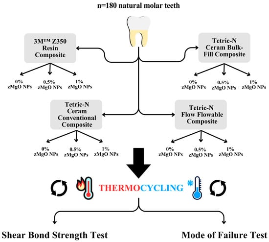
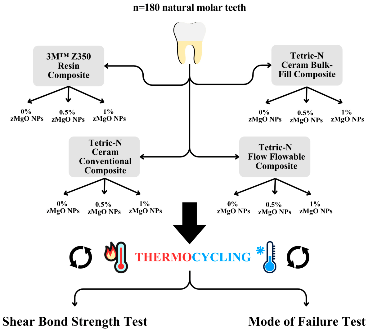
A total of 180 extracted human molars were designated to 4 experimental categories based on composite formulation. This was done using a computer-generated random number assignment to ensure unbiased allocation to the nanoparticle concentration groups. Within each category, specimens were subdivided into three subgroups based on nanoparticle concentration: 0% (unmodified control), 0.5%, and 1% by weight. The samples were also allocated to the zMgO NP concentration groups using a computer-generated random number assignment. Before assessment, the specimens were allotted coded identifiers by an independent investigator who was not involved in the evaluation, thereby blinding the examiners to the composite formulation and zMgO NP concentration. After sample fabrication, all samples underwent thermocycling before mechanical testing and mode-of-failure analysis (Figure 1).
Figure 1.
The 4 composite types were modified with zMgO NPs. The samples were then thermocycled and assessed for shear bond strength and failure mode.
The 0.5% and 1% concentrations were selected based on previous studies demonstrating antimicrobial activity within this concentration range [26,35]. We previously evaluated these two concentrations to determine whether the effect of zMgO NP incorporation was concentration-dependent while maintaining relatively low nanoparticle loading. The nanoparticles were added to the commercial composite without removing or replacing any of its original components. Consequently, nanoparticle incorporation slightly increased the overall inorganic filler-to-resin ratio of the modified materials, whereas the manufacturer-determined proportions of the original filler and resin components within the commercial composite remained unchanged.
2.2. Sample Preparation
Extracted human molars free of caries, restorations, cracks, and developmental defects were selected. After cleaning, teeth were stored in saline mixed with 0.1% thymol solution at 4 °C, with the storage medium replaced every 48 h. The occlusal enamel was removed perpendicular to the long axis of each tooth using a water-cooled diamond saw and an IsoMet machine (Buehler International Inc., Lake Bluff, IL, USA) to expose a flat mid-coronal dentin surface. Each tooth was embedded in acrylic resin, with the prepared dentin surface positioned parallel to and exposed above the acrylic resin. The dentin surface was standardized by wet-polishing with 600-grit silicon-carbide abrasive papers under continuous water irrigation using a micromotor (Marathon Saeyang M4; Saeyang Microtech, Daegu, Republic of Korea) to create uniform bonding substrates.
The zMgO NPs were prepared according to a previously established protocol [25,26]. The MgO NPs (40 nm in diameter and 100 nm in length) were coated with zein particles, which were also 40 nm in diameter and 100 nm in length. Zein was deposited onto the MgO NPs using pH-controlled nanoprecipitation. A solution of ethanol and 0.1 NaOH (93.7% v/v) was prepared, and 0.02 g of zein was added. A 15 mL mixture of 0.02 g MgO and polyvinyl alcohol (0.9% w/v) was infused at 10 °C using 750 W ultrasonic shear at 20 kHz, then stirred at 500 rpm and centrifuged twice at 3000 rpm for 45 min. This separates the nanoparticles from the solution.
Then, to liquefy the pellet, 5 mL of buffer and 2% (w/v) trehalose were added, after which the mixture was lyophilized (VirTis Bench Top Lyophilizer, SP Industries, Gardiner, NY, USA). Zein with polyvinyl alcohol (2:1 by weight) and MgO NPs (4:1 by weight) were magnetically stirred for 30 min. The polyvinyl alcohol was removed, after which the mixture was centrifuged again and freeze-dried.
For each composite formulation, the required mass of nanoparticles was calculated relative to the mass of the composite to obtain concentrations of 0%, 0.5%, and 1% by weight. The composite and nanoparticles were weighed using an analytical balance accurate to 0.0001 g (Precision Balance Scale, BEL Engineering, Monza, Italy). Details on the composite types and composition can be seen in Table 1. Mixing was performed at room temperature using a stainless-steel spatula and glass slab. During mixing, the composite was repeatedly folded and pressed with the nanoparticles until no visible particle clusters remained. The procedure was performed under reduced-light conditions to minimize premature polymerization.
Table 1.
Composition and filler characteristics of the commercial resin composites evaluated in this study.
To initiate bonding, 37% phosphoric acid (Meta Etchant, 37% Total Etching Gel Syringe, Meta Biomed, Cheongju, Republic of Korea) was applied according to the manufacturer’s instructions (15 s), rinsed with water for 10 s, and gently air-dried without desiccating the dentin to leave a visibly moist surface. Scotchbond Universal adhesive (3M™, Solventum, MN, USA) was applied to the dentin surface according to the manufacturer’s instructions (20 s application, 5 s air drying, 10 s light curing).
A cylindrical mold with an internal diameter of 4 mm and a height of 2 mm was positioned over the flat dentin surface. The base of the mold was seated flush with no visible gaps, and its longitudinal axis was kept perpendicular to the bonded surface. The mold was filled with the assigned composite while minimizing air entrapment. A transparent celluloid strip was placed over the mold, and gentle pressure was applied to compact the composite and produce a flat surface. Excess material extending beyond the mold margins was removed before curing, after which the seating and alignment of the mold were rechecked.
The composite was light-cured for 20 s using an LED curing unit (3M™ Elipar DeepCure-L, Solventum, St. Paul, MN, USA) with the following parameters: irradiance of 1470 mW/cm2, wavelength range of 430–480 nm, and tip diameter of 10 mm. The light-guide tip was centered over the composite cylinder, oriented perpendicular to its upper surface, and maintained in direct contact with the celluloid strip, corresponding to a curing distance of 0 mm. The mold was then removed, and the specimens underwent thermocycling.
2.3. Thermocycling
Artificial aging was simulated via repeated thermal cycling to reproduce intraoral temperature fluctuations. Specimens were alternately immersed in cold and hot water baths ranging from 5 °C to 55 °C for 10,000 cycles, with a 30 s dwell time in each bath and a 10 s interval between baths [39,40].
2.4. Shear Bond Strength Test
Mechanical testing was performed using a universal testing machine (Instron 5944, Instron, Norwood, MA, USA). Each acrylic-mounted specimen was secured so that the flat dentin surface was parallel to the direction of crosshead movement and perpendicular to the long axis of the composite cylinder. A knife-edged chisel was positioned against the cylinder’s lateral surface immediately adjacent to the composite–dentin interface, without contacting the samples. The load was applied parallel to the bonded interface at a crosshead speed of 1 mm/min until failure. The maximum failure load was recorded in MPa.
2.5. Mode-of-Failure Test
Following shear bond-strength testing, both the dentin-facing and composite-facing fracture surfaces of each specimen were examined using a stereomicroscope (SZX16, Olympus Corporation, Tokyo, Japan) at ×20 magnification. Failure mode was classified into four categories according to the location and characteristics of the fracture. Adhesive failure was recorded when separation occurred primarily at the dentin-adhesive or adhesive-composite interface, with adhesive or composite remnants predominantly present on only one fractured surface. Cohesive failure was defined as a fracture occurring within the composite or adhesive material, with material remnants visible on both opposing surfaces. Mixed failure was recorded when the specimen exhibited both adhesive and cohesive fracture features. Substrate failure was defined as a fracture within the dentin while the bonded restorative interface remained partially or completely intact.
Two examiners independently classified the failure mode of each specimen while blinded to the experimental groups. Before the formal evaluation, the examiners reviewed the classification criteria to ensure their consistent application. Inter-examiner reliability was determined from their initial independent classifications using Cohen’s kappa coefficient. When the two examiners assigned different classifications, the specimen was referred to a third blinded examiner for adjudication, and the adjudicated classification was used in the final analysis.
2.6. Statistical Analysis
Data were analyzed using SPSS Statistics, version 22 (IBM Corp., Armonk, NY, USA). Shear bond strength was summarized as mean and standard deviation. The normality of the continuous data was assessed using the Shapiro–Wilk test. Homogeneity of variances was assessed using Levene’s test. A two-way analysis of variance was used to evaluate the main effects of composite formulation and zMgO NP concentration, as well as the interaction between composite formulation and nanoparticle concentration. Post hoc pairwise comparisons were performed with Tukey’s test. Failure modes were summarized as frequencies and percentages and compared across the experimental groups using the chi-square test. Statistical significance was set at p < 0.05.
3. Results
3.1. Shear Bond Strength
According to Pallant, a non-significant Levene’s test indicates homogeneity of variances [41]. In this study, Levene’s test was not statistically significant (F = 3.014, p = 0.221), indicating that the error variances were homogeneous across the experimental groups.
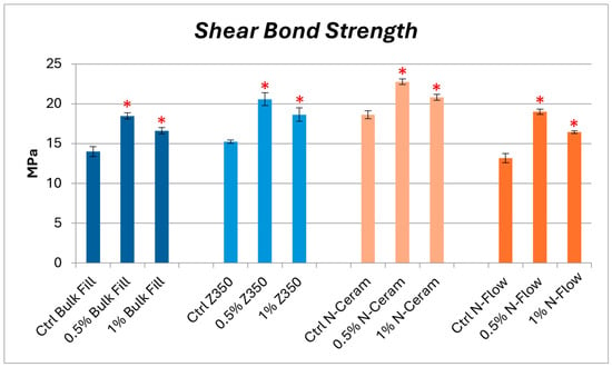
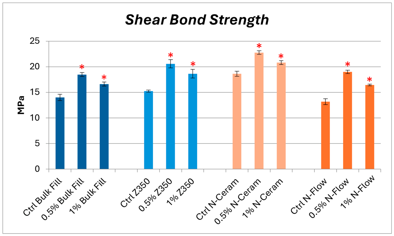
The two-way ANOVA test revealed a statistically significant difference between the groups of different composites (p < 0.001). A t-test was then performed to investigate the differences within the groups. As seen in Figure 2 and Table 2, In the bulk-fill group, there was a significant difference between the control and 0.5% zMgO NPs group (p < 0.001), and between the control and the 1% zMgO group (p = 0.0012). Additionally, there was a significant difference between the 0.5% and 1% zMgO NPs groups (p = 0.0088).
Figure 2.
The bars represent the mean shear bond strength of the samples after thermocycling, measured in MPa. A statistically significant difference was observed between the control and the 0.5% and 1% zMgO NPs samples within each composite group (p < 0.05), denoted by an asterisk (*). The bars (I) indicate the standard deviation.
Table 2.
Shear bond strength test mean and standard deviation across the experimental groups.
In the Z350 group, there was a significant difference between the control and 0.5% zMgO NP group, and between the control and 1% zMgO NP group (p < 0.001 and p < 0.001, respectively). There was also a significant difference between the 0.5% and 1% zMgO NP groups (p = 0.001).
In the N-Ceram group, there was a significant difference between the control and 0.5% zMgO NP composite samples (p < 0.001) and the control and 1% zMgO NP composite samples (p < 0.001). Likewise, there was a significant difference between the 0.5% and 1% zMgO NP groups (p = 0.0156).
As for the N-Flow group, there was a significant difference between the control and 0.5% zMgO NP composite samples (p < 0.001) and between the control and 1% zMgO NP composite samples (p < 0.001). Similarly, a significant difference was observed between the 0.5% and 1% zMgO NP samples (p = 0.00347).
3.2. Mode of Failure
The initial classifications demonstrated high inter-examiner agreement (Cohen’s κ ≥ 0.80). None of the samples underwent mixed or substrate failures. The groups exhibited adhesive and cohesive failures. The chi-squared test showed significant differences among the frequency distributions of the various groups with regard to adhesive and cohesive failure (p < 0.05).
3.2.1. Adhesive Failure
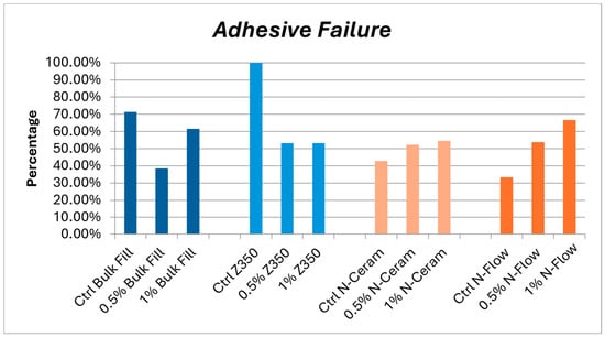
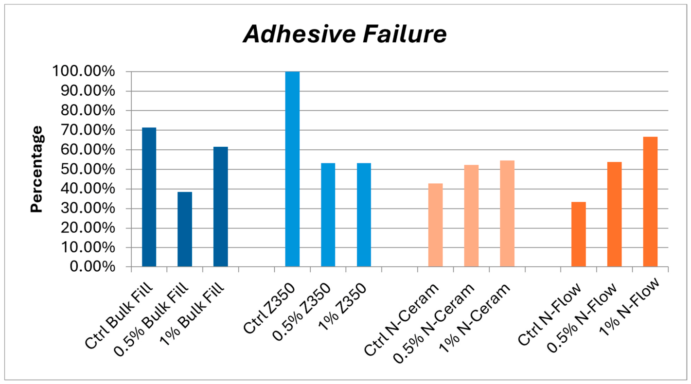
In the bulk-fill group, the control showed 71.4% adhesive failure, the 0.5% zMgO NP group showed 38.5%, and the 1% zMgO NP group showed 61.5%. For the Z350 group, the control had 100% adhesive failure, 0.5% zMgO NP group showed 53.3%, and the 1% zMgO NP group had 53.3% adhesive failure. For the N-Ceram group, the control had 42.8% adhesive failure, the 0.5% zMgO NP group showed 53.3%, and the 1% zMgO NP group had 54.6%. For the N-Flow group, the control had 33.3% adhesive failure, the 0.5% zMgO NP group demonstrated 53.8%, and the 1% zMgO NP group had 66.7% [Figure 3].
Figure 3.
The adhesive failure of the samples after thermocycling. The control Z350 composite demonstrated the highest adhesive failure.
3.2.2. Cohesive Failure
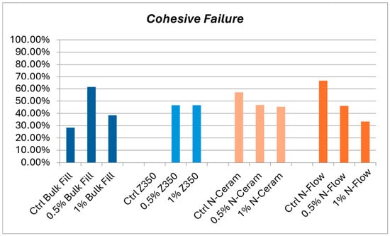
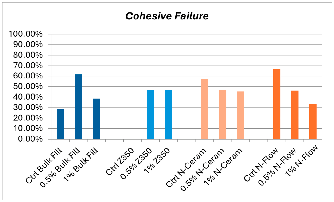
For the bulk-fill group, the control showed 28.6% cohesive failure, the 0.5% zMgO NP group showed 61.5%, and the 1% zMgO NP group had 38.5%. For the Z350 group, the control had 0% cohesive failure, while the 0.5% zMgO NP group demonstrated 46.7% failure, and the 1% zMgO NP group had 46.7% failure. For the N-Ceram group, the control had 57.2% cohesive failure, the 0.5% zMgO NP group had 46.7%, and the 1% zMgO NP group had 45.4%. For the N-Flow group, the control had 66.7% cohesive failure, the 0.5% zMgO NPs group exhibited 46.2% failure, and the 1% zMgO NP group had 33.3% failure [Figure 4].
Figure 4.
The cohesive failure of the samples after thermocycling. The control N-Flow composite demonstrated the highest cohesive failure.
4. Discussion
This study forms part of a broader series of investigations evaluating the biological, physical, and mechanical effects of incorporating zMgO NPs into various dental biomaterials. Under the tested thermocycling conditions, the zMgO NP-modified composites generally exhibited higher mean SBS values than their corresponding unmodified controls. The 0.5% zMgO NP group produced the highest mean SBS within each composite category, whereas increasing the concentration to 1% resulted in a slight reduction, although the 1% zMgO NP group’s SBS was still significantly higher than the control group’s SBS. The N-Ceram conventional composite containing 0.5% zMgO NPs demonstrated the highest mean SBS overall. These findings suggest a concentration-dependent, rather than linear, effect.
Rangrazi et al. studied how MgO NPs affect SBS in orthodontic composites. The study found no significant differences in SBS between the unaltered group, the 0.5% MgO NP group, and the 1% MgO NP group. However, SBS values with 2% and 4% MgO NPs were notably lower [42]. Sodagar et al. reported a progressive reduction in mean SBS after incorporating 1%, 5%, and 10% TiO2 NPs into an orthodontic composite, with the greatest reduction observed at 10% [43]. A comparable non-linear pattern was reported by Toodehzaeim et al., in which CuO NP incorporation produced the highest mean SBS at 0.5% and a lower value at 1%, although the differences from the unmodified orthodontic adhesive were not statistically significant [44]. Similarly, Mirhashemi et al. found that incorporating a combined ZnO-chitosan nanoparticle formulation into an orthodontic composite did not significantly affect SBS at total concentrations of 1% or 5%, whereas 10% significantly reduced SBS after thermocycling [45]. These findings further demonstrate that the influence of metal oxide nanoparticles on SBS is concentration- and material-dependent.
In our previous investigations, the characterization of zMgO NPs incorporated into different types of resin composites showed no distinct X-ray diffraction peaks compared with the unmodified composites. Fourier transform infrared spectroscopy showed no significant spectral changes, and scanning electron microscopy images revealed a consistent, uniform dispersion of the nanoparticles in the different composite types [46,47]. Because the zMgO NPs were incorporated into the composite rather than the adhesive, their effects would be expected to arise primarily within the composite and at the composite-adhesive junction.
The addition of zMgO NPs altered the effective particulate loading and filler-resin balance of the commercial composites. If adequately dispersed and integrated within the resin matrix, the nanoparticles could have occupied spaces between existing filler particles and contributed to more effective stress transfer, greater resistance to local deformation, or interruption of crack propagation [3,15,48]. These effects could increase the load required to produce specimen failure, as seen with previous studies of nanoparticle-modified dental materials [46,47].
However, the net effect of nanoparticle incorporation depends on several interrelated material properties. Stress distribution in bonded restorations is influenced by composite stiffness, polymerization shrinkage, adaptation, and adhesive-interface behavior [49,50]. The addition of zMgO NPs may have altered composite viscosity and adaptation to the adhesive-coated dentin, potentially affecting interfacial void formation. Particle concentration, dispersion, and refractive-index mismatch with the resin matrix could also influence light transmission and polymerization kinetics, thereby affecting the degree of conversion. Resulting changes in the polymer network and filler loading could subsequently influence composite modulus, cohesive strength, polymerization shrinkage, and stress transferred to the bonded interface [51,52]. Although increasing particulate content may reduce the proportion of polymerizable resin and its volumetric shrinkage, an accompanying increase in stiffness could increase interfacial stress [1,15,23,34]. Therefore, filler loading alone cannot predict the overall effect on SBS.
In this study, the SBS represents the nominal failure stress of the complete test assembly and is influenced by the dentin substrate, adhesive layer, composite properties, specimen geometry, loading configuration, and failure location. The higher SBS observed at 0.5% zMgO NPs may represent a favorable balance among particulate reinforcement, viscosity, polymerization, and stress distribution, as seen with other MgO NPs added to dental materials [15,22,53]. Conversely, the slight reduction at 1% could reflect increased viscosity, particle agglomeration, light attenuation, or defect formation at the higher concentration. Direct measurement of the degree of conversion using a validated FTIR or Raman protocol would be required because the estimated values depend on the selected technique, reference band, peak-analysis method, and sampling depth [54,55,56]. Characterization of nanoparticle dispersion, polymerization behavior, and interfacial morphology is therefore required to determine the mechanisms underlying the observed differences.
Analyzing the mode of failure after SBS testing can give insight into the state of the adhesive interface. The unmodified Z350 composite exhibited the highest proportion of adhesive failures (100%), whereas the inclusion of zMgO NPs introduced more cohesive failure. The conventional N-Ceram and flowable N-Flow composites demonstrated lower adhesive failure in the unmodified group, with progressively higher adhesive failure with the increase in zMgO NPs. Interestingly, the bulk-fill composite exhibited an initially higher adhesive failure, with the 0.5% zMgO NP group demonstrating a lower adhesive failure, and the 1% zMgO NP group exhibiting a higher adhesive failure, although less than the initial amount of the control group.
The failure-mode analysis provided qualitative information regarding the location of fracture following thermocycling. The lower proportion of adhesive failures observed in the 0.5% zMgO NP-modified bulk-fill group may indicate that failure occurred less frequently at the bonded interface. However, these findings cannot confirm changes such as filler-matrix debonding, interfacial degradation, or microcrack formation [57]. Failure-mode distributions varied according to composite formulation and zMgO concentration, indicating that nanoparticle addition did not produce a uniform effect across all materials. When increased SBS was accompanied by cohesive failure, the fracture may have shifted away from the bonded interface, suggesting that the interface was no longer the sole weakest component [58].
Nevertheless, no validated percentage of adhesive or cohesive failures constitutes a universally acceptable range or a threshold for poor mechanical performance or durability [59,60]. Failure-mode percentages describe where specimens fractured and are affected by test geometry, loading configuration, and stress distribution [57,58,60]. Adhesive and cohesive failure cannot independently demonstrate improved bonding or material deterioration [58,61].
5. Limitations
The absence of non-aged baseline groups prevented quantification of bond-strength changes attributable to thermal aging. Moreover, thermocycling in distilled water represents only hydrothermal stress and does not reproduce saliva exposure, pH variation, enzymatic degradation, or biofilm activity [51,62,63]. Although zein is susceptible to certain enzymes under non-dental conditions, whether oral enzymes can reach and degrade the embedded zein coating sufficiently to affect adhesion remains unknown [64,65]. Generalizability is also limited by the use of one bonding substrate and one adhesive system.
The study did not assess degree of conversion, viscosity, polymerization shrinkage, entrapped air, water sorption, or solubility, nor did it characterize hybrid-layer morphology, resin-tag formation, or interfacial nanomechanics. Stereomicroscopic failure classification remains qualitative. Therefore, the SBS values represent the nominal failure stress of the complete test assembly, and adhesive and cohesive failure patterns should be interpreted cautiously. Finally, ion and nanoparticle release, monomer elution, local pH and oxidative effects, and cytotoxicity were not assessed. Accordingly, the present findings represent post-thermocycling laboratory performance rather than evidence of long-term clinical durability or biological safety [66,67].
6. Conclusions
Incorporating zMgO NPs to enhance SBS offers a promising approach to improving the durability and reliability of resin-based restorations. Under the tested thermocycling conditions, the zMgO NP-modified composites demonstrated higher post-thermocycling SBS than their corresponding unmodified controls, with material- and concentration-dependent differences in failure behavior. The 0.5% concentration produced the highest mean SBS within each composite category, and the 0.5% zMgO NP-modified N-Ceram conventional composite showed the highest overall value. Confirmation of its clinical suitability requires further extended aging and interfacial characterization.
Author Contributions
Conceptualization: G.H.N. and M.T.H.; Data curation: G.H.N., R.A., S.A., D.A., J.M. and M.T.H.; Formal analysis: G.H.N., R.A., S.A., D.A., J.M., A.A. and M.T.H.; Funding acquisition: A.A. and M.T.H.; Investigation: G.H.N., R.A., S.A., D.A., J.M., A.A. and M.T.H.; Methodology: G.H.N., R.A., S.A., D.A., J.M., A.A. and M.T.H.; Visualization: G.H.N., R.A., S.A., D.A., J.M., A.A. and M.T.H.; Writing—original draft: R.A., S.A., D.A. and J.M.; Writing—review & editing: G.H.N., R.A., S.A., D.A., J.M., A.A. and M.T.H. All authors have read and agreed to the published version of the manuscript.
Funding
This research received no external funding.
Institutional Review Board Statement
Ethical clearance for the experimental protocol was granted by the institutional research ethics committee of King Abdulaziz University Faculty of Dentistry (Approval No. 182-11-23).
Informed Consent Statement
This study utilized previously extracted human teeth and did not involve direct participation of human subjects. The institutional research ethics committee of King Abdulaziz University Faculty of Dentistry determined that the study was exempt from informed consent requirements and did not constitute human-subject research (Approval No. 182-11-23).
Data Availability Statement
Data is available from the corresponding author upon reasonable request.
Acknowledgments
We’d like to extend our sincerest thanks to the King Abdulaziz University Advanced Technology Dental Research Laboratory for their support in conducting our research within their esteemed facility.
Conflicts of Interest
The authors declare no conflicts of interest.
References
- Ferracane, J.L. A Historical Perspective on Dental Composite Restorative Materials. J. Funct. Biomater. 2024, 15, 173. [Google Scholar] [CrossRef] [Scilit] [PubMed]
- Zhou, X.; Huang, X.; Li, M.; Peng, X.; Wang, S.; Zhou, X.; Cheng, L. Development and status of resin composite as dental restorative materials. J. Appl. Polym. Sci. 2019, 136, 48180. [Google Scholar] [CrossRef] [Scilit]
- Santos, M.J.M.C.; Rêgo, H.M.C.; Siddique, I.; Jessani, A. Five-Year Clinical Performance of Complex Class II Resin Composite and Amalgam Restorations—A Retrospective Study. Dent. J. 2023, 11, 88. [Google Scholar] [CrossRef] [Scilit] [PubMed]
- Gronwald, B.; Kozłowska, L.; Kijak, K.; Lietz-Kijak, D.; Skomro, P.; Gronwald, K.; Gronwald, H. Nanoparticles in Dentistry—Current Literature Review. Coatings 2023, 13, 102. [Google Scholar] [CrossRef] [Scilit]
- Dipalma, G.; Inchingolo, A.D.; Guglielmo, M.; Morolla, R.; Palumbo, I.; Riccaldo, L.; Mancini, A.; Palermo, A.; Malcangi, G.; Inchingolo, A.M.; et al. Nanotechnology and Its Application in Dentistry: A Systematic Review of Recent Advances and Innovations. J. Clin. Med. 2024, 13, 5268. [Google Scholar] [CrossRef] [Scilit] [PubMed]
- Glowacka-Sobotta, A.; Ziental, D.; Czarczynska-Goslinska, B.; Michalak, M.; Wysocki, M.; Güzel, E.; Sobotta, L. Nanotechnology for Dentistry: Prospects and Applications. Nanomaterials 2023, 13, 2130. [Google Scholar] [CrossRef] [Scilit] [PubMed]
- Howard, B.; Wilson, N.D.; Newman, S.M.; Pfeifer, C.S.; Stansbury, J.W. Relationships between conversion, temperature and optical properties during composite photopolymerization. Acta Biomater. 2010, 6, 2053–2059. [Google Scholar] [CrossRef] [Scilit] [PubMed]
- Habib, E.; Wang, R.; Zhu, X.X. Correlation of resin viscosity and monomer conversion to filler particle size in dental composites. Dent. Mater. 2018, 34, 1501–1508. [Google Scholar] [CrossRef] [Scilit] [PubMed]
- Ilie, N. Microstructural dependence of mechanical properties and their relationship in modern resin-based composite materials. J. Dent. 2021, 114, 103829. [Google Scholar] [CrossRef] [Scilit] [PubMed]
- Kim, R.J.-Y.; Kim, Y.-J.; Choi, N.-S.; Lee, I.-B. Polymerization shrinkage, modulus, and shrinkage stress related to tooth-restoration interfacial debonding in bulk-fill composites. J. Dent. 2015, 43, 430–439. [Google Scholar] [CrossRef] [Scilit] [PubMed]
- Stape, T.H.S.; Uctasli, M.; Cibelik, H.S.; Tjäderhane, L.; Tezvergil-Mutluay, A. Dry bonding to dentin: Broadening the moisture spectrum and increasing wettability of etch-and-rinse adhesives. Dent. Mater. 2021, 37, 1676–1687. [Google Scholar] [CrossRef] [Scilit] [PubMed]
- Sokolowski, G.; Szczesio, A.; Bociong, K.; Kaluzinska, K.; Lapinska, B.; Sokolowski, J.; Domarecka, M.; Lukomska-Szymanska, M. Dental Resin Cements—The Influence of Water Sorption on Contraction Stress Changes and Hydroscopic Expansion. Materials 2018, 11, 973. [Google Scholar] [CrossRef] [Scilit] [PubMed]
- Mascarenhas-Melo, F.; Mathur, A.; Murugappan, S.; Sharma, A.; Tanwar, K.; Dua, K.; Singh, S.K.; Mazzola, P.G.; Yadav, D.N.; Rengan, A.K.; et al. Inorganic nanoparticles in dermopharmaceutical and cosmetic products: Properties, formulation development, toxicity, and regulatory issues. Eur. J. Pharm. Biopharm. 2023, 192, 25–40. [Google Scholar] [CrossRef] [Scilit] [PubMed]
- Sharma, N.; Kurmi, B.D.; Singh, D.; Mehan, S.; Khanna, K.; Karwasra, R.; Kumar, S.; Chaudhary, A.; Jakhmola, V.; Sharma, A.; et al. Nanoparticles toxicity: An overview of its mechanism and plausible mitigation strategies. J. Drug Target. 2024, 32, 457–469. [Google Scholar] [CrossRef] [Scilit] [PubMed]
- Bourgi, R.; Doumandji, Z.; Cuevas-Suárez, C.E.; Ben Ammar, T.; Laporte, C.; Kharouf, N.; Haikel, Y. Exploring the Role of Nanoparticles in Dental Materials: A Comprehensive Review. Coatings 2025, 15, 33. [Google Scholar] [CrossRef] [Scilit]
- Yin, I.X.; Udduttulla, A.; Xu, V.W.; Chen, K.J.; Zhang, M.Y.; Chu, C.H. Use of Antimicrobial Nanoparticles for the Management of Dental Diseases. Nanomaterials 2025, 15, 209. [Google Scholar] [CrossRef] [Scilit] [PubMed]
- Yudaev, P.; Chuev, V.; Klyukin, B.; Kuskov, A.; Mezhuev, Y.; Chistyakov, E. Polymeric Dental Nanomaterials: Antimicrobial Action. Polymers 2022, 14, 864. [Google Scholar] [CrossRef] [Scilit] [PubMed]
- Umapathy, V.R.; Natarajan, P.M.; SumathiJones, C.; Swamikannu, B.; Johnson, W.M.S.; Alagarsamy, V.; Milon, A.R. Current trends and future perspectives on dental nanomaterials—An overview of nanotechnology strategies in dentistry. J. King Saud Univ.-Sci. 2022, 34, 102231. [Google Scholar] [CrossRef] [Scilit]
- Jandt, K.D.; Watts, D.C. Nanotechnology in dentistry: Present and future perspectives on dental nanomaterials. Dent. Mater. 2020, 36, 1365–1378. [Google Scholar] [CrossRef] [Scilit] [PubMed]
- Al-Dulaijan, Y.A.; AlGhamdi, M.A.; Azmy, E.; Al-Kholy, M.R.Z.; Almulhim, K.S.; Helal, M.A. Color Stability of Nanoparticles-Modified Dental Resin-Based Composites. Appl. Sci. 2023, 13, 3870. [Google Scholar] [CrossRef] [Scilit]
- Azmy, E.; Al-Kholy, M.R.Z.; Fattouh, M.; Kenawi, L.M.M.; Helal, M.A. Impact of Nanoparticles Additions on the Strength of Dental Composite Resin. Int. J. Biomater. 2022, 2022, 1165431. [Google Scholar] [CrossRef] [Scilit] [PubMed]
- Gatou, M.-A.; Skylla, E.; Dourou, P.; Pippa, N.; Gazouli, M.; Lagopati, N.; Pavlatou, E.A. Magnesium Oxide (MgO) Nanoparticles: Synthetic Strategies and Biomedical Applications. Crystals 2024, 14, 215. [Google Scholar] [CrossRef] [Scilit]
- Saberi, A.; Baltatu, M.S.; Vizureanu, P. Recent Advances in Magnesium–Magnesium Oxide Nanoparticle Composites for Biomedical Applications. Bioengineering 2024, 11, 508. [Google Scholar] [CrossRef] [Scilit] [PubMed]
- Sharifian, S.; Loghmani, A.; Nayyerain, S.; Javanbakht, S.; Daneii, P. Application of Magnesium Oxide Nanoparticles in Dentistry: A Literature Review. Eur. J. Gen. Dent. 2023, 12, 001–006. [Google Scholar] [CrossRef] [Scilit]
- Naguib, G.H.; Hassan, A.H.; Al-Hazmi, F.; Kurakula, M.; Al-Dharrab, A.; Alkhalidi, H.M.; Al-Ahdal, A.M.; Hamed, M.T.; Pashley, D.H. Zein Based Magnesium Oxide Nanowires: Effect of Anionic Charge on Size, Release And Stability. Dig. J. Nanomater. Biostruct. 2017, 12, 741–749. [Google Scholar]
- Naguib, G.H.; Hosny, K.M.; Hassan, A.H.; Al Hazmi, F.; Al Dharrab, A.; Alkhalidi, H.M.; Hamed, M.T.; Alnowaiser, A.M.; Pashley, D.H. Zein based magnesium oxide nanoparticles: Assessment of antimicrobial activity for dental implications. Pak. J. Pharm. Sci. 2018, 31, 245–250. [Google Scholar] [CrossRef] [Scilit]
- Naguib, G.H.; Alnowaiser, A.; Mazhar, J.; Mira, A.; Abu-Haimed, T.; Hamed, M.T. A Novel Approach to Enamel Remineralization of White Spot Lesions Using Nanoparticles. Nano Sel. 2025, 7, e70057. [Google Scholar] [CrossRef] [Scilit]
- El Mourad, A.M. Assessment of Bonding Effectiveness of Adhesive Materials to Tooth Structure using Bond Strength Test Methods: A Review of Literature. Open Dent. J. 2018, 12, 664–678. [Google Scholar] [CrossRef] [Scilit] [PubMed]
- Batu Eken, Z.; Ilie, N. A Critical Review on the Factors Affecting the Bond Strength of Direct Restorative Material Alternatives to Amalgam. Materials 2024, 17, 4853. [Google Scholar] [CrossRef] [Scilit] [PubMed]
- Torres-Rosas, R.; Torres-Gómez, N.; García-Contreras, R.; Scougall-Vilchis, R.J.; Domínguez-Díaz, L.R.; Argueta-Figueroa, L. Copper nanoparticles as nanofillers in an adhesive resin system: An in vitro study. Dent. Med. Probl. 2020, 57, 239–246. [Google Scholar] [CrossRef] [Scilit] [PubMed]
- Kheur, M.; Kantharia, N.; Iakha, T.; Kheur, S.; Husain, N.A.H.; Özcan, M. Evaluation of mechanical and adhesion properties of glass ionomer cement incorporating nano-sized hydroxyapatite particles. Odontology 2020, 108, 66–73. [Google Scholar] [CrossRef] [Scilit] [PubMed]
- Mously, H.; Kazim, S.; Nageeb, R.; Naguib, G.; Hamed, M. Influence of Shear Bonding Strength of Lithium Disilicate to Enamel under Different Surface Treatments. J. Evol. Med. Dent. Sci. 2020, 9, 705–710. [Google Scholar] [CrossRef] [Scilit]
- Talan, J.; Gupta, S.; Nikhil, V.; Jaiswal, S. Effect of mechanical alteration of enamel surface on shear bond strength of different bonding techniques. J. Conserv. Dent. 2020, 23, 141–144. [Google Scholar] [CrossRef] [Scilit] [PubMed]
- Ömeroğlu, M.K.; Çam, M.; Doğruer, I.; Kaynar, Z.B. The effect of different surface treatments and adhesive systems on shear bond strength in universal nanohybrid composite resin repair. BMC Oral Health 2025, 25, 459. [Google Scholar] [CrossRef] [Scilit] [PubMed]
- Naguib, G.H.; Nassar, H.; Mira, A.; Alnowaiser, A.; Hamed, M.T. Imparting Antimicrobial Efficacy: Novel Polymer-Coated MgO Nanoparticles Transform Dental Composites. Int. Dent. J. 2025, 75, 100919. [Google Scholar] [CrossRef] [Scilit] [PubMed]
- Ivoclar Vivadent AG. Tetric N-Ceram, Tetric N-Flow, Tetric N-Bond, and N-Etch: Instructions for Use. Manufacturer’s Instructions for Use; Ivoclar Vivadent AG: Schaan, Liechtenstein, 2018; Report No.: 635780/6-spr., Rev. 3. [Google Scholar]
- Ivoclar Vivadent AG. Tetric N-Ceram Bulk Fill: Instructions for Use. Manufacturer’s Instructions for Use; Ivoclar Vivadent AG: Schaan, Liechtenstein, 2012; Contract No.: 651565. [Google Scholar]
- 3M Oral Care. 3M Filtek Z350 XT Universal Restorative: Technical Product Profile; Technical product profile; 3M Oral Care: St. Paul, MN, USA, 2017. [Google Scholar]
- Armstrong, S.; Breschi, L.; Özcan, M.; Pfefferkorn, F.; Ferrari, M.; Van Meerbeek, B. Academy of Dental Materials guidance on in vitro testing of dental composite bonding effectiveness to dentin/enamel using micro-tensile bond strength (μTBS) approach. Dent. Mater. 2017, 33, 133–143. [Google Scholar] [CrossRef] [Scilit] [PubMed]
- Fajardo, J.I.; Paltán, C.A.; León, M.; Matute, A.Y.; Armas-Vega, A.; Puratambi, R.H.; Delgado-Gaete, B.A.; Requena, S.; Benalcazar, A. Impact of Thermal Cycling on the Vickers Microhardness of Dental CAD/CAM Materials: Greater Retention in Polymer-Infiltrated Ceramic Networks (PICNs) Compared to Nano-Filled Resin Composites. Ceramics 2025, 8, 125. [Google Scholar] [CrossRef] [Scilit]
- Pallant, J. SPSS Survival Manual: A Step by Step Guide to Data Analysis Using IBM SPSS; Routledge: Abingdon, UK, 2020. [Google Scholar]
- Rangrazi, A.; Daneshmand, M.S.; Ghazvini, K.; Shafaee, H. Effects of Magnesium Oxide Nanoparticles Incorporation on Shear Bond Strength and Antibacterial Activity of an Orthodontic Composite: An In Vitro Study. Biomimetics 2022, 7, 133. [Google Scholar] [CrossRef] [Scilit] [PubMed]
- Sodagar, A.; Akhoundi, M.S.A.; Bahador, A.; Jalali, Y.F.; Behzadi, Z.; Elhaminejad, F.; Mirhashemi, A.H. Effect of TiO2 nanoparticles incorporation on antibacterial properties and shear bond strength of dental composite used in Orthodontics. Dent. Press J. Orthod. 2017, 22, 67–74. [Google Scholar] [CrossRef] [Scilit] [PubMed]
- Toodehzaeim, M.H.; Zandi, H.; Meshkani, H.; Firouzabadi, A.H. The effect of CuO nanoparticles on antimicrobial effects and shear bond strength of orthodontic adhesives. J. Dent. 2018, 19, 1. [Google Scholar]
- Mirhashemi, A.; Ahmad Akhondi, M.S.; Sodagar, A.; Jalali, Y.F.; Jazi, L. Effect of nano–zinc oxide and nano-chitosan particles on the shear bond strength of dental composites used as orthodontic adhesive. J. World Fed. Orthod. 2021, 10, 172–176. [Google Scholar] [CrossRef] [Scilit] [PubMed]
- Naguib, G.H.; Mazhar, J.; Alnowaiser, A.; Mira, A.; Mously, H.; Aljawi, R.; Abuzinadah, S.H.; Hamed, M.T. Mechanical Behaviour of Novel Nanohybrid Resin Composite Using Two Light Cure Systems. Int. Dent. J. 2024, 75, 1136–1147. [Google Scholar] [CrossRef] [Scilit] [PubMed]
- Naguib, G.H.; Abuelenain, D.; Mazhar, J.; Alnowaiser, A.; Aljawi, R.; Hamed, M.T. Maximizing Dental Composite Performance: Strength and Hardness Enhanced by Innovative Polymer-Coated MgO Nanoparticles. J. Dent. 2024, 149, 105271. [Google Scholar] [CrossRef] [Scilit] [PubMed]
- Yadav, R.; Lee, H.; Lee, J.-H.; Singh, R.K.; Lee, H.-H. A comprehensive review: Physical, mechanical, and tribological characterization of dental resin composite materials. Tribol. Int. 2023, 179, 108102. [Google Scholar] [CrossRef] [Scilit]
- Aliberti, A.; Caggiano, M.; Piscopo, M.; Gasparro, R.; Cernera, M.; Armogida, N.G.; Ausiello, P. Finite Element Analysis in Polymer-Based Adhesive Dental Restorations: A Narrative Review on Material Behavior, Methodological Validity, and Clinical Relevance. Polymers 2026, 18, 580. [Google Scholar] [CrossRef] [Scilit] [PubMed]
- di Lauro, A.E.; Ciaramella, S.; Tribst, J.P.M.; Aliberti, A.; Ausiello, P. Comparison of Bulk Polymeric Resin Composite and Hybrid Glass Ionomer Cement in Adhesive Class I Dental Restorations: A 3D Finite Element Analysis. Polymers 2024, 16, 2525. [Google Scholar] [CrossRef] [Scilit] [PubMed]
- Szczesio-Wlodarczyk, A.; Garoushi, S.; Vallittu, P.; Bociong, K.; Lassila, L. Polymerization shrinkage of contemporary dental resin composites: Comparison of three measurement methods with correlation analysis. J. Mech. Behav. Biomed. Mater. 2024, 152, 106450. [Google Scholar] [CrossRef] [Scilit] [PubMed]
- Zhou, Z.; Guo, D.; Watts, D.C.; Fischer, N.G.; Fu, J. Application and limitations of configuration factor (C-factor) in stress analysis of dental restorations. Dent. Mater. 2023, 39, 1137–1149. [Google Scholar] [CrossRef] [Scilit] [PubMed]
- Balhaddad, A.A.; Garcia, I.M.; Mokeem, L.; Alsahafi, R.; Collares, F.M.; Sampaio de Melo, M.A. Metal Oxide Nanoparticles and Nanotubes: Ultrasmall Nanostructures to Engineer Antibacterial and Improved Dental Adhesives and Composites. Bioengineering 2021, 8, 146. [Google Scholar] [CrossRef] [Scilit] [PubMed]
- Alifui-Segbaya, F. ATR-FTIR assessment of degree of conversion across curing-state transitions in Bis-EMA- and UDMA-based microfilled dental resins for additive manufacturing. J. Mater. Sci. Mater. Med. 2026, 37, 124. [Google Scholar] [CrossRef] [Scilit] [PubMed]
- Bolaños-Carmona, V.; Benavides-Reyes, C.; González-López, S.; González-Rodríguez, P.; Álvarez-Lloret, P. Influence of Spectroscopic Techniques on the Estimation of the Degree of Conversion of Bulk-fill Composites. Oper. Dent. 2019, 45, 92–103. [Google Scholar] [CrossRef] [Scilit] [PubMed]
- Kowalska, A.; Sokolowski, J.; Bociong, K. The Photoinitiators Used in Resin Based Dental Composite—A Review and Future Perspectives. Polymers 2021, 13, 470. [Google Scholar] [CrossRef] [Scilit] [PubMed]
- Cournault, B.; Vedrenne, L.; Roman, T.; Moyaux, V.; Jmal, H.; Etienne, O. Deep Learning Enhances Failure Mode Analysis in Dental Adhesion Studies. J. Dent. Res. 2026, 00220345261437724. [Google Scholar] [CrossRef] [Scilit] [PubMed]
- Franz, A.; Lettner, S.; Watts, D.C.; Schedle, A. Should statistical analysis of bond-strength data include or exclude cohesive failures? Dent. Mater. 2022, 38, 1942–1946. [Google Scholar] [CrossRef] [Scilit] [PubMed]
- Braga, R.R.; Meira, J.B.C.; Boaro, L.C.C.; Xavier, T.A. Adhesion to tooth structure: A critical review of “macro” test methods. Dent. Mater. 2010, 26, e38–e49. [Google Scholar] [CrossRef] [Scilit] [PubMed]
- Fan-Chiang, Y.-S.; Chou, P.-C.; Hsiao, Y.-W.; Cheng, Y.-H.; Huang, Y.; Chiu, Y.-C.; Lin, Y.-J.; Mine, Y.; Feng, S.-W.; Lee, I.-T.; et al. Optimizing Dental Bond Strength: Insights from Comprehensive Literature Review and Future Implications for Clinical Practice. Biomedicines 2023, 11, 2995. [Google Scholar] [CrossRef] [Scilit] [PubMed]
- Scherrer, S.S.; Cesar, P.F.; Swain, M.V. Direct comparison of the bond strength results of the different test methods: A critical literature review. Dent. Mater. 2010, 26, e78–e93. [Google Scholar] [CrossRef] [Scilit] [PubMed]
- Boussès, Y.; Brulat-Bouchard, N.; Bouchard, P.-O.; Tillier, Y. A numerical, theoretical and experimental study of the effect of thermocycling on the matrix-filler interface of dental restorative materials. Dent. Mater. 2021, 37, 772–782. [Google Scholar] [CrossRef] [Scilit] [PubMed]
- Lin, B.A.; Jaffer, F.; Duff, M.D.; Tang, Y.W.; Santerre, J.P. Identifying enzyme activities within human saliva which are relevant to dental resin composite biodegradation. Biomaterials 2005, 26, 4259–4264. [Google Scholar] [CrossRef] [Scilit] [PubMed]
- Hurtado-López, P.; Murdan, S. Zein microspheres as drug/antigen carriers: A study of their degradation and erosion, in the presence and absence of enzymes. J. Microencapsul. 2006, 23, 303–314. [Google Scholar] [CrossRef] [Scilit] [PubMed]
- André de Almeida Campos, L.; Francisco Silva Neto, A.; Cecília Souza Noronha, M.; Ferreira de Lima, M.; Macário Ferro Cavalcanti, I.; Stela Santos-Magalhães, N. Zein nanoparticles for drug delivery: Preparation methods and biological applications. Int. J. Pharm. 2023, 635, 122754. [Google Scholar] [CrossRef] [Scilit] [PubMed]
- Temizci, T.; Bozoğulları, H.N. Effect of thermal cycling on the flexural strength of 3-D printed, CAD/CAM milled and heat-polymerized denture base materials. BMC Oral Health 2024, 24, 357. [Google Scholar] [CrossRef] [Scilit] [PubMed]
- El-Rashidy, A.A.; Abdelraouf, R.M.; Habib, N.A. Effect of two artificial aging protocols on color and gloss of single-shade versus multi-shade resin composites. BMC Oral Health 2022, 22, 321. [Google Scholar] [CrossRef] [Scilit] [PubMed]

Disclaimer/Publisher’s Note: The statements, opinions and data contained in all publications are solely those of the individual author(s) and contributor(s) and not of MDPI and/or the editor(s). MDPI and/or the editor(s) disclaim responsibility for any injury to people or property resulting from any ideas, methods, instructions or products referred to in the content. |
© 2026 by the authors. Licensee MDPI, Basel, Switzerland. This article is an open access article distributed under the terms and conditions of the Creative Commons Attribution (CC BY) license.