


Abstract
Early and accurate skin cancer diagnosis is critical for improving patient outcomes, yet current deep learning classifiers often suffer from limited convergence stability, underutilize patient-level metadata, and lack traceable diagnostic reasoning. To overcome these challenges, this study presents an explainable multimodal deep learning framework optimized via a hybrid Adam–Gray Wolf Optimization (Adam–GWO) strategy. The framework integrates dermoscopic image representations with structured patient metadata (age, sex, anatomical site) through feature-level fusion. The hybrid optimizer combines Adam mini-batch gradient updates with epoch-wise GWO validation-guided swarm exploration for dynamic feature mask gating and hyperparameter tuning. Explainability is established using Grad-CAM++ visual localization maps alongside SHAP metadata feature attributions. To prevent data leakage, evaluation was conducted using group-stratified splits (70% training, 15% validation, 15% testing) based on patient and lesion identifiers across five fixed random seeds on the HAM10000 and SIIM–ISIC 2020 benchmark datasets. Across VGG16, ResNet-50, InceptionV3, and Vision Transformer (ViT-B/16) backbones, the proposed Adam–GWO strategy consistently outperformed baseline optimizers and alternative hybrid methods. On SIIM–ISIC 2020, InceptionV3 with Adam–GWO achieved 89.4 ± 0.6% classification accuracy and 0.945 ± 0.007 AUC, outperforming image-only (87.1%, 0.920 AUC) and metadata-only (72.0%, 0.776 AUC) baselines. On HAM10000, ViT-B/16 achieved 94.0 ± 0.5% accuracy and 0.973 ± 0.004 AUC. Computational profiling indicated a modest training overhead of 9.7–10.7% per epoch, while embedded feature selection reduced single-image real-time inference latency to 11.8 ms/image. Overall, the proposed framework demonstrates a robust balance of predictive performance, empirical reproducibility, computational efficiency, and clinical interpretability.
1. Introduction
Skin cancer remains a major global public health challenge, where early and precise diagnosis is crucial for improving patient survival rates and guiding clinical intervention. Deep learning algorithms have demonstrated remarkable capability in automated skin lesion classification using dermoscopic imagery. However, transitioning these automated systems into routine clinical workflows is hindered by three major hurdles: the predominant reliance on visual data alone to the exclusion of complementary patient metadata, high inter-run variance during optimization, and the opaque black-box nature of modern deep architectures [1,2,3,4].
Multimodal learning addresses information underutilization by unifying visual representations with structured clinical variables, such as age, sex, and anatomical lesion location. Multistage fusion networks have demonstrated clear performance advantages over unimodal architectures [1,5], supported by the emergence of standardized multimodal benchmarks [6]. Concurrently, explainable artificial intelligence (XAI) techniques, including Gradient-weighted Class Activation Mapping (Grad-CAM) variants and SHapley Additive exPlanations (SHAP), have been introduced to visualize localized visual features and quantify feature importance [7,8]. Nevertheless, ensuring the spatial stability and fidelity of these explanations across stochastic optimization trajectories remains a critical, unaddressed challenge in medical decision-support design.
Model optimization significantly dictates generalizability and convergence behavior. While conventional gradient-based optimizers like Adam are widely adopted for deep neural networks, they often suffer from sensitivity to local minima and hyperparameter selection. To overcome these limitations, metaheuristic swarm intelligence algorithms—most notably Gray Wolf Optimization (GWO)—have gained traction for global search, weight selection, and hyperparameter tuning in medical image analysis [9,10,11]. GWO-guided strategies offer distinct advantages in avoiding premature convergence and discovering optimal feature sub-spaces [10,12]. However, seamlessly interleaving global metaheuristic search with local gradient descent for multimodal skin lesion classification represents an open research area, particularly when evaluating predictive accuracy, dynamic feature selection, explainability, and reproducibility in a unified pipeline.
To bridge these gaps, this study introduces an explainable multimodal framework driven by a hybrid Adam–Gray Wolf Optimization (Adam–GWO) strategy. The framework alternates mini-batch gradient descent via Adam with epoch-wise GWO validation-guided search steps to refine candidate feature selection masks and hyperparameter states. Candidate configurations are rigorously assessed using validation feedback prior to parameter integration. This hybrid mechanism retains the local exploitation efficiency of gradient descent while incorporating population-based global search and dynamic weight gating directly into the training loop.
The framework is comprehensively evaluated on two benchmark datasets—HAM10000 and SIIM–ISIC 2020—across four deep learning backbones: VGG16, ResNet-50, InceptionV3, and Vision Transformer (ViT-B/16). Model interpretability is established using Grad-CAM++ for visual feature localization and SHAP for metadata attribution. Five independent experimental runs with fixed random seeds were conducted to rigorously quantify empirical variability. Across all evaluated configurations, the proposed Adam–GWO strategy achieved superior accuracy, AUC, and convergence stability compared to the baseline Adam optimizer. These empirical findings demonstrate improved diagnostic performance under controlled experimental settings, providing a transparent and highly reproducible baseline for clinical decision support.
1.1. Research Gap
While multimodal fusion, explainable artificial intelligence, and metaheuristic optimization have been individually explored for skin lesion classification, their holistic integration remains scarce. Three specific limitations motivate this investigation:
- Existing multimodal classification pipelines rarely incorporate metaheuristic optimization directly into the training loop for joint feature selection and hyperparameter tuning.
- Post-hoc explainability methods are routinely applied to interpret predictions, yet the consistency and stability of these explanations across different optimization strategies and initialization runs receive limited critical evaluation.
- Repeated-run experimental protocols can effectively characterize model variance, but they do not establish statistical significance without appropriate hypothesis testing and multi-run empirical benchmarking.
Addressing these limitations requires a unified evaluation of multimodal image–metadata fusion, hybrid metaheuristic–gradient optimization, and dual visual-quantitative explainability within a single, reproducible framework. To the best of our knowledge, the present study brings these components together with multi-run variability assessment to examine predictive accuracy, computational overhead, and decision transparency in skin lesion diagnosis.
1.2. Research Objective
The primary objective of this study is to design, implement, and evaluate an explainable multimodal framework for skin lesion classification that integrates dermoscopic imagery with structured patient metadata using a hybrid Adam–Gray Wolf Optimization (Adam–GWO) strategy. The framework seamlessly blends gradient-based local learning with metaheuristic global search to achieve simultaneous feature selection and model optimization, while Grad-CAM++ and SHAP are incorporated to deliver visual and quantitative diagnostic rationales, respectively.
The framework is systematically benchmarked on two benchmark datasets—HAM10000 and SIIM–ISIC 2020—across multiple deep learning backbones using five independent experimental runs. The evaluation rigorously assesses predictive accuracy, computational runtime overhead, and inter-run optimization stability. Formal statistical hypothesis testing and prospective clinical validation remain outside the scope of the present study.
1.3. Main Contributions
The main contributions of this study are summarized as follows:
- Explainable Multimodal Classification Framework: Integration of deep visual representations from dermoscopic imagery with structured patient metadata (age, sex, and anatomical site) through dual-stream feature-level concatenation to capture complementary diagnostic information.
- Hybrid Adam–GWO Strategy: Development of an interleaved training protocol that alternates mini-batch Adam gradient descent with epoch-wise Gray Wolf Optimization (GWO) swarm search for dynamic feature-mask gating and adaptive hyperparameter tuning, comprehensively benchmarked against standard, adaptive, and alternative hybrid optimizers.
- Dual Visual and Quantitative Interpretability: Implementation of a post-hoc explainability pipeline combining Grad-CAM++ spatial heatmaps for visual feature localization with SHAP metadata feature attributions to provide transparent and human-interpretable diagnostic rationales.
- Multi-Run Anti-Leakage Experimental Benchmark: Rigorous empirical evaluation across two benchmark datasets (HAM10000 and SIIM–ISIC 2020) and four diverse backbones (VGG16, ResNet-50, InceptionV3, and Vision Transformer ViT-B/16) utilizing group-based patient/lesion splits and five fixed random seeds to eliminate data leakage and quantify inter-run variance.
- Comprehensive Computational Profiling: Detailed quantification of per-epoch training runtime, peak GPU memory footprint, and real-time inference latency to evaluate the practical computational requirements and deployment trade-offs of the proposed framework.
2. Related Work
Recent advancements in automated skin lesion classification have progressed across three primary domains: multimodal feature fusion, explainable artificial intelligence (XAI), and deep learning optimization strategies. While these methodologies have substantially advanced computer-aided diagnostic (CAD) systems, critical challenges persist regarding the effective integration of heterogeneous clinical metadata, the spatial stability and clinical fidelity of model explanations, and the empirical reproducibility of complex optimization routines. This section provides a comprehensive review of relevant literature across these key pillars and articulates the specific research gap addressed by the proposed framework.
2.1. Multimodal Learning for Skin Lesion Analysis
Convolutional neural networks and Transformer-based backbones have achieved remarkable efficacy in image-driven melanoma classification [13,14]. In clinical dermatology, however, diagnostic decisions rarely rely on visual inspection in isolation; patient demographics, anatomical lesion site, and clinical risk factors provide vital context. Consequently, multimodal learning has emerged as a compelling paradigm to synergize dermoscopic visual representations with structured patient metadata.
Early investigations demonstrated the efficacy of this hybrid approach. Adebiyi et al. [15] highlighted classification improvements on the HAM10000 benchmark by incorporating clinical metadata alongside dermoscopic imagery. To optimize computational efficiency, Tang and Lasser [16] engineered an asymmetric fusion network that processes image and clinical modalities through streams of varying network depths. Further refining feature representation, Khurshid et al. [17] integrated auxiliary tasks into multimodal pipelines to enforce feature discrimination.
Building upon these foundations, recent studies have further validated the diagnostic value of heterogeneous clinical inputs. Gabani et al. [18] developed a multimodal deep learning framework for early-stage skin cancer detection, demonstrating how clinical variables compensate for visual ambiguities. Ibrahimi et al. [19] introduced multiscale gated residual blocks coupled with soft-attention mechanisms to refine feature selection and lesion boundary discrimination. Similarly, Das et al. [20] conducted comparative evaluations across multimodal architectures, reporting consistent performance gains when combining clinical and imaging streams. While these findings affirm the utility of multimodal fusion, current architectures frequently lack adaptive mechanisms to dynamically weight heterogeneous streams, leaving model stability and multi-run reproducibility under-explored.
The benefits of combining complementary feature streams extend across broader medical imaging tasks. Toptaş et al. [21] utilized multi-CNN ensembles to enhance feature diversity and classification robustness in leukocyte analysis. Zubair et al. [22] formulated an integrated computer-aided diagnostic framework for diabetic macular edema by merging heterogeneous deep learning representations. Likewise, Zou et al. [23] augmented MobileNetV2 with multiscale feature extraction and attention modules for white blood cell classification. Although applied to distinct clinical tasks, these studies collectively underscore the pivotal role of multi-stream feature aggregation and attention-guided fusion in medical image analysis.
Despite these advancements, elevating raw classification accuracy is insufficient for establishing clinically reliable computer-aided systems. A critical need remains to thoroughly evaluate the interaction between visual and tabular modalities, the impact of optimization strategies on feature integration, and the empirical stability of multimodal networks across stochastic training runs. These limitations motivate the integration of multimodal feature fusion with an adaptive optimization strategy tailored for joint parameter learning and feature selection.
2.2. Explainable Artificial Intelligence (XAI) in Dermatology
Explainability has become a cornerstone in medical artificial intelligence, where model outputs directly inform clinical decision-making. Saliency mapping techniques and feature attribution frameworks—most notably Grad-CAM, Grad-CAM++, and SHAP—are widely employed to visually localize discriminative image regions and quantify input feature contributions [24,25,26]. However, rendering visual or numerical explanations does not inherently validate their clinical fidelity or guarantee their stability across varying model configurations.
Systematic reviews highlight these lingering vulnerabilities. Jan et al. [27] analyzed 22 explainable AI studies in dermatological analysis, noting the ubiquity of post-hoc explanation techniques contrasted against a marked scarcity of empirical reproducibility assessments, while Metta et al. [28] highlighted interpretable AI frameworks for skin lesion classification. Similarly, Surwadkar and Deshmukh [10] emphasized the necessity of evaluating explanation consistency across diverse network initializations and dataset splits. While recent literature, such as a study in Frontiers in Oncology [29], has increasingly integrated visual attribution overlays with deep learning models, the reliability of post-hoc heatmaps requires rigorous analytical verification rather than passive acceptance based on visual appeal.
The broader dermatological domain further reflects the necessity for interpretable frameworks. Behara and Behara [30] examined the role of explainable AI in skin cancer detection, emphasizing algorithmic fairness concerns linked to data scarcity and underrepresented skin phototypes. In parallel, Rajeswari [31] formulated a multimodal XAI pipeline for dermatological and allergic disease diagnosis, demonstrating the clinical utility of combining predictive strength with transparent decision logic. These contributions stress that interpretability must be treated as a primary design objective alongside diagnostic accuracy when building clinical decision-support systems.
The integration of explainability methods is equally evident across other medical imaging disciplines. Zubair et al. [32] developed an interpretable gastric cancer classification architecture leveraging multi-channel attention mechanisms and visual saliency maps. In a related study, Zubair et al. [33] introduced an explainable histopathology-based framework for gastric cancer identification and disease quantification. Furthermore, Hossain et al. [34] comprehensively reviewed medical XAI methodologies, outlining the intrinsic relationships between interpretability, algorithmic robustness, and clinical trust. Collectively, these studies confirm the growing adoption of explanation tools in computational medicine, while reiterating that their utility remains contingent on context and rigorous validation protocols.
In the present framework, Grad-CAM++ and SHAP are deployed synergistically to scrutinize model behavior at both spatial and tabular levels. Grad-CAM++ highlights discriminative image regions, whereas SHAP quantifies the individual attributions of structured metadata variables to predicted output probabilities. Rather than treating these post-hoc maps as definitive evidence of causal clinical reasoning, this study utilizes them as analytical diagnostic tools to inspect network focus, identify potential failure modes, and assess explanation stability across independent optimization trajectories.
2.3. Metaheuristic and Hybrid Optimization in Medical Imaging
Metaheuristic optimization algorithms—such as Gray Wolf Optimization (GWO), Particle Swarm Optimization (PSO), and Genetic Algorithms (GA)—have gained significant traction for feature selection and hyperparameter tuning across diverse medical imaging modalities [35,36,37,38]. For instance, Ou et al. [39] utilized an enhanced GWO variant to perform feature selection on dermoscopic image embeddings. In a comprehensive review, Karnwal et al. [40] examined multimodal artificial intelligence strategies for skin cancer diagnosis and prognosis, surveying convolutional networks, Vision Transformers, and the integration of dermoscopic imagery with histopathological, genomic, and clinical metadata. Their analysis highlighted ongoing hurdles surrounding dataset bias, explanation reliability, and clinical validation. Nevertheless, in most existing frameworks, metaheuristic search is decoupled from the gradient-based optimization loops used to train deep neural representations [37].
The utility of swarm intelligence and metaheuristic search extends to broader clinical diagnostic domains. Özkan et al. [41] proposed a hybrid machine learning paradigm for magnetic resonance brain tumor classification, leveraging metaheuristic feature selection to refine predictive accuracy and feature relevance. Similarly, Şahin and Anka [42] formulated an adaptive hybrid metaheuristic algorithm for pathological lung cancer image segmentation, achieving enhanced boundary delineation and optimization stability. These contributions demonstrate the value of optimization-driven strategies in medical computer vision, though their empirical benefits remain tied to their specific task formulations, architectures, and dataset configurations.
Downstream diagnostic accuracy frequently relies on precise structural segmentation and targeted feature extraction. For example, Pannu et al. [43] developed an advanced optic-disc and optic-cup segmentation methodology for automated glaucoma assessment, enabling the precise extraction of clinical biomarkers. Likewise, Zubair et al. [22] integrated structural analysis and lesion localization into a comprehensive computer-aided diagnostic framework for diabetic macular edema. These implementations underscore the essential synergies between spatial localization, structural representation, and downstream classification in clinical decision-support architectures.
Table 1 presents a qualitative comparison of prominent metaheuristic optimization algorithms commonly integrated into medical imaging pipelines. Notably, empirical convergence trajectories, computational overhead, and optimization efficiency vary based on search space dimensionality, objective function design, and specific implementation parameters.
Table 1.
Qualitative comparison of commonly used metaheuristic optimization algorithms.
The qualitative attributes outlined in Table 1 offer guidance for optimizer selection, yet no metaheuristic algorithm guarantees universal superiority across all search domains. In this study, Gray Wolf Optimization (GWO) was selected primarily due to its hierarchical leadership structure and relatively low control-parameter complexity, which facilitate seamless integration into gradient-based training routines. Nevertheless, its efficacy in multimodal skin lesion diagnosis cannot be assumed a priori and must be empirically validated against competing optimization strategies.
The broader swarm intelligence literature has yielded diverse optimization paradigms. For instance, Agrawal et al. proposed the Yellow Banded Wasp Algorithm (YBWA) [44], leveraging territorial competition dynamics to govern spatial search. Subsequent contributions from the same research group introduced an enhanced Salp Swarm Optimization framework paired with deep CNNs for brain tumor evaluation [45], a quantum-inspired QAMO-PSO algorithm [46], and the Quantum Tunneling-inspired Salp Swarm Algorithm (QT-SSA) [47]. Although these algorithms demonstrate creative exploration mechanisms, their reported efficacy remains domain-dependent, necessitating direct empirical benchmarking within the context of multimodal skin lesion classification.
To leverage the complementary strengths of local gradient descent and global metaheuristic exploration, the proposed Adam–GWO strategy operates through an interleaved optimization scheme. While Adam executes mini-batch parameter updates via adaptive moment estimation, GWO is periodically triggered to refine candidate feature-selection masks and hyperparameter configurations guided by validation feedback. This unified training protocol embeds population-based search directly within the network parameter update process. Its practical merit, however, hinges on balancing search capability against computational overhead and ensuring generalization across unseen clinical splits.
In summary, prior literature has explored multimodal fusion, model interpretability, and metaheuristic optimization as isolated paradigms. The primary research gap addressed by this work lies in the lack of a unified framework that seamlessly integrates all three components alongside multi-run empirical reproducibility assessments. To bridge this gap, this study formulates and evaluates an explainable multimodal Adam–GWO framework that fuses dermoscopic images with structured clinical metadata, provides visual (Grad-CAM++) and quantitative (SHAP) explanations, and rigorously quantifies performance stability across diverse backbones and datasets.
To the best of our knowledge, this study represents the first effort to unify dual-stream image–metadata fusion, Grad-CAM++ and SHAP interpretability pipelines, multi-run anti-leakage benchmarking, and interleaved Adam–GWO within a single skin lesion classification pipeline. The novelty of this integration is empirically demonstrated and substantiated through the controlled experimental protocols detailed in the subsequent sections.
2.4. Distinction from Existing Approaches
The contribution of this study lies in the integration and evaluation of several complementary components within a single skin-lesion classification framework, rather than in proposing a new standalone metaheuristic algorithm. Existing studies have investigated multimodal image–metadata fusion, post-hoc explainability, and metaheuristic optimization, but their combination and evaluation depend on the specific design and experimental setting. In the proposed framework, Adam performs gradient-based network updates, while GWO searches candidate feature-selection masks and hyperparameter configurations using validation feedback. The framework also combines image-level explanations through Grad-CAM++ with metadata-level feature attribution through SHAP.
The evaluation further examines this combination across two independently processed datasets, HAM10000 and SIIM–ISIC 2020, using multiple CNN and Transformer backbones and five independent runs. In addition, the comparison with Adam, AdamW, Lion, SGD, PSO–Adam, and WOA–Adam provides an empirical assessment of the proposed optimizer against conventional and alternative hybrid strategies. This design allows the contribution of the integrated framework to be examined in terms of predictive performance, computational cost, and observed variation across runs. It does not, by itself, establish that GWO is universally superior to other metaheuristic methods or that the generated explanations are clinically validated.
3. Methodology
The methodology develops a robust, interpretable, and deployment-oriented framework for skin cancer detection that combines a multimodal deep learning backbone with hybrid deep–metaheuristic optimization and explicit explainability. In clinical practice, dermatologists rely on both image inspection and contextual patient data; accordingly, the pipeline processes dermoscopic images and structured clinical metadata in parallel, fuses their learned representations, and trains the fused model using a hybrid Adam–Gray Wolf Optimization (Adam–GWO) strategy that combines adaptive gradient updates with global metaheuristic search. This hybridization both stabilizes convergence and performs implicit feature selection by pruning weights associated with low-value features. The final model is explained with heatmaps and feature-attribution values to provide clinicians with traceable, human-interpretable evidence for decisions.
3.1. Dataset Description
To provide a consistent experimental framework, two publicly available dermoscopic image datasets, HAM10000 and SIIM–ISIC 2020, were evaluated independently using binary classification protocols. The target definitions were specified separately for each dataset to preserve the meaning of its original diagnostic labels [48,49,50].
- SIIM–ISIC 2020 Dataset: The SIIM–ISIC 2020 Challenge dataset comprises 33,126 dermoscopic images accompanied by structured patient metadata, including demographic information and anatomical site. The binary target distinguishes melanoma-positive cases (Target = 1; 584 images) from non-melanoma cases (Target = 0; 32,542 images).
- HAM10000 Dataset: The HAM10000 (Human Against Machine with 10,000 training images) dataset contains 10,015 dermoscopic images originally distributed across seven diagnostic categories. For this study, these categories were mapped to two operational classes according to the predefined binary classification protocol: a malignant/potentially malignant class (Target = 1; 1954 images) and a benign class (Target = 0; 8061 images). The exact category-to-class mapping is provided in Section 3.2.
The two datasets were processed and evaluated independently; their images were not merged, and cross-dataset training was not performed. The reported dataset-level counts represent the original distributions before experimental partitioning.
Dataset Provenance
The present study utilized two publicly available benchmark datasets to evaluate the proposed framework. The first dataset was the official SIIM–ISIC 2020 Challenge dataset obtained from the International Skin Imaging Collaboration (ISIC) repository (DOI: 10.34970/2020-DS01), which comprises dermoscopic images collected from multiple international clinical institutions and released for research use through the ISIC Archive.
The second dataset, HAM10000 (Human Against Machine with 10,000 training images), was obtained from the publicly available dataset repository associated with the original HAM10000 study by Tschandl et al. [50]. The dataset contains 10,015 dermoscopic images covering seven diagnostic categories of common pigmented skin lesions. The dataset provides associated diagnostic information and was constructed from multiple clinical sources using histopathological examination, clinical follow-up, expert consensus, and confocal microscopy. Accordingly, HAM10000 was used in this study as a publicly documented and independently established dermoscopic benchmark dataset for skin-lesion classification.
To ensure methodological rigor and eliminate the possibility of information leakage, the two datasets were never merged, and no cross-dataset training, validation, or testing was performed. Instead, each dataset was processed and evaluated independently using separate training, validation, and testing partitions, thereby ensuring that all reported results reflect independent evaluations on each benchmark dataset.
During the editorial revision process, the provenance of this publicly available repository was independently verified through correspondence with both the repository creator and representatives of the ISIC Archive. The verification confirmed that the repository contains processed dermoscopic images originating from the ISIC Archive.
The datasets were accessed by the authors on 2 June 2025 for research purposes. The authors did not have access to information that could identify individual participants during or after data collection. The analysis was conducted using the publicly available dermoscopic images and associated dataset information provided for research purposes.
3.2. Preprocessing
All images are resized to a uniform input of 224 × 224 pixels and normalized to the range [0, 1] to match CNN/Transformer input conventions. Data augmentation is applied to increase robustness and balance class distributions; augmentations include random rotations (±30°), horizontal/vertical flips, scale jittering (±15%), random brightness/contrast adjustments and small translations. To suppress acquisition noise and artifacts, each image is filtered with a combination of median filtering followed by a small Gaussian blur; these steps reduce salt-and-pepper noise and sensor noise while preserving lesion edges. Metadata preprocessing involves standardized imputation for missing values, where numerical attributes (e.g., age) are replaced using the median value computed from the training set, while categorical variables are imputed using the mode prior to encoding. Continuous metadata features are subsequently normalized using z-score normalization, and categorical variables such as lesion site are transformed through one-hot encoding. This preprocessing strategy minimizes information loss, preserves data consistency, and reduces the risk of data leakage between training and testing partitions. Augmented images and processed metadata vectors are paired and shuffled consistently so that each training sample consists of an image tensor and a metadata vector aligned to the same patient/lesion.
3.3. Multimodal Model Architecture and Architectural Specifications
The proposed framework employs a dual-stream architecture that combines visual features extracted from dermoscopic images with structured patient clinical metadata. To ensure full experimental reproducibility, the preprocessing pipeline, layer specifications, vector dimensions, backbone fine-tuning settings, and software execution environment are detailed below.
3.3.1. Metadata Processing and Preprocessing
Missing metadata values are addressed prior to feature transformation using training-set statistics: continuous attributes (age) are imputed via the median, whereas categorical attributes (sex and anatomical lesion site) are imputed using the mode. Continuous age is normalized via z-score scaling. Categorical attributes are encoded into a vector of k = 5 input features: age (1 continuous feature), sex (1 binary-encoded feature: male = 1, female = 0), and anatomical site (3 one-hot encoded categories covering head/neck, torso, and extremities).
The input metadata vector ∈ is processed through a Multilayer Perceptron (MLP) branch structured as: Dense(64, ReLU) → Batch Normalization → Dropout(0.2) → Dense(32, ReLU). This yields a compact metadata embedding vector ∈ .
3.3.2. Image Streams, Backbone Specifications, and Fused Dimensions
Dermoscopic images (resized to 224 × 224 × 3) are processed through pre-trained deep learning backbones. Following global feature pooling (or [CLS] token extraction for Vision Transformers), the image stream generates a visual embedding ∈ . Feature-level fusion concatenates the visual and metadata vectors into a unified representation = [ || ] ∈ . The explicit feature dimensions across the four evaluated backbones are as follows:
- VGG16: Visual embedding = 512; fused vector ∈ .
- ResNet-50: Visual embedding = 2048; fused vector ∈ .
- InceptionV3: Visual embedding = 2048; fused vector ∈ .
- Vision Transformer (ViT-B/16): Visual embedding = 768; fused vector ∈ .
3.3.3. Fine-Tuning Protocols, Framework Specifications, and Classification Head
All image backbones were initialized with ImageNet-1k pretrained weights using TensorFlow v2.15/Keras (Google LLC, Mountain View, CA, USA) in a Python v3.11 environment (Python Software Foundation, Wilmington, DE, USA). To adapt pre-trained representations to dermoscopic features while preserving general feature extraction capabilities, layer-unfreezing protocols were defined per backbone architecture:
- VGG16: Lower convolutional blocks were frozen, while block 5 layers (block5_conv1 through block5_conv3) were unfrozen for fine-tuning.
- ResNet-50: Stages 1 through 3 were frozen, while Stage 4 and Stage 5 residual blocks (conv4_block1 through conv5_block3) were made trainable.
- InceptionV3: Early feature maps were frozen, and higher inception blocks (mixed6a through mixed10) were unfrozen.
- Vision Transformer (ViT-B/16): All 12 Transformer encoder blocks were set to end-to-end trainable from initialization, regulated by a layer-wise learning-rate decay multiplier of 0.95.
The fused representation f_joint is propagated through a post-fusion classification head consisting of: Dense(512, ReLU) → Dropout(0.3) → Dense(128, ReLU) → Dropout(0.2) → Dense(2, Softmax). L2 weight regularization (α ∈ [1 × 10⁻⁵, 1 × 10⁻³]) is applied across dense layers, with dropout rates and α values dynamically optimized during training via the hybrid Adam–GWO framework.
The diagnostic contribution of multimodal fusion was evaluated on the SIIM–ISIC 2020 dataset using the InceptionV3 backbone (Section 4.6. The full multimodal framework achieved 89.4 ± 0.6% classification accuracy and an AUC of 0.945, representing an improvement of +2.3 percentage points in accuracy and +0.025 in AUC over the image-only baseline (87.1 ± 0.9% accuracy, 0.920 AUC), while the metadata-only model achieved 72.0 ± 1.5% accuracy (0.776 AUC). The overall pipeline and detailed architecture of the proposed explainable multimodal skin-lesion classification framework are visually illustrated in Figure 1.
Figure 1.
Architecture of the proposed explainable multimodal skin-lesion classification framework. Dermoscopic images are processed through a CNN/Transformer backbone to generate visual feature representations (fimg), while clinical metadata are encoded using a lightweight multilayer perceptron (MLP) stream to produce metadata representations (fmeta). Both feature streams are fused via feature-level concatenation to form a joint representation (fjoint), which is subsequently processed by dense classification layers. During training, the Hybrid Adam–Gray Wolf Optimization (Adam–GWO) engine seamlessly integrates gradient-based local exploitation with GWO-driven global exploration. Candidate feature-selection masks and hyperparameter configurations are evaluated through validation-guided fitness assessment. The optimizer directly interacts with the image backbone, metadata encoder, and classifier via feedback loops, enhancing convergence stability, feature relevance, and classification accuracy. Asterisks (*) denote the optimal model parameters, feature mask, and hyperparameters selected by the optimization engine.
3.4. Hybrid Optimization and Feature Selection
The training objective combines a standard classification loss with explicit regularization so that (i) the classifier learns discriminative patterns and (ii) the optimization procedure encourages sparsity and feature relevance.
Let the predicted probabilities for sample be and the one-hot ground truth be . For multi-class problems we use categorical cross entropy :
and for binary classification we use binary cross entropy. Where represents the total number of classes, is the ground-truth binary indicator, and denotes the predicted probability for class .
To penalize model complexity, we add an L2 term:
where denotes model parameters and is the regularization coefficient.
The optimization strategy integrates Adam (a gradient-based, adaptive optimizer) with a global metaheuristic to handle feature selection and hyperparameter tuning. The Adam updates are computed per the standard moment estimates:
where
Here, () and () denote the first- and second-moment estimates, respectively, while () and () are their bias-corrected forms. The learning rate is denoted by (), and () is a small constant used to avoid division by zero. Algorithm 1 follows the same bias-corrected Adam update defined in Equation (5).
The Gray Wolf Optimization (GWO) mechanism maintains a population of candidate parameter masks or hyperparameter vectors. Each wolf represents either (a) a binary mask indicating which neuron outputs/feature channels to keep (thus implicitly selecting features), or (b) a small hyperparameter vector (learning rate multiplier, dropout rate, L2 coefficient). GWO updates the positions according to the top three solutions (alpha, beta, delta) and general leadership rules; in simplified form a wolf position update for a dimension can be summarized as:
With and controlling exploitation/exploration and decreasing over time. In practice we alternate Adam steps with discrete GWO refinements that use fitness evaluated on a small validation fold to accept or reject candidate masks/hyperparameters.
To combine these updates, we use a weighted composition:
where λ ∈ [0, 1] is the fusion coefficient controlling the relative contributions of the Adam and GWO-adjusted updates. The value of λ used in the experiments is specified according to the actual implementation. When GWO proposes binary masks, mask application is computed by elementwise multiplication where . This implicitly zeros weak parameters and acts as an embedded feature selection mechanism. Hyperparameters such as per-layer dropout or learning rate multipliers are adjusted by GWO and locked for several epochs to allow the Adam updates to adapt.
Gray Wolf Optimization (GWO) was selected as the metaheuristic component of the proposed framework because its hierarchical leadership mechanism, based on the alpha, beta, and delta solutions, provides a structured approach to guiding the search process. This mechanism is compatible with the proposed hybrid optimization strategy, which combines gradient-based parameter updates with metaheuristic search. However, the suitability of GWO relative to alternative metaheuristic algorithms should be established empirically rather than assumed. Accordingly, PSO–Adam and WOA–Adam were included as alternative hybrid optimization strategies and evaluated under the same experimental conditions. Their comparative results are presented in Section 4.4.
3.4.1. Adam–GWO Workflow
To facilitate the interaction between gradient-based optimization and swarm-intelligence search, the proposed Adam–GWO framework follows an alternating optimization strategy. During each training epoch, Adam performs mini-batch gradient updates using adaptive first- and second-order moment estimates, enabling efficient local exploitation of the loss landscape. After completing the gradient-based update phase, the current model is evaluated on a validation subset to establish a reference fitness score.
Subsequently, the GWO module is activated to perform global exploration. Each wolf represents a candidate optimization state consisting of either a feature-selection mask or a hyperparameter configuration, including dropout rates, learning-rate multipliers, and regularization coefficients. The fitness of each candidate solution is computed using validation performance metrics, and the best-performing solutions are designated as alpha, beta, and delta wolves. These elite solutions guide the search process and update the remaining population according to the standard GWO mechanism.
Candidate solutions generated by GWO are then compared against the current optimization state. If a candidate achieves superior validation performance, the corresponding feature mask or hyperparameter configuration is incorporated into the model; otherwise, the existing configuration is retained. The refined model is subsequently returned to the Adam optimizer for further gradient-based learning. This iterative process continues until convergence or early-stopping criteria are satisfied. By combining Adam’s rapid local refinement with GWO’s global search capability, the proposed framework improves optimization stability, promotes feature relevance, and reduces sensitivity to initialization.
3.4.2. Implementation Details of the Hybrid Optimizer
To ensure full algorithmic transparency, reproducibility, and rigorous execution, the hybrid Adam–GWO framework is implemented according to four explicit design specifications:
- GWO Update Frequency: The GWO module is executed exactly once per training epoch immediately after all mini-batch gradient updates via Adam are completed for that epoch. Executing metaheuristic refinement at the epoch boundary allows Adam to stabilize local weight gradients before global swarm exploration, avoiding the prohibitive computational overhead of per-batch evaluations.
- Feature-Mask Generation and Gating: Each wolf in the population (p = 10) maintains a continuous position vector ∈ , where C is the channel dimension of the joint representation ← Concat(, ). Continuous positions are converted into a binary selection mask ∈ using a Sigmoid thresholding function: if , and 0 otherwise. Feature channel gating is subsequently enforced via element-wise multiplication: .
- Fitness Evaluation and Acceptance Criterion: Candidate configurations proposed by GWO are evaluated on the unaugmented 15% patient-level validation fold . The fitness score is defined as the validation classification accuracy: ← Accuracy(; ). The candidate solution θ_GWO is accepted for hybrid fusion if and only if its validation accuracy strictly exceeds that of the model obtained immediately after the current epoch’s Adam gradient updates (); i.e., Fitness() > Fitness(). If Fitness() ≤ Fitness(), the GWO candidate is discarded for that epoch, and the pure Adam update ( = ) is retained.
- Selection of Fusion Coefficient (λ): The fusion weight λ in Equation (7) balances gradient-based exploitation () with metaheuristic exploration (). We conducted a preliminary sensitivity grid sweep over λ ∈ {0.1, 0.3, 0.5, 0.7, 0.9}. Empirical results demonstrated that λ = 0.5 provided the optimal equilibrium, assigning equal weight to local gradient descent and global search while providing a balanced contribution from the two optimization components.
Training is performed with early stopping on validation loss and a patience of 8 epochs. The hybrid strategy mitigates Adam’s tendency to find sharp minima and GWO’s tendency for slow convergence; together they balance local fine-tuning and global search, leading to models that both generalize well and use a compact set of discriminative features.
The overall processing pipeline is illustrated in Figure 2. The flowchart summarizes the major stages of the proposed framework, including data preprocessing, multimodal feature fusion, hybrid optimization, explainability generation, and final performance evaluation.
Figure 2.
Detailed workflow of the proposed explainable multimodal framework. Dermoscopic images and clinical metadata are processed through parallel feature-extraction streams and fused into a unified representation. A hybrid Adam–Gray Wolf Optimization (Adam–GWO) strategy alternates between gradient-based learning and metaheuristic exploration to perform parameter optimization, feature selection, and hyperparameter refinement. The optimized model is subsequently interpreted using Grad-CAM++ and SHAP before final performance evaluation.
Figure 2 visualizes the sequence of operations and the iterative feedback loop between validation and the metaheuristic optimizer. During training Adam performs per-batch updates to the network parameters while periodic GWO steps evaluate candidate masks and hyperparameter vectors on the validation fold. When a candidate improves validation fitness, it is applied to the model to encourage sparse, discriminative representations. Explainability with Grad-CAM++ and SHAP is performed post-training to produce both visual heatmaps and quantitative attributions, which are used in clinical interpretation and failure-analysis.
To further enhance reproducibility and facilitate implementation of the proposed optimization strategy, the complete training procedure is summarized in Algorithm 1.
To make the process explicit, Algorithm 1 illustrates the training loop where Adam updates and GWO refinements are alternated and where the GWO population encodes feature masks/hyperparameters.
| Algorithm 1. Hybrid Adam–Gray Wolf Optimization (Adam–GWO) Training Framework |
| Input: Training dataset , Validation dataset , Initial parameters θ, Learning rate η, Regularization α, Moments decay (β1, β2), Constant ε, Swarm size P, Fusion weight λ, Maximum epochs E. Output: Optimized network parameters θ*, Selected feature mask M*. 1: Initialize network parameters θ, Adam moment estimates ← 0 ← 0, and GWO population W = {}. 2: for epoch = 1 to E do 3: for each mini-batch B ∈ do 4: Extract image features: ← ImageBackbone() 5: Extract metadata features: ← MetadataMLP() 6: Fuse representations: ← Concat(, ) 7: Generate prediction: ŷ ← Classifier() 8: Compute loss: L ← CrossEntropy(Y, ŷ) + α ||θ||2 9: Compute gradients: 10: Update Adam first moment: β1 + (1−β1) 11: Update Adam second moment: β2 + (1−β2) 12: Compute bias-corrected first moment: 13: Compute bias-corrected second moment: 14: Perform Adam parameter update: 15: end for 16: for each wolf ∈ W do 17: Set ⊙ Mp if encodes feature mask , else apply hyperparameter set 18: Evaluate validation fitness: ← Accuracy(; ) 19: end for 20: Identify leader wolves (α, β, δ), update swarm positions via GWO rules, and generate candidate 21: if > then 22: Perform hybrid parameter fusion: 23: else 24: Retain gradient update: 25: end if 26: Perform early stopping check on ; break if patience limit reached 27: end for 28: Return optimized parameters θ* ← and selected feature mask M*. |
The proposed Adam–GWO framework operates through two complementary optimization phases. Adam performs local gradient-based optimization to rapidly minimize the classification loss, while Gray Wolf Optimization periodically explores alternative feature-selection masks and hyperparameter configurations using validation-guided fitness evaluation. The final parameter update is obtained through a weighted fusion of the Adam and GWO solutions. This hybrid mechanism balances local exploitation and global exploration, leading to improved convergence stability, feature-selection capability, and generalization performance.
3.4.3. Theoretical Computational Complexity
The computational cost of the proposed Adam–GWO framework originates from the integration of adaptive gradient-based learning and validation-guided metaheuristic refinement. During training, Adam performs parameter optimization through efficient gradient updates, while the Gray Wolf Optimization (GWO) module explores alternative feature-selection masks and hyperparameter configurations using population-based search.
The computational overhead associated with the GWO component is influenced by the wolf population size, the dimensionality of the optimization search space, and the cost of evaluating candidate solutions on the validation subset. To maintain computational efficiency, a relatively small population size of 10 wolves was employed throughout all experiments. This configuration provided sufficient exploration capability while limiting the additional computational burden introduced by the metaheuristic search process.
The practical impact of the hybrid optimization strategy was assessed experimentally and is summarized in Section 4.5. Compared with conventional Adam optimization, the proposed Adam–GWO framework increased the average training time per epoch from 41 s to 45 s, corresponding to an overhead of approximately 9.7%, while GPU memory usage increased from 6.2 GB to 6.8 GB. Despite this modest increase in computational cost, the framework consistently achieved improved classification accuracy, AUC, convergence stability, and reproducibility across different datasets and backbone architectures.
These results indicate that the proposed Adam–GWO framework achieves an effective balance between optimization performance and computational efficiency, making it suitable for offline clinical decision-support systems and research-oriented medical image analysis applications.
3.5. Explainability and Interpretation
To better understand the behavior of the proposed model, two complementary explainability techniques were considered: Gradient-weighted Class Activation Mapping++ (Grad-CAM++) and SHapley Additive exPlanations (SHAP) [SHAP library v0.43 (Lundberg Lab, University of Washington, Seattle, WA, USA)]. Grad-CAM++ generates class-specific heatmaps from the activations and gradients of the final convolutional layer, highlighting image regions associated with the model’s prediction. These visualizations provide a qualitative indication of the image areas that contribute to the predicted class, without directly establishing the clinical relevance of the highlighted regions.
SHAP was used to estimate the contributions of the input features to the model’s predictions. For the metadata analysis, the KernelExplainer was applied using 110 background samples drawn from the training partition and 110 evaluation samples drawn from the held-out test partition. The output explained was the predicted probability of the melanoma class. SHAP values provide a feature-level account of the model’s predictions under the evaluated conditions; their interpretation depends on the model, input representation, background data, and evaluation samples.
The two techniques serve complementary purposes within the proposed framework. Grad-CAM++ provides image-level visual explanations, whereas SHAP characterizes the contributions of the metadata variables included in the model input. The resulting explanations were examined to better understand the model’s behavior rather than to establish causal relationships or independently validate its diagnostic reasoning.
Asymmetry and border irregularity are discussed as qualitative morphological characteristics when interpreting dermoscopic images and their corresponding visual explanations. They were not treated as independently measured input features unless explicitly represented in the model’s input data. Accordingly, references to these characteristics describe qualitative interpretations of image-level explanations rather than direct quantitative measurements or independently verified SHAP attributions. The analysis therefore does not establish that the model explicitly measures these characteristics or that its predictions are clinically validated on their basis.
Although Grad-CAM++ and SHAP provide complementary insights into the model’s behavior, no formal clinician-agreement study or user-centered evaluation was conducted as part of the present work. Consequently, the generated explanations should be interpreted as tools for examining model behavior rather than evidence of clinical validation. Further evaluation involving dermatology experts, explanation-fidelity and stability assessments, and user-centered studies is needed to determine the clinical relevance and practical usefulness of these explanations.
3.6. Training Setup, Hyperparameters, and Evaluation
All experiments were implemented in Python 3.11 using TensorFlow/Keras and executed on GPU-enabled environments, including Google Colab and local workstations equipped with NVIDIA GPUs. The proposed framework was trained using mini-batch learning with a batch size of 32, which provided a suitable balance between GPU memory consumption and gradient-estimation stability. Training was performed for a maximum of 50 epochs with an early-stopping mechanism (patience = 8) to prevent overfitting while allowing sufficient opportunity for convergence.
The initial learning rate was set to η = 1 × 10⁻⁴, a value commonly adopted in transfer-learning applications involving pretrained CNN and Transformer architectures. During optimization, this value was further refined through the hybrid Adam–GWO strategy. To improve generalization and reduce overfitting, dropout rates were explored within the range [0.2, 0.5], while the L2 regularization coefficient α was searched within the interval [1 × 10⁻⁵, 1 × 10⁻³]. These ranges were selected based on preliminary experiments and recommendations reported in previous deep-learning studies.
For the Gray Wolf Optimization component, a population size of p = 10 wolves was employed to maintain an effective balance between exploration capability and computational efficiency. The GWO refinement process was activated after each training epoch. The computational overhead was controlled by using a population of 10 wolves and applying the GWO refinement once per training epoch. This configuration was selected because it consistently achieved stable convergence while maintaining reasonable training times.
The main training and optimization hyperparameters, together with their selected values and corresponding justifications, are summarized in Table 2. These settings were chosen to balance predictive performance, convergence stability, computational efficiency, and reproducibility across both datasets.
Table 2.
Hyperparameter configuration and justification used in the proposed Adam–GWO framework. The selected values were determined through preliminary experiments, empirical observations, and recommendations from previous studies to achieve a balance between classification performance, optimization stability, and computational efficiency.
The selected hyperparameter values were determined through preliminary experiments, empirical observations, and recommendations from related literature. Particular attention was given to achieving a favorable trade-off between predictive performance, optimization stability, computational efficiency, and reproducibility across both datasets.
Model performance was evaluated using standard classification metrics, including accuracy, precision, recall (sensitivity), specificity, F1-score, and the area under the receiver operating characteristic curve (AUC). To ensure robust evaluation, each dataset was independently partitioned into training (70%), validation (15%), and testing (15%) subsets at the patient level. All experiments were repeated five times using different random seeds, and the reported results correspond to the mean ± standard deviation across these five independent runs. This evaluation protocol improves reproducibility while minimizing potential bias caused by random initialization.
To reduce data leakage, data partitioning was performed at the group level. For SIIM–ISIC 2020, images were grouped using the patient_id identifier, ensuring that images from the same patient were assigned to a single partition. For HAM10000, the lesion_id identifier was used to group images belonging to the same lesion before partitioning. Each dataset was divided into training (70%), validation (15%), and test (15%) subsets, and a new group-level split was generated for each of the five independent runs using a different random seed. This protocol was designed to prevent overlap between groups across the three subsets while allowing the evaluation to capture variability associated with different data partitions.
Beyond quantitative metrics, the interpretability of the proposed framework was investigated using Grad-CAM++ visualizations and SHAP summary plots. Representative correctly classified and misclassified cases were analyzed to assess explanation consistency and clinical relevance. In addition, the proportion of predictions whose top-ranked SHAP features corresponded to clinically meaningful indicators, such as lesion asymmetry and irregular borders, was examined. To guarantee reproducibility, all random seeds associated with data partitioning, model initialization, and optimization procedures were fixed throughout the experiments.
To ensure reproducibility, all experiments were conducted under a fixed and documented experimental protocol. Dataset partitioning was performed at the patient level to prevent information leakage between training, validation, and testing subsets. Random seeds were fixed for data shuffling, model initialization, and optimization procedures. All preprocessing operations, augmentation settings, hyperparameter configurations, and evaluation protocols were kept consistent across experiments unless explicitly stated otherwise. Experiments were executed using TensorFlow/Keras on NVIDIA Tesla V100 GPU with 32 GB memory (NVIDIA Corporation, Santa Clara, CA, USA), and all reported results were obtained using the same computational environment.
3.7. Dataset Labels, Class Distributions, Data Partitioning, and Augmentation
Both datasets were evaluated independently using binary classification protocols, with dataset-specific operational target definitions. For SIIM–ISIC 2020, the task distinguishes melanoma (Target = 1) from non-melanoma cases (Target = 0). For HAM10000, the seven original diagnostic categories were mapped to the malignant operational class (Target = 1) and the benign operational class (Target = 0), as detailed below. These target definitions establish a common two-class output structure without implying identical clinical meanings across the two datasets.
The SIIM–ISIC 2020 Challenge dataset comprises 33,126 dermoscopic images accompanied by structured patient metadata (demographics and anatomical site). In accordance with the challenge operational target definitions, Target = 1 represents malignant melanoma (584 images), while Target = 0 represents non-melanoma lesions (32,542 images). The HAM10000 dataset contains 10,015 dermoscopic images originally distributed across seven diagnostic categories. To align with our unified binary baseline, an operational binary mapping was established for the experimental scope of this study:
- Malignant Operational Class (Target = 1, Total: 1954 images): Includes melanoma (mel; 1113 images), basal cell carcinoma (bcc; 514 images), and actinic keratoses/intraepithelial carcinoma (akiec; 327 images).
- Benign Operational Class (Target = 0, Total: 8061 images): Includes melanocytic nevi (nv; 6705 images), benign keratosis-like lesions (bkl; 1099 images), vascular lesions (vasc; 142 images), and dermatofibroma (df; 115 images).
This operational binary grouping defines the experimental target labels for model training and should not be construed as a definitive or absolute clinical taxonomy. Both datasets were processed and evaluated entirely independently, with no cross-dataset training or image merging.
To mitigate the risk of data leakage and support generalizability to unseen clinical samples, data partitioning was executed strictly using group-based stratified splits at a 70% training, 15% validation, and 15% testing ratio:
- SIIM–ISIC 2020 Partitioning (33,126 images total): Splitting was enforced prior to training using the patient_id identifier as a grouping key, ensuring that all dermoscopic scans originating from the same individual belonged exclusively to a single split. This resulted in a training set of 23,188 images (22,779 Target 0/409 Target 1), a validation set of 4969 images (4881 Target 0/88 Target 1), and a test set of 4969 images (4882 Target 0/87 Target 1).
- HAM10000 Partitioning (10,015 images total): Partitioning utilized the lesion_id identifier as the grouping key to prevent multiple dermoscopic captures of the same lesion from spanning across partitions. This produced a training set of 7011 images (5643 Target 0/1368 Target 1), a validation set of 1502 images (1209 Target 0/293 Target 1), and a test set of 1502 images (1209 Target 0/293 Target 1).
To evaluate performance stability and maintain strict experimental reproducibility, all models were evaluated across five independent runs using fixed random seeds [42, 101, 2023, 2024, 2025] for data shuffling and network parameter initialization. Data augmentations (rotations, flips, scaling, and contrast adjustments) were applied strictly to the training partition. Validation and testing sets were evaluated without augmentation or synthetic resampling to preserve raw clinical class distributions.
3.8. Evaluation Metrics
To comprehensively evaluate the effectiveness of the proposed framework, multiple performance metrics were employed. These metrics were selected because skin-lesion classification is a medical diagnostic task in which both overall predictive performance and the ability to correctly identify malignant lesions are critically important. The selected evaluation metrics are widely adopted in medical image analysis, diagnostic decision-support systems, and explainable artificial intelligence frameworks because they provide complementary perspectives on model performance. Accordingly, accuracy, sensitivity, specificity, F1-score, and AUC were employed to assess the predictive effectiveness, diagnostic discrimination, and reliability of the proposed framework. Similar evaluation protocols have been successfully employed in recent studies involving emotion recognition [51], explainable anomaly detection systems [52], sign-language recognition [53], and medical image analysis applications [54], demonstrating their suitability for assessing predictive performance and reliability in healthcare-oriented machine-learning models.
The metrics are derived from the confusion matrix shown in Figure 3, where true positives (TP), true negatives (TN), false positives (FP), and false negatives (FN) represent the fundamental outcomes of the classification process.
Figure 3.
Confusion-matrix-based evaluation framework used in this study. TP, TN, FP, and FN denote true positives, true negatives, false positives, and false negatives, respectively. These quantities are used to compute accuracy, sensitivity, specificity, precision, F1-score, and AUC.
To facilitate understanding of the evaluation criteria used throughout this study, Figure 3 illustrates the relationship between the confusion matrix components and the derived performance metrics.
Classification accuracy measures the proportion of correctly classified samples among all evaluated cases and is defined as:
Although accuracy provides an overall measure of predictive performance, it may not fully reflect diagnostic effectiveness when class distributions are imbalanced.
Sensitivity, also referred to as recall or true positive rate (TPR), measures the ability of the proposed model to correctly identify malignant lesions among all malignant cases. It is computed as:
Sensitivity is particularly important in skin-cancer diagnosis because false negatives may result in missed melanoma cases and delayed treatment.
Specificity quantifies the ability of the model to correctly identify benign lesions and is calculated as:
A high specificity reduces unnecessary clinical examinations and minimizes false-positive diagnoses.
Precision evaluates the reliability of positive predictions and is given by:
This metric represents the proportion of lesions predicted as malignant that are actually malignant.
The F1-score combines precision and sensitivity into a single metric and is defined as:
The F1-score is particularly useful when balancing false positives and false negatives is important.
Finally, the Area Under the Receiver Operating Characteristic Curve (AUC) was employed to evaluate the discriminative capability of the model across different classification thresholds. AUC has been extensively utilized as a primary evaluation criterion in several recent AI-based healthcare and intelligent monitoring systems because it provides a robust threshold-independent measure of discriminative capability [51,52]. Unlike accuracy, AUC provides a threshold-independent assessment of classifier performance and is widely regarded as one of the most informative metrics for medical diagnosis tasks.
By jointly considering accuracy, sensitivity, specificity, precision, F1-score, and AUC, the proposed framework can be evaluated from both machine-learning and clinical perspectives. This comprehensive evaluation strategy ensures that the reported results reflect not only predictive effectiveness but also practical diagnostic reliability.
4. Results and Discussion
4.1. Experimental Setup and Evaluation Protocol
To establish a fair and reproducible evaluation, two publicly available benchmark datasets were used. The first was the official SIIM–ISIC 2020 Challenge dataset, whereas the second was the HAM10000 dataset, comprising 10,015 dermoscopic images across seven diagnostic categories of common pigmented skin lesions. The two datasets were evaluated independently and were not merged during experimentation. Separate experiments were therefore conducted on each dataset. Each dataset was partitioned at the patient level into training (70%), validation (15%), and testing (15%) subsets to prevent information leakage, while metadata were partitioned consistently with their corresponding images.
Training was performed on an NVIDIA Tesla V100 GPU with 32 GB memory. We used a batch size of 32, an initial learning rate of 1 × 10⁻⁴ with cosine annealing, and early stopping with a patience of 8 epochs to prevent overfitting. Every experiment was repeated five times with different random seeds, and results are reported as mean ± standard deviation (SD) to capture variability. The evaluation metrics include accuracy, sensitivity, specificity, F1-score, and AUC, as these capture both balanced classification ability and clinical relevance.
This setup ensures that the following results reflect genuine model behavior rather than random fluctuations, and it allows for robust comparison between baselines and the proposed method.
4.2. Baseline Model Performance
Before integrating the hybrid optimizer, we established baseline performance for widely used CNN and Transformer backbones trained with the standard Adam optimizer. Table 3 summarizes the results.
Table 3.
Performance comparison of CNN/Transformer backbones on the HAM10000 and SIIM–ISIC 2020 datasets using Adam and Adam–GWO optimizers. Values represent mean ± standard deviation across five independent runs. The reported differences are descriptive and do not establish statistical significance.
For the HAM10000 dataset, baseline performance was strong, with accuracies exceeding 92% and AUC values approaching 0.97. This reflects the relative simplicity of the binary classification task and the effectiveness of modern deep learning backbones. In contrast, performance on the ISIC 2020 dataset was lower, with the best accuracy around 88% and AUC of 0.931, reflecting the difficulty of the binary classification task and the characteristics of the evaluated dataset.
These results highlight two key insights: (1) backbone architecture alone is insufficient for consistently optimal and stable performance, particularly on complex datasets, and (2) optimization strategies play a critical role in enhancing performance, motivating the use of hybrid optimization methods.
This analysis highlights two points: (1) strong backbones alone are insufficient for consistently stable performance, and (2) optimization may be a limiting factor. This motivates the integration of hybrid optimization to stabilize and improve results beyond what Adam can achieve alone.
4.3. Effect of Hybrid Optimization (Adam–GWO)
Table 3 presents a head-to-head comparison of standard Adam against the proposed Adam–GWO hybrid optimizer for four widely used backbones (VGG16, ResNet-50, InceptionV3, and Vision Transformer) on both the HAM10000 and ISIC 2020 datasets. The results in Table 3 compare the performance of the standard Adam optimizer with that of the proposed Adam–GWO hybrid optimizer across the evaluated backbone models and datasets. On the ISIC 2020 dataset, for example, VGG16 accuracy increased from 86.6% to 88.8% (AUC 0.911 → 0.934), InceptionV3 from 87.9% to 89.4% (AUC 0.927 → 0.945), and Vision Transformer from 88.1% to 89.2% (AUC 0.931 → 0.943). Likewise, on the HAM10000 dataset, the Vision Transformer achieved 94.0% accuracy with an AUC of 0.973, while even the smaller VGG16 backbone improved by 1.5–2.0 percentage points over Adam alone.
These gains were consistent across five independent runs, and the corresponding standard deviations were reduced (see Table 3), indicating enhanced stability and convergence reliability. Importantly, sensitivity and specificity both increased concurrently, which is clinically meaningful because it improves the model’s ability to detect malignant lesions while reducing false-positive predictions that could lead to unnecessary clinical procedures.
The hybrid optimizer achieves this by combining Adam’s fast, gradient-based adaptation with GWO’s global search over feature masks and hyperparameters. This synergy appears to (i) prevent overfitting to local minima, (ii) automatically discard low-value features or neuron outputs, and (iii) dynamically adjust dropout and regularization to better match the underlying data distribution. The net effect is a model that generalizes more effectively across both evaluated skin lesion datasets under group-stratified conditions.
Taken together, Table 3 and the associated narrative show observed improvements in predictive performance with the Adam–GWO approach across the evaluated experiments. The results also show that optimization strategy—not just backbone architecture—can be a decisive factor in state-of-the-art dermatological image analysis systems. Unless otherwise stated, all reported results correspond to the mean performance obtained from five independent runs, while variability is reported using the standard deviation (mean ± SD).
4.4. Comparison with Modern Optimization Algorithms
To assess the effectiveness of the proposed Adam–GWO framework, its performance was compared with that of conventional and adaptive optimizers, including stochastic gradient descent (SGD), Adam, AdamW, and Lion, as well as two hybrid metaheuristic approaches, PSO–Adam and WOA–Adam. All methods were evaluated using the InceptionV3 backbone on the SIIM–ISIC 2020 dataset under the same experimental protocol, with a patient-level split of 70% for training, 15% for validation, and 15% for testing. The reported results summarize five independent runs in terms of mean classification accuracy, area under the receiver operating characteristic curve (AUC), and average training time per epoch.
As shown in Table 4, the conventional and adaptive optimizers achieved mean accuracies of 86.5 ± 1.2% for SGD, 87.9 ± 1.0% for Adam, 88.2 ± 0.9% for AdamW, and 88.3 ± 0.8% for Lion. Their corresponding AUC values were 0.908 ± 0.011, 0.927 ± 0.009, 0.930 ± 0.008, and 0.932 ± 0.008, respectively. These results show modest improvements with AdamW and Lion over the baseline Adam optimizer under the evaluated conditions.
Table 4.
Performance and computational comparison of optimization algorithms using the InceptionV3 backbone on the SIIM–ISIC 2020 dataset across five independent runs.
The hybrid metaheuristic approaches achieved further improvements in predictive performance. PSO–Adam attained a mean accuracy of 88.7 ± 0.7% and an AUC of 0.938 ± 0.008, with an average training time of 44 s per epoch. WOA–Adam achieved a mean accuracy of 88.9 ± 0.7% and an AUC of 0.940 ± 0.007, with a corresponding training time of 46 s per epoch. These results suggest that combining Adam with population-based search can improve classification performance relative to the evaluated conventional and adaptive optimizers.
The proposed Adam–GWO framework achieved the highest mean accuracy (89.4 ± 0.6%) and AUC (0.945 ± 0.007) among the seven evaluated methods, with an average training time of 45 s per epoch. Compared with Adam, the proposed framework improved mean accuracy by 1.5 percentage points and AUC by 0.018. Relative to PSO–Adam and WOA–Adam, it achieved accuracy improvements of 0.7 and 0.5 percentage points, respectively, and AUC improvements of 0.007 and 0.005, respectively. Although PSO–Adam had the shortest training time among the three hybrid approaches, Adam–GWO achieved the best predictive results with a comparable per-epoch training cost.
The performance of Adam–GWO is consistent with the intended complementary roles of its two components: Adam performs gradient-based parameter updates, while GWO provides a population-based search guided by its hierarchical leadership mechanism. The observed results support the effectiveness of this hybrid configuration in the evaluated setting. However, the comparison does not independently establish the contribution of each component or demonstrate that GWO is universally superior to alternative metaheuristic algorithms. Such conclusions would require additional ablation experiments and, where appropriate, statistical significance testing.
Overall, Adam–GWO achieved the highest mean accuracy and AUC among the evaluated optimization methods on the SIIM–ISIC 2020 dataset using InceptionV3. The inclusion of PSO–Adam and WOA–Adam provides a direct comparison with alternative hybrid optimization strategies and offers empirical support for the choice of GWO within the proposed framework. These findings should be interpreted in the context of the dataset, backbone, and experimental protocol used in this study.
4.5. Convergence Analysis
In addition to evaluating the final classification performance, it is essential to examine the optimization dynamics of the proposed framework throughout the training process. To assess the stability and convergence behavior of the hybrid optimizer, we monitored the validation loss across training epochs using the InceptionV3 backbone on both the HAM10000 and ISIC 2020 datasets.
Figure 4 and the quantitative results reported in Table 3 jointly illustrate the convergence behavior and performance trends obtained using the conventional Adam optimizer and the proposed Adam–GWO framework.
Figure 4.
Validation-loss convergence curves of Adam and Adam–GWO using the InceptionV3 backbone on the (a) HAM10000 dataset and (b) ISIC 2020 dataset. The proposed Adam–GWO optimizer consistently converges faster and achieves lower validation loss than the conventional Adam optimizer on both datasets. The smoother convergence trajectories and reduced variability indicate improved optimization stability, robustness, and convergence reliability.
As training progresses, the convergence trajectories reveal a clear difference in optimization stability. While the Adam optimizer exhibits noticeable oscillations around local minima during later epochs, the Adam–GWO framework maintains a smoother and more stable convergence pattern. This behavior suggests that the integration of Gray Wolf Optimization helps guide the search process toward more promising regions of the solution space while reducing sensitivity to unfavorable local minima.
The improved convergence characteristics can be attributed to the complementary interaction between local exploitation and global exploration. Adam efficiently updates network parameters through adaptive gradient-based learning, whereas GWO periodically explores alternative feature-selection masks and hyperparameter configurations. Consequently, the hybrid optimizer is able to balance rapid convergence with effective global search, resulting in a more stable optimization trajectory.
Furthermore, the lower final validation loss achieved by Adam–GWO on both datasets indicates improved generalization capability. The reduced oscillatory behavior observed near convergence suggests enhanced robustness to initialization and training noise, leading to more reliable optimization outcomes. Notably, the consistent separation between the Adam and Adam–GWO convergence curves throughout training demonstrates that the observed performance gains are maintained across the entire optimization process rather than emerging only near the final epochs.
To complement the convergence analysis, Table 5 summarizes the computational overhead associated with the proposed Adam–GWO framework compared with the conventional Adam optimizer. In addition to predictive performance, practical deployment considerations require assessing the computational cost introduced by hybrid optimization. Therefore, we measured the average training time per epoch and GPU memory consumption using the InceptionV3 backbone under identical experimental settings.
Table 5.
Computational cost comparison between Adam and the proposed Adam–GWO framework using the InceptionV3 backbone.
As shown in Table 5, the proposed Adam–GWO framework introduces only a modest computational overhead compared with standard Adam optimization. The average training time per epoch increased by approximately 9.7%, while GPU memory consumption increased marginally from 6.2 GB to 6.8 GB. Considering the consistent improvements observed in classification accuracy, AUC, convergence stability, and reproducibility, the additional computational cost remains acceptable for offline clinical decision-support applications and research-oriented deployment scenarios. These findings indicate that the proposed hybrid optimization strategy achieves a favorable balance between predictive performance and computational efficiency.
From a scalability perspective, the computational overhead introduced by the Adam–GWO framework is primarily influenced by the wolf population size and the frequency of metaheuristic refinements. In the present study, a relatively small population size of 10 wolves was sufficient to achieve stable optimization behavior and consistent performance improvements across all experiments. As reported in Table 5, the additional computational cost remained modest, increasing the average training time per epoch by approximately 9.7% while requiring only a limited increase in GPU memory consumption. These results indicate that the proposed optimization strategy maintains a favorable balance between computational efficiency and predictive performance. Consequently, the framework can be applied to larger datasets and more complex deep-learning architectures, although the computational cost is expected to increase with model size and optimization-search requirements.
Beyond convergence stability, the experimental results reveal consistent performance trends across both datasets and all evaluated backbone architectures. As reported in Table 3, the Adam–GWO optimizer consistently improves classification accuracy, sensitivity, specificity, F1-score, and AUC compared with the baseline Adam optimizer. These improvements are observed for VGG16, ResNet50, InceptionV3, and Vision Transformer models, indicating that the benefits of the proposed optimization strategy are not architecture-dependent. Furthermore, the magnitude of improvement remains relatively consistent across different datasets, suggesting that the proposed framework generalizes effectively under varying data distributions and classification complexities.
Overall, the convergence analysis confirms that the proposed Adam–GWO framework not only improves predictive performance but also accelerates convergence, enhances optimization stability, and provides more reliable training behavior across different skin-lesion datasets.
4.6. Ablation Studies
To quantify the individual contribution of the major components within the proposed framework, an ablation study was conducted using the InceptionV3 backbone on the ISIC 2020 dataset. The objective of this analysis was to quantify the individual contribution of multimodal metadata fusion, data augmentation, feature selection, and the proposed hybrid Adam–GWO strategy to the overall classification performance. Starting from the complete framework, one component was removed at a time while all remaining settings and hyperparameters were kept unchanged. The resulting performance was then compared against that of the full model.
The quantitative results of the ablation study are summarized in Table 6. In addition to the absolute performance values, the corresponding performance degradation relative to the complete framework is reported to provide a clearer assessment of the importance of each component.
Table 6.
Ablation study evaluating the contribution of individual components within the proposed framework using the InceptionV3 backbone on the ISIC 2020 dataset.
As shown in Table 6, removing any individual component results in a measurable decline in both classification accuracy and AUC, demonstrating that the reported performance gains arise from the synergistic interaction of multiple components rather than from a single dominant module. The complete framework consistently achieves the highest performance across all evaluated configurations.
To further investigate the contribution of clinical metadata, an additional metadata-only configuration was evaluated and incorporated into the ablation study. As shown in Table 6, the metadata-only model achieved an accuracy of 72.0% and an AUC of 0.776, indicating that structured patient information contains useful diagnostic information but is insufficient when used independently. In comparison, the image-only configuration achieved an accuracy of 87.1% and an AUC of 0.920, demonstrating the strong discriminative capability of dermoscopic image features. Nevertheless, the proposed multimodal framework achieved the highest overall performance, improving accuracy to 89.4% and AUC to 0.945. These results confirm that patient metadata provides complementary contextual information that enhances classification performance when integrated with image-derived representations through the proposed fusion strategy.
To further visualize the contribution of individual components, Figure 5 presents the variations in classification accuracy and AUC obtained from the ablation study. The observed performance degradation after removing each module highlights the complementary role of metadata fusion, data augmentation, feature selection, and hybrid Adam–GWO in achieving the best overall performance.
Figure 5.
Ablation analysis of the proposed framework using the InceptionV3 backbone on the ISIC 2020 dataset. (a) Classification accuracy obtained by the metadata-only model, image-only configuration, complete multimodal framework, and reduced configurations after removing data augmentation, feature selection, or hybrid Adam–GWO. (b) Corresponding AUC values. The observed performance differences across the evaluated configurations demonstrate the contribution of each component to the overall effectiveness of the proposed framework.
A detailed examination of Table 6 and Figure 5 provides further insight into the contribution of individual components. The metadata-only configuration achieved an accuracy of 72.0% and an AUC of 0.776, indicating that patient-level information contains useful diagnostic information but is insufficient when used independently. In contrast, the image-only configuration achieved an accuracy of 87.1% and an AUC of 0.920, demonstrating the strong discriminative capability of dermoscopic image features for skin-lesion classification.
The results further show that the proposed multimodal framework achieved the highest overall performance, improving accuracy from 87.1% to 89.4% and increasing the AUC from 0.920 to 0.945. These findings demonstrate that patient metadata provides complementary contextual information that enhances lesion discrimination when effectively integrated with image-derived representations through the proposed fusion strategy.
Among the individual framework components evaluated in the ablation study, excluding the data augmentation stage produced the largest reduction in performance, decreasing classification accuracy by 2.6 percentage points and AUC by 0.027. This finding highlights the importance of augmentation for increasing data diversity, mitigating overfitting, and improving generalization to previously unseen skin-lesion images.
The contribution of feature selection is also evident from the ablation results. Removing the feature-selection mechanism reduced classification accuracy from 89.4% to 88.3% and decreased the AUC from 0.945 to 0.933. Although the performance degradation is smaller than that observed for metadata fusion or data augmentation, the results indicate that feature selection facilitates the extraction of more discriminative representations while reducing the influence of redundant or less informative features.
The contribution of the proposed hybrid Adam–GWO strategy is also evident. When the hybrid optimization mechanism was removed, classification accuracy decreased from 89.4% to 87.9%, while AUC declined from 0.945 to 0.927. These findings indicate that the hybrid optimizer contributes to improved convergence behavior, more effective feature selection, and enhanced optimization of the multimodal learning framework.
Overall, the ablation analysis confirms that the superior performance of the proposed framework arises from the complementary integration of multimodal metadata fusion, data augmentation, feature selection, and hybrid Adam–GWO, with each component contributing positively to the final diagnostic performance.
4.7. Explainability Analysis
To gain a better understanding of the proposed model’s predictions, two complementary explainability techniques were considered: Gradient-weighted Class Activation Mapping++ (Grad-CAM++) and SHapley Additive exPlanations (SHAP). Grad-CAM++ provides visual representations of the image regions associated with a model’s prediction, whereas SHAP estimates the contributions of individual metadata variables to the predicted output. Together, these methods offer complementary perspectives on the model’s behavior at the image and feature levels.
4.8. Grad-CAM++ Visual Explanations
Grad-CAM++ was used to generate class-specific heatmaps for selected dermoscopic images. These heatmaps indicate the image regions that contribute to the model’s output through the gradients and feature activations of the selected convolutional layer. In skin lesion classification, examining these regions can help determine whether the model’s attention is concentrated within the lesion or extends to surrounding image areas.
Figure 6 presents seven representative benign cases from the HAM10000 dataset. The upper row shows the original dermoscopic images, while the lower row displays the corresponding Grad-CAM++ overlays. The color scale represents the relative activation intensity, with blue indicating lower activation and red indicating higher activation.
Figure 6.
Grad-CAM++ visual explanations for benign skin lesions from the HAM10000 dataset. Top row: original dermoscopic images for seven representative benign cases. Bottom row: corresponding Grad-CAM++ overlays showing regions associated with higher model activation. Blue indicates lower activation, whereas red indicates higher activation.
As illustrated in Figure 6, the displayed overlays show where the model’s attention is concentrated in the selected examples, primarily within the central lesion regions. These visualizations provide a qualitative view of the spatial patterns associated with the model’s predictions. However, the highlighted regions should not automatically be interpreted as clinically decisive features, nor do they establish that the model has learned valid diagnostic criteria. In particular, characteristics such as asymmetry and border irregularity were not independently measured as input variables in this analysis. Therefore, the heatmaps are interpreted as model-explanation outputs rather than direct measurements of these clinical characteristics or evidence of diagnostic validity.
4.9. SHAP-Based Metadata Attribution
While Grad-CAM++ illustrates the spatial distribution of model attention, SHAP was used to investigate the contribution of metadata variables to the model’s predictions. The analysis employed the KernelExplainer, with 110 background samples drawn from the training partition and 110 evaluation samples drawn from the held-out test partition. The model output explained was the predicted probability of the melanoma class.
Figure 7 summarizes the metadata attribution results obtained in the experiment. Age and lesion site showed the highest relative feature-importance rankings, followed by sex and the variable labeled image acquisition method. This ranking reflects the contributions estimated by SHAP under the evaluated model configuration and data distribution.
Figure 7.
SHAP-based metadata feature attribution for melanoma prediction. Age and lesion site show the highest relative feature-importance rankings, followed by sex and image acquisition method.
The observed ranking indicates that the evaluated model relied on metadata information to different degrees when predicting the melanoma class. The contributions of age and lesion site can be discussed in relation to their relevance in skin lesion assessment; nevertheless, the SHAP results alone do not establish causality or demonstrate that the model’s reasoning is clinically valid. Furthermore, the interpretation of image acquisition method depends on the precise definition and provenance of this variable in the dataset and on its actual inclusion in the model inputs. Its contribution should therefore be understood in the context of the implemented preprocessing pipeline rather than as evidence of an independent biological risk factor.
4.10. Comparison of Explainability Techniques
To place the selected techniques in context, Table 7 compares Grad-CAM++ and SHAP with other commonly used explainability methods. The comparison highlights their different explanation levels and the limitations that should be considered when interpreting their outputs.
Table 7.
Qualitative comparison of explainability techniques commonly used in medical imaging.
The comparison illustrates why no single explainability method provides a complete account of model behavior. Grad-CAM++ and SHAP address different aspects of the prediction process: the former provides spatial information about image-based predictions, whereas the latter estimates the contributions of metadata variables. Their combined use offers a broader description of model behavior than either technique alone, although neither method independently establishes that the resulting explanations are clinically correct.
Overall, the explainability analysis provides complementary visual and feature-level information about the proposed framework. The Grad-CAM++ overlays illustrate the image regions associated with selected predictions, while SHAP identifies the relative contributions of the evaluated metadata variables. These findings should be interpreted within the scope of the experiments conducted. No formal clinician-agreement study or user-centered evaluation was performed; consequently, the explanations do not constitute independent validation of clinical reasoning. Future work should assess explanation fidelity and stability and incorporate dermatology expert evaluation to determine whether the explanations are clinically meaningful and useful in practice.
4.11. Statistical Validation and Reproducibility
The performance of the baseline Adam optimizer and the proposed Adam–GWO framework was compared across five independent runs for each evaluated backbone and dataset. The results are summarized using the mean and standard deviation of the evaluation metrics. The observed differences are descriptive and indicate the direction and magnitude of performance changes across the evaluated configurations; they do not, by themselves, establish statistical significance.
No formal statistical hypothesis tests were conducted for the reported comparisons. Therefore, no exact p-values, test alternatives, tie or zero-difference handling procedures, or multiple-comparison adjustments are reported. Although the Wilcoxon signed-rank test can be used for paired comparisons, applying it retrospectively would require the paired run-level results, which are not reported in the current analysis. Accordingly, the observed improvements should not be interpreted as statistically significant. Further analysis using the paired results from each run would be required to assess statistical significance and account for multiple comparisons.
As shown in Table 8, the Adam–GWO optimizer achieved higher observed accuracy across the evaluated backbone architectures and datasets. The largest improvement was observed for the VGG16 backbone on the ISIC 2020 dataset, where accuracy increased by 2.2 percentage points compared with the baseline Adam optimizer. These results describe the observed performance differences across the evaluated configurations; however, they do not establish statistical significance or demonstrate generalizability beyond the datasets and experimental settings examined in this study. The largest improvement was obtained for the VGG16 backbone on the ISIC 2020 dataset, where Adam–GWO increased classification accuracy by 2.2 percentage points. These results indicate that the benefits of the proposed hybrid optimization strategy are not limited to a specific architecture but remain consistent across different deep-learning backbones and dataset characteristics.
Table 8.
Summary of observed accuracy gains obtained with the Adam–GWO optimizer compared with the baseline Adam optimizer across the evaluated backbone models and datasets.
Figure 8A,B present the mean accuracy and AUC, respectively, with their corresponding standard deviations, based on the five repeated runs reported in Table 3. Across the evaluated backbones and datasets, Adam–GWO shows higher mean accuracy and AUC than the baseline Adam optimizer. The error bars illustrate the observed variability across runs; however, these descriptive results alone do not establish statistical significance or demonstrate that the differences will generalize to other datasets or experimental settings.
Figure 8.
(A) Mean classification accuracy (±SD) for Adam versus Adam–GWO optimizers across four backbone models on the HAM10,000 dataset and three backbone models on the ISIC 2020 dataset. (B) Mean area under the ROC curve (AUC, ±SD) for Adam versus Adam–GWO optimizers across four backbone models on the HAM10,000 dataset and three backbone models on the ISIC 2020 dataset.
The error bars in Figure 8A,B illustrate the variability in performance across the five repeated runs. Although Adam–GWO achieved higher mean performance than the baseline Adam optimizer across the evaluated backbones and datasets, the extent of the observed variability should be considered when interpreting these results. Although Adam–GWO achieved higher mean performance than the baseline Adam optimizer across the evaluated backbones and datasets, the extent of the observed variability should be considered when interpreting these results. Given the limited number of repeated runs, the available evidence supports describing the improvements as consistent observed performance differences rather than as statistically established effects. Further evaluation using a larger number of independent runs and external datasets is needed to assess the statistical robustness and generalizability of these findings. These results provide an initial indication of the potential benefits of the proposed hybrid optimization approach, but they do not independently establish clinical readiness.
Beyond statistical testing, we implemented rigorous reproducibility measures:
- Documented hyperparameters and optimizer settings: all learning-rate schedules, dropout rates, L2 coefficients, and GWO population sizes are explicitly reported in the Methods section and fixed across runs except where metaheuristic tuning is applied.
- Patient-level data splits: images and metadata were partitioned at the patient level (70% training, 15% validation, 15% testing) to prevent information leakage.
- Random-seed control and execution environment: the experimental procedure was repeated five times using different random seeds, as specified in the evaluation protocol. The GPU environment (Tesla V100 with 32 GB memory) is reported to support reproducibility.
- Independent repeated evaluation: each dataset was partitioned at the patient level into training (70%), validation (15%), and testing (15%) subsets, and the complete experimental procedure was repeated five times using different random seeds.
4.12. Empirical Computational Footprint and Resource Analysis
The computational complexity of the proposed multimodal framework was assessed in terms of both training time per epoch and GPU memory footprint. For image processing, convolutional and transformer backbones scale approximately with where is the number of layers and is the feature map dimension. Metadata processing through the MLP adds only where is the number of metadata features and the hidden layer size, making it computationally negligible compared to the image stream.
In practice, training the hybrid Adam–GWO model on the ISIC 2020 dataset required ≈45 s per epoch on a single NVIDIA Tesla V100 GPU, with a peak memory usage of 6.8 GB. This is only ~10% higher than the baseline Adam model, largely due to the population-based operations of GWO, which maintain multiple candidate solutions in parallel. However, the overhead is mitigated by maintaining a population size of 10 wolves and applying GWO refinements at the predefined update frequency.
The overall complexity of the hybrid optimization loop can be approximated as:
where is the per-epoch cost of Adam updates, is the cost of one population-based refinement, and is the frequency of applying the metaheuristic.
The computational cost of the hybrid optimization loop depends on the cost of the Adam updates, the population size, and the frequency of GWO refinement. Let denote the cost of one training epoch and the cost of one population-based refinement. When GWO is executed once after every training epoch, the additional optimization cost per epoch is represented by , and the total per-epoch cost can be expressed as . In this implementation, the GWO population contains 10 wolves and refinement is performed once per epoch. Therefore, the update frequency must not be described as k 1; the frequency is one refinement per epoch. The measured training time and GPU memory usage are reported separately to characterize the resulting computational overhead.
This formulation indicates that the computational overhead introduced by the metaheuristic component scales linearly with the refinement frequency and remains significantly lower than the overall training cost. Consequently, the proposed hybrid optimizer preserves practical training efficiency while benefiting from enhanced global exploration capabilities.
Although the feature-selection process is embedded within the Adam–GWO procedure rather than implemented as a separate filtering stage, the optimizer consistently promotes more discriminative feature representations by reducing the influence of redundant and less informative features during training. Consequently, the proposed framework performs implicit feature refinement and selection while maintaining end-to-end optimization. Future work will investigate explicit quantification of feature-retention ratios and feature-reduction rates to provide a more detailed assessment of feature-selection efficiency.
Empirically, the additional computational overhead introduced by the GWO refinement stage remained modest (approximately 10% per training epoch), while yielding measurable improvements in classification performance, convergence stability, and reproducibility. This trade-off suggests that the proposed hybrid strategy offers a favorable balance between computational cost and predictive benefit.
Table 9 reports the average training time per epoch and peak GPU memory usage for Adam, SGD, and Adam–GWO across the two benchmark datasets. For ISIC 2020, Adam required 41 s per epoch and 6.2 GB of GPU memory, while Adam–GWO required 45 s and 6.8 GB, corresponding to a modest computational overhead of 9.7%. For HAM10000, Adam required 28 s per epoch and 4.1 GB of GPU memory, whereas Adam–GWO required 31 s and 4.5 GB, corresponding to a 10.7% overhead. In comparison, SGD exhibited slightly lower computational requirements, requiring 39 s and 5.9 GB on ISIC 2020 and 26 s and 3.9 GB on HAM10000. However, these computational savings were accompanied by lower predictive performance compared with Adam–GWO. Overall, the results indicate that the proposed Adam–GWO introduces only a modest computational overhead while maintaining the optimization and predictive advantages of the proposed framework.
Table 9.
Computational complexity comparison of optimizers.
Figure 9 visualizes training time and GPU memory usage across optimizers. The Adam–GWO optimizer introduces only a modest computational overhead while consistently delivering superior predictive performance, demonstrating its efficiency and practicality for clinical deployment.
Figure 9.
Training time per epoch (bars, left axis) and GPU memory usage (lines with markers, right axis) for Adam, Adam–GWO, and SGD optimizers on the ISIC 2020 and HAM10000 datasets. The Adam–GWO optimizer shows only a modest computational overhead while retaining high performance.
Figure 9 provides a comparative overview of the computational footprint of the three optimizers used in this study. For both the ISIC 2020 and HAM10000 datasets, the Adam–GWO optimizer required slightly longer training time per epoch and marginally higher GPU memory compared with the baseline Adam optimizer. However, this modest overhead—averaging about 10%—is offset by its superior predictive performance reported in subsequent sections. In contrast, the standard SGD optimizer exhibited slightly lower resource consumption but at the cost of reduced accuracy and stability. These findings highlight that the proposed Adam–GWO strategy offers a balanced compromise between computational efficiency and model effectiveness, making it a practical choice for clinical and industrial deployments where both speed and reliability are critical.
The convergence behavior illustrated in Figure 4 further supports the effectiveness of the proposed optimization strategy, demonstrating faster convergence and reduced oscillatory behavior compared with the baseline Adam optimizer.
It is important to distinguish between the computational requirements during the training phase (Figure 9) and the inference deployment phase. During training, Adam–GWO introduces a modest resource overhead (6.8 GB GPU memory; 45 s/epoch) due to the population-based search and validation evaluations required by the GWO module. Conversely, during clinical inference, the GWO search is deactivated, and the network operates using the optimized binary feature selection mask. This embedded feature pruning eliminates redundant operations, resulting in lower inference latency (11.8 ms/image) and reduced peak memory consumption (2.3 GB at Batch Size = 32) compared to the unpruned baseline model.
4.13. Comparative Analysis with State-of-the-Art
To situate the proposed InceptionV3 + Adam–GWO with metadata fusion framework within the current research landscape, we compared its performance against several recent state-of-the-art approaches (Table 10). The comparative analysis highlights both the diversity of methodologies and the trade-offs that shape their practical utility.
It should be noted that direct comparison with previously published studies is inherently challenging because different works employ distinct datasets, train–test partitions, preprocessing pipelines, augmentation strategies, and evaluation protocols. Therefore, the results reported in Table 10 should be interpreted as a comparative reference rather than a strict head-to-head benchmark. To address this limitation, all backbone architectures investigated in the present study were additionally evaluated under identical experimental conditions, including the same datasets, patient-level data partitions, preprocessing procedures, optimization settings, and evaluation metrics, as reported in Table 3. This controlled experimental setup enables a fair assessment of the contribution of the proposed Adam–GWO framework.
Metaheuristic-driven models have shown strong potential. For instance, Amit Kumar et al. (2024) [55] employed Gray Wolf Optimization with CNNs and reported 95.8% accuracy on a Kaggle dataset. Although the accuracy is high, the binary-class setting and dataset-specific protocol reduce comparability with standardized ISIC benchmarks and clinical scenarios. Similarly, Tang et al. (2024) [56] introduced SkinSwinViT, a transformer-based model that achieved 97.8% accuracy on ISIC2018. While transformers demonstrate remarkable multiclass performance, their reliance on heavy pretraining, extensive augmentation, and significant computational resources raises concerns about scalability and deployment in real-world settings.
Efficiency-driven approaches have also been explored. Sabir and Mehmood (2024) [57] achieved 87.1–91.2% accuracy using EfficientNet-B0/B2, with AUC values up to 0.94. These lightweight CNNs offer competitive performance with lower computational costs, making them attractive for real-time applications, though they lack mechanisms for interpretability. Other optimization-based strategies, such as the dual-optimized deep neural network by Gomathi et al. (2023) [58] and transfer learning models proposed by Kondaveeti and Edupuganti (2020) [59], achieved accuracies in the 84–89% range, but their scope is limited by smaller datasets or lack of explainability.
Integrating metadata has been another avenue to enhance robustness. Pacheco and Krohling (2021) [60] demonstrated that incorporating patient information alongside dermoscopic images improved classification stability, achieving 85.1% accuracy on ISIC 2020. This aligns with our motivation to include metadata fusion; however, our approach advances this concept by combining metadata with optimizer-driven feature selection, achieving both higher predictive accuracy and interpretability.
Challenge benchmarks such as the ISIC 2020 competition provide further context. Top-ranking entries typically surpassed 90% accuracy and AUCs above 0.94 by leveraging large ensembles, external datasets, and heavy preprocessing pipelines. While effective in leaderboard settings, such solutions are resource-intensive and lack transparency, making them less suited for clinical translation.
Against this backdrop, the proposed framework achieved accuracies ranging from 89.4% to 93.5% across the evaluated backbone architectures and the two independently evaluated benchmark datasets, with AUC values ranging from 0.934 to 0.973. This positions it competitively with top single-model approaches and close to ensemble-based methods. More importantly, it incorporates explainability (Grad-CAM++, SHAP) and metadata fusion within a single architecture, providing clinicians not only with accurate predictions but also with interpretable outputs.
A detailed comparison between the proposed framework and representative state-of-the-art methods is presented in Table 10. The comparison highlights differences in datasets, architectural design, optimization strategies, interpretability, and overall predictive performance.
Table 10.
Comparative analysis of the proposed method with recent state-of-the-art approaches for skin lesion classification.
Although exact experimental conditions vary across the literature, the comparison remains valuable because it highlights the relative positioning of the proposed framework with respect to recent state-of-the-art approaches. Unlike several competing methods that rely on large ensembles, extensive pretraining, or external datasets, the proposed framework combines metadata fusion, explainable AI, and hybrid optimization within a single end-to-end architecture while maintaining competitive predictive performance.
Importantly, unlike several competing approaches that prioritize predictive performance alone, the proposed framework simultaneously incorporates multimodal metadata fusion, hybrid optimization, explainable AI mechanisms, and statistical reproducibility analysis within a unified pipeline. This combination is rarely reported in a single skin-lesion classification framework and represents one of the key strengths of the proposed approach.
In summary, the proposed framework strikes a critical balance between accuracy, efficiency, and interpretability—bridging the gap between high-performing research prototypes and clinically viable solutions.
4.14. Clinical Relevance and Limitations
The proposed multimodal framework may support clinical decision-making in skin lesion assessment by combining dermoscopic images with patient metadata and providing predictions accompanied by visual and feature-level explanations. In clinical practice, sensitivity and specificity are both important: missed melanoma cases can have serious consequences, whereas false-positive predictions may lead to unnecessary biopsies and follow-up examinations. The use of patient information, such as age and lesion location, also allows the model to consider factors beyond image appearance. In addition, Grad-CAM++ heatmaps and SHAP feature attributions provide information about the image regions and input features associated with a prediction. These explanations may help clinicians examine model outputs and identify potentially unreliable decisions, although their usefulness in clinical practice requires further evaluation.
The results suggest that the proposed Adam–GWO framework can improve predictive performance while maintaining relatively consistent results across repeated runs. Reduced variation between runs may improve reproducibility when compared with approaches that produce less consistent outcomes under different training conditions. However, these findings should be interpreted within the scope of the datasets and experimental settings used in this study. In particular, the observed improvements do not, on their own, establish that the framework will achieve similar performance in routine clinical practice.
Several limitations should be considered. First, the evaluation was conducted using two publicly available datasets, SIIM–ISIC 2020 and HAM10000. Although these datasets support standardized experimentation, they may not capture the full range of variation encountered in clinical settings, including differences in imaging devices, acquisition protocols, patient populations, and healthcare environments. The current evaluation also relied on retrospective data rather than prospective clinical cohorts. External validation using independently collected, multicenter datasets, followed by prospective evaluation, is therefore needed to assess the generalizability of the framework before it can be considered for routine clinical use. Evaluation on additional dermatological datasets, including PH2, would also help determine whether the observed performance extends to different lesion distributions and acquisition settings.
Potential demographic bias is another concern. Differences in age, sex, skin type, ethnicity, and geographic origin may affect model performance, particularly when some patient groups are underrepresented in the training data. Although using two independent datasets broadens the evaluation, it does not eliminate this risk. Future studies should conduct subgroup analyses and dedicated fairness assessments to determine whether the framework performs consistently across relevant demographic groups. The availability and quality of patient metadata may also differ between healthcare institutions, which could affect the reliability of multimodal predictions in practical deployment.
Second, although Grad-CAM++ and SHAP provide useful information about model predictions, they do not establish causal relationships or fully explain the model’s internal decision-making process. Furthermore, the explanations generated by the proposed framework were not formally evaluated by expert dermatologists in this study. Their consistency, clinical relevance, and usefulness should therefore be assessed through structured expert reviews and user-centered studies before their role in clinical decision support can be established.
Third, the study focused primarily on binary skin lesion classification. Further experiments are needed to evaluate the framework in multiclass diagnostic settings involving a wider range of dermatological conditions. In addition, although the proposed approach incorporates feature selection and hyperparameter optimization through Adam–GWO, the present comparison does not establish that GWO is universally preferable to other metaheuristic algorithms. Future work should investigate alternative strategies, including particle swarm optimization (PSO), genetic algorithms (GA), whale optimization algorithm (WOA), and salp swarm algorithm (SSA), under consistent experimental conditions.
Finally, the computational evaluation focused primarily on training. Although the additional training overhead of the hybrid optimizer was reported to be approximately 10% per epoch, dedicated deployment experiments were not conducted on edge devices or other resource-constrained platforms. Further evaluation should measure inference latency, memory consumption, throughput, and computational requirements under realistic operating conditions. Practical implementation will also require attention to interoperability with hospital information systems, differences in imaging protocols across institutions, and ongoing performance monitoring to detect potential degradation after deployment. The current study also did not include a dedicated federated or privacy-preserving implementation; investigating such approaches may help address data-sharing constraints while extending evaluation across healthcare institutions.
Overall, the proposed framework combines multimodal classification, metaheuristic optimization, and model-explanation methods in a single approach to skin lesion assessment. The reported results provide a basis for further investigation, but external and prospective validation, demographic fairness analysis, expert assessment of explanations, and deployment-oriented testing remain necessary. Addressing these issues will help establish whether the framework can provide reliable and interpretable support for dermatological decision-making in real-world clinical settings.
4.15. Inference-Time Computational Cost
In addition to training time and GPU memory consumption, inference efficiency is an important consideration when assessing the computational requirements of a deep-learning framework. Unlike training, which involves repeated parameter updates, inference focuses on generating predictions from a trained model. We therefore evaluated the baseline Adam and proposed Adam–GWO models using InceptionV3 on the SIIM–ISIC 2020 dataset under two operating conditions: single-image inference and batch inference with a batch size of 32. The evaluation considered average inference latency, throughput, and peak GPU memory consumption. The results are presented in Table 11.
Table 11.
Real-time and batch inference performance comparison between the baseline Adam optimizer and the proposed Adam–GWO framework using the InceptionV3 backbone on the SIIM–ISIC 2020 dataset (NVIDIA Tesla V100 GPU, 32 GB VRAM).
As shown in Table 11, the proposed Adam–GWO model achieved lower inference latency than the baseline Adam model in both evaluation settings. For single-image inference, the average latency decreased from 12.4 to 11.8 ms/image, corresponding to a reduction of approximately 4.84%. Throughput also increased from 80.6 to 84.7 images/s, while peak GPU memory consumption remained unchanged at 1.1 GB. These results indicate that the proposed model required slightly less time to process an individual image under the tested conditions without increasing the measured GPU memory requirement.
A similar pattern was observed during batch inference. With a batch size of 32, the average latency decreased from 4.75 to 4.53 ms/image, representing a reduction of approximately 4.63%. Throughput increased from 210.5 to 220.7 images/s, while peak GPU memory consumption decreased from 2.4 to 2.3 GB. Thus, the proposed model showed favorable latency and throughput measurements in both single-image and batch-processing settings.
These findings complement the training-time analysis by providing a separate assessment of the computational cost of generating predictions after training. In particular, the results suggest that the additional optimization procedure used during training did not translate into higher measured inference costs in the evaluated configuration. However, this observation should be interpreted in the context of the tested hardware, software environment, and evaluation protocol. Further benchmarking would be needed to determine whether similar results hold across different hardware platforms and deployment settings.
4.16. Future Work
An important next step is to evaluate the proposed framework on external datasets collected from different hospitals and clinical settings. The present experiments used publicly available datasets, and further testing is needed to determine whether the reported performance can be maintained across different imaging devices, patient populations, and data acquisition protocols. Multicenter evaluation would also help identify potential sources of bias that may not be apparent when working with benchmark datasets alone.
Another direction is to incorporate additional clinical information into the classification process. Previous reviews have discussed the potential of multimodal medical learning to combine imaging data with complementary clinical information [62,63,64]. Building on these findings, future work will examine whether adding patient history, longitudinal records, and other relevant clinical variables can improve the representation of skin lesions and provide useful information beyond that obtained from dermoscopic images and the metadata currently used.
The framework could also be extended to support collaborative learning across healthcare institutions. Federated learning and other privacy-preserving approaches would allow models to be trained using data held by different institutions without directly transferring sensitive patient records. Further experiments would be needed to assess how these approaches affect classification performance, communication requirements, and generalization across participating institutions.
The interpretability of the model also requires further investigation. Although Grad-CAM++ and SHAP provide information about image regions and input features associated with predictions, their practical value should be assessed rather than assumed. Future studies will explore counterfactual and concept-based explanations and compare the generated explanations with assessments provided by dermatologists. Explanation fidelity, consistency, and agreement with expert assessments will be examined, alongside user studies investigating whether these explanations help clinicians understand model predictions and make informed decisions.
Computational requirements will be another focus of future work. Model pruning, quantization, and knowledge distillation will be investigated as possible ways to reduce memory use and inference time while maintaining classification performance. Testing the framework on resource-constrained hardware will also be necessary to measure inference latency, memory consumption, and throughput under realistic conditions. These measurements will help establish whether the model is suitable for settings where computational resources are limited.
Finally, future research will investigate whether longitudinal patient information, including clinical history and follow-up records, can support tasks beyond binary lesion classification. Such information may be useful for developing models for risk assessment and disease monitoring, provided that appropriate datasets and validation protocols are available.
These investigations will help determine the extent to which the proposed framework generalizes beyond the datasets used in this study and whether its interpretability and computational requirements meet the needs of clinical practice. External validation, expert assessment of explanations, and deployment testing will be particularly important before considering its use in routine dermatological workflows.
5. Conclusions
This study investigated a multimodal deep learning framework for skin-lesion classification that combines dermoscopic images with patient metadata and uses a hybrid Adam–Gray Wolf Optimization (Adam–GWO) strategy. The framework brings together gradient-based learning and metaheuristic search, while Grad-CAM++ and SHAP provide complementary views of the image regions and input features associated with model predictions.
Experiments on the HAM10000 and SIIM–ISIC 2020 datasets showed that Adam–GWO achieved higher mean accuracy and AUC than the baseline Adam optimizer across the evaluated backbone architectures and datasets. Repeated runs were used to examine the consistency of these results, while the ablation experiments provided a comparative assessment of the framework’s components, including metadata fusion, data augmentation, feature selection, and hybrid optimization. These findings support the effectiveness of the proposed approach under the experimental conditions considered. However, additional statistical testing is needed to determine whether the observed differences are statistically significant.
The use of Grad-CAM++ and SHAP adds an interpretability component to the framework by showing which image regions and input features are associated with its predictions. Nevertheless, these explanations have not yet been formally assessed by dermatologists, and their value in clinical decision-making remains to be established. Similarly, although improvements in sensitivity and specificity could be relevant to skin-cancer screening, the current experiments do not demonstrate that the framework reduces missed diagnoses or improves clinical outcomes.
The study also has limitations that should be addressed before practical clinical use can be considered. The evaluation relied on retrospective public datasets, so validation on external multicenter cohorts and prospectively collected clinical data is needed to assess generalizability. Further work should also examine explanation quality through expert assessment, investigate federated and privacy-preserving learning, and evaluate inference latency, memory consumption, and deployment efficiency on relevant hardware. Extending the framework to more challenging multiclass dermatological classification tasks would provide an additional assessment of its capabilities.
Overall, the results support further investigation of multimodal learning combined with hybrid optimization and explainability for skin-lesion classification. External validation, appropriate statistical analysis, and evaluation in realistic clinical settings will be necessary to establish whether the proposed framework can provide reliable and useful support for dermatological decision-making.
Author Contributions
M.A.: Conceptualization, Validation, Methodology, Software, Data Analysis, Writing—Review and Editing, Supervision. N.M.A.: Validation, Methodology, Writing—Review and Editing. All authors have read and agreed to the published version of the manuscript.
Funding
This research received no external funding.
Institutional Review Board Statement
Not applicable.
Informed Consent Statement
Not applicable.
Data Availability Statement
The SIIM–ISIC 2020 Challenge dataset is publicly available through the International Skin Imaging Collaboration repository (https://doi.org/10.34970/2020-DS01). The HAM10000 dataset is available through Harvard Dataverse (https://doi.org/10.7910/DVN/DBW86T). The complete Python implementation of the proposed Adam–GWO multimodal framework, including dataset partitioning routines, model architecture definitions, custom training loops, and fixed random seed configurations, is open-source and publicly accessible at: https://github.com/mohammedaly-tech/SkinCancerAdam_GWO, accessed on 13 January 2026.
Acknowledgments
The author acknowledges that the majority of the research work was conducted at the Egyptian Russian University (ERU), Egypt. The authors confirm that this work did not receive external financial funding or sponsorship from any outside institution or university. The authors would like to thank the Deanship of Scientific Research at Shaqra University for supporting this work. During the preparation of this manuscript, the authors utilized AI-assisted tools strictly for English language editing, grammatical refinement, and improving text readability. The authors thoroughly reviewed, revised, and verified all text, and retain full responsibility for the scientific integrity, accuracy of the findings, interpretation of results, and original contributions of this publication.
Conflicts of Interest
The authors declare no conflicts of interest.
References
- Tang, P.; Yan, X.; Nan, Y.; Xiang, S.; Krammer, S.; Lasser, T. FusionM4Net: A multi-stage multimodal learning algorithm for multi-label skin lesion classification. Med. Image Anal. 2022, 76, 102307. [Google Scholar] [CrossRef] [Scilit]
- Dorathi Jayaseeli, J.D.; Briskilal, J.; Fancy, C.; Vaitheeshwaran, V.; Patibandla, R.L.; Syed, K.; Swain, A.K. An intelligent framework for skin cancer detection and classification using fusion of Squeeze-Excitation-DenseNet with Metaheuristic-driven ensemble deep learning models. Sci. Rep. 2025, 15, 7425. [Google Scholar] [CrossRef] [Scilit]
- Chanda, T.; Hauser, K.; Hobelsberger, S.; Bucher, T.C.; Garcia, C.N.; Wies, C.; Kittler, H.; Tschandl, P.; Navarrete-Dechent, C.; Podlipnik, S.; et al. Dermatologist-like explainable AI enhances trust and confidence in diagnosing melanoma. Nat. Commun. 2024, 15, 524. [Google Scholar] [CrossRef] [Scilit]
- Codella, N.C.; Gutman, D.; Celebi, M.E.; Helba, B.; Marchetti, M.A.; Dusza, S.W.; Kalloo, A.; Liopyris, K.; Mishra, N.; Kittler, H.; et al. Skin lesion analysis toward melanoma detection: A challenge at the 2017 International Symposium on Biomedical Imaging (ISBI), hosted by the International Skin Imaging Collaboration (ISIC). In Proceedings of the IEEE 15th International Symposium on Biomedical Imaging (ISBI 2018), Washington, DC, USA, 4–7 April 2018; pp. 168–172. [Google Scholar]
- Yu, M.; Xu, J.; Liang, W.; Qiu, Y.; Bao, S.; Tang, L. Improved multi-strategy adaptive Gray Wolf Optimization for practical engineering applications and high-dimensional problem solving. Artif. Intell. Rev. 2024, 57, 277. [Google Scholar] [CrossRef] [Scilit]
- Chiou, A.S.; Omiye, J.A.; Gui, H.; Swetter, S.M.; Ko, J.M.; Gastman, B.; Arbesman, J.; Cai, Z.R.; Gevaert, O.; Sadée, C.; et al. Multimodal image dataset for AI-based skin cancer (MIDAS) benchmarking. NEJM AI 2025, 2, AIdbp2400732. [Google Scholar] [CrossRef] [Scilit]
- Giavina-Bianchi, M.; Vitor, W.G.; Fornasiero de Paiva, V.; Okita, A.L.; Sousa, R.M.; Machado, B. Explainability agreement between dermatologists and five visual explanation techniques in deep neural networks for melanoma AI classification. Front. Med. 2023, 10, 1241484. [Google Scholar] [CrossRef] [Scilit]
- Alam, K.N.; Zadeh, P.B.; Sheikh-Akbari, A. Attribution-based explainability in medical imaging: A critical review on explainable computer vision (X-CV) techniques and their applications in medical AI. Electronics 2025, 14, 3024. [Google Scholar] [CrossRef] [Scilit]
- Lai, W.; Kuang, M.; Wang, X.; Ghafariasl, P.; Sabzalian, M.H.; Lee, S. Skin cancer diagnosis using artificial neural network and improved gray wolf optimization. Sci. Rep. 2023, 13, 19377. [Google Scholar] [CrossRef] [Scilit]
- Saleh, N.; Hassan, M.A.; Salaheldin, A.M. Skin cancer classification based on an optimized convolutional neural network and multicriteria decision-making. Sci. Rep. 2024, 14, 17323. [Google Scholar] [CrossRef] [Scilit]
- Li, H.; Lv, T.; Shui, Y.; Zhang, J.; Zhang, H.; Zhao, H.; Ma, S. An improved Gray Wolf optimizer with weighting functions and its application to unmanned aerial vehicles path planning. Comput. Electr. Eng. 2023, 111, 108893. [Google Scholar] [CrossRef] [Scilit]
- Dagnaw, G.H.; Zhu, Y.; Maqsood, M.H.; Yang, W.; Dong, X.; Yin, X.; Liew, A.W.C. Explainable artificial intelligence in biomedical image analysis: A comprehensive survey. arXiv 2025, arXiv:2507.07148. [Google Scholar]
- Esteva, A.; Kuprel, B.; Novoa, R.A.; Ko, J.; Swetter, S.M.; Blau, H.M.; Thrun, S. Dermatologist-level classification of skin cancer with deep neural networks. Nature 2017, 542, 115–118. [Google Scholar] [CrossRef] [Scilit]
- Brinker, T.J.; Hekler, A.; Enk, A.H.; Klode, J.; Hauschild, A.; Berking, C.; Schilling, B.; Haferkamp, S.; Schadendorf, D.; Holland-Letz, T.; et al. Deep learning outperformed 136 of 157 dermatologists in a head-to-head dermoscopic melanoma image classification task. Eur. J. Cancer 2019, 113, 47–54. [Google Scholar] [CrossRef] [Scilit]
- Adebiyi, A.; Abdalnabi, N.; Smith, E.H.; Hirner, J.; Simoes, E.J.; Becevic, M.; Rao, P. Accurate skin lesion classification using multimodal learning on the HAM10000 dataset. medRxiv, 2024; medRxiv:2024.05.30.24308213.
- Tang, P.; Lasser, T. Pay less on clinical images: Asymmetric multimodal fusion method for efficient multi-label skin lesion classification. arXiv 2024, arXiv:2407.09999. [Google Scholar]
- Khurshid, M.; Vatsa, M.; Singh, R. Optimizing skin lesion classification via multimodal data and auxiliary task integration. In Proceedings of the 2024 IEEE International Symposium on Biomedical Imaging (ISBI), Athens, Greece, 27–30 May 2024; pp. 1–5. [Google Scholar]
- Gabani, V.; Navamani, T.M.; Shyamala, K.; Vaswani Rajpal, V.K. Multimodal skin lesion classification for early cancer diagnosis using deep learning. Front. Physiol. 2026, 17, 1717517. [Google Scholar] [CrossRef] [Scilit]
- Ibrahimi, M.A.; Abbad, K.; Ben Abbou, R. A novel multimodal deep learning architecture integrating a multi-scale gated residual block and soft attention for skin lesion classification. Multimed. Tools Appl. 2026, 85, 40. [Google Scholar] [CrossRef] [Scilit]
- Das, A.; Agarwal, V.; Shetty, N.P. Comparative analysis of multimodal architectures for effective skin lesion detection using clinical and image data. Front. Artif. Intell. 2025, 8, 1608837. [Google Scholar] [CrossRef] [Scilit]
- Toptaş, M.; Toptaş, B.; Hanbay, D. Classifying white blood cells using combining different convolutional neural networks. Multimed. Tools Appl. 2025, 84, 44089–44112. [Google Scholar] [CrossRef] [Scilit]
- Zubair, M.; Umair, M.; Naqvi, R.A.; Hussain, D.; Owais, M.; Werghi, N. A comprehensive computer-aided system for an early-stage diagnosis and classification of diabetic macular edema. J. King Saud Univ. Comput. Inf. Sci. 2023, 35, 101719. [Google Scholar] [CrossRef] [Scilit]
- Zou, Y.; Wu, L.; Zuo, C.; Chen, L.; Zhou, B.; Zhang, H. White blood cell classification network using MobileNetV2 with multiscale feature extraction module and attention mechanism. Biomed. Signal Process. Control 2025, 99, 106820. [Google Scholar] [CrossRef] [Scilit]
- Selvaraju, R.R.; Cogswell, M.; Das, A.; Vedantam, R.; Parikh, D.; Batra, D. Grad-CAM: Visual explanations from deep networks via gradient-based localization. Int. J. Comput. Vis. 2020, 128, 336–359. [Google Scholar] [CrossRef] [Scilit]
- Chattopadhay, A.; Sarkar, A.; Howlader, P.; Balasubramanian, V.N. Grad-CAM++: Generalized gradient-based visual explanations for deep convolutional networks. In Proceedings of the 2018 IEEE Winter Conference on Applications of Computer Vision (WACV), Lake Tahoe, NV, USA, 12–15 March 2018; pp. 839–847. [Google Scholar]
- Lundberg, S.M.; Lee, S.I. A unified approach to interpreting model predictions. In Proceedings of the Advances in Neural Information Processing Systems 30: Annual Conference on Neural Information Processing Systems 2017, Long Beach, CA, USA, 4–9 December 2017. [Google Scholar]
- Jan, M.B.; Rashid, M.; Vavekanand, R.; Singh, V. Integrating explainable AI for skin lesion classifications: A systematic literature review. Stud. Med. Health Sci. 2025, 2, 1–14. [Google Scholar] [CrossRef] [Scilit]
- Metta, C.; Beretta, A.; Guidotti, R.; Yin, Y.; Gallinari, P.; Rinzivillo, S.; Giannotti, F. Advancing dermatological diagnostics: Interpretable AI for enhanced skin lesion classification. Diagnostics 2024, 14, 753. [Google Scholar] [CrossRef] [Scilit]
- Ahmad, N.; Shah, J.H.; Khan, M.A.; Baili, J.; Ansari, G.J.; Tariq, U.; Kim, Y.J.; Cha, J.H. A novel framework of multiclass skin lesion recognition from dermoscopic images using deep learning and explainable AI. Front. Oncol. 2023, 13, 1151257. [Google Scholar] [CrossRef] [Scilit]
- Behara, K.; Behara, S. Explainable artificial intelligence in dermatology: Overview of data scarcity for skin cancer detection in black skin. In Measurement, Automation, and Control in Artificial Intelligence and Machine Learning; Springer: Cham, Switzerland, 2025; pp. 267–291. [Google Scholar]
- Rajeswari, M. Multimodal explainable AI predictions for dermatological disease and allergy diagnosis. In Proceedings of the 2026 4th International Conference on Intelligent Data Communication Technologies and Internet of Things (IDCIoT), Ottapalam, India, 4–6 February 2026; pp. 1726–1733. [Google Scholar]
- Zubair, M.; Owais, M.; Hassan, T.; Bendechache, M.; Hussain, M.; Hussain, I.; Werghi, N. An interpretable framework for gastric cancer classification using multi-channel attention mechanisms and transfer learning approach on histopathology images. Sci. Rep. 2025, 15, 13087. [Google Scholar] [CrossRef] [Scilit]
- Zubair, M.; Owais, M.; Mahmood, T.; Iqbal, S.; Usman, S.M.; Hussain, I. Enhanced gastric cancer classification and quantification interpretable framework using digital histopathology images. Sci. Rep. 2024, 14, 22533. [Google Scholar] [CrossRef] [Scilit]
- Hossain, M.I.; Zamzmi, G.; Mouton, P.R.; Salekin, M.S.; Sun, Y.; Goldgof, D. Explainable AI for medical data: Current methods, limitations, and future directions. ACM Comput. Surv. 2025, 57, 1–46. [Google Scholar] [CrossRef] [Scilit]
- Mirjalili, S.; Mirjalili, S.M.; Lewis, A. Grey Wolf optimizer. Adv. Eng. Softw. 2014, 69, 46–61. [Google Scholar] [CrossRef] [Scilit]
- Lu, Y.; Zhang, M.; Chen, Y.; Ju, J.; Chen, F.; Bai, K.; Wang, X. A newly developed multi-strategy optimization algorithm framework based on the adaptive switching approach coupled with grey wolf optimizer. Knowl. Inf. Syst. 2025, 67, 5571–5617. [Google Scholar] [CrossRef] [Scilit]
- Mazhar, F.; Aslam, N.; Naeem, A.; Ahmad, H.; Fuzail, M.; Imran, M. Enhanced diagnosis of skin cancer from dermoscopic images using alignment optimized convolutional neural networks and Gray Wolf Optimization. J. Comput. Theor. Appl. 2025, 2, 3. [Google Scholar] [CrossRef] [Scilit]
- Abdolrazzagh-Nezhad, M.; Izadpanah, S. A new hybrid fuzzy bio-inspired classifier for cancer detection using cuckoo optimization and hyper-planes. Data Technol. Appl. 2025, 59, 416–451. [Google Scholar] [CrossRef] [Scilit]
- Ou, M.; Wu, H.; Liu, H.; Zhu, J. Skin tumor identification by means of convolutional neural network and improved gray wolf optimizer. Front. Oncol. 2026, 16, 1724577. [Google Scholar] [CrossRef] [Scilit]
- Karnwal, A.; Selvaraj, M.; Kumar, G.; Kumar, A.; Al-Tawaha, A.R.M.S.; Nesterova, N. Multimodal artificial intelligence for enhanced skin cancer diagnosis and prognosis. Discov. Oncol. 2026, 17, 515. [Google Scholar] [CrossRef] [Scilit]
- Özkan, Y.; Özçelik, Y.B.; Altan, A. Optimization-driven hybrid machine learning framework for brain tumor classification in MRI with metaheuristic feature selection. Diagnostics 2026, 16, 819. [Google Scholar] [CrossRef] [Scilit]
- Şahin, M.F.; Anka, F. An adaptive hybrid metaheuristic algorithm for lung cancer in pathological image segmentation. Diagnostics 2025, 16, 84. [Google Scholar] [CrossRef] [Scilit]
- Pannu, R.; Zubair, M.; Owais, M.; Hassan, S.; Umair, M.; Usman, S.M.; Albashrawi, M.A.; Hussain, I. Enhanced glaucoma classification through advanced segmentation by integrating cup-to-disc ratio and neuro-retinal rim features. Comput. Med. Imaging Graph. 2025, 123, 102559. [Google Scholar] [CrossRef] [Scilit]
- Agrawal, U.K.; Panda, N.; Aluvalu, R.; Tejani, G.G. Yellow Banded Wasp Algorithm: A competition-driven territorial search optimizer for reliable alignment of point cloud registration. Int. J. Comput. Intell. Syst. 2026, 19, 212. [Google Scholar] [CrossRef] [Scilit]
- Agrawal, U.K.; Panda, N.; Tejani, G.G.; Mousavirad, S.J. Improved salp swarm algorithm-driven deep CNN for brain tumor analysis. Sci. Rep. 2025, 15, 24645. [Google Scholar] [CrossRef] [Scilit]
- Agrawal, U.K.; Panda, N. Quantum-inspired adaptive mutation operator enabled PSO (QAMO-PSO) for parallel optimization and tailoring parameters of Kolmogorov–Arnold networks. J. Supercomput. 2025, 81, 1310. [Google Scholar] [CrossRef] [Scilit]
- Agrawal, U.K.; Panda, N. Quantum tunneling-inspired salp swarm algorithm (QT-SSA) for resilient coalition of point clouds. In Proceedings of the 2025 13th International Conference on Intelligent Systems and Embedded Design (ISED), Raipur, India, 17–19 December 2025; pp. 19–24. [Google Scholar]
- Rotemberg, V.; Kurtansky, N.; Betz-Stablein, B.; Caffery, L.; Chousakos, E.; Codella, N.; Combalia, M.; Dusza, S.; Guitera, P.; Gutman, D.; et al. A patient-centric dataset of images and metadata for identifying melanomas using clinical context. Sci. Data 2021, 8, 34. [Google Scholar] [CrossRef] [Scilit]
- International Skin Imaging Collaboration. SIIM-ISIC 2020 Challenge Dataset. 2020. Available online: https://challenge.isic-archive.com/data/#2020 (accessed on 18 March 2025).
- Tschandl, P.; Rosendahl, C.; Kittler, H. The HAM10000 dataset, a large collection of multi-source dermatoscopic images of common pigmented skin lesions. Sci. Data 2018, 5, 180161. [Google Scholar] [CrossRef] [Scilit]
- Aly, M.; Alotaibi, N.S. A comprehensive deep learning framework for real-time emotion detection in online learning using hybrid models. Sci. Rep. 2025, 15, 42012. [Google Scholar] [CrossRef] [Scilit]
- Aly, M.; Alotaibi, N.M. SHAP-enhanced transformer GWO boosting model for transparent and robust anomaly detection in IIoT environments. Sci. Rep. 2025, 15, 41037. [Google Scholar] [CrossRef] [Scilit]
- Aly, M.; Fathi, I.S. Recognizing American Sign Language gestures efficiently and accurately using a hybrid transformer model. Sci. Rep. 2025, 15, 6344. [Google Scholar] [CrossRef] [Scilit]
- Aly, M. Weakly-supervised thyroid ultrasound segmentation: Leveraging multi-scale consistency, contextual features, and bounding box supervision for accurate target delineation. Comput. Biol. Med. 2025, 186, 109669. [Google Scholar] [CrossRef] [Scilit]
- Amit Kumar, K.; Satheesha, T.Y.; Ahmed, S.T.; Mathivanan, S.K.; Varadhan, S.; Shah, M.A. Trained neural networking framework based skin cancer diagnosis and categorization using Gray Wolf Optimization. Sci. Rep. 2024, 14, 9388. [Google Scholar] [CrossRef] [Scilit]
- Tang, K.; Su, J.; Chen, R.; Huang, R.; Dai, M.; Li, Y. SkinSwinViT: A lightweight transformer-based method for multiclass skin lesion classification with enhanced generalization capabilities. Appl. Sci. 2024, 14, 4005. [Google Scholar] [CrossRef] [Scilit]
- Sabir, R.; Mehmood, T. Classification of melanoma skin cancer based on image dataset using different neural networks. Sci. Rep. 2024, 14, 29704. [Google Scholar] [CrossRef] [Scilit]
- Gomathi, E.; Jayasheela, M.; Thamarai, M.; Geetha, M. Skin cancer detection using dual optimization based deep learning network. Biomed. Signal Process. Control 2023, 84, 104968. [Google Scholar] [CrossRef] [Scilit]
- Kondaveeti, H.K.; Edupuganti, P. Skin cancer classification using transfer learning. In Proceedings of the 2020 IEEE International Conference on Advent Trends in Multidisciplinary Research and Innovation (ICATMRI), Buldhana, India, 30 December 2020; pp. 1–4. [Google Scholar]
- Pacheco, A.G.; Krohling, R.A. An attention-based mechanism to combine images and metadata in deep learning models applied to skin cancer classification. IEEE J. Biomed. Health Inform. 2021, 25, 3554–3563. [Google Scholar] [CrossRef] [Scilit]
- Pangsibidang, R.C.A.; Tuba, S. Optimization detection of skin cancer using deep learning. Fidel. J. Tek. Elektro 2023, 5, 39–42. [Google Scholar] [CrossRef] [Scilit]
- Zubair, M.; Hussain, M.; Al-Bashrawi, M.A.; Bendechache, M.; Owais, M. A comprehensive review of techniques, algorithms, advancements, challenges, and clinical applications of multimodal medical image fusion for improved diagnosis. Comput. Methods Programs Biomed. 2025, 272, 109014. [Google Scholar] [CrossRef] [Scilit]
- Zubair, M.; Ahmad, P.N.; Anwar, M.S.; Ahmad, M.; Masood, S. DECODE PD: Deep explainable composite imaging fusion of MRI, PET, and SPECT for early detection of Parkinson’s disease. In Proceedings of the Medical Imaging 2026: Computer-Aided Diagnosis, Vancouver, BC, Canada, 15–20 February 2026; Volume 13926, pp. 600–603. [Google Scholar]
- Owais, M.; Zubair, M.; Abdul Kareem, D.N.; Naseer, S.A.; Alam, M.; Ahmad, M.; Hussain, I. A comprehensive review tracing the evolution of volumetric medical imaging analysis from classic CNNs to emerging AI-agents. Arch. Comput. Methods Eng. 2026, 1–51. [Google Scholar] [CrossRef] [Scilit]
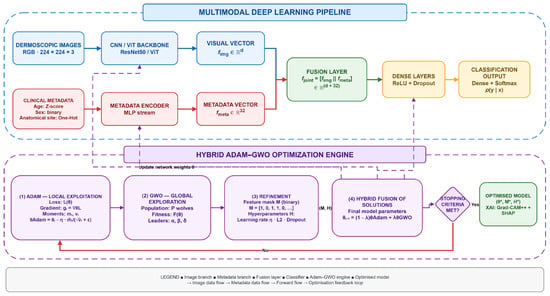
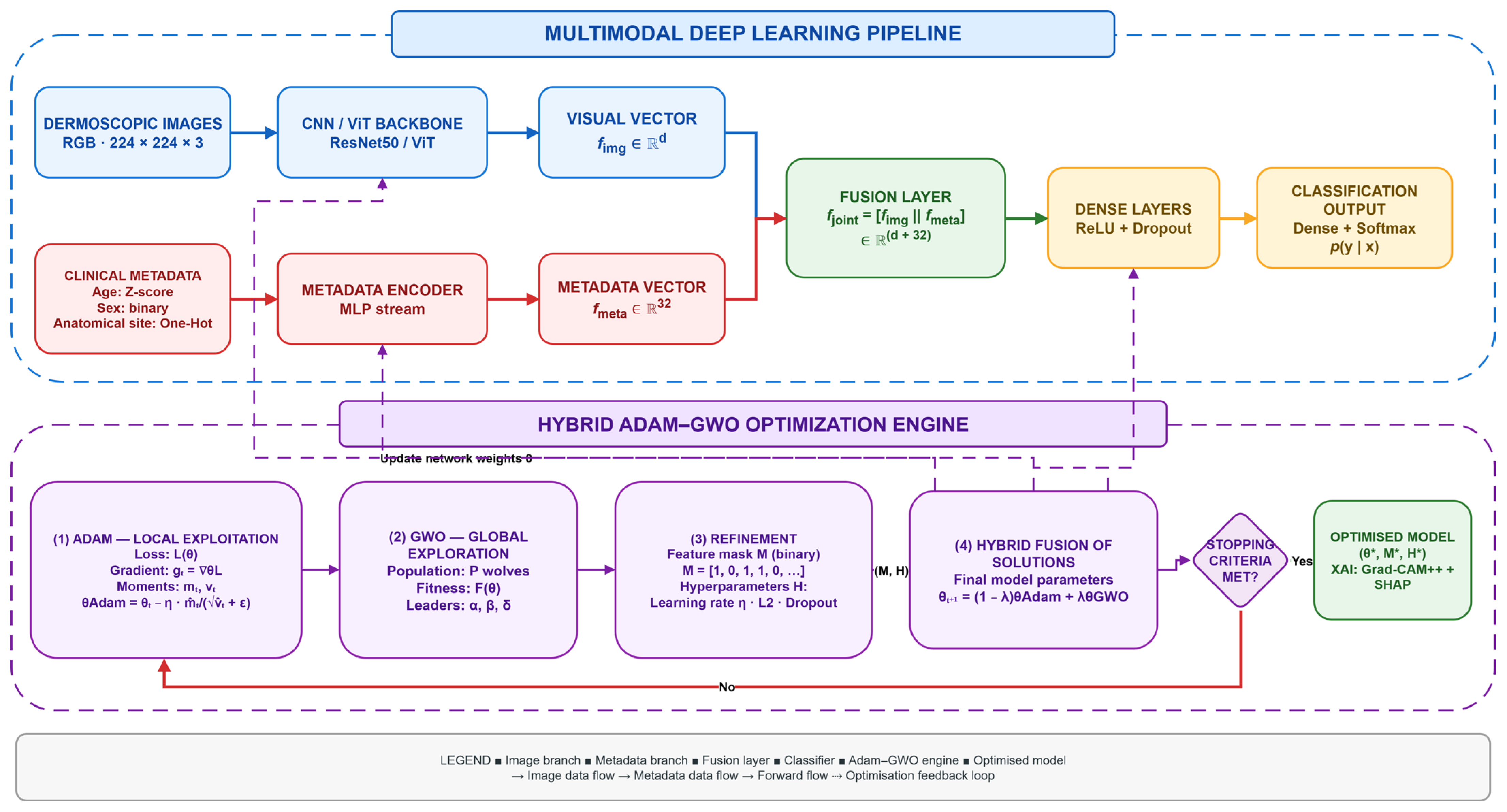
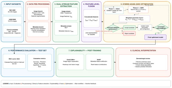
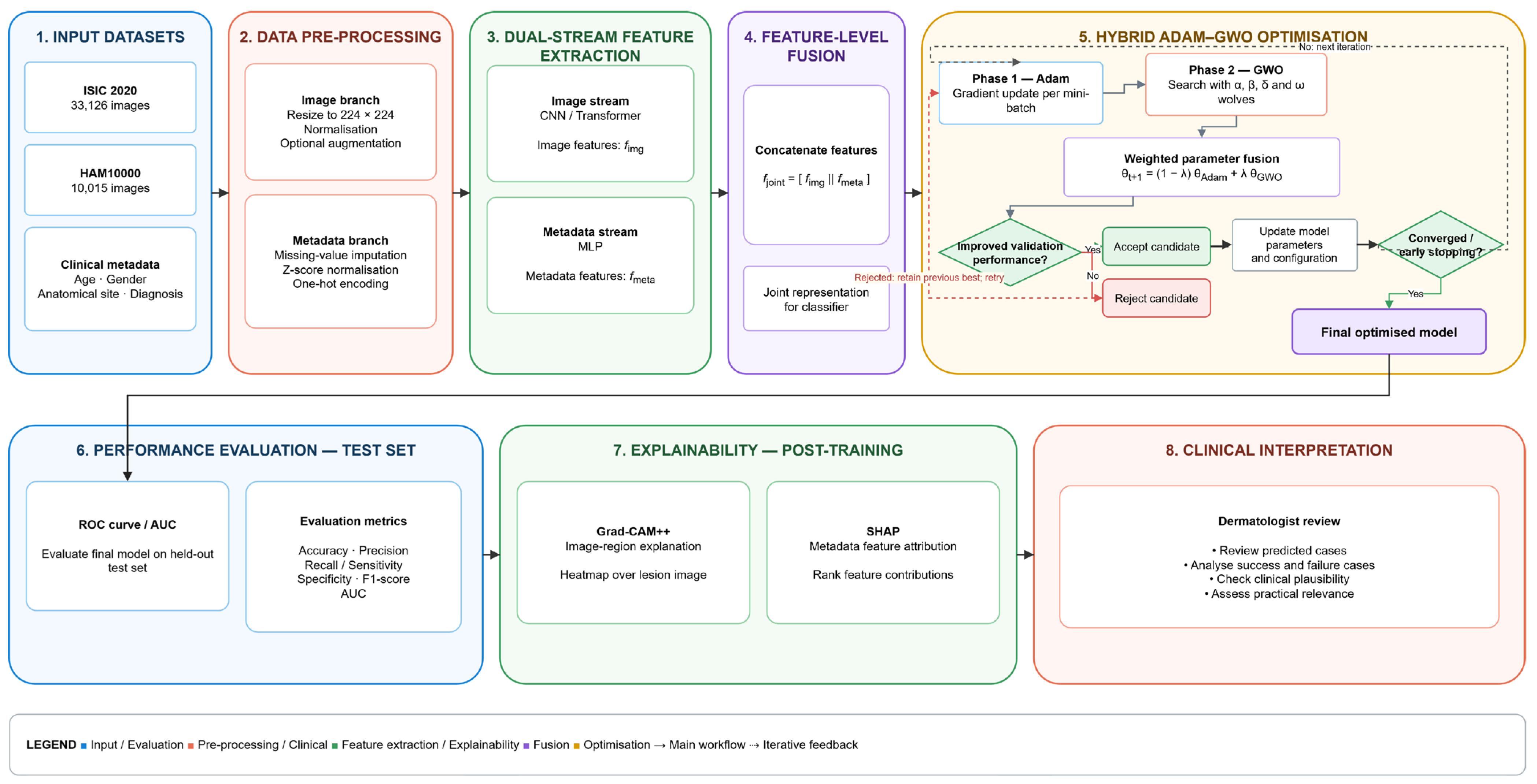
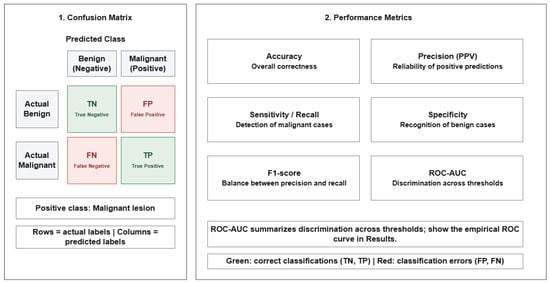
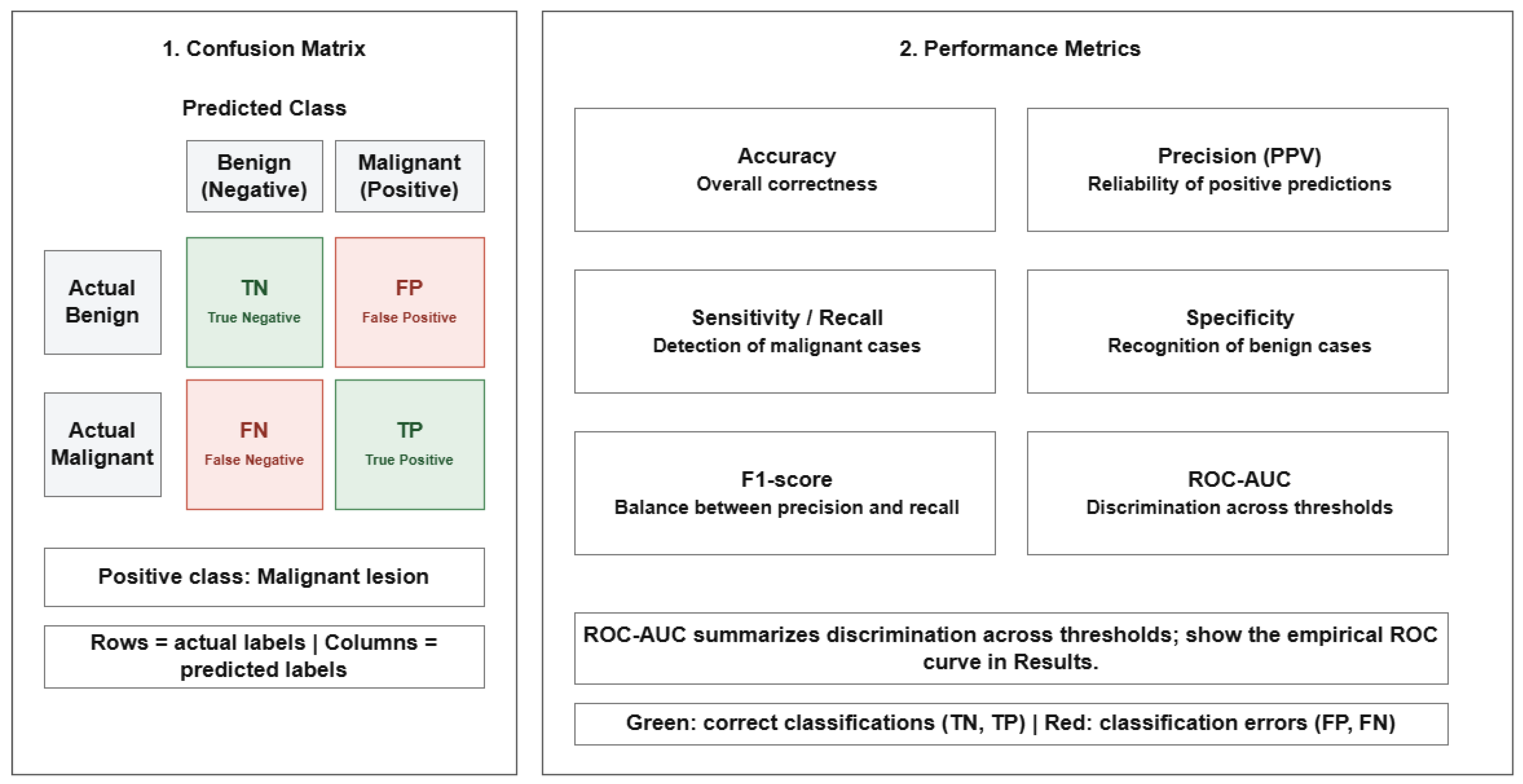
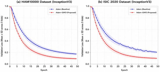
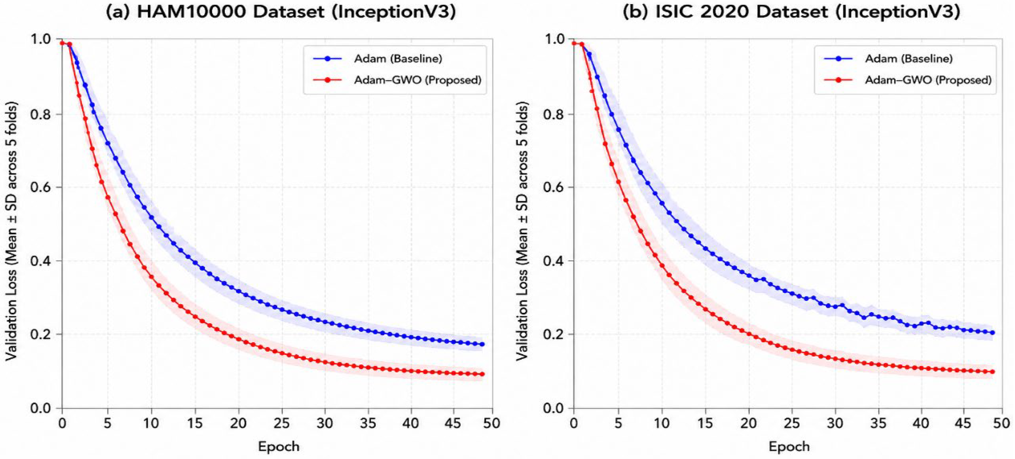
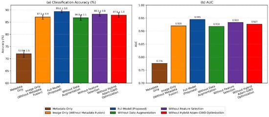
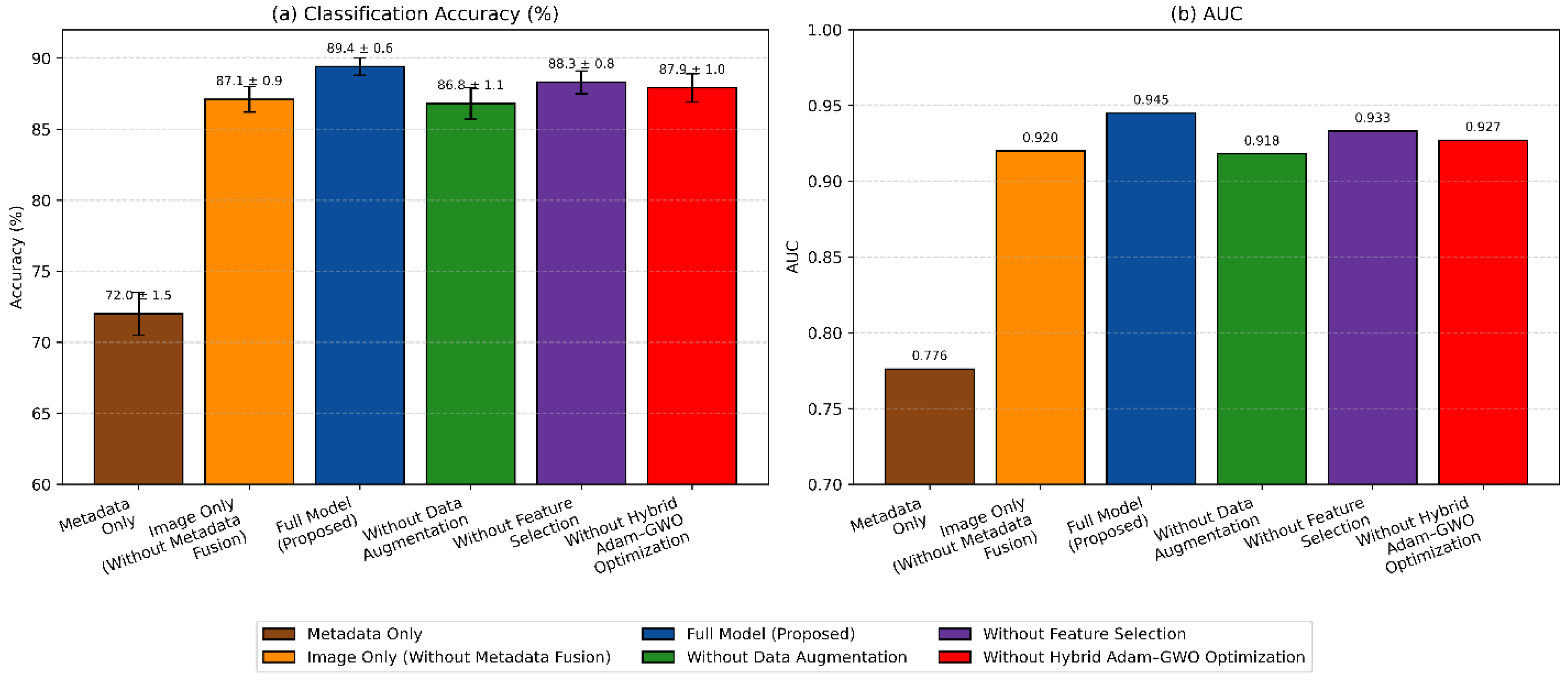
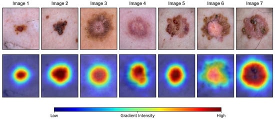
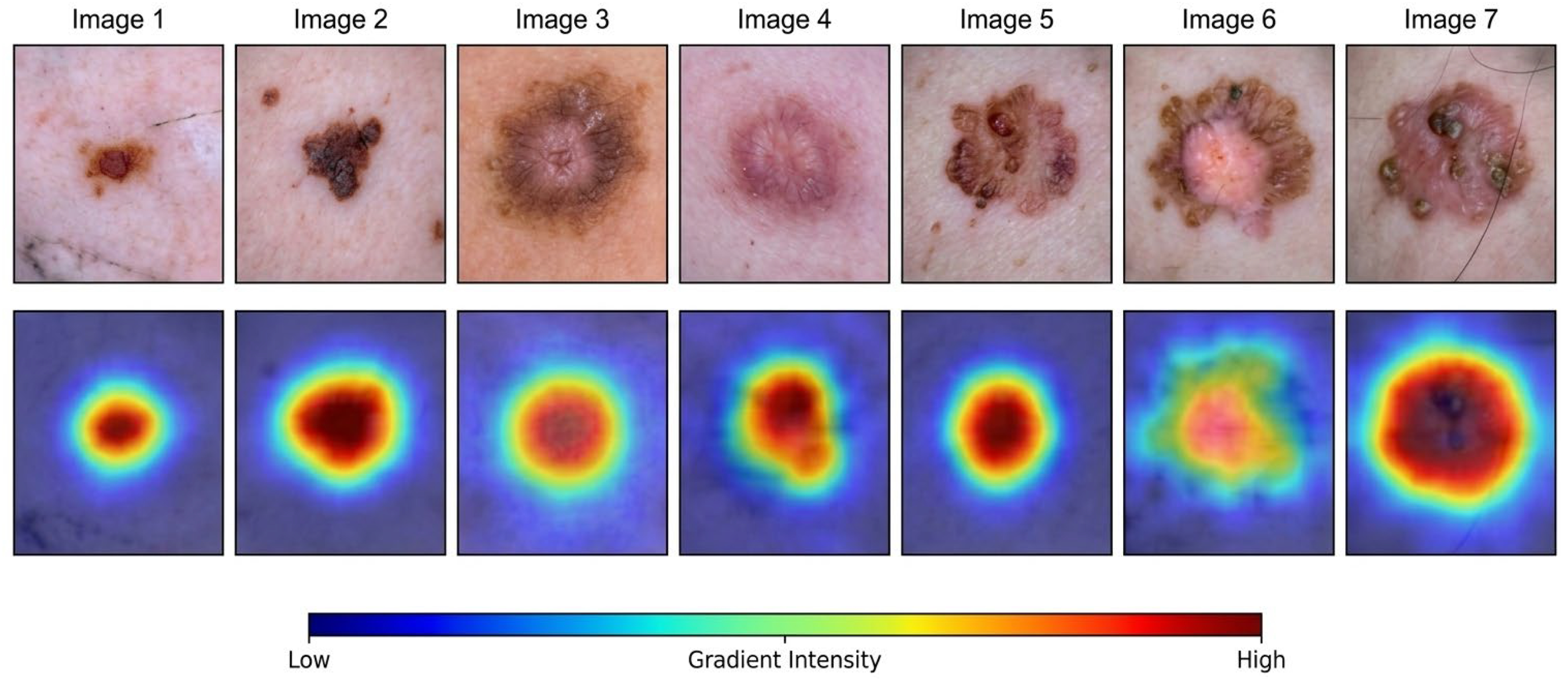
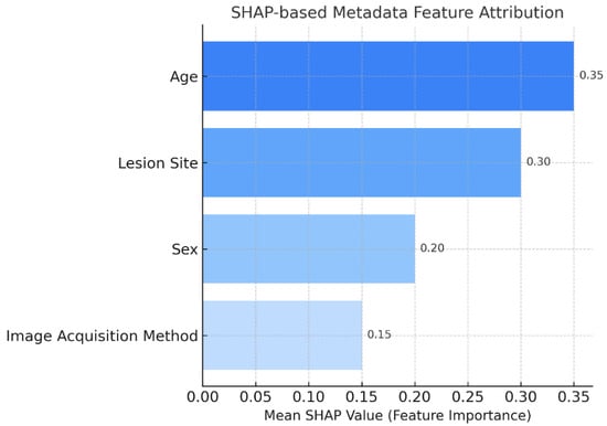
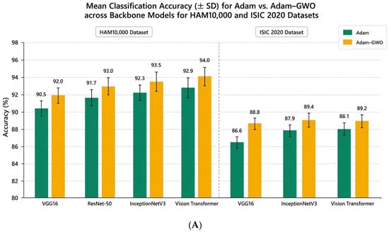
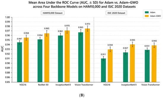
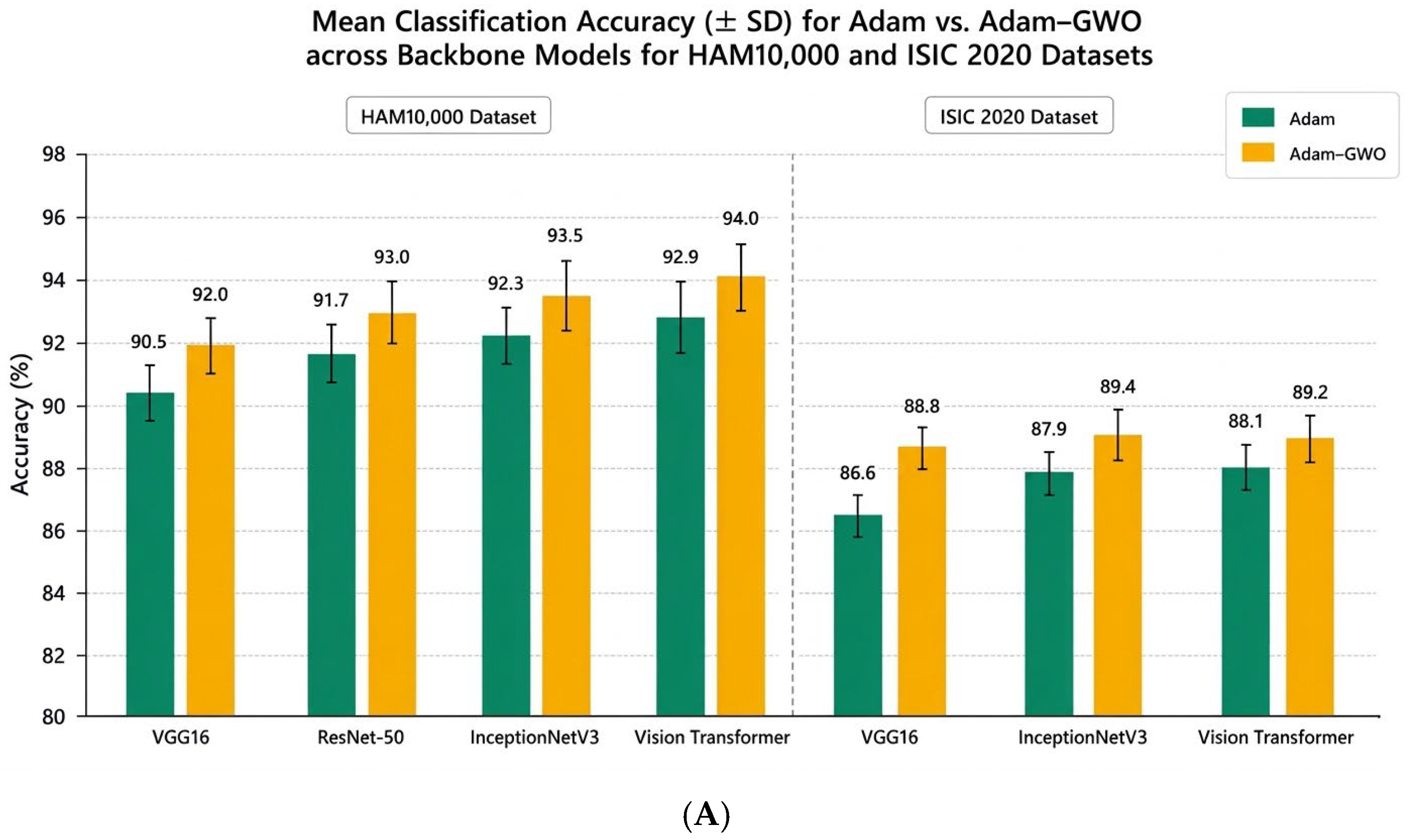
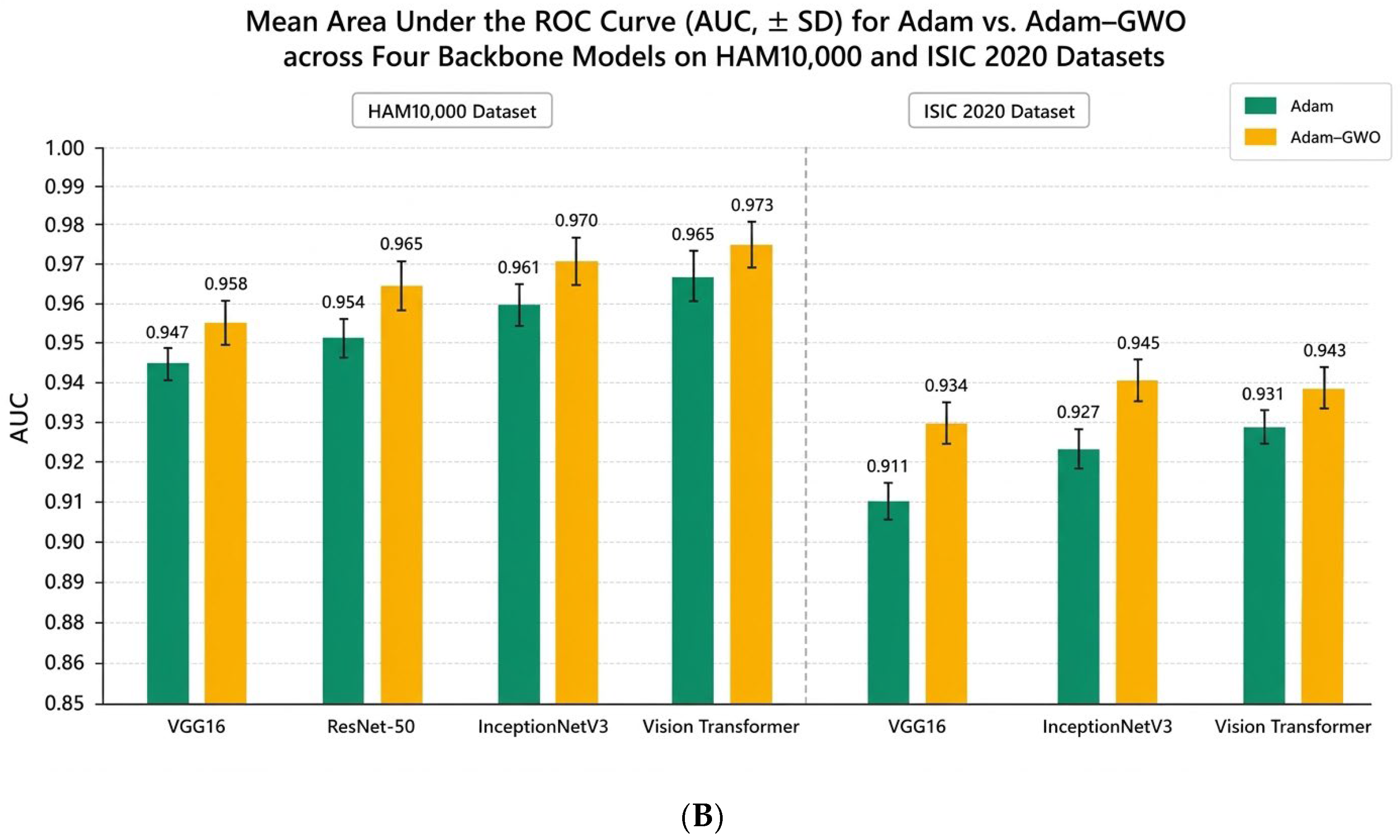
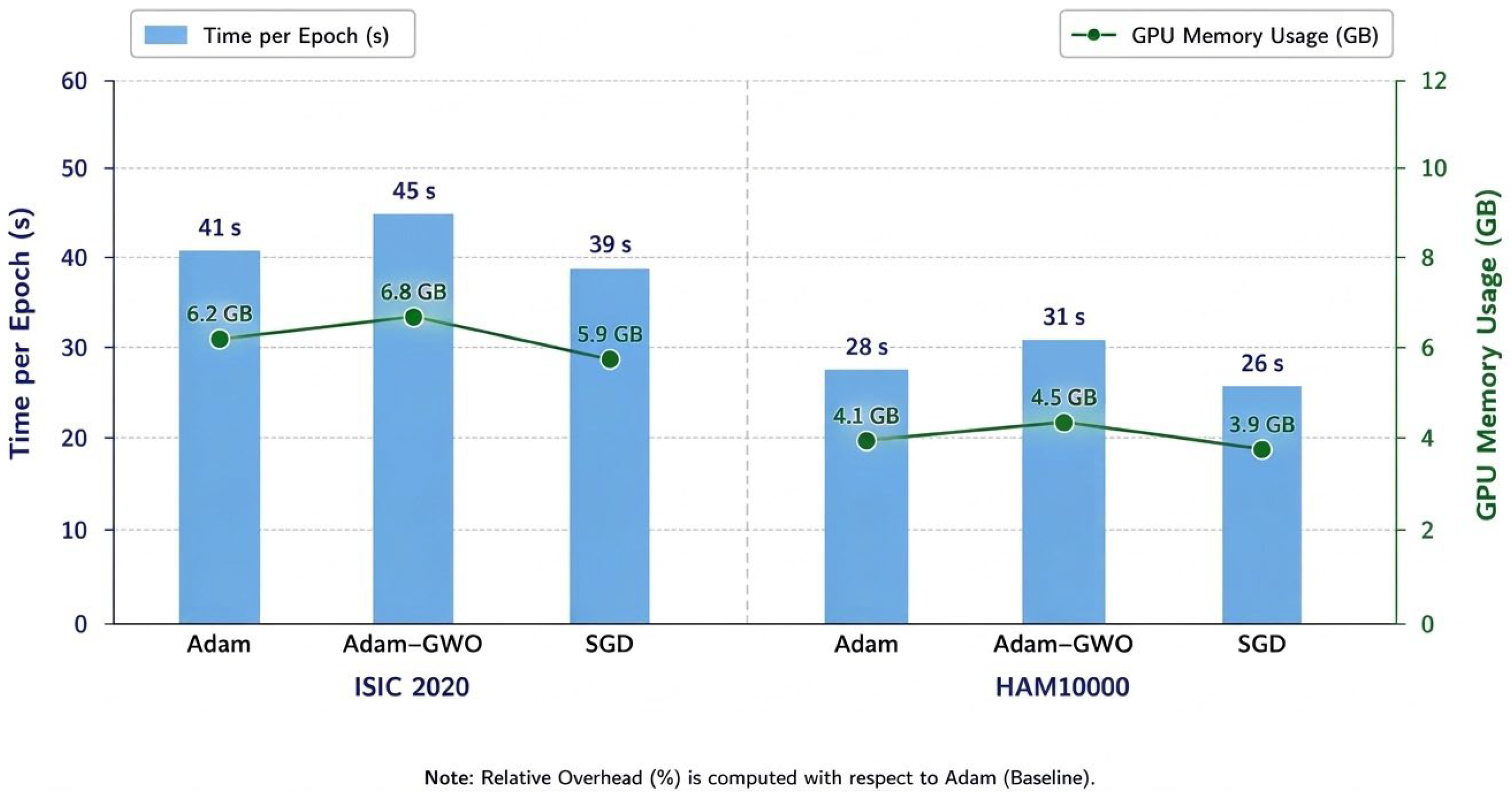

Disclaimer/Publisher’s Note: The statements, opinions and data contained in all publications are solely those of the individual author(s) and contributor(s) and not of MDPI and/or the editor(s). MDPI and/or the editor(s) disclaim responsibility for any injury to people or property resulting from any ideas, methods, instructions or products referred to in the content. |
© 2026 by the authors. Licensee MDPI, Basel, Switzerland. This article is an open access article distributed under the terms and conditions of the Creative Commons Attribution (CC BY) license.