


Abstract
Background: Oxidative stress and disturbed tissue homeostasis contribute to the pathogenesis of gonarthrosis, and trace elements act as cofactors or inhibitors of the antioxidant enzyme systems involved; data on elemental alterations in synovial fluid nevertheless remain scarce. This study aimed to reveal the changes in the elemental composition of synovial fluid in gonarthrosis. Methods: This prospective study included 64 patients: 33 with Kellgren–Lawrence grade 0 knees who underwent arthroscopy for meniscopathy (non-OA comparison group) and 31 with grade 4 gonarthrosis who underwent total knee arthroplasty (osteoarthritis group). Fifteen elements (Ca, Cu, As, Se, Sr, Sb, Hg, Al, Mn, Fe, Co, Ni, Zn, Cd, Pb) were measured in intraoperatively collected synovial fluid by inductively coupled plasma mass spectrometry (ICP-MS). Results: Between-group differences were confined to selenium and strontium. Mean selenium was 49.1 µg/L in the non-OA comparison group versus 37.6 µg/L in the osteoarthritis group (mean difference 11.5 µg/L; 95% CI: 4.15–18.9; p = 0.003), and strontium was 18.6 versus 22.3 µg/L, respectively (mean difference −3.70 µg/L; 95% CI: −6.20 to −1.20; p = 0.005). Mean iron was higher in the osteoarthritis group (3710 versus 3000 µg/L), but the difference was not significant (p = 0.051). Conclusions: In this exploratory cross-sectional comparison, synovial fluid selenium was lower and strontium was higher in grade 4 knees relative to grade 0 knees; however, these differences were attenuated after adjustment for age, and only selenium remained significant after Bonferroni correction. The remaining measured elements, including the toxic metals, did not differ between groups.
1. Introduction
Gonarthrosis, whose incidence increases with advancing age and which poses a growing socioeconomic burden, is a degenerative and progressive form of osteoarthritis affecting the knee joint. Progressive pain and loss of function develop in affected joints, substantially impairing patients’ quality of life. Research has largely focused on elucidating the etiopathogenesis of this disease and on developing treatment modalities. These studies collectively show that the pathogenesis of osteoarthritis is multifactorial, making treatment challenging. Age, obesity, genetic predisposition, physical factors, lifestyle, and biological factors are the principal etiologic contributors.
Increased oxidative stress, one of the biological causes, is known to play a role in the etiopathogenesis of osteoarthritis [1,2,3]. This oxidative stress is known to increase in the pathogenesis of many diseases such as Alzheimer’s disease, Parkinson’s disease, atherosclerosis, and neoplastic diseases [4,5]. Elevated levels of oxidative products damage proteins, lipids, carbohydrates, and nucleic acids—the fundamental macromolecules of the body. Consequently, a predominantly destructive process ensues in the affected tissue. Several antioxidant systems in humans—including glutathione peroxidase (GPx), glutathione-S-transferase (GST), and superoxide dismutase (SOD)—counteract the destructive effects of oxidative stress in tissues. These are complex enzyme systems in which trace elements serve key roles as cofactors or inhibitors. Selenium (Se), zinc (Zn), manganese (Mn), copper (Cu), and iron (Fe) act as cofactors for these enzymes, whereas heavy metals such as cadmium (Cd), mercury (Hg), lead (Pb), arsenic (As), and cobalt (Co) inhibit them [6,7,8,9]. Whether elemental homeostasis is altered in tissues affected by diseases whose etiopathogenesis involves dysfunction of the antioxidant system, such as osteoarthritis, and whether any such alteration contributes to disease pathogenesis remain poorly characterized [10,11]. To better understand this common and multifactorial disease and to inform the development of targeted therapies, each contributing factor in the etiopathogenesis should be systematically characterized.
Furthermore, elements such as calcium, magnesium, zinc, chromium, copper, and manganese have important functions in maintaining bone and cartilage integrity [10]. These elements are crucial for the development of healthy bone and cartilage tissue. Calcium is essential for maintaining bone mineral density, regulating osteoblast–osteoclast coupling, and supporting neuromuscular function [9,12]. Trivalent chromium (Cr3+) has been reported to enhance osteoblast differentiation and collagen synthesis in vitro [9,13]. Magnesium is a cofactor in extracellular matrix synthesis and is required for proper mineralization of bone and cartilage [14]. Copper participates in osteoblast–osteoclast coupling through its role in lysyl oxidase, which is essential for collagen cross-linking [9,15]. Deficiencies of these elements are associated with musculoskeletal disorders including osteoporosis, osteoarthritis, and chondrocalcinosis [9,10].
On this basis, the present study selected a panel of 15 elements that encompasses three functional categories: (a) essential cofactors of antioxidant enzyme systems and bone–cartilage metabolism (Ca, Cu, Zn, Se, Mn, Fe, Co); (b) toxic metals classified as priority environmental pollutants that are known inhibitors of antioxidant defenses and disruptors of bone homeostasis (As, Cd, Pb, Hg, Sb, Al, Ni); and (c) an element with emerging evidence for a role in bone remodeling and treatment of degenerative bone disease (Sr).
Despite the recognized importance of trace elements in antioxidant defense and bone metabolism, data on elemental alterations in synovial fluid during the progression of osteoarthritis remain scarce. Based on the roles of selenium as an antioxidant enzyme cofactor and strontium as a bone-remodeling agent, we hypothesized that synovial fluid selenium would be lower and strontium higher in Kellgren–Lawrence grade 4 knees relative to grade 0 knees, while toxic metals would not differ between groups given the exclusion of occupationally or environmentally exposed individuals. The aim of this study was to compare the concentrations of 15 essential and toxic elements in the synovial fluid of Kellgren–Lawrence grade 0 and grade 4 knees in order to identify elemental alterations associated with advanced knee osteoarthritis.
2. Materials and Methods
2.1. Study Design
This prospective study was conducted using surgically obtained synovial fluid from 64 patients who underwent total knee arthroplasty or interventional knee arthroscopy. Prior to enrollment, detailed medical histories were obtained, physical examinations were performed, and radiological assessments were completed for all patients. Based on the clinical and radiological findings, all knees were classified according to the Kellgren–Lawrence grading system.
2.2. Ethical Approval
The study was approved by the clinical ethics committee (Decision No. 2017-KAEK-189_2019.09.25_13; 25 September 2019). The study was conducted in accordance with the ethical principles of the Declaration of Helsinki. All patients were informed in detail about the study, and written informed consent was obtained prior to enrollment.
2.3. Randomization and Blinding
Randomization was performed by enrolling every third eligible patient from the surgical candidate pool. All patients were coded after sample collection, and the personnel analyzing the samples were blinded to the group allocation of the specimens.
2.4. Patient Selection and Grouping
The exclusion criteria for the study were: (a) presence of an endocrine disorder (obesity, diabetes); (b) smoking history; (c) presence of a dental filling; (d) a history of elemental supplementation therapy (Li, Fe, Zn, Mg, etc.); and (e) a history of occupational heavy metal exposure. Patients with a body mass index between 20 and 25 kg/m2, who had not undergone any trauma or surgery in the knee area (especially implant surgery), no rheumatologic diseases, and no history of heavy physical labor were included in the study.
The non-OA comparison group (Group 1) included 33 patients with Kellgren–Lawrence grade 0 knees who underwent knee arthroscopy after being diagnosed with meniscopathy. Arthroscopic examination confirmed the absence of articular cartilage degeneration or synovitis in this group. The osteoarthritis group (Group 2) included 31 patients with Kellgren–Lawrence grade 4 gonarthrosis who underwent total knee arthroplasty.
2.5. Sample Collection
In both groups, surgical procedures were performed under spinal anesthesia. After limb elevation, a tourniquet was applied to the operative extremity.
Following sterile preparation and draping of the operative lower extremity, a cannulated trocar was inserted into the joint through the arthroscopic lateral portal in the non-OA comparison group (Group 1). After joint entry, the trocar was removed from the cannula, and the synovial fluid draining through the cannula was collected. After sample collection, arthroscopic surgery was completed using the standard technique.
In the osteoarthritis group (Group 2), a midline incision was made following sterile preparation and draping of the lower extremity. After dissection through skin and subcutaneous tissue, the joint was accessed via a medial parapatellar arthrotomy. After arthrotomy, the intra-articular synovial fluid was collected. Following sample collection, the surgical procedure was completed using the standard technique.
2.6. Preparation of Synovial Fluid for Elemental Analysis
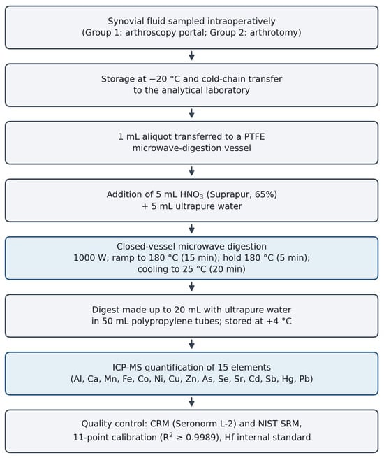
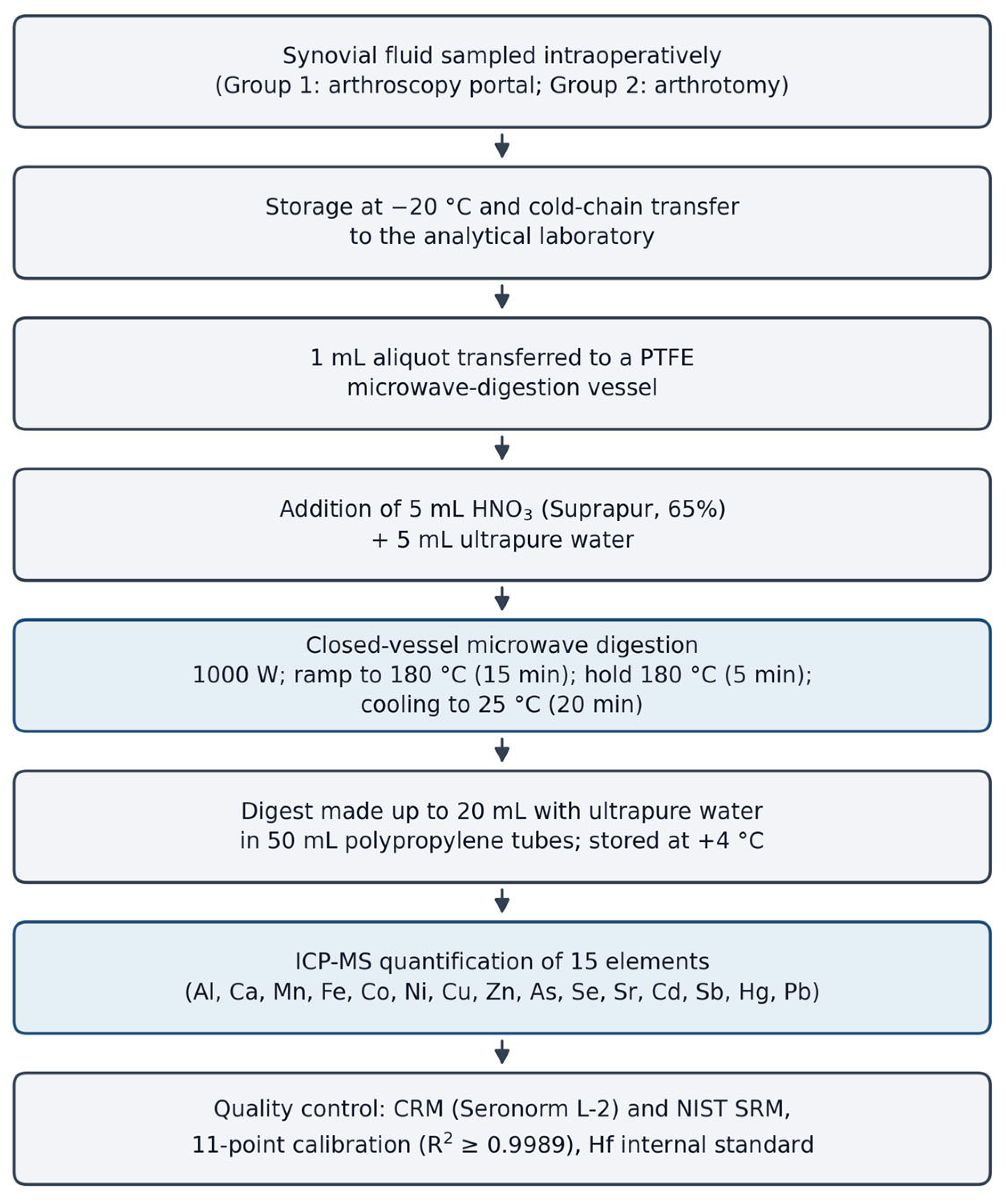
Synovial fluid samples were collected for elemental analysis and stored at −20 °C until transfer. The samples were then transferred to the Yozgat Bozok University Science and Technology Application and Research Center (BILTEM), Yozgat, Türkiye, under cold-chain conditions for elemental analysis. All samples were pretreated before analysis. For this purpose, 1 mL of each sample was transferred into polytetrafluoroethylene (PTFE) tubes of the microwave digestion unit (Milestone Ethos, Milestone Srl, Sorisole, Italy). Subsequently, 5 mL of suprapure nitric acid (HNO3, Suprapur®, 65%, Merck, Darmstadt, Germany) and 5 mL of ultrapure water (Direct-Q®, Millipore, Burlington, MA, USA) were added to these samples, respectively. The samples were digested at 180 °C for 20 min (Table 1). The digested samples were then transferred into 50 mL polypropylene tubes, diluted to 20 mL with ultrapure water, and stored at +4 °C until analysis. The digestion method developed by Aliyev et al. for the analysis of elemental levels in biological samples was used with minor modifications (Figure 1).
Table 1.
Microwave digestion program applied to the synovial fluid samples.
Figure 1.
Workflow of the sample preparation, microwave digestion and ICP-MS measurement procedure applied to the synovial fluid samples.
2.7. Elemental Analysis
All samples were measured for Pb, Cd, As, Hg, Zn, Cu, Ca, Se, Sr, Ni, Co, Al, Fe, Mn and Sb content by inductively coupled plasma mass spectrometry (ICP-MS; iCAP Qc, Thermo Scientific, Bremen, Germany). The operating conditions for the ICP-MS were as follows: RF power, 1500 W; nebulizer pressure, 3 bar; carrier gas, 0.9 L/min; spray chamber temperature, 3.0 °C; and dwell time, 0.01 ms. To ensure the accuracy of the results, samples and standards were measured at least three times, and the R2 values of the calibration curves were at least 0.9989 for all elements. Eleven-point calibration curves were prepared from multi-element standard solutions (ICP multi-element standard solution IV, Merck, Darmstadt, Germany) in 3% nitric acid solution, covering the concentration ranges 0–100 µg/L for trace elements (As, Cd, Co, Hg, Mn, Ni, Pb, Sb, Sr), 0–1000 µg/L for Cu, Se, and Zn, 0–10,000 µg/L for Al and Fe, and 0–100,000 µg/L for Ca. All solutions, including standards and blanks, were prepared in 3% nitric acid solution.
2.8. Method Validation
Two certified reference materials (Seronorm™ Whole Blood L-2, SERO AS, Billingstad, Norway, and a National Institute of Standards and Technology (NIST) Standard Reference Material® (SRM), Gaithersburg, MD, USA) were used for the validation of the Pb, Cd, As, Hg, Zn, Cu, Ca, Se, Sr, Ni, Co, Al, Fe, Mn and Sb methods. The stability and sensitivity of the instrument were validated with these reference materials. Coefficients of variation (CVs) were also calculated for all measurements; the variations in each measurement for CRM and SRM quality controls were 4.7% and 12.0%, respectively. In addition, hafnium (Hf) was used as the internal standard (recovery 97–112%), and relative standard deviations (RSDs) did not exceed 5% for any measurement.
The limits of detection (LODs) and limits of quantification (LOQs) were determined as 3 and 10 times the standard deviation of 10 replicate blank measurements, respectively. LOD values (µg/L) were as follows: Al 0.15, Ca 5.0, Mn 0.005, Fe 0.50, Co 0.002, Ni 0.02, Cu 0.05, Zn 0.10, As 0.01, Se 0.05, Sr 0.005, Cd 0.003, Sb 0.005, Hg 0.01, and Pb 0.01. The complete LOD and LOQ values for all 15 elements are presented in Table S1 (Supplementary Material). All measured values in this study exceeded the LOQ for every element; therefore, no censoring or substitution method was required.
2.9. Statistical Analysis
Data were analyzed using statistical software (JASP 0.14.1, JASP Team, University of Amsterdam, Amsterdam, The Netherlands). Descriptive statistics were first calculated for all elemental concentrations. Normality testing and outlier analysis were then performed. An independent samples t-test was used to compare groups for normally distributed variables. The Mann–Whitney U test was used for variables that did not follow a normal distribution. A value of p < 0.05 was considered significant. To assess the robustness of the primary findings, a post hoc analysis of covariance (ANCOVA) with age as a covariate was performed for elements showing significant between-group differences. Bonferroni correction was applied to account for the 15 simultaneous comparisons, and Cohen’s d effect sizes with 95% confidence intervals for the mean differences were calculated.
3. Results
The mean age of the patients in Group 1 was 46.5 years (range 15–68), whereas the mean age of the patients in Group 2 was 65.9 years (range 56–83) (Table 2). The mean elemental concentrations in the synovial fluid of the non-OA comparison group were as follows: aluminum 20.4 µg/L (4.57–57.39), calcium 32,500 µg/L (23,778.74–43,638.27), manganese 20.7 µg/L (17.34–27.00), iron 3000 µg/L (1047.71–5375.00), cobalt 1.78 µg/L (1.20–3.00), nickel 27.2 µg/L (16.91–45.12), copper 638 µg/L (448.15–961.15), zinc 806 µg/L (489.78–1390.48), arsenic 10.1 µg/L (7.58–12.74), selenium 49.1 µg/L (24.50–99.36), strontium 18.6 µg/L (7.72–29.97), cadmium 4.47 µg/L (3.96–5.44), antimony 0.58 µg/L (0.46–0.73), mercury 0.31 µg/L (0.16–0.50), and lead 23.1 µg/L (0.04–111.17).
Table 2.
Demographic characteristics of the study groups.
The mean elemental concentrations in the synovial fluid of the osteoarthritis group were as follows: aluminum 20.0 µg/L (3.44–65.91), calcium 32,000 µg/L (25,019.54–41,091.73), manganese 21.4 µg/L (17.73–31.94), iron 3710 µg/L (2143.57–6574.58), cobalt 1.75 µg/L (1.23–3.15), nickel 24.5 µg/L (17.75–35.75), copper 647 µg/L (455.23–989.60), zinc 797 µg/L (516.67–1173.28), arsenic 10.2 µg/L (6.93–13.93), selenium 37.6 µg/L (6.39–64.91), strontium 22.3 µg/L (13.98–33.00), cadmium 4.92 µg/L (3.86–7.74), antimony 0.59 µg/L (0.49–0.79), mercury 0.23 µg/L (0.17–0.41), and lead 26.6 µg/L (0.28–85.11).
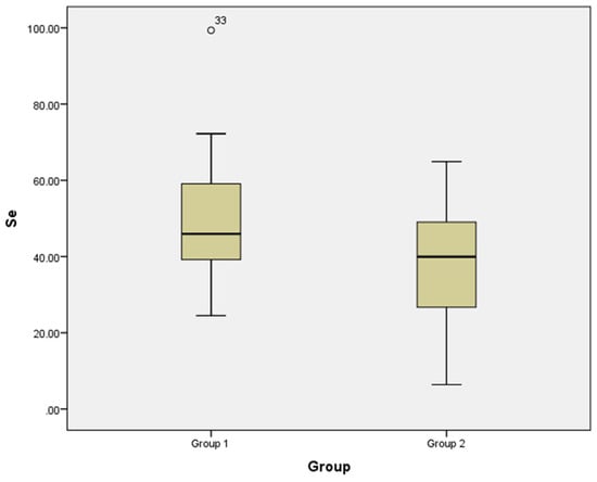
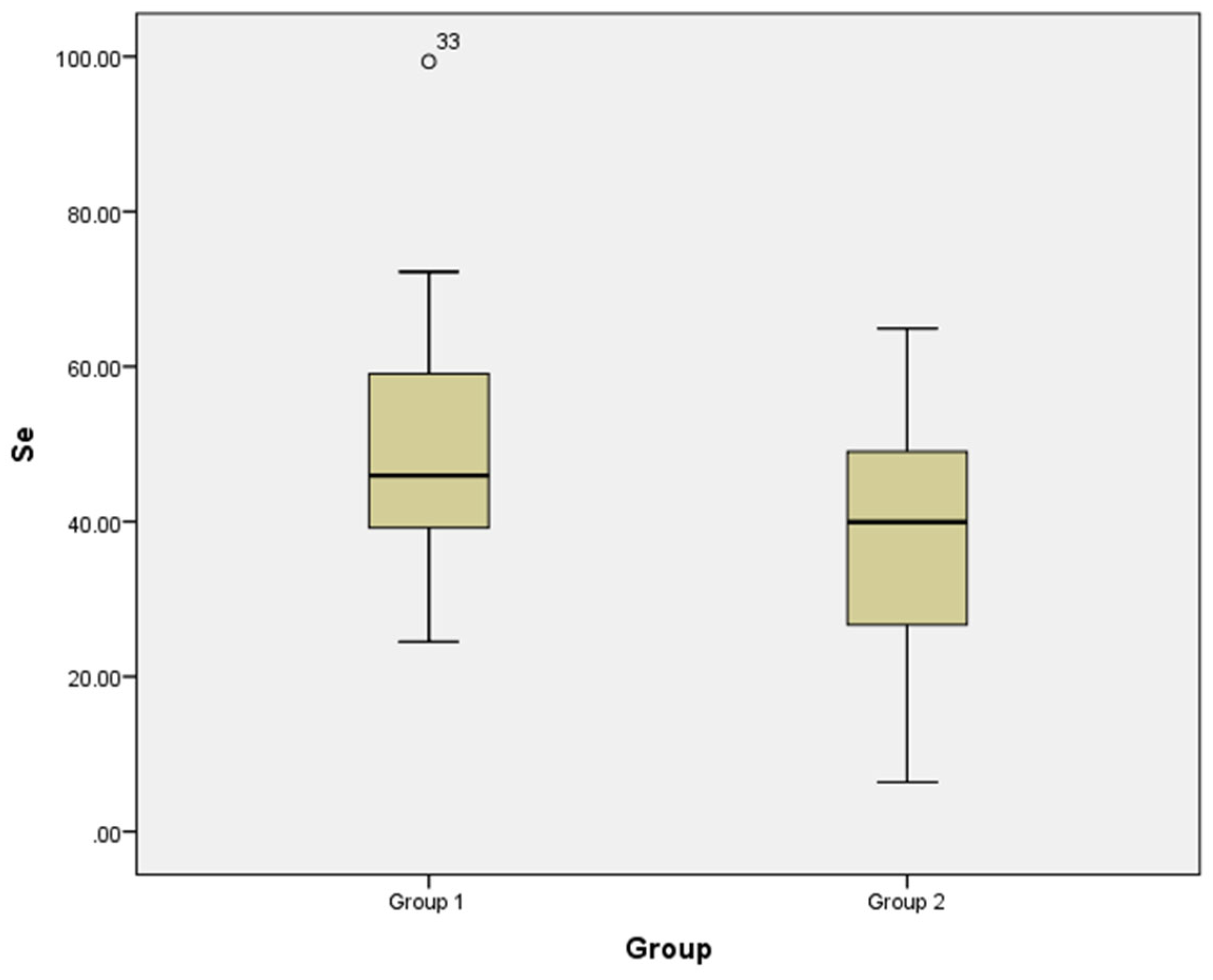
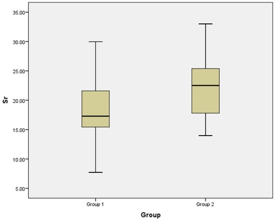
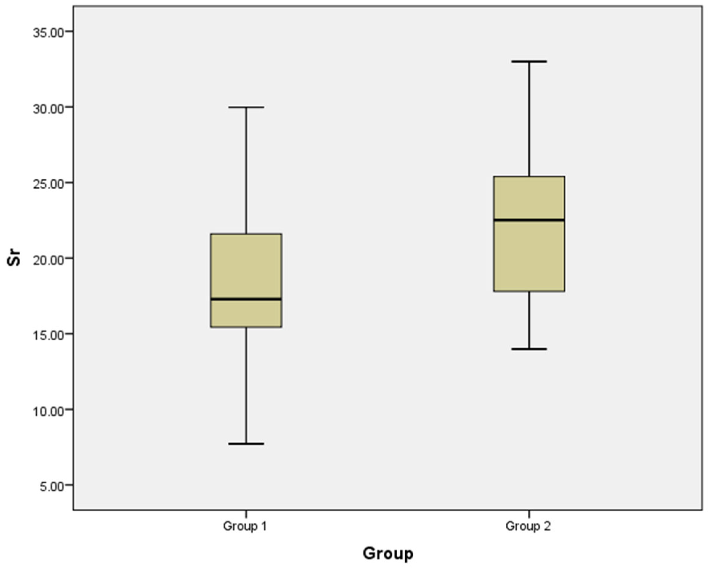
Normality testing revealed that calcium, copper, arsenic, selenium, strontium, antimony, and mercury followed a normal distribution in both groups, whereas aluminum, manganese, iron, cobalt, nickel, zinc, cadmium, and lead did not. The mean ± SD and median (interquartile range) values, along with the differences between the groups, are given in Table 3. Statistically significant differences between the groups were observed only for selenium (Se) and strontium (Sr). In the osteoarthritis group, selenium was significantly lower and strontium was significantly higher than in the non-OA comparison group (Table 3; Figure 2 and Figure 3). The mean difference for selenium was 11.5 µg/L (95% CI: 4.15–18.9; Cohen’s d = 0.77), and for strontium was −3.70 µg/L (95% CI: −6.20 to −1.20; Cohen’s d = 0.73). After Bonferroni correction for 15 comparisons, the selenium difference remained significant (adjusted p = 0.045), whereas the strontium difference became marginally non-significant (adjusted p = 0.075). A post hoc ANCOVA with age as a covariate revealed that the group effect for selenium was attenuated (F = 2.45, p = 0.123) and for strontium was no longer significant (F = 0.22, p = 0.643). A significant positive correlation was observed between age and strontium concentration (Spearman ρ = 0.415, p < 0.001), whereas no significant correlation was found between age and selenium (ρ = −0.200, p = 0.113).
Table 3.
Comparison of synovial fluid element concentrations (mean ± SD and median with interquartile range) between the non-OA comparison group (Kellgren–Lawrence grade 0) and the osteoarthritis group (Kellgren–Lawrence grade 4).
Figure 2.
Distribution of synovial fluid selenium (Se, µg/L) concentrations in the non-OA comparison group (Group 1, Kellgren–Lawrence grade 0) and the osteoarthritis group (Group 2, Kellgren–Lawrence grade 4). Boxes show the interquartile range and median; whiskers show the range excluding outliers; the circle denotes an outlier.
Figure 3.
Distribution of synovial fluid strontium (Sr, µg/L) concentrations in the non-OA comparison group (Group 1, Kellgren–Lawrence grade 0) and the osteoarthritis group (Group 2, Kellgren–Lawrence grade 4). Boxes show the interquartile range and median; whiskers show the range.
4. Discussion
Osteoarthritis is a degenerative joint disease involving cartilage and subchondral bone destruction and synovial inflammation. Its prevalence continues to rise, bringing with it substantial socioeconomic consequences. Current approaches to osteoarthritis focus on understanding the etiopathogenesis and improving treatment [11]. Treatments can be grouped as pharmacological (e.g., nonsteroidal anti-inflammatory drugs), non-pharmacological (e.g., weight loss, lifestyle modification, exercise), and surgical (e.g., knee and hip arthroplasty). The etiopathogenesis involves multiple factors, including age, physical activity, biological processes, nutrition, and lifestyle, in addition to genetic predisposition [16,17]. The incidence of osteoarthritis also increases in some rheumatological diseases, such as rheumatoid arthritis, in which the effects of genetic factors have been demonstrated. Similarly, the high incidence of osteoarthritis among manual laborers, individuals with high body mass indices, and those who subject their joints to excessive physical stress demonstrates that physical factors play a role in the etiopathogenesis [11,18]. Biological factors include cellular dysfunction, enzyme disorders, and impaired DNA synthesis and repair [19]. All of these biological causes disrupt the tissue anabolism–catabolism balance. A shift toward uncontrolled anabolism plays a role in the etiology of neoplastic diseases, whereas a shift toward catabolism contributes to degenerative diseases such as Alzheimer’s disease and gonarthrosis [4].
Trace elements in the body serve as cofactors or inhibitors of numerous enzymatic reactions essential for maintaining homeostasis. Some elements exist in very small amounts in the body, and their concentrations are tightly regulated [20]. Even though they exist in small amounts, these elements play a key role in many enzymatic reactions of vital importance, such as DNA synthesis, antioxidant enzyme systems, oxygen transport systems, and energy metabolism. Disruptions in these enzymatic reactions can impair the biological balance (anabolic and catabolic cycles) in the body. This raises the question: how do elemental levels in affected tissues change in diseases such as osteoarthritis and Alzheimer’s disease? Similarly, how do they change in neoplastic diseases with high anabolic activity? Although numerous studies have examined tissue elemental levels in Alzheimer’s disease, data on elemental changes in osteoarthritis remain limited [21,22].
In their study, Krachler et al. compared the amounts of essential elements in the serum and synovial fluid of patients with osteoarthritis [23]. They showed that elemental concentrations in synovial fluid were lower than those in serum. Yazar et al. compared selenium, zinc, copper, and iron concentrations in the synovial fluid of healthy individuals and patients with osteoarthritis or rheumatoid arthritis [24]. They found that selenium was higher, copper and iron were lower, and zinc was unchanged in the non-OA comparison group relative to the osteoarthritic group. The findings of Yazar et al. for selenium and zinc are consistent with those of the present study. In our study, the analysis demonstrated that the selenium concentration in the synovial fluid was lower in knees with arthrosis and that zinc concentration did not differ. Mean iron concentrations were lower in the non-OA group, but the difference did not reach statistical significance. Taken together, both studies indicate that synovial fluid selenium is lower in osteoarthritic joints. However, direct comparison of absolute concentrations between the two studies should be interpreted with caution because of important methodological differences: the present study compared Kellgren–Lawrence grade 0 versus grade 4 knees, whereas Yazar et al. compared osteoarthritis patients of unspecified grading with healthy volunteers; synovial fluid was collected intraoperatively in the present study versus by aspiration in Yazar et al.; and the analytical methods differed, ICP-MS in the present study versus atomic absorption spectrometry in Yazar et al., with ICP-MS offering lower detection limits and simultaneous multi-element measurement. Furthermore, the age distributions of the study populations differed substantially. Given that selenium is a cofactor of the antioxidant system, its potential role in a destructive disease such as gonarthrosis warrants attention. This inference should be supported by studies measuring synovial tissue antioxidant enzyme activity and evaluating its relationship with selenium concentrations.
It is well established in the literature that heavy metals induce oxidative stress. Jomova and Valko reported that Fe, Cu, Co and Cr triggered the formation of reactive oxygen species and played a role in the pathogenesis of some diseases [4]. Saïdi et al. demonstrated that heavy metals such as cadmium, lead, and mercury increase oxidative stress toxicity [7]. Nelson et al. reported a correlation between elevated blood lead levels and the development of osteoarthritis, suggesting that lead may contribute to its etiology [25]. The review by Dermience et al. is one of the most comprehensive on the functions of elements in the body [9]; it emphasizes the adverse effects of numerous heavy metals on bone and joint metabolism. In the present study, no statistically significant difference was found between normal and osteoarthritic joint synovial fluid in terms of heavy metal levels. The fact that the present results differ from those of the aforementioned studies should not be taken to mean that heavy metals have no effect on the etiopathogenesis of osteoarthritis. This discrepancy may reflect the exclusion of smokers and individuals with occupational heavy metal exposure from the study population.
In the present study, strontium was one of the elements that showed a between-group difference. Strontium is an element whose biological functions have not been fully elucidated. It has been used therapeutically, particularly in the treatment of osteoporosis. Strontium has been reported to promote cartilage formation, osteoblast proliferation, and bone formation [26,27,28,29,30,31,32]. The analysis revealed that the strontium concentration was higher in the synovial fluid of grade 4 knees. However, the ANCOVA sensitivity analysis demonstrated that this difference was no longer significant after adjustment for age (p = 0.643), and a significant positive correlation between age and strontium (Spearman ρ = 0.415, p < 0.001) suggests that the observed elevation may be partly or wholly attributable to the age difference between groups. Moreover, the present study did not assess dietary strontium intake, strontium-containing medications (such as strontium ranelate), or environmental strontium exposure, any of which could contribute to the observed difference. Whether the higher strontium concentration represents a compensatory response during the development of osteoarthritis or reflects age-related cumulative exposure therefore remains to be determined. Future studies should collect dietary questionnaires and medication histories to disentangle these potential confounders.
An important methodological consideration is that ICP-MS measures total element concentrations, which include both free ions and protein-bound species. The biological activity of trace elements in synovial fluid depends on their chemical speciation: for example, selenoproteins such as glutathione peroxidase represent the functionally relevant fraction of total selenium, while protein-bound strontium may differ in its biological effects from free strontium ions. The present study cannot distinguish between these fractions. Speciation analysis using techniques such as size exclusion chromatography coupled with ICP-MS would provide a more complete picture of how trace elements participate in osteoarthritis pathophysiology and is recommended for future investigations.
Kosik-Bogacka et al. assessed the changes in calcium, magnesium, zinc, and lead levels with age in the anatomical structures (meniscus, anterior cruciate ligament, and cartilage tissue) of knees developing osteoarthritis [33]. They observed that the concentrations of these elements in knee structures did not change significantly before or after the age of 60. Kosik-Bogacka et al. compared patients with osteoarthritis grouped by age; however, their study did not include a group with normal knee joints. The present study addresses this gap. Calcium, magnesium, zinc, and lead concentrations not only remain stable with age but also do not change during the osteoarthritis process. The present study demonstrated that the concentrations of these elements were similar in synovial fluid from normal knees and from knees with osteoarthritis.
Strengths and Limitations
The limitations of the present study include the lack of tissue enzyme function assessment, the relatively small sample size, and the single-center design. In addition, the two groups were not comparable in terms of age, which is an inherent consequence of comparing Kellgren–Lawrence grade 0 and grade 4 knees; although the primary analyses were unadjusted, the post hoc ANCOVA sensitivity analysis showed that the selenium and strontium differences were attenuated after adjustment for age, underscoring the need for future studies to recruit age-matched groups or to employ multivariate models from the outset. The 15 simultaneous element comparisons raise the probability of a type I error; after Bonferroni correction, only selenium remained significant, and this result should be interpreted with caution. The study measured total element concentrations at a single time point by ICP-MS and therefore does not distinguish between free ions and protein-bound species; because total concentration and bioavailable fraction may have different biological relevance, speciation analysis would strengthen future studies. Furthermore, the cross-sectional design does not permit any conclusion about environmental exposure, accumulation over time, or causal direction. For strontium specifically, dietary intake, strontium-containing medications, and environmental exposure were not assessed, and any of these could contribute to the observed between-group difference. Multicenter studies with larger patient populations, age- and sex-adjusted models, speciation analyses, dietary and medication questionnaires, and concurrent measurement of enzyme functions are needed to address these limitations. The strengths of the present study include its prospective design, the inclusion of a non-OA comparison group, and the simultaneous measurement of 15 essential and toxic elements by ICP-MS. Moreover, the study provides objective data on elemental changes in synovial fluid during the gonarthrosis process, a topic that has received limited attention.
5. Conclusions
In this exploratory cross-sectional comparison, synovial fluid selenium was lower and strontium was higher in Kellgren–Lawrence grade 4 knees relative to grade 0 knees. After Bonferroni correction for 15 comparisons, only the selenium difference remained statistically significant (adjusted p = 0.045), while the strontium difference became marginally non-significant (adjusted p = 0.075). Importantly, a post hoc ANCOVA sensitivity analysis revealed that both differences were attenuated after adjustment for age, indicating that the age disparity between groups may partly explain the observed elemental differences. The remaining 13 elements, including the toxic metals, did not differ between groups. These findings suggest that altered selenium status may be associated with advanced knee osteoarthritis; however, the cross-sectional design, the loss of significance after age adjustment, and the relatively small sample size preclude causal conclusions. Future studies with age-matched groups, multivariate models, speciation analyses, and concurrent assessment of selenoenzyme activities are warranted to clarify the role of trace elements in osteoarthritis pathophysiology.
Supplementary Materials
The following supporting information can be downloaded at: https://www.mdpi.com/article/10.3390/toxics14110975/s1, Table S1. Limits of detection (LOD) and limits of quantification (LOQ) for the 15 elements measured by ICP-MS (iCAP Qc, Thermo Scientific).
Author Contributions
Conceptualization, S.D. and V.A.T.; methodology, S.D. and V.A.T.; formal analysis, S.D., A.E.P., V.A.T. and M.K.; investigation, S.D., A.E.P. and M.K.; resources, S.D. and V.A.T.; data curation, S.D. and V.A.T.; writing—original draft preparation, S.D., A.E.P., V.A.T. and M.K.; writing—review and editing, S.D., M.K., K.Ö. and H.F.S.; visualization, V.A.T.; supervision, S.D.; funding acquisition, S.D. All authors have read and agreed to the published version of the manuscript.
Funding
This study was supported by the Scientific Research and Project Coordination Center of Yozgat Bozok University, grant number 6602b-TF/19-340.
Institutional Review Board Statement
The study was conducted in accordance with the Declaration of Helsinki and approved by the Clinical Research Ethics Committee of Yozgat Bozok University (Decision No. 2017-KAEK-189_2019.09.25_13; 25 September 2019).
Informed Consent Statement
Written informed consent was obtained from all patients involved in the study for the use of their surgical waste samples.
Data Availability Statement
The original contributions presented in this study are included in the article. Further inquiries can be directed to the corresponding author.
Acknowledgments
The authors thank the staff of the Yozgat Bozok University Science and Technology Application and Research Center (BILTEM) for their support with the ICP-MS analyses.
Conflicts of Interest
The authors declare no conflicts of interest.
References
- Li, D.; Xie, G.; Wang, W. Reactive oxygen species: The 2-edged sword of osteoarthritis. Am. J. Med. Sci. 2012, 344, 486–490. [Google Scholar] [CrossRef] [Scilit] [PubMed]
- Suantawee, T.; Tantavisut, S.; Adisakwattana, S.; Tanavalee, A.; Yuktanandana, P.; Anomasiri, W.; Deepaisarnsakul, B.; Honsawek, S. Oxidative stress, vitamin E, and antioxidant capacity in knee osteoarthritis. J. Clin. Diagn. Res. 2013, 7, 1855–1859. [Google Scholar] [CrossRef] [Scilit] [PubMed]
- Martel-Pelletier, J.; Tardif, G.; Fernandes, J.C.; Pelletier, J.P. Metalloproteases and their modulation as treatment in osteoarthritis. In Principles of Molecular Rheumatology; Tsokos, G.C., Ed.; Humana Press: Totowa, NJ, USA, 2000; pp. 499–513. [Google Scholar] [CrossRef] [Scilit]
- Jomova, K.; Valko, M. Advances in metal-induced oxidative stress and human disease. Toxicology 2011, 283, 65–87. [Google Scholar] [CrossRef] [Scilit] [PubMed]
- Asanuma, M.; Miyazaki, I. Anti-oxidants in astrocytes as target of neuroprotection for Parkinson’s disease. Nihon Yakurigaku Zasshi 2021, 156, 14–20. [Google Scholar] [CrossRef] [Scilit] [PubMed]
- Reyes-Hinojosa, D.; Lozada-Pérez, C.A.; Zamudio Cuevas, Y.; López-Reyes, A.; Martínez-Nava, G.; Fernández-Torres, J.; Olivos-Meza, A.; Landa-Solis, C.; Gutiérrez-Ruiz, M.C.; Rojas del Castillo, E.; et al. Toxicity of cadmium in musculoskeletal diseases. Environ. Toxicol. Pharmacol. 2019, 72, 103219. [Google Scholar] [CrossRef] [Scilit] [PubMed]
- Saïdi, S.A.; Azaza, M.S.; Windmolders, P.; van Pelt, J.; El-Feki, A. Cytotoxicity evaluation and antioxidant enzyme expression related to heavy metals found in tuna by-products meal: An in vitro study in human and rat liver cell lines. Exp. Toxicol. Pathol. 2013, 65, 1025–1033. [Google Scholar] [CrossRef] [Scilit] [PubMed]
- Çatav, Ş.S.; Genç, T.O.; Oktay, M.K.; Küçükakyüz, K. Cadmium toxicity in wheat: Impacts on element contents, antioxidant enzyme activities, oxidative stress, and genotoxicity. Bull. Environ. Contam. Toxicol. 2020, 104, 71–77. [Google Scholar] [CrossRef] [Scilit] [PubMed]
- Dermience, M.; Lognay, G.; Mathieu, F.; Goyens, P. Effects of thirty elements on bone metabolism. J. Trace Elem. Med. Biol. 2015, 32, 86–106. [Google Scholar] [CrossRef] [Scilit] [PubMed]
- Lanocha, N.; Kalisinska, E.; Kosik-Bogacka, D.I.; Budis, H.; Sokolowski, S.; Bohatyrewicz, A. Concentrations of trace elements in bones of the hip joint from patients after hip replacement surgery. J. Trace Elem. Med. Biol. 2012, 26, 20–25. [Google Scholar] [CrossRef] [Scilit] [PubMed]
- Hunter, D.J. Osteoarthritis. Best Pract. Res. Clin. Rheumatol. 2011, 25, 801–814. [Google Scholar] [CrossRef] [Scilit] [PubMed]
- Peacock, M. Calcium metabolism in health and disease. Clin. J. Am. Soc. Nephrol. 2010, 5, S23–S30. [Google Scholar] [CrossRef] [Scilit] [PubMed]
- Zijlstra, W.P.; Bulstra, S.K.; van Raay, J.J.A.M.; van Leeuwen, B.L.; Kuijer, R. Cobalt and chromium ions reduce human osteoblast-like cell activity in vitro, reduce the OPG to RANKL ratio, and induce oxidative stress. J. Orthop. Res. 2012, 30, 740–747. [Google Scholar] [CrossRef] [Scilit] [PubMed]
- Castiglioni, S.; Cazzaniga, A.; Albisetti, W.; Maier, J.A.M. Magnesium and osteoporosis: Current state of knowledge and future research directions. Nutrients 2013, 5, 3022–3033. [Google Scholar] [CrossRef] [Scilit] [PubMed]
- Rodríguez, J.P.; Ríos, S.; González, M. Modulation of the proliferation and differentiation of human mesenchymal stem cells by copper. J. Cell. Biochem. 2002, 85, 92–100. [Google Scholar] [CrossRef] [PubMed]
- Martel-Pelletier, J. Pathophysiology of osteoarthritis. Osteoarthr. Cartil. 2004, 12, 31–33. [Google Scholar] [CrossRef] [Scilit] [PubMed]
- Weinans, H.; Siebelt, M.; Agricola, R.; Botter, S.M.; Piscaer, T.M.; Waarsing, J.H. Pathophysiology of peri-articular bone changes in osteoarthritis. Bone 2012, 51, 190–196. [Google Scholar] [CrossRef] [Scilit] [PubMed]
- Okubo, M.; Okada, Y. Destruction of the articular cartilage in osteoarthritis. Clin. Calcium 2013, 23, 1705–1713. [Google Scholar] [PubMed]
- Malemud, C.J. The biological basis of osteoarthritis: State of the evidence. Curr. Opin. Rheumatol. 2015, 27, 289–294. [Google Scholar] [CrossRef] [Scilit] [PubMed]
- Bertini, I.; Cavallaro, G. Metals in the “omics” world: Copper homeostasis and cytochrome c oxidase assembly in a new light. J. Biol. Inorg. Chem. 2008, 13, 3–14. [Google Scholar] [CrossRef] [Scilit] [PubMed]
- Gerhardsson, L.; Lundh, T.; Minthon, L.; Londos, E. Metal concentrations in plasma and cerebrospinal fluid in patients with Alzheimer’s disease. Dement. Geriatr. Cogn. Disord. 2008, 25, 508–515. [Google Scholar] [CrossRef] [Scilit] [PubMed]
- Basun, H.; Forssell, L.G.; Wetterberg, L.; Winblad, B. Metals and trace elements in plasma and cerebrospinal fluid in normal aging and Alzheimer’s disease. J. Neural Transm. Park. Dis. Dement. Sect. 1991, 3, 231–258. [Google Scholar] [PubMed]
- Krachler, M.; Domej, W.; Irgolic, K.J. Concentrations of trace elements in osteoarthritic knee-joint effusions. Biol. Trace Elem. Res. 2000, 75, 253–263. [Google Scholar] [CrossRef] [Scilit] [PubMed]
- Yazar, M.; Sarban, S.; Kocyigit, A.; Isikan, U.E. Synovial fluid and plasma selenium, copper, zinc, and iron concentrations in patients with rheumatoid arthritis and osteoarthritis. Biol. Trace Elem. Res. 2005, 106, 123–132. [Google Scholar] [CrossRef] [Scilit] [PubMed]
- Nelson, A.E.; Shi, X.A.; Schwartz, T.A.; Chen, J.C.; Renner, J.B.; Caldwell, K.L.; Helmick, C.G.; Jordan, J.M. Whole blood lead levels are associated with radiographic and symptomatic knee osteoarthritis: A cross-sectional analysis in the Johnston County Osteoarthritis Project. Arthritis Res. Ther. 2011, 13, R37. [Google Scholar] [CrossRef] [Scilit] [PubMed]
- Buehler, J.; Chappuis, P.; Saffar, J.L.; Tsouderos, Y.; Vignery, A. Strontium ranelate inhibits bone resorption while maintaining bone formation in alveolar bone in monkeys (Macaca fascicularis). Bone 2001, 29, 176–179. [Google Scholar] [CrossRef] [Scilit] [PubMed]
- Cianferotti, L.; D’Asta, F.; Brandi, M.L. A review on strontium ranelate long-term antifracture efficacy in the treatment of postmenopausal osteoporosis. Ther. Adv. Musculoskelet. Dis. 2013, 5, 127–139. [Google Scholar] [CrossRef] [Scilit] [PubMed]
- Henrotin, Y.; Labasse, A.; Zheng, S.X.; Galais, P.; Tsouderos, Y.; Crielaard, J.M.; Reginster, J.Y. Strontium ranelate increases cartilage matrix formation. J. Bone Miner. Res. 2001, 16, 299–308. [Google Scholar] [CrossRef] [Scilit] [PubMed]
- Marie, P.J. Strontium ranelate: A physiological approach for optimizing bone formation and resorption. Bone 2006, 38, 10–14. [Google Scholar] [CrossRef] [Scilit] [PubMed]
- Reginster, J.Y.; Kaufman, J.M.; Goemaere, S.; Devogelaer, J.P.; Benhamou, C.L.; Felsenberg, D.; Diaz-Curiel, M.; Brandi, M.L.; Badurski, J.; Wark, J.; et al. Maintenance of antifracture efficacy over 10 years with strontium ranelate in postmenopausal osteoporosis. Osteoporos. Int. 2012, 23, 1115–1122. [Google Scholar] [CrossRef] [Scilit] [PubMed]
- Reginster, J.Y.; Bruyère, O.; Sawicki, A.; Roces-Varela, A.; Fardellone, P.; Roberts, A.; Devogelaer, J.P. Long-term treatment of postmenopausal osteoporosis with strontium ranelate: Results at 8 years. Bone 2009, 45, 1059–1064. [Google Scholar] [CrossRef] [Scilit] [PubMed]
- Reginster, J.Y.; Badurski, J.; Bellamy, N.; Bensen, W.; Chapurlat, R.; Chevalier, X.; Christiansen, C.; Genant, H.; Navarro, F.; Nasonov, E.; et al. Efficacy and safety of strontium ranelate in the treatment of knee osteoarthritis: Results of a double-blind, randomised placebo-controlled trial. Ann. Rheum. Dis. 2013, 72, 179–186. [Google Scholar] [CrossRef] [Scilit] [PubMed]
- Kosik-Bogacka, D.I.; Lanocha-Arendarczyk, N.; Kot, K.; Zietek, P.; Karaczun, M.; Prokopowicz, A.; Kupnicka, P.; Ciosek, Z. Calcium, magnesium, zinc and lead concentrations in the structures forming knee joint in patients with osteoarthritis. J. Trace Elem. Med. Biol. 2018, 50, 409–414. [Google Scholar] [CrossRef] [Scilit] [PubMed]

Disclaimer/Publisher’s Note: The statements, opinions and data contained in all publications are solely those of the individual author(s) and contributor(s) and not of MDPI and/or the editor(s). MDPI and/or the editor(s) disclaim responsibility for any injury to people or property resulting from any ideas, methods, instructions or products referred to in the content. |
© 2026 by the authors. Licensee MDPI, Basel, Switzerland. This article is an open access article distributed under the terms and conditions of the Creative Commons Attribution (CC BY) license.