


Abstract
Understanding the mechanisms of placental toxicity provides an important research tool that can help to clarify how early-life exposure may influence adverse pregnancy outcomes, foetal health, and long-term disease risk in adulthood. In the present study we estimated the effects of oral gestational exposure to the pyrethroid insecticide α-cypermethrin (α-cyp), a potential endocrine disruptor, in doses of 1, 10, and 19 mg/kg bw/day on different parameters in placentas of Wistar rats (n = 5 per group; 30 animals in total). We evaluated global DNA methylation within the LINE1 repetitive region using the pyrosequencing method and assessed developmental parameters by immunohistochemical and histopathological analyses and changes in placental weight and oxidative stress biomarkers by high-performance liquid chromatography and spectrophotometry. Global DNA methylation percent decreased dose-dependently, although not significantly. In contrast, a significant decrease in proliferation in placental trophoblast giant cells was observed after exposure to 10 and 19 mg α-cyp/kg bw/day. No significant changes in apoptosis, placental histopathology, placental weight or the examined oxidative stress parameters were observed. These findings suggest that gestational α-cyp exposure may affect placental development through reduced trophoblast giant cell proliferation and subtle epigenetic alterations, in the absence of overt histopathological or oxidative changes. Further studies are warranted to determine the molecular mechanisms underlying these responses and their potential implications for placental function and foetal development.
Keywords:
α-cypermethrin; rat; placenta; DNA methylation; proliferation; histopathology; oxidative damage 1. Introduction
Derived from natural pyrethrins, as constituents of the natural insecticide pyrethrum extracted from Chrysanthemum cinerariaefolium, the pyrethroid insecticides were developed to enhance stability in sunlight and insecticidal activity. Due to their high insecticidal efficacy, lower environmental persistence, and lower toxicity to humans and other mammals, the WHO recommended in late 1980 the use of two synthetic pyrethroids, deltamethrin and permethrin. Since that time, the expansion of pyrethroid use as a substitute for highly toxic, recalcitrant organochlorine and organophosphorus insecticides has begun and has continued to increase [1]. Based on their toxicological and physicochemical properties, i.e., the presence or absence of an α-cyano group in their molecular structure, pyrethroids are classified as either type I or type II compounds. Type I pyrethroids lack an α-cyano group, they generally produce tremor (T) syndrome, and they are less toxic, while type II contain an α-cyano group and produce choreoathetosis with salivation (CS) syndrome and are highly toxic. However, this type I/II and T/CS classification is not strictly adhered to because some type I and type II pyrethroids can produce symptoms characteristic of both T and CS syndromes [1,2]. As one of the first synthetic pyrethroids with high insecticidal activity, cypermethrin belongs to the type II pyrethroids. Alpha-cypermethrin (α-cyp) is composed of two of the most active isomers of cypermethrin, and it is one of the best-selling pyrethroid insecticides [3]. Besides agricultural use against a wide range of insect pests, it is also used in indoor environments (households, warehouses, farms, and public buildings) and in veterinary practice [4]. Exposure to pyrethroids in the general population mostly occurs from dietary intake of fresh and cooked fruit and vegetables, while the main route of occupational exposure is through the skin and, to a lesser extent, by inhalation [5]. Pyrethroids are rapidly metabolised and excreted. In humans, about 36% of orally administered α-cyp is absorbed, and in rats, about 45% is absorbed. It is widely distributed throughout the body, with the highest levels detected in adipose tissue. Excretion is completed by 96 h, and α-cyp is excreted mainly in the urine at low dose levels or via the faeces at higher dose levels due to saturated absorption [6,7].
Pyrethroids are estimated to be approximately 2250 times more toxic to insects than to mammals owing to insects’ smaller size, lower body temperature, and increased sodium channel sensitivity [6]. They disturb the nervous system of insects by altering the kinetics of voltage-sensitive sodium channels, leading to increased sodium ion influx and prolonged neural membrane depolarization. This pyrethroid mechanism of action is generally correlated with different toxicities, but other alternative actions of pyrethroids, such as voltage-gated calcium and chloride channels, GABA-gated chloride channels, nicotinic acetylcholine receptors, and intracellular gap junctions, can also be involved in pyrethroid intoxication [2,8,9]. In addition to effects on the nervous system, investigations in laboratory animals and humans have shown that pyrethroids may act as endocrine-disrupting chemicals in the reproductive system of both females and males at very low exposure levels. They can affect the immune and respiratory systems as well. Exposure to pyrethroids can cause oxidative stress and aberrations in epigenetic regulation that may induce cancer development in many organs [1,10].
As ubiquitous pollutants, pyrethroid residues were found in human breast milk, umbilical cord blood and meconium, indicating possible foetal exposure [5,11]. Accumulating evidence indicating adverse outcomes of pyrethroid exposure in offspring of exposed mothers suggests that pyrethroids can cross the placental barrier and reach the foetal compartment, including foetal cord blood, as shown in both experimental studies and investigations conducted in the general population [11,12,13,14,15,16,17,18,19]. Furthermore, one of the modes of pyrethroid action on the foetus might be indirectly through the placenta. Some pesticides or other xenobiotics can cross the placenta, while others may accumulate in the placenta by affecting its functions. Thus, the placenta, as the interface between the mother and the foetus, provides an important tool for biomonitoring pesticide exposure [20,21].
Some of the most important mechanisms of pesticide action, including their effects on the placenta, involve endocrine disruption, oxidative stress induction, alterations in placental morphology, impairment of placental mitochondrial function, changes in the balance between cell proliferation and apoptosis, which is essential for maintaining placental tissue homeostasis. [20]. In recent years, both epidemiologic and experimental studies have shown that epigenetic mechanisms may contribute to the deleterious effects of pesticides. These changes are heritable, and DNA methylation is the most studied epigenetic mechanism in which a methyl group is covalently attached to the 5’ position of the cytosine base in the cytosine-phosphodiester-guanine (CpG) dinucleotide by the enzyme DNA methyltransferase (DMT) [22]. DNA methyltransferase is highly expressed in placental tissue, and epigenetic changes are the most likely mechanism underlying the association between environmental exposure and pregnancy outcome [23].
It is clear that the placenta is not only a passive organ crucial for the health, growth, and development of the foetus but, as such, may affect foetal programming and induce long-term changes in gene expression, thus leading to disease in adult life [24]. Due to its direct interface with maternal blood flow, its ability to accumulate lipophilic toxicants through protein binding, and its potential to bioactivate toxic compounds via cytochrome P450 enzyme activity, the placenta is a key organ susceptible to toxicant exposure [25].
Accordingly, the objective of this study was to investigate the effects of exposure to the pyrethroid insecticide α-cyp during pregnancy on the placentas of Wistar rats. We evaluated epigenetic changes by analysing global DNA methylation levels using the pyrosequencing method and developmental parameters by determining and quantifying protein expression of proliferative and apoptotic markers using immunohistochemical methods and by assessing histopathological changes using classical histopathological analyses. Also, oxidative stress markers, including the concentration of malondialdehyde (MDA) by high-performance liquid chromatography, the amount of reactive oxygen species (ROS), reduced glutathione (GSH), and the catalytic activity of the enzymes catalase (CAT), superoxide dismutase (SOD), and glutathione peroxidase (GSH-Px) by spectrophotometry, were measured.
Since there is still a lack of knowledge about pesticides and the placenta, our findings about the effects of the potential endocrine-disruptive pyrethroid insecticide α-cyp on different placental parameters will contribute to understanding the possible connection between early-life exposure to environmental factors and the risk for foetal growth and development, as well as disease development in adult life, indirectly through placental changes.
2. Material and Methods
2.1. Chemicals and Reagents
The tested substance α-Cypermethrin (CAS Registry Number 67375-30-8), with a stated purity of 100%, supplied as a Pestanal® analytical standard, positive control compound Diethylstilbestrol ≥99% (HPLC) (CAS-No. 56-53-1), and corn oil as solvent for the stated chemicals (CAS-No. 8001-30-7) were obtained from Sigma-Aldrich Laborchemikalien GmbH (Seelze, Germany). Unless specified otherwise, all reagents and chemicals employed in the biochemical analyses were of analytical grade and purchased from Sigma Chemical Co. (St. Louis, MO, USA). Ultrapure water (18 MΩ.cm) was prepared using a Milli-Q Gradient water purification system (Smart2Pure 3 UV/UF, Thermo Scientific, Langenselbold, Germany).
2.2. Animals
Female Wistar rats (Rattus norvegicus sp.) from an inbred colony maintained at the Institute for Medical Research and Occupational Health (IMROH), Zagreb, Croatia, were used in this study. Three-month-old, sexually mature female rats, with a mean initial body weight of approximately 220 g, were included in the experiment. Animals were housed in standard transparent polycarbonate cages (Ehret, Tulln, Austria) under specific pathogen-free conditions and maintained under controlled environmental conditions with a 12 h light/dark cycle, a temperature of 20–22 °C and a relative humidity of 40–60%. Standard Good Laboratory Practice (GLP)-certified rodent feed (Complete feed for mice and rats 4RF21, Mucedola, Settimo Milanese, Italy) and tap water were provided ad libitum. Throughout the experimental period until euthanasia, animals were maintained in the IMROH Animal Breeding Unit in accordance with established standard operating procedures for laboratory animal treatment. Continuous veterinary supervision was provided. Housing conditions were designed to accommodate the physiological and behavioural needs of the animals, with appropriate group allocation and an optimal living space to promote welfare and minimise stress. All husbandry practices complied with contemporary guidelines and regulatory requirements governing the care and use of laboratory animals [26]. All animal procedures were carried out in accordance with internationally accepted guidelines for animal welfare and were approved by the Institutional Animal Care and Use Committee and the Croatian Ministry of Agriculture. The study complied with the ARRIVE guidelines, relevant national legislation, and international standards governing animal welfare, including Directive 2010/63/EU of the European Parliament and of the Council on the protection of animals used for scientific purposes [27].
2.3. Experimental Design
Females with a regular 4-day oestrous cycle were selected for mating. On the day of proestrus, each female was housed overnight with an unexposed sexually mature male at a 1:1 ratio. Pregnancy was confirmed the following morning by the detection of spermatozoa in vaginal smears and/or the presence of the copulation plug in the vagina, and that day was defined as gestational day 1 (GD1). Pregnant females were randomly allocated to either the control or treatment groups (n = 5 per group), ensuring similar mean body weights.
To assess the effects of α-cyp on placental parameters, pregnant rats were exposed to three doses: 1, 10, and 19 mg/kg bw/day. The lowest dose of α-cyp was 10-fold higher than the human acceptable daily intake (ADI) of 0.02 mg/kg bw/day multiplied by a correction factor of 6.2 to extrapolate the dose from humans to rats [28]. Higher doses were calculated based on NOAEL and LOAEL values of 7.5 mg/kg/day and 37.5 mg/kg/day for reproductive toxicity in rats [29] and on data reported in previous studies.
Appropriate negative controls received water, and solvent controls corn oil. Positive controls received diethylstilbestrol (DES) at a dose of 10 μg/kg bw/day, a potent synthetic oestrogen that has been shown to induce placental changes and intrauterine growth restriction (IUGR) in rodents and humans [30,31,32].
All animal groups were treated orally by gavage from the 6th to 21st day of pregnancy, receiving 1 mL of the appropriate solution, and were handled in the same manner.
Body weight was recorded throughout the whole study: twice weekly during the first week of gestation and every other day during the last two weeks of gestation. The administered doses of α-cyp and DES were adjusted according to body weight. Animals were observed daily for survival, clinical signs of intoxication, and behavioural changes. Pregnant females were additionally monitored for possible vaginal bleeding and evidence of pregnancy loss.
2.4. Tissue Collection and Sample Isolation
At GD21, pregnant rats were anaesthetized by intraperitoneal injection of a ketamine/xylazine mixture (Narketan, Vetoquinol UK Ltd., Towcester, UK, 80 mg/kg; Xylapan, Vetoquinol UK Ltd., 12 mg/kg) and subsequently sacrificed for tissue collection. The gravid uterus was dissected, and placentas from both uterine horns were sampled. All placentas were weighed. Placentas for oxidative stress analyses were frozen at −20 °C. For histopathological and immunohistochemical analyses, half of each placenta was fixed in Sainte Marie solution (96% ethanol containing 1% glacial acetic acid in distilled water), followed by dehydration and paraffin embedding. Serial 4 µm sections were prepared on a microtome (Leica RM2125 RTS, Wetzlar, Germany). The other half of the placental sample was frozen at −80 °C for epigenetic analyses. DNA was isolated as previously described [33]. Briefly, 500 µL of TE buffer (pH 9, 50 mmol/L Tris HCl, 100 mmol/L EDTA, pH 8, 50 mmol/L NaCl, 10% SDS, Milli-Q H2O) with proteinase K (0.1 mg/mL; Carl Roth, Karlsruhe, Germany) was added into the tubes containing the placental specimens. Samples were incubated at 56 °C for 24 h in a thermoblock (Eppendorf ThermoMixer®C, Hamburg, Germany) at 600 rpm, followed by heat inactivation of proteinase K at 95 °C for 10 min. After centrifugation, the supernatants were stored at −20 °C until analysis. DNA concentration and quality were assessed spectrophotometrically with a NanoDrop 2000c instrument (Thermo Fisher Scientific, Waltham, MA, USA).
2.5. Bisulphite Conversion and Polymerase Chain Reaction
Bisulphite conversion was carried out using the EpiTect® Plus DNA Bisulfite Kit (#59124; QIAGEN, Venlo, The Netherlands) following the manufacturer’s protocols. As the kit includes a DNA clean-up step, prior DNA purification was unnecessary. A total of 1000 ng of isolated genomic DNA was used without further purification.
The LINE-1 repetitive region was amplified by polymerase chain reaction (PCR), using the PyroMark PCR Kit (#978705; QIAGEN, Hilden, Germany) and a Bio-Rad C1000 Touch™ Thermal Cycler (Bio-Rad, Hercules, CA, USA). Cycling conditions included an initial denaturation step at 95 °C for 15 min, followed by 50 cycles of denaturation at 94 °C for 30 s, annealing at 53.7 °C for 30 s and extension at 72 °C for 30 s with a final extension step at 72 °C for 10 min with the PCR forward primer 5′-TTGGTGAGTTTGGGATAT-3′ and biotinylated reverse primer 5′-AAATCTAAAAACAAAAAACTACTAC-3′ [34].
2.6. Global DNA Methylation Analysis by Pyrosequencing
One μL of bisulphite-treated DNA, used as a template for the PCR amplification, was run in triplicate, along with commercial methylated and unmethylated control DNA from the EpiTect PCR Control DNA kit (QIAGEN).
The Pyromark Q24 Vacuum Workstation was used for purification of the biotinylated PCR product, following the manufacturer’s recommended steps (QIAGEN, Hilden, Germany). Methylation levels of three CpG islands were measured using the Pyromark Q24 Advanced System with PyroMark Q24 Advanced CpG Reagents (#970922; QIAGEN, Hilden, Germany). 5′-TAGAATTTTTTTAGGAT-3′ (Eurofins Genomics, Louisville, KY, USA) was the sequencing primer for the rat LINE1 repetitive sequence. Methylation levels were calculated as the C/T ratio at each CpG site by the Pyromark Q24 Advanced Software 3.0.1 (QIAGEN, Hilden, Germany), while global methylation was calculated separately as the average of the three analysed CpGs within the LINE sequence. Six placental samples per experimental group were analysed.
2.7. Immunohistochemistry
A primary polyclonal antibody, rabbit histone H3 (pSer10 and pThr11) (1:400; ab32107, Abcam, Cambridge, UK), used as a marker for the M-phase of the cell cycle, and a monoclonal antibody, rabbit cleaved caspase 3 (1:100; #9664, Cell Signaling Technology, Danvers, MA, USA), used as a marker of apoptosis, were incubated with a negative control reagent (V1617; Dako, Glostrup, Denmark). Detection was carried out using LSAB2 System-HRP for rat tissue (K0609; Dako, Glostrup, Denmark), as recommended by the manufacturer. The sections were incubated with a rabbit immunoglobulin-specific secondary antibody (1:1000; ab90751, Abcam, Cambridge, UK) after overnight incubation at 4 °C. Tris-buffered saline (TBS) was used for antibody dilution and washing between steps. Dako REAL EnVision/HRP, Rabbit/Mouse reagent (K5007, Dako, Agilent Technologies Inc., Santa Clara, CA, USA) and 3,3′-diaminobenzidine-tetrahydrochloride (DAB) were used for signal staining and haematoxylin (Hematoxylin M, BioGnost®, Zagreb, Croatia) for counterstaining. A minimum of six placental samples from different groups of treated animals were used [35].
2.8. Histopathology
After deparaffinization and rehydration in a series of alcohol solutions, the sections were routinely stained with haematoxylin and eosin (Eosin Y 0.5% Aqueous, Biognost®, Zagreb, Croatia). The stained sections were then dehydrated and covered [36].
2.9. Qualitative Stereological Analysis
Histopathological analysis of haematoxylin–eosin-stained slides was performed using a Nikon TE300 binocular light microscope (Nikon, Tokyo, Japan) at ×400 augmentation. From each experimental group, three placental samples and ten fields per specimen were analysed. The analysed parameters were: the increase in the trophoblastic giant cell number and size, trophoblastic giant cell nucleus polymorphism, trophoblastic giant cell cytoplasm degeneration, spongiotrophoblast degeneration, basal zone haemorrhage, labyrinth zone haemorrhage, labyrinth zone hyperplasia, reduced and irregular labyrinth zone vessel formation, basal zone lymphocyte infiltration, labyrinth zone lymphocyte infiltration, and glycogen cell cystic degeneration [37]. The severity of histological change was categorised as no change (0), slight change (1), moderate change (2), and severe change (3).
2.10. Quantitative Stereological Analysis
Stereological analysis of cells with proliferative or apoptotic activity in rat placentas at GD21 was performed using an Olympus binocular light microscope CH-BI45-T-2 (Olympus, Tokyo, Japan) with ×400 augmentation and Weibel’s 42-point multipurpose test system (M42) [38]. For each analysed microscopic field, the test line length (Lt) and test area (At) were 1.008 mm and 0.0837 mm2, respectively. Following analysis of the samples on ten fields at a 95% confidence interval using the n = (200/y × s/x)2 formula (n being the number of fields required for analysis, x the arithmetic mean of the orientation sample, s the standard deviation (SD) of the orientation sample, and y the permitted variation from the arithmetic mean), the required sample size was determined [39]. The numerical density (Nv) of histone H3 and cleaved-caspase 3 positive cells was calculated using the formula Nv = N/At × D (N being the number of signal-positive cells within the tested area, and D the mean tangential diameter of the particles). The value of D was determined by measuring 100 cells using the 3D Ellipse stereological software. For rat basal cell nuclei, D was 0.015 mm [40]. The analysis was performed on a minimum of six placental samples from different animal groups, with five fields per sample.
2.11. Biochemical Analyses
For assessment of oxidative stress markers, placental samples were homogenised (10%, w/v) in potassium phosphate buffer (50 mmol/L) supplemented with EDTA (1 mmol/L, pH 7.4). The homogenates were centrifuged at 9610 rpm (10,000× g) for 15 min at 4 °C (Mikro 22R centrifuge, Hettich, Tuttlingen, Germany), and the supernatants were collected for antioxidant enzyme activity analysis. Five placentas from each experimental group were analysed, and all measurements were performed in triplicate.
The method for analysing MDA in placental samples was adapted from [41]. A 50 μL aliquot of the placental sample or standard was mixed with 5 μL of butylated hydroxytoluene (0.2%, w/v), 750 μL of phosphoric acid (1%, v/v) (Kemika, Zagreb, Croatia), 250 μL of thiobarbituric acid (0.6%, w/v), and 445 μL of ultrapure water. The reaction mixture was vortexed and incubated at 90 °C for 30 min in a thermoblock (Eppendorf SmartBlock, Eppendorf, Hamburg, Germany). A Shimadzu high-performance liquid chromatography (HPLC) system equipped with a fluorescence detector (Shimadzu Corporation, Kyoto, Japan) was used. The chromatographic separation was performed using C18 reverse-phase columns (LiChrospher, Merck, Darmstadt, Germany) with a 5 μm particle size, comprising a guard column (4.0 × 4.0 mm) and an analytical column (4.0 × 125.0 mm). The mobile phase consisted of 50 mmol/L KH2PO4 (Merck, Darmstadt, Germany) and methanol in a 60:40 (v/v) ratio (pH 6.8). The analysis was conducted under the following conditions: an injection volume of 20 μL, a flow rate of 1 mL/min, an excitation wavelength (λEx) of 527 nm, an emission wavelength (λEm) of 552 nm, and a column oven temperature of 32 °C. The retention time for MDA under these conditions was approximately 2.5 min. MDA concentrations were calculated using software-generated calibration curves and expressed as μmol/L.
ROS levels in placental tissue homogenates were determined using 2′,7′-dichlorofluorescin diacetate (DCF-DA). Upon hydrolysis by cellular esterase, DCF-DA is converted to 2′,7′-dichlorofluorescin (DCF), which is oxidised by peroxides, including hydrogen peroxide, to a fluorescent DCF detected fluorometrically [42]. Placental tissue homogenate samples were diluted to 1% with ice-cold PBS (pH 7.4) and incubated with 0.12 mmol/L DCFH-DA at 37 °C for 30 min. A reagent blank consisting of PBS and dye only was included to correct for background fluorescence. All samples were analysed using a SpectraMax iD3 multi-mode microplate reader (Molecular Devices, San Jose, CA, USA) with excitation and emission wavelengths set at 485 nm and 520 nm, respectively. Results were expressed as relative fluorescence units (RFU).
GSH levels were determined using monochlorobimane (MBCl), a fluorogenic bimane probe that specifically reacts with GSH to form a fluorescent adduct [43]. Placental tissue homogenates, prepared as previously described for ROS measurement, were incubated with 0.24 mmol/L MBCl dye at 37 °C for 20 min. A reagent blank consisting of PBS and dye only was included to correct for background fluorescence. The amount of GSH in placental tissue homogenate was determined on a SpectraMax iD3 multi-mode microplate reader (Molecular Devices, San Jose, CA, USA) with excitation and emission wavelengths of 355 nm and 460 nm, respectively. Results were expressed as relative fluorescence units (RFU).
SOD activity in undiluted placental supernatants was determined spectrophotometrically at 450 nm with a SpectraMax iD3 multi-mode microplate reader (Molecular Devices, San Jose, CA, USA) using the Superoxide dismutase Assay Kit (Cayman Chemical, Ann Arbor, MI, USA), in accordance with the manufacturer’s protocols. Enzyme activity was expressed as IU/mg protein.
CAT activity in placental supernatants (diluted 1:100) was determined with a Catalase Assay Kit (Cayman Chemical, Ann Arbor, MI, USA), in accordance with the manufacturer’s protocols, with a SpectraMax iD3 multi-mode microplate reader (Molecular Devices, San Jose, CA, USA) at 540 nm. The results were expressed as IU/mg protein.
GSH-Px activity in undiluted placental supernatants was determined using a Glutathione peroxidase Assay Kit (Cayman Chemical, Ann Arbor, MI, USA), in accordance with the manufacturer’s instructions, spectrophotometrically at 340 nm with a SpectraMax iD3 multi-mode microplate reader (Molecular Devices, San Jose, CA, USA). Enzyme activity was expressed as IU/mg protein.
Protein concentration was assessed by the Bradford method [44] using bovine serum albumin as the reference standard.
2.12. Statistical Methods
Statistical analyses of epigenetic changes as well as quantitative and qualitative stereological data were carried out using GraphPad Prism version 10.3.1 for Windows (GraphPad Software, Boston, MA, USA). Since the results were not normally distributed for between-group comparisons of global DNA methylation level and numerical density, and for the evaluation of histological changes, the Kruskal–Wallis test followed by Dunn’s multiple-comparison post hoc test was used. The coefficient of variation (CV) was calculated as a percentage [standard deviation (SD)/mean × 100].
Results for oxidative stress markers were analysed using the licenced statistical software (Dell™ Statistica™, version 14.0.0.15; TIBCO Software Inc., Palo Alto, CA, USA). Data distribution normality was assessed using the Shapiro–Wilk W test. Differences between groups were evaluated using the non-parametric Kruskal–Wallis test, followed by a multiple-comparison test with Bonferroni’s correction.
Results were considered statistically significant at p < 0.05.
3. Results
3.1. Global DNA Methylation in Rat Placental Tissue
The results of global DNA methylation (%) in rat placental tissue, analysed across three CpG islands and their average within the LINE-1 repetitive region, are shown in Figure 1.
Figure 1.
Effects of gestational exposure to α-cypermethrin (α-cyp) on global DNA methylation (%) in three CpG islands and their average inside the LINE-1 repetitive region in placentas of Wistar rats treated orally with three doses of α-cyp (1, 10, and 19 mg/kg bw/day) and in the respective controls. N = 6 samples per group. NC, negative control; SC, solvent control; PC, positive control. The results are presented as the median (line), 25th and 75th percentiles (box), and min–max values (whisker).
Oral exposure to α-cyp during gestation did not significantly affect global DNA methylation percent in placental tissue of rats. Results have shown that exposure to increasing doses of α-cyp decreased the percent of DNA methylation. In all CpG islands, as well as on average, DNA methylation percent was the lowest at the highest α-cyp dose (19 mg /kg bw/day).
3.2. Proliferative and Apoptotic Activity in the Rat Placenta
The effect of oral gestational exposure to α-cyp on cell growth in the rat placenta was investigated by measuring the expression of histone H3 phosphorylated at positions Ser10 and Thr11. Proliferative activity in trophoblast giant cells of the placental basal zone was determined by numerical cell density (Nv). Illustrative images of trophoblast giant cells located in the basal zone of rat placentas are shown in Figure 2.
Figure 2.
Representative images of immunohistochemical staining in rat trophoblast giant cells of the placental basal zone. NC, negative control; PC, positive control. DAB and haematoxylin counterstain, 400× magnification.
The numerical density (Nv/mm3) of phosphorylated histone H3-positive cells in the basal layer of rat placentas is presented in Figure 3.
Figure 3.
Effects of α-cypermethrin (α-cyp) exposure on numerical density (Nv/mm3) of phosphorylated histone H3-positive cells in the placental basal zone of rats treated orally during gestation with three doses of α-cyp (1, 10, and 19 mg/kg bw/day) and in the respective controls. N = 6 samples per group. NC, negative control; SC, solvent control; PC, positive control. The results are presented as the median (line), 25th and 75th percentiles (box), and min–max values (whisker). Statistical significance was assessed using the Kruskal–Wallis test followed by Dunn’s multiple-comparison post hoc test: * p < 0.0005, # p < 0.0001.
A statistically significant decrease in proliferation in trophoblast giant cells was observed after gestational exposure to two higher doses of α-cyp (10 and 19 mg/kg bw/day) compared with the negative control (p ˂ 0.0001 and p ˂ 0.0005, respectively) and to the lowest dose of 1 mg α-cyp/kg bw/day (p ˂ 0.0001; p ˂ 0.0001). Also, exposure to the solvent control significantly decreased proliferation in trophoblast giant cells compared with both the negative control (p ˂ 0.0005) and the lowest dose of 1 mg α-cyp/kg bw/day (p ˂ 0.0001). We observed a statistically significant decrease in trophoblast giant cell proliferation in placentas of rats treated with 10 mg α-cyp/kg bw/day compared with the positive control (p ˂ 0.0001).
The effect of oral gestational exposure to α-cyp on cell apoptosis in the rat placenta was determined by immunohistochemical detection of cleaved caspase-3. The results of cleaved caspase-3 expression indicated that α-cyp does not induce apoptosis at the tested concentrations.
3.3. Histopathological Changes in the Rat Placenta
The histopathological analysis of the placental tissue of rats exposed to α-cyp and to corresponding controls orally during gestation included eleven parameters: increase in trophoblastic giant cell number and size, trophoblastic giant cell nucleus polymorphism, trophoblastic giant cell cytoplasm degeneration, spongiotrophoblast degeneration, basal zone haemorrhage, labyrinth zone haemorrhage, labyrinth zone hyperplasia, reduced and irregular labyrinth zone vessel formation, basal zone lymphocyte infiltration, labyrinth zone lymphocyte infiltration, and glycogen cell cystic degeneration.
Histopathological changes in the placentas of rats are presented in Figure 4.
Figure 4.
Degree of change in 11 histological parameters in placentas of Wistar rats treated orally during gestation with three doses of α-cypermethrin (α-cyp) at doses of 1, 10, and 19 mg/kg bw/day and in the respective controls. 1—increase in trophoblastic giant cell number and size, 2—trophoblastic giant cell nucleus polymorphism (regarding the size and shape), 3—trophoblastic giant cell cytoplasm degeneration, 4—spongiotrophoblast degeneration (necrosis), 5—basal zone haemorrhage, 6—labyrinth zone haemorrhage, 7—labyrinth zone hyperplasia, 8—reduced and irregular vessel formation in labyrinth zone, 9—basal zone lymphocyte infiltration, 10—labyrinth zone lymphocyte infiltration, 11—glycogen cell cystic degeneration. N = 6 samples per group. NC, negative control; SC, solvent control; PC, positive control. Data are presented as the median (line), 25th and 75th percentiles (box), and min–max values (whisker). Statistically significant values were set at p < 0.05 (Kruskal–Wallis test followed by Dunn’s multiple-comparison post hoc test). *—vs. negative control, **—vs. solvent control.
At GD21, statistically significant histopathological changes were observed in four parameters. The negative control and positive control groups differed in four parameters, while the solvent control differed from the positive control in three parameters. Significant increases in degeneration in the trophoblastic giant cell cytoplasm, spongiotrophoblast degeneration (necrosis), and haemorrhage in the basal zone were observed in the positive control group compared with both the negative control and solvent control group. In addition, a significant increase in haemorrhage in the labyrinth zone was observed in the positive control compared with the negative control group.
3.4. Placental Weight
No significant differences in rat placental weight were observed across all experimental groups (Figure 5).
Figure 5.
Effects of gestational exposure to α-cypermethrin (α-cyp) on placental weight in Wistar rats treated orally with three doses of α-cyp (1, 10, and 19 mg/kg bw/day) and in the respective controls. N = 5 samples per group. NC, negative control; SC, solvent control; PC, positive control. The results are presented as the median (line), 25th and 75th percentiles (box), and min–max values (whisker).
3.5. Biochemical Markers
Data on lipid peroxidation expressed as MDA concentration, ROS and GSH levels, as well as activities of antioxidant enzymes (CAT, SOD, and GSH-Px), are presented in Figure 6.
Figure 6.
Effects of gestational exposure to α-cypermethrin (α-cyp) on oxidative stress parameters in placentas of Wistar rats treated orally with three doses of α-cyp (1, 10, and 19 mg/kg bw/day) and in the respective controls. N = 5 rats per group. MDA, malondialdehyde; ROS, reactive oxygen species; GSH, glutathione; CAT, catalase; SOD, superoxide dismutase; GSH-Px, glutathione peroxidase. NC, negative control; SC, solvent control; PC, positive control. The results are presented as the median (square), 25th and 75th percentiles (box), and the min–max values (whisker). Statistically significant values were set at p < 0.05 (Kruskal–Wallis test). *—vs. solvent control.
Gestational oral exposure to all three doses of α-cyp (1, 10, and 19 mg/kg bw/day) decreased MDA concentration in placental tissue, but these decreases were not statistically significant compared with the negative control.
Exposure to the highest dose (19 mg α-cyp/kg bw/day) increased ROS levels, whereas exposure to two lower doses of α-cyp (1 and 10 mg/kg bw/day) during gestation decreased ROS levels in placental tissue, compared with the negative control. However, the observed changes were not significant. The values of ROS measured after exposure to the highest dose of α-cyp (19 mg/kg bw/day) were almost twice as high as the values measured after exposure to the two lower doses of 1 and 10 mg α-cyp/kg bw/day. A statistically significant increase in ROS levels was observed after exposure to the positive control compared to the solvent control (p = 0.0089).
The levels of GSH were slightly increased in placentas of all three groups of rats treated with α-cyp during gestation compared with the negative control, without statistical significance.
No significant changes in CAT activity were observed in the placental tissue of rats after gestational exposure to α-cyp. In placentas of all α-cyp-treated rats, we observed dose-dependent decreases in CAT activity, with the lowest value at the highest dose of α-cyp (19 mg/kg bw/day) compared with the negative control.
The activity of SOD in placentas of rats was not significantly affected by gestational α-cyp exposure. Similar SOD activity values were observed in all α-cyp-treated groups and the negative control.
The values of GSH-Px activity in placentas were not affected significantly after gestational α-cyp exposure. The lowest dose of α-cyp (1 mg/kg bw/day) increased GSH-Px activity, while the two highest doses of 10 and 19 mg/kg bw/day decreased GSH-Px activity slightly.
For nearly all assessed oxidative stress biomarkers (particularly MDA, ROS and GSH), a wide range of concentrations was observed. This may be attributed to both biological heterogeneity and methodological factors. It should also be noted that each experimental group comprised five placental samples derived from five different mother rats.
4. Discussion
The publication of studies, reported in 2005 by the Environmental Working Group [45], that found dozens of chemicals in the umbilical cord of newborns, as well as the knowledge that many chemicals from the environment can cross the placental barrier and reach the foetus, has increased general concern during the last few decades. It is known today that pyrethroid insecticides may cross the placenta and induce different adverse health consequences in the developing foetus [11,16,18,19], which may occur directly or indirectly through the placenta.
Placental tissue is particularly sensitive to the effects of endocrine-disrupting chemicals (EDCs) due to its abundance of hormone receptors [46], and the endocrine-disruptive properties of pyrethroids are recognised today [47]. Exposure to EDCs may inhibit placental development and function [48]. Although interest in studying pyrethroid-mediated placental toxicity and the effects of pyrethroids on the placenta itself has increased, research in this area remains rare. Also, early-life exposure to pyrethroids, especially during the gestation period when critical neurodevelopment happens, serves as a key window to research due to the increased vulnerabilities of both the mother and the developing foetus. Until today, only a limited number of studies describing pyrethroid exposure during gestation have been available in the existing literature, and the modes of action and mechanisms leading to outcomes associated with pyrethroid exposure are not fully clarified. This encouraged us to investigate, for the first time, the effects of oral exposure to α-cyp, a type II pyrethroid insecticide, on different biomarkers of toxicity in the placental tissue of Wistar rats exposed during gestation. We evaluated epigenetic changes, including changes in the percentage of global DNA methylation within the LINE-1 repetitive region, in rat placental tissue. Placental weight changes were assessed. Also, placental development was investigated by quantifying markers for growth and programmed cell death using immunohistochemical methods and by monitoring histopathological changes. The oxidative stress response was also evaluated using various prooxidative and antioxidant biomarkers. Rodent and human placentas share significant morphological and genetic similarities. Both are discoid and haemochorial types, differing in histological structure, the foetal–maternal interface, yolk-sac function, and the steroid-hormone synthesis pathway [49]. As the rat placenta is one of the most suitable models for studying placental processes and mechanisms, the results obtained can be applied to humans but should be extrapolated with caution [50,51].
Placental health is essential for healthy foetal development, and every placental injury induced by xenobiotics may adversely affect the foetus. The strongest proposed mechanism linking environmental factors and placental development and function, and consequently pregnancy outcome, is variation in the placental epigenome [23]. DNA methylation is a crucial mechanism for regulating genes involved in appropriate placental development as well as foetal growth [40]. The placental DNA methylation pattern is different from other somatic tissues, with 14–25% lower levels of global DNA methylation in humans, and with a conserved pattern [52,53]. In this study, we analysed global DNA methylation at three CpG islands within the LINE1 repetitive region in rat placentas. LINE1 elements are highly repeated and widely interspersed retrotransposons throughout the genome, which are normally highly methylated in somatic tissues [54]. They can serve as surrogate markers of global genomic DNA methylation, since more than one-third of DNA methylation occurs in repetitive elements [55,56]. Changes in DNA methylation of repetitive elements have been related to different environmental exposures and severe diseases in humans and experimental animals [56,57]. Given all these characteristics, we found LINE1 elements to be an appropriate marker for our investigation. The results of this research showed that gestational exposure to α-cyp decreased global DNA methylation percentage within the LINE1 repetitive region in rat placentas at increasing doses, but the effect was not significant. In a study investigating the relationship between exposure to persistent organic pollutants (POPs), including organochlorine (OC) pesticides, during pregnancy and DNA methylation in the placenta, hypomethylation of LINE1 in placentas of Korean women was observed, as well as changes in methylation of major imprinted genes (IGF2 and H19) in the placenta [58]. Ouidir et al. [59] found an association between maternal plasma OC pesticide concentrations and changes in placental DNA methylation at 14 CpG sites, whereas the sum of OC concentrations was associated with 25 CpG sites, resulting in differences in neonatal birth size. The results of the investigation conducted on U.S. women by Kappil et al. [60] have shown no significant association between POP exposure, including 1,1-dichloro-2,2-bis(p-chlorophenyl) ethylene (DDE), and IGF2/H19 imprinted control region methylation. So far, these are the only studies investigating the effects of pesticide exposure on placental methylation in humans, and there are almost none in experimental animals. Only one study has investigated the effects of in utero exposure (between 8.5 and 12.5 days post coitum) of pregnant mice to endocrine-disrupting chemicals, together with the pesticide vinclozolin at a dose of 100 mg/kg/day, on the maintenance of imprinted gene expression in the embryo and in extraembryonic organs, including the placenta. Data showed that expression of imprinted genes at 13.5-dpc in the embryo and extraembryonic organs was not affected, but was also not immune to exposure to these substances in pregnant mice [61]. All these results show that exposure to pesticides during the prenatal period may lead to epigenetic modifications in placental tissue, with possible long-term consequences for the newborn.
The rat placenta is histologically composed of foetal and maternal parts. The foetal portion of the placenta primarily facilitates nutrient transport, waste elimination, and glycogen metabolism regulation. It is also recognised as a target of different toxicants [62]. In this investigation, we observed changes in 11 histological parameters in the foetal parts of the rat placenta, including the labyrinth and basal zone. The labyrinth zone is a target for chemical toxicity due to its high blood flow, elevated proliferative activity, and prolonged proliferative phase. It is composed of three trophoblast layers: the cytotrophoblast layer that is in direct contact with maternal blood and two syncytiotrophoblast layers that separate foetal and maternal blood and constitute the placental barrier. Since maternal and foetal blood are very close and separated only by the syncytiotrophoblast layers, maternal–foetal exchange of substances, including nutrients and gases required for foetal development, occurs within the labyrinth zone. Proliferation activity of trophoblast cells reaches its peak at GD13 and gradually decreases by GD21 when it becomes a predominant component of the foetal placenta during late gestation [49,63].
The results of this study showed significantly increased haemorrhage within the labyrinth zone of rat placentas at GD21 after exposure to positive control DES during gestation, while no significant effects were observed after exposure to α-cyp. The haemorrhage in the labyrinth zone of placentas that was observed also in the negative control group could be explained by placental degeneration at the end of pregnancy, since the placentas were sampled at GD21. The presence of other histopathological alterations in the labyrinth zone of placentas from the negative control group could also be explained by sampling at the final stage of gestation, when these changes are expected.
Beneath the labyrinth zone, the basal zone is established. Unlike in the labyrinth zone, foetal capillaries fail to penetrate this region. During normal development, cellular proliferative activity within the basal zone peaks at GD13 and declines markedly by GD15, at which point the basal zone is fully developed and subsequently undergoes regression prior to parturition. Trophoblastic giant cells, as components of the placental basal zone, are large cells with polypoid nuclei. They are involved in numerous processes essential to the successful development of a healthy placenta and pregnancy outcome, including endocrine activity. Due to immunological specialisation, they function as a neutral immunological barrier at the foetal–maternal interface [49,64]. Gestational exposure to α-cyp in our investigation did not reveal significant changes in most of the measured parameters pertaining to these cells. This can be attributed to the fact that placental samples were collected on GD21, a late gestational period when these cells normally diminish. Only in the placentas of rats exposed to DES during gestation was degeneration in the trophoblast giant cells’ cytoplasm significantly increased compared with the negative control. To evaluate the impact of gestational α-cyp exposure on placental development, proliferation rates within the basal zone trophoblast giant cells were quantified by measuring histone H3 expression. Statistical analysis revealed a significant reduction in histone H3-positive cells in groups treated with 10 and 19 mg α-cyp/kg bw/day compared to the negative control. Throughout gestation, alongside cell proliferation, increasing trophoblast apoptosis characterises placental growth and remodelling and is a normal part of placental ageing [20]. In the absence of a detectable signal, we conclude that, at the concentrations tested, α-cyp does not induce programmed cell death under our experimental conditions.
Positioned above the trophoblastic giant cell layer, spongiotrophoblast cells represent the predominant structural element of the basal zone [49]. In our investigation, we assessed spongiotrophoblast degeneration, which was significantly increased only in placentas from rats exposed to the positive control DES during gestation. The effect of exposure to DES during gestation was also observed on haemorrhage in the basal zone of the placentas, which was significantly higher compared with the negative control.
The third type of cells, present in the basal zone, are glycogen cells derived from spongiotrophoblasts, although some evidence suggests they differ. These cells serve as storage of glycogen, which is an important nutrient for the foetus [49]. Cystic degeneration of glycogen cells presents abnormal preservation of extensive cytoplasmic vacuolation within glycogen cells. Under normal developmental conditions, the majority of glycogen cells undergo regression, whereas degenerated cells do not [63]. Exposure to certain chemicals may lead to cystic degeneration of glycogen cells. This effect was also observed in our investigation when exposure to α-cyp during gestation increased cystic degeneration of glycogen cells, which was dose-dependent, as did exposure to DES, but without significance.
In our investigation, we observed mostly adverse effects of positive control DES in the labyrinth and basal zones of placentas, findings that have also been reported in some previous studies. Exposure to 12.5 and 25 μg DES/kg bw/day, s.c., from GD9 to GD12 or GD16 resulted in depletion of the labyrinth zone and accumulation of trophoblast giant cells as well as glycogen cells in the mid- and late-gestation period in mouse placentas [65]. Nagao and Yoshimura [66] reported decidual hypoplasia following oral exposure to DES at a dose of 100 μg/kg/day from GD4 to GD8 and subsequent haemorrhage inside the trophoblast giant cell layer of placentas at 1, 10 and 100 μg DES/kg/day on GD9, which led to foetal death after administration of two higher doses in mice. The same group of authors reported that oral exposure to a dose of 10 μg DES/kg/day from GD4 to GD8 affected placental development through impaired development of the labyrinth zone and poor rough-surfaced endoplasmic reticulum of trophoblast giant cells on GD13, resulting in high embryonic mortality in mice [30]. In their following paper, the authors showed the effects of oral exposure to a dose of 10 μg DES/kg/day from GD4 to GD8 on the development of the labyrinth zone, including histopathological changes in the mouse placentas on GD13, which may be related to embryonic death [31]. A few animal studies have demonstrated that gestational exposure to pyrethroid insecticides in rodents can affect placental development in different ways. In an investigation by Elser et al. [67], oral exposure to α-cyp from GD6 to GD16 to doses of 3 and 10 mg/kg/day altered placental layer morphology by dose-dependent reduction in labyrinth zone size and sinusoidal area, together with producing a larger junctional (basal) zone on GD16 in mice, with consequences for foetal growth and neurodevelopment. Similarly, alterations in the spongiotrophoblast layer and disrupted vascular development, as well as thickened trophoblast layers within the labyrinth zone, were observed in mice on GD12 exposed orally to 1, 10 and 20 mg β-cyp/kg/day from GD1 to GD11 [68]. Also, a reduced blood sinusoidal area in the labyrinth zone of mice placentas on GD18 after oral exposure to fenvalerate at a dose of 2 and 20 mg/kg/day from GD0 to GD17 and after oral exposure to fenvalerate at a dose of 20 mg/kg/day from GD13 to GD17 that may contribute to IUGR was reported by Wang et al. [69] and Guo et al. [70]. Oral gestational exposure to 6.25, 12.5 and 25.0 mg/kg of the pyrethroid insecticide cyfluthrin from GD0 to GD19 impaired placental development in rats, including altered placental structure through abnormal changes in spongiotrophoblast and labyrinth area, dysregulated vascularization and angiogenesis inside the labyrinth zone, and inhibited proliferation and induced apoptosis of placentas. Additionally, inhibited proliferation and induced apoptosis in rat placentas were reported [71].
The timing of placental collection should also be considered when interpreting the present findings. As placentas were collected only on GD21, the present study provides a snapshot of placental morphology and cannot determine whether α-cyp induces histological alterations at earlier stages of placental development, particularly during GD13–15, when placental growth, differentiation, and cellular proliferation are active. Previous studies have demonstrated that exposure to other compounds during gestation can affect placental morphology and cellular processes. For example, Taki et al. [72] reported histopathological changes, including structural alterations in the labyrinth and basal zones and increased apoptosis in the labyrinth zone, in rat placentas following 6-mercaptopurine exposure on GD11–12, with placentas collected on GD13 or GD15. Similarly, Sobočan et al. [73] reported oxidative and nitrosative changes in rat placentas following 5-azacytidine exposure on GD12–13, with placental effects assessed on GD15 and GD20. These studies highlight the importance of considering gestational timing when evaluating placental toxicity. Accordingly, the absence of significant alterations in several histological parameters in the present study does not exclude the possibility that effects may have occurred at earlier stages of placental development.
Together with histopathological changes, placental weight presents an important marker necessary for assessing the indirect effects of placental damage on foetal toxicity. Placental weight is strongly affected by histopathological lesions and during middle and late gestation it reflects changes in the labyrinth zone, which becomes the major part of the placenta as pregnancy advances. With gestational age, the placental weight increases progressively and reaches a plateau on GD19 in rats [74]. Exposure to α-cyp in this investigation did not affect placental weight on GD21. Furthermore, in the investigations of other authors, no effects on placental weight were reported after gestational exposure to the pyrethroid insecticides α-cyp and fenvalerate [67,69,70], while exposure to cyfluthrin reduced placental weight in rats [71] following the aforementioned exposure conditions.
A crucial factor for normal placental development and function is the balance between production of reactive oxygen/nitrogen species (ROS/RNS) and the antioxidant defence system responsible for their removal through detoxifying enzymes, such as SOD, CAT, and GSH-Px, and non-enzymatic antioxidants, such as GSH. Certain conditions, including exposure to environmental factors or xenobiotics, may cause imbalance due to an increase in the production of free radicals and/or a decrease in antioxidant defence mechanisms, thus inducing oxidative stress as an essential factor of placental tissue damage and a number of pregnancy disorders [75,76].
Oxidative stress is considered one of the primary mechanisms of toxicity caused by pesticide exposure, including pyrethroids, across target tissues [1,77]. Increased ROS production and alterations in antioxidant defence parameters have been reported following exposure to other pesticides [78]. The combination of pesticide properties, such as low molecular weight and high lipophilicity, facilitates enhanced absorption and increases pesticide toxicity [79]. The pyrethroid metabolic pathway, which includes hydrolysis and oxidation processes, produces oxygen species and may subsequently induce oxidative stress [80]. During pregnancy, oxidative processes increase in both the mother and the foetus due to elevated oxygen consumption and the physiological involvement of ROS in cellular processes [20]. In humans, the oxygen environment in the placenta shifts from low to high as pregnancy progresses. Increased oxygen exposure in placental tissue can enhance ROS production and induce oxidative stress, which has been implicated in the development of various pregnancy pathologies [76,81]. Unlike in humans, the labyrinth zone in mice and rats develops later in gestation, around the middle of pregnancy, when embryogenesis is largely completed. In this way, the foetus may be protected by maintaining a more constant oxygen environment during early developmental stages [81]. An investigation of mouse placentas confirmed that, as in humans, oxidative stress, which increases with gestational age, is associated with trophoblast apoptosis in the labyrinth zone of placenta [82]. Many physiological functions, including cell growth, development, and metabolism, depend on adequate ROS production and maintaining low intracellular ROS levels. These free radicals are produced mostly in mitochondria and, in addition to their beneficial role, excessive ROS production can harm lipids, proteins, and DNA and cause cellular injury [75,76].
The most commonly used biomarker for assessing lipid oxidation damage is MDA, a product of lipid peroxidation. More stable and membrane-permeable than ROS, its overproduction has been linked with different pathological conditions [83]. Accumulating evidence suggests that lipid peroxidation is a primary mechanism mediating pesticide-induced toxicity [84]. The results obtained in this investigation showed that gestational exposure to α-cyp did not affect free radical production in rat placentas. The highest dose of α-cyp (19 mg/kg bw/day) was associated with elevated ROS production; however, the effect was not significant. No changes in lipid peroxidation, assessed by MDA concentrations, were detected in rat placental tissue under exposure conditions in this investigation. Although studies on pyrethroids and oxidative stress in placental tissue are rare, some results are similar to those from our research. Oral exposure to α-cyp from GD6 to GD16 at doses of 0.3, 3, and 10 mg/kg/day did not significantly induce alterations in MDA levels in the mouse placental tissue [67]. In a previous investigation by the same group of authors, oral exposure of mouse dams from GD11 to GD14 to 10 mg α-cyp/kg/day, combined with restraint stress, increased placental MDA levels and placental expression of oxidative stress-responsive genes [18]. Increased ROS in mouse placentas was reported after exposure to 1, 10, and 20 mg β-cyp/kg/day from GD1 to GD11 [68]. Exposure to both pyrethroid insecticides mentioned above induced oxidative stress, leading to aberrant placental vascular development [18,68].
To counteract ROS-induced damage, cells activate the antioxidant defence system, primarily composed of enzymatic components, of which the most important are SOD, CAT, and GSH-Px. These enzymes efficiently scavenge free radicals and protect against the deleterious effects of lipid peroxidation. SOD catalyses the degradation of superoxide to hydrogen peroxide and oxygen, while CAT and peroxidases convert peroxides into water [76,85]. A non-enzymatic antioxidant, GSH, plays an essential role in protecting cells against free radical-induced damage and in detoxifying diverse xenobiotics. This cysteine-containing antioxidant, characterised by a reactive thiol group, exerts reducing activity by directly scavenging free radicals or by reducing peroxides as a cofactor for the GSH-Px enzyme [85,86]. Many studies have shown that pesticide exposure is associated with increased activities of SOD, CAT, and GSH-Px, as well as enhanced MDA levels, accompanied by decreased GSH levels, indicating an adaptive response to pesticide-induced oxidative stress [84]. In the present study, gestational exposure to α-cyp did not induce significant changes in antioxidant enzyme (SOD and CAT) activities, GSH levels, or GSH-Px enzyme activity in rat placentas. The observed findings may be attributed to a fact that, under the conditions of this study, α-cyp exposure did not enhance ROS generation or activate a compensatory antioxidant mechanism to counteract elevated free radicals and minimise oxidative stress.
Unlike our findings, Guo et al. [70] reported that oral administration of the pyrethroid insecticide fenvalerate in a dose of 20 mg/kg to pregnant mice during the late gestational stage (GD13 to GD17) led to elevated placental MDA levels and reduced GSH levels, alongside increased activity of antioxidant enzymes, including CAT, on GD18. The authors suggested that fenvalerate-induced oxidative stress may contribute to placental damage caused by impaired labyrinthine blood vessel development, ultimately leading to foetal IUGR.
Our experimental approach also has some limitations that should be acknowledged. This investigation is part of a broader project aimed at evaluating the effect of prenatal exposure to α-cyp on epigenetic programming and endocrine disruption of reproduction and development in experimental rats. The study design also includes analyses of α-cyp concentrations in maternal tissues, including the placenta, as well as in the foetal body and amniotic fluid. These findings will contribute to the understanding of the mode of action of α-cyp on foetal development, whether through direct effects on the foetus or indirectly via the placenta. Also, it is well known that both the number of placental cell layers and overall placental thickness decrease as pregnancy progresses, reflecting gestational changes in placental structure that may influence its responses to environmental agents. Thus, assessing the effects of α-cyp exposure at multiple stages of gestation would provide a more comprehensive understanding of its potential impact throughout placental development.
Several limitations should be considered when interpreting the findings of this study. First, the relatively small sample size (n = 5–6 per group), determined based on our previous investigations and in consideration of the 3Rs principles (Replacement, Reduction, and Refinement), may have limited the statistical power to detect differences between groups. This limitation is particularly relevant for the oxidative stress biomarkers (MDA, ROS, and GSH), which also exhibited high variability. Therefore, the lack of statistically significant differences should not be interpreted as evidence that the treatments had no effects on these parameters. In addition, dam-level variability was not modelled separately in the statistical analyses, which should be considered when interpreting the results. Finally, the potential contribution of the solvent vehicle to the observed effects cannot be entirely excluded; thus, the effects observed in the α-cyp-treated groups cannot be attributed exclusively to α-cyp.
Despite these limitations, the findings of the present study provide a basis for further research into the effects of prenatal α-cyp exposure on placental development and function. In particular, the observed reduction in trophoblast giant cell proliferation and the dose-dependent trend in global DNA methylation warrant further investigation to determine their underlying molecular mechanisms and potential functional significance. Future studies should examine whether these placental changes are associated with altered placental function, adverse pregnancy outcomes, or changes in offspring development. Investigating these endpoints at exposure levels more representative of human exposure would further help clarify the biological relevance of prenatal α-cyp exposure. Further experimental and epidemiological studies are needed to establish the relevance of these findings to human health before any clinical applications can be considered.
5. Conclusions
Since placental health is crucial for healthy development of the foetus, any damage induced by exposure to xenobiotics may threaten foetal development and lead to disease in adult life. To date, there is no such comprehensive study in the literature examining the effects of pesticide exposure as an environmental factor during the early-life period on various parameters of placental development, including epigenetic processes. This is the first study to investigate possible epigenetic perturbations via DNA methylation within the LINE1 repetitive region of the placenta, along with assessments of developmental parameters, changes in placental weight, and oxidative stress biomarkers after gestational exposure to the pyrethroid insecticide α-cyp in rats. The obtained results showed that gestational exposure to α-cyp, a potential EDC, altered trophoblast giant cell proliferation and was associated with a dose-related pattern of LINE-1 DNA methylation in the placenta. These findings provide important insights into the potential mode of action by which the pyrethroid insecticide α-cyp may affect placental development, potentially involving epigenetic modifications.
As lipophilic compounds, pyrethroid pesticides accumulate in adipose tissue and may cross the placenta. Studies investigating the placental transfer of pyrethroids have reported limited transplacental transfer of α-cyp to the foetus, with significantly higher concentrations detected both in maternal and placental tissues than in the foetal body and amniotic fluid following oral administration [18,19]. Although limited transplacental transfer may reduce direct foetal exposure, the potential effects of α-cyp on placental development and function remain relevant, as the placenta may be directly exposed to the compound and placental alterations may indirectly affect foetal development. The relevance of the present findings to human health should also be considered in relation to real exposure levels. Specifically, the lowest dose of α-cyp administered in this study (1 mg/kg bw/day) was approximately ten-fold higher than the human ADI, based on the species-adjusted comparison. Consequently, caution is warranted when extrapolating these findings to typical human occupational and environmental exposure scenarios. Further studies using exposure levels more representative of human exposure are needed to clarify the mechanisms underlying α-cyp-induced placental toxicity and assess its potential implications for maternal and foetal health. Elucidating these mechanisms is essential for understanding the pathways contributing to adverse pregnancy outcomes and impaired foetal development. Moreover, environmental exposure to EDCs during critical windows of developmental programming may have lasting health consequences, highlighting the importance of investigating their effects on placental function.
Author Contributions
Conceptualization, A.K. and A.K.B.; Methodology, A.K., S.Ž., V.M., M.N., G.K., A.L.V. and A.K.B.; Formal Analysis, M.H.P., D.K., P.P., M.H., S.Ž. and D.R.; Investigation, A.K., M.H.P., D.K., S.Ž. and V.M.; Resources, V.M. and G.K.; Data Curation, A.K., M.H.P. and M.H.; Writing—Original Draft Preparation, A.K. and M.H.P.; Writing—Review and Editing, D.K., P.P., M.H., S.Ž., D.R., V.M., M.N., G.K., A.L.V. and A.K.B.; Visualisation, D.K., P.P., M.N. and A.L.V.; Validation, M.H., S.Ž., D.R. and A.K.B.; Supervision, A.K. and A.K.B.; Project Administration, A.K.; Funding Acquisition, G.K. and A.L.V. All authors have read and agreed to the published version of the manuscript.
Funding
This study was financially supported by the in-house scientific project “Assessment of the effects of prenatal exposure to α-cypermethrin on epigenetic programming and endocrine disruption of reproduction and development in experimental rats”, which was funded by the Institute for Medical Research and Occupational Health, Croatia and the University North, Croatia. It was conducted using facilities and equipment acquired through the European Regional Development Fund project KK.01.1.1.02.0007 “Research and Education Centre of Environmental Health and Radiation Protection—Reconstruction and Expansion of the Institute for Medical Research and Occupational Health”.
Institutional Review Board Statement
The animal study protocol was approved by the Animal Care and Use Committee of the Institute for Medical Research and Occupational Health, Zagreb, Croatia, and the Croatian Ministry of Agriculture on 17 November 2021 (Class: UP/I-322-01/21-01/41, Reg. No.: 525-10/0543-21-4, Approval No.: HR-POK-021).
Informed Consent Statement
Not applicable.
Data Availability Statement
The data presented in this study are available upon request to the corresponding author.
Acknowledgments
The authors gratefully acknowledge support provided by the Scientific Center of Excellence for Reproductive and Regenerative Medicine, Republic of Croatia, and by the European Union through the European Regional Development Fund, Programme Competitiveness and Cohesion 2021–2027, under the following projects: ‘Reproductive and Regenerative Medicine Exploring New Platforms and Potentials’ (grant agreement No. KK.01.1.1.01.0008) and ‘Development of Innovations and Translational Research in Reproductive and Regenerative Medicine’ (grant agreement PK.1.1.10.0003).
Conflicts of Interest
Marko Hohšteter was employed by the Faculty of Veterinary Medicine, University of Zagreb, Croatia, during the conduct of this study and has since changed his employment to Bioinstitut Ltd., Čakovec, Croatia. The remaining authors declare that the research was conducted in the absence of any commercial or financial relationships that could be construed as a potential conflict of interest.
References
- Ravula, A.R.; Yenugu, S. Pyrethroid based pesticides—Chemical and biological aspects. Crit. Rev. Toxicol. 2021, 51, 117–140. [Google Scholar] [CrossRef] [Scilit]
- Soderlund, D.M. Molecular mechanisms of pyrethroid insecticide neurotoxicity: Recent advances. Arch. Toxicol. 2012, 86, 165–181. [Google Scholar] [CrossRef] [Scilit]
- Manna, S.; Bhattacharyya, D.; Mandal, T.K.; Das, S. Repeated dose toxicity of alfa-cypermethrin in rats. J. Vet. Sci. 2004, 5, 241–245. [Google Scholar] [CrossRef] [Scilit]
- Gajendiran, A.; Abraham, J. An overview of pyrethroid insecticides. Front. Biol. 2018, 13, 79–90. [Google Scholar] [CrossRef] [Scilit]
- Saillenfait, A.M.; Ndiaye, D.; Sabaté, J.P. Pyrethroids: Exposure and health effects—An update. Int. J. Hyg. Environ. Health 2015, 218, 281–292. [Google Scholar] [CrossRef] [Scilit]
- Bradberry, S.M.; Cage, S.A.; Proudfoot, A.T.; Vale, J.A. Poisoning due to pyrethroids. Toxicol. Rev. 2005, 24, 93–106. [Google Scholar] [CrossRef] [Scilit]
- New Zealand Environmental Protection Authority (EPA). Classification and Endpoints Science Memo for the Reassessment of Synthetic Pyrethroids—Alpha-Cypermethrin (APP203936). 2022. Available online: https://www.epa.govt.nz/assets/Uploads/Documents/Hazardous-Substances/Synthetic-Pyrethroids-consultation/APP203936-Draft-Hazard-classification-and-endpoint-memo-Alpha-cypermethrin.pdf?vid=2 (accessed on 20 September 2025).
- Gupta, R.C.; Crissman, J.W. Agricultural chemicals. In Haschek and Rousseaux’s Handbook of Toxicologic Pathology, 3rd ed.; Haschek, W.M., Rousseaux, C.G., Wallig, M.A., Eds.; Academic Press: Boston, MA, USA, 2013; pp. 1349–1372. [Google Scholar] [CrossRef] [Scilit]
- Oyovwi, M.O.; Atere, A.D.; Chimwuba, P.; Joseph, U.G. Implication of pyrethroid neurotoxicity for human health: A lesson from animal models. Neurotox. Res. 2024, 43, 1. [Google Scholar] [CrossRef] [Scilit]
- Burns, C.J.; Pastoor, T.P. Pyrethroid epidemiology: A quality-based review. Crit. Rev. Toxicol. 2018, 48, 297–311. [Google Scholar] [CrossRef] [Scilit]
- Mathiesen, L.; Mørck, T.A.; Poulsen, M.S.; Nielsen, J.K.S.; Mose, T.; Long, M.; Bonefeld-Jørgensen, E.; Bossi, R.; Knudsen, L.E. Placental transfer of pesticides studied in human placental perfusion. Basic Clin. Pharmacol. Toxicol. 2020, 127, 505–515. [Google Scholar] [CrossRef] [Scilit]
- Ostrea, E.M., Jr.; Reyes, A.; Villanueva-Uy, E.; Pacifico, R.; Benitez, B.; Ramos, E.; Bernardo, R.C.; Bielawski, D.M.; Delaney-Black, V.; Chiodo, L.; et al. Fetal exposure to propoxur and abnormal child neurodevelopment at 2 years of age. Neurotoxicology 2012, 33, 669–675. [Google Scholar] [CrossRef] [Scilit]
- Neta, G.; Goldman, L.R.; Barr, D.; Sjödin, A.; Apelberg, B.J.; Witter, F.R.; Halden, R.U. Distribution and determinants of pesticide mixtures in cord serum using principal component analysis. Environ. Sci. Technol. 2010, 44, 5641–5648. [Google Scholar] [CrossRef] [Scilit]
- Liao, H.T.; Hsieh, C.J.; Chiang, S.Y.; Lin, M.H.; Chen, P.C.; Wu, K.Y. Simultaneous analysis of chlorpyrifos and cypermethrin in cord blood plasma by online solid-phase extraction coupled with liquid chromatography-heated electrospray ionization tandem mass spectrometry. J. Chromatogr. B Anal. Technol. Biomed. Life Sci. 2011, 879, 1961–1966. [Google Scholar] [CrossRef] [Scilit]
- Silver, M.K.; Shao, J.; Chen, M.; Xia, Y.; Lozoff, B.; Meeker, J.D. Distribution and predictors of pesticides in the umbilical cord blood of Chinese newborns. Int. J. Environ. Res. Public Health 2016, 13, 94. [Google Scholar] [CrossRef] [Scilit]
- Martin-Reina, J.; Duarte, J.A.; Cerrillos, L.; Bautista, J.D.; Moreno, I. Insecticide reproductive toxicity profile: Organophosphate, carbamate and pyrethroids. J. Toxins 2017, 4, 7. Available online: https://hdl.handle.net/11441/98937 (accessed on 20 September 2025). [CrossRef] [Scilit]
- Liu, Z.; Chandrasekaran, A.; Becker, J.M. Determination of offspring NOAEL for zeta-cypermethrin using internal exposure data from rat developmental neurotoxicity studies. Regul. Toxicol. Pharmacol. 2019, 108, 104425. [Google Scholar] [CrossRef] [Scilit]
- Elser, B.A.; Kayali, K.; Dhakal, R.; O’Hare, B.; Wang, K.; Lehmler, H.J.; Stevens, H.E. Combined maternal exposure to cypermethrin and stress affect embryonic brain and placental outcomes in mice. Toxicol. Sci. 2020, 175, 182–196. [Google Scholar] [CrossRef] [Scilit]
- Elser, B.A.; Simonsen, D.; Lehmler, H.J.; Stevens, H.E. Maternal and fetal tissue distribution of α-cypermethrin and permethrin in pregnant CD-1 mice. Environ. Adv. 2022, 8, 100239. [Google Scholar] [CrossRef] [Scilit]
- Magnarelli, G.; Guiñazú, N. Placental Toxicology of Pesticides. In Recent Advances in Research on the Human Placenta; Zheng, J., Ed.; InTech Open: London, UK, 2012; pp. 95–118. [Google Scholar] [CrossRef] [Scilit]
- Wolfe, J.; Marsit, C. Pyrethroid pesticide exposure and placental effects. Mol. Cell. Endocrinol. 2023, 578, 112070. [Google Scholar] [CrossRef] [Scilit]
- Xin, F.; Susiarjo, M.; Bartolomei, M.S. Multigenerational and transgenerational effects of endocrine disrupting chemicals: A role for altered epigenetic regulation? Semin. Cell Dev. Biol. 2015, 43, 66–75. [Google Scholar] [CrossRef] [Scilit]
- Novakovic, B.; Saffery, R. The ever growing complexity of placental epigenetics—Role in adverse pregnancy outcomes and fetal programming. Placenta 2012, 33, 959–970. [Google Scholar] [CrossRef] [Scilit]
- Myatt, L. Placental adaptive responses and fetal programming. J. Physiol. 2006, 572, 25–30. [Google Scholar] [CrossRef] [Scilit]
- Murayama, N.; Kazuki, Y.; Satoh, D.; Arata, K.; Harada, T.; Shibata, N.; Guengerich, F.P.; Yamazaki, H. Induction of human cytochrome P450 3A enzymes in cultured placental cells by thalidomide and relevance to bioactivation and toxicity. J. Toxicol. Sci. 2017, 42, 343–348. [Google Scholar] [CrossRef] [Scilit]
- National Research Council (US) Committee for the Update of the Guide for the Care and Use of Laboratory Animals. Guide for the Care and Use of Laboratory Animals, 8th ed.; National Academies Press: Washington, DC, USA, 2011. [Google Scholar] [CrossRef] [Scilit]
- Directive 2010/63/EU of the European Parliament and of the Council of 22 September 2010 on the Protection of Animals Used for Scientific Purposes. 2010. Available online: https://data.europa.eu/eli/dir/2010/63/oj (accessed on 30 May 2021).
- Nair, A.B.; Jacob, S. A simple practice guide for dose conversion between animals and human. J. Basic Clin. Pharm. 2016, 7, 27–31. [Google Scholar] [CrossRef] [Scilit]
- World Health Organisation (WHO). Prequalification Team Vector Control Decision Document, Royal Sentry 2.0 (Long Lasting Mosquito Net Treated with Alpha-Cypermethrin). 2019. Available online: https://extranet.who.int/prequal/sites/default/files/vcp-documents/WHOPAR-VC_003-002_DD_20190206.pdf (accessed on 15 March 2021).
- Nagao, T.; Kagawa, N.; Saito, Y.; Komada, M. Developmental effects of oral exposure to diethylstilbestrol on mouse placenta. J. Appl. Toxicol. 2013, 33, 1213–1221. [Google Scholar] [CrossRef] [Scilit]
- Kagawa, N.; Saito, Y.; Nagao, T. Early to middle gestational exposure to diethylstilbestrol impairs the development of labyrinth zone in mouse placenta. Congenit. Anom. 2014, 54, 116–119. [Google Scholar] [CrossRef] [Scilit]
- Wang, Y.; Dong, Y.; Fang, Y.; Lv, Y.; Zhu, Q.; Li, X.; Lian, Q.; Ge, R.S. Diethylstilbestrol inhibits human and rat 11β-hydroxysteroid dehydrogenase 2. Endocr. Connect. 2019, 8, 1061–1069. [Google Scholar] [CrossRef] [Scilit]
- Katušić-Bojanac, A.; Plazibat, M.; Himelreich-Perić, M.; Eck-Raković, K.; Krasić, J.; Sinčić, N.; Jurić-Lekić, G.; Ježek, D.; Bulić-Jakuš, F. Valproate targets mammalian gastrulation impairing neural tissue differentiation and development of the placental source in vitro. Int. J. Mol. Sci. 2022, 23, 8861. [Google Scholar] [CrossRef] [Scilit]
- Sanchez-Hernandez, D.; Poon, A.N.; Kubant, R.; Kim, H.; Huot, P.S.; Cho, C.E.; Pannia, E.; Pausova, Z.; Anderson, G.H. A gestational diet high in fat-soluble vitamins alters expression of genes in brain pathways and reduces sucrose preference, but not food intake, in Wistar male rat offspring. Appl. Physiol. Nutr. Metab. 2015, 40, 424–431. [Google Scholar] [CrossRef] [Scilit]
- Duraiyan, J.; Govindarajan, R.; Kaliyappan, K.; Palanisamy, M. Applications of immunohistochemistry. J. Pharm. Bioallied Sci. 2012, 4, S307–S309. [Google Scholar] [CrossRef] [Scilit]
- Mužić Radović, V.; Bunoza, P.; Marić, T.; Himelreich-Perić, M.; Bulić-Jakuš, F.; Takahashi, M.; Jurić-Lekić, G.; Sinčić, N.; Ježek, D.; Katušić-Bojanac, A. Global DNA methylation and chondrogenesis of rat limb buds in a three-dimensional organ culture system. Bosn. J. Basic Med. Sci. 2022, 22, 560–568. [Google Scholar] [CrossRef] [Scilit]
- Akay, M.T.; Kockaya, E.A. The effects of alcohol on rat placenta. Cell Biochem. Funct. 2005, 23, 435–445. [Google Scholar] [CrossRef] [Scilit]
- Weibel, E.R. Stereological methods. In Practical Methods for Biological Morphometry; Weibel, E.R., Ed.; Academic Press: London, UK, 1979. [Google Scholar]
- Kališnik, M. Basics of Stereology. Acta Stereol. 1985, 4, 1–148. [Google Scholar]
- Šerman, L.; Vlahović, M.; Sijan, M.; Bulić-Jakuš, F.; Serman, A.; Sincić, N.; Matijević, R.; Jurić-Lekić, G.; Katušić, A. The impact of 5-azacytidine on placental weight, glycoprotein pattern and proliferating cell nuclear antigen expression in rat placenta. Placenta 2007, 28, 803–811. [Google Scholar] [CrossRef] [Scilit]
- Drury, J.A.; Nycyk, J.A.; Cooke, R.W.I. Comparison of urinary and plasma malondialdehyde in preterm infants. Clin. Chim. Acta 1997, 263, 177–185. [Google Scholar] [CrossRef] [Scilit]
- Socci, D.J.; Bjugstad, K.B.; Jones, H.C.; Pattisapu, J.V.; Arendash, G.W. Evidence that oxidative stress is associated with the pathophysiology of inherited hydrocephalus in the H-Tx rat model. Exp. Neurol. 1999, 155, 109–117. [Google Scholar] [CrossRef] [Scilit]
- Kamencic, H.; Lyon, A.; Paterson, P.G.; Juurlink, B.H. Monochlorobimane fluorometric method to measure tissue glutathione. Anal. Biochem. 2000, 286, 35–37. [Google Scholar] [CrossRef] [Scilit]
- Bradford, M.M. A rapid and sensitive method for the quantitation of microgram quantities of protein utilizing the principle of protein-dye binding. Anal. Biochem. 1976, 72, 248–254. [Google Scholar] [CrossRef]
- Environmental Working Group (EWG). Body Burden: The Pollution in Newborns. 2005. Available online: https://www.ewg.org/research/body-burden-pollution-newborns (accessed on 5 October 2025).
- Gingrich, J.; Ticiani, E.; Veiga-Lopez, A. Placenta disrupted: Endocrine disrupting chemicals and pregnancy. Trends Endocrinol. Metab. 2020, 31, 508–524. [Google Scholar] [CrossRef] [Scilit]
- Brander, S.M.; Gabler, M.K.; Fowler, N.L.; Connon, R.E.; Schlenk, D. Pyrethroid pesticides as endocrine disruptors: Molecular mechanisms in vertebrates with a focus on fishes. Environ. Sci. Technol. 2016, 50, 8977–8992. [Google Scholar] [CrossRef] [Scilit]
- Hong, S.; Kang, B.S.; Kim, O.; Won, S.; Kim, H.S.; Wie, J.H.; Shin, J.E.; Choi, S.K.; Jo, Y.S.; Kim, Y.H.; et al. The associations between maternal and fetal exposure to endocrine disrupting chemicals and asymmetric fetal growth restriction: A prospective cohort study. Front. Public Health 2024, 12, 1351786. [Google Scholar] [CrossRef] [Scilit]
- Furukawa, S.; Tsuji, N.; Sugiyama, A. Morphology and physiology of rat placenta for toxicological evaluation. J. Toxicol. Pathol. 2019, 32, 1–17. [Google Scholar] [CrossRef] [Scilit]
- Georgiades, P.; Ferguson-Smith, A.C.; Burton, G.J. Comparative developmental anatomy of the murine and human definitive placentae. Placenta 2002, 23, 3–19. [Google Scholar] [CrossRef] [Scilit]
- Šerman, L.; Dodig, D. Impact of DNA methylation on trophoblast function. Clin. Epigenet. 2011, 3, 7. [Google Scholar] [CrossRef] [Scilit]
- Schroeder, D.I.; Jayashankar, K.; Douglas, K.C.; Thirkill, T.L.; York, D.; Dickinson, P.J.; Williams, L.E.; Samollow, P.B.; Ross, P.J.; Bannasch, D.L.; et al. Early developmental and evolutionary origins of gene body DNA methylation patterns in mammalian placentas. PLoS Genet. 2015, 11, e1005442. [Google Scholar] [CrossRef] [Scilit]
- Chatterjee, A.; Macaulay, E.C.; Rodger, E.J.; Stockwell, P.A.; Parry, M.F.; Roberts, H.E.; Slatter, T.L.; Hung, N.A.; Devenish, C.J.; Morison, I.M. Placental hypomethylation is more pronounced in genomic loci devoid of retroelements. G3 2016, 6, 1911–1921. [Google Scholar] [CrossRef] [Scilit]
- Chalitchagorn, K.; Shuangshoti, S.; Hourpai, N.; Kongruttanachok, N.; Tangkijvanich, P.; Thong-ngam, D.; Voravud, N.; Sriuranpong, V.; Mutirangura, A. Distinctive pattern of LINE-1 methylation level in normal tissues and the association with carcinogenesis. Oncogene 2004, 23, 8841–8846. [Google Scholar] [CrossRef] [Scilit]
- Yang, A.S.; Estécio, M.R.; Doshi, K.; Kondo, Y.; Tajara, E.H.; Issa, J.P. A simple method for estimating global DNA methylation using bisulfite PCR of repetitive DNA elements. Nucleic Acids Res. 2004, 32, e38. [Google Scholar] [CrossRef] [Scilit]
- Price, E.M.; Cotton, A.M.; Peñaherrera, M.S.; McFadden, D.E.; Kobor, M.S.; Robinson, W. Different measures of “genome-wide” DNA methylation exhibit unique properties in placental and somatic tissues. Epigenetics 2012, 7, 652–663. [Google Scholar] [CrossRef] [Scilit]
- Byun, H.M.; Motta, V.; Panni, T.; Bertazzi, P.A.; Apostoli, P.; Hou, L.; Baccarelli, A.A. Evolutionary age of repetitive element subfamilies and sensitivity of DNA methylation to airborne pollutants. Part. Fibre Toxicol. 2013, 10, 28. [Google Scholar] [CrossRef] [Scilit]
- Kim, S.; Cho, Y.H.; Lee, I.; Kim, W.; Won, S.; Ku, J.L.; Moon, H.B.; Park, J.; Kim, S.; Choi, G.; et al. Prenatal exposure to persistent organic pollutants and methylation of LINE-1 and imprinted genes in placenta: A CHECK cohort study. Environ. Int. 2018, 119, 398–406. [Google Scholar] [CrossRef] [Scilit]
- Ouidir, M.; Mendola, P.; Buck Louis, G.M.; Kannan, K.; Zhang, C.; Tekola-Ayele, F. Concentrations of persistent organic pollutants in maternal plasma and epigenome-wide placental DNA methylation. Clin. Epigenet. 2020, 12, 103. [Google Scholar] [CrossRef] [Scilit]
- Kappil, M.A.; Li, Q.; Li, A.; Dassanayake, P.S.; Xia, Y.; Nanes, J.A.; Landrigan, P.J.; Stodgell, C.J.; Aagaard, K.M.; Schadt, E.E.; et al. In utero exposures to environmental organic pollutants disrupt epigenetic marks linked to fetoplacental development. Environ. Epigenet. 2016, 2, dvv013. [Google Scholar] [CrossRef] [Scilit]
- Kang, E.R.; Iqbal, K.; Tran, D.A.; Rivas, G.E.; Singh, P.; Pfeifer, G.P.; Szabó, P.E. Effects of endocrine disruptors on imprinted gene expression in the mouse embryo. Epigenetics 2011, 6, 937–950. [Google Scholar] [CrossRef] [Scilit]
- Furukawa, S.; Hayashi, S.; Abe, M.; Hagio, S.; Irie, K.; Kuroda, Y.; Ogawa, I.; Sugiyama, A. Effect of chlorpromazine on rat placenta development. Exp. Toxicol. Pathol. 2014, 66, 41–47. [Google Scholar] [CrossRef] [Scilit]
- Furukawa, S.; Hayashi, S.; Usuda, K.; Abe, M.; Hagio, S.; Ogawa, I. Toxicological pathology in the rat placenta. J. Toxicol. Pathol. 2011, 24, 95–111. [Google Scholar] [CrossRef] [Scilit]
- Nadra, K.; Anghel, S.I.; Joye, E.; Tan, N.S.; Basu-Modak, S.; Trono, D.; Wahli, W.; Desvergne, B. Differentiation of trophoblast giant cells and their metabolic functions are dependent on peroxisome proliferator-activated receptor beta/delta. Mol. Cell. Biol. 2006, 26, 3266–3281. [Google Scholar] [CrossRef] [Scilit]
- Scott, J.N.; Adejokun, F. Placental changes due to administration of diethylstilbestrol (DES). Virchows Arch. B Cell Pathol. 1980, 34, 261–267. [Google Scholar] [CrossRef] [Scilit]
- Nagao, T.; Yoshimura, S. Early embryonic losses in mice induced by diethylstilbestrol. Congenit. Anom. 2009, 49, 269–273. [Google Scholar] [CrossRef] [Scilit]
- Elser, B.A.; Hing, B.; Eliasen, S.; Afrifa, M.A.; Meurice, N.; Rimi, F.; Chimenti, M.; Schulz, L.C.; Dailey, M.E.; Gibson-Corley, K.N.; et al. Maternal α-cypermethrin and permethrin exert differential effects on fetal growth, placental morphology, and fetal neurodevelopment in mice. Toxicol. Sci. 2025, 207, 91–108. [Google Scholar] [CrossRef] [Scilit]
- Tang, X.; Geng, Y.; Gao, R.; Chen, Z.; Mu, X.; Zhang, Y.; Yin, X.; Ma, Y.; Chen, X.; Li, F.; et al. Maternal exposure to beta-cypermethrin disrupts placental development by dysfunction of trophoblast cells from oxidative stress. Toxicology 2024, 504, 153796. [Google Scholar] [CrossRef] [Scilit]
- Wang, B.; Liu, J.J.; Wang, Y.; Fu, L.; Shen, R.; Yu, Z.; Wang, H.; Chen, Y.H.; Zhang, C.; Meng, X.H.; et al. Maternal fenvalerate exposure induces fetal intrauterine growth restriction through disrupting placental thyroid hormone receptor signaling. Toxicol. Sci. 2017, 157, 377–386. [Google Scholar] [CrossRef] [Scilit]
- Guo, C.; Yang, Y.; Shi, M.X.; Wang, B.; Liu, J.J.; Xu, D.X.; Meng, X.H. Critical time window of fenvalerate-induced fetal intrauterine growth restriction in mice. Ecotoxicol. Environ. Saf. 2019, 172, 186–193. [Google Scholar] [CrossRef] [Scilit]
- Ni, W.; Gao, H.; Wu, B.; Zhao, J.; Sun, J.; Song, Y.; Sun, Y.; Yang, H. Gestational exposure to cyfluthrin through endoplasmic reticulum (ER) stress-mediated PERK signalling pathway impairs placental development. Toxics 2022, 10, 733. [Google Scholar] [CrossRef] [Scilit]
- Taki, K.; Fukushima, T.; Ise, R.; Horii, I.; Yoshida, T. 6-Mercaptopurine-induced histopathological changes and xanthine oxidase expression in rat placenta. J. Toxicol. Sci. 2012, 37, 607–615. [Google Scholar] [CrossRef] [Scilit]
- Sobočan, N.; Himelreich-Perić, M.; Katušić-Bojanac, A.; Krasić, J.; Sinčić, N.; Majić, Ž.; Jurić-Lekić, G.; Šerman, L.; Marić, A.; Ježek, D.; et al. Extended Prophylactic Effect of N-tert-Butyl-α-Phenylnitron against Oxidative/Nitrosative Damage Caused by the DNA-Hypomethylating Drug 5-Azacytidine in the Rat Placenta. Int. J. Mol. Sci. 2022, 23, 603. [Google Scholar] [CrossRef] [Scilit]
- Furukawa, S.; Hayashi, S.; Abe, M.; Hagio, S.; Irie, K.; Kuroda, Y.; Ogawa, I.; Sugiyama, A. Background data on developmental parameters during the gestation period in rats. J. Toxicol. Pathol. 2013, 26, 83–88. [Google Scholar] [CrossRef] [Scilit]
- Agarwal, A.; Aponte-Mellado, A.; Premkumar, B.J.; Shaman, A.; Gupta, S. The effects of oxidative stress on female reproduction: A review. Reprod. Biol. Endocrinol. 2012, 10, 49. [Google Scholar] [CrossRef] [Scilit]
- Vornic, I.; Buciu, V.; Furau, C.G.; Gaje, P.N.; Ceausu, R.A.; Dumitru, C.S.; Barb, A.C.; Novacescu, D.; Cumpanas, A.A.; Latcu, S.C.; et al. Oxidative stress and placental pathogenesis: A contemporary overview of potential biomarkers and emerging therapeutics. Int. J. Mol. Sci. 2024, 25, 12195. [Google Scholar] [CrossRef] [Scilit]
- Sule, R.O.; Condon, L.; Gomes, A.V. A common feature of pesticides: Oxidative stress—The role of oxidative stress in pesticide-induced toxicity. Oxid. Med. Cell. Longev. 2022, 2022, 5563759. [Google Scholar] [CrossRef] [Scilit]
- Tariba Lovaković, B.; Kašuba, V.; Katić, A.; Kopjar, N.; Marjanović Čermak, A.M.; Micek, V.; Milić, M.; Pavičić, I.; Pizent, A.; Žunec, S.; et al. Evaluation of Oxidative Stress Responses and Primary DNA Damage in Blood and Brain of Rats Exposed to Low Levels of Tembotrione. Chemosphere 2020, 253, 126643. [Google Scholar] [CrossRef] [Scilit]
- Cortés-Iza, S.C.; Rodríguez, A.I. Oxidative stress and pesticide disease: A challenge for toxicology. Rev. Fac. Med. 2018, 66, 261–267. [Google Scholar] [CrossRef] [Scilit]
- Wang, X.; Martínez, M.A.; Dai, M.; Chen, D.; Ares, I.; Romero, A.; Castellano, V.; Martínez, M.; Rodríguez, J.L.; Martínez-Larrañaga, M.R.; et al. Permethrin-induced oxidative stress and toxicity and metabolism: A review. Environ. Res. 2016, 149, 86–104. [Google Scholar] [CrossRef] [Scilit]
- Burton, G.J.; Jauniaux, E. Placental oxidative stress: From miscarriage to preeclampsia. J. Soc. Gynecol. Investig. 2004, 11, 342–352. [Google Scholar] [CrossRef] [Scilit]
- Burdon, C.; Mann, C.; Cindrova-Davies, T.; Ferguson-Smith, A.C.; Burton, G.J. Oxidative stress and the induction of cyclooxygenase enzymes and apoptosis in the murine placenta. Placenta 2007, 28, 724–733. [Google Scholar] [CrossRef] [Scilit]
- Ayala, A.; Muñoz, M.F.; Argüelles, S. Lipid peroxidation: Production, metabolism, and signaling mechanisms of malondialdehyde and 4-hydroxy-2-nonenal. Oxid. Med. Cell. Longev. 2014, 2014, 360438. [Google Scholar] [CrossRef] [Scilit]
- Banerjee, B.D.; Seth, V.; Ahmed, R.S. Pesticide-induced oxidative stress: Perspectives and trends. Rev. Environ. Health 2001, 16, 1–40. [Google Scholar] [CrossRef] [Scilit]
- Bhardwaj, J.K.; Mittal, M.; Saraf, P.; Kumari, P. Pesticides induced oxidative stress and female infertility: A review. Toxin Rev. 2020, 39, 1–13. [Google Scholar] [CrossRef] [Scilit]
- Hayes, J.D.; McLellan, L.I. Glutathione and glutathione-dependent enzymes represent a co-ordinately regulated defence against oxidative stress. Free Radic. Res. 1999, 31, 273–300. [Google Scholar] [CrossRef] [Scilit]
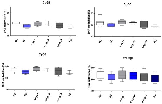
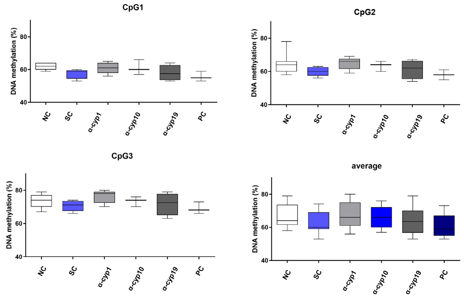
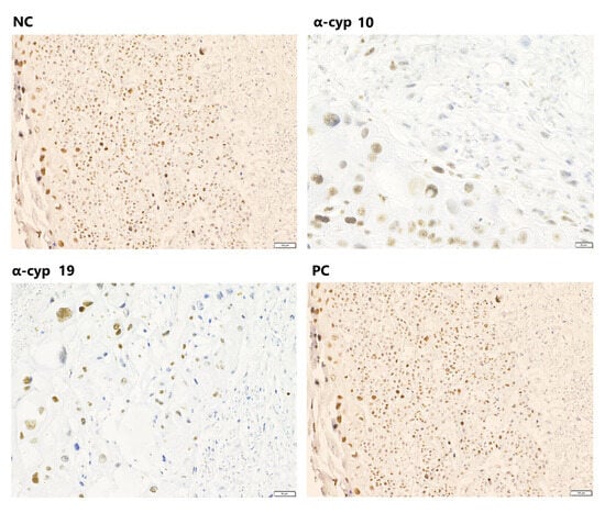
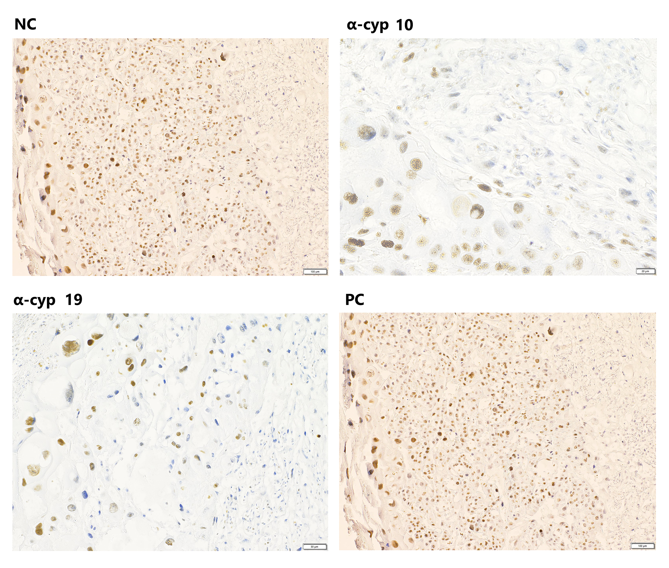
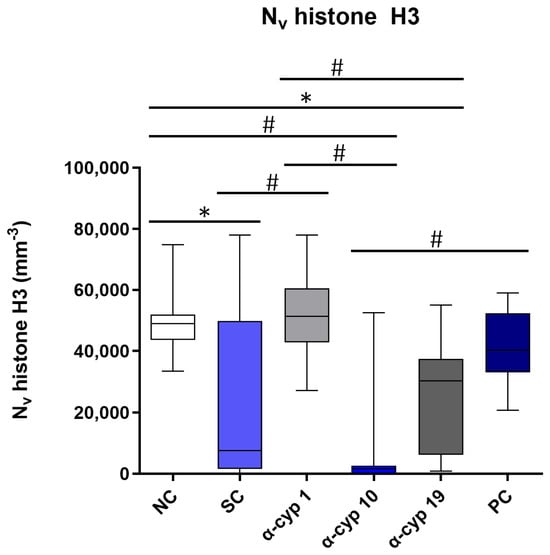
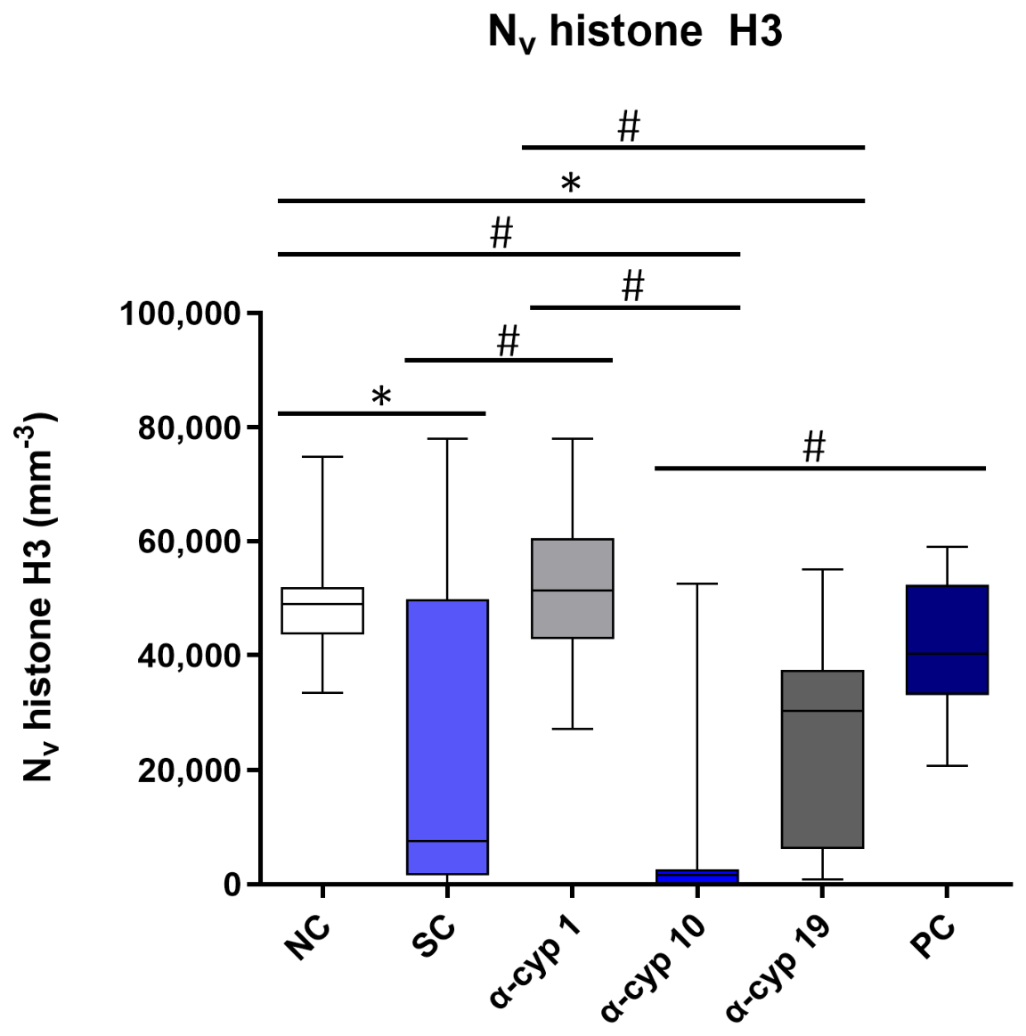
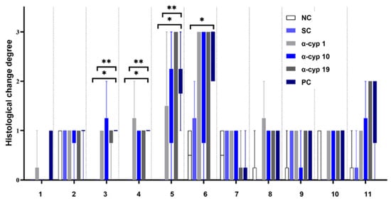
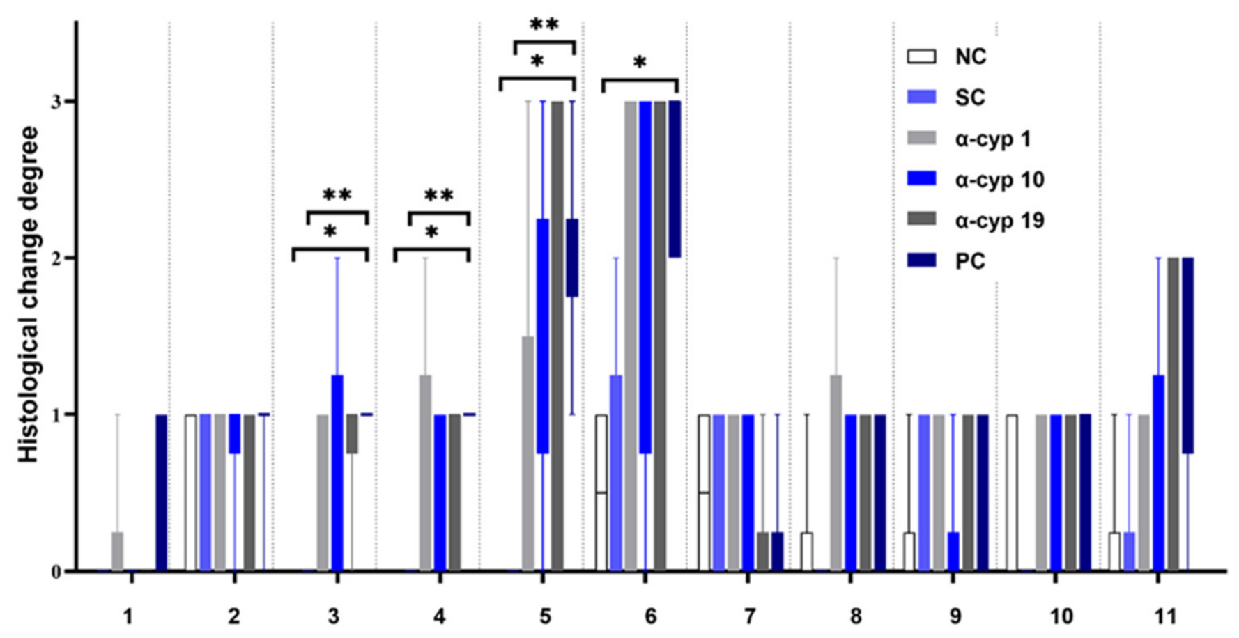
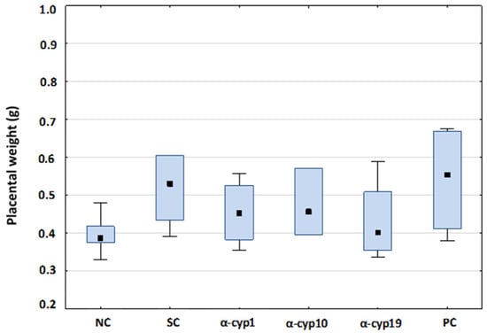
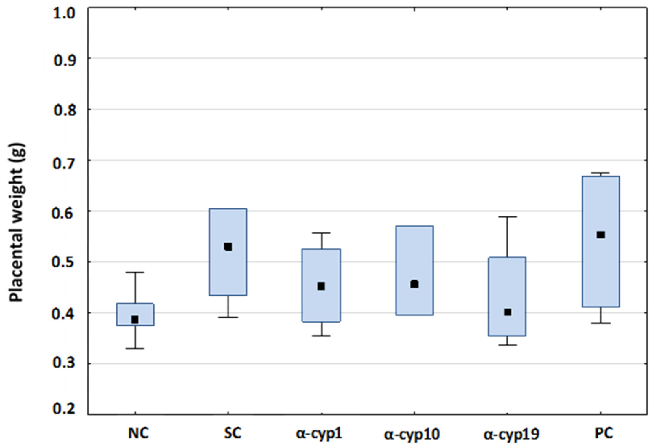
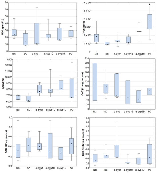
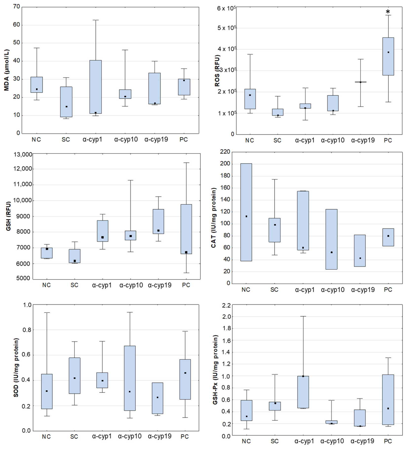
Disclaimer/Publisher’s Note: The statements, opinions and data contained in all publications are solely those of the individual author(s) and contributor(s) and not of MDPI and/or the editor(s). MDPI and/or the editor(s) disclaim responsibility for any injury to people or property resulting from any ideas, methods, instructions or products referred to in the content. |
© 2026 by the authors. Licensee MDPI, Basel, Switzerland. This article is an open access article distributed under the terms and conditions of the Creative Commons Attribution (CC BY) license.