


Abstract
Our skin is colonized by the skin microbiome and provides a physical, chemical, and metabolic barrier to the environment. Although current evidence links dietary pesticide exposure to microbiome disturbance and adverse health effects, the corresponding effects of dermal pesticide exposure on the skin microbiome remain largely unexplored and require further investigation. Using a skin-derived ten-strain model community and the corresponding single-strain cultures, we assessed the impact of the insecticide acetamiprid (ACE) in vitro. We identified strain-specific responses that shaped community-level outcomes. Exposure to ACE significantly altered the growth kinetics, metabolite profiles, and composition of the bacterial community. The most pronounced response was observed for Acinetobacter radioresistens, which exhibited marked growth inhibition upon ACE exposure, resulting in its complete loss from the community. At the single-strain level, susceptibility to ACE varied among isolates, with changes in short-chain fatty acid levels. Although ACE significantly affected only three of the ten skin isolates, these strain-specific responses still led to measurable changes at the community level. Our findings highlight the potential of ACE to modulate the human skin microbiome and underscore the need to integrate single-strain and community-level approaches into pesticide hazard assessment to capture both mechanistic insights and more biologically relevant outcomes.
1. Introduction
The human microbiome, which colonizes externally exposed sites such as the skin, lungs, and gut, is subject to chronic exposure to a wide range of xenobiotic substances. It is well recognized that the microbiome has a critical role in human health. Here, the highly diverse skin microbiome is essential for human health [1,2]. It supports skin barrier function, which forms a physical, chemical, and metabolic barrier against environmental threats [3], protects against pathogens [4,5,6], and regulates the immune system [7,8,9]. Microbial metabolites, such as amino acid derivatives and short-chain fatty acids (SCFAs), mediate these processes [6,10,11]. However, microbiome disturbances caused by xenobiotics are suspected to contribute to the rising incidence of chronic inflammatory diseases [12,13,14,15]. These concerns have recently been acknowledged by regulatory authorities, such as the European Food Safety Authority (EFSA) and the US Environmental Protection Agency (EPA), advocating the integration of microbiome assessments into regulatory procedures to address microbiome-mediated effects on the host [16,17,18].
Pesticides, which include biocides and plant protection products (PPPs), are crucial for meeting the current and increasing demand for food and are among the most ubiquitous environmental contaminants [19,20,21]. Although most pesticides are designed to target specific organisms, there have been reports of unintended effects on non-target organisms, including the human microbiome. Most of these reports focus on changes in community structures and the associated apparent biological functions of these communities. In the human gut, pesticide exposure has been associated with changes in bacterial growth [22,23], community diversity [24,25], and key metabolic pathways, such as amino acid and lipid metabolism [23,26,27], analogous to the reported off-target effects of medication on the gut microbiome [28]. Furthermore, the herbicides glufosinate ammonium and glyphosate have been documented to alter gut microbial composition and function in animals [29,30]. Beyond the gut, changes in microbial composition have also been reported for amphibian skin microbiota upon exposure to the fungicide chlorothalonil [31].
Acetamiprid (ACE) is a common and globally used neonicotinoid insecticide applied for pest management in agricultural, public health, and domestic settings [32,33,34]. It acts by binding nicotinic acetylcholine receptors in the insect nervous system, causing hyperstimulation, paralysis, and subsequent death [35,36]. As a new-generation chloronicotinyl neonicotinoid, ACE is currently the only approved neonicotinoid PPP in the European Union (EU). This approval is mainly attributed to its lower toxicity to humans and non-target organisms along with reduced environmental persistence compared to other neonicotinoids, such as clothianidin and thiamethoxam [32,37,38,39]. Although toxicological evaluations and biomonitoring studies indicate that domestic exposure to ACE is minimal and deemed safe for humans [38,39], the long-term consequences remain unclear. Regulatory authorities demand extensive rodent studies during the approval process for pesticide active substances, which potentially detect severe (acute) microbiome-mediated effects. However, species-specific or difficult-to-detect chronic endpoints might be overlooked [40]. Despite its low toxicity in humans, ACE has been shown to alter microbial community structure of honeybees, silkworms, and mice, affecting host metabolic and immune functions [41,42,43], or pathogen resistance [44]. Conversely, some microbes may alter ACE bioavailability and toxicity [45,46,47,48]. While soil bacteria, such as Stenotrophomonas sp., Pseudomonas sp., or Acinetobacter sp., can degrade ACE [49,50,51,52], its metabolization by human microbiota remains unexplored. Interestingly, these genera have also been isolated from human skin. Therefore, ACE may also affect the human skin microbiome similar to the documented microbiome effects in animals as mentioned above.
While many studies have investigated pesticides that induce microbial disturbances in the gut [22,23,53,54], studies examining interactions of pesticides with the skin microbiome are scarce. Microbial disturbances of the skin have been associated with adverse health effects such as atopic dermatitis, melanoma, and other potential effects [13,47,55]. Thus, pesticide-induced disturbances of the skin microbiome may present a potential health risk. Moreover, previous studies have demonstrated that skin microbiota can modulate the toxicity and dermal absorption of environmental pollutants through metabolization [46,47,56]. Given the critical role of the microbiome in maintaining skin health and its reciprocal interactions with other xenobiotics, it is crucial to understand microbiome-mediated health implications for human hazard assessment.
In this study, we examined the effects of the insecticide ACE on the defined bacterial community Bacillota-Actinomycetota-Pseudomonadota.10 (BAP.10), comprising ten skin-derived bacteria, as well as on the corresponding single-strain cultures. By evaluating growth kinetics, metabolite profiles, and community composition, we gained insights into strain-specific contributions and the translation of effects from a single-strain level to the community level.
2. Materials and Methods
2.1. Chemicals
Unless otherwise specified, all chemicals were obtained from Sigma-Aldrich (Taufkirchen, Germany) in their highest available purity. Both ACE and the internal standard ACE-d3 were sourced from Toronto Research Chemicals (Toronto, ON, Canada) with a purity of ≥98%. Acetone, acetonitrile (ACN), and acetic acid (AcOH) of liquid chromatography-mass spectrometry (LC-MS) grade were purchased from Carl Roth (Karlsruhe, Germany) with a purity of ≥99.9%.
2.2. Bacterial Isolates and Cultivation
We have recently established the BAP.10 community, a defined bacterial community comprising ten bacterial strains from healthy human forearm skin [57,58]. The bacterial strains cover three main phyla commonly isolated from human skin and are listed in Table 1.
Table 1.
Bacterial composition of the skin-derived BAP.10 community.
For all experiments, bacteria were revived from 25% glycerol stocks by streaking onto Lysogenic Broth (LB, Carl Roth) agar plates and incubated at 37 °C. Single colonies were picked and restreaked. The third passage was then transferred into liquid LB medium and cultivated at 37 °C with shaking at 200 rpm overnight. The following day, overnight cultures were used to inoculate the cultures for ACE exposure, either individually or mixed in equal cell numbers within the defined BAP.10 community. The cell concentration was determined by their respective optical density at 600 nm (OD600; FoodALYT/Omnilab, Bremen, Germany) and colony forming units (CFU)/mL values, as characterized previously [58].
2.3. ACE Exposure of Single-Strain Cultures and BAP.10 Community
All experiments were performed in flat-bottomed 96-well plates (Techno Plastics Products AG, Trasadingen, Switzerland) and were conducted in at least duplicates with a minimum of three biological replicates. A final ACE concentration of 3.75 mM (0.835 g/L) was selected as a high-dose exposure scenario while avoiding bactericidal effects under in vitro conditions. Commercial formulations usually contain 200 g/L ACE and are diluted prior to application to approximately 0.05–0.2 g/L [59,60,61]. Therefore, the selected concentration lies within the rage of both the formulation and the applied field concentration. This captures realistic exposure scenarios, such as those occurring during preparation and accidental contact, and may increase the likelihood of detecting treatment-related alterations in skin microbiota under in vitro conditions.
Bacterial cultures were initially adjusted to a final cell concentration of 5 × 10⁵ cells/mL in liquid LB medium supplemented with 3.75 mM ACE and acetone at a final concentration of 1%, or with 1% acetone alone as a solvent control. These cultures were then used either individually or mixed in equal ratios for the BAP.10 community and transferred into a well of a 96-well plate. For metabolite analyses, a respective dead culture was carried along. Dead cultures were derived as controls from the same overnight cultures used for the live cultures as follows. After overnight growth, all ten single-strain cultures and the BAP.10 community were diluted to OD600 = 1, respectively. Cultures were centrifuged at 8000× g for 10 min, the supernatant was removed, and each cell pellet was resuspended in fresh LB medium at a final OD600 = 2. Subsequently, cultures were inactivated at 90 °C for 2 h, followed by two freeze-thawing cycles. To verify inactivation, dead cell suspensions were plated onto LB agar plates. For using the dead cells as controls, 96-well plates were prefilled with LB medium supplemented with 7.5 mM ACE or 2% acetone. The plates were then inoculated at a 1:1 ratio with the dead cultures to achieve a final concentration of either 3.75 mM ACE or 1% acetone. Non-inoculated media served as an aseptic compound control. Empty wells were filled with Dulbecco’s Phosphate Buffered Saline (DPBS; Pan-Biotech, Aidenbach, Germany) to minimize evaporation. Culture plates were incubated at 200 rpm for 24 h at 37 °C in an incubation shaker (Multitron Pro, INFORS HT, Basel, Switzerland). To ensure appropriate bacterial growth of live cultures, their OD600 was measured both before and after the assay using a plate reader (BioTek Synergy Neo2, Agilent Technologies, Waldbronn, Germany). Following 24 h of incubation, whole culture samples and supernatants (centrifugation: 3000× g, 10 min, 4 °C, in round-bottomed 96-well plates) were collected from all samples. Cell-free supernatants for metabolite analyses were stored at −80 °C and cell pellets for BAP.10 community analyses were stored at −20 °C until further processing.
For bacterial growth analyzes, the OD600 was monitored at 30 min intervals for a maximum of 40 h at 37 °C with continuous shaking (BioTek Synergy Neo2, Agilent Technologies). The recorded OD600 values were subsequently used to generate growth curves and calculate growth parameters, including area under the bacterial growth curve (AUC), which summarizes overall bacterial growth over time and is frequently used as a proxy for culture fitness [62], doubling time (tD), and duration of the lag phase (λ) using QurvE v1.1.2 [63].
2.4. Analyzes of Microbial Viability
Microbial viability of single-strain and BAP.10 community cultures was quantified following 24 h of cultivation using the PrestoBlue™ HS Cell Viability Assay (Thermo Fisher Scientific, Waltham, MA, USA), according to manufacturer’s instructions. In brief, 50 µL of each culture was transferred into a 96-well flat bottom plate together with 5 µL of PrestoBlue™ HS reagent. Following incubation for 35 min at 37 °C, absorbance was measured at 570 nm and 600 nm as a reference wavelength (BioTek Synergy Neo2, Agilent Technologies).
2.5. Bacterial DNA Extraction, 16S rRNA Gene Amplicon Sequencing and Data Analyses
For 16S gene analysis, bacterial DNA was isolated alongside negative controls to account for background interference, including water and kit control using the QIAamp UCP DNA Micro Kit (Qiagen, Hilden, Germany), adhering to the manufacturer’s instructions. Nucleic acid concentration was quantified using Qubit (Invitrogen, Carlsbad, CA, USA) and UV-Vis Spectrophotometer (NanoDrop-ND 1000, Peqlab, Erlangen, Germany). To ensure quality, a defined mock microbial community was included for 16S amplicon sequencing (Skin Microbiome Genomic Mix, American Type Culture Collection, Manassas, VA, USA). Preparation of the amplicon library for 16S amplicon sequencing and processing of raw reads was conducted as previously described [58]. Briefly, 16S rRNA genes were amplified from isolated DNA in a two-step polymerase chain reaction (PCR) using primers targeting the V1-V3 regions: 27F-YM (5′-AGA GTT TGA TYM TGG CTC AG-3′) and 534R (5′-ATT ACC GCG GCT GCT GG-3′). Sequencing was conducted on Illumina MiSeq system using PE300 cartridges v3 with 2 × 275 cycles (Illumina Inc., San Diego, CA, USA). Final 16S amplicon sequences (i.e., after denoising, chimera removal and clustering; [64]) were annotated using EzBioCloud’s 16S rRNA gene-based ID [65]. Diversity analyses were conducted using Rhea v1.1.8 [66] with a relative abundance cutoff of 0.5%.
2.6. Quantification of ACE for Evaluating Potential Bioaccumulation Using LC-MS/MS Analysis
For ACE quantification, 50 µL of culture was extracted with 450 µL of ice-cold ACN in amber glass vials (Merck, Darmstadt, Germany) immediately after cultivation and stored at −20 °C until analysis. Subsequently, ACE was quantified using liquid chromatography-tandem-mass spectrometry (LC-MS/MS), as described in the following.
At first, an internal standard dissolved in ice-cold ACN was used for method development and calibration (Table S1). Prior to sample analysis, the method was validated in accordance with the guideline SANTE 11312/2021v2 [67] assessing linearity (Figure S1), sensitivity, accuracy and precision, recovery, matrix effects, robustness, and stability, including sampling and storage conditions. This method was further evaluated by determining the limit of detection (LOD = 0.01 ng/mL) and quantification (LOQ = 0.05 ng/mL), as well as by fortification assessing spike recoveries at 1 ng/mL and 110 ng/mL within the calibration curve (Table S1).
2.6.1. Sample Preparation for LC–MS/MS Analysis
All samples were spiked with the internal standard at a final concentration of 60 ng/mL in ice-cold ACN and diluted in ice-cold ACN to attain a concentration suitable for quantification within the calibration curve. Diluted samples were filtered through a 0.2 µm syringe filter (CHROMAFIL Xtra H-PTFE, Macherey-Nagel, Düren, Germany) into amber glass vials and then injected into the LC-MS/MS. Blank control and calibration curve samples were identically processed.
2.6.2. LC–MS/MS Separation and Detection Conditions
A sample volume of 5 µL was injected into the 1290 Infinity II LC system (Agilent Technologies), which was coupled with a 6475 Triple Quadrupole LC/MS system (Agilent Technologies). The multi-sampler was maintained at 4 °C and the needle was washed with methanol:Milli-Q (70:30, v/v) both prior to and following sampling. Chromatographic separation was conducted using reversed-phase LC with a CORTECS T3 column (C18, 120 Å = 12 nm, 1.6 µm, 2.1 mm × 100 mm, Waters, Milford, MA, USA) and a CORTECS T3 VanGuard pre-column (C18, 120 Å = 12 nm, 1.6 µm, 2.1 mm × 5 mm, Waters) at 30 °C, using a binary eluent system of 0.05% AcOH in Milli-Q (mobile phase A) and 0.05% AcOH in ACN (mobile phase B) and a flow rate of 0.5 mL/min. Separation was performed by gradient elution starting with 95% A (0–3 min), followed by a linear ramp from 3–7 min to 5% A, hold 5% A (7–9 min) and re-equilibration to 95% A (9.5–13.5 min). Following separation, ACE was quantified using the triple quadrupole (QqQ) operating in multi reaction monitoring (MRM) mode via electrospray ionization in positive mode. One transition was used for quantification, and two transitions were used for confirmation (Table S1). The source parameters were as follows: gas temperature, 280 °C; gas flow, 12.5 L/min; nebulizer, 36 psi = 248 kPa; sheath gas temperature, 370 °C; sheath gas flow, 9.5 L/min; capillary voltage, 4100 V; nozzle voltage, 1700 V.
2.6.3. LC–MS/MS Data Analysis
ACE was quantified utilizing MassHunter Quantitative Analysis software v12.1 (Agilent Technologies). Calibration curves, derived from the ratio of the peak area of the pure ACE standard to the peak area of the internal standard, were employed to calculate the ACE concentration (Figure S1). Average analytical deviation was below 6% (Table S2). Thus, an ACE reduction exceeding the analytical deviation by factor two, i.e., 12%, was considered a relevant ACE reduction. A significant ACE reduction above 12% in live cultures compared to the aseptic compound control, together with no ACE reduction in the corresponding dead culture control, was interpreted as indication of biotransformation.
2.7. SCFA, Amino Acid, and Amine Analyses
Each 100 µL bacterial culture supernatant was mixed with five times the µl amount of extraction solvent ACN:Milli-Q (1:1, v/v) and homogenized using a TissueLyser II (30 Hz, 10 min, Retsch, Qiagen). After centrifugation (2 min, 17,968× g), 10 µL and 20 µL were used for amino acid and SCFA derivatization, respectively.
For derivatization of amino acids and biogenic amines, the supernatant was evaporated to dryness (SpeedVac, Eppendorf, Hamburg, Germany), resuspended in 50 µL of 5% phenyl isothiocyanate (PITC) in ethanol:Milli-Q:pyridine (1:1:1, v/v/v) and incubated for 25 min at RT. Subsequently, samples were dried to remove excess PITC and resuspended in 10 µL 5 mM ammonium acetate in methanol. After incubation (10 min, 17,968× g), 90 µL of Milli-Q:ACN + 0.2% formic acid were added.
Prior to measurement, each derivative was diluted 1:25 and 10 µL were injected onto a Waters Acquity UPLC system coupled online with a QTRAP® 5000 mass spectrometer (Sciex, Framingham, MA, USA). Chromatographic separation was achieved with an Agilent Zorbax Eclipse XDB-C18 column (3.5 µm, 3.0 mm × 100 mm) using a constant flow rate of 0.5 mL/min and Milli-Q + 0.2% formic acid and ACN + 0.2% formic acid as mobile phases A and B, respectively. The linear LC gradient was as follows: 0–0.5 min at 0% B, 0.5–4 min from 0–70% B, 4–5.3 min at 70% B, 5.3–5.4 min from 70–0% B, and 5.4–7.3 min at 0% B, and the QTRAP was set to positive ionization mode.
Following the same conditions, all undiluted samples were measured on an LC-MS/MS system using an Agilent 1290 II infinity UPLC system (Agilent Technologies Inc., Santa Clara, CA, USA) coupled with a QTRAP6500+® mass spectrometer (Sciex).
For SCFAs derivatization, the method of Han et al. was modified [68]. The supernatant was combined with 20 µL of 3-nitrophenylhydrazine in 50% ACN (200 mM) and 20 µL of N-(3-dimethylaminopropyl)-N′-ethylcarbodiimid hydrochloride in 50% ACN with 6% pyridine (120 mM) and incubated (30 min, 40 °C) in a thermomixer (Eppendorf).
Prior to measurement, the derivative was diluted 1:50 in 10% ACN. Each sample (10 µL) was injected onto the RSLC UltiMate 3000® system (Thermo Fisher Scientific) coupled online to a QTRAP® 5000 mass spectrometer (Sciex). SCFAs were separated on an Acquity UPLC BEH C18 column (1.7 µm, 2.1 mm × 100 mm, Waters) with solvent A, Milli-Q + 0.01% formic acid, and solvent B, ACN + 0.01% formic acid, as mobile phases. Constant flow rate was set to 0.35 mL and column oven was set to 40 °C. The chromatographic gradient was as follows: 0–2 min at 15% B, 2–15 min from 15–50% B, 15–16 min at 100% B, 16–17 min from 100–15% B, and 17–20 min at 15% B. Mass spectrometric measurement was performed in negative ionization mode.
For identification and quantitation, a scheduled MRM method was used for both amino acids and SCFAs, with specific transitions for every metabolite. External calibration curves were measured for linear and quadratic regression. Peak areas of all samples and standards for linear regression were determined in SciexOS® v3.0.0 Software (Sciex).
2.8. Statistical Data Analyses and Visualization
Data analyses and visualizations were performed in R v4.4.3, utilizing open-source packages from RStudio v2026.07.1, or GraphPad Prism v9.3.1 (Graph Pad, La Jolla, CA, USA). The graphical illustration of the experimental setup was created in BioRender.
Differences between two groups were calculated using two-tailed unpaired t-test and differences between more than two groups were calculated using one-way ANOVA, followed by Tukey’s multiple comparisons test both with significance levels indicated as * p < 0.05, ** p < 0.01, *** p < 0.001, and **** p < 0.0001 in GraphPad Prism. For statistical analysis of the SCFA, amino acid, and biogenic amine profiles, the Mann-Whitney test was applied, and p-values were corrected for multi-testing using the Benjamini–Hochberg method.
Principal Coordinates Analysis (PCoA) was performed in R v4.4.3 using Bray-Curtis dissimilarity in the vegan v2.7-3 package [69]. PCoA were visualized using the ggplot2 v3.5.1 package [70]. Differences between groups were evaluated by PERMANOVA using the adonis function from the vegan package in R v4.4.3. Comprehensive descriptions of each statistical analysis are provided in the corresponding method description and figure legends.
3. Results
We examined the potential effects of the insecticide ACE on skin microbiota using a defined skin-derived microbiota of ten strains, labelled BAP.10. To capture a broader spectrum of potential responses, this model community comprises a defined panel of ten bacterial strains, including common skin isolates such as S. epidermidis and M. luteus, as well as transient bacteria such as P. fulva and P. simplex. The latter two are typically associated with environmental settings. In addition, the bacterial selection was based on experimental requirements for aerobic skin co-cultivation with full-thickness skin models to facilitate a balanced skin colonization with a more complex bacterial community. We used this community and its respective single-strain cultures to assess the influence of ACE on various endpoints, i.e., growth kinetics, metabolism, and for BAP.10 impact on community composition (Figure 1). This enables us to discern which isolates are affected by ACE exposure directly and whether observed effects are mirrored on the community level or if secondary effects might be observed.
Figure 1.
Illustration of the experimental setup. Investigating effects of the insecticide acetamiprid (ACE) on ten skin-derived bacterial strains (colored accordingly) individually and their mix, the BAP.10 community. All single-strain cultures and the BAP.10 community were exposed to 3.75 mM ACE in acetone or 1% acetone (solvent control). Following cultivation, various endpoints were assessed in the BAP.10 community and the single-strain cultures. Created in BioRender. Kissner, M. (2026) https://BioRender.com/nmkif12.
3.1. ACE Exposure Affects Growth and Composition of the BAP.10 Community
First, we investigated how ACE affects the growth of the BAP.10 community by monitoring OD600 compared to the solvent control (Figure 2A,B; Table S3). We found a slightly different growth behavior due to ACE exposure. The AUC was significantly reduced by 4% (Figure 2B). This diminished growth was mirrored by a significant increase in the doubling time of the BAP.10 community of more than 11%, while the lag phase duration (λ) was not affected.
Figure 2.
Impact of ACE exposure on the bacterial BAP.10 skin model community. The impact of ACE on (A) growth kinetics and (B) specific growth metrics, including AUC, doubling time (tD), and duration of the lag phase (λ) was examined for the BAP.10 community across at least four biological replicates and are shown as mean with standard deviation. (C) α-diversity presented as percentage of effective values of the ACE treated BAP.10 community compared to solvent control as mean with standard deviation. (D) β-diversity displayed as PCoA similarity analysis of the BAP.10 community following 24 h of ACE or solvent exposure. Significance levels were determined by PERMANOVA. (E) Relative abundances of the respective genera in both groups shown as mean with standard deviation. M. luteus spp. were not detected in either group. Differences of the ACE treated group from solvent control were calculated using unpaired t-test; * p < 0.05, ** p < 0.01, *** p < 0.001, **** p < 0.0001.
Using 16S rRNA sequencing, we assessed the effect of 24 h ACE exposure on the bacterial diversity and abundance on genus level within the BAP.10 community (Figure 2C–E; Table S4). The α-diversity was significantly reduced upon ACE exposure by 27%, 42%, and 45% for effective richness, effective Shannon, and effective Simpson, respectively, compared to the solvent control (Figure 2C). This change was also obvious when evaluating the β-diversity between the ACE treated BAP.10 community and the solvent control (Figure 2D). Detailed taxonomic analyses revealed that ACE exposure significantly increased the relative abundance of P. fulva and led to a complete loss of A. radioresistens (Figure 2E). In both experiment and control, M. luteus spp. were completely lost from the community, while the strains do grow in single-strain cultures in ACE or acetone carrier alone (Section 3.3).
Overall, ACE exposure slightly inhibited bacterial growth and induced a shift in species composition of the BAP.10 community, with A. radioresistens emerging as the most sensitive community member.
3.2. ACE Exposure Affects Metabolite Profiles of the BAP.10 Community
While some metabolic changes have been found, the overall metabolic activity of the BAP.10 community seems to remain unaffected by ACE exposure (Figure 3A–D, Table S5). Microbial metabolites can influence skin homeostasis and are essential for skin health [6,71]. To understand the functional effects related to ACE exposure of the BAP.10 community, 14 SCFAs, 21 amino acids, and 6 biogenic amines were quantified in culture supernatants (Figure 3B–D, Table S6). Similarity analysis including all measured metabolites revealed significant alterations between the ACE treated group and solvent control (Figure 3B). ACE exposure significantly increased the abundance of the SCFAs isobutyrate, isovalerate, lactate, propionate, and 2-methylbutyrate (Figure 3D; Table S7). Furthermore, ACE exposure altered the abundances of several amino acids and biogenic amines, including tyrosine (Tyr), threonine (Thr), and gamma-aminobutyric acid (GABA), albeit not at a significant level (Figure 3C, Table S6). Next, we quantified the recovery of ACE from the BAP.10 community to gain insights into potential microbial biotransformation of ACE (Figure 3E). A significant ACE reduction in live cultures compared to the aseptic compound control, coupled with the absences of ACE reduction in the corresponding dead culture control, was interpreted as indicator for biotransformation. A significant ACE loss was detected in live cultures of the BAP.10 community (18.9%) compared to the aseptic compound control, while dead culture controls showed only insignificant ACE reduction (1.3%). This may indicate ACE biotransformation by the BAP.10 community.
Figure 3.
Impact of ACE exposure on metabolite profiles of the BAP.10 community and calculated ACE recovery from the BAP.10 community. (A) Quantification of cell viability using PrestoBlue assay. Data are presented as mean with standard deviation and differences of the ACE treated group to solvent control were analyzed using unpaired t-test. (B–D) Following 24 h of ACE exposure, metabolite profiles, including SCFAs, amino acids, and biogenic amines, were analyzed in cell-free supernatants of the BAP.10 community and compared to solvent controls. (B) PCoA of the absolute metabolite abundance was performed using Bray-Curtis dissimilarity and group differences were calculated by PERMANOVA. Log2FC of (C) amino acids and biogenic amines and (D) SCFAs of the ACE treated BAP.10 community compared to the solvent control were visualized in a heatmap. Differences between the ACE-treated BAP.10 community and the BAP.10 solvent control were analyzed using Mann-Whitney test, p-values were corrected for multi-testing using the Benjamini–Hochberg method. Adjusted p-values, or if not applicable, p-values are reported in the grey area; * adj. p < 0.05 and (*) p < 0.05. (E) ACE was quantified in both live and dead (heat inactivated and freeze-thawed) cultures of the BAP.10 community and normalized to the ACE levels obtained from the aseptic compound control. Samples exhibiting > 12% ACE reduction relative to the aseptic compound control were subjected to analysis for differences between (a) the aseptic compound control and (b) live and dead cultures using one-way ANOVA, followed by Tukey’s multiple comparisons test, and adjusted p-values are reported; ** adj. p < 0.01.
3.3. ACE Exposure Affects Growth Kinetics of Certain Skin Isolates
Since ACE exposure affected the overall bacterial growth, composition and metabolism of the BAP.10 community (Figure 2 and Figure 3), we wanted to determine the effect of ACE on each strain (Figure 4A,B; Table S3). For most single skin isolates, the calculated growth parameters showed no significant change upon ACE exposure compared to the respective solvent control. However, growth of K. rhizophila was significantly affected by ACE exposure. Significantly, the AUC was reduced and the doubling time increased compared to the solvent control. Exposure to ACE also significantly reduced the growth of P. fulva, represented by the AUC, accompanied by a significant increase in the duration of the lag phase. The latter was slightly increased for S. capitis, too. A marginal increase in doubling time was also noted for both M. luteus strain 1 and 2, although these changes were not statistically significant.
Figure 4.
Impact of ACE exposure on growth kinetics of skin isolates. (A) Bacterial growth curves and (B) calculated growth parameters, including AUC, doubling time (tD), and duration of the lag phase (λ) are shown as mean with standard deviation of at least four biological replicates. Differences of ACE treated groups from their respective solvent control were analyzed using unpaired t-test; * p < 0.05, ** p < 0.01, **** p < 0.0001.
Notably, alterations in growth kinetics were particularly pronounced for A. radioresistens with a significant and strong increase in doubling time (73%) and duration of the lag phase (223%), accompanied by a significant reduction of the AUC (−34%) upon ACE exposure. This well explains the complete loss of A. radioresistens from the BAP.10 community following ACE exposure (Figure 2E).
3.4. ACE Exposure Affects Viability of Certain Skin Isolates and Results in Isolate-Dependent ACE Recovery
We assessed the ability of each skin isolate to degrade ACE (Figure 5A). Overall, four out of ten skin isolates were found to reduce the amount of ACE in the culture medium (Figure 5A). M. luteus strain 2 exhibited clear signs of ACE biotransformation, evidenced by ACE depletion when using live cells (13.1%), while the ACE concentration in the dead control remained unaffected compared to the aseptic compound control. Differences in ACE recovery between live (17.4%) and dead (5.8%) cultures of P. fulva may also indicate microbial biotransformation of ACE. However, these differences were not statistically significant. Furthermore, a significant reduction of ACE compared to the aseptic compound control was found in live and dead cultures of S. epidermidis (live: 16.9%; dead: 14.2%), K. rhizophila (live: 14.2%; dead: 10.5%), and A. radioresistens (live: 19.5%; dead: 15.4%).
Figure 5.
Quantification of ACE from single-strain cultures and assessment of cell viability. Following 24 h of ACE exposure, (A) ACE was quantified in both live and dead (heat inactivated and freeze-thawed) single-strain cultures and normalized to the ACE levels obtained from the aseptic compound control. Samples exhibiting >12% ACE reduction relative to the aseptic compound control were subjected to analysis for differences between (a) the aseptic compound control and (b) live and dead cultures using one-way ANOVA, followed by Tukey’s multiple comparisons test, and adjusted p-values are reported; * adj. p < 0.05, ** adj. p < 0.01, *** adj. p < 0.001. (B) The absorbance-based PrestoBlue assay was performed for quantification of cell viability. Data are presented as mean with standard deviation for three biological replicates and differences of ACE treated groups to respective solvent control were analyzed using unpaired t-test; * p < 0.05, *** p < 0.001.
Simultaneously, microbial viability upon ACE exposure was evaluated by quantification of their reduction potential relative to the respective solvent control (Figure 5B). To ensure comparable cell concentrations, bacterial growth was assessed by measuring the OD600 both prior to and following cultivation (Table S5). The cell viability of most skin isolates was not affected by ACE exposure. As expected from above results, the viability of A. radioresistens was significantly reduced by 65%. In contrast, viability was significantly increased in S. epidermidis by 49% upon ACE exposure compared to its solvent control. This significant disparity in cell viability is likely due to the considerably reduced cell abundance in A. radioresistens cultures following ACE exposure, as compared to the solvent control (Table S5).
3.5. ACE Exposure Affects Metabolite Profiles of Certain Skin Isolates
As ACE exposure was found to induce alterations in the metabolite profiles of the BAP.10 community (Figure 3B–D), we next analyzed whether these alterations could be attributed to specific skin isolates. The absolute abundances of the number of SCFAs, amino acids, and biogenic amines were quantified in ACE treated microbial cultures and their respective solvent control (Figure 6A–C and Figure S2, Table S6).
Figure 6.
Impact of ACE exposure on metabolite profiles of skin isolates. SCFAs, amino acids, and biogenic amines were analyzed in cell-free supernatants of single-strain cultures following 24 h of ACE exposure and compared to the respective solvent control. (A) PCoA of the relative metabolite abundance was performed using Bray-Curtis dissimilarity and group differences were calculated by PERMANOVA. Heatmap visualization of the log2FC of (B) SCFAs and (C) amino acids and biogenic amines in comparison to the respective solvent control. Differences between the ACE-treated group and the respective solvent group were analyzed using Mann-Whitney test, p-values were corrected for multi-testing using the Benjamini–Hochberg method. Adjusted p-values, or if not applicable, p-values are reported; * adj. p < 0.05 and (*) p < 0.05.
Exposure to ACE significantly altered the global metabolite profile (SCFAs, amino acids, and amines) in three out of ten skin isolates (S. epidermidis, P. simplex, and K. rhizophila), with the effect size being strongest in S. epidermidis (Figure 6A). In S. epidermidis, heatmap visualization of the respective metabolites revealed a significant increase in all SCFAs concentration, except for acetate and lactate, upon ACE exposure compared to the solvent control (Figure 6B). In contrast, ACE exposure significantly reduced most SCFA levels in P. simplex, solely lactate levels were significantly increased. Notably, exposure to ACE affected amino acid and biogenic amine concentrations in the skin isolates; however, these differences were not statistically significant.
4. Discussion
The human microbiome is continuously exposed to xenobiotic substances, including pesticides. Yet, its role in regulatory toxicology, beyond the obvious effects observed in the obligatory in vivo studies required for pesticide authorization, remains largely unexplored. Several studies have indicated that dietary exposure to pesticides can alter the microbial composition and function in the gut [23,24,25,27,41,44]. In many of these cases, the exact toxicological implications remain unclear. However, they clearly show that there is a need to gain a better understanding of the respective toxicology of the gut microbiota. Simultaneously, potential interactions with the skin microbiome remain understudied, despite the fact that exposure to the skin usually occurs at higher concentrations and more frequently, at least for workers and handlers. Although the neonicotinoid ACE is considered safe within the approved uses based on standard risk assessment, evidence of gut microbial disturbance in animals suggests that there is a data gap regarding possible microbiota-mediated effects. This highlights a potential gap in human hazard assessment framework. Our study shows that ACE exerts strain-specific effects in skin isolates that persist to different degrees within the ten-strain model community BAP.10.
4.1. ACE Exposure Induced Structural and Functional Changes Within the BAP.10 Community
ACE significantly reduced bacterial diversity within the BAP.10 community driven by the complete loss of A. radioresistens and a notable increase in the relative abundance of P. fulva. While A. radioresistens exhibited the highest relative abundance in the solvent control, exposure to ACE resulted in its complete absence. An ACE-induced reduction in α- and β-diversity was also observed in studies investigating the gut microbiota of insects [41,42,44]. Consistent with our findings, these studies reported an increased relative abundance of Pseudomonas spp. in the gut microbiota of honeybees and silkworms [42,44], suggesting a selective advantage of Pseudomonas under ACE exposure. This finding is not unexpected, since members of this genus are recognized for their resilience to harsh environmental conditions and their ability to even prosper in high concentrations of pesticides [72,73]. Contrasting our findings, where A. radioresistens was completely void upon ACE exposure, other studies reported an increased relative abundance of other Acinetobacter spp. in honeybees’ intestinal microbiota [42] and a soil consortium [49]. These differences are likely attributed to intra-genus differences [74]. While Acinetobacter spp. can be resistant to environmental stressors [75,76], the neonicotinoid thiamethoxam has also been shown to significantly reduce the relative abundance of another Acinetobacter sp. [77]. The observed complete loss of A. radioresistens in this study may indicate that this strain could not adapt its metabolism quickly enough to the presence of ACE—the lag phase was also prolonged—leading to severe growth disadvantages that consequently led to its absence in the community.
The structural community changes, also reflected in inhibited growth in the presence of ACE, may be explained by the differential susceptibilities of each skin isolate to ACE, including sublethal effects such as membrane disintegration, oxidative stress, and an altered microbial metabolism, which interfere with several cellular processes. This causes growth disadvantages for certain species, but sometimes growth advantages for others [73,78]. The observed effects of ACE in the BAP.10 community could not be attributed to a general cytotoxicity. However, it is important to note that viability assays reflect overall metabolic activity of the community but do not resolve strain-specific responses, including loss or increase of cell viability in different species.
Overall, the observed disturbances in the BAP.10 community regarding its growth kinetics and community structure suggest that ACE may also have the potential to disrupt the human skin microbiota in vivo. Such disruptions have health implications, given that alterations of the skin microbiome are associated with skin diseases, such as acne vulgaris, atopic dermatitis, psoriasis, or melanoma [13], but also allergic disorders [79,80].
Shifts in the abundance of common microbial metabolites can be induced by xenobiotic-mediated changes in microbial composition as well as by xenobiotics themselves [23,81]. Such changes could serve as biomarkers to assess the pesticide’s impact on skin health, as they influence microbial homeostasis, immune regulation, and skin barrier integrity [6,11,71,82]. Although ‘functionality’ is considered more relevant than pure compositional changes for a microbiome, the impact of ACE on microbial metabolism remains poorly investigated. Here, the global metabolite profile (SCFAs, amino acids, and amines) was changed within the BAP.10 community upon ACE exposure with a major difference observed in the SCFA abundance. SCFAs have been shown to modulate oxidative stress [83,84]. For example, isovalerate enhances epithelial barrier and induces gene expression of the antioxidant enzymes glutathione peroxidase 1 (GPX1) and superoxide dismutase 2 (SOD2) [85]. Interestingly, in the study of Yao et al., ACE was found to induce oxidative stress and disrupt antioxidant systems in bacteria. The ACE-induced oxidative stress was accompanied by activation of their antioxidant defense system [86]. Similar effects are also reported from non-bacterial systems [87,88,89]. Consequently, increased levels of SCFAs may be a defensive response to ACE-induced oxidative stress, mitigating adverse effects and enhancing community resilience [90].
Altered bacterial growth and metabolic profiles upon xenobiotic exposure may also be attributed to metabolic reprogramming and microbial biotransformation of the chemicals in question [91,92,93]. The human skin microbiome possesses a high metabolic capacity and may, thus, metabolize ACE, altering its bioavailability and toxicity for the host [45,46,47,48]. In a previous study, we performed functional profiling of the BAP.10 community and found a high functional potential like that found in complete human skin microbiomes [58]. To our knowledge, metabolization of ACE by members of the human skin microbiota has not yet been investigated. However, some environmental bacteria, including Pseudomonas spp. and Acinetobacter spp., have been reported to degrade ACE, and both genera have also been isolated from human skin [49,50,51,94,95]. The present study revealed indications of ACE biodegradation by BAP.10, albeit not at a high degree. ACE recovery was generally high in the BAP.10 community and in all single-strain cultures. This may be attributed to the bacteria being cultivated in a nutrient-rich medium containing alternative carbon sources in addition to ACE, which may have been preferred over ACE [96].
4.2. Single-Strain Cultures Exhibited Different Susceptibility to ACE Exposure
To understand strain-specific contributions to the observed alterations in the BAP.10 community, we also assessed the impact of ACE exposure on each strain. ACE exposure significantly inhibited the growth of A. radioresistens, resulting in its complete elimination from the BAP.10 community. This is likely attributable to ACE-mediated effects on metabolism and other critical physiological processes [73,78,97]. Furthermore, we found indications for ACE bioaccumulation in A. radioresistens, which has been linked with impeded bacterial growth [23,98]. Although the metabolic profiles in A. radioresistens were not significantly affected by ACE exposure, the largest increase in glutamate (Glu) abundance was observed among all skin isolates (Figure S2). Glu is an essential precursor for the synthesis of the antioxidant glutathione (GSH) and thereby contributes to cellular antioxidant capacity and the mitigation of oxidative damage [99]. Furthermore, Glu has been shown to reduce reactive oxygen species (ROS) production and enhance bacterial antioxidant enzyme activities [100]. Consequently, the increased Glu abundance in A. radioresistens following ACE exposure is consistent with an elevated oxidative stress response in this species. Overall, the presence of ACE appears to induce a compensatory state in A. radioresistens, associated with reduced metabolic activity and impaired growth.
Pseudomonadota are known for their metabolic versatility and stress tolerance, especially Pseudomonas spp. [101,102]. Given these characteristics, the marginally prolonged lag phase in P. fulva during ACE exposure may indicate a simple adaptation period. During lag phase, bacteria prepare for exponential growth by metabolic adjustment for carbon source utilization and prioritize adaption over biomass accumulation [97]. Interestingly, we found no evidence of ACE biotransformation, although live cultures of P. fulva exhibited reduced ACE levels after growth. ACE biotransformation has been reported for soil-associated Pseudomonas spp. [51,52]. Although biodegradation capacities are often strain specific, microbial communities can combine complementary metabolic pathways, surpassing capabilities of single strains [103,104]. Overall, ACE appears to have only a marginal effect on P. fulva, possibly due to its high stress tolerance.
In contrast to Pseudomonadota representatives, ACE exposure did not affect the relative abundance of all four Staphylococcus spp., P. simplex, and K. rhizophila in the BAP.10 community. For K. rhizophila, we observed an ACE-induced marginal increase in the doubling time, which may have been caused by bioaccumulation of ACE [23,98]. While Kocuria spp. have been reported to degrade herbicides [105] and neonicotinoids [106], degradation of neonicotinoids by P. simplex or Staphylococcus spp. has not been reported. The present study could not confirm ACE degradation by any of these strains. Notably, S. epidermidis and P. simplex, together with K. rhizophila, were the only skin isolates in which ACE exposure significantly changed their metabolite profiles. This response may suggest metabolic reprogramming, e.g., a stress-induced adaption processes to environmental changes for compensatory survival as discussed previously for the BAP.10 community. These metabolic changes were primarily driven by ACE-induced alterations in SCFA abundances. Interestingly, while the levels of most SCFAs were significantly reduced in P. simplex and K. rhizophila, they were significantly increased in S. epidermidis. Thus, the effects of ACE on the metabolite profiles in S. epidermidis may have been a key driver of the metabolic changes observed in the BAP.10 community, where ACE exposure likewise increased the abundance of all analyzed SCFAs. Notably, while the single-strain cultures exhibited either increased or decreased SCFAs levels, all SCFAs levels were consistently elevated in the BAP.10 community. This suggests that ACE-induced effects observed in single-strain cultures cannot be directly predicted at the community level. Simple additive effects of single strains are not sufficient for predicting community effects, highlighting the complexity of interspecies cross-feeding interactions [62,107,108].
In this study, M. luteus strain 2 was the only skin isolate whose calculated ACE recovery suggested microbial degradation of ACE, which may have contributed to the potential ACE degradation observed in the BAP.10 community. However, both M. luteus strains were absent from the BAP.10 community at the end of cultivation in both the ACE exposed group and the solvent control. This observation is likely attributable to community-level interactions, as both M. luteus strains exhibited normal growth under identical conditions in single-strain cultures. In comparison to most members of the BAP.10 community, M. luteus exhibited a prolonged lag phase and increased doubling time during exposure to solvent and ACE. Consequently, the delayed initiation of growth may have conferred a competitive growth advantage to other species and their dominance, which may have led to the depletion of essential nutrients and resulted in the absence of M. luteus within the BAP.10 community.
Overall, we observed ACE-induced changes in growth kinetics, community composition, and metabolite profiles of the BAP.10 community. These changes were driven by the effects of ACE exposure on specific BAP.10 community members. At the single-strain level, susceptibility to ACE varied among the ten skin isolates, with significant effects in three skin isolates. These findings demonstrate that strain-specific responses can persist in measurable changes at the community level. In our previous study, genome-based functional profiling showed that the BAP.10 community exhibited the highest overall functional potential among the investigated communities and high functional similarity to reference human skin microbiomes [58]. These findings suggest that BAP.10 may provide a useful defined model community for investigating microbiome-related aspects of skin toxicity. Although the BAP.10 community comprises some common members of the core skin microbiota and also transient bacteria, it still lacks other common as well as low abundant aerobic and anaerobic bacteria due to common experimental requirements and the limited community size. The addition of further skin commensals could impact outcomes such as community growth, diversity, metabolite profiles, and community resistance. Therefore, this work represents an initial step towards mimicking complex microbiome-xenobiotic interactions within a defined skin model community.
5. Conclusions
Our findings show that ACE can cause disturbances in a skin-derived model community. Because microbial metabolites contribute to skin homeostasis and systemic signaling, including gut-skin axis interactions, such disturbances may affect not only the skin but also distant organs [71,109]. Our study further demonstrates the limited predictability of single-strain analyses in forecasting community-level responses. Although resistant strains may compensate inhibitory effects on other strains, the loss of functionally relevant strains may still disrupt both microbial and host homeostasis [110]. Given the critical role of the skin microbiome, such disturbances could compensate for epithelial barrier function and host immunity, with potential health consequences [13,71,82]. These findings support the need to integrate single-strain and community-level approaches into microbiome-aware hazard assessments to capture both mechanistic insights and biologically relevant outcomes. Future studies should elucidate the mechanisms by which ACE affects bacterial metabolite synthesis and use more complex models, such as human 3D skin systems, to assess microbial responses alongside microbiome-host interactions.
Supplementary Materials
The following supporting information can be downloaded at https://www.mdpi.com/article/10.3390/toxics14110949/s1, Figure S1: Linearity coefficient of the calibration curve for ACE quantification using LC-MS/MS analysis; Figure S2: Impact of ACE exposure on metabolite profiles of skin isolates; Table S1: Analytical standards and determination of LOD, LOQ, and matrix effects; Table S2: Cutoff determination for distinguishing the observed [ACE] sequestration between potential biotransformation and analytical measurement fluctuations; Table S3: List of growth parameters of single-strain cultures and the BAP.10 community; Table S4: Relative abundance of genera in the BAP.10 inoculum; Table S5: List of OD600 values of single-strain cultures and the BAP.10 community for PrestoBlue assay; Table S6: Statistical analysis of SCFAs, amino acids, and amines abundances; and Table S7: Abundances of SCFAs (log2FC compared to respective solvent control).
Author Contributions
Conceptualization: M.S.K. and J.L.K.; methodology: M.S.K. and M.I.M.; formal analysis: M.S.K., M.I.M., B.E., U.R.-K. and K.N.; investigation: M.S.K.; data curation: M.S.K.; writing—original draft preparation: M.S.K.; writing—review and editing: A.R., K.N., M.v.B., T.T., L.L. and J.L.K.; visualization: M.S.K.; supervision: T.T., L.L. and J.L.K.; L.L. and J.L.K. made equal contributions to this study and share last authorship. All authors have read and agreed to the published version of the manuscript.
Funding
This research was funded by the German Federal Institute for Risk Assessment (BfR) Grant 1323-107. M.v.B. is grateful for funding by the Nordisk Foundation in the framework of the MATOMIC project (NNF21OC0066551), the DFG Priority program SPP 2474, and the DFG CRC 1382, subproject A055.
Institutional Review Board Statement
This study received approval from the SFP Committee of the German Federal Institute for Risk Assessment (BfR) under the reference number 1323-107 (approval date: 25 January 2021). It complies with the Helsinki guidelines and the data generated are protected by data protection laws and are documented in the Record of Processing Activities with the internal procedural reference number BfR-182. All bacterial isolates used in this study were obtained from a previous study [57,58] and no additional human samples were collected. Therefore, no ethical approval was required.
Informed Consent Statement
Not applicable.
Data Availability Statement
Supporting data of this study can be found in the Supplementary Information. Raw 16S rRNA gene sequencing reads have been deposited in the NCBI sequence read archive (SRA) under the BioProject accession number PRJNA1499415. Additional data are available from the corresponding author upon reasonable request.
Acknowledgments
We thank Stephan Bokermann, Janic Gabriel, and Samer-Sebastian Jassim for their excellent support and technical assistance, and Holger Hoffmann for advice on mass spectrometry.
Conflicts of Interest
The authors declare no conflict of interests.
Abbreviations
The following abbreviations are used in this manuscript:
| ACE | acetamiprid |
| ACN | acetonitrile |
| AUC | area under the bacterial growth curve |
| CFU | Colony Forming Unit |
| EFSA | European Food Safety Authority |
| EPA | US Environmental Protection Agency |
| EU | European Union |
| GABA | gamma-aminobutyric acid |
| Glu | glutamate |
| GPX1 | glutathione peroxidase 1 |
| GSH | glutathione |
| AcOH | acetic acid |
| LB | Lysogenic Broth |
| LC-MS/MS | liquid chromatography-tandem-mass spectrometry |
| LOD | limit of detection |
| LOQ | limit of quantification |
| MRM | multi reaction monitoring |
| OD600 | optical density at 600 nm |
| PCoA | Principal Coordinate Analysis |
| PCR | polymerase chain reaction |
| PITC | phenyl isothiocyanate |
| PPPs | plant protection products |
| QqQ | triple quadrupole |
| ROS | reactive oxygen species |
| SCFAs | short-chain fatty acids |
| SOD2 | superoxide dismutase 2 |
| tD | doubling time |
| Thr | threonine |
| Tyr | tyrosine |
| λ | duration of the lag phase |
References
- Grice, E.A.; Kong, H.H.; Conlan, S.; Deming, C.B.; Davis, J.; Young, A.C.; Bouffard, G.G.; Blakesley, R.W.; Murray, P.R.; Green, E.D.; et al. Topographical and temporal diversity of the human skin microbiome. Science 2009, 324, 1190–1192. [Google Scholar] [CrossRef] [Scilit]
- Costello, E.K.; Lauber, C.L.; Hamady, M.; Fierer, N.; Gordon, J.I.; Knight, R. Bacterial community variation in human body habitats across space and time. Science 2009, 326, 1694–1697. [Google Scholar] [CrossRef] [Scilit]
- Proksch, E.; Brandner, J.M.; Jensen, J.M. The skin: An indispensable barrier. Exp. Dermatol. 2008, 17, 1063–1072. [Google Scholar] [CrossRef] [Scilit]
- Nakatsuji, T.; Chen, T.H.; Narala, S.; Chun, K.A.; Two, A.M.; Yun, T.; Shafiq, F.; Kotol, P.F.; Bouslimani, A.; Melnik, A.V.; et al. Antimicrobials from human skin commensal bacteria protect against Staphylococcus aureus and are deficient in atopic dermatitis. Sci. Transl. Med. 2017, 9, eaah4680. [Google Scholar] [CrossRef] [Scilit]
- Flowers, L.; Grice, E.A. The Skin Microbiota: Balancing Risk and Reward. Cell Host Microbe 2020, 28, 190–200. [Google Scholar] [CrossRef] [Scilit]
- Lima, R.D.; Hajiarbabi, K.; Den Ng, B.; Sood, A.; Ferreira, R.B.R. Skin-associated commensal microorganisms and their metabolites. J. Appl. Microbiol. 2025, 136, lxaf111. [Google Scholar] [CrossRef] [Scilit]
- Naik, S.; Bouladoux, N.; Wilhelm, C.; Molloy, M.J.; Salcedo, R.; Kastenmuller, W.; Deming, C.; Quinones, M.; Koo, L.; Conlan, S.; et al. Compartmentalized control of skin immunity by resident commensals. Science 2012, 337, 1115–1119. [Google Scholar] [CrossRef] [Scilit]
- Brown, R.L.; Clarke, T.B. The regulation of host defences to infection by the microbiota. Immunology 2017, 150, 1–6. [Google Scholar] [CrossRef] [Scilit]
- Swaney, M.H.; Kalan, L.R. Living in Your Skin: Microbes, Molecules, and Mechanisms. Infect. Immun. 2021, 89, e00695-20. [Google Scholar] [CrossRef] [Scilit]
- Kim, H.B.; Alexander, H.; Um, J.Y.; Chung, B.Y.; Park, C.W.; Flohr, C.; Kim, H.O. Skin Microbiome Dynamics in Atopic Dermatitis: Understanding Host-Microbiome Interactions. Allergy Asthma Immunol. Res. 2025, 17, 165–180. [Google Scholar] [CrossRef] [Scilit]
- Schwarz, A.; Bruhs, A.; Schwarz, T. The Short-Chain Fatty Acid Sodium Butyrate Functions as a Regulator of the Skin Immune System. J. Investig. Dermatol. 2017, 137, 855–864. [Google Scholar] [CrossRef] [Scilit]
- Shan, Y.; Lee, M.; Chang, E.B. The Gut Microbiome and Inflammatory Bowel Diseases. Annu. Rev. Med. 2022, 73, 455–468. [Google Scholar] [CrossRef] [Scilit]
- Carmona-Cruz, S.; Orozco-Covarrubias, L.; Sáez-de-Ocariz, M. The Human Skin Microbiome in Selected Cutaneous Diseases. Front. Cell. Infect. Microbiol. 2022, 12, 834135. [Google Scholar] [CrossRef] [Scilit]
- Furman, D.; Campisi, J.; Verdin, E.; Carrera-Bastos, P.; Targ, S.; Franceschi, C.; Ferrucci, L.; Gilroy, D.W.; Fasano, A.; Miller, G.W.; et al. Chronic inflammation in the etiology of disease across the life span. Nat. Med. 2019, 25, 1822–1832. [Google Scholar] [CrossRef] [Scilit]
- Lopez, L.R.; Ahn, J.H.; Alves, T.; Arthur, J.C. Microenvironmental Factors that Shape Bacterial Metabolites in Inflammatory Bowel Disease. Front. Cell. Infect. Microbiol. 2022, 12, 934619. [Google Scholar] [CrossRef] [Scilit]
- Ampatzoglou, A.; Gruszecka-Kosowska, A.; Aguilera-Gómez, M. Microbiota analysis for risk assessment of xenobiotics: Toxicomicrobiomics, incorporating the gut microbiome in the risk assessment of xenobiotics and identifying beneficial components for One Health. EFSA J. 2022, 20, e200915. [Google Scholar] [CrossRef] [Scilit]
- Bronzwaer, S.; Devos, Y.; Dorne, J.C.M.; Dujardin, B.; Dumont, A.F.; Heppner, C.; Fernández, E.N.; Paraskevopoulos, K.; das Neves, C.G. Advancing EFSA’s regulatory science: Updated research and innovation needs. EFSA J. 2025, 23, e220401. [Google Scholar] [CrossRef] [Scilit]
- National Academies of Sciences, Engineering and Medicine. Environmental Chemicals, the Human Microbiome, and Health Risk: A Research Strategy; National Academies Press (US): Washington, DC, USA, 2017. [Google Scholar] [CrossRef] [Scilit]
- Tudi, M.; Daniel Ruan, H.; Wang, L.; Lyu, J.; Sadler, R.; Connell, D.; Chu, C.; Phung, D.T. Agriculture Development, Pesticide Application and Its Impact on the Environment. Int. J. Environ. Res. Public Health 2021, 18, 1112. [Google Scholar] [CrossRef] [Scilit]
- Medina-Pastor, P.; Triacchini, G. The 2018 European Union report on pesticide residues in food. EFSA J. 2020, 18, e06057. [Google Scholar] [CrossRef] [Scilit]
- Kaur, R.; Choudhary, D.; Bali, S.; Bandral, S.S.; Singh, V.; Ahmad, M.A.; Rani, N.; Singh, T.G.; Chandrasekaran, B. Pesticides: An alarming detrimental to health and environment. Sci. Total Environ. 2024, 915, 170113. [Google Scholar] [CrossRef] [Scilit]
- Roux, I.; Lindell, A.E.; Grießhammer, A.; Smith, T.; Krishna, S.; Guan, R.; Rad, D.; Faria, L.; Blasche, S.; Patil, K.R.; et al. Industrial and agricultural chemicals exhibit antimicrobial activity against human gut bacteria in vitro. Nat. Microbiol. 2025, 10, 3107–3121. [Google Scholar] [CrossRef] [Scilit]
- Chen, L.; Yan, H.; Di, S.; Guo, C.; Zhang, H.; Zhang, S.; Gold, A.; Wang, Y.; Hu, M.; Wu, D.; et al. Mapping pesticide-induced metabolic alterations in human gut bacteria. Nat. Commun. 2025, 16, 4355. [Google Scholar] [CrossRef] [Scilit]
- Gois, M.F.B.; Fernández-Pato, A.; Huss, A.; Gacesa, R.; Wijmenga, C.; Weersma, R.K.; Fu, J.; Vermeulen, R.C.H.; Zhernakova, A.; Lenters, V.C.; et al. Impact of occupational pesticide exposure on the human gut microbiome. Front. Microbiol. 2023, 14, 1223120. [Google Scholar] [CrossRef] [Scilit]
- Mesnage, R.; Bowyer, R.C.E.; El Balkhi, S.; Saint-Marcoux, F.; Gardere, A.; Ducarmon, Q.R.; Geelen, A.R.; Zwittink, R.D.; Tsoukalas, D.; Sarandi, E.; et al. Impacts of dietary exposure to pesticides on faecal microbiome metabolism in adult twins. Environ. Health 2022, 21, 46. [Google Scholar] [CrossRef] [Scilit]
- Defois, C.; Ratel, J.; Garrait, G.; Denis, S.; Le Goff, O.; Talvas, J.; Mosoni, P.; Engel, E.; Peyret, P. Food Chemicals Disrupt Human Gut Microbiota Activity And Impact Intestinal Homeostasis As Revealed By In Vitro Systems. Sci. Rep. 2018, 8, 11006. [Google Scholar] [CrossRef] [Scilit]
- Sharma, T.; Sirpu Natesh, N.; Pothuraju, R.; Batra, S.K.; Rachagani, S. Gut microbiota: A non-target victim of pesticide-induced toxicity. Gut Microbes 2023, 15, 2187578. [Google Scholar] [CrossRef] [Scilit]
- Maier, L.; Pruteanu, M.; Kuhn, M.; Zeller, G.; Telzerow, A.; Anderson, E.E.; Brochado, A.R.; Fernandez, K.C.; Dose, H.; Mori, H.; et al. Extensive impact of non-antibiotic drugs on human gut bacteria. Nature 2018, 555, 623–628. [Google Scholar] [CrossRef] [Scilit]
- Ruuskanen, S.; Rainio, M.J.; Gómez-Gallego, C.; Selenius, O.; Salminen, S.; Collado, M.C.; Saikkonen, K.; Saloniemi, I.; Helander, M. Glyphosate-based herbicides influence antioxidants, reproductive hormones and gut microbiome but not reproduction: A long-term experiment in an avian model. Environ. Pollut. 2020, 266, 115108. [Google Scholar] [CrossRef] [Scilit]
- Dong, T.; Guan, Q.; Hu, W.; Zhang, M.; Zhang, Y.; Chen, M.; Wang, X.; Xia, Y. Prenatal exposure to glufosinate ammonium disturbs gut microbiome and induces behavioral abnormalities in mice. J. Hazard. Mater. 2020, 389, 122152. [Google Scholar] [CrossRef] [Scilit]
- Jiménez, R.R.; Alvarado, G.; Ruepert, C.; Ballestero, E.; Sommer, S. The Fungicide Chlorothalonil Changes the Amphibian Skin Microbiome: A Potential Factor Disrupting a Host Disease-Protective Trait. Appl. Microbiol. 2021, 1, 26–37. [Google Scholar] [CrossRef] [Scilit]
- Wallace, D.R. Acetamiprid. In Encyclopedia of Toxicology, 4th ed.; Wexler, P., Ed.; Academic Press: Oxford, UK, 2024; pp. 53–59. [Google Scholar] [CrossRef] [Scilit]
- Cloyd, R.A.; Bethke, J.A. Impact of neonicotinoid insecticides on natural enemies in greenhouse and interiorscape environments. Pest Manag. Sci. 2011, 67, 3–9. [Google Scholar] [CrossRef] [Scilit]
- Wrobel, S.A.; Koslitz, S.; Bury, D.; Hayen, H.; Koch, H.M.; Brüning, T.; Käfferlein, H.U. Human biomonitoring of neonicotinoid exposures: Case studies after the use of a spray-agent to ornamental plants and a topical medication to pets. Front. Public Health 2023, 11, 1321138. [Google Scholar] [CrossRef] [Scilit]
- El Hassani, A.K.; Dacher, M.; Gary, V.; Lambin, M.; Gauthier, M.; Armengaud, C. Effects of sublethal doses of acetamiprid and thiamethoxam on the behavior of the honeybee (Apis mellifera). Arch. Environ. Contam. Toxicol. 2008, 54, 653–661. [Google Scholar] [CrossRef] [Scilit]
- Phogat, A.; Singh, J.; Kumar, V.; Malik, V. Toxicity of the acetamiprid insecticide for mammals: A review. Environ. Chem. Lett. 2022, 20, 1453–1478. [Google Scholar] [CrossRef] [Scilit]
- Marchand, P.A. Regulatory Evolution of Neonicotinoid Insecticides as Plant Protection Active Substances in Europe. Agrochemicals 2023, 2, 446–457. [Google Scholar] [CrossRef] [Scilit]
- Hernandez-Jerez, A.; Coja, T.; Paparella, M.; Price, A.; Henri, J.; Focks, A.; Louisse, J.; Terron, A.; Binaglia, M.; Guajardo, I.M.; et al. Statement on the toxicological properties and maximum residue levels of acetamiprid and its metabolites. EFSA J. 2024, 22, e8759. [Google Scholar] [CrossRef] [Scilit]
- European Food Safety Authority (EFSA). Statement on the testing strategy and timelines for the assessment of developmental neurotoxicity and endocrine disruption properties of acetamiprid in the context of the review of the approval of the active substance. EFSA J. 2025, 23, e9639. [Google Scholar] [CrossRef] [Scilit]
- Tralau, T.; Sowada, J.; Luch, A. Insights on the human microbiome and its xenobiotic metabolism: What is known about its effects on human physiology? Expert Opin. Drug Metab. Toxicol. 2015, 11, 411–425. [Google Scholar] [CrossRef] [Scilit]
- Wu, B.; Tian, X.Y.; Ni, W.S.; Gao, H.X.; Wang, Y.W.; Zhang, L.H.; Li, Y.B.; Lv, Y.L.; Song, Y.N.; Yan, Y.C.; et al. Acetamiprid mediates cognitive dysfunction through the gut-brain axis: Synaptic damage and immune-mediated blood-brain barrier dysfunction. J. Hazard. Mater. 2025, 496, 139287. [Google Scholar] [CrossRef] [Scilit]
- Su, Y.; Shi, J.; Hu, Y.; Liu, J.; Wu, X. Acetamiprid Exposure Disrupts Gut Microbiota in Adult and Larval Worker Honeybees (Apis mellifera L.). Insects 2024, 15, 927. [Google Scholar] [CrossRef] [Scilit]
- Cuesta-Maté, A.; Renelies-Hamilton, J.; Kryger, P.; Jensen, A.B.; Sinotte, V.M.; Poulsen, M. Resistance and Vulnerability of Honeybee (Apis mellifera) Gut Bacteria to Commonly Used Pesticides. Front. Microbiol. 2021, 12, 717990. [Google Scholar] [CrossRef] [Scilit]
- Li, F.; Li, M.; Zhu, Q.; Mao, T.; Dai, M.; Ye, W.; Bian, D.; Su, W.; Feng, P.; Ren, Y.; et al. Imbalance of intestinal microbial homeostasis caused by acetamiprid is detrimental to resistance to pathogenic bacteria in Bombyx mori. Environ. Pollut. 2021, 289, 117866. [Google Scholar] [CrossRef] [Scilit]
- Claus, S.P.; Guillou, H.; Ellero-Simatos, S. The gut microbiota: A major player in the toxicity of environmental pollutants? npj Biofilms Microbiomes 2016, 2, 16003. [Google Scholar] [CrossRef] [Scilit]
- Lemoine, L.; Bayrambey, D.; Roloff, A.; Hutzler, C.; Luch, A.; Tralau, T. Commensal-Related Changes in the Epidermal Barrier Function Lead to Alterations in the Benzo[a]Pyrene Metabolite Profile and Its Distribution in 3D Skin. mBio 2021, 12, e0122321. [Google Scholar] [CrossRef] [Scilit]
- Sowada, J.; Lemoine, L.; Schön, K.; Hutzler, C.; Luch, A.; Tralau, T. Toxification of polycyclic aromatic hydrocarbons by commensal bacteria from human skin. Arch. Toxicol. 2017, 91, 2331–2341. [Google Scholar] [CrossRef] [Scilit]
- Phugare, S.S.; Jadhav, J.P. Biodegradation of Acetamiprid by Isolated Bacterial Strain Rhodococcus sp. BCH2 and Toxicological Analysis of Its Metabolites in Silkworm (Bombax mori). CLEAN Soil Air Water 2015, 43, 296–304. [Google Scholar] [CrossRef] [Scilit]
- Xu, B.; Xue, R.; Zhou, J.; Wen, X.; Shi, Z.; Chen, M.; Xin, F.; Zhang, W.; Dong, W.; Jiang, M. Characterization of Acetamiprid Biodegradation by the Microbial Consortium ACE-3 Enriched from Contaminated Soil. Front. Microbiol. 2020, 11, 1429. [Google Scholar] [CrossRef] [Scilit]
- Tang, H.; Li, J.; Hu, H.; Xu, P. A newly isolated strain of Stenotrophomonas sp. hydrolyzes acetamiprid, a synthetic insecticide. Process Biochem. 2012, 47, 1820–1825. [Google Scholar] [CrossRef] [Scilit]
- Satheeshkumar, A.; Duraimurugan, R.; Velu, P.; Muthukumar, B.; Santhosh, S.; Devanesan, S.; AlSalhi, M.S.; Sathishkumar, K.; Rahman, P.K.; Rajasekar, A. Biodegradation pathway and mechanism of new emerging pollutants neonicotinoids. J. Environ. Sci. 2026, 160, 197–205. [Google Scholar] [CrossRef] [Scilit]
- Yao, X.H.; Min, H. Isolation, characterization and phylogenetic analysis of a bacterial strain capable of degrading acetamiprid. J. Environ. Sci. 2006, 18, 141–146. [Google Scholar]
- Chiu, K.; Warner, G.; Nowak, R.A.; Flaws, J.A.; Mei, W. The Impact of Environmental Chemicals on the Gut Microbiome. Toxicol. Sci. 2020, 176, 253–284. [Google Scholar] [CrossRef] [Scilit]
- Harishankar, M.K.; Sasikala, C.; Ramya, M. Efficiency of the intestinal bacteria in the degradation of the toxic pesticide, chlorpyrifos. 3 Biotech 2013, 3, 137–142. [Google Scholar] [CrossRef] [Scilit]
- Lee, S.Y.; Lee, E.; Park, Y.M.; Hong, S.J. Microbiome in the Gut-Skin Axis in Atopic Dermatitis. Allergy Asthma Immunol. Res. 2018, 10, 354–362. [Google Scholar] [CrossRef] [Scilit]
- Platzek, T.; Lang, C.; Grohmann, G.; Gi, U.S.; Baltes, W. Formation of a carcinogenic aromatic amine from an azo dye by human skin bacteria in vitro. Hum. Exp. Toxicol. 1999, 18, 552–559. [Google Scholar] [CrossRef] [Scilit]
- Rosin, A.; Kissner, M.S.; Lemoine, L.; Neuhaus, K. Draft genomes of 10 skin bacteria isolated from human forearm. Microbiol. Resour. Announc. 2026, 15, e0043726. [Google Scholar] [CrossRef] [Scilit]
- Kissner, M.S.; Rosin, A.; Sprenger, H.; Gonnella, G.; Neuhaus, K.; Abbas, A.; Chang, H.-D.; Tralau, T.; Krause, J.L.; Höper, T.; et al. Elucidating the Impact of Bacterial Composition and Diversity on Skin Homeostasis Using a Human 3D In Vitro Skin Model. Microorganisms 2026, 14, 1722. [Google Scholar] [CrossRef] [Scilit]
- Verzeichnis Zugelassener Pflanzenschutzmittel—Carnadine. Available online: https://psm-zulassung.bvl.bund.de/psm/jsp/DatenBlatt.jsp?kennr=00A742-00 (accessed on 30 June 2026).
- Verzeichnis Zugelassener Pflanzenschutzmittel—LEAXO. Available online: https://psm-zulassung.bvl.bund.de/psm/jsp/DatenBlatt.jsp?kennr=00B241-00 (accessed on 30 June 2026).
- Verzeichnis Zugelassener Pflanzenschutzmittel—Schädlingsfrei Careo Spray. Available online: https://psm-zulassung.bvl.bund.de/psm/jsp/DatenBlatt.jsp?kennr=035983-00 (accessed on 30 June 2026).
- Smith, T.P.; Clegg, T.; Ransome, E.; Martin-Lilley, T.; Rosindell, J.; Woodward, G.; Pawar, S.; Bell, T. High-throughput characterization of bacterial responses to complex mixtures of chemical pollutants. Nat. Microbiol. 2024, 9, 938–948. [Google Scholar] [CrossRef] [Scilit]
- Wirth, N.T.; Funk, J.; Donati, S.; Nikel, P.I. QurvE: User-friendly software for the analysis of biological growth and fluorescence data. Nat. Protoc. 2023, 18, 2401–2403. [Google Scholar] [CrossRef] [Scilit]
- Lagkouvardos, I.; Joseph, D.; Kapfhammer, M.; Giritli, S.; Horn, M.; Haller, D.; Clavel, T. IMNGS: A comprehensive open resource of processed 16S rRNA microbial profiles for ecology and diversity studies. Sci. Rep. 2016, 6, 33721. [Google Scholar] [CrossRef] [Scilit]
- Yoon, S.H.; Ha, S.M.; Kwon, S.; Lim, J.; Kim, Y.; Seo, H.; Chun, J. Introducing EzBioCloud: A taxonomically united database of 16S rRNA gene sequences and whole-genome assemblies. Int. J. Syst. Evol. Microbiol. 2017, 67, 1613–1617. [Google Scholar] [CrossRef] [Scilit]
- Lagkouvardos, I.; Fischer, S.; Kumar, N.; Clavel, T. Rhea: A transparent and modular R pipeline for microbial profiling based on 16S rRNA gene amplicons. PeerJ 2017, 5, e2836. [Google Scholar] [CrossRef] [Scilit]
- SANTE 11312/2021v2; Analytical Control and Method Validation Procedures for Pesticides Residues Analysis in Food and Feed. European Commission: Brussels, Belgium, 2021.
- Han, J.; Lin, K.; Sequeira, C.; Borchers, C.H. An isotope-labeled chemical derivatization method for the quantitation of short-chain fatty acids in human feces by liquid chromatography-tandem mass spectrometry. Anal. Chim. Acta 2015, 854, 86–94. [Google Scholar] [CrossRef] [Scilit]
- Oksanen, J.; Simpson, G.; Blanchet, F.; Kindt, R.; Legendre, P.; Minchin, P.; O’Hara, R.; Solymos, P.; Stevens, M.; Szoecs, E.; et al. vegan: Community Ecology Package, R Package Version 2.7-3; R Foundation for Statistical Computing: Vienna, Austria, 2026. [Google Scholar] [CrossRef] [Scilit]
- Wickham, H. ggplot2: Elegant Graphics for Data Analysis; Springer: New York, NY, USA, 2016. [Google Scholar]
- Xiao, X.; Hu, X.; Yao, J.; Cao, W.; Zou, Z.; Wang, L.; Qin, H.; Zhong, D.; Li, Y.; Xue, P.; et al. The role of short-chain fatty acids in inflammatory skin diseases. Front. Microbiol. 2022, 13, 1083432. [Google Scholar] [CrossRef] [Scilit]
- Al-Enazi, N.M.; AlTami, M.S.; Alhomaidi, E. Unraveling the potential of pesticide-tolerant Pseudomonas sp. augmenting biological and physiological attributes of Vigna radiata (L.) under pesticide stress. RSC Adv. 2022, 12, 17765–17783. [Google Scholar] [CrossRef] [Scilit]
- Sodhozai, A.R.; Bibi, S.; Rabia, M.; Jadoon, M.; Akhtar, H.; Ali, N. From growth inhibition to ultrastructural changes: Toxicological assessment of lambda cyhalothrin and fosetyl aluminium against Bacillus subtilis and Pseudomonas aeruginosa. Environ. Res. 2024, 252, 118958. [Google Scholar] [CrossRef] [Scilit]
- Herman, R.; West, D.T.; Meaden, S.; Khan, I.; Cornmell, R.; Hunt, J.; Rudden, M.; Wilkinson, H.N.; Hardman, M.J.; Wilkinson, A.J.; et al. High-resolution Staphylococcus profiling reveals intra-species diversity in a single skin niche. Microb. Genom. 2025, 11, 001531. [Google Scholar] [CrossRef] [Scilit]
- Luo, Y.; Javed, M.A.; Deneer, H.; Chen, X. Nutrient depletion-induced production of tri-acylated glycerophospholipids in Acinetobacter radioresistens. Sci. Rep. 2018, 8, 7470. [Google Scholar] [CrossRef] [Scilit]
- Poirel, L.; Figueiredo, S.; Cattoir, V.; Carattoli, A.; Nordmann, P. Acinetobacter radioresistens as a silent source of carbapenem resistance for Acinetobacter spp. Antimicrob. Agents Chemother. 2008, 52, 1252–1256. [Google Scholar] [CrossRef] [Scilit]
- Tu, J.; Huang, Q.; Chen, Q.; Zhou, Y.; Yi, X.; Qiu, Y.; Wang, X.; Bai, X.; Zhang, C.; Huang, M. Characterization and metabolic analysis of domesticated anaerobic bacteria for effective thiamethoxam bioremediation: Enzymatic, genetic, and pathway insights. Environ. Res. 2025, 279, 121813. [Google Scholar] [CrossRef] [Scilit]
- Wu, C.; Wang, Z.; Ma, Y.; Luo, J.; Gao, X.; Ning, J.; Mei, X.; She, D. Influence of the neonicotinoid insecticide thiamethoxam on soil bacterial community composition and metabolic function. J. Hazard. Mater. 2021, 405, 124275. [Google Scholar] [CrossRef] [Scilit]
- Hörmannsperger, G.; Clavel, T.; Haller, D. Gut matters: Microbe-host interactions in allergic diseases. J. Allergy Clin. Immunol. 2012, 129, 1452–1459. [Google Scholar] [CrossRef] [Scilit]
- Sørensen, M.; Wickman, M.; Sollid, J.U.; Furberg, A.S.; Klingenberg, C. Allergic disease and Staphylococcus aureus carriage in adolescents in the Arctic region of Norway. Pediatr. Allergy Immunol. 2016, 27, 728–735. [Google Scholar] [CrossRef] [Scilit]
- Zhang, Y.; Keerthisinghe, T.P.; Han, Y.; Liu, M.; Wanjaya, E.R.; Fang, M. “Cocktail” of Xenobiotics at Human Relevant Levels Reshapes the Gut Bacterial Metabolome in a Species-Specific Manner. Environ. Sci. Technol. 2018, 52, 11402–11410. [Google Scholar] [CrossRef] [Scilit]
- Gomes, P.W.P.; Mannochio-Russo, H.; Mao, J.; Zhao, H.N.; Ancira, J.; Tipton, C.D.; Dorrestein, P.C.; Li, M. Co-occurrence network analysis reveals the alterations of the skin microbiome and metabolome in adults with mild to moderate atopic dermatitis. mSystems 2024, 9, e0111923. [Google Scholar] [CrossRef] [Scilit]
- Liu, P.; Wang, Y.; Yang, G.; Zhang, Q.; Meng, L.; Xin, Y.; Jiang, X. The role of short-chain fatty acids in intestinal barrier function, inflammation, oxidative stress, and colonic carcinogenesis. Pharmacol. Res. 2021, 165, 105420. [Google Scholar] [CrossRef] [Scilit]
- Mostafavi Abdolmaleky, H.; Zhou, J.R. Gut Microbiota Dysbiosis, Oxidative Stress, Inflammation, and Epigenetic Alterations in Metabolic Diseases. Antioxidants 2024, 13, 985. [Google Scholar] [CrossRef] [Scilit]
- Beaumont, M.; Vicente, C.M.; Plata-Calzado, C.; Lencina, C.; Jones, E.; Lecuelle, S.; Chalvon-Demersay, T. The gut microbiota metabolite isovalerate enhances the epithelial barrier function in cell monolayers derived from porcine ileum organoids. Am. J. Physiol.-Gastrointest. Liver Physiol. 2026, 330, G459–G477. [Google Scholar] [CrossRef] [Scilit]
- Yao, X.H.; Min, H.; Lv, Z.M. Response of superoxide dismutase, catalase, and ATPase activity in bacteria exposed to acetamiprid. Biomed. Environ. Sci. 2006, 19, 309–314. [Google Scholar]
- Zhang, H.X.; Yu, D.; Sun, J.F.; Zeng, L.; Wang, C.Y.; Bai, L.P.; Zhu, G.Y.; Jiang, Z.H.; Zhang, W. An integrated approach to evaluate acetamiprid-induced oxidative damage to tRNA in human cells based on oxidized nucleotide and tRNA profiling. Environ. Int. 2023, 178, 108038. [Google Scholar] [CrossRef] [Scilit]
- Arafa, S.S.; Elnoury, H.A.; Badr El-Din, S.; Sakr, M.A.; Hendawi, F.F.; Masoud, R.A.E.; Barghash, S.S.; Elbehairy, D.S.; Hemeda, A.A.; Farrag, I.M.; et al. Acetamiprid-induced pulmonary toxicity via oxidative stress, epithelial-mesenchymal transition, apoptosis, and extracellular matrix accumulation in human lung epithelial cells and fibroblasts: Protective role of heat-killed Lactobacilli. Food Chem. Toxicol. 2025, 198, 115322. [Google Scholar] [CrossRef] [Scilit]
- Guo, W.; Yang, Y.; Zhou, X.; Ming, R.; Hu, D.; Lu, P. Insight into the toxic effects, bioconcentration and oxidative stress of acetamiprid on Rana nigromaculata tadpoles. Chemosphere 2022, 305, 135380. [Google Scholar] [CrossRef] [Scilit]
- González-Bosch, C.; Boorman, E.; Zunszain, P.A.; Mann, G.E. Short-chain fatty acids as modulators of redox signaling in health and disease. Redox Biol. 2021, 47, 102165. [Google Scholar] [CrossRef] [Scilit]
- Xue, P.; Liu, X.; Zhao, L.; Zhang, J.; He, Z. Integrating high-throughput sequencing and metabolomics to investigate the stereoselective responses of soil microorganisms to chiral fungicide cis-epoxiconazole. Chemosphere 2022, 300, 134198. [Google Scholar] [CrossRef] [Scilit]
- Kamrad, S.; Davis, T.F.; Patil, K.R. Impact of drugs and environmental contaminants on amine production by gut bacteria. Mol. Syst. Biol. 2025, 21, 1351–1370. [Google Scholar] [CrossRef] [Scilit]
- Malwe, A.S.; Sharma, V.K. Application of artificial intelligence approaches to predict the metabolism of xenobiotic molecules by human gut microbiome. Front. Microbiol. 2023, 14, 1254073. [Google Scholar] [CrossRef] [Scilit]
- Yang, H.; Wang, X.; Zheng, J.; Wang, G.; Hong, Q.; Li, S.; Li, R.; Jiang, J. Biodegradation of acetamiprid by Pigmentiphaga sp. D-2 and the degradation pathway. Int. Biodeterior. Biodegrad. 2013, 85, 95–102. [Google Scholar] [CrossRef] [Scilit]
- Wang, G.; Chen, X.; Yue, W.; Zhang, H.; Li, F.; Xiong, M. Microbial degradation of acetamiprid by Ochrobactrum sp. D-12 isolated from contaminated soil. PLoS ONE 2013, 8, e82603. [Google Scholar] [CrossRef] [Scilit]
- Fleming, E.; Lazinski, D.W.; Camilli, A. Carbon catabolite repression by seryl phosphorylated HPr is essential to Streptococcus pneumoniae in carbohydrate-rich environments. Mol. Microbiol. 2015, 97, 360–380. [Google Scholar] [CrossRef] [Scilit]
- Schultz, D.; Kishony, R. Optimization and control in bacterial lag phase. BMC Biol. 2013, 11, 120. [Google Scholar] [CrossRef] [Scilit]
- Klünemann, M.; Andrejev, S.; Blasche, S.; Mateus, A.; Phapale, P.; Devendran, S.; Vappiani, J.; Simon, B.; Scott, T.A.; Kafkia, E.; et al. Bioaccumulation of therapeutic drugs by human gut bacteria. Nature 2021, 597, 533–538. [Google Scholar] [CrossRef] [Scilit]
- Cassier-Chauvat, C.; Marceau, F.; Farci, S.; Ouchane, S.; Chauvat, F. The Glutathione System: A Journey from Cyanobacteria to Higher Eukaryotes. Antioxidants 2023, 12, 1199. [Google Scholar] [CrossRef] [Scilit]
- Zhang, H.; Wang, Z.; Li, Z.; Wang, K.; Kong, B.; Chen, Q. l-glycine and l-glutamic acid protect Pediococcus pentosaceus R1 against oxidative damage induced by hydrogen peroxide. Food Microbiol. 2022, 101, 103897. [Google Scholar] [CrossRef] [Scilit]
- Negi, R.; Sharma, B.; Kaur, T.; Renuka Jyothi, S.; Gupta, A.; Thakur, N.; Jhamta, S.; Rathore, S.; Yadav, N.; Kapoor, M.; et al. Pseudomonadota: Biodiversity, functional annotation for plant growth and biotechnological applications for agro-environmental sustainability. Ecol. Front. 2026, 46, 459–474. [Google Scholar] [CrossRef] [Scilit]
- Antonelli, C.; Narduzzi, M.; Ruzzi, M.; Testa, A.; Vettraino, A.M. Mining Microbial Niches: Sources of Bacteria for Enhancing Plant Growth and Resilience to pH, Salinity, Drought and Phytophthora infestans. Environ. Microbiol. Rep. 2025, 17, e70217. [Google Scholar] [CrossRef] [Scilit]
- Góngora-Echeverría, V.R.; García-Escalante, R.; Rojas-Herrera, R.; Giácoman-Vallejos, G.; Ponce-Caballero, C. Pesticide bioremediation in liquid media using a microbial consortium and bacteria-pure strains isolated from a biomixture used in agricultural areas. Ecotoxicol. Environ. Saf. 2020, 200, 110734. [Google Scholar] [CrossRef] [Scilit]
- Arya, R.; Sharma, A.K. Bioremediation of Carbendazim, a Benzimidazole Fungicide Using Brevibacillus borstelensis and Streptomyces albogriseolus Together. Curr. Pharm. Biotechnol. 2015, 17, 185–189. [Google Scholar] [CrossRef] [Scilit]
- Herrera-González, V.E.; Ruiz-Ordaz, N.; Galíndez-Mayer, J.; Juárez-Ramírez, C.; Santoyo-Tepole, F.; Montiel, E.M. Biodegradation of the herbicide propanil, and its 3,4-dichloroaniline by-product in a continuously operated biofilm reactor. World J. Microbiol. Biotechnol. 2013, 29, 467–474. [Google Scholar] [CrossRef] [Scilit]
- Rodríguez-Castillo, G.; Molina-Rodríguez, M.; Cambronero-Heinrichs, J.C.; Quirós-Fournier, J.P.; Lizano-Fallas, V.; Jiménez-Rojas, C.; Masís-Mora, M.; Castro-Gutiérrez, V.; Mata-Araya, I.; Rodríguez-Rodríguez, C.E. Simultaneous removal of neonicotinoid insecticides by a microbial degrading consortium: Detoxification at reactor scale. Chemosphere 2019, 235, 1097–1106. [Google Scholar] [CrossRef] [Scilit]
- Pande, S.; Kost, C. Bacterial Unculturability and the Formation of Intercellular Metabolic Networks. Trends Microbiol. 2017, 25, 349–361. [Google Scholar] [CrossRef] [Scilit]
- Benomar, S.; Ranava, D.; Cárdenas, M.L.; Trably, E.; Rafrafi, Y.; Ducret, A.; Hamelin, J.; Lojou, E.; Steyer, J.P.; Giudici-Orticoni, M.T. Nutritional stress induces exchange of cell material and energetic coupling between bacterial species. Nat. Commun. 2015, 6, 6283. [Google Scholar] [CrossRef] [Scilit]
- Mishra, A.K.; Verma, S.; Mishra, A.; Khan, G.; Singh, H. Unlocking the role of microbiome through gut-skin axis to alleviate aging: Current perspectives and future scope. Geroscience 2026. [Google Scholar] [CrossRef] [Scilit]
- Banerjee, S.; Schlaeppi, K.; van der Heijden, M.G.A. Keystone taxa as drivers of microbiome structure and functioning. Nat. Rev. Microbiol. 2018, 16, 567–576. [Google Scholar] [CrossRef] [Scilit]
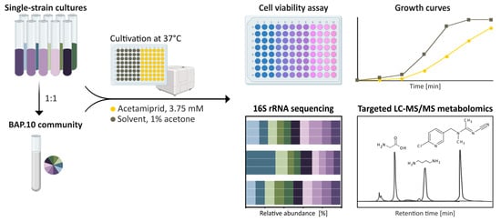
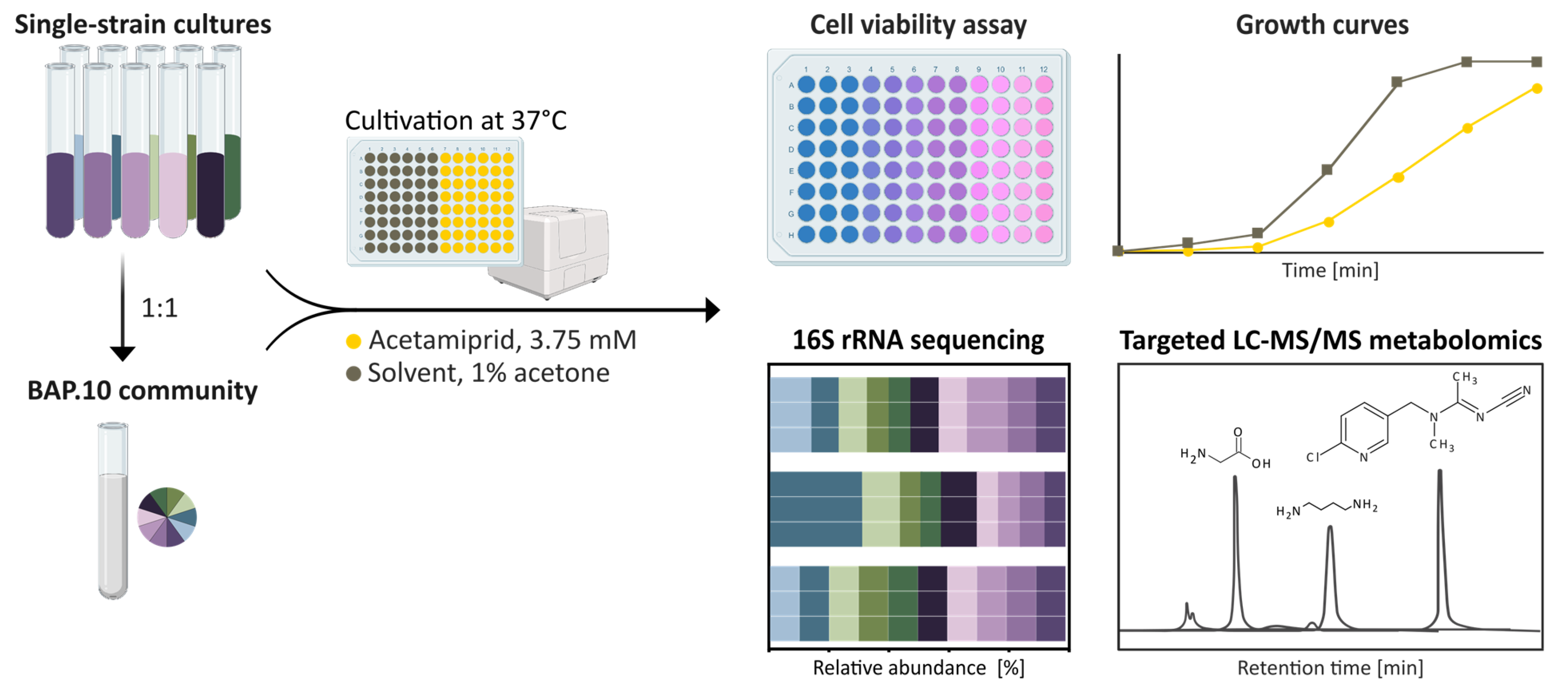
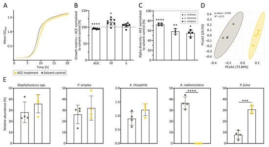
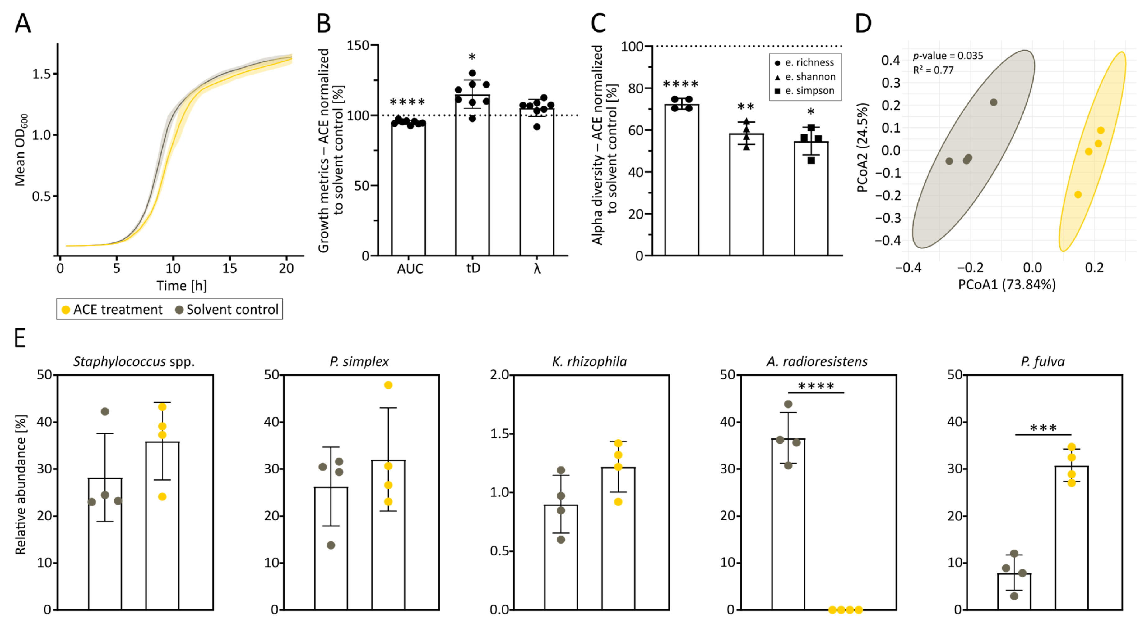
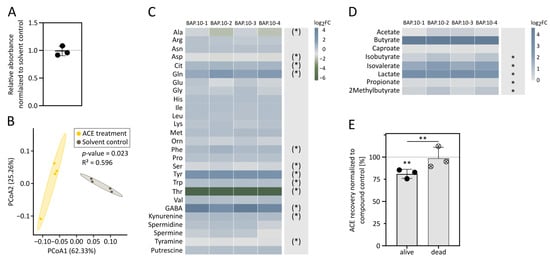
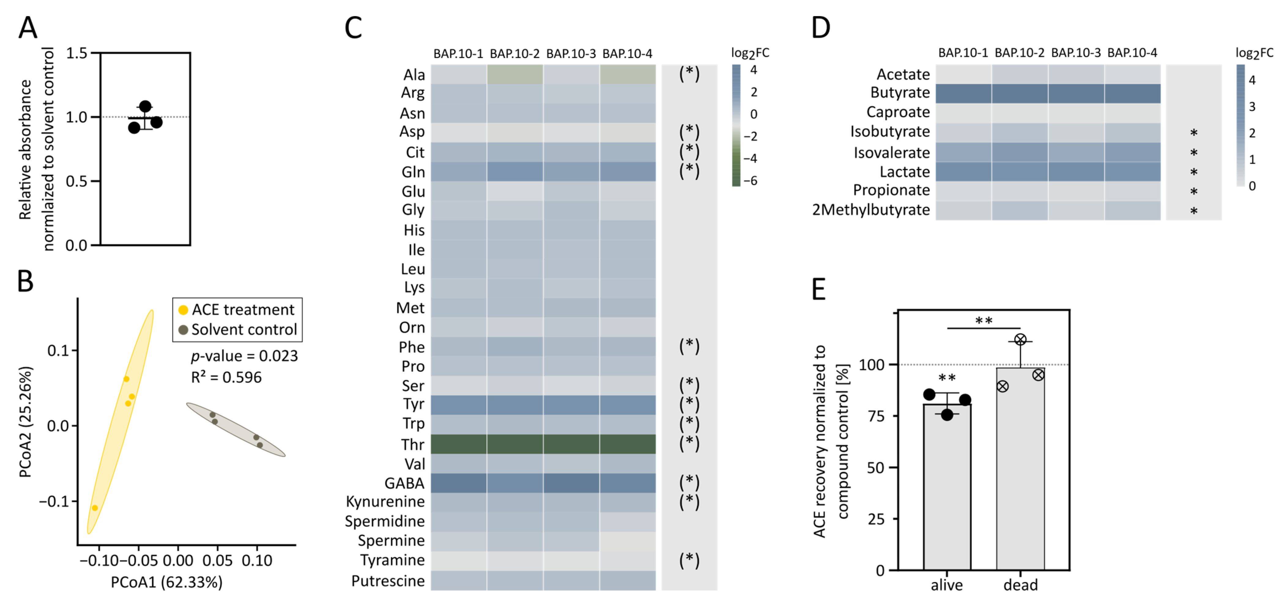
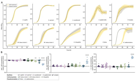
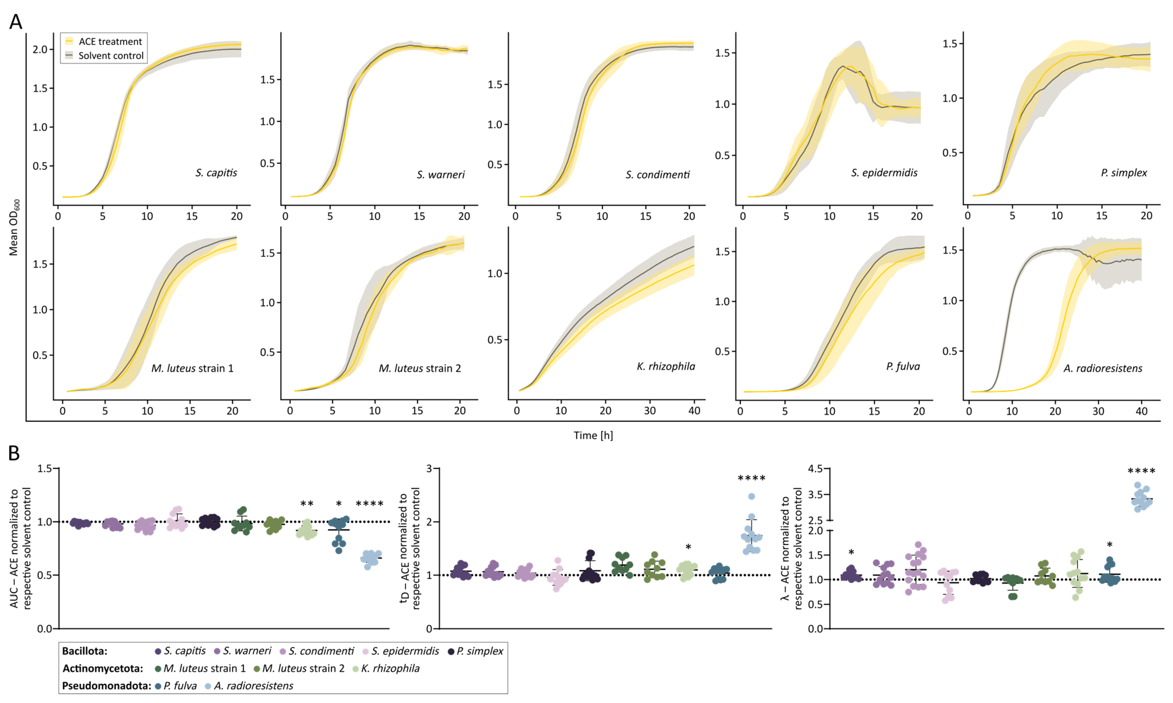
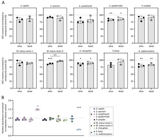
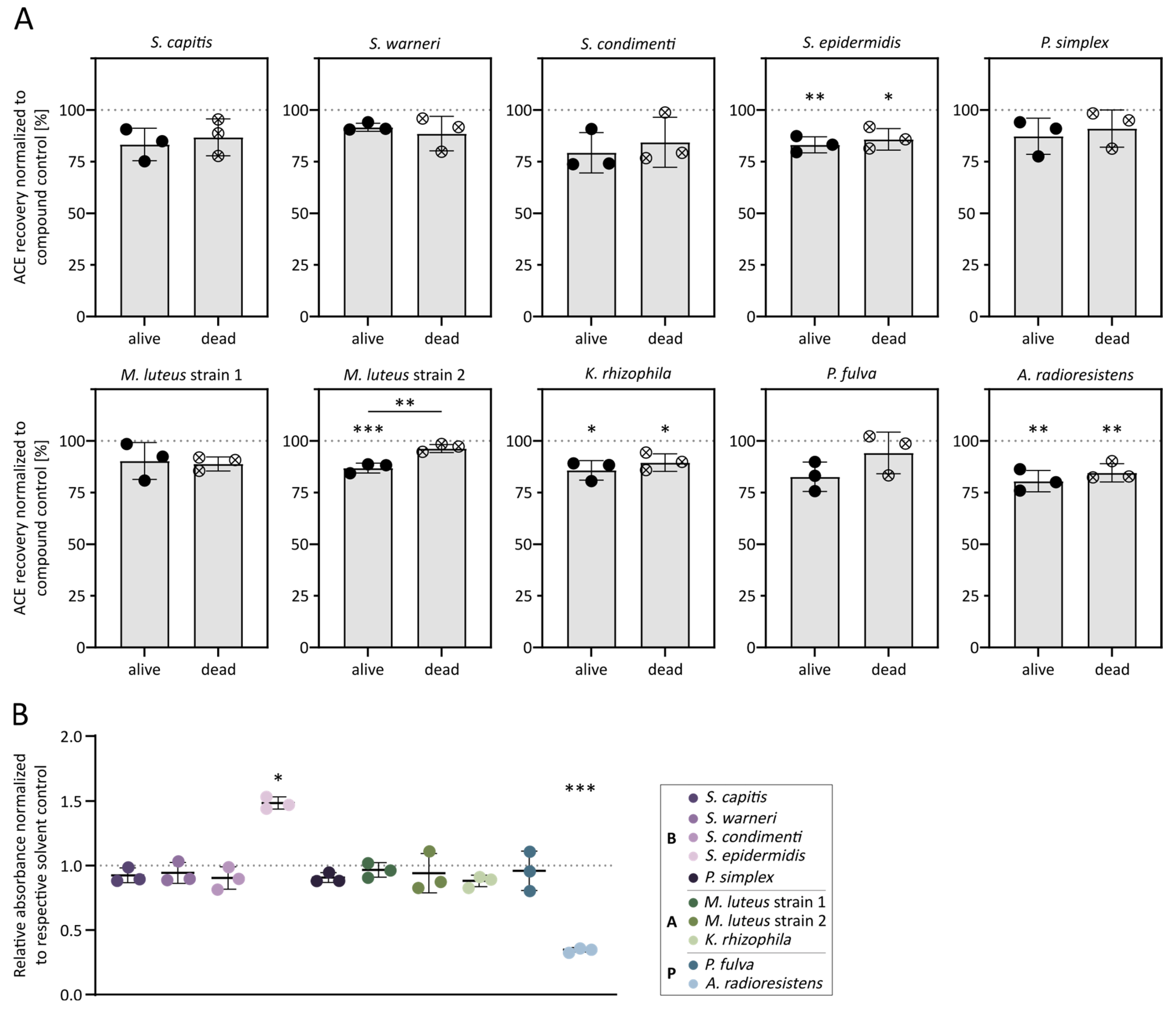
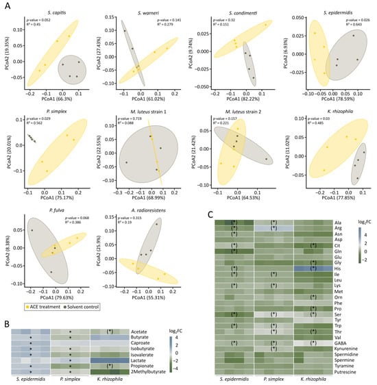
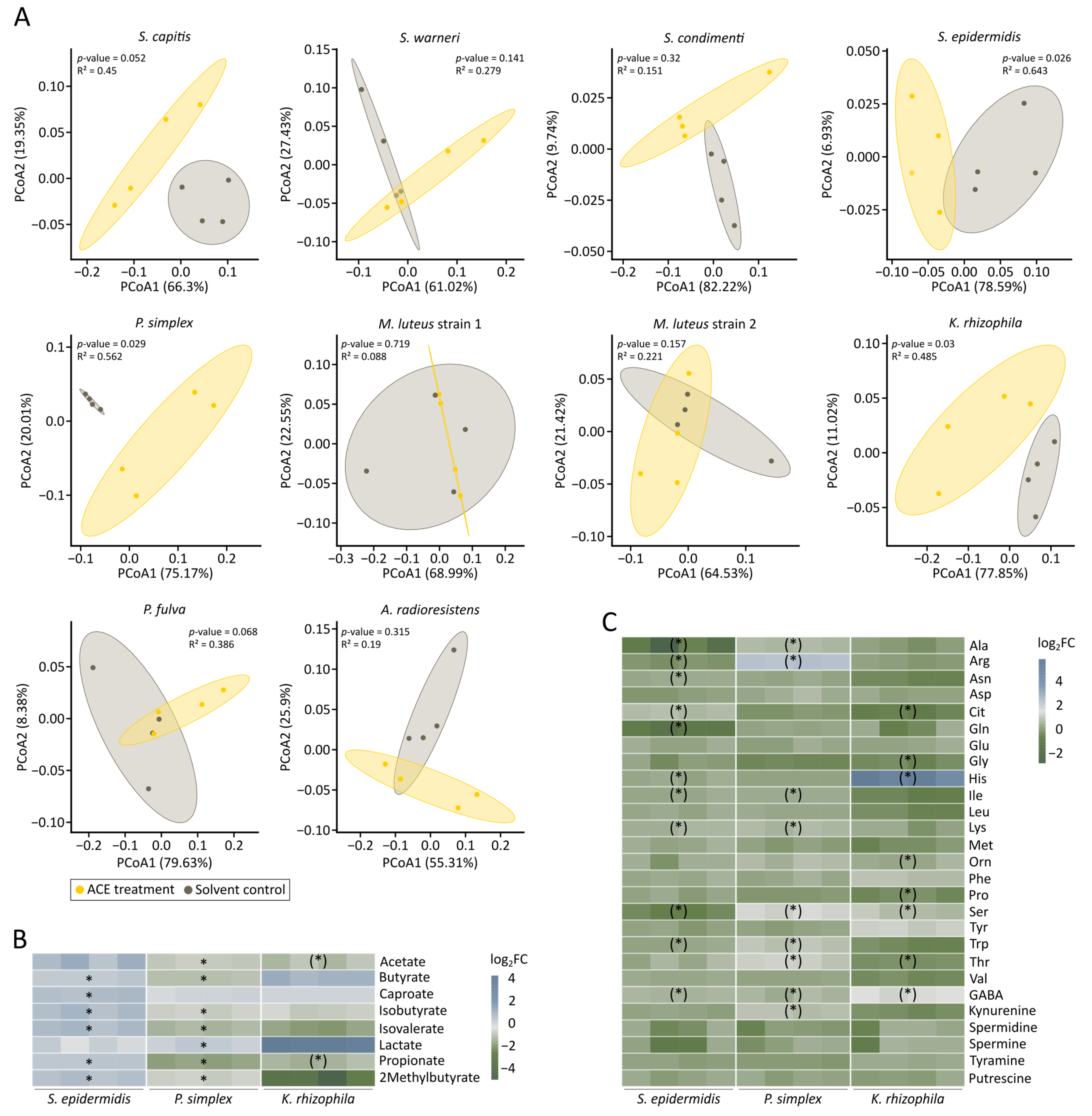

Disclaimer/Publisher’s Note: The statements, opinions and data contained in all publications are solely those of the individual author(s) and contributor(s) and not of MDPI and/or the editor(s). MDPI and/or the editor(s) disclaim responsibility for any injury to people or property resulting from any ideas, methods, instructions or products referred to in the content. |
© 2026 by the authors. Licensee MDPI, Basel, Switzerland. This article is an open access article distributed under the terms and conditions of the Creative Commons Attribution (CC BY) license.