


Abstract
The diverse health benefits of lactic acid bacteria (LAB) have made them a focal point of research in the fields of food and health sciences. Furthermore, probiotics and postbiotics have been demonstrated to directly or indirectly influence food quality and human health. A substantial body of research has been dedicated to the antimicrobial activity of pro- and postbiotics; however, their antioxidant properties remain relatively unexplored. Although LAB are facultative anaerobes, there are several species that have the potential to undergo aerobic respiration, thereby being exposed to the action of reactive oxygen species (ROS). The resultant oxidative stress has been shown to damage all intracellular molecules, thus requiring the presence of antioxidants in order to counteract this effect. The present review discusses the peculiarities of respiration, the role of ROS, the antioxidant potential of LAB, and the mechanisms underlying their activity. Furthermore, the study explores the antioxidant capacity of probiotics and postbiotics, as well as their role in controlling oxidative stress. The objective of the present review is to provide an overview of the current research on the oxidative stress tolerance and antioxidant capacity of LAB and its impact on food.
1. Introduction
Lactic acid bacteria (LAB) are a very important group of microorganisms that play a significant role in various fields, including the food industry, agriculture, and the clinical sector [1]. These bacteria produce lactic acid as a primary metabolic by-product. They are used as starter cultures in the production of many fermented dairy products. Studies have demonstrated that LAB possess significant therapeutic potential, making them indispensable to human health. They exhibit various nutritional, adhesive, and adaptive properties, enabling them to utilize dairy products, fermented foods, fruits, and vegetables as nutrient sources. Furthermore, LAB are known to proliferate extensively within the intestinal tracts of both animals and humans [2].
Many LAB species exhibit probiotic properties. Probiotics (pro–for and bio–life) are defined as live microorganisms that confer health benefits to the host. They boost the immune system and prevent disease through direct interaction with other microorganisms. They also help to detoxify the intestinal tract and inactivate toxins [3,4]. In recent years, alongside probiotics, prebiotics and symbiotics have become increasingly prevalent. The concept of postbiotics has emerged as a result of the limitations of probiotics’ useful effect in cases of intestinal dysbiosis, immunocompromised people, and reduced populations of beneficial bacteria. According to the International Scientific Association for Prebiotics and Probiotics (ISAPP), postbiotics are defined as ‘preparations of inanimate microorganisms and/or their components that confer a health benefit on the host’ [5,6]. Recent research has indicated that postbiotics can positively impact respiratory diseases and mental health and reduce psychological stress [5,7].
LAB-based probiotics and postbiotics have also been found to exhibit antioxidative effects [1,8]. These natural antioxidants can strengthen the human body’s defense mechanisms against oxidative stress (OS) [9]. The term OS refers to an imbalance between the generation of reactive oxygen species (ROS) in cells and their ability to neutralize them in order to avoid serious damage [10]. Superoxide anions (•O2−) and hydroxyl radicals (•OH) are examples of ROS, along with nonradical substances such as singlet oxygen (1O2) and hydrogen peroxide (H2O2) and other related species. Various stress factors can accelerate the production of these radicals, overwhelming the antioxidant defenses of cells and tissues. In this context, antioxidant peptides produced by LAB can be highly beneficial for human health.
While considerable interest exists within the scientific community and the public regarding the antioxidant activity of LAB-derived probiotics, several aspects remain underexplored. Specifically, a comprehensive understanding is lacking concerning the antioxidant activity of postbiotics, the mechanisms underlying their protective effects, and their effective application in probiotic foods and supplements. This review overviews recent advances in the study of antioxidant behavior in LAB, focusing on probiotics and postbiotics. After describing the oxygen respiration process in LAB, we emphasize ROS generation and the cellular mechanisms responding to OS. We then discuss the antioxidant defense systems in LAB, specifically referencing enzymatic and non-enzymatic antioxidants. Furthermore, we review recent findings on the antioxidant potential of probiotics and postbiotics and their impact on food quality. The final section outlines future scientific challenges that must be addressed to fully understand cellular responses to oxidative stress and the link between LAB applications and associated health benefits.
2. Lactic Acid Bacteria as Probiotics and Postbiotics
2.1. LAB as Probiotics
The LAB group includes over 400 species of Gram-positive, non-spore-forming anaerobe to non-respiring, but aerotolerant bacteria, inhabiting natural habitats, as well as found in plants and animals [11]. This physiological group consists of 12–15 genera, including Streptococcus, Lactococcus, Pediococcus, and Enterococcus. The biggest genus, Lactobacillus, was reclassified into 25 new genera belonging to the family Lactobacillaceae. A key common characteristic of LAB is their ability to convert carbohydrates into various lactic acid isomers through fermentation. They are therefore part of the permanent microbiota of fermented foods and the human intestinal tract.
The term “probiotic” has gained significant traction in recent decades and is associated with beneficial effects for humans and animals [1,12]. Probiotic LABs are nonpathogenic microorganisms isolated from various environments, such as foods, animals, plants, and humans [13]. They are commonly considered to be Generally Regarded as Safe (GRAS)/Qualified Presumption of Safety (QPS) and possess distinct characteristics that render them suitable for various applications. Over the years, a variety of probiotic cultures have been administered in the form of fermented dairy products, vegetables, and cereals for the treatment of gastroenteritis and conditions affecting the oral cavity, stomach, small intestine, and large intestine [14].
A significant number of LAB species are used as probiotics to enhance host health. They maintain homeostasis, especially in the gastrointestinal tract (GIT), and positively influence competition with opportunistic bacteria, thereby enhancing digestion, modifying immunity, demonstrating neuromodulation, and producing vitamins and other beneficial substances [4]. Alongside LAB, Bifidobacteria and yeasts have also been successfully used as probiotics. Currently, a number of other types of beneficial microbes are being investigated for their probiotic properties [14]. Probiotic bacteria facilitate the reduction in oxidative stress by exhibiting antioxidant characteristics [15]. The antioxidant capacity of probiotic bacteria, particularly lactobacilli, along with their other health-enhancing properties, has been thoroughly studied in recent years.
According to FAO/WHO recommendations, the human gastrointestinal tract is a traditional source of probiotic strains for human consumption. Notably, Ligilactobacillus salivarius salivarius subsp. salicinius and Lactobacillus acidophilus have been isolated from the human intestine, while Lb. acidophilus, Lb. fermentum, Lb. gasseri, Lb. vaginalis, Lb. reuteri, and Lb. salivarius have been identified in human feces and the human stomach, respectively [16]. Probiotic LAB belonging to the genera Lactococcus, Bifidobacterium, Leuconostoc, Lactobacillales, and Pediococcus have been isolated from fecal samples [17]. These authors highlighted the diet-dependent formation of gut microbiota. The human GIT is a significant reservoir of probiotic bacteria, such as Lb. rhamnosus IMC 501, Lb. paracasei IMC 502 [18], Lb. helveticus BGRA43 [19], Lb. casei/paracasei CTC1677, Lb. casei/paracasei CTC1678, Lb. rhamnosus CTC1679 [20], and Lactobacillus fermentum E-3 and E-18 [21]. Zielińska and Kolohyn-Krajewska [16] reported the isolation of probiotics from breast milk and animal GIT, predominantly identified as Lb. gasseri and Lb. reuteri.
Furthermore, a variety of beneficial microbes have been found in fermented foods, including milk and dairy products, raw fermented meat products, pickled vegetables, and cereal products [2]. Among these, the following have been recognized as effective probiotics: Lb. rhamnosus and Lb. helveticus (from traditionally fermented Xinjiang cheese) [22], Lb. plantarum FC225 (from fermented cabbages) [23], Lb. plantarum O7S1 (from fermented olive) [24], Lb. plantarum C88 and Lb. plantarum MWFLp-182 (from feces) [25,26], and Lb. plantarum NJAU-01 (from traditionally dry-cured meat product Jinhua ham) [27].
2.2. LAB as Postbiotics
The designation “Postbiotic” can be viewed as a comprehensive term that includes all synonyms and related expressions pertaining to these microbial fermentation components [6]. Postbiotics are derived from non-living microorganisms and/or their constituents, which provide a health advantage to the host. A wide range of ingredients can be classified as postbiotics, such as metabolites including short-chain fatty acids, microbial cell fractions, functional proteins, extracellular polysaccharides, cell lysates, teichoic acid, muropeptides, and peptidoglycan-derived structures [15]. To classify a microbial composition as a postbiotic, both the microorganisms and the content must be identified. Salminen et al. [5] proposed the following criteria.
- An elaborate description of the inactivation procedure along with the matrix;
- Confirmation that the inactivation process has been completed;
- Evidence indicating a health benefit for the host from a controlled, high-quality trial;
- A comprehensive description of the ingredients in the postbiotic preparation;
- An evaluation of the safety of the postbiotic preparation for the target host regarding its intended use.
Postbiotics offer several distinct benefits over probiotics [28]. Their non-living status leads to improved shelf stability and resilience across a wider spectrum of pH and temperature conditions and eliminates concerns regarding antibiotic resistance or the form conditions and amines. The specific chemical structures allow for standardized production and may enhance their antimicrobial capabilities, rendering them a promising substitute or complement to live probiotics. Moreover, they can be utilized alongside packaging materials designed to lessen oxidative stress, which in turn enhances the quality of the product [29]. The composition of postbiotics yields positive effects for both the food product and consumer [28]. Furthermore, the suppression of ROS and the decrease in OS have been demonstrated, suggesting a considerable antioxidant potential [6,30,31].
Several genera of LAB have been identified as significant sources of postbiotics [32]. Information about several of these, including Lactobacillus, Lactococcus, Lactiplantibacillus, Ligilactobacillus, Levilactobacillus, Limosilactobacillus, and Latilactobacillus, has been published [29,33,34,35,36].
The beneficial properties of cell-free supernatants (CFSs) derived from LAB have been demonstrated [24,37,38,39]. Similar activity has been observed for exopolysaccharides [34,40,41,42], cell wall fragments (CWF) [43,44], antioxidant enzymes [36,45,46], phenolic compounds [29], and organic acids and short-chain fatty acids [32].
3. Lactic Acid Bacteria and Their Relationship with Oxygen
Although LAB are generally considered anaerobic microorganisms, many species can also practice aerobic respiration, which involves the use of oxygen and the requirement of hemin and menaquinone. The ability to use oxygen promotes their growth, survival, and industrial usefulness, allowing them to prosper in a range of environments and undertake more complex metabolic activities than simple fermentation [47].
LAB respiratory metabolism is heavily reliant on the environment, as these bacteria require exogenous heme and menaquinone. Many of the LAB species lack enzymes necessary for heme production, and some of them do not possess the menFDXBEC gene, which is responsible for menaquinone biosynthesis [48]. In addition, they express cytochrome oxidase (so-called quinol oxidase CydAB), which functions effectively at low oxygen concentrations, removing the oxygen from the surrounding environment [49]. Unlike typical aerobic bacteria, LAB demand a sugar-based carbon source along with glycolytic activity for the recycling of NADH by NADH oxidase generated in the respiratory chain.
Several strains belonging to genera such as Enterococcus, Lactococcus, Leuconostoc, and Weissella have the ability to produce the (ubi/mena)quinone complex (encoded by menFDHBEC, menA and menG/UbiE genes) and generate a respiratory chain in the presence of hemin alone [47,48,50]. This chain contains NADH dehydrogenase that functions as an electron donor; menaquinone, which operates as a quinone shuttle; and haem-binding cytochrome bd-I oxidase, which serves as a terminal oxidase (Figure 1) [51].
Figure 1.
Key elements and processes of respiratory metabolism in lactic acid bacteria.
According to Lechardeur et al. [48], some LAB encode different genes involved in the heme biosynthetic pathway. However, no single strain has been found to encode the entire gene complex. For example, Lb. reuteri possesses early genes involved in the synthesis of vitamin B12, while L. lactis encodes late genes that facilitate the incorporation of iron into protoporphyrin. Furthermore, many LAB can utilize menaquinone (i.e., Vit. K2) from the surrounding environment because they lack genes encoding menaquinone synthesis [52,53]. Duwat et al. [50] noted that L. lactis and Enterococcus faecalis synthesize the enzyme ferrochelatase (HemH), which catalyzes the addition of iron to protoporphyrin-IX (PPIX), resulting in the formation of heme.
Published data based on the highly conserved recA gene indicate that aerobic respiration does not exhibit species-dependent distribution (Table 1) [48]. Huycke et al. [54] reported that E. faecalis possesses genes necessary for respiration, whereas the closely related species E. faecium lacks these genes.
Table 1.
Aerobic respiration among LAB required exogenous heme, or heme and menaquinone.
4. Oxidative Stress and Cell Response in Probiotic LAB
4.1. ROS and Oxidative Stress Generation
Despite the tolerance of LAB to oxygen, its presence in the environment is associated with the formation of ROS. The enzymes NADH oxidase (Nox), pyruvate oxidase (Pox), and lactate oxidase (Lox), located in LAB cells, caused the breakdown of oxygen [60,61]. This process generates •O2−, OH•, 1O2, and ozone (O3) [62]. Of these, •O2− and OH• are free radicals containing unpaired electrons. ROS that lack unpaired electrons, such as H2O2 and O3, are not classified as free radicals, although they are strong oxidizers.
The monovalent reduction of O2 leads to the formation of •O2−, followed by the reduction of a second electron to generate H2O2. Through the Haber–Weiss reaction, •O2− reacts with H2O2 to form HO• [63]. The superoxide radical has an extremely short half-life (10−5–10−6 s). Its role is connected to (i) the site of production in the cell, (ii) the capacity for protonation, and (iii) interaction with a compatible substrate [64]. In microaerophilic bacteria such as LAB, an intracellular source of H2O2 is the superoxide radical dismutation reaction, or the two-electron reduction of oxygen in reactions catalyzed by Nox, Pox, and Lox [60]. H2O2 is an unstable molecule with a half-life of 109 s that accumulates in relatively high concentrations in cells due to its extremely slow rate of reaction with biomolecules. The hydroxyl radical is one of the most reactive radicals present in biological systems (half-life 10−8 to 10−9 s), exerting a direct effect near its point of origin, damaging the first molecule it encounters [65]. Singlet oxygen is a non-radical variant of oxygen, referring to the oxygen molecules where one of the two free electrons is in a higher energy state, leading to the production of free radicals through the energy transfer.
In Lb. acidophilus and Bifidobacterium spp., two types of NADH oxidases have been reported: NADH-H2O2 oxidase and NADH-H2O oxidase, which catalyze the reduction of O2 to H2O2 (R1) and H2O (R2), respectively. Furthermore, NADH oxidase has the potential to cause incomplete reduction of oxygen, producing •O2− that can dismutate to H2O2 or OH• (R3) [66,67].
The overproduction of H2O2 has been observed in Lb. delbrueckii subsp. bulgaricus and Lb. plantarum ATCC 8014T as a result of O2 reduction by a NADH-dependent oxidase [68,69]. Stolz et al. [70] proposed that the accumulation of H2O2 under aerobic conditions and in the absence of catalase results in the activation of NADH-dependent peroxidases, relying on persistent metabolism to produce the needed NADH. Intracellular H2O2 has been shown to react with Fe2+, thereby generating the highly reactive hydroxyl radical (HO•) through the Fenton (R4) and Haber–Weiss (R5) reactions. The first radical in the chain, •O2−, affects cell proteins containing iron–sulfur clusters and enhances the Fe2+ level, stimulating the Fenton reaction [60].
Under conditions of increased oxygen concentration, the process of ROS formation is accelerated, resulting in the manifestation of OS in cells, including probiotic LAB. The factors that cause stress are extremely diverse. Among them, the availability of oxygen, temperature, osmotic pressure, pH levels, ethanol concentration, etc., originates from environmental sources. The presence of bile, antimicrobials, acidity, starvation, or low nutrient availability can also be significant stressors for probiotic LAB [71]. What these factors have in common is the generation of elevated levels of ROS. It has been reported that increased levels of dissolved oxygen during the cultivation of several strains of Lb. acidophilus and Bifidobacterium spp. caused OS [54]. Endogenous ROS generation has been evidenced as a key reason for OS realization in Lb. johnsonii/gasseri strains.
The fundamental cause of OS realization is the balance between the generation and elimination of ROS. The overproduction of ROS can disrupt this balance in favor of oxidants in cells, resulting in damage to biomolecules, including lipids, proteins, and nucleic acids (Figure 2) [64]. Probiotics passing through the intestinal tract are subjected to high levels of ROS, which have the potential to harm all cellular components [72]. Despite the extant data concerning the markedly enhanced growth of L. lactis under aerobic respiration, a number of studies have been published on the negative consequences of oxygen in lactic acid bacteria [73].
Figure 2.
Oxidative damage to intracellular molecules.
Although the DNA molecule is generally stable, it is susceptible to interactions with reactive oxygen species, which can cause various types of damage. ROS can remove electrons from the DNA chain and transform it into an unstable molecule. Effects such as the breakage of one or both strands, the loss or modification of nucleotides resulting in subsequent mutations, and the covalent binding of a protein molecule have been proven to occur [64,74]. HO• is capable of causing DNA fragmentation by breaking the phosphodiester bonds in the molecule. The effect of •O2− on DNA damage has been reported for Lc. lactis grown under aerobic conditions [75].
Many forms of ROS can harm intracellular proteins in LAB by various mechanisms. The most common reactions include the removal of hydrogen, transfer of electrons, fragmentation, rearrangement, disproportionation, and substitution in amino acids, peptides, and proteins, among others. HO• has the capacity to damage proteins, which can lead to a reduction in ATP and, as a result, a decrease in energy levels within the LAB cells [60,76]. It has been reported that H2O2 directly damaged enzyme proteins containing cysteinyl residues, inactivating their activity [77]. Similar results have been reported for Lb. johnsonii NCC 533 strain [78].
In addition, other molecules in LAB, such as lipids, are also susceptible to oxygen [60]. It has been established that lipid oxidation of membranes is the primary deleterious effect of ROS attack on lipids [79]. Many serious damages have been found in cellular lipids within the plasma membrane of LAB [60]. Unsaturated linoleic and linolenic fatty acids have been identified as the most sensitive to oxidation in LAB [80].
Lactic acid bacteria experience significant metabolic disruptions when exposed to stress conditions. For instance, LAB can modify specific alternative sources of pyruvate, consume different carbon sources, engage the proteolytic system, and enhance the catabolism of free amino acids. According to Zhai et al. [81], the rise in oxidative stress modifies the carbon source profile of Lb. plantarum and stimulates glycolysis, thereby producing more ATP.
4.2. Cell Response to Oxidative Stress
To overcome the deleterious effects of ROS, LAB have developed various responses to OS, depending on the levels and types of antioxidative mechanisms available. As facultative aerobes, they possess an incomplete antioxidant system, including the production of antioxidant compounds and the activation of redox regulatory and repair systems [60,82]. Published reports demonstrated that the aerobic cultivation of some LAB in the presence of haem and menaquinone resulted in increased biomass content due to the formation of a respiratory electron transport chain [56,83]. There are several closely related mechanisms allowing them to neutralize ROS (Figure 3).
Figure 3.
Cell response mechanisms to oxidative stress.
- Direct counteraction of ROS (Figure 3, Point 1). LAB can directly scavenge some ROS, such as •O2−, H2O2, HO•, using several mechanisms. They can prevent the generation of ROS, break the chain of their generation, quench them, or transform H2O2 to H2O through the dismutation of •O2−. For this purpose, LAB utilize cell wall components such as peptidoglycans, exopolysaccharides, teichoic and lipoteichoic acids, as well as protein.
- Chelation of metal ions (Figure 3, Point 2). Certain structures within the cell walls of LAB facilitate the chelation of metal ions (e.g., Fe3+ and Cu2+) via the Fenton reaction, thereby decreasing their availability and the opportunity for them to initiate a chain of radical generation [65].
- Mn2+ homeostasis and superoxide tolerance (Figure 3, Point 3). Many Lb. casei and Lb. plantarum require the accumulation of Mn2+. Despite functioning as a cofactor for several enzymes, these ions protect cells from the harmful effects of ROS [84]. Intracellular manganese participates in the scavenging of •O2− under aerobic conditions.
- Production of antioxidant enzymes (Figure 3, Point 4). LAB can produce enzymes with antioxidant activity, such as superoxide dismutase, haem-dependent and haem-independent catalase, NADH oxidase, NADH peroxidase, and glutathione reductase [56,85].
- Modifications on the metabolite level (Figure 3, Point 5). Under aerobic conditions, a significant number of probiotic LAB have been observed to reduce the production of lactic acid while simultaneously increasing the synthesis of NADH oxidase and NADH peroxidase [86]. For example, Lb. plantarum required NADH dehydrogenase for the oxygen respiration [47]. In the presence of oxygen, L. lactis exhibited an inhibitory effect on lactic acid fermentation, utilizing cytochrome bd oxidase-mediated NADH oxidation within the electron transport chain [87]. Consequently, an alteration in glycolytic flux towards the synthesis of acetate, ethanol, acetoin, diacetyl, and CO2 has been observed [85].
5. Antioxidants and Antioxidant Defense in LAB
Antioxidants are described as “any substance that delays, prevents, or removes oxidative damage to a target molecule” [88]. They are stable molecules that can donate or accept an electron to each free radical, thereby restoring their paired state (Figure 4).
Figure 4.
Antioxidant action against free radicals.
The effectiveness of the cellular defense system’s antioxidant activity depends on the strategy employed, including prevention, scavenging and destroying radicals, repairing damage, and adaptation (Figure 5). Antioxidant activity involves decreasing the ability of ROS to inflict injury or interacting with ROS to prevent the cascade of their production before essential cellular components are damaged. Furthermore, antioxidants can be categorized into natural and synthetic types, enzymatic and non-enzymatic forms, low- and high-molecular-weight structures, intracellular and extracellular locations, and water-soluble and lipid-soluble varieties [89].
Figure 5.
Antioxidant systems in LAB strains.
5.1. Enzymatic Antioxidant Defense in Probiotic LAB
O2-consuming enzymes. It has been demonstrated that some probiotic LAB possess O2-consuming enzymes participating in the cell response against OS. These enzymes, including NADH oxidase (NOX), pyruvate oxidase (POX), and lactate oxidase (LOX), facilitate the rapid elimination of oxygen and are crucial for maintaining the intracellular redox balance [60]. NOX plays a key role in aerobic growth. There are two forms of NOX: NADH-H2O2 oxidase (NOX-1) and NADH-H2O oxidase (NOX-2). These can be found in LAB strains, either separately or together. NOX-2 enables a direct four-electron reduction to H2O, bypassing the two-electron reduction of O2 to H2O2 [82].
POX and LOX also contributed to the accumulation of H2O2 in several LAB strains growing in the presence of O2 [78,90,91]. Both enzymes are expressed throughout growth; however, POX is responsible for producing most of the H2O2 in the early and logarithmic phases, whereas LOX predominantly contributes to H2O2 production in the stationary phase. In addition, POX is highly effective in catalyzing the reduction of H2O2 to H2O in Lb. casei under oxidative stress conditions [90].
A lack of NADH oxidase production decreases the growth and oxidative stress tolerance of the Fructilactobacillus sanfranciscensis mutant [92]. This enzyme has been reported to play a beneficial role in several species of bifidobacteria, such as Bifidobacterium longum [93] and B. animalis [94]. Concurrently, LAB can express the NADH-oxidase/peroxidase system, which facilitates the metabolism of molecular oxygen and enhances ATP production. Similar results have been published for Lb. panis PM1 [95], B. longum, and B. animalis subsp. lactis [96], among others. Reduction of O2 to H2O2 with NADH oxidase has been demonstrated in Lb. delbrueckii and Lb. plantarum [68,97]. Shimamura et al. [98] also demonstrated the protective role of NADH oxidase and NADH peroxidase in B. infantis, B. breve, B. adolescentis, and B. longum against oxygen toxicity.
Superoxide dismutase (EC 1.15.1.1, SOD) is the main enzyme in the first line of antioxidant defense. This enzyme catalyzes the dismutation of •O2− to molecular oxygen and H2O2 according to the following equation [85,99].
SOD is a metalloenzyme and contains a metal atom in the active site, forming four different types of isoenzymes: Fe-SOD, Mn-SOD, Cu/Zn-SOD, and Ni-SOD [90]. Manganese- and Fe-SOD are considered to be evolutionarily more ancient than Cu/Zn-SOD, originating from a common precursor enzyme [100]. During subsequent phases of phylogenetic evolution, these enzymes diverged, leading to the formation of two separate groups based on their amino acid sequences. The evolution of the Cu/Zn SOD family occurred independently, resulting in a notable deviation from the properties of the original two enzymes.
Superoxide dismutase is essential for eliminating oxidative stress in LAB. The basic isoenzymes are Mn- or Fe-containing SODs [101]. However, a cambalistic SOD (Fe/Mn family, containing various amounts of both metals) has also been found in some species [101,102]. According to Feng and Wang [60], MnSOD represents the predominant form found in LAB, in contrast to the relatively infrequent occurrence of FeSOD. The presence of Mn-SOD is determined by the intracellular concentration of Mn2+. This isoenzyme has been detected in many members of the genera Streptococcus and Lactococcus [103]. For example, a high SOD activity has been demonstrated in Lb. brevis P68 and Lb. fermentum strains E-3 and E-18 [21,104]. The presence of Mn-SOD in Lb. casei and Lb. rhamnosus strains markedly improved the ROS tolerance of the lactic acid cultures [105,106].
It is noteworthy that Mn-SOD can protect LAB from peroxide stress through the elimination of •O2−, which disrupts the iron reduction cycle, and decreases the possibility of HO• generation [85]. This suggestion has been confirmed by heterologous expression of the sodA gene encoding Mn-SOD of S. thermophilus in Lactobacillus species that lack this enzyme [107].
Catalases (EC 1.11.1.6, CAT) are antioxidant enzymes that are present in nearly all aerobic organisms and a variety of microaerophilic microorganisms [108]. They can break down intracellular H2O2 before it escapes through the cell membrane in the following reaction:
Catalases are categorized into four types based on structure and mechanism: mono-functional haem catalases (classical catalase), catalase-peroxidases (atypical catalase), non-haem catalases (Mn-containing), and minor catalases [109]. In the historical context, LAB were traditionally classified as microorganisms that lacked CAT activity. Nonetheless, there are certain exceptions to this. In the LAB species, heme-dependent or Mn-containing enzymes have been detected. The homotetrameric heme-dependent CAT has been found in cultures grown on the medium supplemented with heme or hematin. Mn-CAT, which does not require heme or hematin, is rarely detected in LAB [85]. The protective effects of Mn-CAT against H2O2 have been evaluated in Lb. plantarum [110], S. thermophilus AO54 [111], Lb. mali and P. pentosaceus [112], and so on. Furthermore, there is evidence to suggest the presence of a heme-dependent catalase in Lb. pentosus, Lb. sake, Lb. delbrueckii, and Lb. plantarum WCFS1 [113,114,115]. Engesser et al. [112] characterized CAT in 71 spp. of LAB. The authors detected Mn-CAT activity in Lb. plantarum, Lb. mali, and P. pentosaceus, while heme-CAT has been found in 21 species belonging to the different genera of LAB. It has been demonstrated that the maximum non-heme CAT activity is produced in stationary phase cultures following the complete consumption of glucose.
Thioredoxin reductase (EC 1.8.1.9, TrxR) is a flavoprotein with a NADPH cofactor that has the ability to oxidize thioredoxin. The TrxR-selenium complex regulates the entire thioredoxin system and participates in cellular antioxidant defenses [85,89].
The thioredoxin system has been demonstrated to facilitate direct electron transfer to antioxidant enzymes. This process has been shown to enhance their efficiency in scavenging ROS, reduce damaged biomolecules, restore enzyme activity by breaking disulfide bonds, and regulate redox-sensitive transcription factors. The L. casei strain Shirota responds to OS by utilizing its thioredoxin (Trx) system to reduce disulfide bonds, thereby preserving the equilibrium of protein dithiol/disulfide bonds [116]. Due to their near-constant exposure to oxidative stress, LAB have evolved genetically encoded antioxidant mechanisms. These mechanisms fundamentally rely on the expression of genes that code for antioxidant enzymes [60,85]. The key genes present in LAB consist of sodA, predominantly encoding Mn-SOD; katA and katB, responsible for encoding catalase; npr, ahpC, and ahpF, which encode peroxidases; gshR, which encodes glutathione reductase; and gor, which encodes glutathione oxidoreductase [85]. The expression levels of thioredoxin genes depend on conditions related to oxidative stress. As Serata et al. [116] indicated, the trxB gene has been demonstrated to play a pivotal role in facilitating the aerobic growth of L. casei. Serrano et al. [117] discovered the trxB1 gene, which encodes TrxR in L. plantarum WCFS1, and showed that the heterologous overexpression of TrxR significantly improved the strain’s ability to withstand oxidative stress.
5.2. Non-Enzymatic Antioxidant Defense in Probiotic LAB
Glutathione (γ-Glu-Cys-Gly, GSH) is a non-protein thiol compound that has been found in almost all living organisms. It has the potential to protect cells from ROS and maintain the redox status in the intracellular environment under conditions of abiotic stress [118,119]. Moreover, GSH has also been found to participate in the cell response against oxidative stress of LAB [21,85,89,120]. GSH is essential for the regeneration of other antioxidants, such as vitamins C and E. It preserves –SH groups in their reduced form and contributes to xenobiotic detoxification processes. Adequate intracellular levels of glutathione have been shown to prevent the accumulation of H2O2, thereby protecting cellular biomolecules from oxidative damage [121]. Comparative proteomic analysis of L. lactis SK11 cells, cultivated in the presence or absence of GSH, exhibited enhanced stability of glycolytic enzymes under various stress factors [122]. The authors proposed that glutathione functions as a multifaceted protective agent, promoting LAB survival and cellular integrity.
Exopolysaccharides (EPSs) are high-molecular-weight extracellular carbohydrates composed of long-chain biopolymers linked by α- or β-glycosidic bonds [89]. Chemically, EPSs are classified as homopolysaccharides (HoPSs) or heteropolysaccharides (HePSs). They exhibit diverse physiological functions, including antioxidant properties. According to Zhang et al. [123], the majority of LABs are capable of producing EPSs at varying levels, often demonstrating significant antioxidant potential. Similar data have been published for EPS from S. thermophilus DSM 24731-EPS1, Lb. delbrueckii ssp. bulgaricus DSM 20081T-EPS5, Lb. fermentum DSM 20049-EPS6, Bifidobacterium longum ssp. longum DSM 200707-EPS10 [124].
Strains such as Lactiplantibacillus sp. ME2b, Lactococcus sp. ME7, Lacticaseibacillus sp. ME17 and Lactobacillus sp. ME27a, isolated from bovine milk, has been shown to produce EPSs capable of neutralizing free radicals, including DPPH and HO• [125]. Similarly, Yu et al. [126] demonstrated that crude EPSs from Lb. plantarum A63 effectively scavenged DPPH, ABTS+, and OH• radicals, exhibiting high antioxidant activity.
Short-chain fatty acids (SCFAs) are by-products of carbohydrate fermentation. LAB produce acetic acid, propionic acid, and butyric acid, which contribute to the reduction in OS through direct scavenging of ROS [89]. Among them, butyric acid is particularly noteworthy, as it modulates the expression of genes involved in antioxidant enzyme synthesis [127].
Carotenoids are natural pigments responsible for red and yellow coloration and are widely recognized for their health-promoting properties, largely attributed to their antioxidant activity [60]. Certain LAB belonging to the genera Enterococcus and Lactiplantibacillus, known as producers of a deep-yellow C30 carotenoid, have been reported to possess resistance to oxidative stress [128]. Analysis of cell-free supernatants from LAB cultures revealed DPPH radical-scavenging activity ranging from 84.1% to 99.5%. Kim et al. [129] found that Lb. pentosus strain KCCP11226, harboring the crtM and crtN genes responsible for C30 carotenoid synthesis, exhibited higher oxidative stress resistance. Under aerobic cultivation, although bacterial growth was slower, carotenoid production increased due to the induced synthesis of mevalonate, a key carotenoid precursor.
Beyond the antioxidant mechanisms described above, additional LAB-derived metabolites—including phenolic compounds, flavonoids, ferulic acid, and histamine—have also been reported to possess free radical-scavenging activity [60]. Furthermore, LABs employ multiple repair mechanisms that facilitate the restoration of oxidatively damaged DNA, proteins, and lipids, thereby collectively enhancing resistance to oxidative stress.
6. Antioxidant Potential of Probiotics and Postbiotics
6.1. Antioxidant Potential of Probiotic LAB Strains
The antioxidant activity of probiotics and their cell-free fractions (CFS), commonly referred to as postbiotics, has attracted considerable interest in both theoretical research and practical applications. Numerous studies have demonstrated the beneficial effects of probiotics in humans and animals [6,15,130,131]. Probiotic bacteria enhance the host’s cellular response to OS through their intrinsic antioxidant defense systems. These mechanisms include direct scavenging of ROS, metal ion chelation, modulation of cell receptors, and the regulation of signal transduction pathways in eukaryotic cells (Table 2). In addition, probiotics have been observed to stimulate the transcription of antioxidant enzymes [13].
Simultaneously, probiotic bacteria modulate gut microbiota composition by inhibiting the overgrowth of pathogenic microorganisms and influencing intestinal barrier permeability [132]. They have also been reported to contribute to the restoration of the body’s antioxidant status. LeBlanc et al. [45] successfully applied Lb. casei BL23, a strain producing SOD and CAT, to alleviate pathological inflammatory processes in mice. Another probiotic strain, Lb. reuteri, has demonstrated the ability to colonize the gut and activate specific regulatory systems—such as heat shock protein Lo18, polyphosphate kinase 2, and the regulatory protein RsiR—in response to oxidative stress [133].
Table 2.
Antioxidant activity of LAB-based probiotic strains.
Probiotic LABs are capable of scavenging defined amounts of ROS and protecting against free-radical-induced damage, thereby mitigating oxidative stress. The antioxidant activity of Lb. plantarum FC225 has been attributed to its capacity to neutralize •O2−, OH•, and DPPH radicals. LAB strains with probiotic properties, isolated from fermented foods and feces, have demonstrated strong antioxidant capacity by scavenging DPPH+, •O2−, and OH• radicals, as well as chelating ferrous ions [65]. Comparable findings have been reported for Lb. fermentum [136], three LAB isolates (LS09, LS10, and LS17) from Algerian fermented products [141], B. animalis 01 isolated from centenarians [142], and intestinal lactic acid bacteria B. longum (ATCC 15708) and Lb. acidophilus (ATCC 4356) [138].
Similarly, Lb. plantarum FC225 isolated from fermented cabbages exhibited inhibitory activity against •O2−, OH•, and DPPH+ radicals [23]. Four LAB species (Lb. gasseri ATCC 33323, Lb. rhamnosus GG ATCC 53103, Lb. brevis ATCC 8287, and Lb. plantarum ATCC 14917) have been shown to scavenge ROS and prevent peroxyl radical-induced DNA damage [135]. Furthermore, the production of bacteriocins may contribute to their overall antioxidant capacity. Lb. rhamnosus and Lb. helveticus isolated from traditionally fermented Xinjiang cheese exhibited a high DPPH+ and hydroxyl scavenging activities [22]. Rosales Cavaglieri et al. [30] also reported remarkable antioxidant properties of P. pentosaceus RC007 isolated from the gastrointestinal contents of juvenile rainbow trout, as evidenced by ABTS•+, scavenging activity, and elevated TEAC and FRAP values.
The cell response of probiotic LAB to OS also involves modulation of host enzyme activity, including upregulation of antioxidant enzymes and downregulation of ROS-generating systems. Gao et al. [23] reported that probiotic administration enhanced SOD and GPX synthesis while reducing lipid peroxidation levels in mouse liver homogenates. By increasing the activity of SOD, CAT, and GPX, the probiotic strains Lb. plantarum C88 and MA2 [25,26] or Lb. brevis MG000874 [146] enhanced the health of elderly mice exposed to OS. Elevated SOD activity and increased cellular content of GSH and GSSG have also been reported for Lactococcus strains [137].
The probiotic strain Lb. fermentum E-3 exhibited prolonged survival associated with high glutathione content and Mn-SOD expression [21]. Similarly, Watthanasakphuban et al. [61] indicated that Mn-SOD and Mn-CAT expression play a key role in mitigating ROS effects in probiotic Lb. reuteri KUB-AC5. Several LAB species, such as Lb. plantarum, Lb. sakei, and Lb. plantarum ATCC 1443, possesses manganese-dependent antioxidant enzymes such as Mn-pseudocatalase and Mn-SOD [110,139]. In vivo studies have confirmed high SOD activity in animals administered Lb. brevis, Lb. acidophilus, and B. animalis subsp. lactis [147].
The antioxidant capacity is strongly associated with intracellular manganese accumulation [84,89]. While Fe3+ and Cu2+ promote hydroxyl radicals via the Fenton reaction, Mn2+ facilitates superoxide anion elimination during aerobic growth [148]. Manganese ions are involved in numerous cellular processes, necessitating tight regulation of their intracellular concentration. This regulation is achieved through the coordinated activity of metal transporters, transcription factors, and oxidative stress-responsive riboswitches. Mn-SOD requires manganese as an essential cofactor [99]. Interestingly, LAB lacking classical SOD enzymes often accumulate high intracellular Mn2+ levels, which functionally substitute for SOD activity. The ability to accumulate manganese is frequently associated with the absence of genuine superoxide dismutase activity [143]. Non-enzymatic manganese-mediated oxidative stress management is common in LAB species that lack SODs and/or catalases. For example, Lb. paracasei ATCC 55544 retains high viability during storage due to its capacity to accumulate manganese ions acting as free radical scavengers [144]. Enhanced survival under oxidative stress conditions resulting from high intracellular manganese accumulation has also been demonstrated in Lb. plantarum [149], Lb. plantarum WCFS1 [115], and Lb. rhamnosus, Lb. fermentum 36, Lc. mesenteroides 8293 [145]. The precise mechanism by which Mn2+ mitigates oxidative damage in the absence of SOD remains incompletely understood. The prevailing hypothesis suggests that Mn2+ forms complexes with low-molecular-weight (LMW) metabolites, which act as ROS scavengers and functionally replace SOD activity [84].
6.2. Antioxidant Potential of Postbiotics
Recently, increasing attention has been directed toward the antioxidant properties of postbiotics, including cell-free extracts (CFSs, supernatants), exopolysaccharides released from microbiota, antioxidant enzymes, cell wall components, metabolites, and intracellular extracts (Table 3). Evidence supporting these effects has been obtained from both in vitro and in vivo [15,150].
Table 3.
Overview of antioxidant activity of postbiotics derived from LAB probiotic strains.
CFSs derived from LAB are regarded as natural protective agents due to their antimicrobial, antioxidant, and anti-inflammatory properties [37]. They contain residual nutrients and a broad range of bioactive metabolites, including organic acids, bacteriocins, vitamins, extracellular polysaccharides, hydrogen peroxide, enzymes, and ethanol. Moreover, CFSs demonstrated high stability and extended shelf life, which enhances their potential applications in human health and food preservation. Hu et al. [65] reported that CFSs from LAB exhibit significantly greater antioxidant capacity than intact bacterial cells. Similarly, CFS from cultures of Lb. delbrueckii ssp. lactis, Lb acidophilus, and Lb. casei displayed higher antioxidant activity than whole-cell preparations [33]. This phenomenon can be attributed to secreted metabolites such as glutathione, butyric acid, folic acid, extracellular polysaccharides, antioxidant peptides, and isoflavone glycosides. Notably, a substantial proportion of antioxidant components in probiotic strains is associated with the cell surface.
Numerous studies have documented the antioxidant properties of LAB-derived supernatant from diverse sources. Huang et al. [37] demonstrated that supernatants and CFSs mixture from Lb. plantarum API6, Lb. plantarum API1, Lb. fermentum 22A, Lb. salivarius 26C, P. acidilactici 46A, and Lb. curvatus L-A1 contain organic acids, volatile compounds, and bacteriocin-like substances. This CFS cocktail exhibited potent antioxidant, antibacterial, and antibiofilm activities. Strong antioxidant capacity has also been reported for CFSs from LAB isolated from endophytic plants (Lb. plantarum ngue16, Lb. plantarum ng10, Lb. brevis w6) [151]. Proton nuclear magnetic resonance (1H-NMR) analysis identified several metabolites contributing to antioxidant activity, including γ-aminobutyric acid (GABA), acetic acid, lactic acid, uracil, uridine, propylene glycol, isopropanol, serine, histidine, and indole-3-acetic acid. Radical-scavenging activity has likewise been demonstrated for CFSs from Lb. paracasei B1 and Lb. plantarum O24 [39], various Lactobacillus species [38], and Lb. plantarum O7S1 [24]. Tariq et al. [29] further reported high antioxidant activity of postbiotics (CFSs and cell wall components) from several LAB species, attributing these effects primarily to their phenolic compound content. A comprehensive review of the antioxidant properties of LAB-derived postbiotics has been provided by Aghebati-Maleki et al. [31].
EPS produced by LAB are increasingly recognized as promising antioxidants due to their functional and bioactive properties. As key components of postbiotics, they are considered effective and non-toxic agents with demonstrated antioxidant effects both in vitro and in vivo. Although LAB-derived EPS have been extensively characterized structurally, their specific role as postbiotic effectors remains less thoroughly explored [34]. Multiple studies have demonstrated their ability to inhibit ROS generation [154]. Purified EPSs from Lb. paracasei ET-22 exhibited strong postbiotic activity through effective scavenging of ABTS2+, DPPH, OH•, and •O2− radicals [34]. Sulfated EPSs isolated from Lb. plantarum WLPL04 significantly inhibited ROS production and lipid peroxidation under H2O2-induced oxidative stress [35], while also enhancing SOD, CAT, and GPx activities in H2O2-damaged Caco-2 cells. In vitro studies with EPSs from W. cibaria GA44, composed of glucose and rhamnose units, confirmed HO• and •O2− scavenging activity [42]. EPSs from Lb. plantarum RJF4 demonstrated considerable antioxidant capacity and DPPH radical scavenging activity, along with notable thermal stability (up to 225 °C) [40]. Similar antioxidant properties have been reported for EPSs from Lb. plantarum NTMI05 and Lb. plantarum NTMI20 [152], Lb. acidophilus LA5, and B. animalis subsp. lactis BB12 [41], and Lb. plantarum KX041 [155].
The antioxidant activity of postbiotics is largely attributed to antioxidant enzymes such as SOD, GPx, and CAT, which act as the first line of defense for cells [150,156]. High GPx activity has been identified as a key contributor to the antioxidant effects of postbiotics derived from Lb. plantarum RG14 [36]. LeBlanc et al. [45] reported that CAT and SOD were produced by engineered Lb. casei BL23 effectively reduced the severity of intestinal pathologies. Tomusiak-Plebanek et al. [46] further highlighted the importance of SOD and CAT in the anti-inflammatory and antioxidant effects of Lb. acidophilus 900 and Lb. plantarum 30B. Additionally, oral administration of postbiotic CAT from Lactococcus lactis has been shown to exert a preventative effect against colorectal cancer in mice [153]. Amobonye et al. [32] reviewed the beneficial antioxidant roles of probiotic enzymes, including SOD, CAT, and GPx, produced by Lc. pseudomesenteroides, Lb. paracasei, Lb. plantarum, etc.
Peptides represent another important class of postbiotic components [32]. These biologically active molecules consist of defined amino acid sequences of varying lengths. Specific peptides, such as leucine-leucine-proline (LLP) and valine-tyrosine-proline (VYP), have been identified as antioxidants due to their ability to scavenge free radicals and thereby reduce oxidative stress.
Cell wall fragments (CWFs) further contribute to the functional complexity of postbiotic preparations. Antioxidant activity of CWFs has been demonstrated in postbiotics derived from various LAB species [15]. For example, CWFs from Lb. casei CRL-431 have been shown to act synergistically with lipid compounds, with GHS identified as the main contributor to the antioxidant activity of this postbiotic preparation [43]. Additionally, cell wall-bound phenolic compounds isolated from Lb. plantarum ATCC 8014 have been reported to enhance the antioxidant capacity of postbiotic formulations [44].
6.3. Comparison of Antioxidant Mechanisms of Action
The antioxidant potential of LAB-derived probiotics and their postbiotic functions is strain-specific and context-dependent. The benefits are not uniform across all strains, and individual responses can vary [157]. This observation is supported by Łepecka et al. [89], who analyzed twenty-one LAB probiotic strains belonging to the species Lb. plantarum, Lb. pentosus, P. pentosaceus, and Lb. paracasei. Conversely, Abduxukur et al. [158] found that probiotic Lactobacillus strains exhibited the highest hydroxyl and superoxide anion scavenging rate in their IC, followed by CFS.
Furthermore, differences in antioxidant components have been demonstrated between IC, CFS, and metabolites in CFE [159]. The authors reported that the CFS exhibited a higher free radical scavenging rate and greater lipid peroxidation inhibition compared to IC and CFE. Moreover, CFS was distinguished from other fractions by higher antioxidant enzyme activity (SOD and GSH-Px). These strong antioxidant properties and activities can be attributed to metabolites produced during strain cultivation.
According to Wu et al. [160], four LAB probiotic strains (Lb. plantarum ZJ316, Lb. sakei LZ217, Lb. paracasei ZFM54, and Lb. rhamnosus ZFM202) displayed varying degrees of H2O2 tolerance, which correlated with their antioxidant activity. Lb. plantarum ZJ316, a strain with strong H2O2 tolerance, exhibited significant ABTS+, DPPH, and HO• scavenging activity. However, its scavenging rate against •O2− was weaker, potentially due to less efficient binding of antioxidant components to the superoxide anion target on the intact cell surface. Analysis also revealed significant expression of genes associated with SOD and GSH-Px activity in the CFE. Similar results demonstrating synergistic effects between enzymatic and non-enzymatic systems have been reported for the probiotic strain Lb. plantarum GXL94 [161]. The combined utilization of both enzymatic and non-enzymatic antioxidants by LAB has been demonstrated in IC and CFS fractions [162]. In recent years, there has been a notable increase in reports highlighting the ability of lactic acid bacteria to manage oxidative stress through the combined action of these antioxidant systems [89]. For example, Łepecka et al. [89] reviewed such activity for Lb. fermentum R6, Lb. plantarum, Lb. paracasei, E. faecium, Lb. helveticus, and W. paramesenteroides.
7. Probiotics and Postbiotics from LAB and Their Impact on Foods
LAB probiotics can be incorporated into a variety of food and beverage products, including both dairy and non-dairy options or administered as supplements. Fermented foods often contain active microbes with genetic similarities to probiotic strains. Research indicates that consuming fermented foods can enhance their functional and nutritional properties by modifying substrates and generating bioactive and bioavailable end-products [72].
The application of probiotic bacteria in fermented food production is a significant area of research, particularly regarding their ability to enhance the physical, chemical, sensory, and functional properties of these products, notably their antioxidant qualities [14]. Several probiotic strains with antioxidant properties have been used as supplements to improve the nutritional qualities of foods, thereby mitigating the impact of oxidative stress (Figure 6) [163].
Figure 6.
LAB-based probiotics used as supplements to foods.
The addition of the probiotic strain Lb. fermentum ME-3 to the cheese Pikantne has been shown to increase its total antioxidant properties [164]. Furthermore, novel probiotic cheese supplements exhibiting high radical scavenging activity have been documented, including Lb. brevis BJ20 (isolated from traditional Korean fermented food), Leuconostoc mesenteriodes, and Lb. plantarum (from fermented Chinese cabbage) [165], and L. lactis KC24 (from Korean kimchi) [166]. Supplementing cottage cheese with the probiotic strains Lb. casei and Lb. rhamnosus GG has been demonstrated to enhance its antioxidant and antimicrobial properties [167]. Islam et al. [168] reported four probiotic strains (Lb. fermentum SCJ26, Lb. plantarum SCJ27, Lb. fermentum SCJ28, and Lb. pentosus SCJ29 isolated from sugarcane juice as candidates for natural food preservatives.
Probiotics have demonstrated the capability to enhance the quality of fermented milk, with their radical scavenging activity contributing to the antioxidant properties of the products [14]. Consumption of fermented milk containing Lb. fermentum ME-3 has been shown to have a beneficial effect on human health, attributed to its substantial antioxidant properties [164]. A comparative analysis demonstrated enhanced total antioxidant activity, plasma GSH, and GPx in yogurt supplemented with probiotic Lb. acidophilus LA5 compared to control yogurt [169]. Laali et al. [170] found that incorporating L. plantarum in the fermentation of coconut water enhanced the antioxidant properties of the resulting beverage. Similar results have been reported for the supplementation of Cornelian cherry juice fermentation with probiotic Lb. plantarum 14917 [171]. Incorporating Lb. plantarum CCMA 0743 into the production of functional plant-fermented beverages enhances the antioxidant capacity of the final product [72]. The positive effects of adding LAB to foods to increase their antioxidant capacity have been documented in several types of fermentation. Hunaefi et al. [44] published findings regarding Lb. plantarum ATCC 8014 and Lb. acidophilus NCFM during red cabbage fermentation. Similar findings have been reported for Lb. plantarum in kiwi fruit and cereal grain fermentation [172], as well as for Lb. plantarum, Lb. acidophilus, Lb. casei, Lb. delbrueckii subsp. bulgaricus, and Lb. helveticus in soy product fermentation [132].
Postbiotics have recently garnered increasing interest as nutritional supplements due to their valuable properties, including antioxidant activity. They are potentially more beneficial than probiotics because they can produce similar results even when inactive, thus avoiding the challenges associated with living microorganisms [32]. Unlike live probiotics, postbiotics are unaffected by antibiotics, eliminating the risk of disseminating drug-resistant genes. Furthermore, the bioactive characteristics of postbiotics, including antioxidant activity, and the number of patented postbiotic formulations, promote their application in diverse fields, such as medicine, cosmetics, and the food and pharmaceutical industries. Market investigations project the global postbiotics market to exceed USD 2 billion by 2030, reflecting growing consumer demand for microbiome-supporting products that offer safety, stability, and regulatory transparency [32]. Postbiotics are stable across a wide range of temperatures and pH levels, allowing their addition to foods and ingredients before thermal processing [173]. This offers postbiotic manufacturers technical and financial advantages.
The integration of postbiotics into diverse food matrices has gained popularity across various categories (Figure 7) [174]. Traditional dairy products, mainly yogurts, cheeses, and fermented milk, have been used as potential carriers. Additionally, postbiotics have been incorporated into a variety of beverages, including fruit juices, functional drinks, and smoothies, providing health benefits without compromising taste or texture. Numerous studies have documented the beneficial effects of postbiotics in food and beverages. They can be added to functional foods and/or drug formulations in controlled amounts under conditions that ensure sufficient survival and effective storage [175]. A postbiotic derived from the novel strain Lb. plantarum O7S1 has been recommended as an alternative agent for mitigating oxidative stress in both the food and pharmaceutical industries [24].
Figure 7.
LAB-based postbiotics used in foods.
The addition of postbiotics derived from Lb. acidophilus and Lb. helveticus to non-fermented dairy beverages enhanced their antioxidant activity and potential as health-promoting dairy products [176]. Several LAB-based postbiotics have been registered as promoters of antioxidant capacity in various fermented foods. For example, Wu et al. [34] demonstrated that incorporating postbiotics derived from Lb. plantarum into fermented blackberry and blueberry juices increased antioxidant activity. Aguilar-Toala et al. [43] characterized the EPS produced by Lb. rhamnosus CRL-431 as a natural antioxidant, suggesting it may improve the quality of food products. The authors reported that its addition to milk enhances the physicochemical and sensory qualities of the products. The application of EPS from Lb. plantarum YML007 in soybean fermentation extended its shelf life up to two months [177]
Anvar et al. [178] have recommended a combination of plant extract nanoemulsion and postbiotic from Lb. plantarum subsp. plantarum for butter treatment, which facilitated peroxide radical scavenging and thereby increased its shelf life. Postbiotics derived from a combination of the cell-free extracts and cell wall components of Lb. plantarum, Lb. paraplantarum, Lb. brevis, and L. lactis strains have been found to contain high levels of antioxidants and phenolic compounds, as well as acetic and formic acids [29]. These properties contribute to their increased antimicrobial activity against Escherichia coli.
Lactobacillus rhamnosus and Lb. reuteri produce postbiotics with antioxidant qualities that have been described as an efficient natural preservative for use in the meat industry [28]. The results of Jalali et al. [28] confirmed the potential of Lactobacilli-derived postbiotics in red meat preservation. Furthermore, the combination of postbiotics from Lb. plantarum and essential oil nanoemulsion from Thymus daenensis Celak extended the cold storage of rainbow trout filets due to its antioxidant effects [179]. Ozma et al. [180] reported improved quality of lamb meat slices after treatment with the postbiotic from Lb. paracasei.
Postbiotics have emerged as valuable ingredients in functional foods [181,182,183]. These foods are rich in metabolites that have been proven to promote gut health, modulate immune responses, and deliver antioxidant and antimicrobial properties. In addition, they can be employed as a strategy in addressing food allergies [173]. As Wang et al. [184] suggested, the postbiotic of Lb. plantarum 70810 containing EPSs has the potential to improve the texture, taste, flavor, and shelf life of functional foods [185]. Similarly, the addition of LAB-derived postbiotics has been shown to significantly improve the antioxidant activity of strawberry juice [186], cheese whey and skim milk during yogurt production [187], bread [188], and sheep’s milk yogurt [189], among other foods. Beyond their antimicrobial effects, postbiotics can inhibit lipid peroxidation, preventing rancidity and discoloration, which are major quality concerns in meat preservation [177].
The strong antioxidant and antimicrobial properties of LAB postbiotics have led to their use as natural preservatives against foodborne pathogens [190]. The application of these natural compounds for bio-preservation is gaining attention as a viable alternative to chemical preservatives. Their compatibility with modern processing technologies, combined with their natural origin, makes them exceptionally well-suited for inclusion in clean-label preservation strategies [191]. The presence of enzymes and non-enzyme antioxidants in postbiotics plays a pivotal role in scavenging ROS, slowing lipid oxidation and protein damage, and mitigating OS in foods [174]. Evidence supports their efficacy in various food sectors, including dairy, meat, beverages, and plant-based foods [192]. Avchi et al. [193] proposed that postbiotics function as lipid peroxidation inhibitors, helping to prevent rancidity and discoloration in meat products. Furthermore, the CFS of three probiotic LAB strains (Lb. sakei, Lb. plantarum and P. pentosaceus), employed as starters in ] sausage fermentation, exhibited a significant rate of radical scavenging and inhibition of lipid peroxidation, thereby improving the quality of the meat products [194]. Comparable results regarding the antioxidant properties of CFS from Lb. plantarum and L. lactis subsp. lactis, used as starter cultures in fermented meat products, have been obtained Bilecen Şen [195].
Reported data indicate that the antioxidant potential of postbiotics can also be exploited for incorporation into oral supplements for humans and animals [6,173,196]. Diets containing LAB-based postbiotics can improve the antioxidant status of sows subjected to heat stress [197], post-weaned lambs [36], and young calves [198].
8. Conclusions and Future Scientific Challenges
A thorough analysis of over 180 scientific publications underscores the strategic importance of lactic acid bacteria (LAB) as microorganisms with well-established probiotic and postbiotic potential, including significant antioxidant activity. Recent research highlights the specific adaptations of LAB to utilize oxygen respiration and the mechanisms they employ to mitigate oxidative stress by managing generated free radicals. LAB have developed specialized stress response mechanisms involving both enzymatic and non-enzymatic antioxidant components. These include direct scavenging of reactive oxygen species (ROS), the production of antioxidant enzymes, the synthesis of bioactive metabolites, and the modulation of cellular redox status. Moreover, the incorporation of LAB-derived probiotics and postbiotics into food matrices contributes to improved oxidative stability, extended shelf life, and enhanced functional value.
Despite considerable progress in this field, several key scientific challenges persist. A primary obstacle is the lack of standardized methodologies for evaluating antioxidant activity across in vitro, in vivo, and cell-based models, which hinders comparability between studies. International harmonization of protocols for assessing the antioxidant capacity of probiotics and postbiotics is therefore urgently needed. Furthermore, much of the current evidence is derived from laboratory models. Well-designed, controlled clinical trials demonstrating antioxidant effects in humans remain limited, particularly concerning dose–response relationships, bioavailability, and long-term safety.
While key antioxidant enzymes and metabolites have been identified, the detailed molecular mechanisms regulating redox homeostasis in LAB are only partially understood. Integrating genomics, transcriptomics, proteomics, and metabolomics approaches is crucial to achieve a deeper understanding of cellular responses to oxidative stress. The effects of LAB and their postbiotic metabolites are also known to vary depending on the food matrix (e.g., dairy, plant-based, or meat products). A more comprehensive understanding of the interactions between bacterial metabolites and food components (lipids, proteins, polyphenols) is required, as these interactions may either enhance or diminish antioxidant efficacy. Finally, individual differences in gut microbiota composition may influence the effectiveness of probiotics and postbiotics. Future research should therefore explore the relationship between the antioxidant potential of LAB and personalized nutrigenomic strategies.
Author Contributions
Conceptualization, S.D. and M.A.; writing—original draft preparation, S.D. and M.A.; writing—review and editing, R.A. and E.K.; visualization, N.A. and L.D.; funding acquisition, S.D. All authors have read and agreed to the published version of the manuscript.
Funding
This study was funded by project No. PVU-63 of 16.12.2024 BG-RRP-2.017-0047-C01 “New approaches for selection and integration of BIOTICS (pro/post and metabiotics) in functional formulas for food supplements with high biological potential based on agro and industrial waste”, under the Recovery and Sustainability Mechanism for the implementation of investment C2I2.
Institutional Review Board Statement
Not applicable.
Informed Consent Statement
Not applicable.
Data Availability Statement
No new data were created or analyzed in this study. Data sharing is not applicable to this article.
Conflicts of Interest
The authors declare no conflicts of interest.
Abbreviations
The following abbreviations are used in this manuscript:
| LAB | Lactic acid bacteria |
| OS | Oxidative Stress |
| ROS | Reactive Oxygen Species |
| •O2− | Superoxide anion |
| •OH | Hydroxyl radical |
| 1O2 | Singlet oxygen |
| NOX | NADH oxidases |
| POX | Pyruvate oxidases |
| LOX | Lactate oxidases, |
| SOD | Superoxide dismutase |
| CAT | Catalase |
| TrxR | Thioredoxin reductase |
| GPx | Glutathione peroxidase |
| GSH | Glutathione |
| GIT | gastrointestinal tract |
| CFSs | cell-free supernatants |
| CWF | cell wall fragments |
| EPS | Exopolysaccharides |
| SCFA | Short-chain fatty acids |
References
- Al-Habsi, N.; Al-Khalili, M.; Haque, S.A.; Elias, M.; Olqi, N.A.; Al Uraimi, T. Health benefits of prebiotics, probiotics, synbiotics, and postbiotics. Nutrients 2024, 16, 3955. [Google Scholar] [CrossRef] [Scilit]
- Ayivi, R.D.; Gyawali, R.; Krastanov, A.; Aljaloud, S.O.; Worku, M.; Tahergorabi, R.; Silva, R.C.D.; Ibrahim, S.A. Lactic acid bacteria: Food safety and human health applications. Dairy 2020, 1, 202–232. [Google Scholar] [CrossRef] [Scilit]
- Lourens-Hattingh, A.; Viljoen, B.C. Yogurt as probiotic carrier food. Int. Dairy J. 2001, 11, 1–17. [Google Scholar] [CrossRef] [Scilit]
- Plaza-Diaz, J.; Ruiz-Ojeda, F.J.; Gil-Campos, M.; Gil, A. Mechanisms of action of probiotics. Adv. Nutr. 2019, 10, S49–S66. [Google Scholar] [CrossRef] [Scilit] [PubMed]
- Salminen, S.; Collado, M.C.; Endo, A.; Hill, C.; Lebeer, S.; Quigley, E.M.M.; Sanders, M.E.; Shamir, R.; Swann, J.R.; Szajewska, H.; et al. The International Scientific Association of Probiotics and Prebiotics (ISAPP) Consensus Statement on the Definition and Scope of Postbiotics. Nat. Rev. Gastroenterol. Hepatol. 2021, 18, 649–667. [Google Scholar] [CrossRef] [Scilit] [PubMed]
- Zhao, X.; Liu, S.; Li, S.; Jiang, W.; Wang, J.; Xiao, J.; Chen, T.; Ma, J.; Khan, M.Z.; Wang, W.; et al. Unlocking the Power of postbiotics: A revolutionary approach to nutrition for humans and animals. Cell Metab. 2024, 36, 725–744. [Google Scholar] [CrossRef] [Scilit]
- Homayouni-Rad, A.; Houshyar, J.; Alikhah, H.; Kamalledin Moghadam, S.; Osouli, Z.; Sarabi-Aghdam, V.; Asghari, A. Could postbiotics improve mental health? BioSocial Health J. 2024, 1, 170–172. [Google Scholar] [CrossRef] [Scilit]
- Hosseinzadeh, N.; Asqardokht-Aliabadi, A.; Sarabi-Aghdam, V.; Hashemi, N.; Dogahi, P.R.; Sarraf-Ov, N.; Homayouni-Rad, A. Antioxidant properties of postbiotics: An overview on the analysis and evaluation methods. Probiotics Antimicrob. Prot. 2025, 17, 606–624. [Google Scholar] [CrossRef] [Scilit]
- Sadishkumar, V.; Jeevaratnam, K. In vitro probiotic evaluation of potential antioxidant lactic acid bacteria isolated from Idli batter fermented with Piper betle leaves. Int. J. Food Sci. Technol. 2017, 52, 329–340. [Google Scholar] [CrossRef] [Scilit]
- McCord, J.M. The evolution of free radicals and oxidative stress. Am. J. Med. 2000, 108, 652–659. [Google Scholar] [CrossRef] [Scilit]
- Martín-Miguélez, J.M.; Peromingo, B.; Castaño, C.; Córdoba, J.J.; Delgado, J.; Martín, I. Lactic acid bacteria isolated from traditional dry-cured fermented foods with probiotic effect: Selection, mechanisms of action and applications. Foods 2025, 14, 4332. [Google Scholar] [CrossRef] [Scilit] [PubMed]
- Swain, M.R.; Anandharaj, M.; Ray, R.C.; Parveen Rani, R. Fermented fruits and vegetables of asia: A potential source of probiotics. Biotechnol. Res. Int. 2014, 2014, 250424. [Google Scholar] [CrossRef] [Scilit] [PubMed]
- Yeboah, P.J.; Wijemanna, N.D.; Eddin, A.S.; Williams, L.L.; Ibrahim, S.A. Lactic acid bacteria: Review on the potential delivery system as an effective probiotic. In Food Science and Nutrition; Ibrahim, S.A., Ed.; IntechOpen: London, UK, 2023; Volume 1, ISBN 978-1-83768-092-4. [Google Scholar]
- Salminen, S.; Kenifel, W.; Ouwehand, A.C. Bacteria, beneficial probiotics, applications in dairy products. In Encyclopedia of Dairy Sciences; Elsevier: Amsterdam, The Netherlands, 2011; pp. 412–419. ISBN 978-0-12-374407-4. [Google Scholar]
- Blazheva, D.; Mihaylova, D.; Averina, O.V.; Slavchev, A.; Brazkova, M.; Poluektova, E.U.; Danilenko, V.N.; Krastanov, A. Antioxidant potential of probiotics and postbiotics: A biotechnological approach to improving their stability. Russ. J. Genet. 2022, 58, 1036–1050. [Google Scholar] [CrossRef] [Scilit]
- Zielińska, D.; Kolożyn-Krajewska, D. Food-origin lactic acid bacteria may exhibit probiotic properties: Review. BioMed Res. Int. 2018, 2018, 5063185. [Google Scholar] [CrossRef] [Scilit]
- Bamal, A.; Garg, A.P. Lactic acid bacteria isolated from human feces of vegetarian and non-vegetarians. Biosci. Biotechnol. Res. Asia 2025, 22, 779–788. [Google Scholar] [CrossRef] [Scilit]
- Verdenelli, M.C.; Silvi, S.; Cecchini, C.; Orpianesi, C.; Cresci, A. Influence of a combination of two potential probiotic strains, Lactobacillus rhamnosus IMC 501® and Lactobacillus paracasei IMC 502® on bowel habits of healthy adults. Lett. Appl. Microbiol. 2011, 52, 596–602. [Google Scholar] [CrossRef] [Scilit]
- Golić, N.; Veljović, K.; Popović, N.; Djokić, J.; Strahinić, I.; Mrvaljević, I.; Terzić-Vidojević, A. In vitro and in vivo antagonistic activity of new probiotic culture against clostridium difficile and Clostridium perfringens. BMC Microbiol. 2017, 17, 108. [Google Scholar] [CrossRef] [Scilit]
- Rubio, R.; Jofré, A.; Martín, B.; Aymerich, T.; Garriga, M. Characterization of Lactic acid bacteria isolated from infant faeces as potential probiotic starter cultures for fermented sausages. Food Microbiol. 2014, 38, 303–311. [Google Scholar] [CrossRef] [Scilit]
- Kullisaar, T.; Zilmer, M.; Mikelsaar, M.; Vihalemm, T.; Annuk, H.; Kairane, C.; Kilk, A. Two antioxidative lactobacilli strains as promising probiotics. Int. J. Food Microbiol. 2002, 72, 215–224. [Google Scholar] [CrossRef] [Scilit]
- Azat, R.; Liu, Y.; Li, W.; Kayir, A.; Lin, D.; Zhou, W.; Zheng, X. Probiotic properties of lactic acid bacteria isolated from traditionally fermented Xinjiang cheese. J. Zhejiang Univ. Sci. B 2016, 17, 597–609. [Google Scholar] [CrossRef] [Scilit]
- Gao, D.; Gao, Z.; Zhu, G. Antioxidant effects of Lactobacillus plantarum via activation of transcription factor Nrf2. Food Funct. 2013, 4, 982–989. [Google Scholar] [CrossRef] [Scilit]
- Aliouche, N.; Sifour, M.; Ouled-Haddar, H. Exploring the Antioxidant, antidiabetic, and antibacterial potential of postbiotic compounds derivedfrom Lactiplantibacillus plantarum O7S1. BioTechnologia 2024, 105, 215–225. [Google Scholar] [CrossRef] [Scilit] [PubMed]
- Niea, H.; Maa, X.; Konga, F.; Luoc, Y.; Mua, G.; Wua, X. Improvement of Lactiplantibacillus plantarum MWFLp-182 on oxidative deficits induced by in 2,2’-azobis(2-methylpropionamidine) dihydrochloride and the relating key gene analysis. Food Sci. Hum. Wellness 2025, 14, 9250026. [Google Scholar] [CrossRef] [Scilit]
- Tang, W.; Xing, Z.; Hu, W.; Li, C.; Wang, J.; Wang, Y. Antioxidative effects in vivo and colonization of Lactobacillus plantarum MA2 in the murine intestinal tract. Appl. Microbiol. Biotechnol. 2016, 100, 7193–7202. [Google Scholar] [CrossRef] [Scilit] [PubMed]
- Ge, Q.; Yang, B.; Liu, R.; Jiang, D.; Yu, H.; Wu, M.; Zhang, W. Antioxidant activity of Lactobacillus plantarum NJAU-01 in an animal model of aging. BMC Microbiol. 2021, 21, 182. [Google Scholar] [CrossRef] [Scilit]
- Jalali, S.; Mojgani, N.; Haghighat, S.; Sanjabi, M.R.; Sarem-Nezhad, S. Investigation of antimicrobial and antioxidant properties of postbiotics produced by Lactobacillus rhamnosus and Limosilactobacillus reuteri and their potential application in surface decontamination of red meat. LWT Food Sci. Technol. 2024, 209, 116758. [Google Scholar] [CrossRef] [Scilit]
- Tariq, A.; Tahran, M.N.; Ozturkoglu-Budak, S. Bioprotective and functional activities of postbiotics from lactic acid bacteria derived from artisanal dairy products. Food Sci. Nutr. 2025, 13, e70647. [Google Scholar] [CrossRef] [Scilit]
- Rosales Cavaglieri, L.A.; Isgro, M.C.; Aminahuel, C.; Parada, J.; Poloni, V.L.; Montenegro, M.A.; Alonso, V.; Falcone, R.D.; Cavaglieri, L.R. Exploring the potential of lactic acid bacteria to produce postbiotics with antimicrobial and antioxidant properties: Focus on the probiotic strain Pediococcus pentosaceus RC007 for industrial-scale production. Int. J. Food Sci. Technol. 2025, 60, vvae003. [Google Scholar] [CrossRef] [Scilit]
- Aghebati-Maleki, L.; Hasannezhad, P.; Abbasi, A.; Khani, N. Antibacterial, antiviral, antioxidant, and anticancer activities of postbiotics: A review of mechanisms and therapeutic perspectives. Biointerface Res. Appl. Chem. 2021, 12, 2629–2645. [Google Scholar] [CrossRef] [Scilit]
- Amobonye, A.; Pillay, B.; Hlope, F.; Asong, S.T.; Pillai, S. Postbiotics: An insightful review of the latest category in functional biotics. World J. Microbiol. Biotechnol. 2025, 41, 293. [Google Scholar] [CrossRef] [Scilit]
- Saide, J.A.O.; Gilliland, S.E. Antioxidative activity of lactobacilli measured by oxygen radical absorbance capacity. J. Dairy Sci. 2005, 88, 1352–1357. [Google Scholar] [CrossRef] [Scilit]
- Wu, J.; Li, Z.; Zhang, Z.; Zhang, J.; Hu, H.; Lan, H.; Hong, W.; Yang, Z. Characterization of a postbiotic exopolysaccharide produced by Lacticaseibacillus paracasei ET-22 with antioxidant and anti-inflammatory efficacy. Int. J. Biol. Macromol. 2025, 306, 141608. [Google Scholar] [CrossRef] [Scilit]
- Liu, Y.; Qin, S.; Song, Y.; Feng, Y.; Lv, N.; Xue, Y.; Liu, F.; Wang, S.; Zhu, B.; Ma, J.; et al. The perturbation of infant gut microbiota caused by cesarean delivery is partially restored by exclusive breastfeeding. Front. Microbiol. 2019, 10, 598. [Google Scholar] [CrossRef] [Scilit]
- Izuddin, W.I.; Humam, A.M.; Loh, T.C.; Foo, H.L.; Samsudin, A.A. Dietary postbiotic Lactobacillus plantarum improves serum and ruminal antioxidant activity and upregulates hepatic antioxidant enzymes and ruminal barrier function in post-weaning lambs. Antioxidants 2020, 9, 250. [Google Scholar] [CrossRef] [Scilit] [PubMed]
- Huang, X.; Nzekoue, F.K.; Wang, J.; Attili, A.R.; Coman, M.M.; Verdenelli, M.C.; Fiorini, D.; Rossi, G.; Marchini, C.; Miceli, C.; et al. A study of bioactivities and composition of a cocktail of supernatants derived from lactic acid bacteria for potential food applications. Probiotics Antimicrob. Prot. 2025, 17, 5012–5029. [Google Scholar] [CrossRef] [Scilit] [PubMed]
- Xing, J.; Wang, G.; Zhang, Q.; Liu, X.; Gu, Z.; Zhang, H.; Chen, Y.Q.; Chen, W. Determining Antioxidant activities of Lactobacilli cell-free supernatants by cellular antioxidant assay: A comparison with traditional methods. PLoS ONE 2015, 10, e0119058. [Google Scholar] [CrossRef] [Scilit]
- Karbowiak, M.N.; Kruk, M.; Szymański, P.; Zielińska, D. Novel synergistic combination of lactic acid and acetic acid bacteria cell-free extracts for enhanced food biopreservation. Ann. Agric. Environ. Med. 2025. [Google Scholar] [CrossRef] [Scilit]
- Dilna, S.V.; Surya, H.; Aswathy, R.G.; Varsha, K.K.; Sakthikumar, D.N.; Pandey, A.; Nampoothiri, K.M. Characterization of an exopolysaccharide with potential health-benefit properties from a probiotic Lactobacillus plantarum RJF4. LWT Food Sci. Technol. 2015, 64, 1179–1186. [Google Scholar] [CrossRef] [Scilit]
- Amiri, S.; Rezaei Mokarram, R.; Sowti Khiabani, M.; Rezazadeh Bari, M.; Alizadeh Khaledabad, M. Exopolysaccharides production by Lactobacillus acidophilus LA5 and Bifidobacterium animalis subsp. lactis BB12: Optimization of fermentation variables and characterization of structure and bioactivities. Int. J. Biol. Macromol. 2019, 123, 752–765. [Google Scholar] [CrossRef] [Scilit]
- Adesulu-Dahunsi, A.T.; Sanni, A.I.; Jeyaram, K. Production, characterization and in vitro antioxidant activities of exopolysaccharide from Weissella cibaria GA44. LWT 2018, 87, 432–442. [Google Scholar] [CrossRef] [Scilit]
- Aguilar-Toalá, J.E.; Estrada-Montoya, M.C.; Liceaga, A.M.; Garcia, H.S.; González-Aguilar, G.A.; Vallejo-Cordoba, B.; González-Córdova, A.F.; Hernández-Mendoza, A. An insight on antioxidant properties of the intracellular content of Lactobacillus casei CRL-431. LWT Foods Sci. Technol. 2019, 102, 58–63. [Google Scholar] [CrossRef] [Scilit]
- Hunaefi, D.; Akumo, D.N.; Smetanska, I. Effect of fermentation on antioxidant properties of red cabbages. Food Biotechnol. 2013, 27, 66–85. [Google Scholar] [CrossRef] [Scilit]
- LeBlanc, J.G.; del Carmen, S.; Miyoshi, A.; Azevedo, V.; Sesma, F.; Langella, P.; Bermúdez-Humarán, L.G.; Watterlot, L.; Perdigon, G.; de Moreno de LeBlanc, A. Use of superoxide dismutase and catalase producing lactic acid bacteria in tnbs induced crohn’s disease in mice. J. Biotechnol. 2011, 151, 287–293. [Google Scholar] [CrossRef] [Scilit]
- Tomusiak-Plebanek, A.; Heczko, P.; Skowron, B.; Baranowska, A.; Okoń, K.; Thor, P.J.; Strus, M. Lactobacilli with superoxide dismutase-like or catalase activity are more effective in alleviating inflammation in an inflammatory bowel disease mouse model. Drug Des. Dev. Ther. 2018, 12, 3221–3233. [Google Scholar] [CrossRef] [Scilit]
- Pedersen, M.B.; Gaudu, P.; Lechardeur, D.; Petit, M.-A.; Gruss, A. Aerobic respiration metabolism in lactic acid bacteria and uses in biotechnology. Annu. Rev. Food Sci. Technol. 2012, 3, 37–58. [Google Scholar] [CrossRef] [Scilit]
- Lechardeur, D.; Cesselin, B.; Fernandez, A.; Lamberet, G.; Garrigues, C.; Pedersen, M.; Gaudu, P.; Gruss, A. Using heme as an energy boost for lactic acid bacteria. Curr. Opin. Biotechnol. 2011, 22, 143–149. [Google Scholar] [CrossRef] [Scilit]
- Dinarieva, T.Y.; Klimko, A.I.; Cherdyntseva, T.A.; Bryukhanov, A.L.; Netrusov, A.I. Vitamin K2 mediates electron transport from NADH dehydrogenase 2 to Bd-Type quinol oxidase in Lacticaseibacillus rhamnosus CM MSU 529. Moscow Univ. Biol. Sci. Bull. 2022, 77, 172–177. [Google Scholar] [CrossRef] [Scilit]
- Duwat, P.; Sourice, S.; Cesselin, B.; Lamberet, G.; Vido, K.; Gaudu, P.; Le Loir, Y.; Violet, F.; Loubière, P.; Gruss, A. Respiration Capacity of the fermenting bacterium Lactococcus lactis and its positive effects on growth and survival. J. Bacteriol. 2001, 183, 4509–4516. [Google Scholar] [CrossRef] [Scilit]
- Zotta, T.; Parente, E.; Ricciardi, A. Aerobic metabolism in the genus Lactobacillus: Impact on stress response and potential applications in the food industry. J. Appl. Microbiol. 2017, 122, 857–869. [Google Scholar] [CrossRef] [Scilit] [PubMed]
- Yamamoto, Y.; Poyart, C.; Trieu-Cuot, P.; Lamberet, G.; Gruss, A.; Gaudu, P. Respiration metabolism of group B Streptococcus is activated by environmental haem and quinone and contributes to virulence. Mol. Microbiol. 2005, 56, 525–534. [Google Scholar] [CrossRef] [Scilit] [PubMed]
- Yamamoto, Y.; Poyart, C.; Trieu-Cuot, P.; Lamberet, G.; Gruss, A.; Gaudu, P. Roles of environmental heme, and menaquinone, in Streptococcus agalactiae. Biometals 2006, 19, 205–210. [Google Scholar] [CrossRef] [Scilit]
- Huycke, M.M.; Moore, D.; Joyce, W.; Wise, P.; Shepard, L.; Kotake, Y.; Gilmore, M.S. Extracellular superoxide production by Enterococcus faecalis Requires demethylmenaquinone and is attenuated by functional terminal quinol oxidases. Mol. Microbiol. 2001, 42, 729–740. [Google Scholar] [CrossRef] [Scilit] [PubMed]
- Zotta, T.; Ricciardi, A.; Ianniello, R.G.; Storti, L.V.; Glibota, N.A.; Parente, E. Aerobic and respirative growth of heterofermentative lactic acid bacteria: A screening study. Food Microbiol. 2018, 76, 117–127. [Google Scholar] [CrossRef] [Scilit] [PubMed]
- Ricciardi, A.; Ianniello, R.G.; Parente, E.; Zotta, T. Factors affecting gene expression and activity of heme- and manganese-dependent catalases in Lactobacillus casei strains. Int. J. Food Microbiol. 2018, 280, 66–77. [Google Scholar] [CrossRef] [Scilit]
- Jääskeläinen, E.; Johansson, P.; Kostiainen, O.; Nieminen, T.; Schmidt, G.; Somervuo, P.; Mohsina, M.; Vanninen, P.; Auvinen, P.; Björkroth, J. Significance of heme-based respiration in meat spoilage caused by Leuconostoc gasicomitatum. Appl. Environ. Microbiol. 2013, 79, 1078–1085. [Google Scholar] [CrossRef] [Scilit] [PubMed]
- Zotta, T.; Ricciardi, A.; Ianniello, R.G.; Parente, E.; Reale, A.; Rossi, F.; Iacumin, L.; Comi, G.; Coppola, R. Assessment of aerobic and respiratory growth in the Lactobacillus casei Group. PLoS ONE 2014, 9, e99189. [Google Scholar] [CrossRef] [Scilit]
- Brooijmans, R.; Smit, B.; Santos, F.; van Riel, J.; de Vos, W.M.; Hugenholtz, J. Heme and menaquinone induced electron transport in lactic acid bacteria. Microb. Cell Fact. 2009, 8, 28. [Google Scholar] [CrossRef] [Scilit]
- Feng, T.; Wang, J. Oxidative stress tolerance and antioxidant capacity of lactic acid bacteria as probiotic: A systematic review. Gut Microbes 2020, 12, 1801944. [Google Scholar] [CrossRef] [Scilit]
- Watthanasakphuban, N.; Srila, P.; Pinmanee, P.; Sompinit, K.; Rattanaporn, K.; Peterbauer, C. Development of high cell density Limosilactobacillus reuteri KUB-AC5 for cell factory using oxidative stress reduction approach. Microb. Cell Fact. 2023, 22, 86. [Google Scholar] [CrossRef] [Scilit]
- Halliwell, B. Antioxidants in human health and disease. Annu. Rev. Nutr. 1996, 16, 33–50. [Google Scholar] [CrossRef]
- de Almeida, A.J.P.O.; de Oliveira, J.C.P.L.; da Silva Pontes, L.V.; de Souza Júnior, J.F.; Gonçalves, T.A.F.; Dantas, S.H.; de Almeida Feitosa, M.S.; Silva, A.O.; de Medeiros, I.A. ROS: Basic concepts, sources, cellular signaling, and its implications in aging pathways. Oxidative Med. Cell Longev. 2022, 2022, 1225578. [Google Scholar] [CrossRef] [Scilit] [PubMed]
- Juan, C.A.; Pérez De La Lastra, J.M.; Plou, F.J.; Pérez-Lebeña, E. The chemistry of reactive oxygen species (ROS) revisited: Outlining their role in biological macromolecules (DNA, lipids and proteins) and induced pathologies. Int. J. Mol. Sci. 2021, 22, 4642. [Google Scholar] [CrossRef] [Scilit] [PubMed]
- Hu, Y.; Zhao, Y.; Jia, X.; Liu, D.; Huang, X.; Wang, C.; Zhu, Y.; Yue, C.; Deng, S.; Lyu, Y. Lactic acid bacteria with a strong antioxidant function isolated from “Jiangshui,” pickles, and feces. Front. Microbiol. 2023, 14, 1163662. [Google Scholar] [CrossRef] [Scilit] [PubMed]
- Talwalkar, A.; Kailasapathy, K. The role of oxygen in the viability of probiotic bacteria with reference to L. acidophilus and Bifidobacterium spp. Curr. Issues Intest. Microbiol. 2004, 5, 1–8. [Google Scholar]
- Condon, S. Responses of lactic acid bacteria to oxygen. FEMS Microbiol. Lett. 1987, 46, 269–280. [Google Scholar] [CrossRef]
- Götz, F.; Sedewitz, B.; Elstner, E.F. Oxygen utilization by Lactobacillus plantarum: I. oxygen consuming reactions. Arch. Microbiol. 1980, 125, 209–214. [Google Scholar] [CrossRef] [Scilit]
- Marty-Teysset, C.; De La Torre, F.; Garel, J.-R. Increased production of hydrogen peroxide by Lactobacillus delbrueckii subsp. bulgaricus upon aeration: Involvement of an NADH oxidase in oxidative stress. Appl. Environ. Microbiol. 2000, 66, 262–267. [Google Scholar] [CrossRef] [Scilit]
- Stolz, P.; Vogel, R.F.; Hammes, W.P. Utilization of electron acceptors by Lactobacilli isolated from sourdough: II. Lactobacillus pontis, L. reuteri, L. amylovorus, and L. fermentum. Z. Lebensm. Unters. Forch. 1995, 201, 402–410. [Google Scholar] [CrossRef] [Scilit]
- Franz, C.M.A.P.; Holzapfel, W.H. The importance of understanding the stress physiology of lactic acid bacteria. In Stress Responses of Lactic Acid Bacteria; Tsakalidou, E., Papadimitriou, K., Eds.; Springer: Boston, MA, USA, 2011; pp. 3–20. ISBN 978-0-387-92770-1. [Google Scholar]
- He, X.; Cui, Y.; Jia, Q.; Zhuang, Y.; Gu, Y.; Fan, X.; Ding, Y. Response mechanisms of lactic acid bacteria under environmental stress and their application in the food industry. Food Biosci. 2025, 64, 105938. [Google Scholar] [CrossRef] [Scilit]
- van de Guchte, M.; Serror, P.; Chervaux, C.; Smokvina, T.; Ehrlich, S.D.; Maguin, E. Stress responses in lactic acid bacteria. Antonie Van Leeuwenhoek 2002, 82, 187–216. [Google Scholar] [CrossRef] [Scilit]
- Jomova, K.; Raptova, R.; Alomar, S.Y.; Alwasel, S.H.; Nepovimova, E.; Kuca, K.; Valko, M. Reactive oxygen species, toxicity, oxidative stress, and antioxidants: Chronic diseases and aging. Arch. Toxicol. 2023, 97, 2499–2574. [Google Scholar] [CrossRef] [Scilit] [PubMed]
- Sanders, J.W.; Leenhouts, K.J.; Haandrikman, A.J.; Venema, G.; Kok, J. Stress response in Lactococcus lactis: Cloning, expression analysis, and mutation of the Lactococcal superoxide dismutase gene. J. Bacteriol. 1995, 177, 5254–5260. [Google Scholar] [CrossRef] [Scilit] [PubMed]
- Hong, Y.; Boiti, A.; Vallone, D.; Foulkes, N.S. Reactive oxygen species signaling and oxidative stress: Transcriptional regulation and evolution. Antioxidants 2024, 13, 312. [Google Scholar] [CrossRef] [Scilit] [PubMed]
- Miyoshi, A.; Rochat, T.; Gratadoux, J.-J.; Le Loir, Y.; Oliveira, S.C.; Langella, P.; Azevedo, V. Oxidative stress in Lactococcus lactis. Genet. Mol. Res. 2003, 2, 348–359. [Google Scholar]
- Maresca, D.; Zotta, T.; Mauriello, G. Adaptation to aerobic environment of Lactobacillus johnsonii/gasseri strains. Front. Microbiol. 2018, 9, 157. [Google Scholar] [CrossRef] [Scilit]
- Santivarangkna, C.; Kulozik, U.; Foerst, P. Storing lactic acid bacteria: Current methodologies and physiological implications. In Stress Responses of Lactic Acid Bacteria; Tsakalidou, E., Papadimitriou, K., Eds.; Springer: Boston, MA, USA, 2011; pp. 479–504. ISBN 978-0-387-92770-1. [Google Scholar]
- Yao, A.A.; Coulibaly, I.; Lognay, G.; Fauconnier, M.-L.; Thonart, P. Impact of polyunsaturated fatty acid degradation on survival and acidification activity of freeze-dried Weissella paramesenteroides LC11 during storage. Appl. Microbiol. Biotechnol. 2008, 79, 1045–1052. [Google Scholar] [CrossRef] [Scilit]
- Zhai, Z.; Yang, Y.; Wang, H.; Wang, G.; Ren, F.; Li, Z.; Hao, Y. Global transcriptomic analysis of Lactobacillus plantarum CAUH2 in response to hydrogen peroxide stress. Food Microbiol. 2020, 87, 103389. [Google Scholar] [CrossRef] [Scilit]
- Gao, Y.; Liang, Q.; Sun, J.; Wu, X.; Song, Y.; Xu, Y.; Nie, H.; Huang, J.; Mu, G. Probiotic potential of lactic acid bacteria with antioxidant properties in modulating health: Mechanisms, applications, and future directions. Food Biosci. 2025, 66, 106181. [Google Scholar] [CrossRef] [Scilit]
- Guidone, A.; Ianniello, R.G.; Ricciardi, A.; Zotta, T.; Parente, E. Aerobic metabolism and oxidative stress tolerance in the Lactobacillus plantarum Group. World J. Microbiol. Biotechnol. 2013, 29, 1713–1722. [Google Scholar] [CrossRef] [Scilit]
- Bosma, E.F.; Rau, M.H.; van Gijtenbeek, L.A.; Siedler, S. Regulation and distinct physiological roles of manganese in bacteria. FEMS Microbiol. Rev. 2021, 45, fuab028. [Google Scholar] [CrossRef] [Scilit]
- Bryukhanov, A.L.; Klimko, A.I.; Netrusov, A.I. Antioxidant properties of lactic acid bacteria. Microbiology 2022, 91, 463–478. [Google Scholar] [CrossRef] [Scilit]
- Talwalkar, A.; Kailasapathy, K. Metabolic and biochemical responses of probiotic bacteria to oxygen. J. Dairy Sci. 2003, 86, 2537–2546. [Google Scholar] [CrossRef] [Scilit] [PubMed]
- Arioli, S.; Zambelli, D.; Guglielmetti, S.; De Noni, I.; Pedersen, M.B.; Pedersen, P.D.; Dal Bello, F.; Mora, D. Increasing the heme-dependent respiratory efficiency of Lactococcus lactis by inhibition of lactate dehydrogenase. Appl. Environ. Microbiol. 2013, 79, 376–380. [Google Scholar] [CrossRef] [Scilit] [PubMed]
- Halliwell, B.; Gutteridge, J.M.C. The definition and measurement of antioxidants in biological systems. Free Rad. Biol. Med. 1995, 18, 125–126. [Google Scholar] [CrossRef] [Scilit]
- Łepecka, A.; Kołożyn-Krajewska, D. Antioxidant properties of food-derived lactic acid bacteria: A Review. Microb. Biotechnol. 2025, 18, e70229. [Google Scholar] [CrossRef] [Scilit]
- Barstad, L. Purification and Characterization of NADH Oxidase and Peroxidase from Lactobacillus casei. Ph.D. Thesis, Portland State University, Portland, OR, USA, 1978. [Google Scholar] [CrossRef] [Scilit]
- Pridmore, R.D.; Pittet, A.-C.; Praplan, F.; Cavadini, C. Hydrogen peroxide production by Lactobacillus johnsonii NCC 533 and Its role in anti-Salmonella activity: Hydrogen peroxide production by L. johnsonii NCC 533. FEMS Microbiol. Lett. 2008, 283, 210–215. [Google Scholar] [CrossRef] [Scilit]
- Jänsch, A.; Freiding, S.; Behr, J.; Vogel, R.F. Contribution of the NADH-oxidase (Nox) to the aerobic life of Lactobacillus sanfranciscensis DSM20451T. Food Microbiol. 2011, 28, 29–37. [Google Scholar] [CrossRef] [Scilit]
- Xiao, M.; Xu, P.; Zhao, J.; Wang, Z.; Zuo, F.; Zhang, J.; Ren, F.; Li, P.; Chen, S.; Ma, H. Oxidative stress-related responses of Bifidobacterium longum subsp. longum BBMN68 at the proteomic level after exposure to oxygen. Microbiology 2011, 157, 1573–1588. [Google Scholar] [CrossRef] [Scilit]
- Ruiz, L.; Gueimonde, M.; Ruas-Madiedo, P.; Ribbera, A.; De Los Reyes-Gavilán, C.G.; Ventura, M.; Margolles, A.; Sánchez, B. Molecular clues to understand the aerotolerance phenotype of Bifidobacterium animalis subsp. lactis. Appl. Environ. Microbiol. 2012, 78, 644–650. [Google Scholar] [CrossRef] [Scilit]
- Kang, T.S.; Korber, D.R.; Tanaka, T. Influence of oxygen on NADH recycling and oxidative stress resistance systems in Lactobacillus panis PM1. AMB Express 2013, 3, 10. [Google Scholar] [CrossRef] [Scilit]
- Oberg, T.S.; Broadbent, J.R. Hydrogen peroxide resistance in Bifidobacterium animalis subsp. lactis and Bifidobacterium longum. In Stress and Environmental Regulation of Gene Expression and Adaptation in Bacteria; De Bruijn, F.J., Ed.; Wiley: Hoboken, NJ, USA, 2016; pp. 638–646. ISBN 978-1-119-00488-2. [Google Scholar]
- De Angelis, M.; Gobbetti, M. Environmental stress responses in Lactobacillus: A review. Proteomics 2004, 4, 106–122. [Google Scholar] [CrossRef] [Scilit]
- Shimamura, S.; Abe, F.; Ishibashi, N.; Miyakawa, H.; Yaeshima, T.; Araya, T.; Tomita, M. Relationship between oxygen sensitivity and oxygen metabolism of Bifidobacterium species. J. Dairy Sci. 1992, 75, 3296–3306. [Google Scholar] [CrossRef] [Scilit]
- McCord, J.M.; Fridovich, I. Superoxide dismutase. an enzymic function for erythrocuprein (hemocuprein). J. Biol. Chem. 1969, 244, 6049–6055. [Google Scholar] [CrossRef] [Scilit]
- Valenti, R.; Jabłońska, J.; Tawfik, D.S. Characterization of ancestral Fe/Mn superoxide dismutases indicates their cambialistic origin. Protein Sci. 2022, 31, e4423. [Google Scholar] [CrossRef] [Scilit] [PubMed]
- Kong, L.; Xiong, Z.; Song, X.; Xia, Y.; Zhang, H.; Yang, Y.; Ai, L. Enhanced antioxidant activity in Streptococcus thermophilus by high-level expression of superoxide dismutase. Front. Microbiol. 2020, 11, 579804. [Google Scholar] [CrossRef] [Scilit]
- Lancaster, V.L.; LoBrutto, R.; Selvaraj, F.M.; Blankenship, R.E. A cambialistic superoxide dismutase in the thermophilic photosynthetic bacterium Chloroflexus aurantiacus. J. Bacteriol. 2004, 186, 3408–3414. [Google Scholar] [CrossRef] [Scilit]
- Marques Da Silva, W.; Oliveira, L.C.; Soares, S.C.; Sousa, C.S.; Tavares, G.C.; Resende, C.P.; Pereira, F.L.; Ghosh, P.; Figueiredo, H.; Azevedo, V. Quantitative proteomic analysis of the response of probiotic putative Lactococcus lactis NCDO 2118 strain to different oxygen availability under temperature variation. Front. Microbiol. 2019, 10, 759. [Google Scholar] [CrossRef] [Scilit]
- Arasu, M.V.; Al-Dhabi, N.A.; Rejiniemon, T.S.; Lee, K.D.; Huxley, V.A.J.; Kim, D.H.; Duraipandiyan, V.; Karuppiah, P.; Choi, K.C. Identification and characterization of Lactobacillus brevis P68 with antifungal, antioxidant and probiotic functional properties. Indian J. Microbiol. 2015, 55, 19–28. [Google Scholar] [CrossRef] [Scilit]
- An, H.; Zhai, Z.; Yin, S.; Luo, Y.; Han, B.; Hao, Y. Coexpression of the superoxide dismutase and the catalase provides remarkable oxidative stress resistance in Lactobacillus rhamnosus. J. Agric. Food Chem. 2011, 59, 3851–3856. [Google Scholar] [CrossRef] [Scilit] [PubMed]
- Lin, J.; Zou, Y.; Cao, K.; Ma, C.; Chen, Z. The impact of heterologous catalase expression and superoxide dismutase overexpression on enhancing the oxidative resistance in Lactobacillus casei. J. Ind. Microbiol. Biotechnol. 2016, 43, 703–711. [Google Scholar] [CrossRef] [Scilit] [PubMed]
- Bruno-Bárcena, J.M.; Andrus, J.M.; Libby, S.L.; Klaenhammer, T.R.; Hassan, H.M. Expression of a heterologous manganese superoxide dismutase gene in intestinal Lactobacilli provides protection against hydrogen peroxide toxicity. Appl. Environ. Microbiol. 2004, 70, 4702–4710. [Google Scholar] [CrossRef] [Scilit]
- Zámocký, M.; Koller, F. Understanding the structure and function of catalases: Clues from molecular evolution and in vitro mutagenesis. Prog. Biophys. Mol. Biol. 1999, 72, 19–66. [Google Scholar] [CrossRef] [Scilit]
- Sooch, B.S.; Kauldhar, B.S.; Puri, M. Recent insights into microbial catalases: Isolation, production and purification. Biotechnol. Advan. 2014, 32, 1429–1447. [Google Scholar] [CrossRef] [Scilit]
- Peacock, T.; Hassan, H.M. Role of the Mn-catalase in aerobic growth of Lactobacillus plantarum ATCC 14431. Appl. Microbiol. 2021, 1, 615–625. [Google Scholar] [CrossRef] [Scilit]
- Andrus, J.M.; Bowen, S.W.; Klaenhammer, T.R.; Hassan, H.M. Molecular characterization and functional analysis of the manganese-containing superoxide dismutase gene (sodA) from Streptococcus thermophilus AO54. Arch. Biochem. Biophys. 2003, 420, 103–113. [Google Scholar] [CrossRef] [Scilit] [PubMed]
- Engesser, D.M.; Hammes, W.P. Non-heme catalase activity of lactic acid bacteria. Syst. Appl. Microbiol. 1994, 17, 11–19. [Google Scholar] [CrossRef] [Scilit]
- Wolf, G.; Strahl, A.; Meisel, J.; Hammes, W.P. Heme-dependent catalase activity of lactobacilli. Int. J. Food Microbiol. 1991, 12, 133–140. [Google Scholar] [CrossRef] [Scilit]
- Knauf, H.J.; Vogel, R.F.; Hammes, W.P. Cloning, sequence, and phenotypic expression of katA, which encodes the catalase of Lactobacillus sake LTH677. Appl. Environ. Microbiol. 1992, 58, 832–839. [Google Scholar] [CrossRef] [Scilit]
- Watanabe, M.; Van Der Veen, S.; Nakajima, H.; Abee, T. Effect of respiration and manganese on oxidative stress resistance of Lactobacillus plantarum WCFS1. Microbiology 2012, 158, 293–300. [Google Scholar] [CrossRef] [Scilit]
- Serata, M.; Iino, T.; Yasuda, E.; Sako, T. Roles of thioredoxin and thioredoxin reductase in the resistance to oxidative stress in Lactobacillus casei. Microbiology 2012, 158, 953–962. [Google Scholar] [CrossRef] [Scilit]
- Serrano, L.M.; Molenaar, D.; Wels, M.; Teusink, B.; Bron, P.A.; De Vos, W.M.; Smid, E.J. Thioredoxin reductase is a key factor in the oxidative stress response of Lactobacillus plantarum WCFS1. Microb. Cell Fact. 2007, 6, 29. [Google Scholar] [CrossRef] [Scilit]
- Carmel-Harel, O.; Storz, G. Roles of the glutathione- and thioredoxin-dependent reduction systems in the Escherichia coli and Saccharomyces cerevisiae responses to oxidative stress. Annu. Rev. Microbiol. 2000, 54, 439–461. [Google Scholar] [CrossRef] [Scilit]
- Allocati, N.; Federici, L.; Masulli, M.; Di Ilio, C. Distribution of glutathione transferases in gram-positive bacteria and archaea. Biochimie 2012, 94, 588–596. [Google Scholar] [CrossRef] [Scilit] [PubMed]
- Fahey, R.C.; Brown, W.C.; Adams, W.B.; Worsham, M.B. Occurrence of glutathione in bacteria. J. Bacteriol. 1978, 133, 1126–1129. [Google Scholar] [CrossRef] [Scilit] [PubMed]
- Brodzka, S.; Baszyński, J.; Rektor, K.; Hołderna-Bona, K.; Stanek, E.; Kurhaluk, N.; Tkaczenko, H.; Malukiewicz, G.; Woźniak, A.; Kamiński, P. The role of glutathione in age-related macular degeneration (AMD). Int. J. Mol. Sci. 2024, 25, 4158. [Google Scholar] [CrossRef] [Scilit] [PubMed]
- Zhang, Y.; Li, Y. Engineering the antioxidative properties of lactic acid bacteria for improving its robustness. Curr. Opin. Biotechnol. 2013, 24, 142–147. [Google Scholar] [CrossRef] [Scilit]
- Zhang, K.; Liu, S.; Liang, S.; Xiang, F.; Wang, X.; Lian, H.; Li, B.; Liu, F. Exopolysaccharides of lactic acid bacteria: Structure, biological activity, structure-activity relationship, and application in the food industry: A review. Int. J. Biol. Macromol. 2024, 257, 128733. [Google Scholar] [CrossRef] [Scilit]
- Khalil, M.A.; Sonbol, F.I.; Al-Madboly, L.A.; Aboshady, T.A.; Alqurashi, A.S.; Ali, S.S. Exploring the therapeutic potentials of exopolysaccharides derived from Lactic Acid Bacteria and Bifidobacteria: Antioxidant, antitumor, and periodontal regeneration. Front. Microbiol. 2022, 13, 803688. [Google Scholar] [CrossRef] [Scilit]
- Tarannum, N.; Hossain, T.J.; Ali, F.; Das, T.; Dhar, K.; Nafiz, I.H. Antioxidant, antimicrobial and emulsification properties of exopolysaccharides from lactic acid bacteria of bovine milk: Insights from biochemical and genomic analysis. LWT Food Sci. Technol. 2023, 186, 115263. [Google Scholar] [CrossRef] [Scilit]
- Yu, J.; Zhang, L.; Hu, S.; Qin, M.; Ma, L.; Li, D.; Duan, C. Screening of high-yield exopolysaccharide lactic acid bacteria in fermented pickles in changbai mountain and antioxidant properties of polysaccharides. Sci. Technol. Food Ind. 2024, 45, 110–117. [Google Scholar] [CrossRef]
- Kang, C.-H.; Kim, J.-S.; Park, H.M.; Kim, S.; Paek, N.-S. Antioxidant activity and short-chain fatty acid production of lactic acid bacteria isolated from korean individuals and fermented foods. 3 Biotech 2021, 11, 217. [Google Scholar] [CrossRef] [Scilit]
- Hwang, C.Y.; Cho, E.-S.; Yoon, D.J.; Seo, M.-J. Probiotic and antioxidant properties of C30 Carotenoid-producing Lactiplantibacillus plantarum Isolated from Kimchi. Food Sci. Biotechnol. 2023, 32, 543–552. [Google Scholar] [CrossRef] [Scilit]
- Kim, M.; Seo, D.-H.; Park, Y.-S.; Cha, I.-T.; Seo, M.-J. Isolation of Lactobacillus plantarum subsp. plantarum producing C30 carotenoid 4,4-diaponeurosporene and the assessment of its antioxidant activity. J. Microbiol. Biotechnol. 2019, 29, 1925–1930. [Google Scholar] [CrossRef] [Scilit]
- Nowak, A.; Paliwoda, A.; Błasiak, J. Anti-proliferative, pro-apoptotic and anti-oxidative activity of Lactobacillus and Bifidobacterium strains: A review of mechanisms and therapeutic perspectives. Crit. Rev. Food Sci. Nutr. 2019, 59, 3456–3467. [Google Scholar] [CrossRef] [Scilit]
- Averina, O.V.; Poluektova, E.U.; Marsova, M.V.; Danilenko, V.N. Biomarkers and utility of the antioxidant potential of probiotic Lactobacilli and Bifidobacteria as representatives of the human gut microbiota. Biomedicines 2021, 9, 1340. [Google Scholar] [CrossRef] [Scilit] [PubMed]
- Wang, Y.; Wu, Y.; Wang, Y.; Xu, H.; Mei, X.; Yu, D.; Wang, Y.; Li, W. Antioxidant properties of probiotic bacteria. Nutrients 2017, 9, 521. [Google Scholar] [CrossRef] [Scilit] [PubMed]
- Basu Thakur, P.; Long, A.R.; Nelson, B.J.; Kumar, R.; Rosenberg, A.F.; Gray, M.J. Complex responses to hydrogen peroxide and hypochlorous acid by the probiotic bacterium Lactobacillus reuteri. mSystems 2019, 4, e00453-19. [Google Scholar] [CrossRef] [Scilit] [PubMed]
- Fardet, A.; Rock, E. In vitro and in vivo antioxidant potential of milks, yoghurts, fermented milks and cheeses: A narrative review of evidence. Nutr. Res. Rev. 2018, 31, 52–70. [Google Scholar] [CrossRef] [Scilit]
- Vougiouklaki, D.; Tsironi, T.; Tsantes, A.G.; Tsakali, E.; Van Impe, J.F.M.; Houhoula, D. Probiotic properties and antioxidant activity in vitro of Lactic Acid Bacteria. Microorganisms 2023, 11, 1264. [Google Scholar] [CrossRef] [Scilit] [PubMed]
- Jasim, S.A.; Hafsan, H.; Saleem, H.D.; Kandeel, M.; Khudhair, F.; Yasin, G.; Iswanto, A.H.; Mohammed, H.T.; Izzat, S.E.; Dadras, M. The synergistic effects of the probiotic (Lactobacillus fermentum) and cinnamon, Cinnamomum sp. powder on growth performance, intestinal microbiota, immunity, antioxidant defence and resistance to Yersinia ruckeri infection in the rainbow trout (Oncorhynchus mykiss) under high rearing density. Aquac. Res. 2022, 53, 5957–5970. [Google Scholar] [CrossRef] [Scilit]
- Amaretti, A.; Di Nunzio, M.; Pompei, A.; Raimondi, S.; Rossi, M.; Bordoni, A. Antioxidant properties of potentially probiotic bacteria: In vitro and in vivo activities. Appl. Microbiol. Biotechnol. 2013, 97, 809–817. [Google Scholar] [CrossRef] [Scilit]
- Lin, M.-Y.; Chang, F.-J. Antioxidative effect of intestinal bacteria Bifidobacterium longum ATCC 15708 and Lactobacillus acidophilus ATCC 4356. Dig. Dis. Sci. 2000, 45, 1617–1622. [Google Scholar] [CrossRef] [Scilit] [PubMed]
- An, H.; Zhou, H.; Huang, Y.; Wang, G.; Luan, C.; Mou, J.; Luo, Y.; Hao, Y. High-level expression of heme-dependent catalase gene kata from Lactobacillus sakei protects Lactobacillus rhamnosus from oxidative stress. Mol. Biotechnol. 2010, 45, 155–160. [Google Scholar] [CrossRef] [Scilit]
- Coda, R.; Rizzello, C.G.; Pinto, D.; Gobbetti, M. Selected Lactic Acid Bacteria synthesize antioxidant peptides during sourdough fermentation of cereal flours. Appl. Environ. Microbiol. 2012, 78, 1087–1096. [Google Scholar] [CrossRef] [Scilit]
- Mahrouk, A.; Mosbah, A.; Chaouche, N.K.; Hamdi, M.; Slimani, A.; Badaoui, M.Y.Z.; Amrani, Y. Characterization of novel lactic acid bacteria from Algerian ecosystems with probiotic potential and antioxidant-antidiabetic properties. Folia Microbiol 2025. [Google Scholar] [CrossRef] [Scilit]
- Shen, Q.; Shang, N.; Li, P. In vitro and in vivo antioxidant activity of Bifidobacterium animalis 01 isolated from centenarians. Curr. Microbiol. 2011, 62, 1097–1103. [Google Scholar] [CrossRef] [Scilit]
- Serata, M.; Yasuda, E.; Sako, T. Effect of superoxide dismutase and manganese on superoxide tolerance in Lactobacillus casei strain Shirota and analysis of multiple manganese transporters. Biosci. Microbiota Food Health 2018, 37, 31–38. [Google Scholar] [CrossRef] [Scilit]
- Poddar, D.; De Jonge, M.D.; Howard, D.L.; Palmer, J.; Ainscough, E.W.; Singh, H.; Haverkamp, R.G.; Jameson, G.B. Manganese accumulation in probiotic Lactobacillus paracasei ATCC 55544 analyzed by synchrotron X-Ray fluorescence microscopy and impact of accumulation on the bacterial viability following encapsulation. Food Res. 2021, 147, 110528. [Google Scholar] [CrossRef] [Scilit] [PubMed]
- Varvara, R.A.; Budde, H.; Ley, R.; Vodnar, D.C. Optimizing magnesium uptake in Lacticaseibacillus rhamnosus to advance nutribiotic strategies. Curr. Microbiol. 2026, 83, 132. [Google Scholar] [CrossRef] [Scilit]
- Noureen, S.; Riaz, A.; Arshad, M.; Arshad, N. In vitro selection and in vivo confirmation of the antioxidant ability of Lactobacillus brevis MG000874. J. Appl. Microbiol. 2019, 126, 1221–1232. [Google Scholar] [CrossRef] [Scilit] [PubMed]
- Hoffmann, A.; Kleniewska, P.; Pawliczak, R. Antioxidative activity of probiotics. Arch. Med. Sci. 2021, 17, 792–804. [Google Scholar] [CrossRef] [Scilit]
- Lingappa, U.F.; Monteverde, D.R.; Magyar, J.S.; Valentine, J.S.; Fischer, W.W. How manganese empowered life with dioxygen (and vice versa). Free Rad. Biol. Med. 2019, 140, 113–125. [Google Scholar] [CrossRef] [Scilit]
- Barnese, K.; Gralla, E.B.; Cabelli, D.E.; Selverstone Valentine, J. Manganous phosphate acts as a superoxide dismutase. J. Am. Chem. Soc. 2008, 130, 4604–4606. [Google Scholar] [CrossRef] [Scilit]
- Żółkiewicz, J.; Marzec, A.; Ruszczyński, M.; Feleszko, W. Postbiotics—A step beyond pre- and probiotics. Nutrients 2020, 12, 2189. [Google Scholar] [CrossRef] [Scilit]
- Qadi, W.S.M.; Mediani, A.; Kasim, Z.M.; Misnan, N.M.; Sani, N.A.; Jamar, N.H. Biological characterization and metabolic variations among cell-free supernatants produced by selected plant-based lactic acid bacteria. Metabolites 2023, 13, 849. [Google Scholar] [CrossRef] [Scilit]
- Imran, M.Y.M.; Reehana, N.; Jayaraj, K.A.; Ahamed, A.A.P.; Dhanasekaran, D.; Thajuddin, N.; Alharbi, N.S.; Muralitharan, G. Statistical optimization of exopolysaccharide production by Lactobacillus plantarum NTMI05 and NTMI20. Int. J. Biol. Macromol. 2016, 93, 731–745. [Google Scholar] [CrossRef] [Scilit] [PubMed]
- De Moreno De LeBlanc, A.; LeBlanc, J.G.; Perdigón, G.; Miyoshi, A.; Langella, P.; Azevedo, V.; Sesma, F. Oral administration of a catalase-producing Lactococcus lactis can prevent a chemically induced colon cancer in mice. J. Med. Microbiol. 2008, 57, 100–105. [Google Scholar] [CrossRef] [Scilit]
- Jurášková, D.; Ribeiro, S.C.; Silva, C.C.G. Exopolysaccharides produced by lactic acid bacteria: From biosynthesis to health-promoting properties. Foods 2022, 11, 156. [Google Scholar] [CrossRef] [Scilit] [PubMed]
- Wang, X.; Shao, C.; Liu, L.; Guo, X.; Xu, Y.; Lü, X. Optimization, partial characterization and antioxidant activity of an exopolysaccharide from Lactobacillus plantarum KX041. Int. J. Biol. Macromol. 2017, 103, 1173–1184. [Google Scholar] [CrossRef] [Scilit] [PubMed]
- Alan, Y.; Savcı, A.; Koçpınar, E.F.; Ertaş, M. Postbiotic metabolites, antioxidant and anticancer activities of probiotic Leuconostoc pseudomesenteroides strains in natural pickles. Arch. Microbiol. 2022, 204, 571. [Google Scholar] [CrossRef] [Scilit]
- Caliman-Sturdza, O.A.; Vaz, J.A.; Lupaescu, A.V.; Lobiuc, A.; Bran, C.; Gheorghita, R.E. Antioxidant and anti-inflammatory activities of probiotic strains. Int. J. Mol. Sci. 2026, 27, 1079. [Google Scholar] [CrossRef] [Scilit]
- Abduxukur, D.; Tursuntay, A.; Zhu, X.; Wang, X.; Rahman, N. Antioxidant capacity of lactic acid bacteria and yeasts from Xinjiang traditional fermented dairy products. Fermentation 2023, 9, 639. [Google Scholar] [CrossRef] [Scilit]
- Gu, X.; Wang, H.; Wang, L.; Zhang, K.; Tian, Y.; Wang, X.; Xu, G.; Guo, Z.; Ahmad, S.; Egide, H.; et al. The antioxidant activity and metabolomic analysis of the supernatant of Streptococcus alactolyticus strain FGM. Sci. Rep. 2024, 14, 8413. [Google Scholar] [CrossRef] [Scilit]
- Wu, S.; Chen, Y.; Chen, Z.; Zhou, Q.; Wei, F.; Li, P.; Gu, Q. Antioxidant properties and molecular mechanisms of Lactiplantibacillus plantarum ZJ316: A potential probiotic resource. LWT Foods Sci. Technol. 2023, 187, 115269. [Google Scholar] [CrossRef] [Scilit]
- Zhou, Y.; Gong, W.; Xu, C.; Zhu, Z.; Peng, Y.; Xie, C. Probiotic assessment and antioxidant characterization of Lactobacillus plantarum GXL94 isolated from fermented chili. Front. Microbiol. 2022, 13, 997940. [Google Scholar] [CrossRef] [Scilit] [PubMed]
- Wu, T.; Hu, F.; Tang, S.; Xu, X.; Li, D. Enhancing antioxidant activity and modulating gut microbiota through Lactiplantibacillus plantarum-fermented processing wastewater of yuba (FPWY). Fermentation 2025, 11, 212. [Google Scholar] [CrossRef] [Scilit]
- Faraki, A.; Rahmani, F. The antioxidant activity of lactic acid bacteria and probiotics: A review. J. Food Saf. Hyg. 2021, 6, 168–182. [Google Scholar] [CrossRef] [Scilit]
- Songisepp, E.; Kals, J.; Kullisaar, T.; Mändar, R.; Hütt, P.; Zilmer, M.; Mikelsaar, M. Evaluation of the functional efficacy of an antioxidative probiotic in healthy volunteers. Nutr. J. 2005, 4, 22. [Google Scholar] [CrossRef] [Scilit]
- Lee, B.-J.; Kim, J.-S.; Kang, Y.M.; Lim, J.-H.; Kim, Y.-M.; Lee, M.-S.; Jeong, M.-H.; Ahn, C.-B.; Je, J.-Y. Antioxidant activity and γ-aminobutyric acid (GABA) content in sea tangle fermented by Lactobacillus brevis BJ20 isolated from traditional fermented foods. Food Chem. 2010, 122, 271–276. [Google Scholar] [CrossRef] [Scilit]
- Lee, N.-K.; Han, K.J.; Son, S.-H.; Eom, S.J.; Lee, S.-K.; Paik, H.-D. Multifunctional effect of probiotic Lactococcus lactis KC24 isolated from kimchi. LWT Food Sci. Technol. 2015, 64, 1036–1041. [Google Scholar] [CrossRef] [Scilit]
- Abadía-García, L.; Cardador, A.; Martín Del Campo, S.T.; Arvízu, S.M.; Castaño-Tostado, E.; Regalado-González, C.; García-Almendarez, B.; Amaya-Llano, S.L. Influence of probiotic strains added to cottage cheese on generation of potentially antioxidant peptides, anti-listerial activity, and survival of probiotic microorganisms in simulated gastrointestinal conditions. Int. Dairy J. 2013, 33, 191–197. [Google Scholar] [CrossRef] [Scilit]
- Islam, S.; Hossain, T.J.; Afrin, S.; Paul, S.; Bappi, M.S.H.; Ali, F.; Ferdouse, J. Multifunctional lactic acid bacteria from sugarcane juice with probiotic properties for gut adaptation, health benefits, food preservation and quality enhancement. Sci. Rep. 2026, 16, 9994. [Google Scholar] [CrossRef] [Scilit] [PubMed]
- Asemi, Z.; Jazayeri, S.; Najafi, M.; Samimi, M.; Mofid, V.; Shidfar, F.; Shakeri, H.; Esmaillzadeh, A. Effect of daily consumption of probiotic yogurt on oxidative stress in pregnant women: A randomized controlled clinical trial. Ann. Nutr. Metab. 2012, 60, 62–68. [Google Scholar] [CrossRef] [Scilit]
- Laali, K.K.; Greves, W.J.; Correa-Smits, S.J.; Zwarycz, A.T.; Bunge, S.D.; Borosky, G.L.; Manna, A.; Paulus, A.; Chanan-Khan, A. Novel fluorinated curcuminoids and their pyrazole and isoxazole derivatives: Synthesis, structural studies, computational/docking and in-vitro bioassay. J. Fluor. Chem. 2018, 206, 82–98. [Google Scholar] [CrossRef] [Scilit]
- Mantzourani, I.; Nouska, C.; Terpou, A.; Alexopoulos, A.; Bezirtzoglou, E.; Panayiotidis, M.I.; Galanis, A.; Plessas, S. Production of a novel functional fruit beverage consisting of cornelian cherry juice and probiotic bacteria. Antioxidants 2018, 7, 163. [Google Scholar] [CrossRef] [Scilit]
- Zhou, Y.; Wang, R.; Zhang, Y.; Yang, Y.; Sun, X.; Zhang, Q.; Yang, N. Biotransformation of phenolics and metabolites and the change in antioxidant activity in kiwifruit induced by Lactobacillus plantarum fermentation. J. Sci. Food Agric. 2020, 100, 3283–3290. [Google Scholar] [CrossRef] [Scilit]
- Thorakkattu, P.; Khanashyam, A.C.; Shah, K.; Babu, K.S.; Mundanat, A.S.; Deliephan, A.; Deokar, G.S.; Santivarangkna, C.; Nirmal, N.P. Postbiotics: Current trends in food and pharmaceutical industry. Foods 2022, 11, 3094. [Google Scholar] [CrossRef] [Scilit]
- Mafe, A.N.; Sharifi-Rad, J.; Calina, D.; Ogunyemi, A.J.; Tubi, A.O. Postbiotics in functional foods: Microbial derivatives shaping health, immunity and next-generation nutrition. Food Front. 2026, 7, e70205. [Google Scholar] [CrossRef] [Scilit]
- Rad, A.H.; Abbasi, A.; Kafil, H.S.; Ganbarov, K. Potential pharmaceutical and food applications of postbiotics: A review. Curr. Pharm. Biotechnol. 2020, 21, 1576–1587. [Google Scholar] [CrossRef] [Scilit] [PubMed]
- Bolivar-Jacobo, N.A.; Reyes-Villagrana, R.A.; Arévalos-Sánchez, M.M.; Rentería-Monterrubio, A.L.; Santellano-Estrada, E.; Salas-Salazar, N.A.; Chávez-Martínez, A. Antioxidant and ACE-inhibition activities after in vitro digestion of a non-fermented dairy beverage enriched with postbiotics of Lactobacillus spp. Fermentation 2025, 11, 223. [Google Scholar] [CrossRef] [Scilit]
- Ahmad Rather, I.; Seo, B.J.; Rejish Kumar, V.J.; Choi, U.-H.; Choi, K.-H.; Lim, J.H.; Park, Y.-H. Isolation and characterization of a proteinaceous antifungal compound from Lactobacillus plantarum YML007 and its application as a food preservative. Lett. Appl. Microbiol. 2013, 57, 69–76. [Google Scholar] [CrossRef] [Scilit]
- Anvar, S.A.; Rahimyan, D.; Golestan, L.; Shojaee, A.; Pourahmad, R. Butter fortified with spray-dried encapsulated Ferulago angulata extract nanoemulsion and postbiotic metabolite of Lactiplantibacillus plantarum subsp. plantarum improves its physicochemical, microbiological and sensory properties. Int. J. Dairy Technol. 2023, 76, 381–392. [Google Scholar] [CrossRef] [Scilit]
- Sharafi, H.; Razavi, R.; Alipashaeihalabi, A.; Hassani, D.; Hosseini, S.H.; Marhamati, H.; Karimidastjerd, A.; Liu, S.-Q.; Moradi, M. Lactiplantibacillus plantarum postbiotics and thyme essential oil nanoemulsion-based edible spray: An innovative approach to extending shelf life of rainbow trout fillets. Food Measure 2025, 19, 876–885. [Google Scholar] [CrossRef] [Scilit]
- Ozma, A.; Abbasi, A.; Sabani, S. Characterization of postbiotics derived from Lactobacillus paracasei ATCC 55544 and its application in Malva sylvestris seed mucilage edible coating to the improvement of the microbiological, and sensory properties of lamb meat during storage. Biointerface Res. Appl. Chem. 2022, 13, 267. [Google Scholar] [CrossRef] [Scilit]
- Khani, N.; Shirkhan, F.; Ashkezary, M.R.; Sarabi Aghdam, V.; Soleimani, R.A.; Shokouhian, S.M.J.; Hosseinzadeh, N.; Homayouni-Rad, A. Functional foods based on postbiotics as a food allergy treatment. Foods 2025, 14, 3584. [Google Scholar] [CrossRef] [Scilit]
- Bodjrenou, D.M.; Dhital, S.; Li, W.; Zeng, H.; Huang, Q.; Wang, Q.; Mackay, C.R.; Zhang, B. Promises and challenges of postbiotics: From bioactive mechanisms to functional food applications. Food Wellness 2026, 2, 100041. [Google Scholar] [CrossRef] [Scilit]
- Pawar, S.V.; Saldinger, S.S.; Shemesh, M. Postbiotics for improving food quality and safety: From mechanistic insights to industrial applications. LWT Food Sci. Technol. 2026, 241, 119066. [Google Scholar] [CrossRef] [Scilit]
- Wang, K.; Li, W.; Rui, X.; Chen, X.; Jiang, M.; Dong, M. Characterization of a novel exopolysaccharide with antitumor activity from Lactobacillus plantarum 70810. Int. J. Biol. Macromol. 2014, 63, 133–139. [Google Scholar] [CrossRef] [Scilit]
- Erdinc, A.N.; Cufaoglu, G. Postbiotics as natural antimicrobials: A novel biocontrol strategy for food safety. Front. Bacteriol. 2026, 4, 1712139. [Google Scholar] [CrossRef] [Scilit]
- Chen, W.; Xie, C.; He, Q.; Sun, J.; Bai, W. Improvement in color expression and antioxidant activity of strawberry juice fermented with lactic acid bacteria: A phenolic-based research. Food Chem X 2023, 17, 100535. [Google Scholar] [CrossRef] [Scilit]
- Sadighbathi, S.; Saris, P.E.J.; Amiri, S.; Yousefvand, A. Development and properties of functional yoghurt enriched with postbiotic produced by yoghurt cultures using cheese whey and skim milk. Front. Microbiol. 2023, 14, 1276268. [Google Scholar] [CrossRef] [Scilit]
- Morán, J.; Kilasoniya, A. Integration of postbiotics in food products through attenuated probiotics: A case study with lactic acid bacteria in bread. Foods 2024, 13, 2042. [Google Scholar] [CrossRef] [Scilit] [PubMed]
- Ramos, I.M.; Navajas-Porras, B.; Delgado-Osorio, A.; Rufián-Henares, J.Á.; Poveda, J.M. Bioactive compounds and antioxidant properties of sheep’s milk yogurt enriched with a postbiotic extract from Lactiplantibacillus plantarum UCLM56: Effects of in vitro digestion and fermentation. J. Agric. Food Chem. 2025, 73, 7325–7334. [Google Scholar] [CrossRef] [Scilit]
- Abbasi, A.; Hashemi, M.; Pourjafar, H.; Hosseini, S.M.; Kafil, H.S.; Rad, A.H.; Taghizadeh, M.; Hosseini, H. Chemical characterization, cell-based safety, and antioxidant assessments of Lactobacillus helveticus postbiotics and their potential antibacterial effects and mode of action against food-borne multidrug-resistant Staphylococcus aureus and enterohaemorrhagic Escherichia coli O157:H7. J. Food Saf. 2024, 44, e13174. [Google Scholar] [CrossRef] [Scilit]
- Makut, M.D.; Mafe, A.N.; Owuna, J.E.; Calina, D.; Sharifi-Rad, J. Postbiotics from indigenous lactic acid bacteria: Mechanisms, biopreservation and functional food applications. J. Sci. Food Agric. 2026; early access. [CrossRef] [Scilit]
- Sivamaruthi, B.S.; Kesika, P.; Rafeek, S.B.S.; Sivakumar, K.; Ramasamy, S.P.; Chaiaysut, C.; Fukngoen, P.; Alagarsamy, K. Lactic acid bacteria in the meat industry: Flavor, function, and food safety. Front. Microbiol. 2025, 16, 1703213. [Google Scholar] [CrossRef] [Scilit] [PubMed]
- Avci, D.; Gillarová, S.; Henke, S.; Bubník, Z.; Sluková, M. Probiotic lactic acid bacteria in biotechnology and the food industry: A review. Czech J. Food Sci. 2025, 43, 75–89. [Google Scholar] [CrossRef] [Scilit]
- Liu, Y.; Gao, S.; Cui, Y.; Wang, L.; Duan, J.; Yang, X.; Liu, X.; Zhang, S.; Sun, B.; Yu, H.; et al. Characteristics of lactic acid bacteria as potential probiotic starters and their effects on the quality of fermented sausages. Foods 2024, 13, 198. [Google Scholar] [CrossRef] [Scilit] [PubMed]
- Bilecen Şen, D.; Ertürkmen, P.; Alp Baltakesmez, D. Microbiota and quality profiling of fermented goat meat sausages (sucuk) under nitrite-reduced and mixed-culture strategies. World J. Microbiol. Biotechnol. 2026, 42, 64. [Google Scholar] [CrossRef] [Scilit]
- Moghadam, S.K.; Homayouni-Rad, A.; Shokouhian, S.M.J.; Kazemi, S.; Allahmadadi, S.; Khaleghi, M.; Homayouni-Rad, H. Postbiotics in food, feed, and culture media: Mechanisms and applications of microbial metabolites in sustainable systems. Food Prod. Proc. Nutr. 2026, 8, 5. [Google Scholar] [CrossRef] [Scilit]
- Chen, J.; Zhang, Y.; You, J.; Song, H.; Zhang, Y.; Lv, Y.; Qiao, H.; Tian, M.; Chen, F.; Zhang, S.; et al. The effects of dietary supplementation of Saccharomyces cerevisiae fermentation product during late pregnancy and lactation on sow productivity, colostrum and milk composition, and antioxidant status of sows in a subtropical climate. Front. Vet. Sci. 2020, 7, 71. [Google Scholar] [CrossRef] [Scilit] [PubMed]
- Thorsteinsson, M.; Vestergaard, M. Performance and health of young rosé veal calves supplemented with yeast (Saccharomyces cerevisiae) and a postbiotic from Lactobacillus acidophilus. J. Anim. Feed Sci. 2020, 29, 115–124. [Google Scholar] [CrossRef] [Scilit]
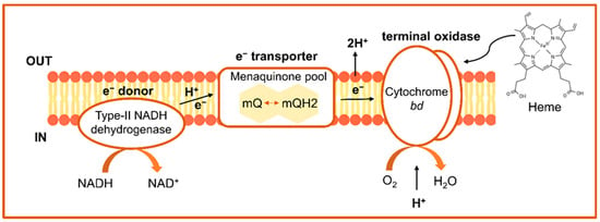
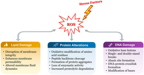
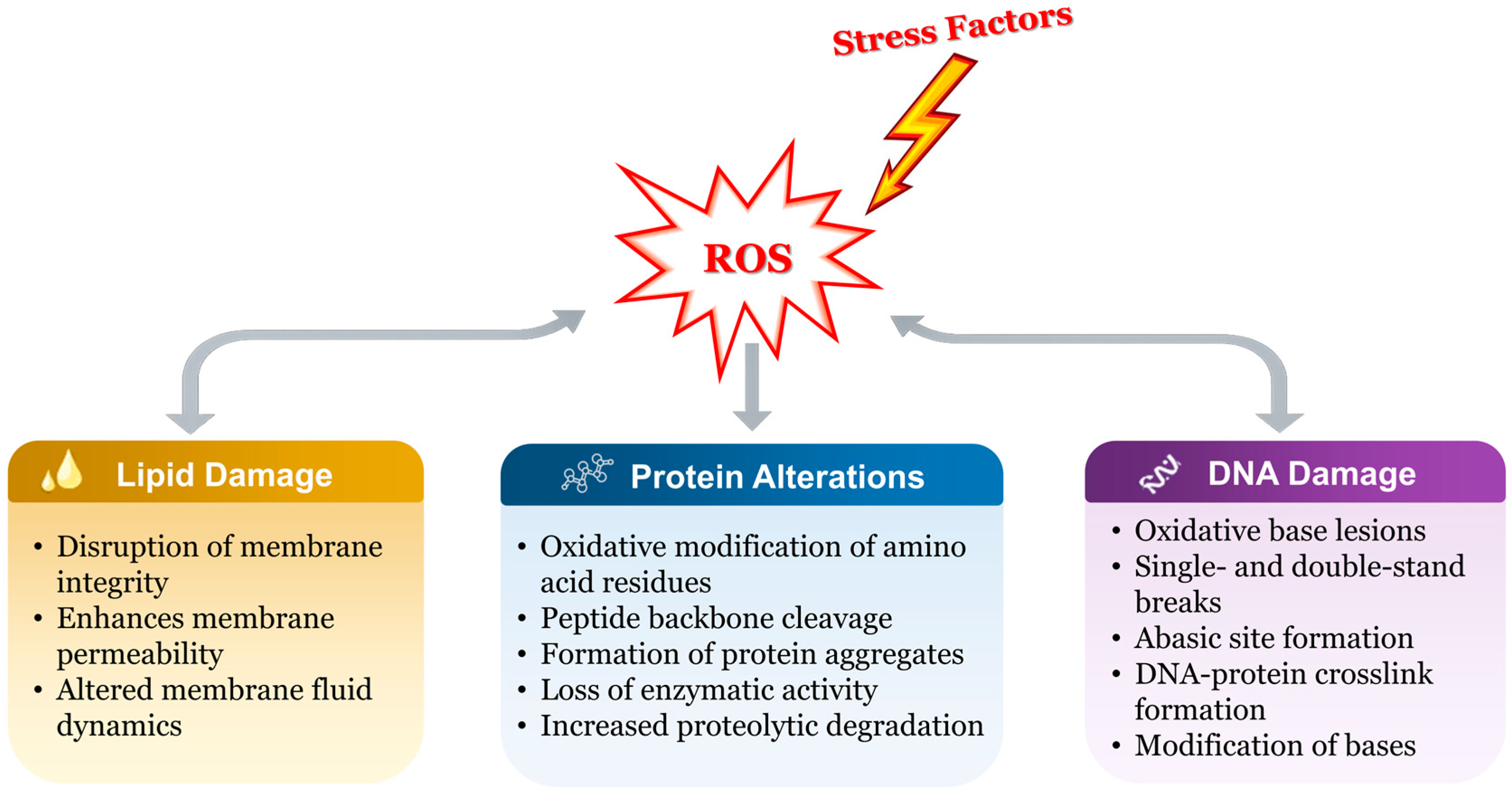
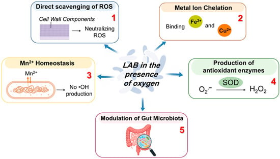
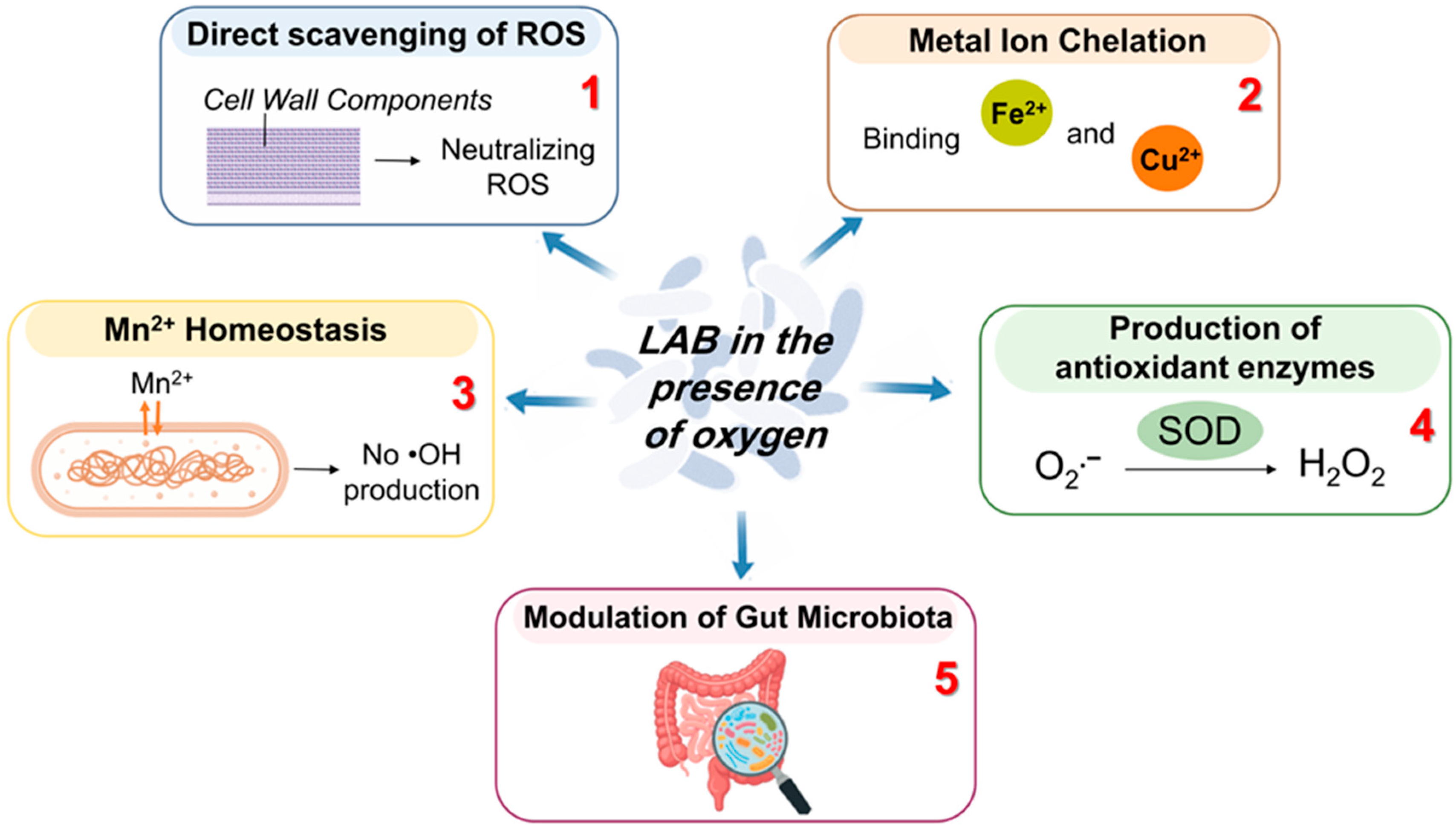
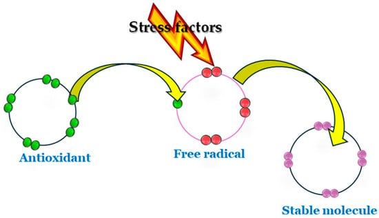
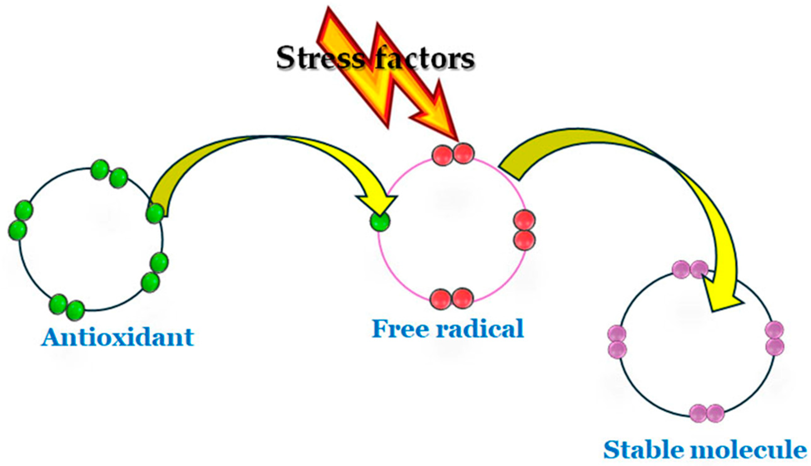
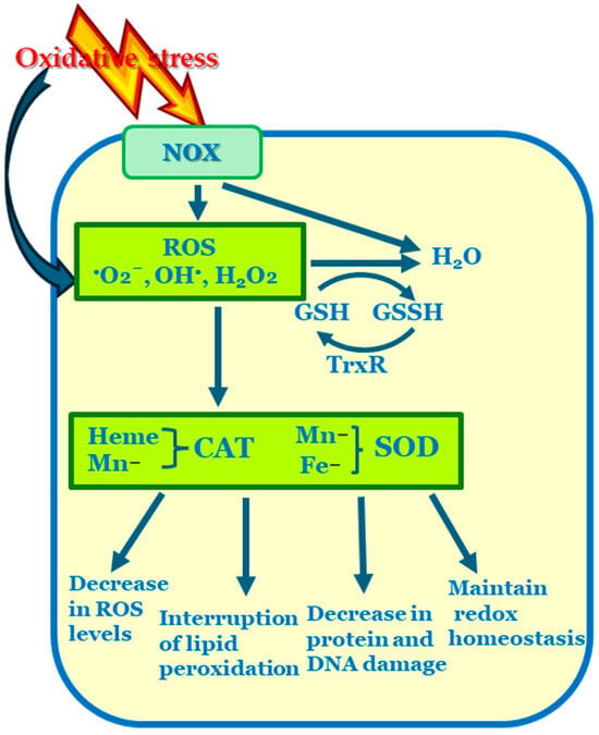
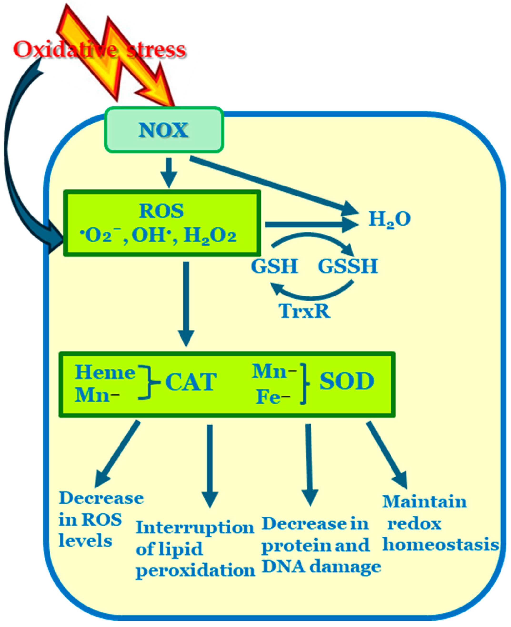
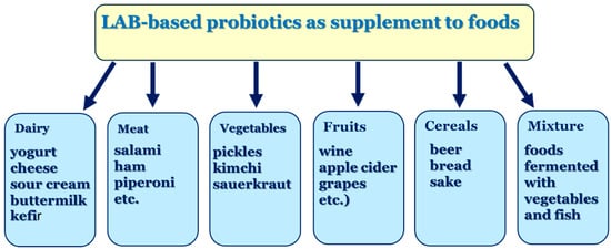
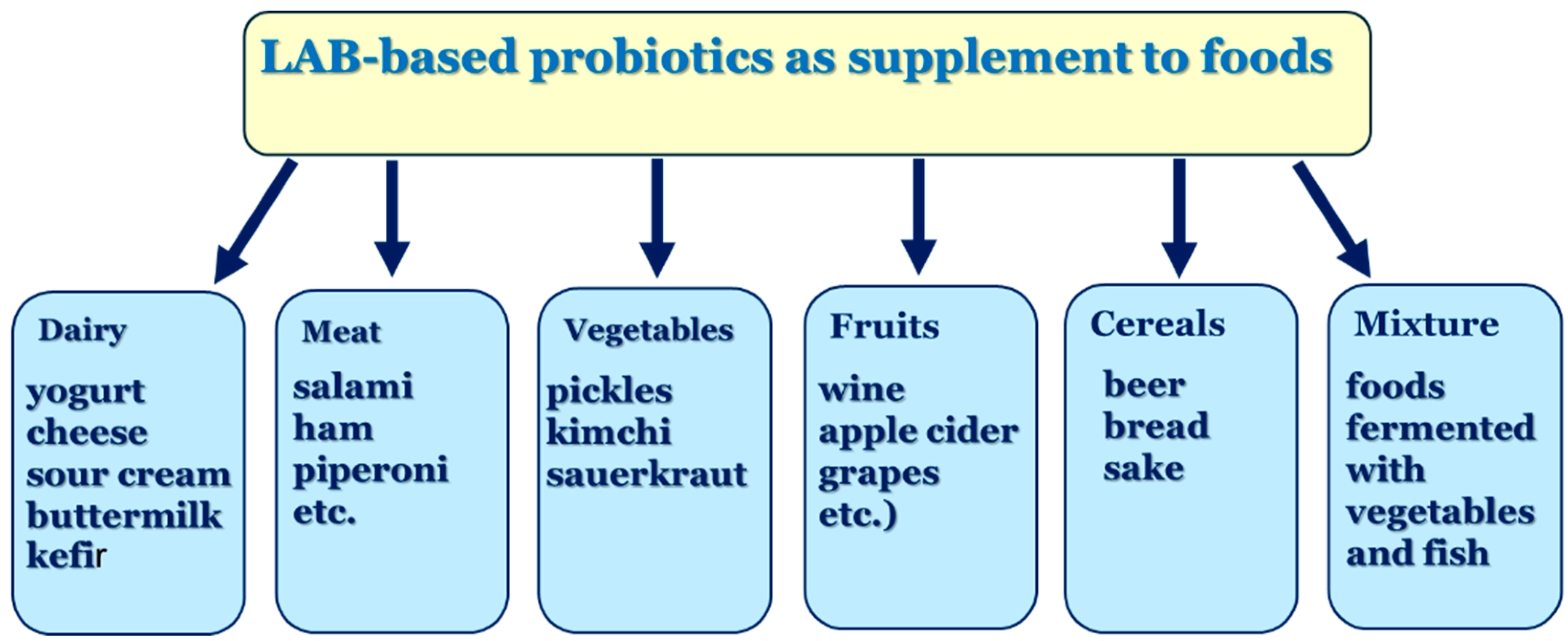
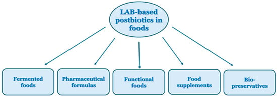
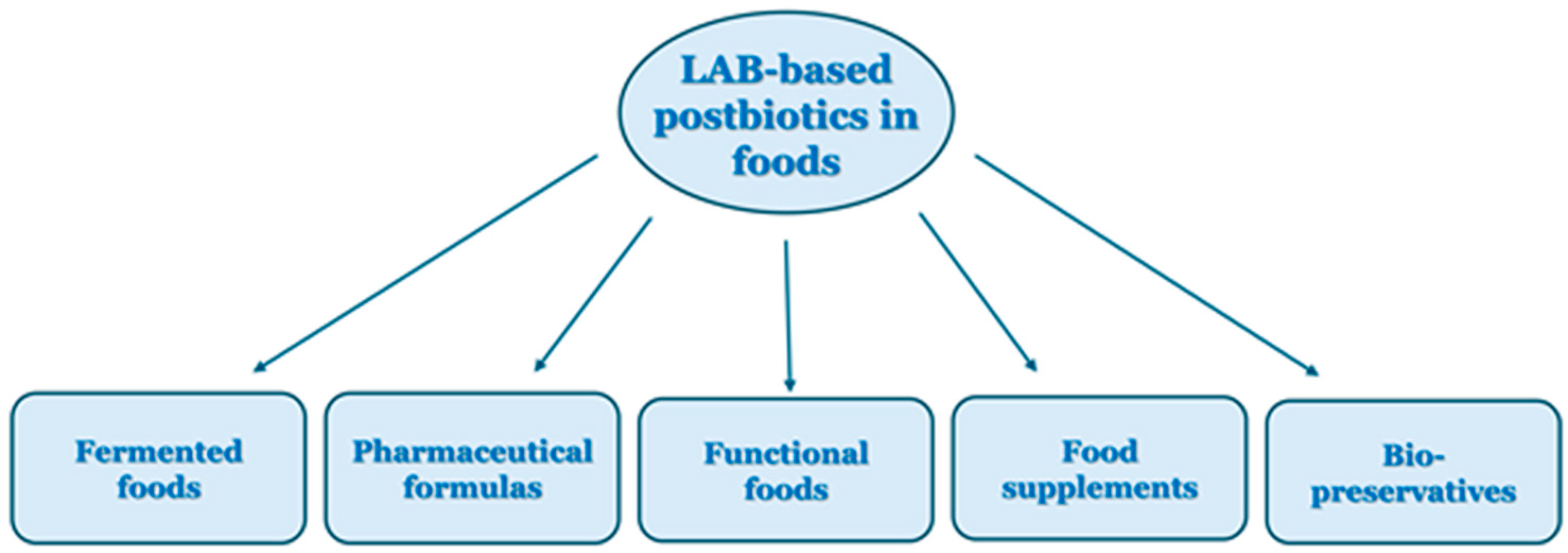

Disclaimer/Publisher’s Note: The statements, opinions and data contained in all publications are solely those of the individual author(s) and contributor(s) and not of MDPI and/or the editor(s). MDPI and/or the editor(s) disclaim responsibility for any injury to people or property resulting from any ideas, methods, instructions or products referred to in the content. |
© 2026 by the authors. Licensee MDPI, Basel, Switzerland. This article is an open access article distributed under the terms and conditions of the Creative Commons Attribution (CC BY) license.