


Abstract
Biofilms contribute significantly to the persistence and transmission of Campylobacter jejuni, yet enzymatic strategies for disrupting these structures remain underexplored. In this study, we evaluated the activity of a glycosyl hydrolase, CAase, against both planktonic and biofilm-associated C. jejuni. CAase showed no inhibitory effect on planktonic growth at concentrations up to 1 mg/mL. However, at 0.1 mg/mL, CAase reduced mature biofilm biomass by more than 93% in the wild-type strain and produced similar reductions in a luxS mutant, demonstrating substantial degradation of conserved glycan components within the extracellular polymeric substance (EPS). Scanning electron microscopy (SEM) imaging revealed substantial extracellular matrix loss, structural disruption, and altered cell morphology following CAase treatment. Rheological measurements demonstrated marked decreases in kinematic viscosity, further reflecting weakened biofilm cohesion. Together, these results indicate that CAase effectively disrupts multiple components of C. jejuni biofilm architecture despite lacking direct antimicrobial activity. This level of biofilm removal suggests potential utility in poultry-processing or surface-sanitation applications where targeted enzymatic disruption could enhance cleaning efficacy.
1. Introduction
Campylobacter jejuni is a leading cause of bacterial gastroenteritis worldwide and remains a significant challenge to food safety, particularly within poultry production and processing environments [1,2]. As a microaerobic, thermophilic organism, C. jejuni thrives in low-oxygen environments such as the avian gastrointestinal tract and can persist in protected microenvironments on food-contact surfaces and equipment. These physiological traits enable its survival in processing facilities despite its fragility under fully aerobic conditions [3,4,5]. Its low infectious dose and ability to contaminate carcasses, processing water, and equipment surfaces further contribute to its significance as a foodborne pathogen [6,7]. In addition, Campylobacter may enter dormancy and integrate into resident biofilms, making surface colonization and environmental persistence significant concerns. Therefore, effective strategies to limit biofilm formation and persistence are essential for reducing Campylobacter-associated illness.
Biofilm formation is a major contributor to the environmental resilience of C. jejuni. Biofilms enable attachment to stainless steel, plastic, rubber, and other industrial materials, protecting cells from desiccation, temperature fluctuations, and standard sanitation procedures [8,9,10,11]. This protection arises from the extracellular polymeric substance (EPS) matrix, which promotes cell aggregation, adhesion, and resistance to shear forces [12,13]. Unlike many bacteria with polysaccharide-dominated matrices, C. jejuni biofilms are comparatively protein-rich, contributing to greater mechanical stability, lower permeability, and reduced susceptibility to carbohydrate-targeted sanitizers [14]. Consistent with this, C. jejuni in mixed-culture biofilms shows reduced susceptibility to quaternary ammonia, peracetic acid, and peracetic acid/peroctanoic acid mixtures, with incomplete inactivation under these treatments [15]. Even so, C. jejuni EPS contains key glycan components, including capsular polysaccharides, glycoproteins, and LOS-associated carbohydrates, that support matrix cohesion and remain promising enzymatic targets [16,17].
Biofilm formation varies considerably among different Campylobacter strains, with C. jejuni 81-176, YH001, and YH027 forming robust monoculture biofilms [18]. Quorum sensing further modulates biofilm development via LuxS, which produces the interspecies signaling molecule autoinducer-2 (AI-2) [19]. luxS mutation disrupts AI-2 production and alters multiple biofilm-related phenotypes, including EPS composition, surface attachment, and matrix architecture. As a result, luxS mutants often produce biofilms with reduced biomass or modified structural properties [20,21]. These phenotypic differences make luxS-deficient strains valuable tools for evaluating whether biofilm-disrupting agents target conserved matrix components or vary in effectiveness across different EPS backgrounds.
Glycosyl hydrolases (GHs) have emerged as effective antibiofilm agents because they specifically cleave carbohydrate linkages within the EPS, targeting polysaccharide components essential for matrix integrity [22,23,24]. By hydrolyzing glycosidic bonds, GHs weaken the biofilm scaffold, enhance antimicrobial penetration, and facilitate detachment of sessile cells [25,26]. CAase, a bioengineered GH, disrupts EPS glycans and has been shown to remove mature biofilms of Escherichia coli O157:H7, Salmonella enterica serotype Typhimurium, and Listeria monocytogenes, achieving 17.4–34.6% greater removal than saline controls in crystal violet assays [27]. Despite its demonstrated efficacy across multiple foodborne pathogens, CAase has never been evaluated against C. jejuni. Given that C. jejuni produces a protein-rich yet glycan-containing EPS, it remains unclear whether glycan-cleaving enzymes can effectively disrupt its biofilms.
This study addresses this gap by evaluating CAase activity against C. jejuni 81-176 wild-type and luxS-mutant strains to determine whether glycan-directed disruption is effective across differences in EPS composition. Specifically, we assessed CAase effects on planktonic growth and mature biofilms and characterized structural and mechanical alterations using biomass quantification, scanning electron microscopy, and rheometry. By examining how CAase targets glycan components within a predominantly protein-rich matrix, this work provides critical insight into the feasibility and applied potential of glycan-cleaving enzymes for controlling persistent C. jejuni biofilms.
2. Materials and Methods
2.1. Minimal Inhibitory Concentration (MIC) Assay
Inhibition of C. jejuni growth by CAase was assessed using a broth microdilution assay as previously described [28]. The C. jejuni 81-176 strain was used because it is a well-characterized, highly virulent, and robust biofilm-forming strain, making it a standard and reliable model for evaluating CAase activity. Briefly, C. jejuni 81-176 was grown overnight in Mueller–Hinton (MH) broth (Becton Dickinson and Company, Franklin Lakes, NJ, USA), consisting of a standard dehydrated formulation containing beef extract, acid hydrolysate of casein, and starch, at 42 °C under microaerophilic conditions generated using a CampyPak gas pack (Becton Dickinson and Company), which produces an atmosphere of approximately 5% O2 and 10% CO2 (balance N2) in a sealed Nalgene Bio-Safe Carrier container (Rochester, NY, USA). The resulting culture was diluted in MH broth to approximately 104 CFU/mL. An aliquot of this suspension was heat-killed at 99 °C for 10 min, cooled to room temperature, and used as the nonviable control.
Serial two-fold dilutions of CAase (0.001953125–1.0 mg/mL; 100 µL per well) were prepared in MH broth in sterile 96-well microtiter plates. An additional 100 µL of the bacterial suspension (~104 CFU/mL) was added to each well, with column 11 receiving the heat-killed inoculum and all remaining wells receiving live cells. After incubating the plates at 42 °C for 48 h under microaerobic conditions, optical density at 600 nm was measured using a Safire 2 microplate reader (Tecan Group Ltd., Männedorf, Switzerland). Each CAase concentration was tested using four biological replicates, and growth inhibition in each treatment well was compared with that of the CAase-free control wells. Statistical significance of differences between treatment and control groups was evaluated using an all-pairs Tukey–Kramer test (α = 0.05) in JMP Pro (version 18.2, SAS Institute Inc., Cary, NC, USA).
2.2. Biofilm Sampling
Fresh colonies of C. jejuni 81-176 and its luxS deletion mutant [29] were resuspended in 2 mL of MH broth and incubated overnight at 42 °C under microaerophilic conditions. One milliliter of the overnight culture was then inoculated into 100 mL of MH broth and mixed thoroughly. From this inoculum, three different setups with distinct substrate type and surface area were used to develop biofilms: (1) six 2 mL aliquots were dispensed into 14 mL sterile polypropylene tubes (17 × 100 mm, Becton, Dickinson and Company) to evaluate biofilm formation in a small-volume, limited-surface-area environment; (2) six 5 mL aliquots were transferred into 10 mL polystyrene tissue culture tubes with a flat surface of 10 cm2 growth area and vent cap (Techno Plastic Products AG, Trasadingen, Canton Schaffhausen, Switzerland); and (3) six 5 mL aliquots were transferred into 40 µm nylon cell strainers (Becton, Dickinson and Company) positioned within a 6-well plate. These disposable mesh filters, designed to retain larger particulates, were used to provide a uniform, high-porosity surface that supports consistent biofilm attachment and enables efficient biomass recovery for downstream processing. Biofilms were allowed to develop for 5 days under microaerophilic conditions [18]. After incubation, biofilm biomass was harvested by filtering each sample through a 40 µm cell strainer fitted onto a 50 mL Falcon tube. Samples grown in the flat tubes were scraped with a silicon-blade cell scraper (Corning Life Sciences, Corning, NY, USA) to facilitate release of the biofilms.
2.3. Biofilms Quantification
Following CAase treatment, biofilms retained on the cell strainers were quantified using the crystal violet (CV) staining method as previously described [18]. Briefly, each biofilm sample was gently rinsed with 2 mL of MH broth, stained with 5 mL of 0.1% CV for 30 min, and washed three times with 5 mL of 18 MΩ water. The CV-stained biofilms were then solubilized by adding 6 mL of 100% ethanol and incubating at room temperature for 30 min with gentle shaking. Absorbance was measured in triplicate at 590 nm using a Cytation 5 plate reader (BioTek/Agilent Life Sciences, Winooski, VT, USA).
2.4. CAase Production
CAase was produced following the method of Mayton et al. [27]. Briefly, the enzyme, derived from a truncated Salmonella phage hydrolase (NCBI YP_004893855.1) and engineered with an N-terminal His6 tag, was expressed in E. coli BL21(DE3) under a T7 promoter (pET28) using IPTG induction and purified by Ni-NTA affinity chromatography. The purity of CAase was verified by SDS–PAGE, confirming successful purification of the final enzyme preparation. Enzyme concentration was determined by measuring absorbance at 280 nm, using a NanoDrop 1000 Spectrophotometer (Thermo Fisher Scientific, Waltham, MA, USA) and a calculated extinction coefficient of 121,990 M−1 cm−1 derived from its primary amino acid sequence.
2.5. CAase Treatment
Biofilm cells harvested on a cell strainer were rinsed with 2 mL of fresh MH broth and transferred to a 6-well plate. An aliquot of the culture filtrate was serially diluted in MH broth and enumerated using the 6 × 6 drop plate method [30] to quantify CFU in the planktonic fraction of the pre-treatment culture. For each biofilm growth setup, three biofilm samples in cell strainers were submerged in 5 mL of 0.1 mg/mL CAase. As untreated controls, three additional biofilms were submerged in 5 mL of MH broth alone. The 6-well plate containing the treated and untreated biofilms was incubated at 42 °C under microaerobic conditions generated with a CampyPak in an airtight container for 2 h.
2.6. Scanning Electron Microscope (SEM) Examination
Biofilms treated with or without 0.1 mg/mL CAase for 2 h under microaerobic conditions were collected by centrifugation (6000× g, 2 min) and applied directly to cleaned glass coverslips. Planktonic cells grown for 24 h at 42 °C under microaerobic conditions were included as a control. Samples were air-dried for 1 h at room temperature and fixed with 2.5% glutaraldehyde for 2 h. After rinsing with 0.1 M imidazole buffer, samples were dehydrated in 50%, 80%, and 100% ethanol (three changes per step) and dried by CO2 critical-point drying. Coverslips were mounted on SEM stubs and sputter-coated with a thin gold layer using a Scancoat Six sputter coater (BOC Edwards, Wilmington, MA, USA). SEM images were acquired at room temperature using a Quanta 200 FEG scanning electron microscope (FEI, Inc., Hillsboro, OR, USA) [28].
2.7. Viscosity Measurement of C. jejuni Biofilms
The kinematic viscosity of C. jejuni biofilms treated with or without 0.1 mg/mL CAase for 2 h under microaerobic conditions was measured using a calibrated glass kinematic viscometer tube (0.1 cSt/s constant flow rate; size 200; Thermo Fisher Scientific, Waltham, MA, USA) following the manufacturer’s instructions. After incubation, 5 mL of each biofilm sample was loaded into the viscometer tube. A vacuum pump was used to gently draw the sample up the capillary until the meniscus was slightly above the upper timing mark, after which the vacuum was released to allow the sample to flow downward under gravity. The efflux time, the time required for the meniscus to pass between the timing marks, was recorded at room temperature using video capture. Kinematic viscosity (ν in centistokes (cSt)) was calculated using the equation:
where t is the efflux time (seconds), and C is the viscometer’s calibration constant (0.1 cSt/s). All measurements were performed on triplicate samples.
ν = t × C
3. Results
3.1. Effect of CAase on C. jejuni Growth
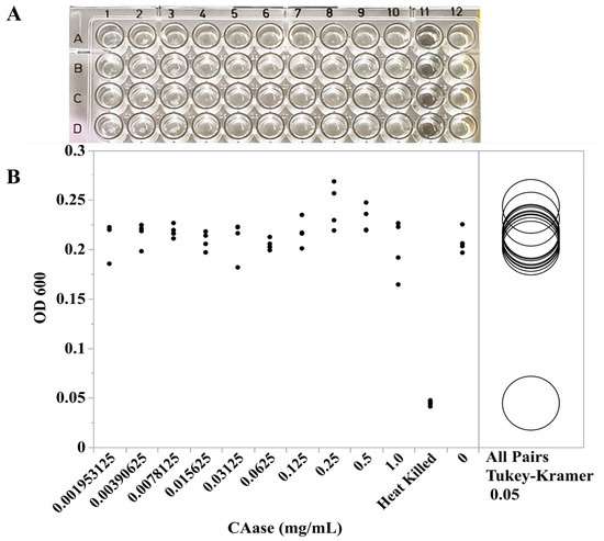
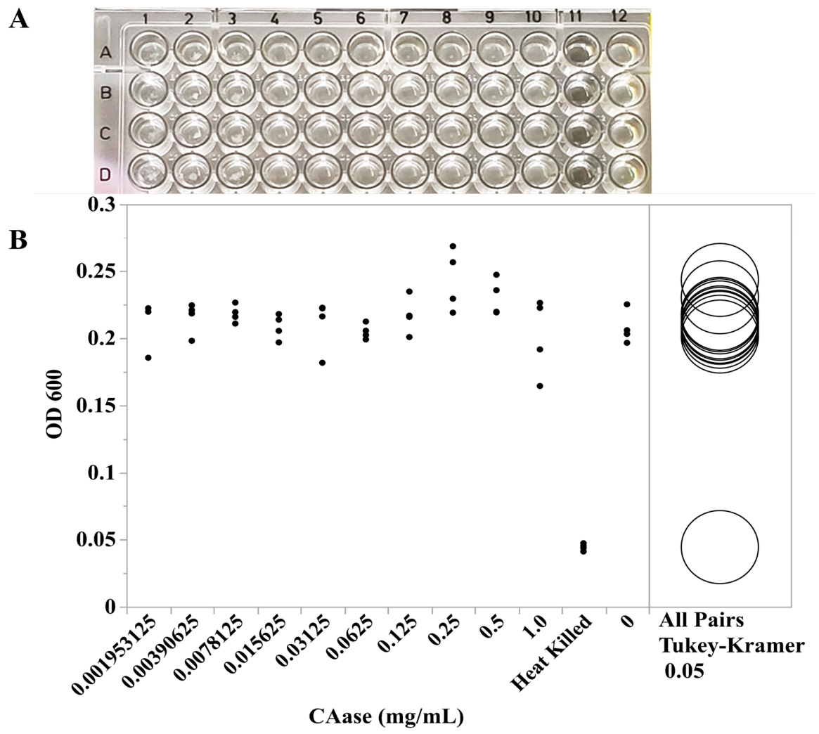
Minimal inhibitory concentration (MIC) testing demonstrated that CAase did not inhibit the growth of C. jejuni 81-176 at concentrations up to 1 mg/mL (Figure 1). After 48 h of microaerobic incubation, OD600 values across the full CAase concentration range (0.001953125–1.0 mg/mL) were indistinguishable from the untreated control. Statistical analysis using the all-pairs Tukey–Kramer test (α = 0.05) confirmed that none of the CAase treatments resulted in a significant reduction in bacterial growth. As expected, only the heat-killed control showed no detectable growth (p ≤ 0.05). An independent trial conducted after 24 h of microaerobic incubation produced similar results. Collectively, these findings indicate that CAase lacks measurable antimicrobial activity against C. jejuni under the conditions tested, suggesting that any observed biological effects are likely due to mechanisms other than direct growth inhibition.
Figure 1.
Effect of CAase on C. jejuni growth. Growth of C. jejuni 81-176 was evaluated across CAase concentrations ranging from 0.001953125 to 1.0 mg/mL. After 48 h of microaerobic incubation, cultures were photographed (A), showing CAase-treated wells (columns 1–10), a heat-killed control (column 11), and an untreated control (column 12). (B) Optical density (OD600) was measured following incubation. Data represent four biological replicates, and statistical comparisons were performed using the Tukey–Kramer test (α = 0.05).
3.2. CAase-Mediated Disruption of C. jejuni Biofilms
The ability of CAase, a glycoside hydrolase that targets carbohydrate components of the EPS matrix, to disrupt biofilms was evaluated using C. jejuni 81-176 and its luxS mutant. Mature biofilms were exposed to 0.1 mg/mL CAase for 2 h under static, microaerobic conditions, and residual biomass was quantified using crystal violet staining (OD590). All experiments were performed in triplicate independent cultures.
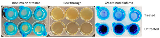
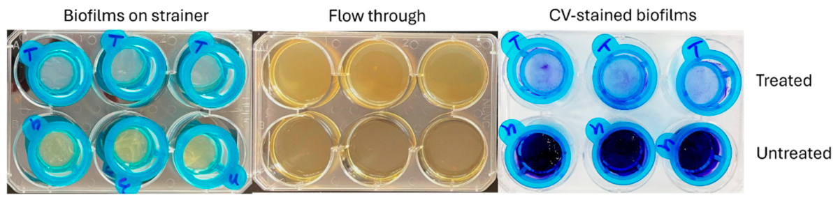
CAase treatment significantly reduced biofilm biomass in both strains and under all growth setups. Representative images in Figure 2 show robust biofilms formed by C. jejuni 81-176 retained on 40 µm cell strainers. Following CAase exposure, the biofilms were visibly disrupted and released through the strainer, resulting in increased turbidity in the flow-through (Figure 2).
Figure 2.
CAase-treated and untreated C. jejuni biofilms. Representative images of biofilms formed by C. jejuni 81-176 following treatment with 0.1 mg/mL CAase (upper row) compared with untreated controls (lower row). Biofilms were collected using nylon strainers. The left panel shows intact biofilms retained on the strainer surface, the middle panel displays the flow-through fractions collected beneath the strainer, and the right panel presents crystal violet–stained biofilms used for visualization and biomass quantification. Each treatment was performed in triplicate across three independent biological replicates.
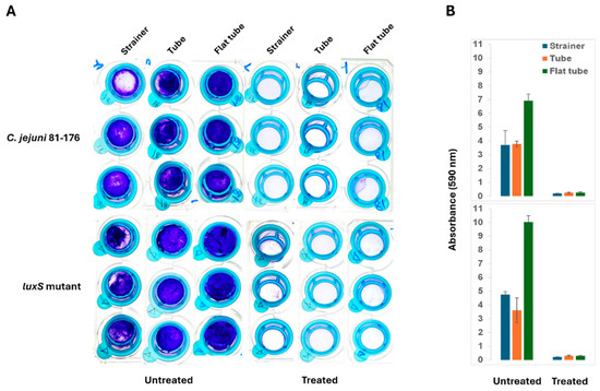
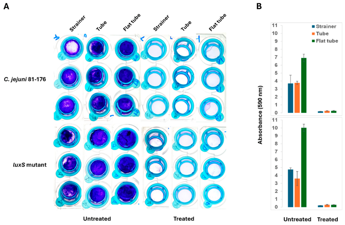
Quantitative crystal violet assays showed that treatment with 0.1 mg/mL CAase at 42 °C for 2 h reduced biofilm biomass in C. jejuni 81-176 by more than 93%. The luxS mutant, despite its altered EPS composition resulting from disrupted quorum sensing, remained highly susceptible, exhibiting over a 91% reduction in biomass (Figure 3). These findings indicate that CAase effectively removes mature biofilms regardless of genetic or structural variation. Overall, the results here suggest that CAase functions as a potent biofilm-disrupting enzyme capable of degrading biofilms across C. jejuni strains with distinct EPS backgrounds.
Figure 3.
CAase-treated and untreated biofilms of C. jejuni 81-176 and its luxS mutant. (A) Biofilms were developed using three growth setups: a 14 mL round-bottom tube, a 10-cm2 flat-surface tissue culture tube, and a 40 µm cell strainer. (B) Biofilm biomass was quantified by crystal violet staining followed by OD590 measurement. Statistical differences between CAase-treated and untreated samples were analyzed using the Tukey–Kramer test (α = 0.05). Each sample included three biological replicates and two technical replicates.
3.3. SEM Examination of C. jejuni Biofilms
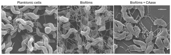
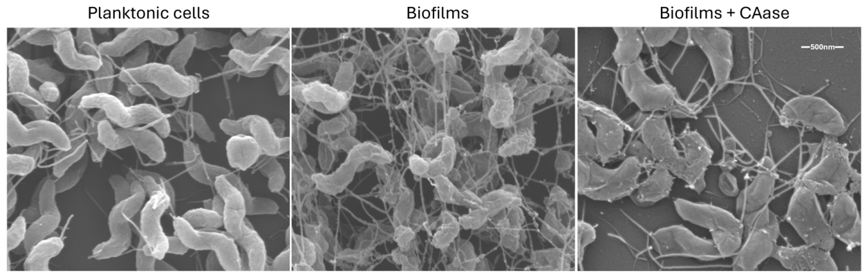
SEM imaging was used to evaluate the effects of CAase on C. jejuni 81-176 cell morphology and biofilm structure. As a control, planktonic cells lacking biofilm and extensive EPS displayed the typical spiral morphology of Campylobacter (Figure 4). In contrast, mature biofilms exhibited abundant EPS and a more three-dimensional architecture, with spiral cells embedded within a dense matrix.
Figure 4.
Scanning electron microscopy images of C. jejuni 81-176 biofilms under CAase-treated and untreated conditions. A planktonic cell control, lacking biofilm and extensive EPS, was included and processed identically. The images depict differences in cell morphology, surface attachment, and extracellular matrix architecture following 0.1 mg/mL CAase treatment, illustrating the potential structural effects of CAase on Campylobacter biofilms.
CAase-treated biofilms exhibited clear structural deterioration. SEM imaging showed a marked reduction in extracellular matrix material and noticeable changes in cell morphology, indicating compromised matrix integrity and diminished overall biofilm stability. These observations collectively highlight the disruptive impact of CAase on C. jejuni biofilms.
3.4. Effect of CAase on C. jejuni Biofilm Kinematic Viscosity
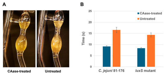
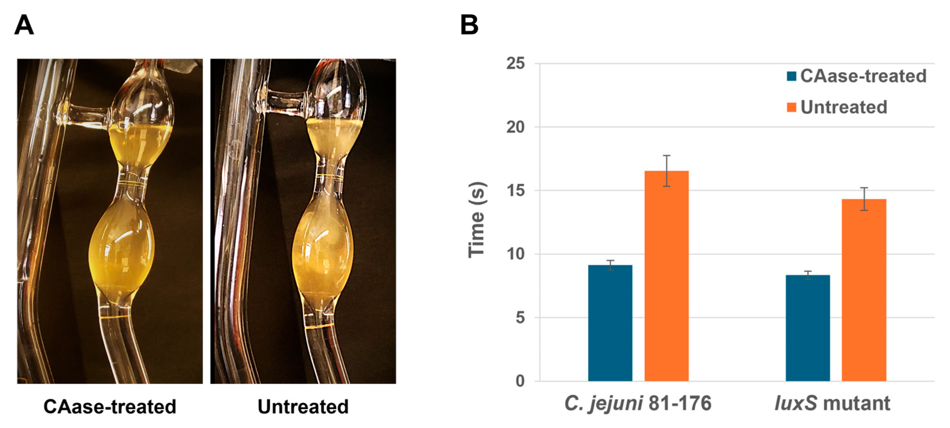
To assess changes in mechanical properties, the flow behavior of Campylobacter biofilms with and without CAase treatment was evaluated by measuring kinematic viscosity. As shown in Figure 5 and Supplemental Video S1 (untreated) and Video S2 (treated), CAase significantly altered the rheological properties of both C. jejuni 81-176 and its luxS mutant. CAase-treated samples appeared more homogeneous and exhibited a marked reduction in “gelatinous” biomass. Flow time decreased from 16.5 to 9.1 s (corresponding to a viscosity reduction from 1.65 to 0.91 cSt) for the wild type and from 14.3 to 8.3 s (1.43 to 0.83 cSt) for the luxS mutant (p < 0.05, one-way ANOVA). These reductions in viscosity are consistent with CAase-mediated degradation of the biofilm matrix. This reduction reflects decreased biofilm viscosity consistent with CAase-mediated matrix disruption.
Figure 5.
Kinematic viscosity of C. jejuni biofilms treated with and without CAase. (A) Flow times of the biofilm samples were measured using a glass kinematic viscometer tube to compare rheological properties following enzymatic degradation. (B) CAase treatment significantly decreased flow time for both C. jejuni 81-176 and the luxS mutant, reducing flow times from 16.5 s to 9.1 s and from 14.3 s to 8.3 s, respectively (p < 0.05, one-way ANOVA). Error bars represent the mean ± standard error of the mean (SEM) from three biological replicates.
Lower viscosity reflects a weakened, less cohesive biofilm structure, likely increasing susceptibility to shear forces and promoting cell dispersion. This mechanical destabilization may also enhance penetration of antimicrobials or host defenses, ultimately reducing biofilm persistence under environmental or processing conditions.
4. Discussion
CAase treatment resulted in pronounced structural degradation of C. jejuni biofilms. SEM imaging revealed a clear reduction in extracellular matrix material, disrupted surface architecture, and altered cell morphology, indicating substantial loss of matrix integrity and overall biofilm stability. These structural changes were consistent with the marked reductions in biomass observed in crystal-violet assays and reinforce the conclusion that CAase effectively destabilizes the biofilm scaffold.
Because crystal violet staining and SEM do not differentiate viable from non-viable cells, we evaluated cell survival using CFU enumeration following CAase treatment. These results confirmed that CAase did not affect cell viability, indicating that the enzyme disrupts the biofilm matrix without killing the bacteria. This distinction is now explicitly acknowledged, clarifying that CAase’s activity reflects structural, not bactericidal, effects.
The viscosity measurements provide additional but indirect evidence for CAase-mediated weakening of the biofilm matrix. Both wild-type and luxS-mutant exhibited reduced flow time and kinematic viscosity following CAase treatment, suggesting loss of mechanical cohesion. These rheological changes may arise from multiple factors, including EPS degradation, decreased biomass, altered aggregation, and disruption of three-dimensional architecture, and therefore should not be interpreted as direct evidence of glycan hydrolysis. Instead, they support the broader conclusion that CAase compromises the biofilm’s mechanical integrity.
Importantly, CAase did not inhibit planktonic growth, underscoring that its activity is directed toward extracellular matrix components rather than cellular metabolism. Its effectiveness in both wild-type and luxS-mutant strains indicates that CAase can disrupt biofilms across distinct EPS backgrounds. While this consistency suggests possible interaction with glycan elements important for biofilm stability, such mechanistic interpretations remain inferential, as EPS composition and glycan degradation products were not directly measured.
The effects of CAase align with those of other glycosyl hydrolases that disrupt biofilms by cleaving matrix polymers. DNase I, for example, degrades extracellular DNA and enhances dispersal in C. jejuni and multispecies biofilms [31]. while cellulase and α-amylase disperse Pseudomonas aeruginosa and Staphylococcus aureus biofilms in a context-dependent manner [25]. GHs from Pseudomonas fluorescens also efficiently dismantle polysaccharide-rich biofilms at low concentrations [32]. Within this framework, CAase exhibits comparable biofilm-disrupting activity and shows promise as part of enzyme-based biofilm-control strategies.
Together, the structural, rheological, and quantitative data demonstrate that CAase compromises multiple aspects of C. jejuni biofilm integrity, biochemically by altering matrix composition and mechanically by reducing cohesive strength, while leaving cell viability unaffected. These findings support CAase as a promising antibiofilm candidate that may enhance sanitation effectiveness by improving antimicrobial penetration and reducing reliance on harsh chemical treatments. Its consistent activity across genetically distinct C. jejuni strains, as well as other foodborne pathogens such as Escherichia coli O157:H7, Salmonella enterica serotype Typhimurium, and Listeria monocytogenes [27], further underscores its potential utility in diverse industrial and environmental settings.
However, the present study was conducted using laboratory-grown, pure-culture C. jejuni biofilms under controlled conditions. Mixed-species biofilms typical of food-processing environments were not evaluated, and the enzyme’s stability across fluctuating temperatures, pH conditions, oxidizing sanitizers, detergents, or organic residues remains unknown. Industrial applicability is therefore prospective. Future studies should assess CAase stability and performance under processing-relevant conditions, evaluate its compatibility with commonly used sanitizers, and examine its activity on biofilms formed on industry-relevant materials. Expanding these analyses to additional foodborne and environmental pathogens will further clarify the enzyme’s practical utility and inform its integration into real-world biofilm-control interventions.
5. Conclusions
This study demonstrates that although CAase does not inhibit planktonic growth of C. jejuni, it exerts a strong disruptive effect on established biofilms by degrading key extracellular matrix components. CAase treatment substantially reduced biofilm biomass, weakened structural cohesion, altered rheological properties, and produced marked architectural damage observable by SEM. Its consistent activity against both wild-type and luxS-mutant strains suggests that CAase may act on glycan elements important for biofilm stability, although this remains a hypothesis rather than a mechanism directly demonstrated here. Collectively, these findings support CAase as a promising antibiofilm candidate for disrupting persistent C. jejuni biofilms. Further mechanistic and applied studies will be necessary to define its full utility and evaluate its effectiveness under broader environmental and industrial conditions.
Supplementary Materials
The following supporting information can be downloaded at https://www.mdpi.com/article/10.3390/foods15162849/s1. Supplemental Video S1. Kinematic viscosity of untreated C. jejuni 81-176 biofilms was measured using a glass kinematic viscometer tube; Supplemental Video S2. Kinematic viscosity of CAase-treated C. jejuni 81-176 biofilms was measured using a glass kinematic viscometer tube.
Author Contributions
Y.H., C.-Y.C., G.D. and J.C. designed and performed experiments, analyzed data, interpreted results, and prepared the manuscript. H.K., K.L. and B.B. conducted experiments, analyzed results, and reviewed the manuscript. All authors have read and agreed to the published version of the manuscript.
Funding
This research was supported by the U.S. Department of Agriculture, Agricultural Research Service (USDA-ARS), National Program 108, Current Research Information System numbers 8072-42000-093 and 8072-42000-094.
Institutional Review Board Statement
Not applicable.
Informed Consent Statement
Not applicable.
Data Availability Statement
The original contributions presented in this study are included in the article/Supplementary Materials. Further inquiries can be directed to the corresponding author.
Acknowledgments
We would like to thank Joe Uknalis of the Microscopy Imaging Facility of the USDA, ARS, ERRC, for technical assistance in acquiring the SEM images. The mention of trade names or commercial products in this publication is solely for the purpose of providing specific information and does not imply recommendation or endorsement by the USDA. The USDA is an equal opportunity provider and employer.
Conflicts of Interest
The authors declare no conflicts of interest.
References
- Centers for Disease Control and Prevention. About Campylobacter Infection. Available online: https://www.cdc.gov/campylobacter/about/index.html (accessed on 28 January 2026).
- World Health Organization (WHO). Fact Sheet on Campylobacter. Available online: https://www.who.int/news-room/fact-sheets/detail/campylobacter (accessed on 28 January 2026).
- Humphrey, S.; Chaloner, G.; Kemmett, K.; Davidson, N.; Williams, N.; Kipar, A.; Humphrey, T.; Wigley, P. Campylobacter jejuni is not merely a commensal in commercial broiler chickens and affects bird welfare. mBio 2014, 5, e01364-14. [Google Scholar] [CrossRef] [Scilit] [PubMed]
- Ica, T.; Caner, V.; Istanbullu, O.; Nguyen, H.D.; Ahmed, B.; Call, D.R.; Beyenal, H. Characterization of mono- and mixed-culture Campylobacter jejuni biofilms. Appl. Environ. Microbiol. 2012, 78, 1033–1038. [Google Scholar] [CrossRef] [Scilit] [PubMed]
- Pokhrel, D.; Thames, H.T.; Zhang, L.; Dinh, T.T.N.; Schilling, W.; White, S.B.; Ramachandran, R.; Theradiyil Sukumaran, A. Roles of Aerotolerance, Biofilm Formation, and Viable but Non-Culturable State in the Survival of Campylobacter jejuni in Poultry Processing Environments. Microorganisms 2022, 10, 2165. [Google Scholar] [CrossRef] [Scilit] [PubMed]
- Veronese, P.; Dodi, I. Campylobacter jejuni/coli Infection: Is It Still a Concern? Microorganisms 2024, 12, 2669. [Google Scholar] [CrossRef] [Scilit] [PubMed]
- Nguyen, V.T.; Fegan, N.; Turner, M.S.; Dykes, G.A. Role of attachment to surfaces on the prevalence and survival of Campylobacter through food systems. J. Food Prot. 2012, 75, 195–206. [Google Scholar] [CrossRef] [Scilit] [PubMed]
- Silha, D.; Sirotkova, S.; Svarcova, K.; Hofmeisterova, L.; Korycanova, K.; Silhova, L. Biofilm Formation Ability of Arcobacter-like and Campylobacter Strains under Different Conditions and on Food Processing Materials. Microorganisms 2021, 9, 2017. [Google Scholar] [CrossRef] [Scilit] [PubMed]
- Reeser, R.J.; Medler, R.T.; Billington, S.J.; Jost, B.H.; Joens, L.A. Characterization of Campylobacter jejuni biofilms under defined growth conditions. Appl. Environ. Microbiol. 2007, 73, 1908–1913. [Google Scholar] [CrossRef] [Scilit] [PubMed]
- Olsen, I. Biofilm-specific antibiotic tolerance and resistance. Eur. J. Clin. Microbiol. Infect. Dis. 2015, 34, 877–886. [Google Scholar] [CrossRef] [Scilit] [PubMed]
- Teh, A.H.; Lee, S.M.; Dykes, G.A. Does Campylobacter jejuni form biofilms in food-related environments? Appl. Environ. Microbiol. 2014, 80, 5154–5160. [Google Scholar] [CrossRef] [Scilit] [PubMed]
- Flemming, H.C.; Wingender, J.; Szewzyk, U.; Steinberg, P.; Rice, S.A.; Kjelleberg, S. Biofilms: An emergent form of bacterial life. Nat. Rev. Microbiol. 2016, 14, 563–575. [Google Scholar] [CrossRef] [Scilit] [PubMed]
- Bundurus, I.A.; Balta, I.; Pet, I.; Stef, L.; Popescu, C.A.; McCleery, D.; Lemon, J.; Callaway, T.; Douglas, A.; Corcionivoschi, N. Mechanistic concepts involved in biofilm associated processes of Campylobacter jejuni: Persistence and inhibition in poultry environments. Poult. Sci. 2024, 103, 104328. [Google Scholar] [CrossRef] [Scilit] [PubMed]
- Pavlinjek, N.; Klancnik, A.; Sabotic, J. Evaluation of physical and chemical isolation methods to extract and purify Campylobacter jejuni extracellular polymeric substances. Front. Microbiol. 2024, 15, 1488114. [Google Scholar] [CrossRef] [Scilit] [PubMed]
- Trachoo, N.; Frank, J.F. Effectiveness of chemical sanitizers against Campylobacter jejuni-containing biofilms. J. Food Prot. 2002, 65, 1117–1121. [Google Scholar] [CrossRef] [Scilit] [PubMed]
- Ma, L.; Feng, J.; Zhang, J.; Lu, X. Campylobacter biofilms. Microbiol. Res. 2022, 264, 127149. [Google Scholar] [CrossRef] [Scilit] [PubMed]
- Melo, R.T.; Mendonca, E.P.; Monteiro, G.P.; Siqueira, M.C.; Pereira, C.B.; Peres, P.; Fernandez, H.; Rossi, D.A. Intrinsic and Extrinsic Aspects on Campylobacter jejuni Biofilms. Front. Microbiol. 2017, 8, 1332. [Google Scholar] [CrossRef] [Scilit] [PubMed]
- He, Y.; Dykes, G.; Koppenhofer, H.; Capobianco, J.; Chen, C.Y. Comparative Genomics Reveals Unique Genetic Determinants of Biofilm Formation in Campylobacter. Int. J. Mol. Sci. 2026, 27, 2543. [Google Scholar] [CrossRef] [Scilit] [PubMed]
- Plummer, P.J. LuxS and quorum-sensing in Campylobacter. Front. Cell. Infect. Microbiol. 2012, 2, 22. [Google Scholar] [CrossRef] [Scilit] [PubMed]
- Puning, C.; Su, Y.; Lu, X.; Golz, G. Molecular Mechanisms of Campylobacter Biofilm Formation and Quorum Sensing. Curr. Top. Microbiol. Immunol. 2021, 431, 293–319. [Google Scholar] [CrossRef] [Scilit] [PubMed]
- Li, S.; Chan, K.K.; Hua, M.Z.; Golz, G.; Lu, X. Inhibition of AI-2 Quorum Sensing and Biofilm Formation in Campylobacter jejuni by Decanoic and Lauric Acids. Front. Microbiol. 2021, 12, 811506. [Google Scholar] [CrossRef] [Scilit] [PubMed]
- Al-Madboly, L.A.; Aboulmagd, A.; El-Salam, M.A.; Kushkevych, I.; El-Morsi, R.M. Microbial enzymes as powerful natural anti-biofilm candidates. Microb. Cell. Fact. 2024, 23, 343. [Google Scholar] [CrossRef] [Scilit] [PubMed]
- Wang, S.; Zhao, Y.; Breslawec, A.P.; Liang, T.; Deng, Z.; Kuperman, L.L.; Yu, Q. Strategy to combat biofilms: A focus on biofilm dispersal enzymes. npj Biofilms Microbiomes 2023, 9, 63. [Google Scholar] [CrossRef] [Scilit] [PubMed]
- Rumbaugh, K.P.; Sauer, K. Biofilm dispersion. Nat. Rev. Microbiol. 2020, 18, 571–586. [Google Scholar] [CrossRef] [Scilit] [PubMed]
- Redman, W.K.; Welch, G.S.; Rumbaugh, K.P. Differential Efficacy of Glycoside Hydrolases to Disperse Biofilms. Front. Cell. Infect. Microbiol. 2020, 10, 379. [Google Scholar] [CrossRef] [Scilit] [PubMed]
- Ellis, J.R.; Bull, J.J.; Rowley, P.A. Fungal Glycoside Hydrolases Display Unique Specificities for Polysaccharides and Staphylococcus aureus Biofilms. Microorganisms 2023, 11, 293. [Google Scholar] [CrossRef] [Scilit] [PubMed]
- Mayton, H.M.; Walker, S.L.; Berger, B.W. Disrupting Irreversible Bacterial Adhesion and Biofilm Formation with an Engineered Enzyme. Appl. Environ. Microbiol. 2021, 87, e0026521. [Google Scholar] [CrossRef] [Scilit] [PubMed]
- Xie, Y.; He, Y.; Irwin, P.L.; Jin, T.; Shi, X. Antibacterial activity and mechanism of action of zinc oxide nanoparticles against Campylobacter jejuni. Appl. Environ. Microbiol. 2011, 77, 2325–2331. [Google Scholar] [CrossRef] [Scilit] [PubMed]
- He, Y.; Frye, J.G.; Strobaugh, T.P.; Chen, C.Y. Analysis of AI-2/LuxS-dependent transcription in Campylobacter jejuni strain 81-176. Foodborne Pathog. Dis. 2008, 5, 399–415. [Google Scholar] [CrossRef] [Scilit] [PubMed]
- Chen, C.Y.; Nace, G.W.; Irwin, P.L. A 6 × 6 drop plate method for simultaneous colony counting and MPN enumeration of Campylobacter jejuni, Listeria monocytogenes, and Escherichia coli. J. Microbiol. Methods 2003, 55, 475–479. [Google Scholar] [CrossRef] [Scilit] [PubMed]
- Brown, H.L.; Hanman, K.; Reuter, M.; Betts, R.P.; van Vliet, A.H. Campylobacter jejuni biofilms contain extracellular DNA and are sensitive to DNase I treatment. Front. Microbiol. 2015, 6, 699. [Google Scholar] [CrossRef] [Scilit] [PubMed]
- Wang, D.; Naqvi, S.T.A.; Lei, F.; Zhang, Z.; Yu, H.; Ma, L.Z. Glycosyl hydrolase from Pseudomonas fluorescens inhibits the biofilm formation of Pseudomonads. Biofilm 2023, 6, 100155. [Google Scholar] [CrossRef] [Scilit] [PubMed]
Disclaimer/Publisher’s Note: The statements, opinions and data contained in all publications are solely those of the individual author(s) and contributor(s) and not of MDPI and/or the editor(s). MDPI and/or the editor(s) disclaim responsibility for any injury to people or property resulting from any ideas, methods, instructions or products referred to in the content. |
© 2026 by the authors. Licensee MDPI, Basel, Switzerland. This article is an open access article distributed under the terms and conditions of the Creative Commons Attribution (CC BY) license.