


Abstract
Background/Objectives: To summarize evidence on whether anodization of titanium implant abutments modifies abutment surface/physical properties, peri-implant soft tissue esthetics, and biological outcomes compared with machined titanium and alternative abutment materials. Methods: This focused narrative review used a structured, reproducible literature search with dual-reviewer screening and data extraction. The study selection pathway was summarized using a PRISMA-style flow diagram for transparency. Searches of PubMed/MEDLINE, EMBASE, CENTRAL, and Google Scholar were performed, supplemented by gray literature screening and manual searching. Laboratory, animal, and human clinical studies evaluating anodized titanium abutments were eligible. Data were synthesized qualitatively across prespecified domains (surface or physical properties, esthetic outcomes, and biological outcomes). Because the work was designed as a narrative review, protocol registration and formal risk of bias appraisal were not undertaken. Results: Thirty-five studies were included (twenty-five in vitro, three animal, and seven human clinical). Anodization modified oxide layer characteristics and surface chemistry, and was commonly associated with increased hydrophilicity. Esthetically, pink or yellow anodized titanium generally reduced discoloration compared with uncolored titanium, particularly when soft tissue and restorative material thickness were sufficient, whereas zirconia most consistently produced the most favorable color outcomes. In vitro studies frequently reported improved early soft tissue cell responses and, in selected protocols, reduced bacterial adhesion. Reported clinical differences in inflammation indices and peri-implant marginal bone changes were small or inconsistent. Conclusions: Anodization can improve the optical masking of titanium and alter surface wettability and chemistry, but evidence for sustained clinical biological benefit remains limited. Current evidence supports anodization primarily as an esthetic adjunct, while long-term peri-implant biological outcomes appear to be driven by multiple clinical factors beyond surface modification.
1. Introduction
Titanium implant abutments are commonly used due to their biocompatibility, strength, and predictable clinical performance. However, their inherent gray color can negatively affect esthetic outcomes, especially in sites with a thin gingival phenotype. Several surface modifications have been developed to improve the appearance of titanium, including anodization (anodic oxidation), which specifically targets this limitation [1,2,3,4,5,6].
Anodization is an electrolytic surface modification technique in which the metal’s natural oxide layer is thickened (approximately 30–150 nm) by applying specific voltages in an electrolytic solution [7,8]. The titanium component functions as the anode, and the applied current promotes oxide growth [9,10]. The resulting thickened oxide layer can display different colors due to light interference, which can improve esthetic outcomes [11,12]. Thus, titanium implant abutments are generally anodized to display a pink or gold color to better blend with peri-implant tissues and reduce the gray appearance [13,14,15].
In addition to aesthetic changes, anodization can modify the titanium’s physical properties, such as surface roughness, wettability, oxide layer thickness, and surface topography [5,6,8]. These physical modifications may subsequently influence biological outcomes at the implant-abutment interface. Some clinical studies have reported that anodized abutments may lower the bleeding index at abutment removal and may increase the height of the keratinized mucosa. It is important to establish an adequate band of keratinized mucosa around the implants, as insufficient height may be associated with greater soft tissue discomfort during hygiene [16,17]. Additionally, other research suggests that anodic oxidation of titanium may even improve corrosion resistance and durability because of the metal’s increased oxide-layer thickness [18,19,20]. However, it is shown that anodic oxidation performed at lower temperatures results in oxide layers with higher hardness [18]. Because anodization influences esthetic, physical, and biological parameters, a thorough review of the existing literature is needed to clarify its clinical significance.
The aim of this study was to conduct a narrative review, supported by a structured study-identification and selection process, to evaluate the effects of anodized titanium abutments compared with machined titanium and alternative abutment materials on (1) abutment surface/physical properties, (2) peri-implant soft tissue esthetics, and (3) biological outcomes.
2. Materials and Methods
This focused narrative review was conducted using a structured literature search and predefined eligibility criteria, and the study selection pathway was summarized using a PRISMA-style flow diagram to enhance transparency. Because this was a narrative (qualitative) synthesis, no meta-analysis was planned. The focused question was formulated using the PICO (Population, Intervention, Comparison, Outcome) format: “For patients receiving dental implants, does anodization of titanium abutments, compared with machined titanium or alternative abutment materials, improve (1) abutment surface or physical properties, (2) peri-implant soft tissue esthetics, and (3) biological outcomes?” The review was not prospectively registered, and a protocol was not prepared.
Studies were included if they were laboratory, animal, or human clinical investigations published between 2002 and 2025 that evaluated anodized titanium implant abutments (or transmucosal abutment surfaces) and reported outcomes relevant to at least one of the three prespecified domains (surface/physical, esthetic, and/or biological). Studies were excluded if they were methodology/technique papers or review articles, involved duplicate publications from the same dataset, lacked identifiable information relevant to implant abutments or transmucosal components, did not report extractable outcomes aligned with the review question, or were published in a non-English language.
The review search strategy was conducted electronically on PubMed/MEDLINE, Cochrane Central Register of Controlled Trials (CENTRAL), EMBASE, and Google Scholar by using MeSH terms and keywords associated with the focused question (Table 1). Gray literature was searched through electronic screening using the New York Academy of Medicine Grey Literature report (greylit.org) and through Google Scholar. All records were imported into a review management software (Covidence; Melbourne, Australia) to aid data collection and extraction. Additionally, manual searching was conducted in Clinical Oral Implants Research, International Journal of Oral Maxillofacial Implants, Clinical Implant Dentistry and Related Research, Journal of Oral Implantology, Implant Dentistry, Journal of Prosthetic Dentistry, Journal of Prosthodontics, and International Journal of Prosthodontics, as well as relevant dental materials journals and personal communications identified through gray literature. The final electronic search was completed on 15 January 2026.
Table 1.
Focus question and search strategy.
Two reviewers independently screened titles/abstracts and full texts to determine eligibility, and any difference in opinion was resolved via personal discussion and consensus. Regarding data extraction, the following information was extracted and collected from the selected studies: general study characteristics (authors, publication year, types of study, sample size, types of implants, types of abutments, anodization methods), physical properties (Scanning Electron Microscopy [SEM] imaging, X-ray diffractometry [XRD], chemical elemental analysis, surface characteristics [Sa, Sq, Sz, Sdr, Sdq, Sal, Str], and water contact angle [WCA] measurement), esthetic outcomes (color difference [ΔL*, Δa*, Δb*, ΔE] among abutments), and biological properties (soft tissue outcomes, radiographic analysis, biocompatibility, and histometric analysis). Results were summarized in tables and synthesized qualitatively within the three outcome domains rather than by study model.
3. Results
3.1. Study Screening and Selection
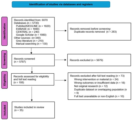
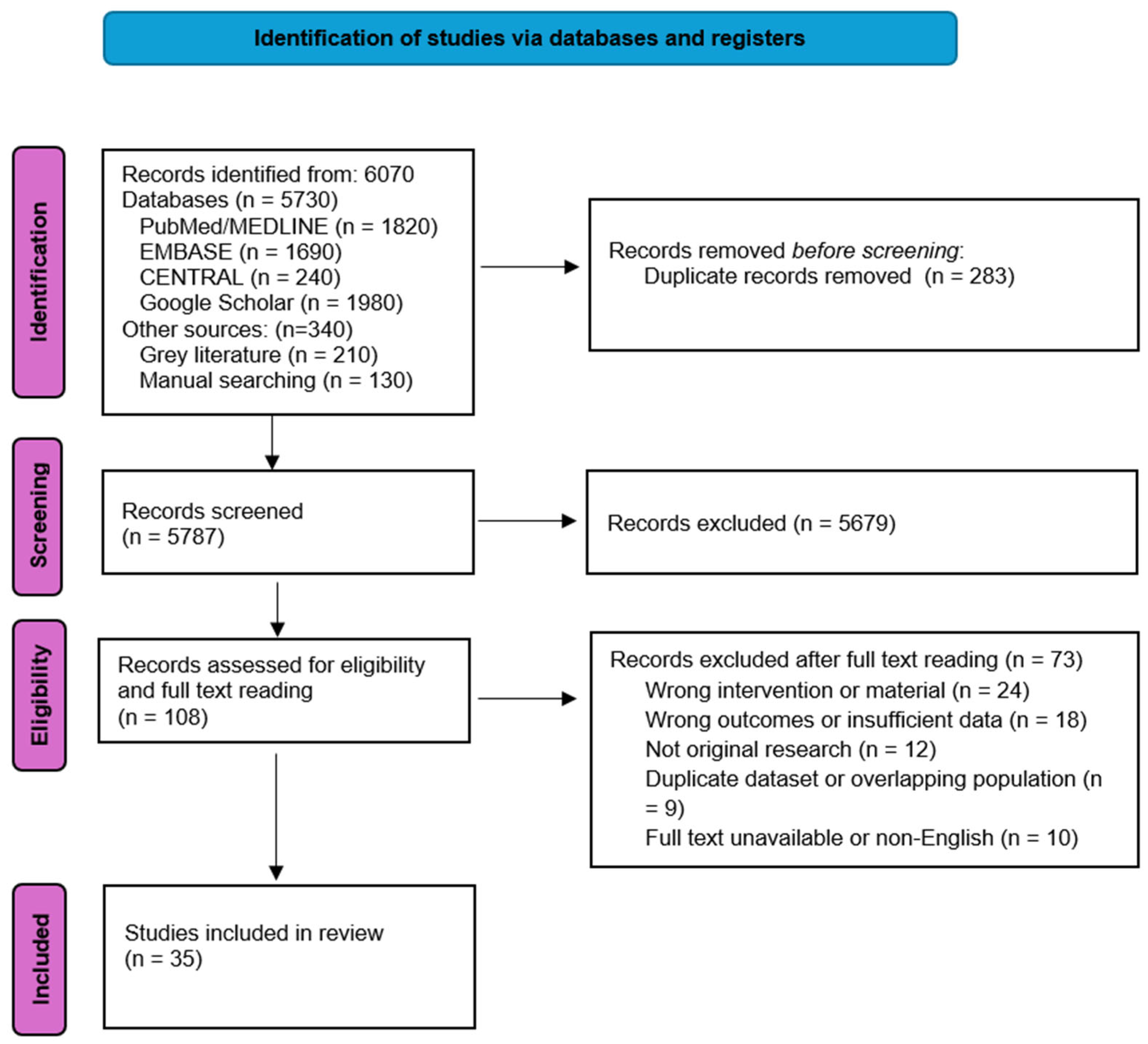
After the initial electronic and manual search, 6070 studies were found. After removing 283 duplicate studies, 5787 studies were included in the initial screening. After title and abstract screening, 5679 studies were excluded as they did not meet the inclusion criteria. A total of 108 studies were included in the full-text review, while 73 studies were excluded. A total of 35 studies were included in the review. The included evidence comprised 25 in vitro studies, 3 animal studies, and 7 human clinical studies . Two reviewers (E.Y.L. and A.A.) independently assessed the full texts to determine eligibility (inclusion/exclusion), and disagreements were resolved by discussion and consensus. The PRISMA flow diagram visually summarizes the inclusion process (Figure 1).
Figure 1.
Study selection flow diagram (PRISMA-style) for records identified, screened, and included.
3.2. Data Extraction and Analysis
Data extraction and qualitative comparisons were completed from the 35 included studies. The summarized results can be found in Table 2 and Table 3.
Table 2.
Physical Properties of Anodized Titanium Abutments.
Table 3.
Esthetic and Biological Outcomes of Anodized Titanium Abutments.
3.3. Physical Properties
3.3.1. Scanning Electron Microscopy (SEM) Imaging
SEM was used to evaluate how anodization changes the titanium surface topography and its effect on the surrounding soft tissue. Multiple studies demonstrated how an increase in voltage during the anodization process produced a more organized nanotube structure, with 80 V showing the highest level of soft tissue integration. Some authors reported micro-cracks and grain formation in the oxide layer, which may be a result of oxygen bubble formation occurring during the anodization process. This may suggest possible changes within the oxide layer itself during the anodization process, not just changes in the surface topography [23].
Increased change in surface topography was reported in studies where roughening techniques were used, whereas minimal change was reported from just the anodization process alone [21,30]. SEM analysis showed increased fibroblast and osteoblast attachment on anodized surfaces compared with smooth machined surfaces, and smaller pore diameters were associated with reduced biofilm formation [22,27,36]. Additionally, biofilm imaging was obtained with SEM and showed that an atomic-layer-deposited coating resisted bacteria due to its smooth and dense surface, whereas porous anodized surfaces did not consistently reduce bacterial attachment [31]. Across studies, SEM was used both to confirm surface morphology/material identity and to visualize cell adhesion and morphology [24,25,26,28,29,31,33].
3.3.2. X-Ray Diffractometry (XRD)
XRD was used by Mendez et al. and Mühl et al. to identify the crystalline phases of the oxide layer after anodization [29,36].
3.3.3. Chemical Elemental Analysis
Chemical elemental analyses generally reported higher oxygen signals after anodization; however, these findings are best interpreted as reflecting growth and thickening of the TiO2 layer within the analytical sampling depth, rather than true oxygen enrichment of the outermost titanium dioxide surface. Such oxide-layer growth contributes to the interference colors observed after anodization [7,21,22,23,24,30,31].
3.3.4. Surface Characteristics
Surface characteristic measurements frequently showed lower contact-angle values after anodization, suggesting increased hydrophilicity under the specific tested conditions and, in some protocols, altered roughness, which may contribute to increased fibroblast attachment [23,25,26,29,36,37].
3.3.5. Water Contact Angle (WCA)
Most of the papers found that anodization lowered the WCA and increased hydrophilicity, which may support early soft tissue healing. However, WCA measurements should be interpreted cautiously because they can be influenced by cleaning and storage conditions, surface roughness, adventitious contamination, liquid type, and the time elapsed after surface preparation. This trend applied up to approximately 60 V, while higher applied voltages resulted in irregular oxide layers with decreased hydrophilicity [7,21,22,23,25,26,28,29,30,33,36,37]. Additional reported physical effects included increased tensile bond strength between lithium disilicate crowns and titanium abutments after anodization, in some studies without substantial change in surface topography [32].
3.4. Esthetic Outcomes
Most studies reported that zirconia abutments displayed the lowest ΔE values and produced superior esthetic outcomes. However, yellow- or pink-anodized titanium abutments often improved esthetic outcomes compared with uncolored titanium, particularly when ≥2 mm of ceramic or soft tissue was present. Overall, anodization tended to reduce soft tissue discoloration relative to unmodified titanium, although zirconia most consistently demonstrated the most favorable color outcomes across conditions [7,8,14,16,21,40]. More opaque, lighter-colored cements also contributed to reduced ΔE values [7,8,14,16,34,38,40,41,42,43,44,45,46,47,49].
3.5. Biological Properties
3.5.1. Soft Tissue Outcomes
Across the in vitro studies the material that consistently displayed the best fibroblast proliferation and attachment was zirconia. However, anodized titanium displayed increased fibroblast alignment, viability, and adhesion compared with machined titanium. Additionally, anodized titanium with nanoporous surfaces displayed improved tissue sealing and soft tissue outcomes in several laboratory models, and reduced bacterial adhesion was reported in some protocols [22,23,24,25,26,27,28,29,30,31,33,36]. In animal and clinical studies, reported differences between anodized and machined titanium were generally small and not consistently significant, and factors such as abutment geometry (e.g., slim versus regular) and soft tissue thickness may have a greater influence on soft tissue outcomes [8,14,16,17,35,37,39,48,49].
3.5.2. Radiographic Analysis
Across most studies, radiographic analysis showed that there was no significant difference between implant stability and peri-implant marginal bone remodeling associated with the anodized titanium abutments [8,14,16,17,27,30,37,39,40,48,49].
3.5.3. Biocompatibility
Across most studies, zirconia was frequently reported as highly biocompatible, and anodized titanium generally performed similarly to conventional titanium with respect to fibroblast adhesion and proliferation, with some protocols reporting reduced bacterial adhesion and improved early soft tissue healing markers. Some studies reported improved mechanics associated with the machined abutments; however, the anodized abutments have consistently shown to be compatible in the esthetic zone with superior esthetics and soft tissue integration [7,8,14,16,17,21,23,27,30,31,33,35,37,39,48,49,50].
3.5.4. Histometric Analysis
Studies utilized cell-based assays and histometric analysis to examine the soft tissue response surrounding anodized abutments. The results consistently showed that there was enhanced connective tissue arrangement and fibroblast activity associated with the anodized titanium abutments. Although one study found there were better early outcomes with slim machined abutments, the overall soft tissue findings associated with anodized abutments were generally favorable [17,23,25,26,30,37,39,48].
4. Discussion
The outcomes of this narrative review demonstrate that anodized titanium abutments affect physical, esthetic, and biological properties via surface chemistry, nano and micro topographic changes, and optical masking. However, the strength of evidence varies by outcome and study design, and long-term clinical data remain limited.
4.1. Physical Properties
Anodization of titanium abutments modifies physicochemical characteristics primarily by increasing the thickness of the surface TiO2 layer and is commonly associated with lower water contact angles and increased hydrophilicity [22,23,26,27,28,29,30,36]. Therefore, higher oxygen signals reported by EDX or related elemental analyses should be interpreted as an apparent increase caused by a thicker oxide layer within the probed analytical depth, rather than as an increase in the oxygen content of the outermost titanium dioxide surface. These effects may be relevant at the transmucosal junction, where protein adsorption and fibroblast behavior contribute to soft tissue integration. Lower contact angles have been correlated with greater fibroblast proliferation, alignment, and focal adhesion formation, which may support more favorable early soft tissue interactions than machined titanium [23,25,26,29]. However, reported hydrophilicity changes should be interpreted in relation to contact-angle testing protocols, because wettability measurements can be affected by surface preparation, storage, contamination, liquid type, and measurement timing. Several studies suggest that anodization alone does not consistently improve fibroblast function unless accompanied by additional micro-roughening [21,23,30]. Voltage effects also appear important, with lower anodization voltages (≤60 V) producing more homogeneous oxide layers and improved hydrophilicity, whereas higher voltages have been associated with micro-cracking and irregular oxide formation [7,23,29]. From a surface property standpoint, voltages at or below 60 V are preferable, as they produce more uniform oxide layers, greater hydrophilicity, and more favorable surface characteristics for soft tissue integration. However, from an esthetic standpoint, higher voltages, particularly in the range of 65 to 75 V, are often necessary to achieve the pink coloration that most closely mimics the peri-implant soft tissue. Clinicians should be aware of this voltage-dependent trade-off when selecting anodization parameters for esthetic zone restorations. Overall, anodization predictably changes wettability and surface chemistry, but these physical changes should be interpreted as contributory rather than determinative of long-term clinical outcomes.
4.2. Esthetic Outcomes
Although zirconia abutments demonstrated the most esthetic outcomes, anodized titanium abutments exhibited notable improvements compared to the untreated titanium in scenarios where adequate soft tissue thickness was provided [7,8,14,38,40,42,43,45,50]. These findings suggest that anodization improves titanium’s esthetic outcomes, but zirconia remains the material most consistently associated with superior color outcomes.
The predominant reason for these improved esthetic outcomes is the presence of a thicker oxide layer, which is created during the anodization process. This oxide layer modifies the L*, a*, and b* values by altering the light wave interference. This process minimizes titanium’s inherent gray color, especially in the anodized yellow and pink spectrum [7,12,21,24,46]. Other reasons why anodized titanium abutments display improved esthetics, specifically in soft tissue masking, are changes in the refractive index and decreased contrast between the mucosa and the abutments.
The thickness of the peri-implant soft tissue also contributes to the esthetic outcomes. Multiple studies have demonstrated that soft tissue with ≥2 mm has been shown to decrease the ΔE values regardless of the abutment material [8,40,44,50]. This emphasizes the significance of soft tissue phenotype in establishing appropriate esthetic outcomes. Additionally, other variables such as cement translucency and ceramic thickness also contribute to esthetic outcomes [34,38,47]. This highlights that esthetics are not only reliant on the abutment material alone but on the compound effects of substrate color, soft tissue characteristics, and restorative design limitations. Accordingly, esthetic outcomes should be interpreted as a combined effect of abutment color, soft tissue thickness, and restorative design.
4.3. Biological Outcomes
Across the reviewed literature, anodized titanium abutments were frequently associated with improved early soft tissue responses, including fibroblast alignment, adhesion, and viability, and in some protocols reduced bacterial adhesion compared with machined titanium [21,22,23,25,26,27,28,30]. These findings have been linked to higher hydrophilicity, altered surface topography, and changes in surface energy after anodization. However, animal and human clinical studies generally reported small, inconsistent, or non-significant differences between anodized and machined abutments for probing depth, inflammation indices, and marginal bone changes [17,37,39,48,49]. The divergence between laboratory and clinical findings likely reflects the multifactorial nature of peri-implant healing, including abutment geometry (regular versus slim), surgical technique, host factors, and soft tissue phenotype.
Similarly, although several in vitro models reported reduced bacterial adhesion on anodized titanium, clinical studies did not show consistent differences in plaque accumulation or inflammatory parameters [16,21,22,27,28,49]. These observations suggest that anodization may influence early biological events under controlled conditions, but its clinical effect on long-term peri-implant health is limited. Accordingly, anodization is best interpreted as an adjunctive surface modification rather than a primary determinant of peri-implant stability.
4.4. Study Limitations
Several limitations should be accounted for when analyzing the findings of this narrative review. First, many of the included studies were in vitro investigations, which may not fully replicate the true biological environment of the peri-implant soft tissue in a clinical study. Second, there was quite a difference with respect to anodization methods, applied voltage, surface preparation, and electrolyte composition amongst the included studies. This may have contributed to variability in the findings. As a result, some findings, especially those based on physicochemical characterization, are descriptive and should be interpreted as mechanistic or surrogate evidence rather than direct proof of clinical superiority. Additionally, there were no standardized outcome measures across studies associated with biological and esthetic outcomes, and long-term clinical studies were limited. The restriction to English-language publications may have introduced language bias. In addition, publication bias cannot be excluded, particularly for laboratory studies where negative findings may be less likely to be published. Because this review used a narrative (qualitative) synthesis, no meta-analysis was performed, and a formal risk-of-bias assessment was not undertaken; therefore, the certainty of evidence for several outcomes cannot be fully quantified. Finally, factors such as soft tissue thickness, abutment geometry, patient variability, and surgical techniques may influence the outcomes independently, regardless of the surface modification. This emphasizes the need for long-term and well-designed clinical studies to determine the effects of anodized titanium abutments.
5. Conclusions
Collectively, anodization is a reproducible surface modification that can improve the optical masking of titanium and may support early soft tissue interactions under selected protocols. Anodized titanium abutments are associated with altered oxide-layer characteristics, wettability, and surface topography, which may contribute to enhanced soft tissue interactions. Esthetically, anodization can improve the gray show-through by altering light interference patterns, with pink and yellow anodized abutments demonstrating lower color differences. However, soft tissue thickness, cement opacity, and material thickness also play a contributing role. Anodized titanium abutments are an improvement over machined titanium abutments, although zirconia abutments have shown to provide the most favorable esthetic outcomes. While in vitro studies suggest that anodization may improve early soft tissue responses, in vivo studies show that there are minimal differences found between machined and anodized abutments in probing depth, inflammation, and bone remodeling. Therefore, anodization should be considered primarily an esthetic adjunct rather than a definitive strategy for improving long-term peri-implant biological outcomes or peri-implant soft tissue stability.
Author Contributions
Conceptualization, W.-S.L.; methodology, E.Y.L., A.A.-F., C.-C.Y.,, M.L.K., J.A.L. and W.-S.L.; investigation, E.Y.L., A.A.-F., C.-C.Y., , M.L.K., J.A.L. and W.-S.L.; writing—original draft preparation, E.Y.L., A.A.-F. and W.-S.L.; writing—review and editing, E.Y.L., A.A.-F., C.-C.Y., , M.L.K., J.A.L. and W.-S.L.; supervision, W.-S.L. All authors have read and agreed to the published version of the manuscript.
Funding
This review received no external funding.
Institutional Review Board Statement
IRB approval was not required for this review.
Informed Consent Statement
Not applicable.
Data Availability Statement
Data supporting the findings of this study are available from the corresponding author upon reasonable request.
Conflicts of Interest
The authors declare no conflicts of interest.
References
- Dede, D.O.; Armaganci, A.; Ceylan, G.; Celik, E.; Cankaya, S.; Yilmaz, B. Influence of implant abutment material on the color of different ceramic crown systems. J. Prosthet. Dent. 2016, 116, 764–769. [Google Scholar] [CrossRef] [Scilit] [PubMed]
- Aslan, M.A.; Ural, C.; Arici, S. Investigation of the effect of titanium alloy surface coating with different techniques on titanium-porcelain bonding. J. Prosthet. Dent. 2016, 115, 115–122. [Google Scholar] [CrossRef] [Scilit] [PubMed]
- Kim, A.; Campbell, S.D.; Viana, M.A.; Knoernschild, K.L. Abutment Material Effect on Peri-implant Soft Tissue Color and Perceived Esthetics. J. Prosthodont. 2016, 25, 634–640. [Google Scholar] [PubMed]
- Ajlouni, K.; Elshahawy, W.; Ajlouni, R.; Sadakah, A. Color masking measurement for ceramic coating of titanium used for dental implants. J. Prosthet. Dent. 2018, 119, 426–431. [Google Scholar] [CrossRef] [Scilit] [PubMed]
- Diamanti, M.V.; Del Curto, B.; Pedeferri, M. Anodic oxidation of titanium: From technical aspects to biomedical applications. J. Appl. Biomater. Biomech. 2011, 9, 55–69. [Google Scholar] [CrossRef] [Scilit] [PubMed]
- Mazzarolo, A.; Curioni, M.; Vicenzo, A.; Skeldon, P.; Thompson, G.E. Anodic growth of titanium oxide: Electrochemical behaviour and morphological evolution. Electrochim. Acta 2012, 75, 288–295. [Google Scholar] [CrossRef] [Scilit]
- Wang, T.; Wang, L.; Lu, Q.; Fan, Z. Changes in the esthetic, physical, and biological properties of a titanium alloy abutment treated by anodic oxidation. J. Prosthet. Dent. 2019, 121, 156–165. [Google Scholar] [CrossRef] [Scilit] [PubMed]
- Martínez-Rus, F.; Prieto, M.; Salido, M.P.; Madrigal, C.; Özcan, M.; Pradíes, G. A Clinical Study Assessing the Influence of Anodized Titanium and Zirconium Dioxide Abutments and Peri-implant Soft Tissue Thickness on the Optical Outcome of Implant-Supported Lithium Disilicate Single Crowns. Int. J. Oral Maxillofac. Implant. 2017, 32, 156–163. [Google Scholar] [CrossRef] [Scilit]
- Napoli, G. Colouring titanium alloys by anodic oxidation. Metalurgija 2018, 57, 111–113. [Google Scholar]
- Roberge, P.R. Handbook of Corrosion Engineering, 3rd ed.; McGraw-Hill Education: New York, NY, USA, 2019. [Google Scholar]
- Paschalis, E.I.; Chodosh, J.; Spurr-Michaud, S.; Cruzat, A.; Tauber, A.; Behlau, I.; Gipson, I.; Dohlman, C.H. In vitro and in vivo assessment of titanium surface modification for coloring the backplate of the Boston keratoprosthesis. Investig. Ophthalmol. Vis. Sci. 2013, 54, 3863–3873. [Google Scholar] [CrossRef] [Scilit]
- Wadhwani, C.; Brindis, M.; Kattadiyil, M.T.; O’Brien, R.; Chung, K.H. Colorizing titanium-6aluminum-4vanadium alloy using electrochemical anodization: Developing a color chart. J. Prosthet. Dent. 2018, 119, 26–28. [Google Scholar] [CrossRef] [Scilit] [PubMed]
- Wadhwani, C.P.K.; Schoenbaum, T.; King, K.E.; Chung, K.H. Techniques to Optimize Color Esthetics, Bonding, and Peri-implant Tissue Health With Titanium Implant Abutments. Compend. Contin. Educ. Dent. 2018, 39, 110–119. [Google Scholar] [PubMed]
- Gil, M.S.; Ishikawa-Nagai, S.; Elani, H.W.; Da Silva, J.D.; Kim, D.M.; Tarnow, D.; Schulze-Späte, U.; Bittner, N. A prospective clinical trial to assess the optical efficacy of pink neck implants and pink abutments on soft tissue esthetics. J. Esthet. Restor. Dent. 2017, 29, 409–415. [Google Scholar] [CrossRef] [Scilit] [PubMed]
- Huang, J.W.; Chen, W.C.; Huang, T.K.; Fu, P.S.; Lai, P.L.; Tsai, C.F.; Hung, C.C. Using a spectrophotometric study of human gingival colour distribution to develop a shade guide. J. Dent. 2011, 39, e11–e16. [Google Scholar] [CrossRef] [Scilit] [PubMed]
- Hall, J.; Neilands, J.; Davies, J.R.; Ekestubbe, A.; Friberg, B. A randomized, controlled, clinical study on a new titanium oxide abutment surface for improved healing and soft tissue health. Clin. Implant Dent. Relat. Res. 2019, 21, 55–68. [Google Scholar] [CrossRef] [Scilit] [PubMed]
- Susin, C.; Finger Stadler, A.; Fiorini, T.; de Sousa Rabelo, M.; Ramos, U.D.; Schüpbach, P. Safety and efficacy of a novel anodized abutment on soft tissue healing in Yucatan mini-pigs. Clin. Implant Dent. Relat. Res. 2019, 21, 34–43. [Google Scholar] [CrossRef] [Scilit] [PubMed]
- Yetim, A. Investigation of wear behavior of titanium oxide films, produced by anodic oxidation, on commercially pure titanium in vacuum conditions. Surf. Coat. Technol. 2010, 205, 1757–1763. [Google Scholar] [CrossRef] [Scilit]
- Dong, H.; Bell, T. Enhanced wear resistance of titanium surfaces by a new thermal oxidation treatment. Wear 2000, 238, 131–137. [Google Scholar] [CrossRef] [Scilit]
- Kahar, M.S.; Macwan, A.; Oza, M.R.; Oza, V.; Shah, S. Characterization and Corrosion Study of Titanium Anodized Film Developed in KOH Bath. J. Eng. Res. Appl. 2013, 3, 441–445. [Google Scholar]
- Genova, T.; Chinigò, G.; Munaron, L.; Rivolo, P.; Luganini, A.; Gribaudo, G.; Cavagnetto, D.; Mandracci, P.; Mussano, F. Bacterial and Cellular Response to Yellow-Shaded Surface Modifications for Dental Implant Abutments. Biomolecules 2022, 12, 1718. [Google Scholar] [CrossRef] [Scilit] [PubMed]
- Golalipour, S.; Jalalian, E.; Koosha, S.; Khorshidi, S.; Torshabi, M.; Sayyari, M. In vitro effect of anodization on surface roughness and bacterial adhesion to titanium abutments. J. Prosthet. Dent. 2025, 133, 291.e1–291.e8. [Google Scholar] [CrossRef] [Scilit] [PubMed]
- Gulati, K.; Moon, H.J.; Kumar, P.T.S.; Han, P.; Ivanovski, S. Anodized anisotropic titanium surfaces for enhanced guidance of gingival fibroblasts. Mater. Sci. Eng. C Mater. Biol. Appl. 2020, 112, 110860. [Google Scholar] [CrossRef] [Scilit] [PubMed]
- Hulka, I.; Florido-Suarez, N.R.; Mirza-Rosca, J.C.; Saceleanu, A. Ti–Ta dental alloys and a way to improve gingival aesthetic in contact with the implant. Mater. Chem. Phys. 2022, 287, 126343. [Google Scholar] [CrossRef] [Scilit]
- Kim, Y.S.; Ko, Y.; Kye, S.B.; Yang, S.M. Human gingival fibroblast (HGF-1) attachment and proliferation on several abutment materials with various colors. Int. J. Oral Maxillofac. Implant. 2014, 29, 969–975. [Google Scholar] [CrossRef] [Scilit]
- Kim, Y.S.; Shin, S.Y.; Moon, S.K.; Yang, S.M. Surface properties correlated with the human gingival fibroblasts attachment on various materials for implant abutments: A multiple regression analysis. Acta Odontol. Scand. 2015, 73, 38–47. [Google Scholar] [PubMed]
- Lima, J.H.C.; Robbs, P.C.M.; Tude, E.M.O.; De Aza, P.N.; Costa, E.M.D.; Scarano, A.; Prados-Frutos, J.C.; Fernandes, G.V.O.; Gehrke, S.A. Fibroblasts and osteoblasts behavior after contact with different titanium surfaces used as implant abutment: An in vitro experimental study. Heliyon 2024, 10, e25038. [Google Scholar] [CrossRef] [Scilit] [PubMed]
- Liu, W.; Gregory, R.L.; Yang, C.C.; Hamada, Y.; Lin, W.S. The effects of anodization and instrumentation on titanium abutment surface characteristics and biofilm formation. J. Prosthodont. 2025. [Google Scholar] [CrossRef] [Scilit] [PubMed]
- Mühl, A.; Szabó, P.; Krafcsik, O.; Aigner, Z.; Kopniczky, J.; Ákos Nagy Marada, G.; Turzó, K. Comparison of surface aspects of turned and anodized titanium dental implant, or abutment material for an optimal soft tissue integration. Heliyon 2022, 8, e10263. [Google Scholar] [CrossRef] [Scilit] [PubMed]
- Mussano, F.; Genova, T.; Laurenti, M.; Zicola, E.; Munaron, L.; Rivolo, P.; Mandracci, P.; Carossa, S. Early Response of Fibroblasts and Epithelial Cells to Pink-Shaded Anodized Dental Implant Abutments: An In Vitro Study. Int. J. Oral Maxillofac. Implant. 2018, 33, 571–579. [Google Scholar] [CrossRef] [Scilit]
- Pan, Y.; Cao, L.; Chen, L.; Gao, L.; Wei, X.; Lin, H.; Jiang, L.; Wang, Y.; Cheng, H. Enhanced Bacterial and Biofilm Adhesion Resistance of ALD Nano-TiO2 Coatings Compared to AO Coatings on Titanium Abutments. Int. J. Nanomed. 2024, 19, 11143–11159. [Google Scholar] [CrossRef] [Scilit]
- Pour, H.K.; Ansari, H.; Tehrani, A.M. In vitro effect of anodization of titanium abutments on their tensile bond strength to implant-supported lithium disilicate all-ceramic crowns. Dent. Res. J. 2023, 20, 99. [Google Scholar] [CrossRef] [Scilit]
- Selamet, S.M.; Kumbuloglu, O.; Sahiner, A.; Ozdemir, G. Assessment of surface roughness and microbiological retention on custom abutments produced by different techniques and materials. Eur. J. Oral Sci. 2025, 133, e13038. [Google Scholar] [CrossRef] [Scilit] [PubMed]
- Sirawuttipong, C.; Palanuwech, M. Effect of Zirconia Thickness, Cement Color, and Titanium Implant Abutment Surface Treatment Type on the Esthetic Outcomes of High-Translucency Monolithic Zirconia. Int. J. Oral Maxillofac. Implant. 2024, 39, 765–775. [Google Scholar]
- Squier, R.S.; Psoter, W.J.; Taylor, T.D. Removal torques of conical, tapered implant abutments: The effects of anodization and reduction of surface area. Int. J. Oral Maxillofac. Implant. 2002, 17, 24–27. [Google Scholar]
- Traver-Méndez, V.; Camps-Font, O.; Ventura, F.; Nicolau-Sansó, M.A.; Subirà-Pifarré, C.; Figueiredo, R.; Valmaseda-Castellón, E. In Vitro Characterization of an Anodized Surface of a Dental Implant Collar and Dental Abutment on Peri-Implant Cellular Response. Materials 2023, 16, 6012. [Google Scholar] [CrossRef] [Scilit] [PubMed]
- Wang, C.; Wang, X.; Lu, R.; Cao, X.; Yuan, D.; Chen, S. Influence of surface nanotopography and wettability on early phases of peri-implant soft tissue healing: An in-vivo study in dogs. BMC Oral Health 2023, 23, 651. [Google Scholar] [CrossRef] [Scilit] [PubMed]
- Bas, B.B.; Cakan, U. Evaluation of the effect of anodization-colored titanium abutments and zirconia substructure thickness on zirconia substructure color: An In vitro study. Niger. J. Clin. Pract. 2022, 25, 2024–2029. [Google Scholar] [CrossRef] [Scilit] [PubMed]
- Dib-Zaitum, I.; Guadilla-González, Y.; Flores-Fraile, J.; Dib-Zakkour, J.; Benito-Garzón, L.; Montero, J. Effect Morphology and Surface Treatment of the Abutments of Dental Implants on the Dimension and Health of Peri-Implant Biological Space. Materials 2022, 15, 4422. [Google Scholar] [CrossRef] [Scilit] [PubMed]
- Farrag, K.M.; Khamis, M.M. Effect of anodized titanium abutment collars on peri-implant soft tissue: A split-mouth clinical study. J. Prosthet. Dent. 2023, 130, 59–67. [Google Scholar] [CrossRef] [Scilit] [PubMed]
- Farrag, K.; Bakry, S.; Aly, Y. Effect of anodic oxidation of titanium substrate on the shade of lithium disilicate ceramic with different thicknesses (in vitro study). Alex. Dent. J. 2022, 47, 125–131. [Google Scholar]
- Khorshidi, S.; Zarbakhsh, A.; Lawaf, S.; Golalipour, S.; Sayyari, M.; Nahavandi, A.M. In Vitro Effect of Anodization of Titanium Abutments on Color Parameters and Color Difference of Lithium Disilicate All-Ceramic Crowns. Clin. Exp. Dent. Res. 2024, 10, e70002. [Google Scholar] [CrossRef] [Scilit] [PubMed]
- Kilinc, H.; Sanal, F.A. Influence of Abutment Material, Cement Thickness, and Crown Type on the Final Color of Implant-Supported Restorations. Int. J. Prosthodont. 2024, 37, 211–222. [Google Scholar] [CrossRef] [Scilit] [PubMed]
- Şen, N.; Şermet, I.B. Influence of Gingival Color and Abutment Material on the Final Color of Peri-Implant Soft Tissue: An In Vitro Analysis. J. Oral Implantol. 2025, 51, 111–116. [Google Scholar] [CrossRef] [Scilit] [PubMed]
- Senkiw, K. Shade outcome of 3Y, 4Y, 5Y monolithic zirconia on colored anodized titanium. Dent. Rev. 2024, 4, 100133. [Google Scholar] [CrossRef] [Scilit]
- Seyidaliyeva, A.; Rues, S.; Evagorou, Z.; Hassel, A.J.; Büsch, C.; Rammelsberg, P.; Zenthöfer, A. Predictability and outcome of titanium color after different surface modifications and anodic oxidation. Dent. Mater. J. 2022, 41, 930–936. [Google Scholar] [CrossRef] [Scilit] [PubMed]
- Seyidaliyeva, A.; Zenthöfer, A.; Rues, S. Evaluation of the Color of Zirconia in Different Substrates of Osseointegrated Implants, Thickness of Materials and Types of Resin Cements. Int. J. Dent. 2024, 2024, 8696008. [Google Scholar] [CrossRef] [Scilit] [PubMed]
- Susin, C.; Finger Stadler, A.; Musskopf, M.L.; de Sousa Rabelo, M.; Ramos, U.D.; Fiorini, T. Safety and efficacy of a novel, gradually anodized dental implant surface: A study in Yucatan mini pigs. Clin. Implant Dent. Relat. Res. 2019, 21, 44–54. [Google Scholar] [CrossRef] [Scilit] [PubMed]
- Vazouras, K.; Gholami, H.; Margvelashvili-Malament, M.; Kim, Y.J.; Finkelman, M.; Weber, H.P. An Esthetic Evaluation of Different Abutment Materials in the Anterior Maxilla: A Randomized Controlled Clinical Trial Using a Crossover Design. J. Prosthodont. 2022, 31, 673–680. [Google Scholar] [CrossRef] [Scilit] [PubMed]
- Wang, T.; Wang, L.; Lu, Q.; Fan, Z. Influence of anodized titanium abutments on the esthetics of the peri-implant soft tissue: A clinical study. J. Prosthet. Dent. 2021, 125, 445–452. [Google Scholar] [CrossRef] [Scilit] [PubMed]

Disclaimer/Publisher’s Note: The statements, opinions and data contained in all publications are solely those of the individual author(s) and contributor(s) and not of MDPI and/or the editor(s). MDPI and/or the editor(s) disclaim responsibility for any injury to people or property resulting from any ideas, methods, instructions or products referred to in the content. |
© 2026 by the authors. Licensee MDPI, Basel, Switzerland. This article is an open access article distributed under the terms and conditions of the Creative Commons Attribution (CC BY) license.