


Abstract
Background/Objectives: Alveolar ridge preservation (ARP) is commonly performed following tooth extraction to minimize post-extraction dimensional changes. However, limited information is available regarding root-volume variations among different tooth types. This study aimed to assess the root volumes of maxillary and mandibular teeth using cone-beam computed tomography (CBCT) and to compare the measured root volumes with commonly available graft-material package volumes in the context of ARP. Methods: CBCT scans from 53 patients meeting the eligibility criteria were retrospectively evaluated. Fourteen teeth per scan were assessed using a split-mouth approach, resulting in a total of 742 teeth. Root boundaries were identified by two trained and calibrated dentists, and root volumes were calculated using OsiriX MD v13.0.3 software. Statistical comparisons were performed using paired and independent t-tests. Results: Root volumes varied according to tooth type and arch. The maxillary first molar demonstrated the highest mean root volume (0.55 ± 0.10 cm3), whereas the mandibular central incisor demonstrated the lowest (0.11 ± 0.02 cm3). Maxillary teeth showed significantly greater root volumes than their mandibular counterparts. Gender-related differences were limited to the maxillary second premolar and mandibular second molar and were considered of limited clinical relevance. For illustrative comparison, the measured root volumes were considered in relation to commonly available graft-material package volumes, including 0.25 and 0.50 cc; these comparisons do not represent clinically validated graft requirements. Conclusions: This study provides CBCT-derived reference root-volume values for different tooth types and demonstrates variations according to tooth type and arch. Comparison of these measurements with commonly available graft-material package volumes may provide a descriptive volumetric reference in the context of ARP. However, root volume does not directly represent post-extraction socket volume or the quantity of graft material required clinically. Therefore, these comparisons should not be interpreted as recommendations for graft quantity, and prospective clinical studies comparing root volume with actual socket volume and graft utilization are required before clinical application.
1. Introduction
The integrity of alveolar bone and soft tissues is essential to achieve satisfactory outcomes when replacing missing teeth with dental implants [1]. It is well documented that the alveolar ridge undergoes local alterations and dimensional changes as part of the physiological remodeling process post tooth extraction [2]. The most commonly encountered post-extraction bone loss occurs in the buccal plate in a horizontal dimension [3]. Wah Le Tan et al. revealed in a systematic review that the mean magnitudes of horizontal and vertical bone collapse six months following extraction were 3.79 ± 0.23 mm (29–63%) and 1.24 ± 0.11 mm (11–22%), respectively. Further dimensional changes were reported with mean loss of 0.84 ± 0.62 mm mesially and 0.80 ± 0.71 mm distally in vertical dimension [4]. It was recommended that a combination of minimally traumatic extraction and delivering bone graft material in the extraction site will result in less dimensional changes [5].
Alveolar ridge preservation (ARP) is a procedure that involves the use of bone graft materials with or without using a barrier membrane, aiming to minimize alveolar ridge remodeling and facilitate the future placement of a dental implant [5]. The cost-benefit ratio of ARP outweighs that of guided bone regeneration of the alveolar ridge and decreases the potential need for a sinus augmentation procedure [6]. When comparing ARP with spontaneous socket healing, ARP gives superior and more favorable outcomes in terms of horizontal and vertical bone dimensions as well as volumetric soft tissue changes [7]. Furthermore, if the ARP procedure was done prior to dental implant placement, the need for additional bone grafting material at the time of implant fixture insertion is reduced [7].
Different bone substitutes are used for alveolar ridge preservation, including allografts, xenografts of bovine or porcine origin, and synthetic (alloplastic) materials. Although these materials can all be used for ARP, they differ in their composition, structure, degradation, and resorption characteristics and therefore should not be considered completely interchangeable [8]. Particle size may also influence graft behavior; particulate grafts are available in different granule sizes, with small particles commonly ranging from approximately 0.25–1.0 mm and larger particles from 1 to 2 mm [9]. Smaller particles provide greater surface area, whereas larger particles may provide greater interparticle spaces and exhibit different remodeling characteristics. However, particle-size classifications vary among materials and manufacturers, and there are currently no universally established indications for small, medium, or large particles specifically for ARP. Therefore, graft type and particle size should be selected according to the characteristics of the biomaterial, socket morphology, and the intended clinical approach [8,9].
Despite the widespread use of alveolar ridge preservation (ARP), scientific evidence remains limited regarding the approximate volume of bone graft material required for individual extraction sites. Variations in the size, shape, and morphology of tooth roots may influence the volume of the resulting extraction socket and, consequently, the quantity of graft material required. A previous study assessed root volume using a water displacement technique and highlighted the potential importance of predetermining the amount of bone graft material required for ARP procedures [10].
Cone-beam computed tomography (CBCT) has been widely used for three-dimensional assessment of alveolar and dental structures and provides valuable information for implant treatment planning [11]. Three-dimensional imaging also enables volumetric assessment of intraoral structures with reproducible quantitative measurements [12]. Furthermore, volumetric assessment using three-dimensional imaging has previously been applied to estimate the amount of bone graft required for maxillary sinus augmentation, demonstrating the potential clinical utility of imaging-based volumetric measurements for graft planning [13].
However, to the authors’ knowledge, no previous study has utilized CBCT-derived root volume measurements specifically to estimate the approximate quantity of bone graft material required for ARP. Addressing this gap may provide clinicians with a practical reference for anticipating graft requirements before the procedure, potentially facilitating treatment planning and reducing unnecessary material waste. Therefore, the aim of this study was to assess the volume of human tooth roots using semi-automated segmentation on CBCT and to explore its potential utility in estimating the approximate amount of bone graft material required for ARP. The null hypothesis was that there would be no significant differences in root volume between corresponding maxillary and mandibular teeth, between first and second premolars and molars, or between male and female participants.
2. Materials and Methods
2.1. Ethics Declaration
This study was conducted using previously acquired cone-beam computed tomography (CBCT) data. The study protocol was reviewed and approved by the Imam Abdulrahman bin Faisal University Institutional Review Board, approval number IRB 2025–02–0277. In accordance with institutional and national ethical standards and in compliance with the Declaration of Helsinki (2013 revision). All data were handled in accordance with the ethical standards outlined in the International Ethical Guidelines for Health-related Research Involving Humans (Council for International Organizations of Medical Sciences–CIOMS, 2016) and local data protection regulations. This study received no funding.
2.2. CBCT Selection
All radiographs screened were taken between 20 April 2024 and 20 May 2025, using a CBCT machine (Carestream CS 9300, Carestream Health Inc., Kodak, Rochester, NY, USA). The CBCT settings were 70 kVp, 10 mA, and a 14.3-s scan time according to the manufacturer and vendor recommendations, with a large field of view of 16.5 cm in height by 13 cm in width.
The eligibility criteria for CBCT selection were as follows:
Inclusion criteria:
- Fully dentate patients; Fully dentate patients were selected to maintain a standardized study population and to minimize potential anatomical variability associated with previous tooth loss.
- Teeth with normal root morphology;
- Healthy periodontal condition; and
- CBCT scans of sufficient diagnostic quality to permit clear identification and segmentation of the root boundaries.
Exclusion criteria:
- History of systemic diseases or medications that may influence bone metabolism, including bisphosphonates and RANKL inhibitors;
- Periodontal disease;
- Active orthodontic treatment;
- Mixed dentition;
- Periapical or odontogenic lesions affecting the evaluated teeth;
- Abnormal or altered root morphology;
- Teeth with full-coverage restorations;
- Root canal-treated teeth; and
- CBCT scans containing streak artifacts that could interfere with accurate root segmentation.
An a priori sample size calculation was not performed due to the retrospective nature of the study. All available CBCT scans acquired during the predefined study period were screened for eligibility. A total of 356 scans were assessed, of which 53 fulfilled the predefined eligibility criteria and were included in the analysis. Further data extraction for the 53 subjects included age, gender, and medical history.
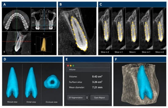
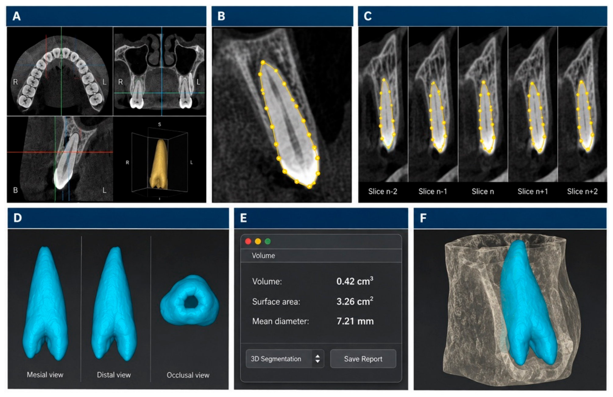
All CBCT scans were analyzed to calculate root volumes. For each patient, 14 teeth were evaluated, representing seven tooth positions from the central incisor through the second molar in each of the maxillary and mandibular arches. For each tooth position, either the right- or left-side tooth was randomly selected for assessment; consequently, the 14 evaluated teeth could include a combination of right- and left-side teeth within the same patient. The cementoenamel junction (CEJ) was used as the coronal anatomical boundary for root segmentation. The selected teeth were segmented using OsiriX MD software (version 14.1.0; Pixmeo SARL, Bernex, Switzerland) to generate a three-dimensional image of the root separated from its surrounding periodontal structures. Root boundaries were identified by placing reference points along the root outline on every other slice, using the axial plane for single-rooted teeth and the sagittal plane for multi-rooted teeth. Following completion of the segmentation, OsiriX MD generated a three-dimensional reconstruction of the segmented root and automatically calculated its volume (Figure 1).
Figure 1.
Representative workflow for root-volume measurement using cone-beam computed tomography (CBCT) and OsiriX MD software. (A) Multiplanar CBCT views (axial, coronal, and sagittal) used for tooth identification and anatomical assessment. (B) Manual delineation of the tooth boundaries using reference points (yellow dots) on a selected CBCT slice. (C) Sequential CBCT slices illustrating the segmentation process, with yellow dots and lines indicating the manually defined boundaries. (D) Three-dimensional reconstruction of the segmented structure (blue), displayed from mesial, distal, and occlusal perspectives. (E) Automatic calculation of the segmented volume and associated geometric parameters using OsiriX MD software. (F) Three-dimensional visualization of the segmented structure (blue) within the surrounding anatomical structure (translucent gray). The yellow dots and lines indicate segmentation reference points and boundaries, respectively, while blue represents the reconstructed three-dimensional model.
As this was a retrospective study using previously acquired CBCT scans, information regarding voxel size and specific patient positioning was not consistently retrievable from the available imaging records and therefore could not be reliably reported. Only CBCT scans of sufficient diagnostic quality to permit clear identification and segmentation of the root boundaries were included. Root volume was used as a volumetric approximation of the space occupied by the extracted root within an intact alveolar socket. The measured root volumes for the different tooth types were subsequently considered in relation to commonly available graft-material quantities (0.25, 0.5, 1.0, and 2.0 cc) to provide approximate estimates of the graft quantity required for alveolar ridge preservation. No specific graft-material brand or particle size was standardized, as graft materials were not clinically placed or evaluated in the present study. As no clinical extractions or alveolar ridge preservation procedures were performed as part of this retrospective study, these estimates were not directly compared with actual post-extraction socket volumes or the quantities of graft material used clinically. Therefore, the estimates were intended as a practical preliminary guide rather than an exact clinical requirement, as the amount of graft material required may vary according to individual socket anatomy and local clinical conditions.
Root-volume measurements were performed independently by two trained and calibrated dentists according to the standardized segmentation protocol described above. Each examiner measured the root volumes of 742 teeth from 53 CBCT scans. Inter-examiner reliability was assessed using the intraclass correlation coefficient (ICC), which demonstrated excellent agreement between the two examiners (ICC = 0.957; 95% CI: 0.903–0.976; p < 0.001). Following establishment of inter-examiner reliability, measurements obtained by one examiner were used for the final statistical analyses. The mean root volume and standard deviation were calculated for each tooth type.
2.3. Statistical Analysis
Continuous variables were summarized as means and standard deviations. Prior to inferential analysis, the assumptions underlying parametric testing were assessed, including evaluation of the data distribution for normality. Paired-samples t-tests were used to assess differences in root volume between corresponding maxillary and mandibular teeth, as well as between first and second molars and between first and second premolars within the same arch. Independent-samples t-tests were used to assess differences in root volume according to gender. All statistical tests were two-tailed, and statistical significance was set at p < 0.05. Given the exploratory nature of the study, no formal adjustment for multiple comparisons was applied; therefore, findings from multiple comparisons were interpreted cautiously.
Potential confounding was primarily addressed through the predefined eligibility criteria, which excluded clinical and radiographic conditions that could potentially influence root morphology or volumetric assessment. Gender was additionally evaluated as a potential source of variation. Given the retrospective and exploratory nature of the study and the limited sample size, multivariable adjustment for additional potential confounders was not performed; therefore, the possibility of residual confounding cannot be excluded. All statistical analyses were performed using SPSS version 25 (IBM Corp., Armonk, NY, USA).
3. Results
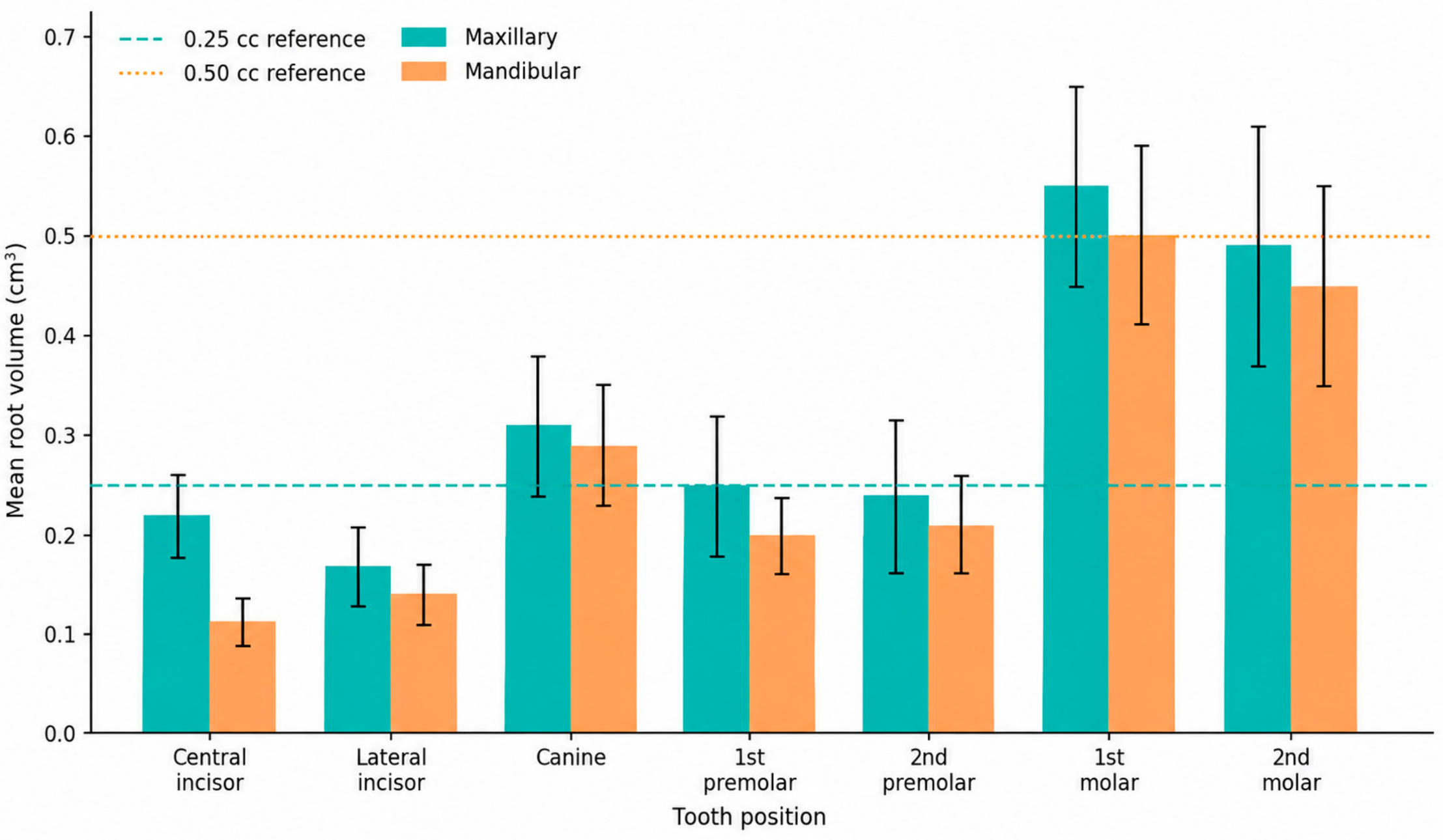
The study included a total of 53 patient records, of which 31 were males and 22 were females. Table 1 presents the mean root volumes of maxillary and mandibular teeth. Among anterior teeth, mean root volumes ranged from 0.11 to 0.22 cm3 for incisors and from 0.29 to 0.31 cm3 for canines, while posterior teeth ranged from 0.20 to 0.25 cm3 for premolars and from 0.45 to 0.55 cm3 for molars. When considered in relation to commonly available graft-material quantities, the mean volumes of incisors and premolars generally corresponded to approximately 0.25 cc, whereas molars corresponded most closely to approximately 0.5 cc. Canine mean volumes (0.29–0.31 cm3) were slightly greater than 0.25 cc but remained below 0.5 cc. These correlations represent volumetric approximations based on root volume and should not be interpreted as clinically validated graft requirements.
Table 1.
Mean root volumes of maxillary and mandibular teeth (cm3) ± standard deviation.
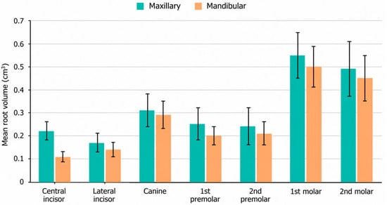
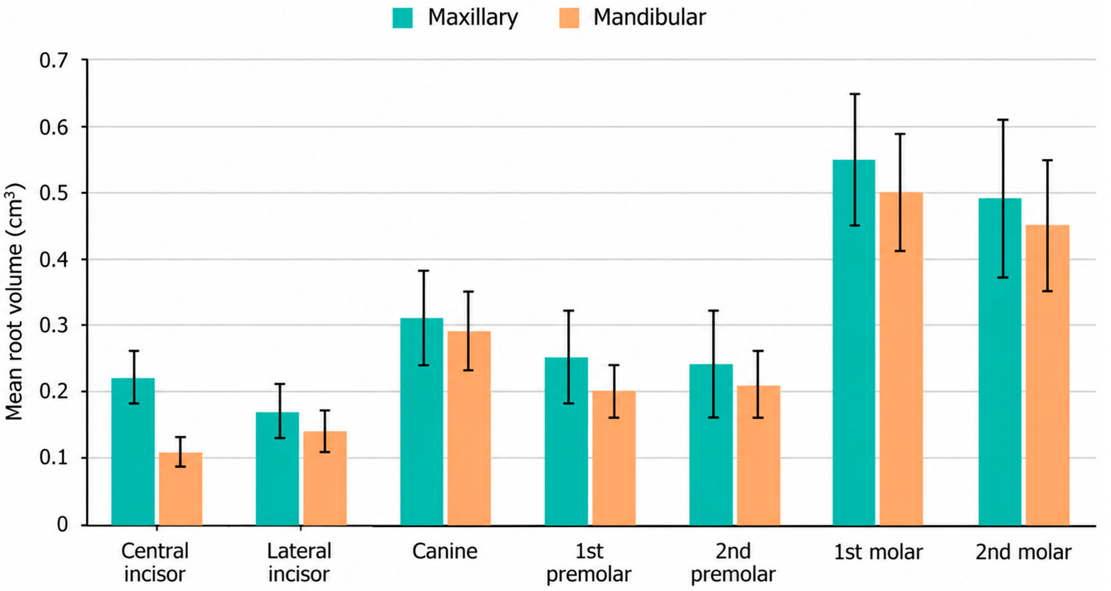
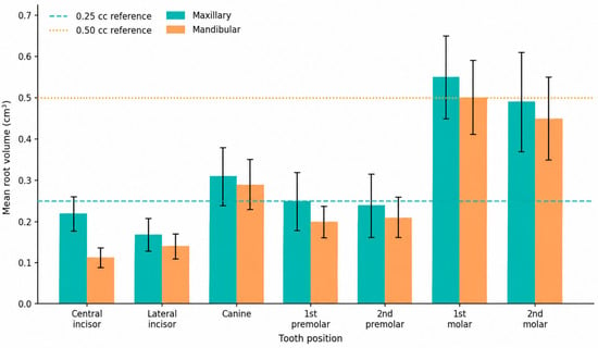
Comparison of mean root volumes between corresponding maxillary and mandibular teeth using paired-samples t-tests demonstrated significantly greater root volumes for all maxillary teeth (Figure 2). The differences were statistically significant for the central incisor (MD = 0.11 cm3, 95% CI: 0.10–0.12, p < 0.001), lateral incisor (MD = 0.03 cm3, 95% CI: 0.02–0.04, p < 0.001), canine (MD = 0.02 cm3, 95% CI: 0.01–0.03, p = 0.004), first premolar (MD = 0.05 cm3, 95% CI: 0.04–0.06, p < 0.001), second premolar (MD = 0.03 cm3, 95% CI: 0.01–0.04, p = 0.001), first molar (MD = 0.05 cm3, 95% CI: 0.03–0.07, p < 0.001), and second molar (MD = 0.04 cm3, 95% CI: 0.02–0.06, p < 0.001) (Table 2 and Figure 3).
Figure 2.
Comparison of mean root volumes (cm3) between maxillary and mandibular teeth across different tooth types. Error bars represent variability in root volume measurements.
Table 2.
Comparison of mean root volumes between maxillary and mandibular teeth using a paired t-test.
Figure 3.
Mean root volumes of maxillary and mandibular teeth according to tooth position. Error bars represent standard deviations. Horizontal reference lines at 0.25 cc and 0.50 cc indicate commonly available graft-material quantities for volumetric comparison. These reference values do not represent clinically validated graft requirements.
No significant difference was observed between the maxillary first and second premolars (MD = −0.01 cm3, 95% CI: −0.02–0.01, p = 0.379). In contrast, the mandibular second premolar demonstrated a significantly greater root volume than the first premolar (MD = 0.02 cm3, 95% CI: 0.01–0.03, p < 0.001). First molars demonstrated significantly greater root volumes than second molars in both arches (mean difference = 0.06 cm3, p < 0.001) (Table S1).
Independent-samples t-tests demonstrated significantly greater root volumes in males only for the maxillary second premolar (MD = 0.04 cm3, p = 0.037) and mandibular second molar (MD = 0.06 cm3, p = 0.031); no statistically significant gender differences were observed for the remaining teeth (Table S2). Despite these statistically significant findings, the magnitude of the observed mean differences was small and considered to have limited clinical significance.
4. Discussion
The present study aimed to measure root volumes of human teeth to help clinicians in predicting bone graft volume needed when preparing for alveolar ridge preservation. The term “Socket Preservation” is often used to describe this procedure throughout the literature. For the sake of standardization, we refer to the same procedure as “Ridge preservation”, as the aim is to preserve the Alveolar bone ridge and not to preserve a socket. Knowing root volumes may aid clinicians in preparing the approximate amount of bone graft material, thus minimizing waste and reducing potential cost, especially when planning for multiple teeth extractions and ridge preservations. Maxillary teeth scored significantly greater volumes than anticipated because of their bi-rooted and tri-rooted nature in premolars and molars, respectively, when compared with single-rooted and bi-rooted mandibular posterior teeth.
Various biomaterials are available for alveolar ridge preservation, including autogenous grafts, allografts of human origin, xenografts of animal origin such as bovine and porcine materials, and synthetic/alloplastic substitutes. These materials differ in composition, microstructure, resorption behavior, and patterns of new bone formation, and are available in different particle sizes and package volumes [8,14]. Particle-size classifications are not universally standardized and vary among manufacturers; therefore, materials of different origins and particle sizes should not be considered directly interchangeable. In clinical practice, particulate graft materials are supplied in different volumes, commonly including 0.25, 0.5, 1.0, and 2.0 cc. In the present study, the measured root volumes were related to these commonly available quantities, with approximately 0.25 cc corresponding to the mean volumes of incisors and premolars and approximately 0.5 cc more closely corresponding to canines and molars. CBCT-based segmentation using OsiriX MD may therefore provide a useful three-dimensional approach for preoperative estimation of root volume and preparation of an approximate graft quantity, particularly when multiple extraction sites are planned, potentially reducing unnecessary material preparation and waste. However, graft requirements are also influenced by socket morphology, socket-wall integrity, graft characteristics, particle size, and packing density; therefore, these estimates should be considered preliminary and require prospective clinical validation against actual post-extraction socket volumes and graft quantities used.
From a practical and patient-care perspective, preoperative estimation of root volume may assist clinicians in anticipating and preparing an appropriate quantity of commercially available graft material before alveolar ridge preservation, potentially reducing unnecessary material preparation and waste. More informed estimation of graft requirements may also support procedural planning and clearer preoperative discussions with patients regarding treatment requirements and anticipated costs, particularly when multiple extraction sites are planned. However, the present study did not directly quantify graft-material waste, treatment costs, procedural efficiency, patient-reported outcomes, or clinical outcomes following ridge preservation. Therefore, these potential practical and patient-centered benefits should be considered preliminary and require validation in prospective clinical studies incorporating actual graft utilization, cost-related measures, and patient-centered outcomes.
The clinical relevance of estimating graft quantity should be considered within the broader context of the dimensional changes that occur following tooth extraction. Previous systematic reviews have demonstrated that unassisted socket healing is associated with substantial horizontal and vertical reduction in the alveolar ridge, with horizontal changes generally being more pronounced [3,4]. Alveolar ridge preservation procedures have been shown to reduce, although not completely prevent, these post-extraction dimensional changes [5,7]. More recent evidence further supports these findings. Bassir et al. [15], in a systematic review and meta-analysis, demonstrated that alveolar ridge preservation significantly reduced post-extraction hard-tissue dimensional loss compared with unassisted healing and identified several clinical and treatment-related factors that may influence treatment outcomes. Similarly, a systematic review and network meta-analysis by Canullo et al. [14], including 88 randomized controlled trials and 3,073 extraction sockets, demonstrated that alveolar ridge preservation significantly reduced horizontal and vertical ridge shrinkage compared with spontaneous healing. These findings emphasize that the amount of graft material required cannot be determined solely from tooth-root volume and should be adapted to the anatomical and clinical characteristics of the individual extraction site.
CBCT-based volumetric assessment has been applied to different dentoalveolar structures and provides methodological support for the approach used in the present study. Oberoi et al. [16] demonstrated the utility of CBCT for three-dimensional volumetric assessment of alveolar bone grafts. Baldwin [17] investigated CBCT- and micro-CT-based root-volume estimation and limited segmentation approaches, highlighting the relevance of segmentation methodology in volumetric assessment. Liu et al. [18] validated in vivo CBCT tooth-volume measurements against physical volumes, supporting the feasibility of CBCT-based dental volumetric assessment. Agbaje et al. [19] similarly demonstrated that CBCT provided reliable volumetric estimates of extraction sockets compared with direct physical measurements. Ye et al. [20] further evaluated the accuracy of CBCT-derived tooth-volume measurements, while Forst et al. [21] compared different in vivo CBCT tooth-volume measurement protocols, emphasizing the influence of segmentation methodology on volumetric assessment. Hourany [22] also evaluated CBCT-derived tooth surface-area and volume measurements. Collectively, these studies support the feasibility of CBCT-based dental volumetric assessment while emphasizing the importance of acquisition and segmentation methodology.
Previous studies have specifically evaluated root volume using CBCT-based volumetric methods. Fadili et al. [23] demonstrated that semi-automated CBCT segmentation could provide accurate and highly reproducible root-volume measurements, reporting excellent agreement between semi-automated and manual segmentation methods. Shahabi et al. [24] applied CBCT-based volumetric assessment to maxillary canines and lateral incisors in patients with unilateral maxillary canine impaction, further supporting the feasibility of three-dimensional root-volume quantification in different clinical settings. More recently, Su et al. [25] quantified intrabony root volumes across different tooth positions in 100 adults and reported a distribution broadly consistent with the present findings: the maxillary first molar demonstrated the greatest root volume, the mandibular central incisor the smallest, and maxillary teeth generally showed greater volumes than their mandibular counterparts. However, differences in absolute values between studies should be interpreted in light of variations in patient populations, tooth types, the anatomical definition of the measured root segment, CBCT acquisition parameters, and segmentation techniques. Collectively, these findings support the reproducibility of the general anatomical pattern observed in the present study while highlighting the importance of methodological standardization when comparing absolute root-volume measurements across studies.
The root-volume values obtained in the present study were generally comparable to those reported by Thousand et al. [10], who investigated adult tooth-root volumes specifically for ridge-preservation purposes using a physical water-displacement technique. Nevertheless, differences between the values reported across studies may be related to variations in patient populations, tooth types and extraction sites, and individual anatomical characteristics, including root number, morphology, and dimensions. Methodological differences should also be considered. Thousand et al. [10] determined root volume using physical water displacement, whereas the present study used CBCT-based semi-automated segmentation. Previous studies have demonstrated that CBCT-derived tooth-volume measurements may differ from physical measurements and that image acquisition, segmentation, and processing protocols can influence volumetric estimates [17,18,20,21,22].
Furthermore, the present study estimated potential graft requirements from root volume rather than directly measuring extraction-socket volume or the quantity of graft material placed clinically. Consequently, direct comparison with studies evaluating clinical grafting protocols should be interpreted cautiously, as the actual graft quantity may additionally depend on socket morphology, integrity of the alveolar housing, graft-particle characteristics, and the specific ridge-preservation technique employed. Future prospective studies directly comparing CBCT-derived root volume, actual extraction-socket volume, and clinically utilized graft volume across different patient populations and extraction sites are warranted.
The accuracy of CBCT measurements may also be influenced by imaging, viewing, and acquisition conditions. Tihanyi et al. [26] demonstrated that radiographic measurement accuracy may be influenced by image display and adjustment conditions, although their investigation involved radiographic measurements of infrabony defects rather than CBCT-derived root volumes. Kim et al. [27] demonstrated high accuracy and reliability of three-dimensional CT measurements using OsiriX software, supporting its use as a quantitative measurement platform. OsiriX has also been applied for volumetric analysis in the medical field, with van der Vorst et al. [28] demonstrating its utility for CT-based liver volumetry. Field of view represents another potential source of measurement variability; Elshenawy et al. [29] evaluated linear measurement accuracy using different CBCT fields of view. As the present study used scans with a large field of view, future studies using a limited field of view and optimized acquisition parameters may further evaluate their influence on identification of the PDL space and root-surface boundaries.
Several additional limitations of the present study should be acknowledged. The retrospective design and absence of an a priori sample size calculation represent important limitations. Although all available CBCT scans within the predefined study period were screened, only 53 patients fulfilled the eligibility criteria, which may have limited the statistical power and generalizability of the findings. The restriction to fully dentate patients, together with the exclusion of teeth with periodontal disease, periapical lesions, altered root morphology, and other conditions that could affect volumetric assessment, resulted in a relatively homogeneous study population but may limit the applicability of the findings to partially dentate patients and extraction sites with compromised alveolar anatomy. Furthermore, because inclusion was limited to available CBCT scans that met the predefined eligibility and imaging-quality criteria, the possibility of selection bias cannot be excluded. Although measurements were performed by two trained and calibrated examiners and demonstrated excellent inter-examiner agreement (ICC = 0.957; 95% CI: 0.903–0.976), some examiner-dependent variability in identifying root boundaries may remain. Future prospective studies should include larger and more diverse populations, including eligible teeth from partially dentate patients with intact surrounding periodontal structures, to validate and improve the generalizability of these findings.
The presented method may have potential as a supplementary tool for preoperative graft-material planning by providing volumetric information that could assist in estimating the amount of biomaterial to be prepared. In addition, the reference root-volume values generated in this study may have potential applications in other dental fields, including orthodontics and endodontics. Further clinical validation is required before routine clinical application.
5. Conclusions
This study provides reference root-volume values for different tooth positions using CBCT-based three-dimensional assessment with OsiriX MD. Root volumes varied according to tooth type and arch, with greater volumes generally observed in maxillary teeth and posterior teeth. These findings may support a preliminary approach for estimating the approximate graft-material volume to be prepared for alveolar ridge preservation and may assist in preoperative planning. However, as root volume does not directly represent post-extraction socket volume or actual clinical graft requirements, prospective clinical validation is necessary before this approach can be routinely applied in clinical practice.
Supplementary Materials
The following supporting information can be downloaded at: https://www.mdpi.com/article/10.3390/dj14110724/s1, Table S1: Comparison of mean root volumes of 1st and 2nd premolars, 1st and 2nd molars, Table S2: Correlation between gender and mean root volumes using an independent t-test.
Author Contributions
A.A. (Abdulkareem Alhumaidan): Conceptualization, Methodology, Writing—Review & Editing; M.A. (Mohammed Alsaati): Methodology, Supervision, Software, Writing—Review & Editing; F.A.-q.: Methodology, Writing—Review & Editing; A.A. (Ali Alqahtani): Formal Analysis, Writing—Review & Editing; E.A. (Esra’a Alabdurubalnabi): Investigation, Data Curation, Software, Writing—Original Draft; S.A.: Investigation, Data Curation, Software, Writing—Original Draft; E.A. (Eman Aljoghaiman): Writing—review & editing; M.A. (Mishali AlSharief): Methodology, Writing—Review & Editing, Correspondence. All authors have read and agreed to the published version of the manuscript.
Funding
This research received no external funding.
Institutional Review Board Statement
The study was conducted in accordance with the Declaration of Helsinki and approved by the Institutional Review Board of the Imam Abdulrahman bin Faisal University (protocol code IRB 2025–02–0277 and date of approval on 20 April 2025).
Informed Consent Statement
Patient consent was waived because this is a retrospective study using pre-existing complete blood count (CBC) records obtained as part of routine clinical care.
Data Availability Statement
The data presented in this study are available on request from the corresponding author to protect patient confidentiality.
Conflicts of Interest
The authors declare no conflicts of interest.
References
- Caplanis, N.; Lozada, J.L.; Mesquida, J. Tooth Extraction and Site Preservation. In Principles and Practice of Single Implant and Restorations; Elsevier: Amsterdam, The Netherlands, 2013; pp. 87–106. [Google Scholar] [CrossRef] [Scilit]
- Araújo, M.G.; O Silva, C.; Misawa, M.; Sukekava, F. Alveolar socket healing: What can we learn? Periodontol. 2000 2015, 68, 122–134. [Google Scholar] [CrossRef] [Scilit]
- Van der Weijden, F.; Dell’Acqua, F.; Slot, D.E. Alveolar bone dimensional changes of post-extraction sockets in humans: A systematic review. J. Clin. Periodontol. 2009, 36, 1048–1058. [Google Scholar] [CrossRef] [Scilit]
- Tan, W.L.; Wong, T.L.T.; Wong, M.C.M.; Lang, N.P. A systematic review of post-extractional alveolar hard and soft tissue dimensional changes in humans. Clin. Oral Implant. Res. 2012, 23, 1–21. [Google Scholar] [CrossRef] [Scilit]
- A Atieh, M.; Alsabeeha, N.H.M.; Payne, A.G.T.; Duncan, W.; Faggion, C.M.; Esposito, M. Interventions for replacing missing teeth: Alveolar ridge preservation techniques for dental implant site development. Cochrane Database Syst. Rev. 2015, 2015, CD010176. [Google Scholar] [CrossRef] [Scilit]
- Kalsi, A.S.; Kalsi, J.S.; Bassi, S. Alveolar ridge preservation: Why, when and how. Br. Dent. J. 2019, 227, 264–274. [Google Scholar] [CrossRef] [Scilit]
- Avila-Ortiz, G.; Chambrone, L.; Vignoletti, F. Effect of alveolar ridge preservation interventions following tooth extraction: A systematic review and meta-analysis. J. Clin. Periodontol. 2019, 46, 195–223, Erratum in J. Clin. Periodontol. 2020, 47, 129. https://doi.org/10.1111/jcpe.13212. [Google Scholar] [CrossRef] [Scilit]
- Majzoub, J.; Ravidà, A.; Starch-Jensen, T.; Tattan, M.; Del Amo, F.S.-L. The Influence of Different Grafting Materials on Alveolar Ridge Preservation: A Systematic Review. J. Oral Maxillofac. Res. 2019, 10, e6. [Google Scholar] [CrossRef] [Scilit]
- Shokry, A.M.; Gamal, A.; Sarhan, S. The Effect of Xenogenic Bone Graft Particle Size on the Quality of Newly Formed Tissue in Alveolar Ridge Preservation (Clinical and Histomorphometry Study). Adv. Dent. J. 2023, 5, 368–383. [Google Scholar] [CrossRef] [Scilit]
- Thousand, J.; Datar, J.; Font, K.; A Powell, C. A root volume study of the adult dentition for ridge preservation purposes. Gen. Dent. 2017, 65, 21–23. [Google Scholar]
- Walter, C.; Schmidt, J.C.; Rinne, C.A.; Mendes, S.; Dula, K.; Sculean, A. Cone beam computed tomography (CBCT) for diagnosis and treatment planning in periodontology: Systematic review update. Clin. Oral Investig. 2020, 3, 2943–2958. [Google Scholar] [CrossRef] [Scilit]
- E Jansen, C. CBCT technology for diagnosis and treatment planning: What general practitioners should consider. Compend. Contin. Educ. Dent. 2014, 35, 749. [Google Scholar]
- Uchida, Y.; Goto, M.; Katsuki, T.; Soejima, Y. Measurement of maxillary sinus volume using computerized tomographic images. Int. J. Oral Maxillofac. Implant. 1998, 13, 811–818. [Google Scholar]
- Canullo, L.; Del Fabbro, M.; Khijmatgar, S.; Panda, S.; Ravidà, A.; Tommasato, G.; Sculean, A.; Pesce, P. Dimensional and histomorphometric evaluation of biomaterials used for alveolar ridge preservation: A systematic review and network meta-analysis. Clin. Oral Investig. 2021, 26, 141–158. [Google Scholar] [CrossRef] [Scilit]
- Bassir, S.; Alhareky, M.; Wangsrimongkol, B.; Jia, Y.; Karimbux, N. Systematic Review and Meta-Analysis of Hard Tissue Outcomes of Alveolar Ridge Preservation. Int. J. Oral Maxillofac. Implant. 2018, 33, 979–994. [Google Scholar] [CrossRef] [Scilit]
- Oberoi, S.; Chigurupati, R.; Gill, P.; Hoffman, W.Y.; Vargervik, K. Volumetric Assessment of Secondary Alveolar Bone Grafting Using Cone Beam Computed Tomography. Cleft Palate Craniofacial J. 2009, 46, 503–511. [Google Scholar] [CrossRef] [Scilit]
- Baldwin, T.C. Estimating Root Volumes by Limited Segmentation: A Volumetric Analysis of CBCT and Micro-CT Data. Master’s Thesis, Loma Linda University, Loma Linda, CA, USA, 2018. [Google Scholar]
- Liu, Y.; Olszewski, R.; Alexandroni, E.S.; Enciso, R.; Xu, T.; Mah, J.K. The Validity of In Vivo Tooth Volume Determinations From Cone-Beam Computed Tomography. Angle Orthod. 2010, 80, 160–166. [Google Scholar] [CrossRef] [Scilit]
- Agbaje, J.O.; Jacobs, R.; Maes, F.; Michiels, K.; Van Steenberghe, D. Volumetric analysis of extraction sockets using cone beam computed tomography: A pilot study on ex vivo jaw bone. J. Clin. Periodontol. 2007, 34, 985–990. [Google Scholar] [CrossRef] [Scilit]
- Ye, N.; Jian, F.; Xue, J.; Wang, S.; Liao, L.; Huang, W.; Yang, X.; Zhou, Y.; Lai, W.; Li, J.; et al. Accuracy of in-vitro tooth volumetric measurements from cone-beam computed tomography. Am. J. Orthod. Dentofac. Orthop. 2012, 142, 879–887. [Google Scholar] [CrossRef] [Scilit]
- Forst, D.; Nijjar, S.; Flores-Mir, C.; Carey, J.; Secanell, M.; Lagravere, M. Comparison of in vivo 3D cone-beam computed tomography tooth volume measurement protocols. Prog. Orthod. 2014, 15, 69. [Google Scholar] [CrossRef] [Scilit]
- Hourany, J. Accuracy of Surface Area and Volume Measurements of Teeth Determined from CBCT. Master’s Thesis, Loma Linda University, Ann Arbor, MI, USA, 2019. [Google Scholar]
- Fadili, A.; Halimi, A.; Benyahia, H.; Zaoui, F. Stereology volume analysis to evaluate teeth’s root using CBCT images. Rep. Med. Imaging 2018, 11, 31–39. [Google Scholar] [CrossRef] [Scilit]
- Shahabi, M.; Zarch, H.H.; Shadman, Z.; Ahrari, F. Root volume measurements of maxillary canines and lateral incisors in patients with unilateral maxillary canine impaction. Dent. Press J. Orthod. 2024, 29, e242416. [Google Scholar] [CrossRef] [Scilit]
- Su, J.; Du, W.; Yang, Y.; Wang, Z.; Li, S.; Fan, Y.; Cai, Y.; Peng, X.; Ye, P. CBCT-based quantification of intrabony root volumes in adult natural teeth: An exploratory study. Med. Oral Patol. Oral Cir. Bucal 2026, 31, e378–e385. [Google Scholar] [CrossRef] [Scilit]
- Tihanyi, D.; Gera, I.; Eickholz, P. Influence of individual brightness and contrast adjustment on accuracy of radiographic measurements of infrabony defects. Dentomaxillofac. Radiol. 2011, 40, 177–183. [Google Scholar] [CrossRef] [Scilit]
- Kim, G.; Jung, H.-J.; Lee, H.-J.; Lee, J.-S.; Koo, S.; Chang, S.-H. Accuracy and Reliability of Length Measurements on Three-Dimensional Computed Tomography Using Open-Source OsiriX Software. J. Digit. Imaging 2012, 25, 486–491. [Google Scholar] [CrossRef] [Scilit] [PubMed]
- van der Vorst, J.R.; van Dam, R.M.; van Stiphout, R.S.A.; van den Broek, M.A.; Hollander, I.H.; Kessels, A.G.H.; Dejong, C.H.C. Virtual liver resection and volumetric analysis of the future liver remnant using open source image processing software. World J. Surg. 2010, 34, 2426–2433. [Google Scholar] [CrossRef] [Scilit]
- Elshenawy, H.; Aly, W.; Salah, N.; Nasry, S.; Anter, E.; Ekram, K. The Influence of Small, Midi, Medium and Large Fields of View on Accuracy of Linear Measurements in CBCT Imaging: Diagnostic Accuracy Study. Open Access Maced. J. Med. Sci. 2019, 7, 1037–1041. [Google Scholar] [CrossRef] [Scilit]

Disclaimer/Publisher’s Note: The statements, opinions and data contained in all publications are solely those of the individual author(s) and contributor(s) and not of MDPI and/or the editor(s). MDPI and/or the editor(s) disclaim responsibility for any injury to people or property resulting from any ideas, methods, instructions or products referred to in the content. |
© 2026 by the authors. Licensee MDPI, Basel, Switzerland. This article is an open access article distributed under the terms and conditions of the Creative Commons Attribution (CC BY) license.