


Abstract
Background/Objectives: Maxillary sinus floor elevation is a well-established procedure for enabling implant placement in the posterior maxilla when residual bone height is insufficient. Minimally invasive transcrestal techniques, including the use of the Crestal Approach Sinus (CAS) kit, have been developed to reduce surgical morbidity while maintaining predictable clinical outcomes. The aim of this retrospective comparative study was to evaluate the clinical and radiographic outcomes of transcrestal sinus floor elevation performed using the CAS kit in patients presenting with residual bone height below or above 4 mm. Methods: Forty-five partially edentulous patients who received single implant-supported rehabilitations in the posterior maxilla between September 2021 and October 2024 were included. Patients were allocated to a test group (residual bone height < 4 mm; n = 20) and a control group (residual bone height ≥ 4 mm; n = 25). All procedures were performed using a standardized CAS-kit protocol with hydraulic membrane elevation and xenogeneic bone grafting. The primary outcomes were implant and prosthetic survival rates and the occurrence of biological or technical complications. Secondary outcomes included the radiographic evaluation of residual bone height, final bone height, vertical bone gain, and bone height above the implant apex at implant placement, prosthetic delivery (9 months) and at the 2 years follow-up. Results: No implant or prosthetic failures and no biological or technical complications were recorded, resulting in 100% implant and prosthetic survival rates. Residual bone height was significantly lower in the test group (2.8 ± 0.8 mm) than in the control group (5.3 ± 1.0 mm) (p < 0.001). Despite significantly greater vertical bone gain in the test group (10.9 ± 1.6 mm vs. 8.2 ± 0.6 mm; p < 0.001), comparable final bone heights were observed immediately after augmentation (13.7 ± 1.7 mm vs. 13.5 ± 1.5 mm; p = 0.742) and at prosthetic delivery (12.6 ± 1.6 mm vs. 12.9 ± 1.6 mm; p = 0.556). Bone height above the implant apex was also similar between the groups at both time points. A modest but statistically significant reduction in augmented bone height was observed during healing in both groups. Conclusions: Within the limitations of this retrospective study, transcrestal sinus floor elevation using the hydraulic CAS-kit technique showed favorable clinical and radiographic outcomes in both groups, including sites with residual bone height below 4 mm. These preliminary findings suggest that the crestal approach may represent a treatment option in carefully selected patients with limited residual bone height. However, larger prospective controlled studies with standardized radiographic assessments and longer follow-up periods are needed to confirm these findings.
1. Introduction
Maxillary sinus augmentation procedures have been a cornerstone in dental implantology for over five decades, playing a pivotal role in managing patients with insufficient bone volume in the posterior maxilla. Over the years, maxillary sinus floor augmentation has progressively evolved, with refinements in surgical techniques and treatment protocols aimed at improving predictability, reducing invasiveness, and minimizing complications [1]. These interventions have since become essential for ensuring the successful placement and long-term stability of dental implants, particularly in cases where the natural bone height is compromised due to sinus pneumatization and progressive alveolar ridge resorption.
Anatomical and physiological characteristics of the maxillary sinus often pose significant challenges in implant dentistry. Following tooth loss, the sinus cavity tends to expand inferiorly (pneumatization) while the alveolar bone simultaneously undergoes resorption. This dual process results in a diminished vertical bone height, making it difficult or even impossible to achieve the primary stability required for conventional implant placement. Sinus lift techniques, therefore, aim to augment the available bone volume by elevating the Schneiderian membrane and introducing grafting material into the subantral space. This not only facilitates the placement of dental implants but also contributes to improved osseointegration and overall treatment outcomes [2,3]. Two principal surgical approaches have been established for sinus membrane elevation: the lateral window technique and the crestal (or transalveolar) approach. The lateral window technique, originally introduced by Tatum, involves creating an access point on the lateral wall of the maxillary sinus. This allows direct visualization and controlled elevation of the Schneiderian membrane, beneath which grafting material is placed to stimulate new bone formation [4]. Despite its high success rate, this technique is associated with a number of potential drawbacks, including increased risk of membrane perforation, postoperative discomfort, extended healing time, and the requirement for advanced surgical skills [5]. In contrast, the crestal approach, first proposed by Summers in 1994 [6], represents a less invasive alternative. Using osteotomes or other specialized instruments, the sinus floor is accessed through the alveolar ridge, and the membrane is carefully elevated by fracturing the sinus floor in a controlled manner. Bone grafts may be introduced through this crestal access to promote bone regeneration. Over time, several innovations have enhanced this method, incorporating piezoelectric surgery, balloon-assisted elevation, and hydraulic pressure systems to improve safety and precision while minimizing trauma [7,8,9,10].
A landmark randomized controlled trial conducted by Xhanari et al. [7] provided valuable insight into the comparative effectiveness of the lateral and crestal techniques. The study found that while both methods yielded successful clinical outcomes, the crestal approach offered significant advantages in terms of reduced surgical time, lower incidence of postoperative complications, and improved patient tolerance. These findings have contributed to a growing preference among clinicians for more minimally invasive sinus lift protocols, especially in cases with favorable anatomical conditions.
Further contributions to the evolution of sinus lift techniques came from Better et al. [8], and were subsequently expanded upon by Tallarico et al. [9,10], who introduced a staged, minimally invasive sinus lift technique using a specially designed dental implant system. This approach employs hydraulic pressure to gently elevate the sinus membrane while concurrently introducing a flowable bone substitute. The dual action simplifies the surgical workflow, reduces the likelihood of membrane perforation, and improves the predictability of graft integration. However, its adoption has been limited by the need for customized implant systems and a steep learning curve for clinicians new to the technique. Recent advancements have also emphasized hydraulic sinus lift technologies as an effective evolution of the crestal technique. Notably, a study by Yassin Alsabbagh et al. [11] compared balloon-assisted elevation and the use of the Crestal Approach Sinus (CAS) kit with traditional osteotome techniques. The results indicated that these modern tools offer superior control, reduced risk of complications, and enhanced clinical outcomes. Multiple studies have since validated the CAS kit’s effectiveness, especially when used in combination with bone-level implants [12,13,14], reflecting a broader trend toward precision-driven, patient-friendly surgical protocols.
In recent years, the choice of sinus augmentation technique largely determined by residual alveolar bone height. The crestal approach was generally recommended when the residual bone height is equal or less than 4 mm [15]. Simultaneous implant placement may be considered even in sites with limited residual bone height, provided that adequate primary stability can be achieved. Primary implant stability is influenced by multiple factors, including bone quality and density, implant macrogeometry, osteotomy design, and surgical underpreparation, rather than by residual bone height alone. Therefore, treatment planning should be based on a comprehensive evaluation of anatomical and biomechanical factors.
When the residual bone height is less than 4 mm, the clinical challenges increase significantly. The risk of membrane perforation increases, and achieving sufficient implant stability through the crestal approach alone becomes difficult. In such cases, the lateral window technique—potentially followed by a delayed or staged implant placement—is generally considered the standard of care. These cases demand meticulous planning, careful patient selection, and considerable surgical expertise. Additional contraindications to crestal sinus elevation include chronic sinus conditions, uncontrolled systemic diseases (such as diabetes mellitus), active periodontal infection, poor oral hygiene, and anatomical factors like an extremely thin Schneiderian membrane [16,17].
Despite ongoing technological and procedural improvements, several challenges remain in the field of sinus augmentation. These include the prevention of membrane perforation, ensuring long-term graft stability, and maintaining predictable implant integration in augmented sites. Current research efforts are directed toward optimizing biomaterials, refining instrumentation, and enhancing surgical workflows to address these challenges. Looking ahead, the future of sinus lift surgery lies in the development of standardized, evidence-based protocols and the accumulation of robust long-term clinical data. With the increasing emphasis on minimally invasive techniques, patient-centered care, and digital guided approaches (such as 3D imaging and guided surgery), the field continues to evolve toward safer, more effective, and more predictable outcomes for patients requiring implant-supported rehabilitation in the posterior maxilla.
The aim of this retrospective study is to evaluate the differences in the increased vertical bone levels, measured in millimeters, following crestal sinus lift procedures using the CAS kit technique in combination with tissue-level implants in patients with residual bone greater or less than 4 mm. This manuscript has been prepared in accordance with the STROBE Statement guidelines for reporting observational studies.
2. Materials & Methods
2.1. Study Design and Ethical Principles
This investigation was designed as a retrospective comparative cohort study based on the analysis of routinely collected clinical data. Patients were retrospectively stratified into two groups according to their baseline residual bone height, without any prospective allocation or modification of the surgical treatment. Patients were enrolled and treated at the San Pietro Hospital, “Fatebenefratelli” in Rome, Italy. Data were analyzed at the Department of Medicine, Surgery, and Pharmacy, at the University of Sassari, Italy. All the patients were informed about the nature of the treatment, and their written consent was obtained as part of general good practice. Data collection was designed to preserve patient anonymity. Due to this paper being a retrospective case series in which the best possible treatment was delivered, and no prospective/experimental research was carried out, in accordance with European and international guidelines (see below), no ethical concern was present, so no ethical approval was requested (Directive 2001/20/EC of the European Parliament and of the Council of 4 April 2001 on the approximation of the laws, regulations and administrative provisions of the member states relating to the implementation of good clinical practice in the conduct of clinical trials on medicinal products for human use) [18]. Moreover, any Institutional Review Board approval was not required for this case series, since we did not test any experimental technique, in accordance with relevant legislation (Artt. 10 e320 cod.civ. and artt. 96 e 97 legge 22.4.1941, n. 633). All the surgical and prosthetic procedures were performed by expert clinician (FMC) in accordance with the ethical principles reported in the Declaration of Helsinki, regarding human experimentation developed originally in 1964 and amended at the General Assembly in October 2024. For this publication, medical data were anonymized so that patients could not be identified.
2.2. Inclusion and Exclusion Criteria
This retrospective study evaluated data collected from any partially edentulous patient, aged 18 years or older who underwent single implant-supported rehabilitation in the posterior maxilla (premolars and molars) with tissue level implant in combination with sinus lift by a crestal approach, with a residual bone height (RBH) ranging between 2 to 8 mm, between September 2021 and October 2024, and with a minimum follow-up of 2 years. Exclusion criteria included: symptomatic sinus membrane pathology and/or obstruction of the maxillary ostium; immediate implants (at least 4 months after tooth extraction); heavy smoking (≥10 cigarettes/day); untreated periodontitis; poor oral hygiene and motivation (full-mouth bleeding on probing and full-mouth plaque index 25%); general medical contraindication to oral surgery (American Society of Anesthesiologists class III or IV); irradiation in the head and neck area less than 5 years before implantation; psychiatric problems; alcohol or drug abuse; pregnancy or nursing; any interfering medication (steroid or bisphosphonate therapy). For comparative analysis, patients were stratified into two groups according to baseline RBH: <4 mm and ≥4 mm.
2.3. Surgical and Prosthetic Procedures
Preoperative evaluation included orthopantomography and cone-beam computed tomography (CBCT) for initial screening and treatment planning. All surgical procedures were performed under local anesthesia. A full-thickness mucoperiosteal flap was elevated to expose the alveolar ridge, and implant site preparation was carried out using the CAS-kit system (Osstem Implant Co., Ltd., Seoul, Republic of Korea) according to the manufacturer’s protocol. Cortical bone marking was first performed to identify the drilling position using a guide drill (2.0/2.7 mm) combined with a 2-mm stopper at a rotational speed of 1000–1500 rpm. Subsequently, a 2.2-mm twist drill was used with a stopper positioned 1 mm short of the working length, defined as the distance between the crestal bone level and the sinus floor along the planned implant axis. This drill was not used in cases presenting with residual bone height of 2–3 mm. The CAS drills with rounded edges were then employed with depth-limiting stoppers to approach the sinus floor at a rotational speed of 400–800 rpm, with the aim of minimizing the risk of Schneiderian membrane perforation. Membrane elevation was achieved using a hydraulic membrane lifter, through the gradual infusion of sterile saline. The volume of saline injected depended on the difference between the planned implant length and the measured residual bone height, with incremental injections of approximately 0.5 cc up to a maximum of 1.0–1.5 cc. The osteotomy was subsequently enlarged using CAS drills according to the planned implant diameter and bone density (2.8-mm drill for 3.8-mm diameter implants and 3.1-mm drill for 4.25-mm diameter implants), allowing access to the sinus cavity for graft placement.
Sinus augmentation was performed using an anorganic bovine bone substitute (Bio-Oss®, granule size 0.25–1 mm; Geistlich Pharma AG, Wolhusen, Switzerland), delivered through a dedicated bone carrier. The amount of grafting material was determined according to the planned elevation height. Tissue-level implants (PRAMA®, Sweden & Martina, Due Carrare, Italy) were then inserted. These implants are characterized by a 2.8-mm convergent transmucosal neck with a microtextured UTM surface and a Zirconium-Titanium (ZirTi) implant body surface, designed to support the biologically oriented preparation technique (BOPT). All implants were placed with a minimum insertion torque of 25 Ncm. Healing abutments were connected immediately after implant placement, and the surgical flaps were sutured without tension.
Postoperative care included written instructions, analgesic medication (ibuprofen 600 mg as needed, up to three times daily), and antibiotic therapy (amoxicillin 1 g twice daily for six days). After a healing period of six months, conventional impressions were taken, and single screw-retained porcelain-fused-to-metal crowns were delivered 3–4 weeks later. All patients were subsequently enrolled in a maintenance program that included professional oral hygiene sessions and periodic radiographic and clinical evaluation of the restorations.
2.4. Outcome Measures
- Primary outcome measures were implant and prosthetic survival rates, and any biological and technical complications. Implant failure was defined as mobility, infection requiring removal, implant fracture, or any other mechanical complication rendering the implant useless. Prosthesis failure was defined as the need for replacement of the prosthesis. Biological complications included pain, swelling, mobility, and suppuration; technical complications included abutment or veneering material fracture, screw loosening, or screw fracture.
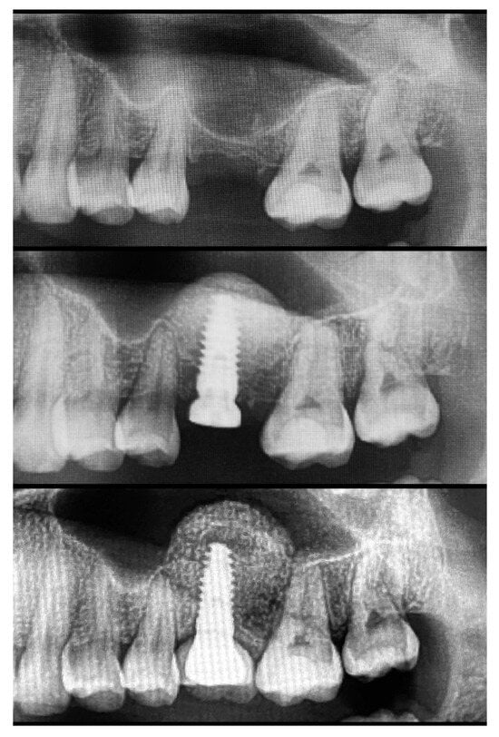
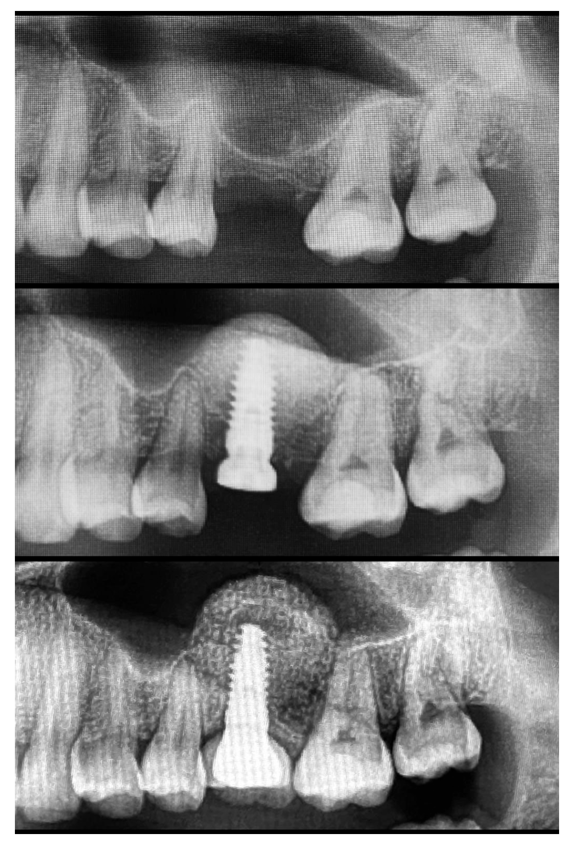
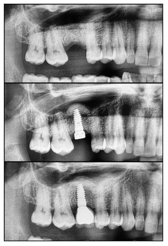
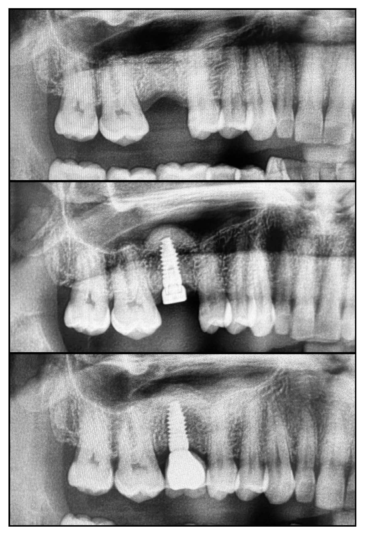
- Secondary outcomes measures were the overall tending effect (residual bone height + implant + graft) evaluated immediately after implant placement, and the final overall height of the elevated sinus evaluated at the crown delivery, 9 months after implant placement, and with a clinical follow up of 2 years. All the post-operative measures were taken on the panoramic radiography (Figure 1 and Figure 2).
Figure 1.
Test group. From top to bottom: pre-operative X-Ray, Implant placement, and 2 year follow-up.
Figure 2.
Control group. From top to bottom: pre-operative X-Ray, Implant placement, and 2-year follow-up.
2.5. Statistical Analysis
Data were recorded and analyzed using Numbers software (version 14.2, build 7041.0.109; Apple Inc., Cupertino, CA, USA). Statistical analysis was performed according to a pre-established analysis plan by a biostatistician with expertise in dentistry research. Descriptive statistics were calculated as mean ± standard deviation (SD), median, and 95% confidence interval (CI). The normality of paired differences was assessed using the Shapiro–Wilk test, supporting the use of paired t-tests for within-group comparisons. Within-group differences between time points were assessed using paired t-tests, while between-group comparisons were performed using the Mann–Whitney U test for independent samples, through the online statistical calculator StatsKingdom (https://www.statskingdom.com/170median_mann_whitney.html, accessed on 13 September 2026). The implant was the statistical unit of the analyses. All statistical comparisons were conducted at the 0.05 level of significance.
3. Results
A total of 45 patients were included in this retrospective study. Twenty patients were allocated to the test group (residual bone height < 4 mm), while twenty-five patients were included in the control group (residual bone height ≥ 4 mm).
In the test group, 9 males and 11 females were treated, with a mean age of 59.0 years. In the control group, 12 males and 13 females were included, with a mean age of 61.4 years. Implants were predominantly placed in molar sites. In the control group, 2 implants were placed in premolar positions, while the remaining implants were placed in molar areas.
No implant failures, prosthetic failures, or biological or technical complications were recorded during the observation period, resulting in a 100% survival rate.
At implant placement, the residual bone height (RBH) was significantly lower in the test group (2.8 ± 0.8 mm; 95% CI 2.4–3.2 mm) compared with the control group (5.3 ± 1.0 mm; 95% CI 4.9–5.7 mm) (p < 0.001). The final bone height (FBH) immediately after sinus augmentation was 13.7 ± 1.7 mm (95% CI 12.9–14.4 mm) in the test group and 13.5 ± 1.5 mm (95% CI 12.9–14.1 mm) in the control group, with no statistically significant difference between groups (p = 0.742). The vertical bone gain, calculated as the difference between initial and final bone height, was significantly greater in the test group (10.9 ± 1.6 mm; 95% CI 10.2–11.6 mm) compared with the control group (8.2 ± 0.6 mm; 95% CI 7.6–8.8 mm) (p < 0.001). The bone height above the implant apex at implant placement was 3.4 ± 1.6 mm (95% CI 2.7–4.1 mm) in the test group and 3.0 ± 1.2 mm (95% CI 2.6–3.5 mm) in the control group, with no statistically significant difference (p = 0.420).
At prosthetic delivery (9 months), the final bone height was 12.6 ± 1.6 mm (95% CI 11.9–13.3 mm) in the test group and 12.9 ± 1.6 mm (95% CI 12.2–13.5 mm) in the control group, showing no statistically significant inter-group difference (p = 0.556). A significant reduction in bone height compared with baseline was observed in both groups. This reduction was 1.1 ± 0.8 mm (95% CI 0.7–1.4 mm; p < 0.001) in the test group and 0.6 ± 0.6 mm (95% CI 0.4–0.9 mm; p < 0.001) in the control group. The inter-group comparison showed a slightly greater reduction in the test group (p = 0.040). At the same follow-up, the bone height above the implant apex was 2.8 ± 1.3 mm (95% CI 2.2–3.4 mm) in the test group and 2.8 ± 1.3 mm (95% CI 2.3–3.2 mm) in the control group, with no statistically significant difference (p = 0.901). The reduction from baseline in this parameter was 0.6 ± 0.8 mm (95% CI 0.3–1.0 mm; p < 0.001) in the test group and 0.3 ± 0.7 mm (95% CI 0–0.6 mm; p < 0.001) in the control group, with no statistically significant inter-group difference (p = 0.176). Data are reported in Table 1.
Table 1.
Radiographic outcomes according to baseline residual bone height.
4. Discussion
The present retrospective study evaluated the 2-year clinical outcomes and early radiographic changes of transcrestal sinus floor elevation performed using the CAS-kit technique in patients with different levels of residual bone height. The main finding was that comparable clinical outcomes and similar final bone heights were obtained regardless of whether the initial residual bone height was <4 mm or ≥4 mm. Although greater vertical bone gain was observed in sites with reduced residual bone height, the final augmented bone level and the amount of bone maintained above the implant apex were similar between groups at prosthetic delivery. These findings suggest that, when appropriate surgical protocols are followed, the crestal sinus lift approach may represent a predictable treatment option even in cases with limited residual bone height [9,10,11,12,13,14,19,20,21,22].
Traditionally, residual bone height has been considered one of the main parameters for selecting the surgical approach for maxillary sinus floor elevation [5,15,21]. Transcrestal sinus floor elevation has generally been recommended when approximately 4–5 mm or more of residual bone is available, whereas markedly reduced RBH has traditionally favored the use of a lateral window approach because of concerns regarding insufficient primary implant stability and an increased risk of membrane perforation [5,15,21,23]. However, recent evidence has progressively challenged the concept of a rigid RBH threshold. A recent systematic review and meta-analysis comparing transcrestal and lateral sinus floor augmentation in sites with RBH ≤ 6 mm found no significant differences in implant survival, sinus membrane perforation, or marginal bone loss between the two approaches, while reporting high implant survival for the transcrestal approach [22]. The results of the present investigation are consistent with this evolving evidence, indicating that predictable vertical augmentation and implant survival can also be achieved in sites with RBH < 4 mm, provided that adequate surgical planning and strategies to achieve primary implant stability are adopted. Notably, the mean baseline RBH in the test group was only 2.8 mm, indicating that this group included sites with very limited residual bone availability. Nevertheless, comparable final bone heights and 100% implant survival were obtained in both groups.
One of the factors potentially contributing to these favorable outcomes is the achievement of adequate primary implant stability through controlled osteotomy underpreparation. In the posterior maxilla, where bone density is often limited, implant macrodesign and site preparation protocols aimed at preserving or condensing peri-implant bone may improve mechanical stability at placement. However, the potential benefits of underpreparation and osseodensification should be balanced against their possible effects on peri-implant bone remodeling, and the available clinical evidence remains heterogeneous [22,24].
Furthermore, the possibility of engaging both the crestal cortical bone and the cortical layer of the sinus floor may provide additional anchorage, particularly in cases with reduced residual bone height. This biomechanical concept has been previously proposed as a rationale for simultaneous implant placement in augmented sinuses and may partially explain the comparable outcomes observed between groups in the present study [9,10,21].
The use of dedicated surgical instrumentation may also have played an important role in reducing intraoperative complications and improving the predictability of the procedure. The CAS-kit system enables controlled, depth-limited preparation of the sinus floor combined with hydraulic membrane elevation, allowing gradual separation of the Schneiderian membrane while minimizing the risk of perforation [11,12,13,14]. Previous investigations have demonstrated that hydraulic or balloon-assisted techniques may reduce surgical trauma and provide favorable clinical outcomes compared with conventional osteotome approaches [8,9,10,11,12,13,14]. More recently, a systematic review specifically evaluating hydraulic pressure sinus floor elevation reported a pooled implant survival rate of 98.4%, a sinus membrane perforation rate of 2.05%, and a mean endo-sinus bone gain of 6.72 mm, supporting the clinical predictability of hydraulic elevation techniques [25]. The present findings are consistent with these observations, as no biological or technical complications were recorded during the follow-up period. Furthermore, the presence and morphology of sinus septa were not systematically recorded, representing an additional limitation of the present study. Although depth-controlled instrumentation and hydraulic membrane elevation may facilitate membrane detachment in anatomically challenging conditions, their effectiveness and safety in the presence of sinus septa require further investigation [26,27].
Another relevant aspect influencing the clinical predictability of transcrestal sinus elevation is operator experience and the associated learning curve. Limited tactile feedback and indirect visualization may increase the technical sensitivity of the procedure, especially in cases with minimal residual bone [5,21]. Therefore, adequate training, strict adherence to surgical protocols, and careful case selection remain essential to optimize outcomes and reduce the risk of complications [21].
In addition to surgical technique, the anatomical characteristics of the maxillary sinus may significantly affect the success of membrane elevation procedures [17]. Previous studies have shown that sinus width, floor morphology, membrane characteristics, and the presence of anatomical variations may influence membrane elevation, graft containment, bone formation, and the risk of membrane perforation [17]. Narrower and more tapered sinus configurations may provide more favorable conditions for bone formation and graft containment, whereas wider sinus cavities may represent a more challenging anatomical environment. Consequently, preoperative three-dimensional radiographic assessment of sinus anatomy should be considered an integral component of treatment planning rather than relying on RBH alone when selecting the surgical approach [17].
The decision to use grafting material during transcrestal sinus elevation remains a topic of debate. While satisfactory outcomes have been reported with graftless techniques, grafting materials may contribute to space maintenance beneath the elevated membrane and facilitate radiographic monitoring of bone remodeling [20,21]. In the present study, all sites were grafted with a xenogeneic bone substitute to enhance volumetric stability, which may have contributed to the limited reduction in augmented bone height observed during healing. Nevertheless, the available evidence suggests that implant survival following sinus floor elevation may depend on multiple biological, anatomical, implant-related, and surgical factors rather than on the use of grafting material alone [19,20].
Despite the favorable outcomes reported, several limitations must be considered when interpreting the results of this study. First, the retrospective design may have introduced selection bias and limits the ability to establish causal relationships. Second, no a priori sample size calculation was performed, and the relatively small number of treated patients may reduce the external validity of the findings. Third, the use of panoramic radiographs for linear measurements may have affected the accuracy of bone height evaluation compared with cone-beam computed tomography. Furthermore, potential confounding factors such as sinus morphology, bone density, implant distribution per patient, and prosthetic loading conditions were not controlled. Finally, the absence of a control group treated with the lateral window technique prevents direct comparison between different surgical approaches.
Within the limitations of this retrospective study, transcrestal sinus floor elevation using the hydraulic CAS-kit technique showed favorable clinical and radiographic outcomes in patients with different residual bone heights, including sites with RBH below 4 mm. These preliminary findings suggest that residual bone height alone should not necessarily be considered an absolute criterion for selecting the surgical approach. However, treatment planning should also consider sinus anatomy, bone quality, implant design, and the possibility of achieving adequate primary stability. Given the retrospective design, limited sample size, and use of two-dimensional radiographic measurements, these findings should be interpreted with caution and cannot establish the long-term predictability of the technique. Further prospective controlled studies with larger sample sizes, standardized radiographic assessments, and longer follow-up periods are warranted.
Author Contributions
Conceptualization, F.M.C. and M.T.; Methodology, F.M.C., F.P. and M.G.; Investigation, F.M.C. and A.M.; Formal Analysis, A.M. and C.C.; Data Curation, F.Z. and C.D.F. and E.X.; Writing—Original Draft Preparation, F.P. and C.D.F.; Writing—Review and Editing, M.T. and C.C.; Visualization, F.Z.; Supervision, M.G. and E.X. All authors have read and agreed to the published version of the manuscript.
Funding
This research received no external funding.
Institutional Review Board Statement
This study was prepared in accordance with the principles of the Declaration of Helsinki (1975, revised in 2013). No additional research-related interventions were performed, and the patient’s treatment was conducted according to standard practice. In accordance with Directive 2001/20/EC of the European Parliament and of the Council of 4 April 2001 on the approximation of the laws, regulations and administrative provisions of the member states relating to the implementation of good clinical practice in the conduct of clinical trials on medicinal products for human use. Moreover, since we did not test any experimental technique, in accordance with relevant legislation, artt. 10 e320 cod. civ. and artt. 96 e 97 legge 22.4.1941, n. 633. This study does not require ethical approval. All patient data were anonymized to protect patient privacy and confidentiality.
Informed Consent Statement
Written informed consent was obtained from all patients as part of standard clinical practice. For the present publication, all patient data were anonymized in accordance with applicable data protection regulations.
Data Availability Statement
All relevant data are included in the manuscript. Additional clinical data, radiographic images, and raw data are available from the corresponding author upon reasonable request, in accordance with applicable data protection regulations.
Conflicts of Interest
M.T. and E.X. serve as Key Opinion Leaders for the manufacturer of the CAS-KIT used in this study. Their contributions were limited to manuscript review, editing, and scientific supervision. The study was entirely self-funded, and the manufacturer had no involvement in the study design, data collection, analysis, interpretation, manuscript preparation, or decision to publish. The authors declare no other conflicts of interest.
References
- Lyu, M.; Xu, D.; Zhang, X.; Yuan, Q. Maxillary sinus floor augmentation: A review of current evidence on anatomical factors and a decision tree. Int. J. Oral Sci. 2023, 15, 41. [Google Scholar] [CrossRef] [Scilit] [PubMed]
- Chen, J.; Lu, Y.; Xu, J.; Hua, Z. Clinical evaluation of maxillary sinus floor elevation with or without bone grafts: A systematic review and meta-analysis of randomised controlled trials with trial sequential analysis. Arch. Med. Sci. 2024, 20, 384–401. [Google Scholar] [CrossRef] [Scilit] [PubMed]
- Raghoebar, G.M.; Onclin, P.; Boven, G.C.; Vissink, A.; Meijer, H.J.A. Long-term effectiveness of maxillary sinus floor augmentation: A systematic review and meta-analysis. J. Clin. Periodontol. 2019, 46, 307–318. [Google Scholar] [CrossRef] [Scilit] [PubMed]
- Sirinirund, B.; Rodriguez Betancourt, A.B.; Scaini, R.; Wu, Y.C.; Chan, H.L. Minimally invasive sinus augmentation: A systematic review. Clin. Implant Dent. Relat. Res. 2025, 27, e13403. [Google Scholar] [CrossRef] [Scilit] [PubMed]
- Pjetursson, B.E.; Tan, W.C.; Zwahlen, M.; Lang, N.P. A systematic review of the success of sinus floor elevation and survival of implants inserted in combination with sinus floor elevation. Part I: Lateral approach. J. Clin. Periodontol. 2008, 35, 216–240. [Google Scholar] [CrossRef] [Scilit] [PubMed]
- Summers, R.B. A new concept in maxillary implant surgery: The osteotome technique. Compendium 1994, 15, 152–158. [Google Scholar] [PubMed]
- Farina, R.; Riccardi, O.; Schincaglia, G.P.; Severi, M.; Trombelli, L.; Simonelli, A. Six-year extension results of a randomized trial comparing transcrestal and lateral sinus floor elevation at sites with 3–6 mm of residual bone. Clin. Oral Implant. Res. 2023, 34, 813–821. [Google Scholar] [CrossRef] [Scilit] [PubMed]
- Farina, R.; Franzini, C.; Minenna, L.; Trombelli, L.; Simonelli, A. Effectiveness, morbidity, and costs of transcrestal and lateral sinus floor elevation at sites with different residual bone heights: A re-analysis of data from a parallel-arm randomized trial. Int. J. Oral Maxillofac. Implant. 2023, 38, 1123–1138. [Google Scholar] [CrossRef] [Scilit] [PubMed]
- Ceruso, F.M.; Lumbau, A.I.; Pernice, F.; Mastroianni, A.; Miranda, M.; Meloni, S.M.; Gargari, M.; Tallarico, M.; Pisano, M. Crestal hydraulic sinus lift with simultaneous implant insertion: A retrospective case series. Dent. J. 2025, 13, 193. [Google Scholar] [CrossRef] [Scilit] [PubMed]
- Tallarico, M.; Cochran, D.L.; Xhanari, E.; Dellavia, C.; Canciani, E.; Mijiritsky, E.; Meloni, S.M. Crestal sinus lift using an implant with an internal L-shaped channel: 1-year after loading results from a prospective cohort study. Eur. J. Oral Implantol. 2017, 10, 325–336. [Google Scholar] [PubMed]
- Mamidi, A.R.; Gottumukkala, S.N.V.S.; Mantena, S.R.; Penmetsa, G.S.; Ramesh, K.S.V.; Pasupuleti, M.K. Comparison of clinical and radiological outcomes using crestal approach sinus kit and piezoelectric surgery for sinus membrane elevation: A prospective clinical trial. Dent. Res. J. 2022, 19, 88. [Google Scholar] [CrossRef] [Scilit]
- Cobo-Vázquez, C.M.; García-Rodríguez, S.; Colmenares-Otero, M.E.; Cortés-Bretón-Brinkmann, J.; Madrigal Martínez-Pereda, C.; Meniz-Garcia, C. Clinical and radiographic evaluation for two crestal sinus lift techniques: Osteotome versus osseodensification. A systematic review and meta-analysis. Int. J. Implant Dent. 2025, 11, 36. [Google Scholar] [CrossRef] [Scilit] [PubMed]
- Kim, Y.K.; Cho, Y.S.; Yun, P.Y. Assessment of dentists’ subjective satisfaction with a newly developed device for maxillary sinus membrane elevation by the crestal approach. J. Periodontal Implant Sci. 2013, 43, 308–314. [Google Scholar] [CrossRef] [Scilit] [PubMed]
- Gatti, F.; Gatti, C.; Tallarico, M.; Tommasato, G.; Meloni, S.M.; Chiapasco, M. Maxillary sinus membrane elevation using a special drilling system and hydraulic pressure: A 2-year prospective cohort study. Int. J. Periodontics Restor. Dent. 2018, 38, 593–599. [Google Scholar] [CrossRef] [Scilit] [PubMed]
- Tent, A.P.; Țig, I.A.; Bran, S.; Zlotu, A.; Mester, A.; Onisor, F. Long-Term Effects of Sinus Floor Elevation with and Without Bone Graft: A Systematic Analysis of Randomized Clinical Trials. Medicina 2025, 61, 104. [Google Scholar] [CrossRef] [Scilit] [PubMed]
- Soares, L.F.F.; Malzoni, C.M.A.; da Silveira, M.L.; Marcantonio Junior, E.; Pigossi, S.C. Evaluation of different approaches for sinus membrane perforation repair during sinus elevation: A systematic review and meta-analysis. Int. J. Oral Maxillofac. Implant. 2024, 39, 107–118. [Google Scholar] [CrossRef] [Scilit] [PubMed]
- Farina, R.; Franzini, C.; Trombelli, L.; Simonelli, A. Minimal invasiveness in the transcrestal elevation of the maxillary sinus floor: A systematic review. Periodontol. 2000 2023, 91, 145–166. [Google Scholar] [CrossRef] [Scilit] [PubMed]
- European Parliament and Council. Directive 2001/20/EC of 4 April 2001. Off. J. Eur. Communities 2001, L121, 34–44. [Google Scholar]
- Shi, S.; Han, L.; Su, J.; Guo, J.; Yu, F.; Zhang, W. Clinical efficacy of transcrestal sinus floor augmentation, in comparison with lateral approach, in sites with residual bone height ≤ 6 mm: A systematic review and meta-analysis. Clin. Oral Implant. Res. 2023, 34, 1151–1175. [Google Scholar] [CrossRef] [Scilit] [PubMed]
- Guruprasad, M.; Kulloli, A.; Mehta, V.; Fiorillo, L.; Cicciù, M. Maxillary sinus floor augmentation with allograft alone compared with alternate grafting materials: A systematic review and meta-analysis. J. Craniofac. Surg. 2024, 35, 686–691. [Google Scholar] [CrossRef] [Scilit] [PubMed]
- Abesi, F.; Yousefi, M.J.; Zamani, M. Prevalence and anatomical characteristics of maxillary sinus septa: A systematic review and meta-analysis of cone-beam computed tomography studies. J. Oral Maxillofac. Surg. Med. Pathol. 2023, 35, 501–507. [Google Scholar] [CrossRef] [Scilit]
- Kalra, J.; Dhawan, P.; Jain, N. Implant stability and crestal bone level in osseodensification and conventional drilling protocols: A systematic review and meta-analysis. J. Prosthet. Dent. 2025, 133, 1462.e1–1462.e7. [Google Scholar] [CrossRef] [Scilit] [PubMed]
- Decker, A.M.; Stuhr, S.; Testori, T.; Wang, H. Clinical and radiographic changes following transcrestal sinus augmentation: A scoping review of the last 25 years. Clin. Implant Dent. Relat. Res. 2024, 26, 1338–1353. [Google Scholar] [CrossRef] [Scilit] [PubMed]
- Antonacci, D.; Del Fabbro, M.; Bollero, P.; Stocchero, M.; Jinno, Y.; Canullo, L. Clinical effects of conventional and underprepared drilling preparation of the implant site based on bone density: A systematic review and meta-regression. J. Prosthodont. Res. 2023, 67, 23–34. [Google Scholar] [CrossRef] [Scilit] [PubMed]
- Hughes, J.; Sharma, A.; Sharma, L.A. The safety and effectiveness of the hydraulic pressure sinus floor elevation technique: A systematic review. Aust. Dent. J. 2026, 71, 101–118. [Google Scholar] [CrossRef] [Scilit] [PubMed]
- Yang, B.; Wang, T.; Wen, Y.; Liu, X. Association between sinus septa and lateral wall thickness with risk of perforation during maxillary sinus lift surgery: A systematic review and meta-analysis. PLoS ONE 2024, 19, e0308166. [Google Scholar] [CrossRef] [Scilit] [PubMed]
- Ro, J.H.; Moon, J.H.; Cheon, M.C.; Park, C.J. Measurement of hydraulic pressure on the sinus membrane for safer control during transcrestal sinus lifting. Int. J. Oral Maxillofac. Surg. 2021, 50, 1267–1270. [Google Scholar] [CrossRef] [Scilit] [PubMed]

Disclaimer/Publisher’s Note: The statements, opinions and data contained in all publications are solely those of the individual author(s) and contributor(s) and not of MDPI and/or the editor(s). MDPI and/or the editor(s) disclaim responsibility for any injury to people or property resulting from any ideas, methods, instructions or products referred to in the content. |
© 2026 by the authors. Licensee MDPI, Basel, Switzerland. This article is an open access article distributed under the terms and conditions of the Creative Commons Attribution (CC BY) license.