


Abstract
Psychoactive nitrogen-containing compounds represent a structurally diverse group of natural and synthetic molecules whose interactions with metal ions influence their physicochemical properties, biological activity, and analytical behavior. Although numerous studies have reported the synthesis of metal complexes, solution interactions, and metal-assisted analytical methods, the available knowledge remains scattered across compound classes and has not been comprehensively evaluated. This review summarizes advances in the coordination chemistry of psychoactive nitrogen-containing compounds from ScienceDirect, Google Scholar, and the Cambridge Structural Database (CSD), emphasizing donor atoms, coordination modes, structural diversity, crystallographic characterization, spectroscopic and computational investigations, and the biological properties of the resulting metal complexes. The current literature demonstrates that metal complexation can alter molecular geometry, electronic structure, redox behavior, biomolecular recognition, and pharmacological activity, while also offering opportunities to develop compounds with enhanced antimicrobial, anticancer, antioxidant, and DNA-binding properties. By integrating coordination behavior, structural, and biological aspects within a single framework, this review highlights the central role of metal interactions in psychoactive compounds and identifies emerging opportunities to develop advanced analytical methodologies and functional metal-based systems.
Keywords:
psychoactive compounds; metal interactions; cocaine; morphine; promazine; mescaline; codeine; caffeine; metal complexes; SERS 1. Introduction
Psychoactive nitrogen-containing compounds comprise a structurally diverse group of naturally occurring and synthetic molecules capable of altering the function of the central nervous system through interactions with neurotransmitter receptors, ion channels, transporters, and enzymes [1,2]. This broad class includes naturally occurring alkaloids, methylxanthines, phenethylamines, indoleamines, and synthetic phenothiazine derivatives that have found widespread applications as stimulants, analgesics, antipsychotics, antidepressants, anxiolytics, sedatives, and hallucinogens. Some of these biologically active compounds or their derivatives also have antioxidant, anticancer, anti-inflammatory, and antiviral activities, while others remain substances of abuse that continue to pose significant medical, social, and forensic challenges [2,3,4,5,6]. Their identification, characterization, and understanding of their molecular interactions therefore remain important topics in medicinal chemistry, toxicology, forensic science, and analytical chemistry [7].
In addition to their pharmacological properties, these compounds share another important structural characteristic. Psychoactive molecules selected for this review contain one or more heteroatoms capable of interacting with metal ions, as presented in Figure 1 [8,9,10,11]. Depending on their chemical structure, potential donor sites include primary, secondary, tertiary, or heterocyclic nitrogen atoms, as well as oxygen-containing functional groups such as hydroxyl, carbonyl, ether, amide, or carboxylate moieties. In aromatic systems of promazine derivatives described in this review, sulfur atoms and extended π-electron systems may additionally participate in coordination either directly or through secondary interactions. Consequently, many psychoactive compounds behave as mono-, bi-, or polydentate ligands capable of forming coordination compounds with transition metals, main-group elements, and lanthanides [12,13,14,15]. The preferred coordination mode depends on the nature of both the ligand and the metal ion, frequently producing square-planar, tetrahedral, octahedral, trigonal-bipyramidal, or polymeric structures that differ substantially in their physicochemical and biological properties.
Figure 1.
Chemical structures of psychoactive nitrogen-containing compounds included in the review.
Metal complexation profoundly influences the chemical behavior of biologically active molecules. Coordination may modify molecular geometry, electronic structure, lipophilicity, solubility, redox potential, acid-base properties, and kinetic stability. These changes often alter ligand interactions with biomolecular targets, including nucleic acids, proteins, membranes, and enzymes. Numerous studies have demonstrated that metal complexation can enhance antimicrobial, antioxidant, anticancer, anti-inflammatory, or DNA-binding properties compared with the parent organic compounds [16,17,18,19,20,21,22,23,24,25,26]. Transition-metal ions such as Cu(II), Zn(II), Fe(III), Co(II), Ni(II), Pd(II), Pt(II), Au(III), and Ru(II), together with biologically relevant alkali, alkaline-earth, and lanthanide ions, have therefore become valuable tools for modulating the biological activity of nitrogen-containing drugs [27,28,29]. At the same time, understanding the preferred donor atoms and coordination geometries provides important insight into possible interactions between therapeutic agents and endogenous metal ions under physiological conditions.
Although the coordination chemistry of individual classes of psychoactive compounds has attracted considerable attention, the available information remains highly fragmented. Certain molecules, such as caffeine and cocaine, have generated extensive literature describing numerous transition-metal complexes and mixed-ligand systems, whereas others, including atropine, mescaline, serotonin, or morphine, have been only sporadically investigated [13,30,31,32,33]. Furthermore, studies differ substantially in their objectives, ranging from the synthesis and crystallographic characterization of discrete coordination compounds to spectroscopic investigations of solution equilibria, computational studies, biological evaluation, and analytical applications. As a consequence, direct comparisons between different classes of psychoactive compounds are often difficult, and general trends governing their coordination behavior have not been systematically discussed.
Interactions between psychoactive compounds and metal ions are also highly relevant to forensic chemistry. Numerous forensic methods rely directly or indirectly on metal complex formation. Presumptive color tests frequently involve coordination reactions that produce characteristic chromogenic species, while ion-association compounds have been exploited for the extraction, separation, and spectrophotometric determination of illicit drugs [34]. Metal-containing reagents have also been employed to mask controlled substances, as illustrated by so-called black cocaine [35], where the formation of stable transition-metal thiocyanate complexes substantially complicates routine forensic identification. In addition, metal ions and metal-based nanomaterials have become increasingly important in modern analytical techniques, including UV–VIS spectroscopy, fluorescence spectroscopy, Raman and surface-enhanced Raman spectroscopy, and electrochemical sensing [36,37,38,39,40]. These approaches enable improved sensitivity, selectivity, and detection limits for numerous psychoactive compounds encountered in forensic investigations.
Beyond analytical applications, the coordination chemistry of psychoactive compounds provides valuable opportunities for the rational design of new functional materials and therapeutics. Complexation with biologically active metal ions may enhance pharmacological efficacy, modify toxicity profiles, improve cellular uptake, or introduce entirely new mechanisms of action. Recent studies have demonstrated that several metal complexes derived from psychoactive compounds exhibit significantly improved anticancer, antimicrobial, antioxidant, and DNA-binding properties compared with the parent ligands [41,42,43,44,45,46]. Moreover, advances in computational chemistry, molecular spectroscopy, crystallography, and bioinorganic chemistry have considerably improved our understanding of the structural factors governing metal binding and structure–activity relationships [47,48,49,50,51]. The integration of experimental and theoretical approaches, therefore, represents an increasingly important strategy for developing new medicinal and analytical systems based on metal–drug interactions.
Despite the growing number of publications describing metal complexes of individual psychoactive compounds, no comprehensive review has systematically compared their coordination chemistry across the major structural classes of psychoactive nitrogen-containing molecules. This review summarizes current knowledge of the coordination chemistry of psychoactive nitrogen-containing compounds. The published articles were searched on Google Scholar, ScienceDirect, and the Cambridge Structural Database (CSD) without any specific time frame. The search terms used Boolean logic. They included “metal complexes”/”metal interactions” AND the specific psychoactive compounds: “cocaine”, “atropine”, “morphine”, “codeine”, “papaverine”, “berberine”, “caffeine”, “ephedrine”, “tyramine”, “mescaline”, “tryptamine”, “serotonin”, “melatonin”, “promazine”, “promethazine”, and “chlorpromazine”. The articles were selected based on their relevance to the topic through initial screening of the title and abstract. All available articles were included for most compounds, whereas only representative studies were selected for caffeine and cocaine, given the extensive literature on their metal complexation. Particular attention is devoted to the identification of donor atoms, coordination modes, crystallographically characterized structures, spectroscopic and computational characterization, and the influence of metal complexation on biological activity. Because the degree of structural characterization varies considerably among the available studies, a distinction is made throughout this review between crystallographically confirmed structures and structural models proposed based on indirect experimental and/or computational evidence. Structures determined by single-crystal X-ray diffraction are regarded as structurally established, whereas coordination geometries, donor-atom assignments, and nuclearities inferred from elemental analysis, spectroscopy, magnetic measurements, thermal analysis, or computational calculations are described as proposed models. In the latter cases, the reported structures are discussed in the context of the evidence provided by the original authors, recognizing that alternative structural arrangements cannot be excluded without direct crystallographic characterization. The major challenges and future perspectives are highlighted.
2. Tropane Alkaloids
2.1. Cocaine
Cocaine (COC) is a bioproduct extracted from Erythroxylum coca leaves and contains an azobicyclo [3.2.1] moiety [1]. The same structural parameter is present within a range of compounds with antibacterial, analgesic, and anti-inflammatory properties [52,53,54]. Glichrist presented the possibility of the treatment of cancer cells with cocaine in 1909, as explained in the reference [6]. The presence of the carbonyl oxygen and the oxygen atoms of the aliphatic chain make this structure an attractive ligand for the complexation of transition-metal complexes. Notable examples are listed in Table 1.
In the study by Abdallah and coworkers, a series of compounds with cocaine was prepared [47]. The authors based their structural predictions on elemental percentages, although they did not obtain crystallographic structures. In the case of Cr(III), Fe(III), Zn(II), and Cd(II) complexes, there is one cocaine ligand together with chlorido and aqua ligands, with the general formula [M(COC)Clx(H2O)y]∙nH2O. The proposed structure of the Cd(II) complex is shown in Figure 2. On the other hand, complexes containing Co(II) and Ni(II) ions contain two cocaine ligands, next to aqua and chlorido ligands ([M(COC)2Clx(H2O)y]∙nH2O). Structural optimizations of the predicted structures at the B3LYP/LanL2DZ level of theory revealed that the donor atoms include the carbonyl and etheric oxygen atoms of cocaine, with the donor set adopting octahedral geometry around Fe(III) and tetrahedral geometry around Cd(II) ions. These findings were in line with the experimental FTIR spectrum, which showed a shift to lower wavenumbers in the C=O vibration from 1732 cm−1 to 1706–1721 cm−1. The formation of the M−O bond was observed at 509–569 cm−1. Further structural studies included thermogravimetric analysis and electronic spectra. In the examination of anticancer activity, the Co(II) complex showed promising results, with specific selectivity towards hepatoblastoma (HepG-2) cells. The number of cocaine molecules in the structures is reduced to one in complexes containing previously mentioned d-block metal ions when the TMEDA (N,N,N′,N′-tetramethylethylenediamine) ligand is present, leading to the formula M(COC)(TMEDA)Cl(OH2)]·2Cl·xH2O [55]. These conclusions were drawn from the elemental analysis and mass spectra, which confirmed the formation of the proposed species. The authors also discussed that fragmentation was observed in the mass spectra, with peaks attributed to the pure ligands. TMEDA is a bidentate ligand with two donor nitrogen atoms. In the theoretical analysis, all of the complexes adopted an octahedral geometry around the central metal ion. Among these compounds, the Fe(III) and Cd(II) complexes were the most active against HepG-2 and MCF-7 cancer cell lines, with minimal effects on HFB4 human normal cells.
Figure 2.
Proposed structure of the cocaine-Cd(II) complex from ref. [47] based on elemental analysis and DFT calculations.
The same type of monoligand complexes with cocaine were obtained with La(III), Er(III), and Yb(III) in the reference [49]. The proposed structures contained three water molecules as ligands, one chlorido ligand, and two chlorine counterions. The structural assumptions were based on the mass spectrum of the La(III) complex and elemental analysis. Shifts in the FTIR spectra were the same as previously discussed, confirming that carbonyl and etheric oxygen atoms were included in the complex. Thermogravimetric analysis indicated that water and chlorine molecules were released first from the complex, followed by cocaine decomposition. The final product was the oxide of each lanthanide. The Er(III) complex also showed promising anticancer activity against the MCF-7 cell line, comparable to that of cisplatin, and high safety towards healthy cells. Lanthanide elements were also included in the formation of Ln(COC)(TMEDA)Cl(OH2)]·2Cl·xH2O complexes [56]. Within this group of compounds, the Yb(III) complex showed anticancer activity comparable to that of cisplatin, with minimal effect on normal cell lines. This compound induced apoptosis by increasing cellular caspase-3 and caspase-9 levels.
Laussmann et al. reported the structural analysis of the compound containing the copper thiocyanato complex and protonated cocaine molecules (COCH) in so-called “black cocaine” [35]. Although this compound does not involve cocaine directly in the inner sphere of a complex species, it is illustrative of the activities that are performed to cover the presence of this drug in samples. The addition of metal ions and SCN− disables analytical tests for cocaine. Anionic SCN− complexes with Fe(III), Co(II), Cu(II), and Hg(I) form ion pairs with protonated forms of alkaloids [57,58,59]. The dark crystal structure of (COCH)2[Cu(NCS)4] contained square planar anionic species and COCH+ cations [35]. This experimentally determined structure was composed of protonated cocaine molecules as cations and negatively charged copper(II) anions. Previously, a similar crystallographic structure was obtained for the gold complex of the formula (COCH)[AuCl4] in [50], stabilized by the intermolecular C−H∙∙∙O (between two COCH+) and C−H∙∙∙Cl interactions. In (COCH)2[Cu(NCS)4], only with the use of 1H NMR was the identification of cocaine possible. The mixture of “black cocaine” also contained complex species formed between Fe(III) and the thiocyanate anion, which interact with COCH and form a dark red solution. Mohammadzai and Hinze reported an indirect atomic absorption determination of cocaine based on interactions between COCH and [FeCl4]−, similar to those observed with other anionic transition-metal complexes [59].
The reactions with transition metal ions and cocaine influenced the development of the various tests for its detection in street samples. One of the most famous examples involves complexation of this alkaloid with [Co(SCN)2(H2O)4], which produces an intense blue color. Based on the results of FTIR spectroscopy and theoretical analysis, Haddoub and coworkers discussed that the color change is a consequence of the formation of the complex [Co(COC)(NCS)(H2O)3]SCN, as suggested by molecular modeling and FTIR spectra [57]. It should be noted that the formation of other species cannot be excluded. In the water/glycerol solution, the band attributed to the C=N vibration (2100 cm−1) should be very sharp, but two intense bands at 2069 and 2105 cm−1 were observed. In the proposed structure, coordination occurred through a protonated nitrogen atom and an ether oxygen atom. The previously proposed structure was employed in the discussion of the results, in which the formation constant of the complex was determined electrochemically [60]. The low value of the formation constant was consistent with the need for concentrated solutions in colorimetric tests for cocaine. The coordination of cocaine to cobalt ions was used for the development of a voltammetric method for its detection [58]. The platinum disc electrode was chemically modified with a cobalt-hexacyanoferrate film. Because the interactions between cocaine and cobalt ions were reversible, the surface was partially passivated, leading to a linear dependence of cocaine detection over the range 2.4 × 10⁻⁴ to 1.5 × 10⁻³ mol L−1, with a detection limit of 1.4 × 10⁻⁴ mol L−1 and a high correlation coefficient.
2.2. Atropine
Atropine is a tropane alkaloid containing a tertiary amine together with ester and hydroxyl oxygen atoms that may participate in metal binding [2]. Nevertheless, unlike many other alkaloids, its coordination chemistry has received very little attention, and no structurally characterized transition-metal complexes have been reported. The only detailed study investigated the ionophoric properties of atropine toward biologically relevant alkali and alkaline-earth metal ions [30]. Spectrophotometric extraction and liquid membrane transport experiments demonstrated that atropine forms complexes with Na+, K+, Mg2+, and Ca2+ ions, exhibiting the highest affinity for Ca2+, while the transport efficiency followed the order Mg2+ > Ca2+ > K+ > Na+. The direct interaction between atropine and the metal ions presented was not proven but was inferred mainly from the change from water to organic solvent when atropine was present in the latter. The authors proposed that these ionophoric properties may contribute to atropine’s pharmacological activity by modulating calcium-dependent cellular processes and membrane permeability.
Table 1.
Selected studies on metal complexes of tropane alkaloids.
3. Morphinan and Isoquinoline Alkaloids
3.1. Morphine
Morphine is the principal representative of the morphinan alkaloids and contains a tertiary amine together with phenolic and secondary alcoholic hydroxyl groups that can potentially participate in metal binding [3,61,62]. Despite the presence of these donor sites, the coordination chemistry of morphine remains relatively limited compared with many other naturally occurring alkaloids. The studies presented focused on solution interactions with biologically relevant metal ions rather than on synthesizing well-defined coordination compounds, reflecting an interest in how metal ions influence opioid pharmacology and receptor recognition.
The most comprehensive studies have examined the interaction of morphine with Mn(II), employed as a spectroscopic probe for biologically relevant divalent cations such as Ca(II) and Mg(II) [31]. Fourier-transform NMR relaxation experiments demonstrated that morphine forms a relatively weak 1:1 complex (Kass ≈ 10−2 M−1), in which the metal ion binds simultaneously to the two hydroxyl groups. Analysis of proton relaxation rates indicated fast exchange for most morphine protons, whereas the N-methyl group exhibited slow exchange with the coordination sphere. The interaction was shown to be predominantly enthalpy-driven, with a dissociation rate constant of approximately 2.25 × 10⁴ s−1 at 27 °C, suggesting a dynamic complex relevant to biological systems rather than a highly stable coordination compound. The authors proposed that such interactions may influence the binding of divalent metal ions to biological membranes and modulate opioid–receptor recognition, thereby providing a molecular explanation for the involvement of Ca(II) and Mg(II) in drug dependence and tolerance [63].
Kalinin and co-workers described the preparation of the first π-allyl palladium complexes of morphine alkaloid derivatives, obtained from 6β-chloro-6-deoxycodeine under Pd(PPh3)4-mediated conditions [64]. These organopalladium intermediates enabled stereoselective cross-coupling reactions and further functionalization of the morphinan skeleton, illustrating that transition metals have been exploited primarily as synthetic tools rather than as components of stable morphine coordination compounds.
3.2. Codeine
Codeine (COD) is a naturally occurring opioid and the 3-O-methyl ether derivative of morphine. Kalinin et al. mentioned that the formation of transition metal complexes with morphine alkaloids is important for the stereocontrolled introduction of substituents and the conservation of the double bonds [65]. It has analgesic properties with a mild sedative effect.
Zayed and coworkers prepared Co(II), Ni(II), Cu(II), Fe(II), Fe(III), Co(III), Zn(II) complexes with codeine, of the general formula [M(COD)(OH)x(H2O)y](H2O)zH3PO4, in which x, y, and z depend on the atoms and overall geometries, that included octahedral, tetrahedral, and square planar structures [66] (Table 2). The conclusion regarding structural features was obtained through mass spectrometry and elemental analysis, and it should be taken with caution, as the crystallographic structures were not obtained. Under the examined conditions, the OH group of codeine was deprotonated, and its vibrational intensity decreased, confirming coordination through this group. The change in the position and intensity of the methoxy group oxygen also suggests coordination through this atom. Additionally, bands attributed to metal-oxygen bonds were observed in the FTIR spectrum between 489 and 445 cm−1. The magnetic measurements also confirmed the proposed geometries: octahedral for Fe(II), square-planar for Co(II), and tetrahedral for Ni(II). The structural analyses also included mass spectrometry and thermal degradation. The same structures were further examined by Zayed and coworkers using the semi-empirical molecular orbital (MO) calculations in [67]. The relative stability of complexes depended on the charge distributed on the donor oxygen atoms that interact with the central metal ion. This also influenced the mass fragmentation pattern and removal of specific groups.
Another Cu(II)-containing compound, presented in [44], was obtained in the mixture of copper(II)-acetate and codeine, as outlined in Table 2. The authors proposed the following formula, supported by elemental analysis and NMR experiments: [Cu(COD)2(H2O)(OAc)]OAc. This compound was not further examined by crystallographic methods, and different arrangements of ligands cannot be excluded. Codeine acted as a bidentate ligand through the deprotonated phenolic OH group and the oxygen of the methoxy group, as determined from the NMR experiments. The authors have also concluded that mechanochemical and solvent-based synthetic routes yielded the same compounds. Complexation with copper(II) also increased the compound’s antimicrobial and antifungal potency against the bacteria E. coli, S. aureus, and K. pneumoniae, and the fungi Candida albicans and A. niger, as assessed by the diffusion method.
3.3. Papaverine
Papaverine (PAP) is an opiate alkaloid isolated from Papaver somniferum and Rauwolfia serpentina plants. It contains a benzylisoquinoline moiety that is substituted by two methoxy groups and a 3,4-dimethoxybenzyl group. Papaverine is an antispasmodic, smooth muscle relaxant, vasodilator, and narcotic drug [68,69,70,71]. Other biological activities, including antiviral and antitumor effects, have been described in the literature [72,73,74]. Gaber and coworkers reported the synthesis and anticancer activities of novel papaverine-metal complexes, including V(III), Ru(III), and Au(III) ions [41], as presented in Table 2. The proposed structure of the Ru(III) complex, as shown in Figure 3, was determined using FTIR, UV–VIS, NMR, TGA, and XRD. The crystallographic results are missing from this study, and the structure can be considered approximate. The coordination occurred through the nitrogen atom of the benzylisoquinoline group, as verified by the shift in the C=N vibration from 1517/1458 cm−1 towards higher or lower values, while the other metal-ligand bonds were present at 408–467 and 364–372 cm−1 for M−N and M−Cl bonds. The OCH3 peaks in the NMR spectrum were unaffected by the presence of metal ions, indicating that these groups were not coordinated with the metal ions. Other results also indicated an octahedral geometry of ligands around the central metal ion. The complex with Au(III) ions showed the highest anticancer and antimicrobial activity, and in MCF-7 cells, this activity was comparable to cisplatin.
Figure 3.
Proposed structure of the papaverine-Ru(III) complex from ref. [41] based on the FTIR, NMR, TGA, and XRD results.
The first crystal structure in which papaverine, with CCDC reference 233116, was present as a ligand was determined in reference [75]. This complex, [Zn2(C7H5O2)4(PAP)2], contained two papaverine molecules and two benzoate ions, with square pyramidal geometry around the central metal ion. The bond distance between Zn and the nitrogen atom is 2.0812(16) Å. The second structure containing papaverine was published by Valach et al. [51]. Another Zn(II) complex with papaverine of the formula [Zn(isobut)2(PAP)], in which isobut is (CH3)2CHCOO−, is described in reference [76]. The removal of papaverine was the first step in thermal decomposition, followed by the pyrolysis of Zn(II) isobutirate.
Complexes between papaverine and Cu(II) ions were also mentioned in the literature. Melink and coworkers examined the spectroscopic properties of [Cu(flu)2(PAP)] (flu-fenate) [77]. Elemental analysis confirmed the predicted composition. The shift in the C=N band was also taken as evidence that the compound was formed by complexation at the nitrogen atom. The EPR spectrum of the complex was recorded, and, based on comparisons with similar data, a binuclear structure ([Cu2(flu)4(PAP)2]) is proposed, with a square pyramidal configuration around Cu(II) [27]. EPR was also used in the paper by Williams et al. to study Cu(II) complexes with differently substituted carboxylic acids and papaverine. Both monomeric and dimeric complexes formed, and the authors concluded that dimer formation is more likely with weaker acids.
3.4. Berberine
Berberine is a protoberberine isoquinoline alkaloid whose coordination chemistry differs from that of classical N-donor alkaloids because the quaternary isoquinolinium nitrogen is unavailable for coordination. Berberine and its derivatives strongly interact with DNA, including quadruplex and triplex structures [78,79]. Consequently, metal complexation is generally achieved by chemical functionalization of the berberine scaffold with additional donor groups such as carboxylates, pyridyl, or phenanthroline moieties, whereas the extended aromatic π-system of berberine primarily contributes to DNA binding and molecular recognition. Early work demonstrated that a 9-O-carboxybenzyl derivative of berberine (CBB) readily formed a 2:1 Cu(II) complex, [Cu(CBB)2](NO3)2·2H2O, in which the deprotonated carboxylate groups coordinate the metal center predominantly in a monodentate fashion, as determined from the difference in wavenumbers of symmetric and asymmetric C=O vibrations (Figure 4) [80]. This structure was proposed based on elemental analysis, mass spectrometry, NMR, FTIR, and TGA, but efforts to crystallize the compound were unsuccessful. This complex exhibited remarkably enhanced oxidative DNA cleavage under physiological conditions, attributed to cooperative DNA binding by the two berberine units, while the Cu(II) center acted as the catalytic cleavage site.
Figure 4.
Proposed structure of the berberine-Cu(II) complex from ref. [80] based on the results of mass spectrometry, NMR, FTIR, elemental analysis, and TGA.
More recently, functionalized berberine ligands have enabled the preparation of several transition-metal complexes with improved biological properties. Zn(II) complexes incorporating pyridyl and phenanthroline-type donor ligands displayed submicromolar antiproliferative activity, selective mitochondrial localization, and high affinity toward the antiapoptotic protein Mcl-1, leading to apoptosis and synergistic activity with the Bcl-2 inhibitor ABT-199 [45]. Likewise, a recently reported Pt(II) complex containing a berberine-derived ligand exhibited preferential binding toward G-quadruplex DNA over duplex DNA, induced apoptosis in tumor cells, and represents a promising strategy for the development of platinum-based anticancer agents with mechanisms distinct from cisplatin [81].
Besides therapeutic applications, berberine has also been explored as a selective fluorescent chemosensor for Ag+ ions, where predicted complex formation induces characteristic absorption changes and strong fluorescence quenching, demonstrating the ability of the berberine scaffold to participate in metal-ion recognition despite the absence of direct coordination through the isoquinolinium nitrogen [82]. It should be noted that the structural characterization of the formed species was discussed in the reference.
Table 2.
Selected studies on metal complexes of morphinan and isoquinoline alkaloids.
4. Caffeine
Caffeine (1,3,7-trimethylxanthine, CAF) belongs to the xanthine group of molecules, compounds naturally produced by plants [83,84]. It is found in coffee, tea, and other beverages and is used medicinally to treat headaches and respiratory depression in neonates. In the body, caffeine is an antagonist of adenosine receptors and modulates GABAA receptors, which underlie its central nervous system effects. Selected compounds formed between caffeine and metal complexes are shown in Table 3.
In 2019 Hamdani and Amane prepared complexes of CAF and Fe(II), Co(II), Mn(II), Cd(II), Zn(II), Cu(II), and Ni(II), of the proposed formula [M(CAF)4](PF6)2 [46]. The multiplicity of signals in the 1H NMR spectrum proved that four CAF ligands surrounded the central metal ion. The proposed structure should be taken with caution, as only NMR spectra were taken for the determination of the ligand-to-metal ratio, while the molar conductance measurements showed that the complexes are electrolytes of the type 2:1. Without the crystallographic analysis, these structures should be taken as presumptive. Based on the electronic spectra, it was concluded that the complexes have a tetrahedral geometry, except in the case of Cu(II), for which the square planar geometry was proposed. The authors suggested that coordination occurred through the nitrogen atom, as indicated in the FTIR spectrum by the loss of overall symmetry of CAF. Out of the obtained complexes, only Ni(II) and Cd(II) complexes showed an increase in antibacterial activity.
The interactions with the nitrogen atom of divalent cadmium, mercury, strontium, and barium ions were observed in the paper by Nafisi and coworkers [32]. The bands assigned to C=N vibrations shifted to higher frequencies with increasing concentration, leading to band splitting due to the formation of an M−N interaction. The binding constants between the mentioned ions and CAF were determined by UV–VIS spectroscopy, with the highest values observed for the Cd and Hg complexes, followed by Sr and Ba. The same group later examined the formation of complexes with potassium, and the shifts in both the carbonyl group, C=N group, and skeletal vibrations were dependent on the concentration of the metal ion [85]. The association constants were much lower in the case of monovalent metal ions compared to the divalent ones. This article does not contain any structural analysis of the formed species.
The complexes containing divalent cations of Ni, Cu, Cd, and Zn with azo dye ([disodium (7-hydroxyl-8-phenylazo-1,3-naphthalenedisulfonate)]), sulfamate, and caffeine were obtained in the paper by Bouhdada and coworkers [86]. The proposed formula of the species contained one azo dye, one caffeine, and one sulfamate ligand. The authors did not present a crystallographic structure, but the composition was determined by NMR, EPR, UV–VIS, and XRD methods; therefore, alternative structures cannot be excluded. These compounds were characterized by tetrahedral geometry around the central metal ions: the dye is bidentate through N and O atoms, while CAF and sulfamate coordinate through N and oxygen atoms, respectively. The Zn and Cd complexes showed significant activity against several bacterial strains, including Klebsiella pneumoniae, Bacillus cereus, Pseudomonas aeruginosa, Staphylococcus aureus, Escherichia coli, and Staphylococcus saprophyticus, while the biological activity of the ligands was not observed, indicating the importance of complex formation. The same metals, when complexed with CAF or halides, yielded a tetrahedral geometry.
The complex formed between gold(III) and caffeine in the ratio 1:1, with the other three positions occupied by chlorido ligands, was prepared by Ivanova in [33]. The author provided a detailed discussion of changes in the FTIR spectrum of CAF upon complexation, but these results are not definitive for structural assignment.
Other precious metals also interact with CAF. The simplest complexes, [Pt(CAF)2Cl2] and [Pd(CAF)2Cl2], described by Jovanović-Stević and coworkers, showed a substitution reaction in which chlorido ligands were exchanged with 9-methylguanine and guanosine-5′-monophosphate [87]. These compounds also interacted with CT-DNA via the minor groove, and their binding affinity for HSA was high. The anticancer activity of the Pt(II) complex was higher compared to the Pd(II) complex. The complexes with platinum(II) and palladium(II) were formed with structurally modified caffeine through the formation of N-heterocyclic carbenes (NHCs) [88]. Caffeine was an appropriate target as it contains the imidazole unit leading to a tridentate ligand, as shown in Figure 5a. This structure was crystallographically analyzed and deposited in the CCDC under reference number 2157149. NHC ligand derived from CAF and its complex with Au are discussed in the reference [89], and its structure, determined by mass spectrometry and NMR, is presented in Figure 5b. A Pt(II) compound was also fluorescent. Another platinum(II) complex, trans-[Pt(CAF)Cl2(DMSO)], was obtained by Censi et al. in reference [90]. It is important to note that coordination with DMSO occurred through the sulfur atom. This compound interacted with DNA via groove binding through electrostatic forces and was inactive against several cancer cell lines.
Figure 5.
Structure of NHC ligand obtained from caffeine with (a) Pt(II) and (b) Au(I) from ref. [88,89]. The first structure is obtained by crystallographic analysis, while the second is proposed based on mass spectrometry and NMR.
A mixed-ligand mononuclear complex formed between copper(II), bipyridine (bpy), and two CAF molecules of the formula [Cu(bpy)(CAF)2(SCN)2] was described in reference [91]. The complexation behavior was determined from FTIR spectra and the shift in the C=N vibration to lower wavenumbers. Additionally, the structure was supported by mass spectra and thermal analysis data. The obtained complex was active towards CT-DNA and BSA, as examined by spectrofluorimetry. The activity against cancer cells was higher than that of pure caffeine, with reduced cell growth and in vitro cell apoptosis. Several transition metal complexes with Cu(II), Ni(II), Cd(II), and Zn(II) were prepared with a Schiff base glycinate (N-salicylidene-glycinato) and CAF [92]. This ligand acted as a bidentate ligand with nitrogen and oxygen donor atoms, while CAF interacted via the nitrogen atom. All compounds had octahedral geometry, were non-electrolytes, and thermogravimetric measurements indicated that, in the first step, CAF molecules were removed, while the second step led to the oxides of the respective metal ions. The activities of complexes were much more pronounced towards microorganisms. The activity of the Ni-complex was explained by molecular docking with the CK2 receptor and the presence of various interactions.
Complexes with Other Bioactive Compounds
Several complexes formed between CAF and other metal ions, including other bioactive molecules, have been described in the literature. The examples include compounds with adenine, such as those in the paper by Shaker and coworkers [93]. In this contribution, complexes with Co(II), Ni(II), Cu(II), Zn(II), and Cd(II) were characterized by FTIR, UV–VIS, elemental analysis, and atomic absorption techniques. All of the complexes had octahedral geometry, with two CAF molecules coordinated through the nitrogen atom, while the adenine molecule acted as a bidentate ligand. The proposed general formula of the complexes was [M(Ad)(CAF)2(SCN)2] in the neutral form, as confirmed by molar conductivity measurements, although the authors noted that crystallographic determination would be important for more detailed structural elucidation.
Inspired by the organometallic complexes approved for medical applications, Borówka and collaborators prepared silver and copper complexes with ibuprofen and caffeine, one of which is shown in Figure 6 [94]. They were examined by FTIR, thermogravimetry, UV–VIS spectroscopy, elemental analysis, and various bioassays. FTIR results showed that ibuprofen was a bidentate bridging donor in the copper complex, whereas in the silver complexes it was a bidentate chelating donor; however, because experimental X-ray diffraction data were not presented, the structure of the complex cannot be fully characterized. In both complexes, CAF coordination occurred through the N atom. Experimental and theoretical methods showed that the Cu complex was less toxic to human fibroblasts, while both complexes exhibited strong antibacterial activity against several strains.
Table 3.
Selected studies on metal complexes of caffeine.
5. Phenethylamine Derivatives
5.1. Ephedrine and Pseudoephedrine
Ephedrine (EPH) is a phenylalkylamine alkaloid structurally related to amphetamine and methamphetamine. It can be found in food products, pharmaceuticals, and drinks for athletes [95]. Ephedrine is considered an illicit drug as it can be found in pharmacological drugs listed by the Olympic Committee. This compound has a similar structure to methamphetamine and, therefore, is classified as its precursor [9,96]. The stimulant properties of ephedrine were discussed in the literature, with special concern being put on the transport workers using it to alleviate the effects of fatigue [46,97,98]. In contrast to amphetamine compounds, ephedrine contains an additional hydroxyl group and can therefore coordinate metal ions via the amino nitrogen and the alcohol oxygen. Depending on the metal ion and experimental conditions, ephedrine was proposed to act as either a monodentate nitrogen donor or an N, O-bidentate ligand, in its neutral or deprotonated form.
The first crystallographically characterized ephedrine derivative was obtained in the reaction of l-ephedrine with trialkylaluminum compounds [99]. The resulting complexes had dimeric structures of the general formula [R2 Al(O-EPH)]2, in which the deprotonated oxygen atoms bridged two aluminum centers. Additional coordination through the amino nitrogen formed five-membered chelate rings, resulting in a distorted five-coordinate geometry around the aluminum atoms.
Copper(II) formed both 1:1 and 1:2 complexes with ephedrine and norephedrine [100]. Their geometry depended on ligand concentration and solution conditions, changing from octahedral or tetragonally distorted structures containing coordinated water molecules to square-planar bis(ephedrine) species. In these complexes, the ligands were proposed to coordinate through nitrogen and oxygen donor atoms, but the authors could not reach definitive conclusions because they relied on spectrophotometric and potentiometric methods and elemental analysis. A related norephedrine complex contained two trans N,O-bidentate ligands and exhibited a square-pyramidal coordination environment. Pseudoephedrine, a diastereomer of ephedrine, was included in the mixed-ligand Cu(II) complex [Cu(PSE)(phen)Cl2 ], where phen denotes 1,10-phenanthroline [101]. This structure was proposed based on mass spectrometric and FTIR analyses, combined with computational methods. Spectroscopic and computational results suggested N,O-bidentate coordination of pseudoephedrine and an octahedral environment around the Cu(II) ion. The resulting complex showed strong binding to CT-DNA via groove binding. Interactions between copper(II) ions and ephedrine in solution can also enhance the performance of electrochemical methods for detecting this compound, as described in [102].
Complex formation of ephedrine with Ni(II), Zn(II) and Cd(II) was also investigated spectrophotometrically, and species with 1:1 and 1:2 metal-to-ligand ratios were detected [11]. Further discussion of the mode of complexation was limited by the methods used, and the author only commented on the possible binding site. The formation of a coloured Co(II)–ephedrine species was applied to the spectrophotometric determination of ephedrine in pharmaceutical preparations [103]. A 1:2 metal-to-ligand ratio was proposed, although the precise coordination structure of this analytical complex was not established. This method proved to be simple, rapid, accurate, and sensitive, with no interferences observed. Complex [VO(EPH)2] adsorbed on magnetic nanoparticles was successfully used for the efficient and green oxidation of alcohols in reference [104]. The authors only stated that they repeated the procedure used to obtain the pseudoephedrine complex, without additional structural characterization.
The Fe(III) complex was assigned the formula [Fe(EPH)2Cl2]Cl⋅H2O [105]. The geometrical environment around the central metal ion was determined from a paramagnetic doublet in the Mössbauer spectra. The overall structure was proposed based on elemental analysis and shifts in FTIR spectra, while thermogravimetric methods were used to assess the number of co-crystallized water molecules. The distribution of ligands in outer and counterions was not determined by X-ray crystallography, so alternative structures are possible.
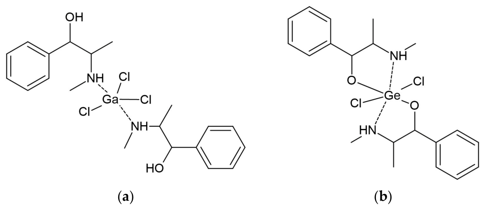
Ephedrine complexes with Mo(V), Nb(V), Ga(III) and Ge(IV) were reported with the general compositions [Mo(EPH)2Cl4]Cl, [Nb(EPH)2 Cl3 ], [Ga(EPH)2 Cl3 ] and [Ge(EPH)2 Cl2 ], as deduced from the microanalysis, FTIR spectra, molar conductance, and thermal analysis [106]. The structures of Ga(III) and Ge(IV) complexes are given in Figure 6. Ephedrine was proposed to act as a monodentate nitrogen donor in the Mo(V) and Ga(III) compounds, whereas N,O-bidentate coordination involving the deprotonated hydroxyl group was proposed for the Nb(V) and Ge(IV) complexes. The assigned coordination numbers ranged from five for Ga(III) to seven for Nb(V). As crystallographic structures were not obtained, a different arrangement of ligands cannot be excluded.
Figure 6.
Proposed structures of complexes between ephedrine and (a) Ga(III) and (b) Ge(IV), in which ephedrine acts as a monodentate and bidentate ligand [106]. The results were obtained from microanalysis, FTIR, molar conductance, and thermal analysis data.
5.2. Tyramine
Tyramine (TYR), 4-(2-aminoethyl)phenol, is a naturally occurring biogenic amine containing amino and phenolic groups. It is commonly used to examine the role of the sympathetic nervous system in the pathology and physiology of the human body [107]. Nevertheless, direct coordination of unmodified tyramine has rarely been investigated. In most reported complexes, tyramine is chemically modified, especially by condensing its amino group with aldehydes or ketones to form Schiff bases. This modification introduces an azomethine nitrogen atom and, depending on the carbonyl precursor, additional nitrogen or oxygen donor atoms, which provide more favorable chelating arrangements than those of the parent molecule.
To the best of our knowledge, the only complex containing unmodified tyramine is the Ru(II) complex [Ru(bpy)2(TYR)2]2+, in which two tyramine molecules coordinate monodentately through their amino nitrogen atom [108]. The resulting octahedral coordination sphere contains two bidentate bipyridine ligands and two tyramine ligands. Irradiation with visible light releases one tyramine molecule and forms the corresponding monoaqua complex, allowing the Ru(II) fragment to act as a photolabile protecting group. The structures of starting complexes and photodegrading products were examined by NMR spectroscopy and spectrofluorimetry, while no further spectroscopic characterization was performed. This research was motivated by the need for caged metal complexes that are photolabile and can deliver biomolecules to the desired position by using wavelengths that do not harm the surrounding tissue. [109,110].
More commonly, tyramine has been incorporated into larger ligand frameworks without directly coordinating the primary metal center. Tyramine-derived dipicolylamine units were attached to tricarbonylrhenium(I) polypyridine complexes of the general formula [Re(N∧N)(CO)3(py-TU-DPAT)]+ (py-TU-DPAT = 3-(2-(4-hydroxy-3-(2,20-dipicolylaminomethyl)phenyl)ethylthioureidyl)pyridine; N∧N=1,10-phenanthroline (phen)) [111] (Table 4). In these structures, Re(I) adopts the usual facial tricarbonyl environment and is coordinated by a bidentate polypyridine ligand and a pyridine nitrogen atom. The modified tyramine fragment contains a dipicolylamine binding site that subsequently coordinates Zn(II) or Cd(II), thereby altering the luminescence properties of the Re(I) complex. This type of Re(I) complex possesses desirable properties: it can be internalized by cancer cells and exhibits interesting emission properties [112,113].
Several Cu(II), Ni(II), Co(II), and Zn(II) complexes were prepared with a Schiff base obtained from tyramine, 4-aminoantipyrine, and 3,4-dimethoxybenzaldehyde [114]. The complexes had the general formula [ML2Cl2] (3,4-dimethoxybenzylidene-4-aminoantipyrinyl-4-aminoethylphenol (L)), and spectroscopic results supported octahedral geometries. In this case, the coordination properties are mainly determined by the newly introduced azomethine and carbonyl donor groups rather than by the original amino group of tyramine. The obtained complexes interacted with CT-DNA through minor groove binding, as one of the primary cellular targets for many anticancer drugs [115].
An isatin–tyramine Schiff base (L) was similarly used for the preparation of [CuL2Cl2], [NiL2Cl2], and [ZnL2Cl2] complexes [116]. The modified tyramine ligand acted as a bidentate donor, while two chlorido ligands completed the octahedral coordination sphere. A distorted octahedral geometry was proposed for the Cu(II) compound, whereas the Ni(II) and Zn(II) complexes were assigned regular octahedral arrangements. These compounds promoted the cleavage of pYC19 DNA from supercoiled form I to form II (open circular form) in the presence of hydrogen peroxide. Similar Schiff base complexes were described in [15,117,118].
5.3. Mescaline
Mescaline (3,4,5-trimethoxyphenethylamine) is a naturally occurring psychedelic phenethylamine alkaloid [119] whose coordination chemistry has received very limited attention. To the best of our knowledge, only one study has reported the synthesis and characterization of Cu(II) complexes of mescaline [28]. The authors prepared several chloride- and bromide-containing complexes, including Cu2OCl6(MES)4, Cu(MES)2Cl2·H2O, Cu(MES)2Br2, and the ionic salt (MH+)2(CuCl42−) (MES = mescaline), which were characterized by elemental analysis, molar conductivity, infrared and electronic spectroscopy, mass spectrometry, and powder X-ray diffraction, as given in Table 4. Spectroscopic evidence indicated that coordination occurs predominantly through the terminal amino nitrogen, whereas the three methoxy substituents remain largely uninvolved in direct metal binding. The authors proposed these complexes as model systems for investigating the possible role of essential transition-metal ions in the biological activity of hallucinogenic compounds. Despite the historical significance of this work, no further studies on mescaline coordination compounds have been reported, making its coordination chemistry largely unexplored.
Table 4.
Selected studies on metal complexes of phenethylamine derivatives.
6. Indole Alkaloids
6.1. Tryptamine
Tryptamine (TRA) is an indole alkaloid and biogenic amine containing a terminal primary amino group and an indole heterocycle, both of which contribute to its coordination chemistry and synthesis of other bioactive compounds [120,121]. It shows antioxidant, antibacterial, and anti-inflammatory activity [122]. Tryptamine is a parent molecule for a large class of ligands used for the synthesis of transition and main group metal complexes [123]. In most reported complexes, direct coordination occurs through the terminal amino nitrogen, while the indole nitrogen generally remains non-coordinating [14]. Nevertheless, the indole ring plays an important structural role by participating in π-stacking, hydrogen bonding, cation–π interactions, and, in some cases, direct metal–π interactions that influence the geometry, electronic structure, and reactivity of the complexes [124,125].
An example of a simple coordination compound is a palladium(II) complex of composition cis-[PdCl2(TRA)2], in which tryptamine acts as a monodentate ligand through the amino nitrogen [14]. This inclusion of bioactive components as ligands is performed to reduce the toxicity and increase the efficacy of the obtained compounds [126]. Elemental analysis, ESI-TOF mass spectrometry, and thermogravimetric studies, together with DFT calculations, confirmed the cis geometry and amino-nitrogen coordination, as shown in Figure 7. The preferred isomer of the obtained complex was proven by comparison with cisplatin, as the same type of interactions around the central metal ion are formed [127]. DFT results should be taken with caution, as optimization usually involves species in vacuum that neglect solid-state or solution interactions; therefore, differences between crystallographic and theoretical structures are possible.
Figure 7.
Proposed structure of tryptamine-Pd(II) complex from ref. [14]. The structure is proposed based on elemental analysis, ESI-TOF mass spectrometry, thermogravimetric studies, and DFT.
Lomenzo and coworkers presented an interesting discussion on the formation of a complex between tryptamine and the (η5-C5Me5)Ru moiety, as previous research showed that the interaction between the central metal ion and the six-electron arene of the indole moiety is stable [128]. Based on the NMR results, it was concluded that the nucleophilic reaction of the amino group of tryptamine with ruthenium occurs. The attempts to convert the obtained σ complex to an entropically favored structure with the η6-tryptamine ligand were not successful.
Beyond simple amino-coordinated complexes, most reported studies have focused on chemically modifying the tryptamine scaffold to generate multidentate ligands [129]. Condensation of the terminal amino group with aldehydes or ketones affords Schiff bases capable of coordinating through N, O- or N, N, O-donor sets, leading to stable complexes with transition metals including Cu(II), Mn(II), Co(II), Zn(II), Cd(II), Fe(III), Ru(III), Rh(III), Re(V), and Ni(II) [42,48,130,131,132,133]. In other approaches, incorporation of pyridyl- or imidazolyl-containing donor groups into the tryptamine framework has produced copper complexes that serve as structural and functional models for copper-containing metalloenzymes, highlighting the importance of secondary interactions between the indole ring and the metal coordination sphere [129].
Beyond expanding the structural diversity of coordination compounds, these modifications have been driven primarily by medicinal applications. Tryptamine-derived Schiff base complexes and ligands have been investigated for antimicrobial, antioxidant, anticancer, gastroprotective, and radical-scavenging activities, while several studies demonstrated that complexation can substantially alter the biological properties of the parent ligand [42,130,131,133,134].
6.2. Serotonin
Serotonin (5-hydroxytryptamine) is an indoleamine neurotransmitter containing a primary amino group, an indole ring, and a phenolic hydroxyl group, all of which may potentially contribute to metal binding [135,136]. Nevertheless, reports describing well-defined coordination compounds of serotonin remain relatively scarce. Most studies have focused on serotonin interactions with biologically relevant metal ions or metallomacrocycles rather than on the synthesis of discrete coordination complexes. For example, Oni and Nyokong suggested that serotonin interacts with iron(II) tetrasulfophthalocyanine, followed by internal electron transfer to afford a reduced iron(I) phthalocyanine species [13]. Spectroscopic and kinetic investigations showed that the coordination process is first-order in both serotonin and the iron complex, while the resulting metal–indoleamine framework stabilizes serotonin adducts. The authors concluded that the complex formed solely based on changes in UV–VIS spectra and NMR chemical shifts in a dried mixture of the iron(II) complex and serotonin, which limits further discussion of the possible interaction type or the geometry of any newly formed species.
In addition to transition-metal complexes, serotonin has been shown to interact with several physiologically relevant metal ions, including Li+, K+, Na+, and Al3+, whereas Ca2+ exhibits only weak affinity [137]. The interactions were studied by NMR spectroscopy in DMSO solution of a dried mixture containing serotonin and metal salts, which does not prove that new species formed, only that interactions were present in the solid state. Electrochemical studies demonstrated that lithium and potassium form the most stable serotonin complexes, suggesting that metal coordination may influence serotonin stability and metabolism. These interactions have been proposed to contribute to the pharmacological effects of lithium in the treatment of mood disorders by stabilizing serotonin against enzymatic degradation, while complexation with aluminum has been discussed in relation to its possible involvement in neurodegenerative disorders. Although the exact coordination sites remain unresolved, these findings highlight the biological significance of serotonin–metal interactions beyond classical coordination compounds.
6.3. Melatonin
Melatonin (N-acetyl-5-methoxytryptamine, MLT) is one of the most extensively investigated indoleamines in coordination chemistry owing to the presence of an amide carbonyl group, methoxy oxygen, indole nitrogen, and a flexible side chain capable of interacting with a variety of metal ions. It is involved in several important functions, such as regulating the sleep cycle, circadian rhythm, and blood pressure, and acts as an antidepressant [4,10,138]. Research has shown that endogenous melatonin acts as an antioxidant and protects DNA, proteins, and membrane lipids from free radical species [139,140,141]. In contrast to serotonin, numerous experimental and theoretical studies have shown that melatonin forms stable complexes with both transition and main-group metals and acts as an efficient metal-chelating agent [29].
In the previously mentioned paper by Romero and coworkers, initial electrochemical studies demonstrated that melatonin interacts with biologically relevant alkali metal ions, showing the strongest affinity toward K+ and Li+, followed by Na+ and Al3+, while the coordination of Ca2+ was weak [137]. Although the exact coordination sites could not be unequivocally assigned due to the mentioned limitations of the study, these investigations established melatonin as an effective endogenous metal-binding ligand under physiological conditions.
Subsequent spectroscopic and computational studies significantly improved the understanding of melatonin coordination. Gas-phase infrared spectroscopy combined with DFT calculations revealed that alkali and alkaline-earth metal ions preferentially coordinate through the amide carbonyl oxygen and methoxy oxygen atoms, whereas alternative binding modes involving π-interactions are energetically less favorable [29]. For Mg2+ complexes, deprotonation at the amide nitrogen may also occur, giving rise to different coordination motifs. Density functional calculations further demonstrated that melatonin and, in particular, its metabolites (3-hydroxymelatonin, AFMK, and AMK) efficiently chelate Cu(II) through direct or coupled deprotonation–chelation mechanisms, thereby suppressing Cu-catalyzed oxidative stress and Fenton-type chemistry [142]. Similar computational investigations suggested that melatonin metabolites may also form thermodynamically stable Pb(II) complexes, supporting their potential application as endogenous chelating agents [143]. This procedure was proposed as an alternative to EDTA treatment. These theoretical studies and structural elucidations are approximate because the results depend significantly on the starting geometry for optimization.
Several discrete coordination compounds of melatonin have also been reported. A nanostructured Au(III) complex, formulated as [Au(MLT)2Cl(H2O)] (Figure 8 and Table 5), was synthesized in which melatonin behaves as a monodentate ligand around a square-planar Au(III) center, while exhibiting enhanced antioxidant and anti-inflammatory properties compared with the free ligand [144]. These conclusions were drawn from FTIR spectra and elemental analyses, while the binding position of chlorine in the complex was confirmed by measuring molar conductance. No additional crystallographic structures were obtained, leaving the possibility of alternative structures. More recently, Cu(II) complexes containing melatonin together with amino acid ligands were prepared and characterized by FTIR spectroscopy [145]. The observed spectral changes suggested the participation of the amide functionality in coordination, although the authors noted that melatonin metal complexes remain relatively uncommon because their synthesis is experimentally demanding. The prepared compound also showed a high antioxidant capacity.
Figure 8.
Proposed structure of melatonin-Au(III) complex from ref. [144], based on FTIR, elemental analysis and molar conductance.
A significant advance was reported in 2026 with the first single-crystal structure of a melatonin-coordinated magnesium porphyrin [146]. In this complex, melatonin coordinates axially through the carbonyl oxygen atom to the Mg(II) center (Mg–O = 2.008 Å), yielding a five-coordinate square-pyramidal geometry. Structural, spectroscopic, electrochemical, and DFT studies showed that axial coordination substantially modifies the photophysical properties and enhances the photostability of the magnesium porphyrin, illustrating melatonin’s ability to act as an effective neutral O-donor ligand.
Table 5.
Selected studies on metal complexes of indole alkaloids.
7. Phenothiazine Psychoactive Drugs
7.1. Promazine
Promazine is one of the earliest investigated phenothiazine derivatives in coordination and analytical chemistry. This group of chemicals includes versatile anticholinergic and antihistamine compounds [147]. Similar to other members of this class, promazine contains a tricyclic phenothiazine scaffold incorporating sulfur and nitrogen heteroatoms together with a dimethylaminopropyl side chain, providing several potential interaction sites for metal ions [5,8]. However, compared with promethazine, its coordination chemistry remains relatively limited, and much of the available literature focuses on analytical applications rather than on the synthesis of well-defined coordination compounds.
One of the earliest studies demonstrated that Pd(II) readily interacts with promazine hydrochloride in acetate buffer to form stable colored complexes exhibiting absorption maxima at approximately 460 and 540 nm [148]. The proposed structure had a ratio of 1:1 (metal: ligand) determined by Job’s method, although the authors did not present additional chemical characterization. Because of their relatively high stability and sensitivity, these complexes were successfully applied to the spectrophotometric determination of palladium, demonstrating the phenothiazine framework’s strong affinity for soft transition-metal ions.
Promazine readily forms sparingly soluble ion-association compounds with metal complex anions, including ferricyanide, ammonium reineckate, and cobaltinitrite [149]. These ion associates were characterized by elemental analysis, infrared spectroscopy, thermogravimetric analysis, and solubility measurements, allowing the determination of their thermodynamic stability. Although these species do not represent classical coordination compounds, they are structurally well-defined supramolecular assemblies that have found extensive application in analytical chemistry owing to their low solubility and reproducible stoichiometry.
More generally, phenothiazine derivatives, including promazine, readily produce binary and ternary ion-association compounds with halide and thiocyanate complexes of numerous transition metals, such as Co(II), Pd(II), Fe(III), Cr(III), Au(III), Ti(IV), Pt(IV), Mo(V), W(V), and U(VI) [150]. These intensely colored species extract easily into organic solvents and have served as the basis for numerous spectrophotometric procedures for determining both phenothiazine drugs and metal ions.
7.2. Promethazine
Promethazine (PRO) exhibits considerably richer coordination chemistry than promazine and represents one of the best-studied phenothiazine ligands. In addition to the phenothiazine sulfur atom, the molecule contains a tertiary amino nitrogen in the dimethylaminopropyl side chain and a heterocyclic ring nitrogen, allowing several coordination modes depending on the metal ion and reaction conditions.
Geary and coworkers described a series of complexes with formula [M(PROH)X3], in which PROH is the protonated form of promethazine, X is Cl or Br, and M is Pd(II) or Pt(II) [151]. Single-crystal X-ray diffraction of the palladium complex revealed an unusual coordination mode in which the Pd(II) center binds exclusively to the sulfur atom of the phenothiazine ring, while the tertiary amino group remains protonated (Figure 9 and Table 6). The side chain adopts a folded conformation that enables intramolecular N–H···Cl hydrogen bonding with the coordinated palladium chloride fragment, stabilizing the crystal structure. Infrared, NMR, and electronic spectroscopic studies indicated that the corresponding platinum(II) complexes possess closely related structural features. Additionally, polynuclear Rh(II/III) chloride complexes with formula [Rh3(PRO)2(OH)2Cl8]Cl have been synthesized and characterized spectroscopically in reference [152]. Complexes with Zn(II) and Cd(II) with PRO contained protonated species and yielded compounds of the following formulas: [ZnCl(PROH)2]Cl3 [153] and [CdCl(PROH)2]Cl3, although these formulas were determined by elemental analyses, molar conductivity, and spectroscopy [154].
Figure 9.
Proposed structure of promethazine-Pd(II) complex from ref. [151].
An important extension of promethazine coordination chemistry involves lanthanide(III) nitrates. A complete series of complexes having the general formula [Ln(PRO)2(NO3)2]NO3 (Ln = La–Lu) has been successfully characterized [155]. Infrared spectroscopic analysis demonstrated that promethazine behaves as a bidentate ligand, coordinating through the heterocyclic ring nitrogen and the tertiary amino nitrogen of the side chain, whereas two nitrate ions coordinate in a bidentate fashion and the third nitrate remains outside the coordination sphere. As crystallographic data were not presented, other structures cannot be excluded.
Solution studies further expanded the coordination chemistry of promethazine through mixed-ligand Cu(II) complexes [156]. Potentiometric and spectroscopic investigations demonstrated the formation of stable binary and ternary complexes with amino acids, dicarboxylic acids, amides, and DNA constituents. Some of these complexes were isolated and, by infrared spectroscopy, thermal analysis, and magnetic measurements, were shown to adopt square-planar coordination around Cu(II). Equilibrium studies further revealed that complexes containing aromatic amino acids are more thermodynamically stable than those formed with aliphatic amino acids, highlighting the importance of secondary ligand structure in mixed-ligand systems. Similar to promazine, promethazine readily forms ion-association compounds with the previously mentioned ions in [149].
7.3. Chlorpromazine
Chlorpromazine (CPR), a first-generation phenothiazine antipsychotic, represents one of the most extensively investigated phenothiazine ligands in coordination chemistry. The structure contains the same possible donor positions as in promazine, although an additional chlorine atom is present [157,158]. Early studies on platinum-group metals demonstrated that chlorpromazine readily coordinates to Pd(II) and Pt(II), predominantly through the sulfur atom of the phenothiazine ring. More recently, researchers have also prepared platinum(IV) complexes. Reaction of chlorpromazine hydrochloride with hexachloroplatinate(IV) afforded a coordinated Pt(IV) complex in which direct coordination to the central metal ion was observed (Figure 10). These structural assignments were based on detailed spectroscopic analysis and molar conductivity, although no crystallographic data supported them, as shown in Table 6. Moreover, the Pt(IV) complex displayed antibacterial activity against both Gram-positive and Gram-negative bacteria, illustrating that metal coordination can modify the biological properties of the parent drug.
Figure 10.
Proposed structure of chlorpromazine-Pt(IV) complex from ref. [159], as elucidated from spectroscopic and elemental analyses and molar conductivity measurements.
One of the most remarkable coordination compounds reported for chlorpromazine is a μ-oxo tetranuclear copper(II) complex obtained by reacting chlorpromazine with CuCl2 in the presence of traces of water [160]. Single-crystal X-ray diffraction revealed a Cu4OCl6 core in which four copper atoms surround a central μ4-oxo bridge, while each chlorpromazine molecule coordinates through the terminal nitrogen atom of the dimethylaminopropyl side chain rather than through the sulfur atom of the phenothiazine ring. This represented the first structurally characterized example demonstrating that chlorpromazine can coordinate through the side-chain nitrogen, thereby establishing an alternative coordination mode for phenothiazine ligands. Each copper center adopts a distorted trigonal bipyramidal environment, and chloride bridges between adjacent copper atoms further stabilize the tetranuclear core.
Beyond structurally characterized complexes, chlorpromazine has also been investigated in solution through potentiometric studies. Binary complexes with Cu(II), Zn(II), Co(II), Ni(II), and UO22+ have been characterized together with corresponding ternary systems containing biologically relevant amino acids such as glycine, glutamate, and histidine [161]. As the author only applied pH measurements, it is difficult to determine the actual binding mode of CPR and other structural features of the obtained species. Stability constant measurements demonstrated the formation of both binary and mixed-ligand species, providing valuable models for understanding the interaction of chlorpromazine with biologically important metal ions under physiological conditions. Chlorprenazine readily forms colored species with Ru(III), allowing rapid and sensitive spectrophotometric determination of ruthenium without heating or solvent extraction [162]. The resulting red complex exhibits a characteristic absorption maximum near 530 nm and forms rapidly at room temperature under acidic conditions.
Similarly, chlorpromazine reacts with thiocyanatomolybdenum(V) species to produce an extractable orange ion-association compound of composition (CPR–H)[MoO(SCN)4], which has been successfully applied for the spectrophotometric determination of molybdenum [163]. In addition, the reversible oxidation of chlorpromazine to a colored radical cation has enabled its use as a redox indicator in cerium(IV)-based titrations of molybdenum(V), illustrating the combined importance of both coordination and redox chemistry in analytical applications.
More recently, chlorpromazine has become an important component of biologically active ruthenium complexes. Ruthenium(II)–N-alkylphenothiazine compounds containing chlorpromazine as the counter-ion have demonstrated enhanced cytostatic and prooxidant activity toward cancer cells compared with the free ligand, while simultaneously exhibiting lower genotoxicity toward normal lymphocytes [43]. These studies indicate that combining chlorpromazine with biologically active metal centers may offer promising strategies for developing novel metal-based therapeutics that leverage both the pharmacological activity of the phenothiazine scaffold and the unique properties of transition metals.
Table 6.
Selected studies on metal complexes of phenothiazine psychoactive drugs.
8. Conclusions and Future Prospects
Psychoactive nitrogen-containing compounds constitute a structurally diverse family of molecules that exhibit remarkably different coordination behavior depending on the nature, number, and spatial arrangement of their donor atoms. As demonstrated throughout this review, interactions with metal ions extend far beyond the formation of classical coordination compounds and include solution equilibria, ion-association systems, supramolecular assemblies, metal-mediated analytical reactions, and functional nanomaterials. Despite these differences, several general trends emerge. Oxygen-containing alkaloids, such as morphine, codeine, cocaine, and papaverine, predominantly coordinate through hydroxyl, carbonyl, ether, or isoquinoline nitrogen atoms, whereas phenothiazines and phenethylamines rely mainly on tertiary amino nitrogen atoms and sulfur atoms. In contrast, molecules such as berberine frequently require structural modification before acting as efficient ligands, illustrating how subtle differences in molecular architecture govern metal-binding behavior. The reviewed studies collectively demonstrate that transition metals, lanthanides, and biologically relevant metal ions provide a versatile platform for tuning the structural and physicochemical properties of psychoactive compounds.
Metal complexation frequently produces changes that extend beyond simple structural modification. Coordination may alter molecular geometry, electronic structure, lipophilicity, acid-base properties, and redox behavior, ultimately influencing interactions with biological targets. Numerous complexes summarized in this review exhibited enhanced antimicrobial, antioxidant, anticancer, DNA-binding, or protein-binding properties compared with the parent ligands, indicating that metal coordination represents a valuable strategy for modulating biological activity. However, these reported biological effects were obtained using different experimental models, cell lines or microorganisms, concentrations, exposure conditions, and assay protocols. Consequently, the activities reported in different studies should not be directly compared, and apparent differences in potency should be interpreted within the context of the individual experimental conditions. At the same time, studies of solution interactions with physiologically relevant metal ions improve our understanding of the possible roles of endogenous metals in drug transport, metabolism, receptor recognition, and pharmacological response. Although these relationships remain incompletely understood for many psychoactive compounds, they clearly demonstrate that coordination chemistry provides valuable mechanistic insight into their biological behavior.
Despite the considerable progress achieved during the past decades, significant challenges remain. For many psychoactive compounds, only a limited number of structurally characterized complexes have been reported, while systematic comparisons of coordination preferences across different classes are still lacking. Future investigations should therefore combine single-crystal X-ray diffraction, advanced spectroscopic techniques, computational chemistry, and solution-equilibrium studies to establish reliable structure–property relationships. Particular attention should also be devoted to the stability of the complexes and to metal–ligand speciation under physiologically relevant conditions, since the species present in biological media may differ from those isolated and characterized in the solid state. It is advisable to examine biologically relevant metal ions under physiological conditions, competitive binding in multicomponent biological environments, and the influence of metal complexation on pharmacokinetics and metabolism. The integration of experimental and theoretical approaches will undoubtedly accelerate the rational design of new metal-based therapeutic agents, functional materials, and analytical platforms, ensuring that coordination chemistry remains an important component of future research on psychoactive nitrogen-containing compounds.
Funding
The author acknowledges receiving financial support from the Ministry of Science, Technological Development, and Innovation of the Republic of Serbia (Nos. 451-03-34/2026-03/200146 and 451-03-33/2026-03/200146).
Institutional Review Board Statement
Not applicable.
Informed Consent Statement
Not applicable.
Data Availability Statement
No new data were created or analyzed in this study. Data sharing is not applicable to this article.
Conflicts of Interest
The author declares no conflicts of interest. The funders had no role in the design of the study; in the collection, analyses, or interpretation of data; in the writing of the manuscript; or in the decision to publish the results.
Abbreviations
The following abbreviations are used in this manuscript:
| Ad | Adenine |
| AFMK | N1-acetyl-N2-formyl-5-methoxykynuramine |
| AMK | N1-acetyl-5-methoxykynuramine |
| BSA | Bovine serum albumin |
| bpy | 2,2′-bipyridine |
| CAF | Caffeine |
| CBB | 9-O-carboxybenzyl derivative of berberine |
| CD | Circular dichroism |
| COC | Cocaine |
| COCH | Protonated cocaine |
| COD | Codeine |
| CPR | Chlorpromazine |
| CT-DNA | Calf thymus DNA |
| DFT | Density functional theory |
| DMSO | Dimethyl sulfoxide |
| DNA | Deoxyribonucleic acid |
| DSC | Differential scanning calorimetry |
| DTA | Differential thermal analysis |
| DTG | Derivative thermogravimetry |
| EDTA | Ethylenediaminetetraacetic acid |
| EPH | Ephedrine |
| EPR | Electron paramagnetic resonance |
| FTIR | Fourier-transform infrared spectroscopy |
| HRMS | High-resolution mass spectrometry |
| HSA | Human serum albumin |
| Ibu | Ibuprofen |
| MALDI-TOF | Matrix-assisted laser desorption/ionization time-of-flight mass spectrometry |
| MES | Mescaline |
| MLT | Melatonin |
| MO | Molecular orbital |
| NHC | N-heterocyclic carbene |
| NMR | Nuclear magnetic resonance spectroscopy |
| PAP | Papaverine |
| phen | 1,10-phenanthroline |
| PRO | Promethazine |
| PROH | Protonated promethazine |
| PSE | Pseudoephedrine |
| SEM | Scanning electron microscopy |
| SERS | Surface-enhanced Raman spectroscopy |
| TEM | Transmission electron microscopy |
| TG | Thermogravimetry |
| TGA | Thermogravimetric analysis |
| TMEDA | N,N,N′,N′-tetramethylethylenediamine |
| TRA | Tryptamine |
| TYR | Tyramine |
| UV–Vis | Ultraviolet–visible spectroscopy |
| XRD | X-ray diffraction |
References
- Arbo, M.D.; Bulcão, R.P.; Rossato, L.G.; Callegari, E.; Cendron, L.H.; Cabrera, E.R.; Freitas, M.A.D.; Leal, M.B. Drugs of Abuse and the Internet. In Neuropathology of Drug Addictions and Substance Misuse; Elsevier: Amsterdam, The Netherlands, 2016; pp. 50–60. [Google Scholar]
- Lakstygal, A.M.; Kolesnikova, T.O.; Khatsko, S.L.; Zabegalov, K.N.; Volgin, A.D.; Demin, K.A.; Shevyrin, V.A.; Wappler-Guzzetta, E.A.; Kalueff, A.V. DARK Classics in Chemical Neuroscience: Atropine, Scopolamine, and Other Anticholinergic Deliriant Hallucinogens. ACS Chem. Neurosci. 2019, 10, 2144–2159. [Google Scholar] [CrossRef] [Scilit] [PubMed]
- Gülçin, I. In Vitro Antioxidant Properties of Morphine. Pharmacol. Res. 2004, 49, 59–66. [Google Scholar] [CrossRef] [Scilit] [PubMed]
- Simko, F.; Paulis, L. Melatonin as a Potential Antihypertensive Treatment. J. Pineal Res. 2007, 42, 319–322. [Google Scholar] [CrossRef] [Scilit] [PubMed]
- Manzur, M.E.; Brandán, S.A. S(-) and R(+) Species Derived from Antihistaminic Promethazine Agent: Structural and Vibrational Studies. Heliyon 2019, 5, e02322. [Google Scholar] [CrossRef] [Scilit] [PubMed]
- Gilchrist, R.M. The Treatment of Cancer with Cocaine. BMJ 1909, 1, 274–275. [Google Scholar] [CrossRef] [Scilit] [PubMed][Green Version]
- Dimić, D. Emerging Novel Psychoactive Substances (2020–2025): GC-MS Approaches for Separation, Detection, and Characterization. Chemosensors 2025, 13, 426. [Google Scholar] [CrossRef] [Scilit]
- Falkenberg, G.; Ringertz, H. Crystallographic Data of Promazine and Chlorpromazine. Acta Cryst. 1967, 23, 1111. [Google Scholar] [CrossRef] [Scilit] [PubMed]
- Iwata, Y.T.; Inoue, H.; Kuwayama, K.; Kanamori, T.; Tsujikawa, K.; Miyaguchi, H.; Kishi, T. Forensic Application of Chiral Separation of Amphetamine-Type Stimulants to Impurity Analysis of Seized Methamphetamine by Capillary Electrophoresis. Forensic Sci. Int. 2006, 161, 92–96. [Google Scholar] [CrossRef] [Scilit] [PubMed]
- Hardeland, R.; Pandi-Perumal, S.R.; Cardinali, D.P. Melatonin. Int. J. Biochem. Cell Biol. 2006, 38, 313–316. [Google Scholar] [CrossRef] [Scilit] [PubMed]
- Guindi, E.L.; Nabawia, M. Composition and Stability of Complexes of Ephedrine Hydrochloride with Ni2+, Zn2+, and Cd2+ as Toxic Ions. Sci. Pharm. 2003, 71, 75–88. [Google Scholar] [CrossRef] [Scilit]
- Ghamami, S.; Golzani, M.; Lashgari, A. Synthesis, Characterization and Biological Properties of New Codeine Fe(III) Complex. J. Chil. Chem. Soc. 2016, 61, 2954–2957. [Google Scholar] [CrossRef] [Scilit]
- Oni, J.; Nyokong, T. Interaction between Iron(II) Tetrasulfophthalocyanine and the Neurotransmitters, Serotonin and Dopamine. Polyhedron 2000, 19, 1355–1361. [Google Scholar] [CrossRef] [Scilit]
- Carvalho, M.A.; Arruda, E.G.R.; Profirio, D.M.; Gomes, A.F.; Gozzo, F.C.; Formiga, A.L.B.; Corbi, P.P. Chemical and Spectroscopic Characterizations, ESI-QTOF Mass Spectrometric Measurements and DFT Studies of New Complexes of Palladium(II) with Tryptamine and Mefenamic Acid. J. Mol. Struct. 2015, 1100, 6–13. [Google Scholar] [CrossRef] [Scilit]
- Asha, V.A.; Jaabil, G.; Alphonse, N.R.; Anusha, A. Spectroscopic, Thermal, Computational and Ligand Field Investigations of Co(II), Ni(II), Cu(II) and Zn(II) Complexes of a Vanillin–Tyramine Schiff Base with Antimicrobial Evaluation. Polyhedron 2026, 297, 118297. [Google Scholar] [CrossRef] [Scilit]
- Sakurai, H. A New Concept: The Use of Vanadium Complexes in the Treatment of Diabetes Mellitus. Chem. Rec. 2002, 2, 237–248. [Google Scholar] [CrossRef] [Scilit] [PubMed]
- Mir, I.A.; Ain, Q.U.; Qadir, T.; Malik, A.Q.; Jan, S.; Shahverdi, S.; Nabi, S.A. A Review of Semicarbazone-Derived Metal Complexes for Application in Biomedicine and Related Fields. J. Mol. Struct. 2024, 1295, 136216. [Google Scholar] [CrossRef] [Scilit]
- Turecka, K.; Chylewska, A.; Rychłowski, M.; Zakrzewska, J.; Waleron, K. Antibacterial Activity of Co(III) Complexes with Diamine Chelate Ligands against a Broad Spectrum of Bacteria with a DNA Interaction Mechanism. Pharmaceutics 2021, 13, 946. [Google Scholar] [CrossRef] [Scilit] [PubMed]
- Poladian, Q.; Şahin, O.; Karakurt, T.; İlhan-Ceylan, B.; Kurt, Y. A New Zinc(II) Complex with N2O2-Tetradentate Schiff-Base Derived from Pyridoxal-S-Methylthiosemicarbazone: Synthesis, Characterization, Crystal Structure, DFT, Molecular Docking and Antioxidant Activity Studies. Polyhedron 2021, 201, 115164. [Google Scholar] [CrossRef] [Scilit]
- Rudra, S.; Dasmandal, S.; Patra, C.; Kundu, A.; Mahapatra, A. Binding Affinities of Schiff Base Fe(II) Complex with BSA and Calf-Thymus DNA: Spectroscopic Investigations and Molecular Docking Analysis. Spectrochim. Acta—Part A Mol. Biomol. Spectrosc. 2016, 166, 84–94. [Google Scholar] [CrossRef] [Scilit] [PubMed]
- Rana, M.S.; Rayhan, N.M.A.; Emad, A.H.; Hossain, M.I.; Shah, M.M.; Kudrat-E-Zahan, M.; Hossen, M.F.; Asraf, M.A. Salicylaldehyde-Based Schiff Bases and Their Transition Metal Complexes: An Overview on Synthesis and Biological Activities. J. Coord. Chem. 2025, 78, 937–1006. [Google Scholar] [CrossRef] [Scilit]
- Alayyafi, A.A.; Diab, M.A.; El-Sonbati, A.Z.; Negm, E.; El-Zahed, M.M.; Morgan, S.M. Synthesis of Metal(II) Acetate Complexes with a Heterocyclic Azo Dye Ligand: Characterization, Geometrical Molecular Structures, Antioxidant, Biological Activities, DNA Binding and Molecular Docking Studies. J. Mol. Struct. 2025, 1322, 140223. [Google Scholar] [CrossRef] [Scilit]
- Lv, L.; Zheng, T.; Tang, L.; Wang, Z.; Liu, W. Recent Advances of Schiff Base Metal Complexes as Potential Anticancer Agents. Coord. Chem. Rev. 2025, 525, 216327. [Google Scholar] [CrossRef] [Scilit]
- Mahmoud, N.H.; Fahim, A.M. The Effect of Anion, Steric Factors on the Catalytic Activity of Hydrogen Peroxide, Biological Activities, Docking, and DFT Calculations of Novel Mixed Ligand of Copper Complexes. Appl. Organomet. Chem. 2025, 39, e70220. [Google Scholar] [CrossRef] [Scilit]
- Felemban, M.F.; Tayeb, F.J.; Alqarni, A.; Adnan Ashour, D.A.; Shafie, D.A. Recent Advances in Schiff Base Coinage Metal Complexes as Anticancer Agents: A Comprehensive Review (2021–2025). Dye. Pigment. 2025, 237, 112710. [Google Scholar] [CrossRef] [Scilit]
- Kumar, N.; Kaushal, R.; Awasthi, P. Transition Metal Complexes as Antibacterial Agents: An Overview. J. Fluoresc. 2025, 35, 10445–10461. [Google Scholar] [CrossRef] [Scilit] [PubMed]
- Williams, C.J.; Morris, H.; Svorec, J.; Valková, M.; Valko, M.; Moncol, J.; Mazur, M.; Valach, F.; Melnik, M. A Study of Copper(II) Carboxylato Complexes with the Biological Ligands Nicotinamide and Papaverine. J. Mol. Struct. 2003, 659, 53–60. [Google Scholar] [CrossRef] [Scilit]
- Watt, G.W.; Durney, M.T. Copper(II) Complexes of β-Phenethylamine and Mescaline. Bioinorg. Chem. 1974, 3, 315–338. [Google Scholar] [CrossRef] [Scilit] [PubMed]
- Chakrabarty, S.; DiTucci, M.J.; Berden, G.; Oomens, J.; Williams, E.R. Structural Investigation of the Hormone Melatonin and Its Alkali and Alkaline Earth Metal Complexes in the Gas Phase. J. Am. Soc. Mass Spectrom. 2018, 29, 1835–1847. [Google Scholar] [CrossRef] [Scilit] [PubMed]
- Rabi, L.; Moutaouakkil, A.; Blaghen, M. Ionophoric Properties of Atropine: Complexation and Transport of Na+, K+, Mg2+ and Ca2+ Ions across a Liquid Membrane. Nat. Prod. Res. 2008, 22, 547–553. [Google Scholar] [CrossRef] [Scilit] [PubMed]
- Comparini, I. Conformational and Kinetic Features of the Morphine-Mn(II) Interaction as Probed by Nuclear Paramagnetic Resonance Investigations. J. Inorg. Biochem. 1983, 18, 349–360. [Google Scholar] [CrossRef] [Scilit]
- Nafisi, S.; Sadjadi, A.S.; Zadeh, S.S.; Damerchelli, M. Interaction of Metal Ions with Caffeine and Theophylline: Stability and Structural Features. J. Biomol. Struct. Dyn. 2003, 21, 289–296. [Google Scholar] [CrossRef] [Scilit] [PubMed]
- Ivanova, B. Gold(III) Complex of Caffeine: Synthesis, Isolation and Spectroscopic Characterization. Turk. J. Chem. 2007, 31, 97–103. [Google Scholar]
- Nash, D.J.; Siddhanti, D.A.; Wendt, A.F.; Penabade, R.A.; Bertrand, K.; Andino, A.; Smith, K.; Hick, S.; Kawamoto, K.; Ortiz, A.C.; et al. A Novel Method for the Presumptive Identification of Heterocyclic Amines of Forensic Interest Using Photoluminescent Copper(I) Iodide Cluster Compounds. Anal. Methods 2018, 10, 2217–2225. [Google Scholar] [CrossRef] [Scilit]
- Laussmann, T.; Grzesiak, I.; Krest, A.; Stirnat, K.; Meier-Giebing, S.; Ruschewitz, U.; Klein, A. Copper Thiocyanato Complexes and Cocaine—A Case of “Black Cocaine.”. Drug Test. Anal. 2015, 7, 56–64. [Google Scholar] [CrossRef] [Scilit] [PubMed]
- Smeliková, V.; Kopal, I.; Člupek, M.; Dendisová, M.; Švecová, M. Unveiling the Crucial Role of Chemical Enhancement in the SERS Analysis of Amphetamine–Metal Interactions on Gold and Silver Surfaces: Importance of Selective Amplification of the Narrow Interval of Vibrational Modes. Anal. Chem. 2024, 96, 5416–5427. [Google Scholar] [CrossRef] [Scilit] [PubMed]
- Murali, K.; Murugan, D.; Pathak, R.; Chauhan, A.; Munjal, S.; Usha, S.P.; Shrivastav, A.M. Forensic Detection: A Comprehensive Review of SERS Mechanisms and Applications. ACS Appl. Opt. Mater. 2026, 4, 695–722. [Google Scholar] [CrossRef] [Scilit]
- Kyndait, S.; Das, T.; Bokotial, D.; Chowdhury, A. Recognition of Illicit Date Rape Drugs: A Luminescence Sensing Perspective. Sens. Diagn. 2026, 5, 488–521. [Google Scholar] [CrossRef] [Scilit]
- Roig-Rubio, J.; Hernandez-Contreras, J.; Parra, M.; Gil, S.; Arroyo, P.; Sáez, J.A.; Gaviña, P. A “Turn-on” Fluorogenic Probe for the Rapid and on-Site Detection of Synthetic Cathinones, Ketamine and Scopolamine in Water and Beverages. Sens. Bio-Sens. Res. 2026, 51, 100987. [Google Scholar] [CrossRef] [Scilit]
- Garrido, E.; Pla, L.; Lozano-Torres, B.; El Sayed, S.; Martínez-Máñez, R.; Sancenón, F. Chromogenic and Fluorogenic Probes for the Detection of Illicit Drugs. ChemistryOpen 2018, 7, 401–428. [Google Scholar] [CrossRef] [Scilit] [PubMed]
- Gaber, A.; Alsanie, W.F.; Kumar, D.N.; Refat, M.S.; Saied, E.M. Novel Papaverine Metal Complexes with Potential Anticancer Activities. Molecules 2020, 25, 5447. [Google Scholar] [CrossRef] [Scilit] [PubMed]
- Apfelbaum, H.C.; Blum, J.; Mandelbaum-Shavit, F. Preparation and Anticancer Activity of Two Tryptamine Derived Platinum Complexes. Inorganica Chim. Acta 1991, 186, 243–246. [Google Scholar] [CrossRef] [Scilit]
- Leskovac, A.; Petrovic, S.; Lazarevic-Pasti, T.; Krstic, M.; Vasic, V. Ruthenium(II)-N-Alkyl Phenothiazine Complexes as Potential Anticancer Agents. J. Biol. Inorg. Chem. 2018, 23, 689–704. [Google Scholar] [CrossRef] [Scilit] [PubMed]
- Tella, A.C.; Obaleye, J.A.; Eke, U.B.; Isaac, A.Y.; Ameen, O.M. Solvent-Free Synthesis, X-Ray Studies and In Vitro Inhibitory Activities of Copper(II) Complexes of Non-Steroidal Anti-Inflammatory Drugs. Res. Chem. Intermed. 2014, 40, 1441–1457. [Google Scholar] [CrossRef] [Scilit]
- Wang, Z.-F.; Huang, X.-Q.; Wu, R.-C.; Qin, Q.-P.; Qin, A.; Zhang, S.-H.; Tan, M.-X.; Liang, H. Zinc(II)–Berberine-Based Complexes Targeting Mitochondria Exhibit Enhanced Antiproliferative Activity as Single Drugs or in Combination with ABT-199. Bioorg. Chem. 2026, 169, 109440. [Google Scholar] [CrossRef] [Scilit] [PubMed]
- Hamdani, H.E.L.; Amane, M.E.L. Preparation, Spectral, Antimicrobial Properties and Anticancer Molecular Docking Studies of New Metal Complexes [M(Caffeine)4] (PF6)2; M = Fe(II), Co(II), Mn(II), Cd(II), Zn(II), Cu(II), Ni(II). J. Mol. Struct. 2019, 1184, 262–270. [Google Scholar] [CrossRef] [Scilit]
- Abdallah, A.M.; Zaki, N.G.; El Kerdawy, A.M.; Mahmoud, W.H.; Mohamed, G.G. Coordination Behavior of Cocaine toward D-Block Metal Ions: Synthesis, Spectral Analysis, Density Functional Theory (DFT) Studies, and Chemotherapeutic Activity. J. Mol. Struct. 2023, 1293, 136301. [Google Scholar] [CrossRef] [Scilit]
- Buran, K. Benzoin-Tryptamine Schiff Base-Metal Complexes: Synthesis, DFT Calculation and Antimicrobial Activities. J. Mol. Struct. 2025, 1330, 141499. [Google Scholar] [CrossRef] [Scilit]
- Zaki, N.G.; Mahmoud, W.H.; El Kerdawy, A.M.; Abdallah, A.M.; Mohamed, G.G. Structural Characterization, Thermal, DFT, Cytotoxicity, and Antimetastatic Properties of Cocaine Complexes with La(III), Er(III), and Yb(III). Res. Chem. Intermed. 2020, 46, 3193–3216. [Google Scholar] [CrossRef] [Scilit]
- Wood, M.R.; Brettell, T.A.; Lalancette, R.A. The Gold(III) Tetrachloride Salt of L -Cocaine. Acta Crystallogr. C Cryst. Struct. Commun. 2007, 63, m33–m35. [Google Scholar] [CrossRef] [Scilit] [PubMed]
- Valach, F.; Pavlik, J.; Šalitroš, I.; Melník, M.; Kožíšek, J. Novel Trinuclear Copper(II) Complex: Crystal Structure at 100 K and Magnetic Properties of (R, S)-Di[(6,7-Dimethoxy-Isoquinolin-1-Yl)-(3,4-Dimethoxy-Phenyl)-Methanolato]-Tetra(2-Hydroxybenzoato)-Diaqua-Tricopper Dihydrate, [Cu3(C7H5O3)4(C20H20NO5)2(H2O)2]·2(H2O). J. Chem. Crystallogr. 2024, 54, 163–172. [Google Scholar] [CrossRef] [Scilit]
- Rageot, D.; Bohnacker, T.; Melone, A.; Langlois, J.-B.; Borsari, C.; Hillmann, P.; Sele, A.M.; Beaufils, F.; Zvelebil, M.; Hebeisen, P.; et al. Discovery and Preclinical Characterization of 5-[4,6-Bis({3-Oxa-8-Azabicyclo[3.2.1]Octan-8-Yl})-1,3,5-Triazin-2-Yl]-4-(Difluoromethyl)Pyridin-2-Amine (PQR620), a Highly Potent and Selective mTORC1/2 Inhibitor for Cancer and Neurological Disorders. J. Med. Chem. 2018, 61, 10084–10105. [Google Scholar] [CrossRef] [Scilit] [PubMed]
- Wang, Y.-J.; Zuo, Z.-X.; Zhang, M.; Feng, Z.-H.; Yan, M.; Li, X.-Y. The Analgesic Effects of (5R,6R)6-(3-Propylthio-1,2,5-Thiadiazol-4-Yl)-1-Azabicyclo[3.2.1] Octane on a Mouse Model of Neuropathic Pain. Anesth. Analg. 2017, 124, 1330–1338. [Google Scholar] [CrossRef] [Scilit] [PubMed]
- Liu, Y.; Lang, Y.; Patel, N.K.; Ng, G.; Laufer, R.; Li, S.-W.; Edwards, L.; Forrest, B.; Sampson, P.B.; Feher, M.; et al. The Discovery of Orally Bioavailable Tyrosine Threonine Kinase (TTK) Inhibitors: 3-(4-(Heterocyclyl)Phenyl)-1 H -Indazole-5-Carboxamides as Anticancer Agents. J. Med. Chem. 2015, 58, 3366–3392. [Google Scholar] [CrossRef] [Scilit] [PubMed]
- Abdallah, A.M.; Zaki, N.G.; Mahmoud, W.H.; El Kerdawy, A.M.; Mohamed, G.G. Synthesis, Structural Characterization, Density Functional Theory Calculations, and Antimicrobial, Anticancer, and Antimetastatic Properties of Nanosized Heteroleptic Complexes of Cocaine/TMEDA with D-block Metal Ions. Appl. Organomet. Chem. 2021, 35, e6441. [Google Scholar] [CrossRef] [Scilit]
- Zaki, N.G.; Mahmoud, W.H.; El Kerdawy, A.M.; Abdallah, A.M.; Mohamed, G.G. Heteroleptic Complexes of Cocaine/TMEDA with Some f Block Metals: Synthesis, DFT Studies, Spectral, Thermal, Cytotoxicity and Antimetastatic Properties. Spectrochim. Acta Part A Mol. Biomol. Spectrosc. 2020, 229, 117938. [Google Scholar] [CrossRef] [Scilit] [PubMed]
- Haddoub, R.; Ferry, D.; Marsal, P.; Siri, O. Cobalt Thiocyanate Reagent Revisited for Cocaine Identification on TLC. New J. Chem. 2011, 35, 1351. [Google Scholar] [CrossRef] [Scilit]
- Oiye, É.N.; Figueiredo, N.B.D.; Andrade, J.F.D.; Tristão, H.M.; Oliveira, M.F.D. Voltammetric Determination of Cocaine in Confiscated Samples Using a Cobalt Hexacyanoferrate Film-Modified Electrode. Forensic Sci. Int. 2009, 192, 94–97. [Google Scholar] [CrossRef] [Scilit] [PubMed]
- Mohammadzai, I.U.; Hinze, W.L. Indirect Atomic Absorption Determination of Cocaine via Formation and Liquid-liquid Extraction of the Iron(III)-cocaine Complex. J. Chin. Chem. Soc. 2004, 51, 1297–1302. [Google Scholar] [CrossRef] [Scilit]
- Vázquez, D.; Garcinuño, B.; Tejedor, J.; Pardo, R.; Deban, L. Polarographic Determination of the Stability Constant of the Ternary Complex Formed between Cocaine and Cobalt Thiocyanate. Toxicol. Environ. Chem. 2017, 99, 10–16. [Google Scholar] [CrossRef] [Scilit]
- Brook, K.; Bennett, J.; Desai, S.P. The Chemical History of Morphine: An 8000-Year Journey, from Resin to de-Novo Synthesis. J. Anesth. Hist. 2017, 3, 50–55. [Google Scholar] [CrossRef] [Scilit] [PubMed]
- Ahrar, J.; Nematullah, M.; Rahman, M.A. Insights into Morphine: Extraction, Structural Characterization, and Pharmacological Properties. LabMed Discov. 2025, 2, 100043. [Google Scholar] [CrossRef] [Scilit]
- Gaggelli, E.; Comparini, I.B.; Picchi, M.P. Divalent Metal Ions in the Pharmacodynamics of Morphine-like Opiates. Inorganica Chim. Acta 1983, 79, 303. [Google Scholar] [CrossRef] [Scilit]
- Kalinin, V.N.; Belyakova, I.L.; Derunov, V.V.; Park, J.K.; Schmidhammer, H. Synthesis and Properties of π-Complexes of Morphine Alkaloids with Palladium. Mendeleev Commun. 1995, 5, 22–23. [Google Scholar] [CrossRef] [Scilit]
- Kalinin, V.N.; Derunov, V.V.; Maat, L.; Park, J.K.; Schmidhammer, H. Synthesis of π-Complexes of Codeine with Iron and Molybdenum. Mendeleev Commun. 1995, 5, 222–223. [Google Scholar] [CrossRef] [Scilit]
- Zayed, M.A.; El-shahat, M.F.; Abdullah, S.M. The Use of IR, Magnetism, Reflectance, and Mass Spectra Together with Thermal Analyses in Structure Investigation of Codeine Phosphate Complexes of d-Block Elements. Spectrochim. Acta Part A Mol. Biomol. Spectrosc. 2005, 61, 1955–1964. [Google Scholar] [CrossRef] [Scilit] [PubMed]
- Zayed, M.A.; Hawash, M.F.; Fahmey, M.A. Structure Investigation of Codeine Drug Using Mass Spectrometry, Thermal Analyses and Semi-Emperical Molecular Orbital (MO) Calculations. Spectrochim. Acta Part A Mol. Biomol. Spectrosc. 2006, 64, 363–371. [Google Scholar] [CrossRef] [Scilit] [PubMed]
- Liu, J.K.; Couldwell, W.T. Intra-Arterial Papaverine Infusions for the Treatment of Cerebral Vasospasm Induced by Aneurysmal Subarachnoid Hemorrhage. Neurocrit Care 2005, 2, 124–132. [Google Scholar] [CrossRef] [Scilit] [PubMed]
- Sayama, C.M.; Liu, J.K.; Couldwell, W.T. Update on Endovascular Therapies for Cerebral Vasospasm Induced by Aneurysmal Subarachnoid Hemorrhage. Neurosurg. Focus 2006, 21, 1–11. [Google Scholar] [CrossRef] [Scilit] [PubMed]
- Chappie, T.A.; Humphrey, J.M.; Allen, M.P.; Estep, K.G.; Fox, C.B.; Lebel, L.A.; Liras, S.; Marr, E.S.; Menniti, F.S.; Pandit, J.; et al. Discovery of a Series of 6,7-Dimethoxy-4-Pyrrolidylquinazoline PDE10A Inhibitors. J. Med. Chem. 2007, 50, 182–185. [Google Scholar] [CrossRef] [Scilit] [PubMed]
- Yildiz, N.; Gokkaya, N.K.O.; Koseoglu, F.; Gokkaya, S.; Comert, D. Efficacies of Papaverine and Sildenafil in the Treatment of Erectile Dysfunction in Early-Stage Paraplegic Men. Int. J. Rehabil. Res. 2011, 34, 44. [Google Scholar] [CrossRef] [Scilit] [PubMed]
- Nokta, M.; Albrecht, T.; Pollard, R. Papaverine Hydrochloride: Effects on HIV Replication and T-Lymphocyte Cell Function. Immunopharmacology 1993, 26, 181–185. [Google Scholar] [CrossRef] [Scilit] [PubMed]
- Afzali, M.; Ghaeli, P.; Khanavi, M.; Parsa, M.; Montazeri, H.; Ghahremani, M.H.; Ostad, S.N. Non-Addictive Opium Alkaloids Selectively Induce Apoptosis in Cancer Cells Compared to Normal Cells. DARU J. Pharm. Sci. 2015, 23, 16. [Google Scholar] [CrossRef] [Scilit] [PubMed]
- Goto, T.; Matsushima, H.; Kasuya, Y.; Hosaka, Y.; Kitamura, T.; Kawabe, K.; Hida, A.; Ohta, Y.; Simizu, T.; Takeda, K. The Effect of Papaverine on Morphologic Differentiation, Proliferation and Invasive Potential of Human Prostatic Cancer LNCaP Cells. Int. J. Urol. 1999, 6, 314–319. [Google Scholar] [CrossRef] [Scilit] [PubMed]
- Zeleňák, V.; Sabo, M.; Massa, W.; Černák, J. Tetra-μ-Benzoato-κ8O:O′-Bis({1-[(3,4-Dimethoxyphenyl)Methyl]-6,7-Dimethoxyisoquinoline-κN }zinc(II)): The First Crystal Structure with Papaverine as a Ligand. Acta Crystallogr. C Cryst. Struct. Commun. 2004, 60, m85–m87. [Google Scholar] [CrossRef] [Scilit] [PubMed]
- Györyová, K.; Kovářová, J.; Andogová, E.; Zeleňák, V.; Nour El-Dien, F.A. Thermal, Spectral and Antimicrobial Properties of New Zinc(II) Isobutyrate Compounds: Part I. Papaverine, Phenazone. J. Therm. Anal. Calorim. 2002, 67, 119–128. [Google Scholar] [CrossRef] [Scilit]
- Melni’k, M.; Potocˇnˇak, I.; Macásˇková, L.; Miklosˇ, D.; Holloway, C.E. Spectral Study of Copper(II) Flufenamates: Crystal and Molecular Structure of Biis(Flufenamato)DI(N,N-Diethylnicotinamide)Di(Aqua)Copper(II). Polyhedron 1996, 15, 2159–2164. [Google Scholar] [CrossRef] [Scilit]
- Basu, A.; Jaisankar, P.; Kumar, G.S. 9-O-N-Aryl/Arylalkyl Amino Carbonyl Methyl Substituted Berberine Analogues Induce Self-Structure in Polyadenylic Acid. RSC Adv. 2012, 2, 7714. [Google Scholar] [CrossRef] [Scilit]
- Giri, P.; Kumar, G.S. Self-Structure Induction in Single Stranded Poly(A) by Small Molecules: Studies on DNA Intercalators, Partial Intercalators and Groove Binding Molecules. Arch. Biochem. Biophys. 2008, 474, 183–192. [Google Scholar] [CrossRef] [Scilit] [PubMed]
- Chen, J.-X.; Lin, W.-E.; Chen, M.-Z.; Zhou, C.-Q.; Lin, Y.-L.; Chen, M.; Jiang, Z.-H.; Chen, W.-H. Synthesis, Characterization and Potent DNA-Cleaving Activity of Copper(II)-Complexed Berberine Carboxylate. Bioorg. Med. Chem. Lett. 2012, 22, 7056–7059. [Google Scholar] [CrossRef] [Scilit] [PubMed]
- Zhang, S.-L.; Fu, H.; Ma, Y.; Lin, Q.; Xu, Y.; Yang, Q.; He, P.; Wei, Z. A Novel Platinum(II) Complex with a Berberine Derivative as a Potential Antitumor Agent Targeting G-Quadruplex DNA. Org. Biomol. Chem. 2025, 23, 1112–1119. [Google Scholar] [CrossRef] [Scilit] [PubMed]
- Affrose, A.; Parveen, S.D.S.; Kumar, B.S.; Pitchumani, K. Selective Sensing of Silver Ion Using Berberine, a Naturally Occurring Plant Alkaloid. Sens. Actuators B Chem. 2015, 206, 170–175. [Google Scholar] [CrossRef] [Scilit]
- Francescato, G.; Leitão, M.I.P.S.; Orsini, G.; Petronilho, A. Synthesis and Medicinal Applications of N-Heterocyclic Carbene Complexes Based on Caffeine and Other Xanthines. ChemMedChem 2024, 19, e202400118. [Google Scholar] [CrossRef] [Scilit] [PubMed]
- Franklin, M.; Das, R. Carbohydrate-Decorated NHC–Metal Complexes: From Bioorganometallic Design to Biomedical Function. Coord. Chem. Rev. 2026, 565, 218102. [Google Scholar] [CrossRef] [Scilit]
- Nafisi, S.; Monajemi, M.; Ebrahimi, S. The Effects of Mono- and Divalent Metal Cations on the Solution Structure of Caffeine and Theophylline. J. Mol. Struct. 2004, 705, 35–39. [Google Scholar] [CrossRef] [Scilit]
- Bouhdada, M.; Amane, M.E.L.; El Hamzaoui, N. Synthesis, Spectroscopic Studies, X-Ray Powder Diffraction Data and Antibacterial Activity of Mixed Transition Metal Complexes with Sulfonate Azo Dye, Sulfamate and Caffeine Ligands. Inorg. Chem. Commun. 2019, 101, 32–39. [Google Scholar] [CrossRef] [Scilit]
- Jovanović-Stević, S.; Ćoćić, D.; Puchta, R.; Bogojeski, J.; Jurišević, M.; Gajović, N.; Jakovljević, S.; Arsenijević, N.; Jovanović, I.; Petrović, B. Assessment of Biological Activity of the Caffeine-Derived Pt (II) and Pd (II) Complexes. Appl. Organomet. Chem. 2022, 36, e6532. [Google Scholar] [CrossRef] [Scilit]
- Bysewski, O.; Winter, A.; Liebing, P.; Schubert, U.S. Noble Metal Complexes of a Bis-Caffeine Containing NHC Ligand. Molecules 2022, 27, 4316. [Google Scholar] [CrossRef] [Scilit] [PubMed]
- Mariconda, A.; Iacopetta, D.; Sirignano, M.; Ceramella, J.; D’Amato, A.; Marra, M.; Pellegrino, M.; Sinicropi, M.S.; Aquaro, S.; Longo, P. Silver and Gold Complexes with NHC-Ligands Derived from Caffeine: Catalytic and Pharmacological Activity. Int. J. Mol. Sci. 2024, 25, 2599. [Google Scholar] [CrossRef] [Scilit] [PubMed]
- Censi, V.; Caballero, A.B.; Pérez-Hernández, M.; Soto-Cerrato, V.; Korrodi-Gregório, L.; Pérez-Tomás, R.; Dell’Anna, M.M.; Mastrorilli, P.; Gamez, P. DNA-Binding and in Vitro Cytotoxic Activity of Platinum(II) Complexes of Curcumin and Caffeine. J. Inorg. Biochem. 2019, 198, 110749. [Google Scholar] [CrossRef] [Scilit] [PubMed]
- Kisku, T.; Paul, K.; Singh, B.; Das, S.; Mukherjee, S.; Kundu, A.; Rath, J.; Sekhar Das, R. Synthesis of Cu(II)-Caffeine Complex as Potential Therapeutic Agent: Studies on Antioxidant, Anticancer and Pharmacological Activities. J. Mol. Liq. 2022, 364, 119897. [Google Scholar] [CrossRef] [Scilit]
- Bouhdada, M.; Amane, M.E.; Hamdani, H.E.; Khiya, Z. Synthesis, Characterization, Biological Evaluation and Molecular Docking Studies of Salicylidene-Aniline and Their Metal Mixed Ligand Complexes with Caffeine. J. Mol. Struct. 2023, 1271, 134026. [Google Scholar] [CrossRef] [Scilit]
- Shaker, D.S.; Farina, Y.; Mahmmod, S.; Eskender, M. Synthesis and Characterization of Mixed Ligand Complexes of Caffeine, Adenine and Thiocyanate with Some Transition Metal Ions. Sains Malays. 2010, 39, 957–962. [Google Scholar]
- Borówka, A.; Sierosławska, A.; Baier, A.; Rymuszka, A.; Olszewska, E. Silver and Copper Complexes with Ibuprofen and Caffeine—Preparation and Evaluation of Their Selected Biological Effects. Molecules 2024, 29, 506. [Google Scholar] [CrossRef] [Scilit] [PubMed]
- Refat, M.S.; Ibrahim, O.B.; Saad, H.A.; Adam, A.M.A. Usefulness of Charge–Transfer Complexation for the Assessment of Sympathomimetic Drugs: Spectroscopic Properties of Drug Ephedrine Hydrochloride Complexed with Some π-Acceptors. J. Mol. Struct. 2014, 1064, 58–69. [Google Scholar] [CrossRef] [Scilit]
- Stoica, E.; Gavrila, A.-M.; Branger, C.; Brisset, H.; Dyshlyuk, A.; Vitrik, O.; Iovu, H.; Miron, A.; Sarbu, A.; Iordache, T.-V. Evaluation of Molecularly Imprinted Thin Films for Ephedrine Recognition. Mater. Plast. 2019, 56, 865–874. [Google Scholar] [CrossRef] [Scilit]
- Bents, R.T.; Tokish, J.M.; Goldberg, L. Ephedrine, Pseudoephedrine, and Amphetamine Prevalence in College Hockey Players: Most Report Performance-Enhancing Use. Physician Sportsmed. 2004, 32, 30–34. [Google Scholar] [CrossRef] [Scilit]
- Couper, F.; Pemberton, M.; Jarvis, A.; Hughes, M.; Logan, B. Prevalence of Drug Use in Commercial Tractor-Trailer Drivers. J. Forensic Sci. 2002, 47, 562–567. [Google Scholar] [CrossRef] [Scilit]
- Sierra, M.L.; De Mel, V.S.J.; Oliver, J.P. Synthesis and Spectroscopic Investigations of Optically Active Alkylaluminum Compounds of I-Ephedrine: The Crystal Structure of the [Me2AlO-(R)-CHC6H5(S)-CH((S)-NHCH3)CH3]2 Dimer: An Optically Active Five-Coordinate Aluminum Complex. Organometallics 1989, 8, 2486–2488. [Google Scholar] [CrossRef] [Scilit]
- Abu-Eittah, R.H.; Abdel Razik, A.; Mobarak, Z.; Kamel, L.T. A Study on the Complex Formation between Copper(II) and Some Ephedrines. Polyhedron 1993, 12, 2027–2037. [Google Scholar] [CrossRef] [Scilit]
- Shahabadi, N.; Akhtarshenas, S.; Hadidi, S. Synthesis, Characterization and DNA Interaction Studies of New Copper Complex Containing Pseudoephedrine Hydrochloride Drug. Nucleosides Nucleotides Nucleic Acids 2019, 38, 680–699. [Google Scholar] [CrossRef] [Scilit] [PubMed]
- Platts, M.; Smith, R.B.; Mould, N.; Davis, J. Ephedrine–Copper–Carbon Interactions: An Electroanalytical Investigation. Electrochem. Commun. 2006, 8, 633–637. [Google Scholar] [CrossRef] [Scilit]
- Alminshid, A.H.; Alalwan, H.A.; Abdulghani, H.A.; Mohammed, M.M. Spectrophotometric Study of Ephedrine Hydrochloride in Drug Using Molecular Absorption UV–Visible. Spectrochim. Acta Part A Mol. Biomol. Spectrosc. 2022, 270, 120828. [Google Scholar] [CrossRef] [Scilit] [PubMed]
- Rostami, A.; Pourshiani, O.; Darvishi, N.; Atashkar, B. Efficient and Green Oxidation of Alcohols with Tert-Butyl Hydrogenperoxide Catalyzed by a Recyclable Magnetic Core-Shell Nanoparticle-Supported Oxo-Vanadium Ephedrine Complex. Comptes Rendus. Chim. 2016, 20, 435–439. [Google Scholar] [CrossRef] [Scilit]
- AL-Majthoub, M.M.; Refat, M.S.; Adam, A.M.A.; Ahmed, M.A. Spectroscopic and Intermolecular Electron-Transfer Mechanism of Ferric (III) Ephedrine Complex as a Model of Drug Design. Int. J. Electrochem. Sci. 2013, 8, 7053–7061. [Google Scholar] [CrossRef] [Scilit]
- El-Habeeb, A.A.; Refat, M.S. Metal-Drug Interactions: Synthesis and Spectroscopic Characteristics, Surface Morphology, and Pharmacological Activity of Ephedrine–HCl Complexes with Mo(V), Nb(V), Ga(III), and Ge(IV). Russ. J. Gen. Chem. 2018, 88, 2163–2169. [Google Scholar] [CrossRef] [Scilit]
- Jacob, G.; Gamboa, A.; Diedrich, A.; Shibao, C.; Robertson, D.; Biaggioni, I. Tyramine-Induced Vasodilation Mediated by Dopamine Contamination: A Paradox Resolved. Hypertension 2005, 46, 355–359. [Google Scholar] [CrossRef] [Scilit] [PubMed][Green Version]
- Zayat, L.; Salierno, M.; Etchenique, R. Ruthenium(II) Bipyridyl Complexes as Photolabile Caging Groups for Amines. Inorg. Chem. 2006, 45, 1728–1731. [Google Scholar] [CrossRef] [Scilit] [PubMed]
- Adams, S.R.; Tsien, R.Y. Controlling Cell Chemistry with Caged Compounds. Annu. Rev. Physiol. 1993, 55, 755–784. [Google Scholar] [CrossRef] [PubMed]
- Zayat, L.; Calero, C.; Alborés, P.; Baraldo, L.; Etchenique, R. A New Strategy for Neurochemical Photodelivery: Metal−Ligand Heterolytic Cleavage. J. Am. Chem. Soc. 2003, 125, 882–883. [Google Scholar] [CrossRef] [Scilit] [PubMed]
- Louie, M.-W.; Liu, H.-W.; Lam, M.H.-C.; Lau, T.-C.; Lo, K.K.-W. Novel Luminescent Tricarbonylrhenium(I) Polypyridine Tyramine-Derived Dipicolylamine Complexes as Sensors for Zinc(II) and Cadmium(II) Ions. Organometallics 2009, 28, 4297–4307. [Google Scholar] [CrossRef] [Scilit]
- Kam-Wing Lo, K.; Chun-Ming Ng, D.; Hui, W.-K.; Cheung, K.-K. Luminescent Rhenium(i) Polypyridine Complexes with an Isothiocyanate Moiety–Versatile Labelling Reagents for Biomolecules. J. Chem. Soc. Dalton Trans. 2001, 30, 2634–2640. [Google Scholar] [CrossRef] [Scilit]
- Lo, K.K.-W.; Tsang, K.H.-K.; Hui, W.-K.; Zhu, N. Luminescent Rhenium(I) Diimine Indole Conjugates—Photophysical, Electrochemical and Protein-Binding Properties. Chem. Commun. 2003, 39, 2704–2705. [Google Scholar] [CrossRef] [Scilit] [PubMed]
- Raman, N.; Sobha, S.; Thamaraichelvan, A. A Novel Bioactive Tyramine Derived Schiff Base and Its Transition Metal Complexes as Selective DNA Binding Agents. Spectrochim. Acta Part A Mol. Biomol. Spectrosc. 2011, 78, 888–898. [Google Scholar] [CrossRef] [Scilit] [PubMed]
- Hecht, S.M. Bleomycin: New Perspectives on the Mechanism of Action. J. Nat. Prod. 2000, 63, 158–168. [Google Scholar] [CrossRef] [Scilit] [PubMed]
- Raman, N.; Sobha, S.; Mitu, L. Synthesis, Structure Elucidation, DNA Interaction, Biological Evaluation, and Molecular Docking of an Isatin-Derived Tyramine Bidentate Schiff Base and Its Metal Complexes. Monatsh Chem. 2012, 143, 1019–1030. [Google Scholar] [CrossRef] [Scilit]
- Jailani, A.K.; Gowthaman, N.S.K.; Kesavan, M.P. Synthesis, Characterisation and Biological Evaluation of Tyramine Derived Schiff Base Ligand and Its Transition Metal(II) Complexes. Karbala Int. J. Mod. Sci. 2020, 6, 15. [Google Scholar] [CrossRef] [Scilit]
- Mukherjee, A.; Acharya, S.; Purkait, K.; Chakraborty, K.; Bhattacharjee, A.; Mukherjee, A. Effect of N,N Coordination and RuII Halide Bond in Enhancing Selective Toxicity of a Tyramine-Based RuII (p-Cymene) Complex. Inorg. Chem. 2020, 59, 6581–6594. [Google Scholar] [CrossRef] [Scilit] [PubMed]
- Vamvakopoulou, I.A.; Narine, K.A.D.; Campbell, I.; Dyck, J.R.B.; Nutt, D.J. Mescaline: The Forgotten Psychedelic. Neuropharmacology 2023, 222, 109294. [Google Scholar] [CrossRef] [Scilit] [PubMed]
- Zhu, Y.-S.; Wang, H.-Y.; Shu, P.-H.; Wang, Q.-L. Construction of Indolines From Tryptamine and Tryptophol Derivatives Through Dearomatative Strategies. Asian J. Org. Chem. 2026, 15, e70394. [Google Scholar] [CrossRef] [Scilit]
- Singh, Y.P.; Kumar, H. Tryptamine: A Privileged Scaffold for the Management of Alzheimer’s Disease. Drug Dev. Res. 2023, 84, 1578–1594. [Google Scholar] [CrossRef] [Scilit] [PubMed]
- Tittarelli, R.; Mannocchi, G.; Pantano, F.; Romolo, F.S. Recreational Use, Analysis and Toxicity of Tryptamines. Curr. Neuropharmacol. 2015, 13, 26–46. [Google Scholar] [CrossRef] [Scilit] [PubMed]
- Al Zoubi, W.; Al-Hamdani, A.A.S.; Kaseem, M. Synthesis and Antioxidant Activities of Schiff Bases and Their Complexes: A Review. Appl. Organomet. Chem. 2016, 30, 810–817. [Google Scholar] [CrossRef] [Scilit]
- Shimazaki, Y.; Yajima, T.; Takani, M.; Yamauchi, O. Metal Complexes Involving Indole Rings: Structures and Effects of Metal–Indole Interactions. Coord. Chem. Rev. 2009, 253, 479–492. [Google Scholar] [CrossRef] [Scilit]
- García-Raso, Á.; Fiol, J.J.; Bádenas, F.; Lago, E.; Molins, E. Crystal Structures of Two Copper(II) Ternary Complexes of N-Salicylidene-Tryptophanato with 2-Aminopyridine and 2-Aminopyrimidine. Polyhedron 2001, 20, 2877–2884. [Google Scholar] [CrossRef] [Scilit]
- Vranec, P.; Potočňák, I.; Kavečanský, V. Low-Dimensional Compounds Containing Bioactive Ligands. Part II: Palladium(II) and Platinum(II) Complexes with Clioquinol as Carboplatin Analogues. Polyhedron 2012, 39, 118–123. [Google Scholar] [CrossRef] [Scilit]
- Michalska, D.; Wysokiński, R. The Prediction of Raman Spectra of Platinum(II) Anticancer Drugs by Density Functional Theory. Chem. Phys. Lett. 2005, 403, 211–217. [Google Scholar] [CrossRef] [Scilit]
- Lomenzo, S.A.; Nolan, S.; Trudell, M.L. Synthesis of (H5-C5Me5)Ru(H6-Tryptamine)(CF3SO3) Complexes: Chemospecific H6 Coordination of the (H5-C5Me5)Ru+ Moiety. Organometallics 1994, 13, 676–681. [Google Scholar] [CrossRef] [Scilit]
- Lee, S.-T.; Craig, D.C.; Colbran, S.B. Copper(II) Complexes of Ligands Derived from Tryptamine. Polyhedron 2009, 28, 1097–1102. [Google Scholar] [CrossRef] [Scilit]
- Kareem, M.J.; Al-Hamdani, A.A.S.; Ko, Y.G.; Al Zoubi, W.; Mohammed, S.G. Synthesis, Characterization, and Determination Antioxidant Activities for New Schiff Base Complexes Derived from 2-(1H-Indol-3-Yl)-Ethylamine and Metal Ion Complexes. J. Mol. Struct. 2021, 1231, 129669. [Google Scholar] [CrossRef] [Scilit]
- Ibrahim, M.M.; Ali, H.M.; Abdullah, M.A.; Hassandarvish, P. Acute Toxicity and Gastroprotective Effect of the Schiff Base Ligand 1H-Indole-3-Ethylene-5-Nitrosalicylaldimine and Its Nickel (II) Complex on Ethanol Induced Gastric Lesions in Rats. Molecules 2012, 17, 12449–12459. [Google Scholar] [CrossRef] [Scilit] [PubMed]
- Mustafa, I.M.; Hapipah, M.A.; Abdulla, M.A.; Ward, T.R. Synthesis, Structural Characterization, and Anti-Ulcerogenic Activity of Schiff Base Ligands Derived from Tryptamine and 5-Chloro, 5-Nitro, 3,5-Ditertiarybutyl Salicylaldehyde and Their Nickel(II), Copper(II), and Zinc(II) Complexes. Polyhedron 2009, 28, 3993–3998. [Google Scholar] [CrossRef] [Scilit]
- Malik, M.A.; Raza, M.K.; Dar, O.A.; Amadudin; Abid, M.; Wani, M.Y.; Al-Bogami, A.S.; Hashmi, A.A. Probing the Antibacterial and Anticancer Potential of Tryptamine Based Mixed Ligand Schiff Base Ruthenium(III) Complexes. Bioorg. Chem. 2019, 87, 773–782. [Google Scholar] [CrossRef] [Scilit] [PubMed]
- Kızılkaya Saltık, P.; Şenkuytu, E.; Yilmaz Ozguven, S.; Beynek, N.; Yenilmez Çiftçi, G. Novel Tryptamine Decorated Cyclotriphosphazene Derived Schiff Base Compounds: Synthesis, Characterization and Molecular Docking Studies. J. Mol. Struct. 2025, 1321, 140024. [Google Scholar] [CrossRef] [Scilit]
- Olivier, B. Serotonin: A Never-Ending Story. Eur. J. Pharmacol. 2015, 753, 2–18. [Google Scholar] [CrossRef] [Scilit] [PubMed]
- Berger, M.; Gray, J.A.; Roth, B.L. The Expanded Biology of Serotonin. Annu. Rev. Med. 2009, 60, 355–366. [Google Scholar] [CrossRef] [Scilit] [PubMed]
- Lack, B.; Daya, S.; Nyokong, T. Interaction of Serotonin and Melatonin with Sodium, Potassium, Calcium, Lithium and Aluminium. J. Pineal Res. 2001, 31, 102–108. [Google Scholar] [CrossRef] [Scilit] [PubMed]
- Cardinali, D.P.; Srinivasan, V.; Brzezinski, A.; Brown, G.M. Melatonin and Its Analogs in Insomnia and Depression. J. Pineal Res. 2012, 52, 365–375. [Google Scholar] [CrossRef] [Scilit] [PubMed]
- Zang, L.-Y.; Cosma, G.; Gardner, H.; Vallyathan, V. Scavenging of Reactive Oxygen Species by Melatonin. Biochim. Biophys. Acta (BBA) —Gen. Subj. 1998, 1425, 469–477. [Google Scholar] [CrossRef] [Scilit] [PubMed]
- Romero, A.; Ramos, E.; De Los Ríos, C.; Egea, J.; Del Pino, J.; Reiter, R.J. A Review of Metal-catalyzed Molecular Damage: Protection by Melatonin. J. Pineal Res. 2014, 56, 343–370. [Google Scholar] [CrossRef] [Scilit] [PubMed]
- Galano, A.; Tan, D.-X.; Reiter, R. Melatonin: A Versatile Protector against Oxidative DNA Damage. Molecules 2018, 23, 530. [Google Scholar] [CrossRef] [Scilit] [PubMed]
- Galano, A.; Medina, M.E.; Tan, D.X.; Reiter, R.J. Melatonin and Its Metabolites as Copper Chelating Agents and Their Role in Inhibiting Oxidative Stress: A Physicochemical Analysis. J. Pineal Res. 2015, 58, 107–116. [Google Scholar] [CrossRef] [Scilit] [PubMed]
- Díaz-Cervantes, E.; García-Revilla, M.A.; Soto-Arredondo, K.; Villaseñor-Granados, T.; Martínez-Alfaro, M.; Robles, J. Computational Study of Metal Complexes Formed with EDTA, Melatonin, and Its Main Metabolites: Implications in Lead Intoxication and Clues to a Plausible Alternative Treatment. J. Mol. Model. 2019, 25, 18. [Google Scholar] [CrossRef] [Scilit] [PubMed]
- El-Megharbel, S.M.; Almalki, A.S.; Hamza, R.Z.; Gobouri, A.A.; Alhadhrami, A.A.; Al-Humaidi, J.Y.; Refat, M.S. Synthesis and Suggestion of a New Nanometric Gold(III) Melatonin Drug Complex: An Interesting Model for Testicular Protection. Future Med. Chem. 2018, 10, 1693–1704. [Google Scholar] [CrossRef] [Scilit] [PubMed]
- Ljubljankić, S.; Galijašević, S.; Plavšić-Davidović, B.; Balaban, M.; Ljubijankić, N. Synthesis, IR Characterization and Antioxidant Capacity of Cu(II)Complexes with Amino Acids and Melatonin. Bull. Chem. Technol. Bosnia Herzeg. 2022, 58, 19–32. [Google Scholar] [CrossRef] [Scilit]
- Choudhury, A.K.; Bhuyan, J. Axial Coordination of Melatonin in Magnesium Porphyrin: Structural Insights, Excited-State Dynamics, and Photostability. Inorg. Chem. 2026, 65, 13793–13806. [Google Scholar] [CrossRef] [Scilit] [PubMed]
- Snyder, S.H. The Dopamine Hypothesis of Schizophrenia: Focus on the Dopamine Receptor. Am. J. Psychiatry 1976, 133, 197–202. [Google Scholar] [CrossRef] [Scilit] [PubMed]
- Puzanowska-Tarasiewicz, H.; Tarasiewicz, M.; Basińska, H. Colorimetric Determination of Palladium (II) by Means of Promazine Hydrochloride. Microchem. J. 1974, 19, 353–361. [Google Scholar] [CrossRef] [Scilit]
- El-Ansary, A.L.; El-Hawary, W.F.; Issa, Y.M.; Ahmed, A.F. Preparation, Characterization, and Thermodynamic Studies of Promazine, Chlorpromazine, Promethazine, Imipramine, and Ciprofloxacin Ion-Associates with Some Metal Complex Ions. Synth. React. Inorg. Met.-Org. Chem. 2001, 31, 441–456. [Google Scholar] [CrossRef] [Scilit]
- Puzanowska-Tarasiewicz, H.; Kuzmicka, L.; Misiuk, W.; Regulska, E. Analytical Application of the Binary and Ternary Complexes of 2,10-Disubstituted Phenothiazines. Anal. Lett. 2008, 41, 2893–2911. [Google Scholar] [CrossRef] [Scilit]
- Geary, W.J.; Mason, N.J.; Nowell, I.W.; Nixon, L.A. Interactions between Phenothiazine Drugs and Metal Ions. Part 1. Palladium(II) and Platinum(II) Complexes. Crystal and Molecular Structure of Protonated Trichloro[10-(2′-Dimethylaminopropyl)Phenothiazine-S]Palladium(II). J. Chem. Soc. Dalton Trans. 1982, 11, 1103–1107. [Google Scholar] [CrossRef] [Scilit]
- Made Gowda, N.M.; Phyu, H.P. Polynuclear Rhodium Complexes of Phenothiazines: Synthesis and Characterization. Transit. Met. Chem. 1992, 17, 467–470. [Google Scholar] [CrossRef] [Scilit]
- Gowda, N.M.M.; Pacquette, H.L.; Kim, D.-H.; Jayaram, B. Coordination Compounds of Zinc(II) and N-Alkylphenothiazines: Synthesis and Characterization. J. Mol. Struct. 1996, 382, 129–135. [Google Scholar] [CrossRef] [Scilit]
- M MadeGowda, N.; Kim, D.-H.; Raju, B.; Sherigara, B.S. Coordination Compounds of Cadmium Chloride and N-Alkylphenothiazines: Synthesis and Characterization. Synth. React. Inorg. Met.-Org. Chem. 1996, 26, 1421–1435. [Google Scholar] [CrossRef] [Scilit]
- Keshavan, B.; Chandrashekara, P.G.; Made Gowda, N.M. Synthesis, Characterization, and Spectral Studies of Lanthanide(III) Nitrate Complexes of Promethazine. J. Mol. Struct. 2000, 553, 193–197. [Google Scholar] [CrossRef] [Scilit]
- Shoukry, A.A. Complex Formation Reactions of Promethazine Copper(II) and Various Biologically Relevant Ligands. Synthesis, Equilibrium Constants, Spectroscopic Characterization and Biological Activity. J. Solut. Chem. 2011, 40, 1796–1818. [Google Scholar] [CrossRef] [Scilit]
- López-Muñoz, F.; Alamo, C.; Cuenca, E.; Shen, W.W.; Clervoy, P.; Rubio, G. History of the Discovery and Clinical Introduction of Chlorpromazine. Ann. Clin. Psychiatry 2005, 17, 113–135. [Google Scholar] [CrossRef] [Scilit] [PubMed]
- McDowell, J.J.H. The Crystal and Molecular Structure of Chlorpromazine. Acta Cryst. B 1969, 25, 2175–2181. [Google Scholar] [CrossRef] [Scilit]
- Poljarević, J.M.; Krstić, M.P.; Grgurić-Šipka, S.; Sovilj, S.P.; Mišić, D.R.; Sabo, T.J. Platinum(IV) Complexes with N-Alkylphenothiazines: Synthesis, Characterization, and Antibacterial Activity. J. Coord. Chem. 2013, 66, 3760–3769. [Google Scholar] [CrossRef] [Scilit]
- Yamada, K.; Oguma, E.; Nakagawa, H.; Kawazura, H. Structure of μ-Oxo-Tetranuclear Copper(II) Complex Produced from Chlorpromazine(Cpz) and Cu(II)Cl2. An Alternative Binding Site of the Cpz Molecule to Metal. Chem. Pharm. Bull. 1994, 42, 368–370. [Google Scholar] [CrossRef] [Scilit]
- Khodari, M. Electrometric Studies on the Ternary Complexes of M(II) With Chlorpromazine and Some Amino Acids. Microchim. Acta 1999, 131, 231–235. [Google Scholar] [CrossRef] [Scilit]
- Gowda, H.S.; Keshavan, B. Chlorpromazine Hydrochloride as a New Reagent for the Spectrophotometric Determination of Ruthenium (III). Mikrochim. Acta 1977, 67, 211–217. [Google Scholar] [CrossRef] [Scilit]
- Puzanowska-Tarasiewicz, H.; Grudniewska, A.; Tarasiewicz, M. An Examination of Chlorpromazine Hydrochloride as Indicator and Spectrophotometric Reagent for the Determination of Molybdenum(V). Anal. Chim. Acta 1977, 94, 435–442. [Google Scholar] [CrossRef] [Scilit]
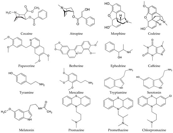
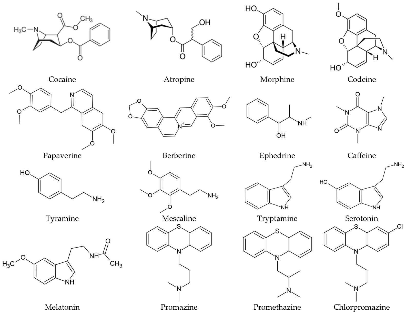
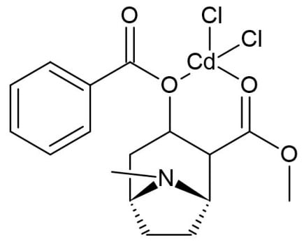
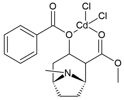
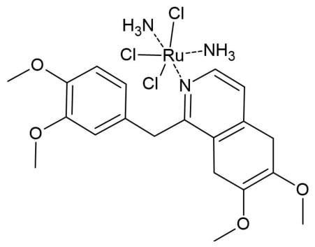
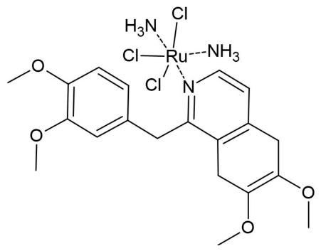
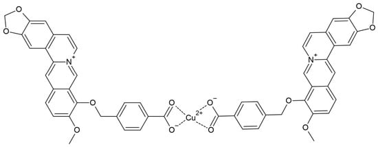
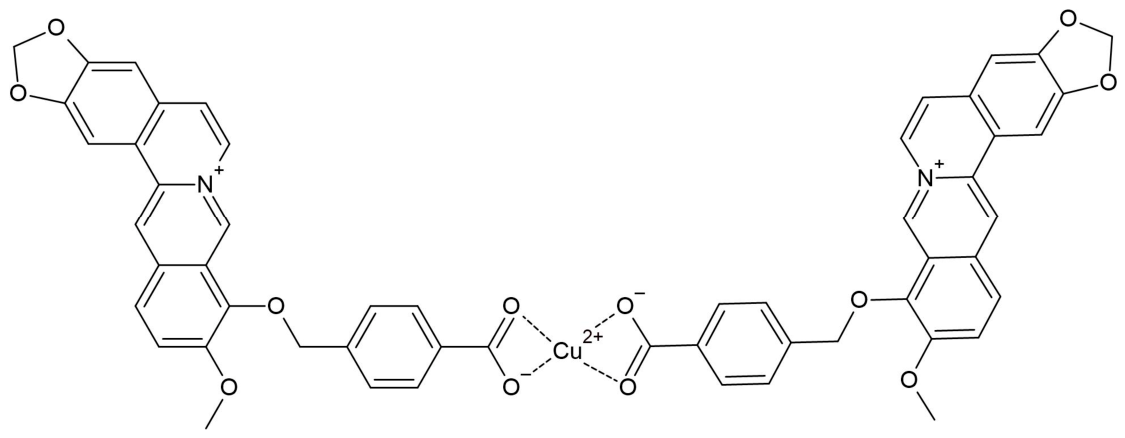
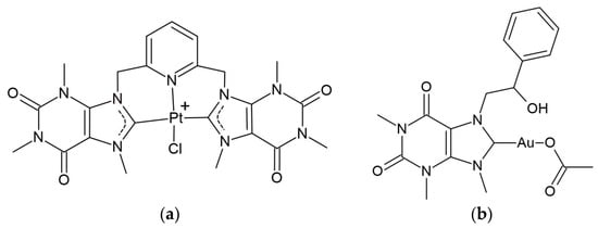
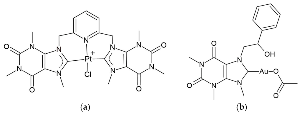
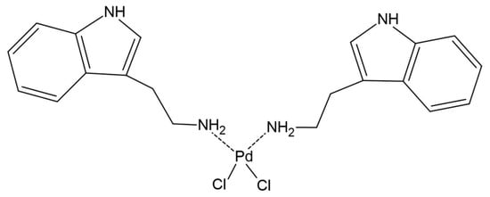
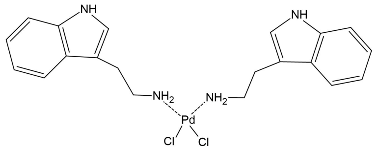
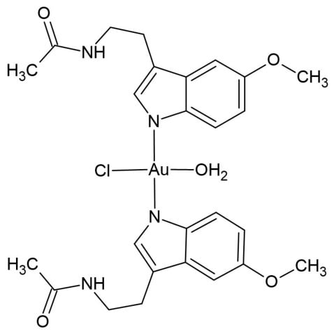
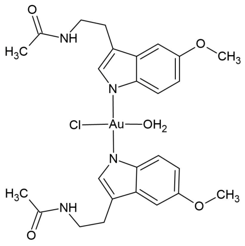
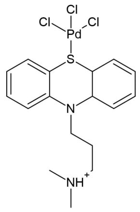
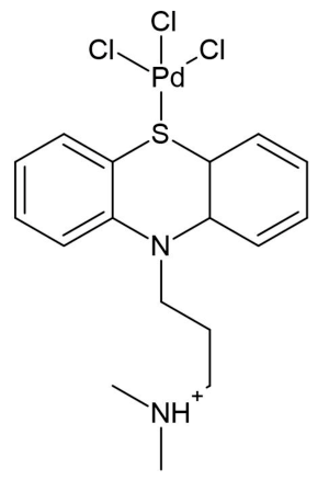
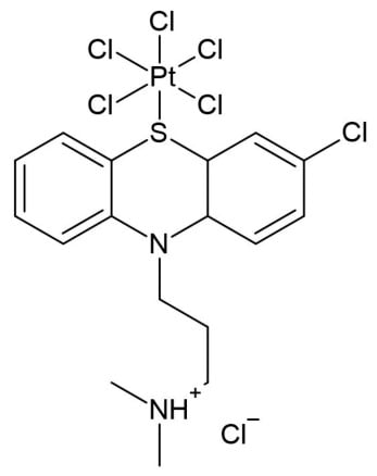
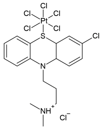
Disclaimer/Publisher’s Note: The statements, opinions and data contained in all publications are solely those of the individual author(s) and contributor(s) and not of MDPI and/or the editor(s). MDPI and/or the editor(s) disclaim responsibility for any injury to people or property resulting from any ideas, methods, instructions or products referred to in the content. |
© 2026 by the author. Licensee MDPI, Basel, Switzerland. This article is an open access article distributed under the terms and conditions of the Creative Commons Attribution (CC BY) license.