


Abstract
Janus nanoparticles (JNPs) synthesis has caught the scientific community’s attention due to their amphiphilic properties and extensive areas of application. In this work, different new copper–silica-based and silica-based JNPs were synthesized using a novel masking methodology and a self-assembly method based on sol–gel procedures, respectively. Moreover, various techniques were used to characterize the developed nanomaterials, including scanning electron microscopy (SEM), X-ray diffraction (XRD), Fourier Transform Infrared Spectroscopy (FTIR), and X-ray photoelectron spectroscopy (XPS). Two types of copper–silica-based Janus nanoparticles were synthesized with a 40 to 70 nm size, while SiO2-based JNPs of around 135 nm were obtained. The duality of different JNPs was confirmed by SEM and by a simple and economical route based on an emulsion stabilization path: analyzing dispersion/aggregation and associated behavior at the immiscible solvent interface. JNPs exhibited an extended residence time over 20 days at an immiscible solvent interface, thereby enhancing the resulting emulsion interface stability. This behavior highlighted their amphiphilic characteristics in comparison to conventional nanoparticles. Consequently, a procedure to determine nanoparticle amphiphilicity could be further standardized.
1. Introduction
Janus nanoparticles (JNPs) are a unique class of nanomaterials characterized by their asymmetrical structure due to two distinct chemical compositions or functional groups within a single nanoparticle (NP). This dual composition confers amphiphilic properties, which generate fascinating performances, e.g., distinctive surface properties between both sides and a capacity to develop clusters with an engineered size and shape [1,2]. Therefore, different applications have been proposed for energy storage [3], biomedicine [4], environmental remediation [5], and oriented catalysts [6], among many others [7,8,9].
Nanomaterial synthesis is categorized into top-down and bottom-up methods. In the top-down approach, NPs are obtained by modifying existing bulk particles through general physical techniques such as ball milling, lithography, and laser ablation. In the bottom-up approach, nanomaterials are synthesized from atomic or molecular species through chemical reactions for nucleus formation in a controlled growth process, allowing nanoparticle size and morphology control. The most used methods are chemical vapor deposition, hydrothermal, and sol–gel techniques [10]. Amphiphilic JNPs are usually synthesized by masking, self-assembling, and phase separation. One of the most common pathways is a self-assembly process, where building blocks associate spontaneously [11]. Silica NPs are popular for their ease of modification, high porosity, and excellent adsorption capacity. They offer superior colloidal stability, mechanical strength, and thermal stability compared to polymer-based NPs. Furthermore, they are chemically inert, biocompatible, and non-toxic [12].
The amphiphilic SiO2 JNPs leverage their dual interfacial properties in a range of applications, functioning as solid surfactants and interface stabilizers. In oil–water separation, JNPs enable efficient demulsification and pollutant removal. Magnetic Janus-like SiO2/polymer nanoparticles adsorb at oil–water interfaces and, under a magnetic field, coalesce and remove emulsified droplets, achieving >99% separation efficiency and >98% recyclability [13]. Likewise, Janus-based membranes exhibit high performance: a lignin–nanoparticle-bridged Janus paper achieved >99% separation efficiency for emulsified oils, delivered ultra-high fluxes (~5500–6300 L·m−2·h−1·bar−1), and maintained antifouling properties over 10 cycles [14]. In enhanced oil recovery (EOR), Janus SiO2 materials significantly improve displacement efficiency. Temperature-responsive Janus SiO2 particles formed stable emulsions and reduced interfacial tension, boosting oil recovery by ~16.5% with just 0.03 wt. % addition [15]. Similarly, Janus-reinforced polymer gel microspheres (conformance agents + interfacial activity) achieved ~19.7% extra recovery in core flooding, outperforming conventional polymer systems [16]. Janus polymer nanosheets at 1–50 mg∙L−1 acted as ultra-efficient emulsifiers, lowering IFT and altering core wettability, resulting in enhanced flooding and reduced injection pressures [17]. Amphiphilic Janus carbon nanotubes formed stable O/W nano-emulsions, reduced oil viscosity by ~99%, and converted oil-wet rock toward neutral-wet, enabling >20% EOR efficiency [18]. In foam-assisted gas injection EOR, SiO2 JNPs enhanced foam stability across CH4, CO2, and air media. Oleic acid-modified JNPs reduced surface tension by ~25 mN∙m−1 compared to bare SiO2 and maintained foam height (~80%) for over 2 h [19]. Beyond oil applications, Janus SiO2 has been integrated into coatings and colloidal systems: snowman-shaped polymer JNPs encapsulated silicone oil droplets in a thin shell, which, when added to paint, improved stain resistance without typical cratering effects [20]. More broadly, JNPs act as hybrid surfactant–particle emulsifiers in nanofluids, stabilizing Pickering emulsions and dispersions that require both steric and amphiphilic effects [13,21].
Metallic hybrid JNPs are also being exploited for several applications, such as Ag-TiO2-L-arginine for pathogenic bacteria suppression [22] and TiO2-Au JNPs for antitumor therapy [23]. However, the use of abundant, non-toxic metals such as copper remains merely promising. Across the compiled Cu-based Janus nanoparticle literature, the reported systems cluster into three material families defined by the bimetallic Janus domain, “soft–hard” hybrid architectures, and a template-mediated interfacial growth family; each is typically associated with a characteristic synthesis route. Cu–Fe “Janus”-like bimetallic nanoparticles were produced by the simultaneous electrical explosion of twisted Cu and Fe wires in argon [24]. CuO–Ag JNPs were generated by the simultaneous electrical explosion of twisted Cu–Ag wires in an argon/oxygen buffer gas mixture [25]. Cu–Ag JNPs were prepared by a wet-chemical route mixing AgNO3(aq), CuCl2, glucose, and hexadecylamine and heating at 100 °C for 2 h [26]. Au–Cu Janus nanostructures appear in two works via seeded growth [27,28]. In Cu2−xSe–Au Janus nanoparticles, Au cores were first formed, followed by surface functionalization [29]. Cu3P–Cu2O JNPs embedded in porous carbon were enabled at scale via the carbonization of Cu2+-containing ion-exchange resins with KOH [30]. CuS–ZnS JNPs were synthesized by thermolysis in octadecene/dodecanethiol [31]. In the second category, soft–hard hybrid JNPs include Prussian Blue/CuS [32], and polydopamine–Cu (PDA/Cu) JNPs [33]. Beyond these, the template-mediated interfacial growth group can be illustrated by Cu-sSiO2&rPMO mesoporous JNPs (sSiO2 means hydrophilic SiO2 nanospheres and rPMO refers to periodic mesoporous organosilica) [34]; and a biomedical “decorated Janus” variant couples copper peroxide (CP) nanodots onto a gold nanostructure/silica nanorod Janus scaffold, assembled via silica nanorod sol–gel growth [35]. The current development of mesoporous JNPs is primarily constrained to functionally inert SiO2 and polymer materials. Furthermore, the synthesis of metal compound Janus architectures, including the selective assembly of Cu on the hydrophilic SiO2 compartment, is deemed “urgently needed” yet poses considerable challenges. Further research into new and versatile synthesis methods for Cu–SiO2 Janus nanoparticles is necessary, as the functionalization of the silica side holds significant potential for applications across various fields.
Moreover, JNP applications face two main challenges: high synthesis costs and large-scale production complexity. To address these issues, research on silica JNPs can be focused on optimizing reactants, characterization techniques, and scalability [2,36]. Various advanced techniques are employed to characterize Janus nanomaterials, exhibiting distinct physical and chemical properties on opposite sides. These techniques include scanning electron microscopy (SEM), Transmission Electron Microscopy (TEM), X-ray diffraction, Fourier Transform Infrared Spectroscopy (FTIR), and X-ray photoelectron spectroscopy (XPS). In addition, wettability tests frequently allow for the demonstration of their two-faced amphiphilicity through water contact angle measurements [37]. These results could also be supported by dynamic light scattering (DLS) and zeta potential measurements (ζ), which allow for assessing size distributions and colloidal stability [14,19], providing light upon the size and charge differences. Other methods are based on how Janus SiO2 particles adsorb strongly at oil–water interfaces; tensiometry confirms that they dramatically reduce interfacial tension. Despite the efficacy of these methods, their inefficiencies in accurately determining the duality of some specific JNPs have been documented in the literature [2]. Notably, their ability to disperse in both aqueous and oily phases—demonstrated via colloidal dispersion tests—is directly linked to their oppositely functionalized hemispheres [20].
This limitation underscores the pressing need to develop innovative, cost-effective methodologies to reliably assess the unique behaviors and functionalities of Janus nanomaterials. Different strategies have been proposed, e.g., employing a measurable hydrophilic–lipophilic balance (HLB) (estimated with surfactants) [38], and assessing the impact on interfacial tension between two immiscible liquids [39]. Two other cost-effective approaches, solubility tests and interface stability tests, have recently been discussed to meet the previously mentioned requirements [2].
In this work, two types of JNPs were synthesized and systematically characterized. The first type, novel copper–silica JNPs, was produced using a novel masking method and a self-assembly procedure based on the sol–gel technique, where the Cu nanoparticle seeds were modified with functionalized SiO2 nanoparticles in a single step. The masking technique employed hexadecyltrimethylammonium bromide (CTAB) to stabilize the emulsion and enable the deposition of modified silica on the metallic seeds, yielding JNPs named GPTMOS-SiO2@Cu and PhTMOS-SiO2@Cu. The second type, denoted as SiO2 JNPs, comprised fully silica-based JNPs obtained through a sol–gel self-assembly strategy employing (3-glycidyloxypropyl) trimethoxysilane (GPTMOS) and trimethoxy (2-phenylethyl) silane (PhTMOS) to introduce chemically distinct hydrophilic and hydrophobic functionalities, respectively, in several steps. The asymmetric character of the resulting SiO2@Cu JNPs was evidenced by SEM, while two different cost-effective interfacial stability assays in immiscible solvent systems were employed to determine the Janus character of the second type of SiO2 JNPs, thereby demonstrating their potential for EOR. These methodologies enabled accurate assessment of the structural asymmetry and interfacial behavior of the synthesized nanoparticles.
2. Results and Discussions
2.1. SEM and EDS Characterization
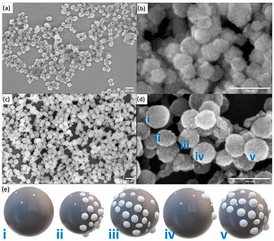
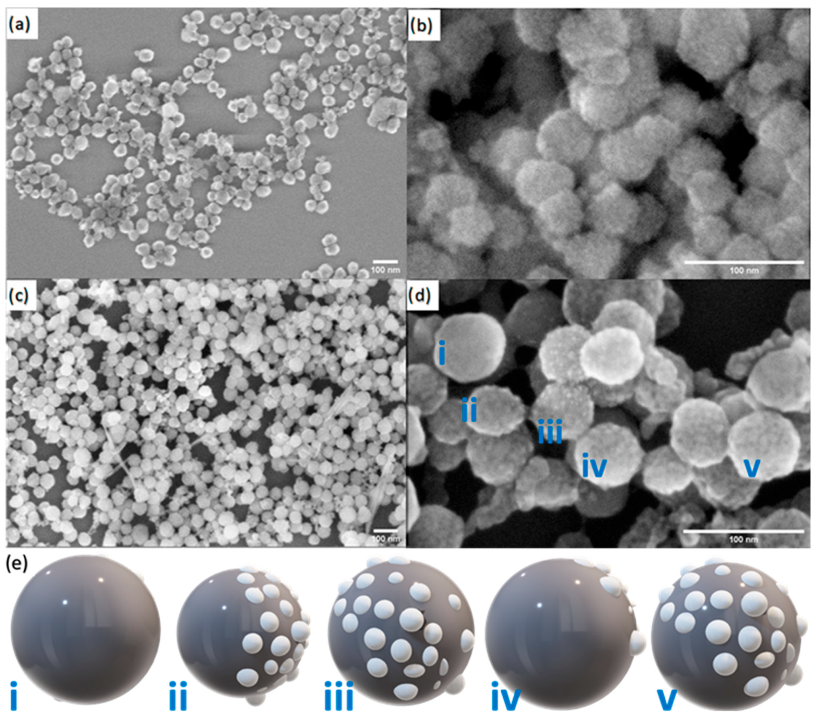
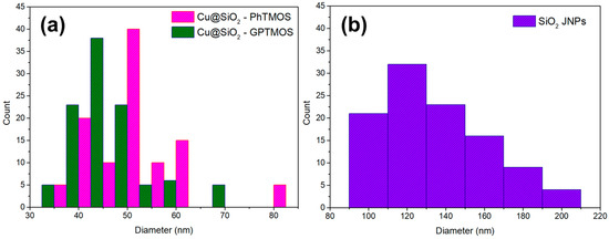
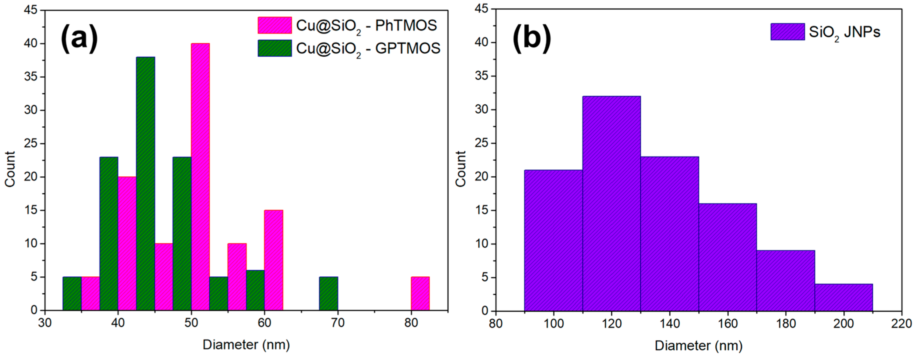
Nanomaterials were characterized using SEM to assess size distribution and morphology. Copper-based nanoparticles displayed spherical shapes, suggesting the presence of a rough surface covered by smaller spherical particles (Figure 1). Similarly, spherical SiO2-based JNPs were achieved (Figure S1a). Smaller spherical particles were attributed to modified silicon oxide nanoparticles, as revealed by the EDS and XPS analysis discussed in the next sections. For some particles, it was observed that a rough surface is concentrated on one side of the nanoparticle, as expected in nanoparticles with Janus character (Figure 1b,d); during the preparation of samples for SEM, the random positioning of nanoparticles resulted in a heterogeneous JNP orientation. This variability provides distinct perspectives on their Janus characteristics, as illustrated in Figure 1e. In addition, fine, filamentous wires were observed connecting some nanoparticles (e.g., in Figure 1c). These intriguing structures were hypothesized to originate from the formation of silica-based linkages between different copper-based JNPs. It was advisable to mitigate this network through ultrasonication prior to any application to ensure uniform dispersion. Size distributions of SiO2-JNPs and SiO2@Cu-JNPs for a population of 100 nanoparticles (each) were as displayed in Figure 2.
Figure 1.
SEM images of GPTMOS-SiO2@Cu (a,b), and PhTMOS-SiO2@Cu JNPs (c,d). Representation of different Janus orientations (i–v) of SiO2@Cu JNPs (e); adapted with permission from [2].
Figure 2.
Size distribution of the synthesized nanoparticles: (a) SiO2@Cu-based Janus nanoparticles functionalized with PhTMOS and GPTMOS, and (b) SiO2 Janus nanoparticles.
The results revealed that GPTMOS-SiO2@Cu JNPs had an average size of 44.0 ± 7.8 nm. PhTMOS-SiO2@Cu JNPs presented an average size of 53.0 ± 9.8 nm. This finding suggested that the silane monomer choice did not significantly impact either the morphology or size distribution for both types of SiO2@Cu JNPs.
The polydispersity of the SiO2 Janus nanoparticles observed in Figure 3 arises from the intrinsic kinetics of the sol–gel synthesis, in which variations in precursor concentration, hydrolysis and condensation rates, and mixing efficiency lead to overlapping nucleation and growth processes [40]. These effects, together with minor fluctuations in temperature and pH, result in particles forming at different growth stages, giving rise to a broad size distribution.
Figure 3.
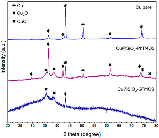
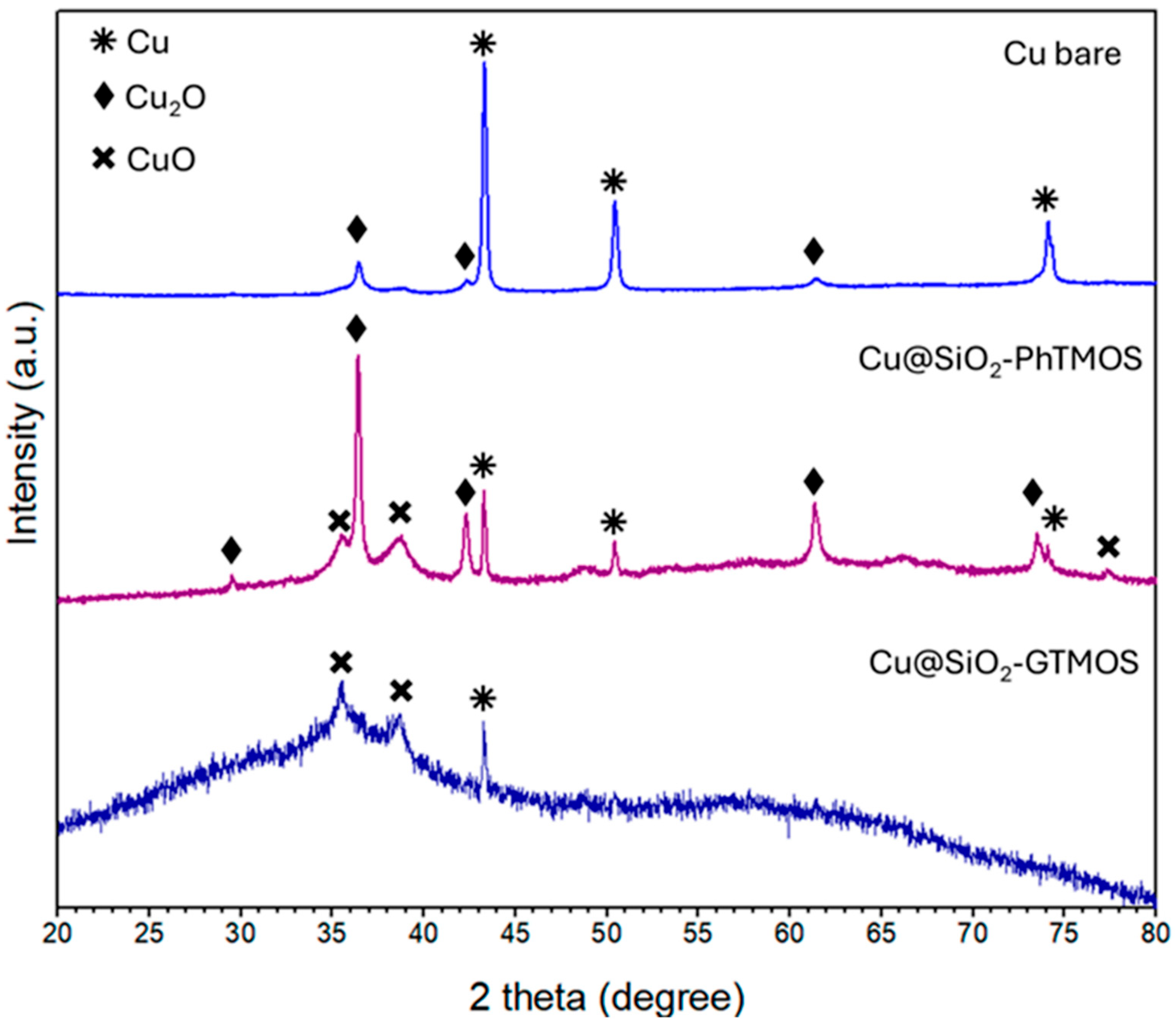
XRD patterns for Cu NPs without modification (light blue line, up pattern), PhTMOS-SiO2@Cu JNPs (purple line), and GPTMOS-SiO2@Cu JNPs (dark blue line).
In addition, post-synthesis processing plays a significant role. During drying, capillary forces and interparticle interactions can promote agglomeration, particularly in the presence of residual water, further broadening the apparent size distribution [41]. This behavior was quantified by analyzing SEM micrographs of over 200 particles, from which the mean diameter, standard deviation, and coefficient of variation were determined.
All JNPs were subjected to analysis via SEM and EDS to ascertain their Janus character and evaluate sample composition (Figure S1). SiO2-JNPs presented notable complexities for analysis using this technique. These complexities primarily stemmed from the nanoparticles’ small size and non-conductive nature, which hindered the acquisition of clear images and precise measurements. Furthermore, during SEM examination, the nanoparticles tended to become charged due to interaction with the electron beam. This charging effect not only distorted the imaging results but also compromised analysis stability. Despite the complexity of the analysis, spherical nanoparticles of approximately 134.2 ± 30.6 nm in size were imaged.
Nanomaterial agglomeration impeded an accurate Janus character assessment, and the EDS results did not show conclusive evidence in determining the Janus character. Therefore, it was necessary to employ an alternative technique to provide more robust evidence regarding this characteristic, such as solubility and interface tests [2]. Further EDS analysis of the copper-based Janus nanoparticles confirmed the presence of copper and silicon (Si) dispersed across the sample (Figure S1c,d), as opposed to commercial copper nanoparticles (Figure S1b).
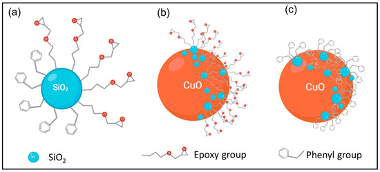
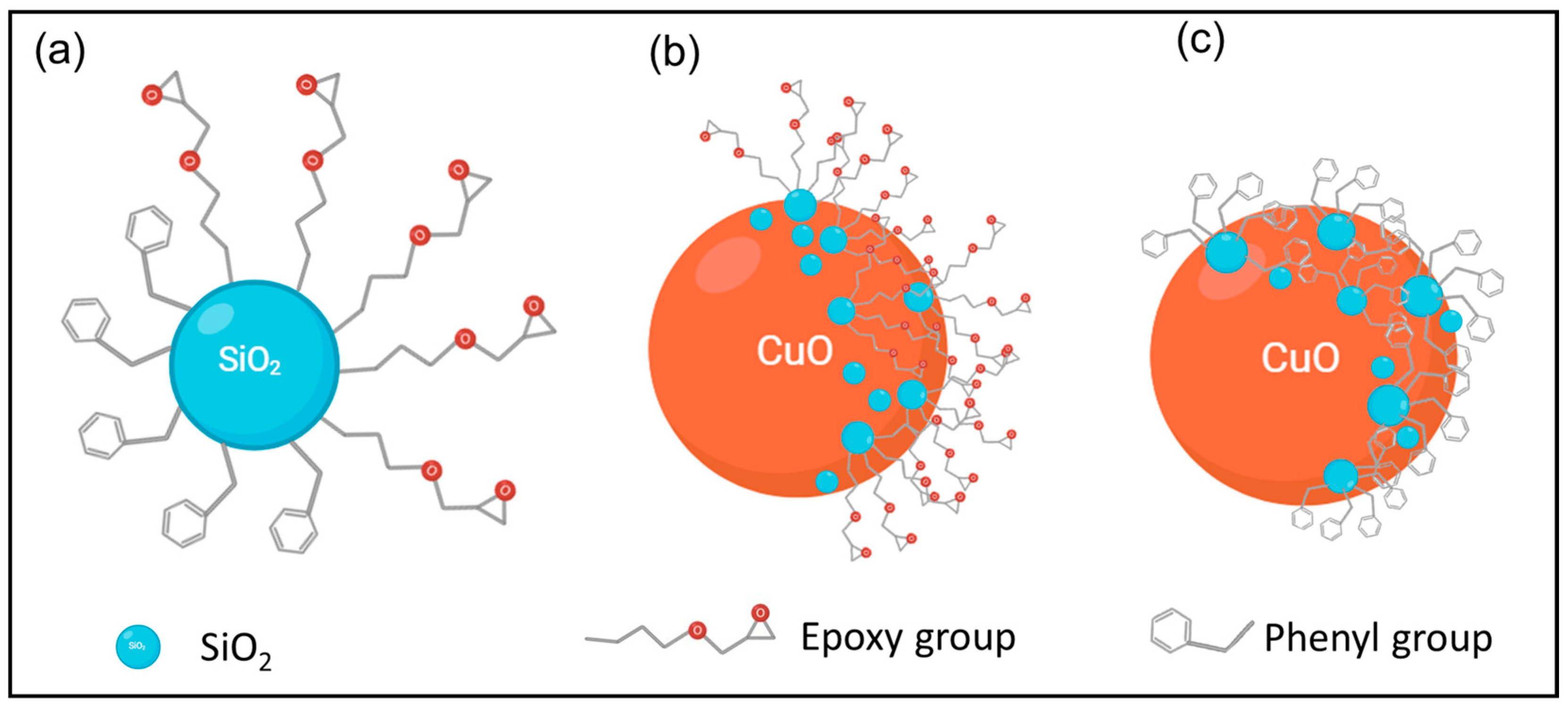
Synthesized amphiphilic Janus nanoparticles are schematically represented in Scheme 1, where SiO2 with epoxy and phenyl group functionalization derived from the sol–gel method provided SiO2 nanoparticles with their Janus characteristic (Scheme 1a). Copper oxide nanoparticles functionalized with SiO2-based epoxy or phenyl group modification obtained by a modified solvothermal method are represented in Scheme 1b and Scheme 1c, respectively.
Scheme 1.
Hypothetical schematic representation of Janus nanoparticles. (a) GPTMOS-PhTMOS-SiO2 JNP, (b) GPTMOS-SiO2@Cu JNP, and (c) PhTMOS-SiO2@Cu JNP.
2.2. XRD Analysis
XRD diffraction patterns of copper nanoparticles before and after modification were obtained, as seen in Figure 3, while the SiO2 JNPs’ XRD pattern showed an amorphous phase (Figure S2). In the copper-based JNPs’ XRD spectra, it is possible to observe the presence of metallic copper, copper (I) oxide, and copper (II) oxide. In the case of bare copper, intense peaks at 43.34° 2θ, 50.48° 2θ and 74.17° 2θ are assigned to the (111), (002), and (022) planes of metallic copper (ICSD card N° 98-005-3247), whereas peaks located at 36.42° 2θ and 61.37° 2θ correspond to the copper planes (111) and (022) of (I) oxide (ICSD card N° 98-005-2043). The presence of oxide is related to Cu NPs’ ambient oxidation.
The XRD patterns also show that modifying Cu NPs with either PhTMOS or GPTMOS leads to copper oxide appearance. Specifically, Cu modified with PhTMOS (PhTMOS-SiO2@Cu JNPs) presents intense peaks located at 36.49° 2θ, 42.39° 2θ, and 61.50° 2θ, associated with the (111), (002), (022) planes, respectively, of copper (I) oxide (ICSD card N° 98-002-6963). In addition, other peaks corresponding to copper (II) oxide at 35.55° 2θ (11-1), 38.67° 2θ (111) and 48.84° 2θ (20-2) (Tenorite, ICSD card N° 98-004-3179) are visible. In contrast, in copper nanoparticle modification with GPTMOS silane (GPTMOS-SiO2@Cu JNPs), the XRD pattern exhibits a mostly amorphous character due to a high proportion of amorphous silica, similar to the results obtained in refs. [42,43], higher than in PhTMOS-SiO2@Cu JNPs. However, this revealed the presence of two crystalline peaks corresponding to (11-1) and (111) planes, at 35.54 and 38.64° 2θ, which are attributed to the presence of copper (II) oxide (ICSD card N° 98-004-3179), and the peak located at 43.19° 2θ corresponding to metallic copper (ICSD card N° 98-005-3247). The phenyl group exhibits a passivating effect on nanomaterial surfaces by forming a protective layer, which limits copper Cu NPs’ oxidation. In contrast, GPTMOS groups possess hydrophilic properties, which results in an increased rate of Cu nanoparticle oxidation.
2.3. XPS Characterization
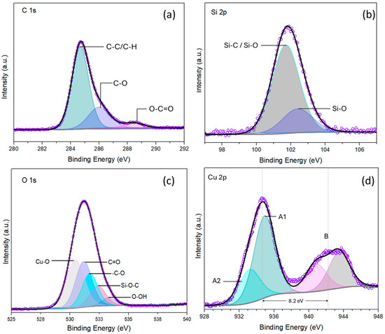
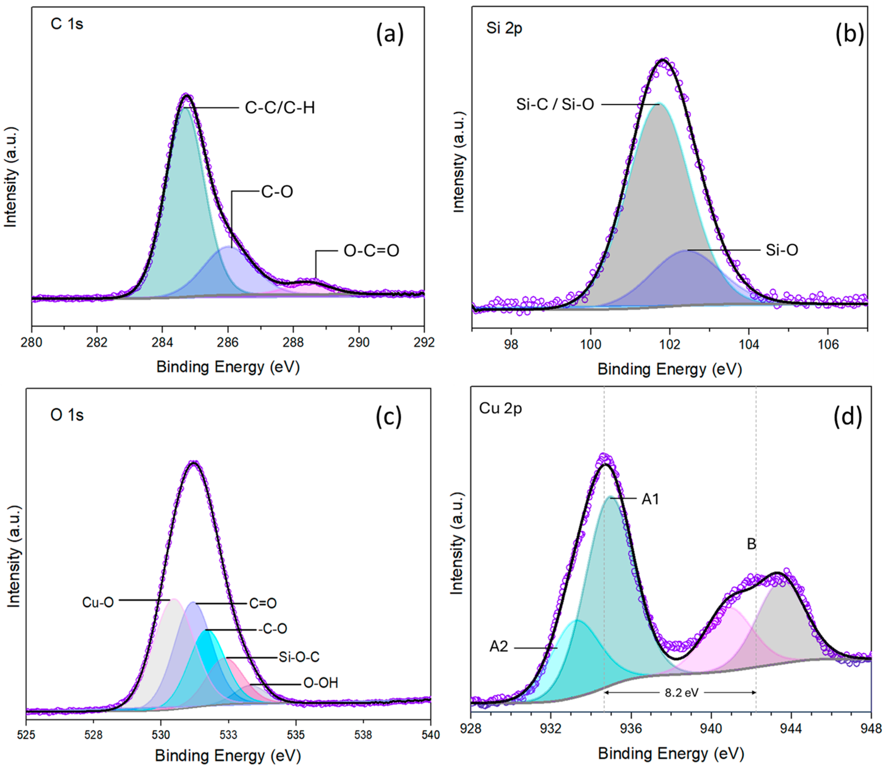
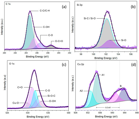
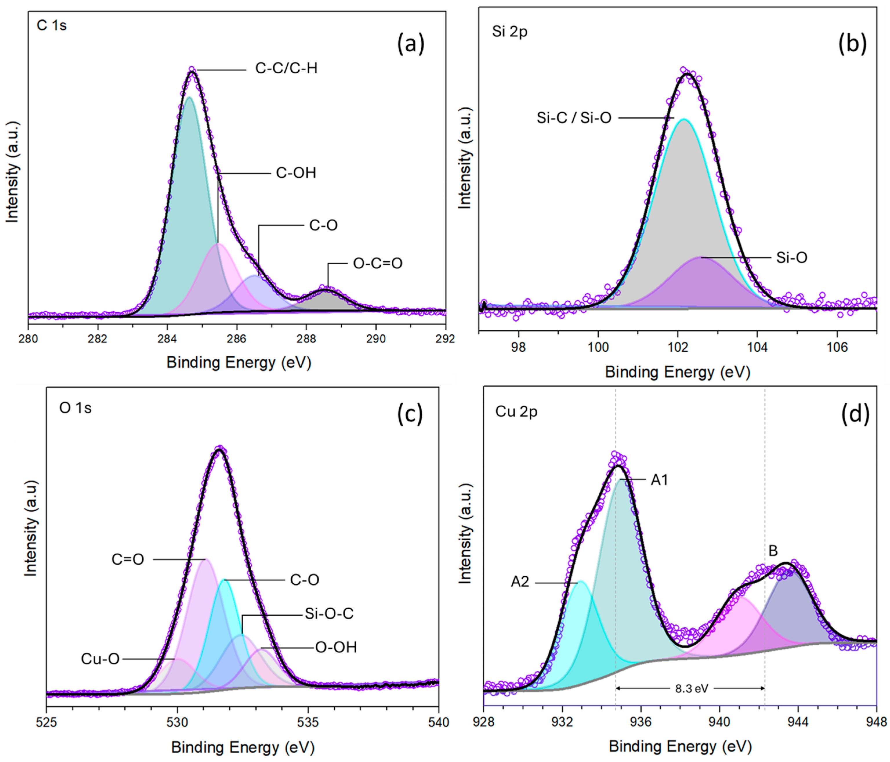
Both functional groups in GPTMOS-SiO2@Cu and PhTMOS-SiO2@Cu JNPs were attached to the silicon dioxide surface via silanization to obtain JNPs. XPS analyses of C 1s, O 1s, Si 2p, and Cu 2p were conducted on both samples to obtain comprehensive and high-resolution spectra (Figure 4 and Figure 5, respectively). The C 1s spectra of the GPTMOS-SiO2@Cu JNP sample was deconvoluted into three distinctive peaks (Figure 4a). The first peak, located at 284.68 eV, corresponds to C-C/C-H bonds. The peak at 286.07 eV is assigned to the C–O bond, attributed to the epoxy group presence [44,45]. The third at 288.39 eV is ascribed to the O–C=O bond [46]. Similarly, the C 1s spectra of the PhTMOS-SiO2@Cu JNP sample was deconvoluted into four curves (Figure 5a). The peak located at 284.62 eV is assigned to the C-C/C-H bonds and the one at 284.46 eV is related to the C-OH bond, which is in agreement with the aromatic ring corresponding to the phenyl group [47,48]. These findings are in agreement with the representation proposed in Scheme 1b, which displays a short hydrocarbon chain with a terminal epoxide moiety for GPTMOS-SiO2@Cu JNPs, and a phenyl group termination for PhTMOS-SiO2@Cu JNPs.
Figure 4.
(a) C 1s, (b) Si 2p, (c) O 1s, and (d) Cu 2p core levels’ XPS spectra of the GPTMOS-SiO2@Cu JNPs. Purple hollow dots correspond to raw data, the black solid line corresponds to envelope/fitting curve, and colored areas correspond to deconvoluted components.
Figure 5.
(a) C 1s, (b) Si 2p, (c) O 1s, and (d) Cu 2p core levels’ XPS spectra of PhTMOS-SiO2@Cu JNPs. Purple hollow dots correspond to raw data, black solid line corresponds to envelope/fitting curve, and colored areas correspond to deconvoluted components.
Silicon recorded in the XPS spectra was attributed to silicon in silanes. The Si 2p spectrum is displayed in Figure 4b and Figure 5b. After Gaussian–Lorentzian fitting, two main components were observed in both samples.
For GPTMOS-SiO2@Cu JNPs, the peak located at 101.73 eV is assigned to Si-O bonding, and the one at 102.30 eV is attributed to the combined contribution of Si-C and Si-O bonding [49]. The peak contribution to the Si 2p XPS spectra is 77.97% and 22.03%, respectively. Peak deconvolutions for the PhTMOS-SiO2@Cu JNP sample were found at 102.15 eV and 102.59 eV. These peaks are attributed to Si-O and Si-C/Si-O bonding, respectively.
Figure 4c and Figure 5c show the O 1s spectra, for GPTMOS-SiO2@Cu and PhTMOS-SiO2@Cu JNPs, respectively. For both spectra, each curve was deconvoluted into five distinctive curves, corresponding to Si-O-C, C-O, C=O, Cu-O, and O-OH bond groups (for detailed information, see Table S1). Although the same bonds are suggested for both samples, the Cu-O bond peak for the GPTMOS-SiO2@Cu JNP sample has a higher peak area (33.55%) compared to that of PhTMOS-SiO2@Cu JNPs (9.78%). This could be related to different copper oxidation states coexisting in both samples, also indicating a slight Cu2+ increase in the GPTMOS-SiO2@Cu JNP sample, in line with the XRD results (Figure 3).
In the Cu 2p3/2 spectra for GPTMOS-SiO2@Cu and PhTMOS-SiO2@Cu JNPs (Figure 4d and Figure 5d, respectively), it was observed that the characteristic peak and its corresponding satellite were separated by a binding energy of ~8 eV, as reported before [50]. Previous studies indicated that these satellites were related to the partially filled Cu2+ oxidation state d-orbital (d9), suggesting that their presence and intensity were directly correlated with the Cu2+ surface concentration [51,52,53]. This corroborated the XRD results, which showed that JNPs are composed of copper and its oxides.
The relative concentrations are calculated by the following equations:
where B is the total area of the satellite peak, and A is the total area of the main peak, which are contributions of the Cu2+ (A1)-related area and area related to (Cu0 + Cu+) (A2). (A1S/BS) is the ratio of the main peak/shake-up peak areas for the 100% pure Cu2+ sample equal to 1.89 for the CuO reference at 20 eV pass energy [51]. Area peaks were calculated using Casa XPS Version 2.3.25PR1.0 software (peak area/relative sensitivity factor; 16.73 for Cu 2p3/2). The (Cu0 + Cu+) and Cu2+ calculated content was 23.53% and 76.46%, respectively, for PhTMOS-SiO2@Cu JNPs. Similarly, for Cu in GPTMOS-SiO2@Cu JNPs, the percentage of (Cu0 + Cu+) was 18.65% and 81.34% for Cu2+.
From these results, it was concluded that the oxidation of copper nanoparticles to copper oxides occurred in both cases. Still, higher oxidation with GPTMOS-SiO2@Cu modification was observed (Cu2+). This enhanced oxidation may be related to the hydrophilic character of the GPTMOS [54], which contains epoxy functional groups known for their ability to interact with environmental water, thereby promoting the oxidation of the copper nanoparticles. The formation of these oxide layers can enhance chemical stability by passivating the surface, while also modifying surface chemistry and interactions with silane groups. Therefore, the resulting particles can be more accurately described as copper-based nanoparticles with oxide-rich cores.
2.4. FTIR Characterization (Silica-Based JNPs, Cu-Based JNPs)
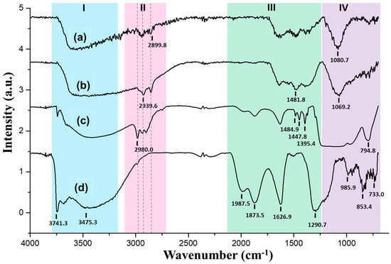
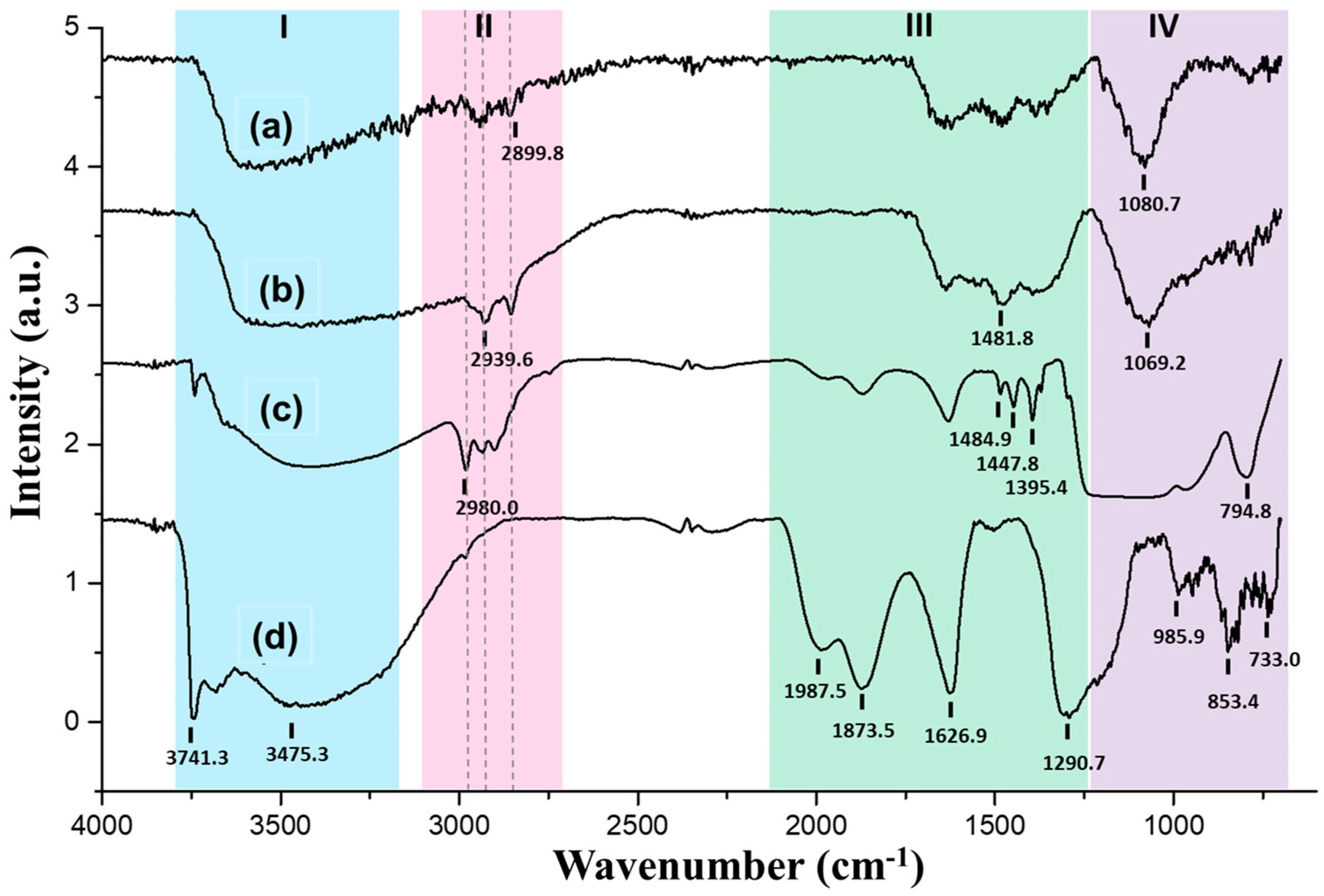
The FTIR spectra and band summaries for different vibration modes and corresponding chemical functional groups for silica (Scheme 2) and SiO2@Cu-based JNPs are shown in Figure 6 and Table 1, respectively. Analysis of these results, starting from Zone IV, where the asymmetric and symmetric stretching vibration of the Si–O–Si bond, denoted as ν(SiOSi), at approximately 1080.0 and 795.0 cm−1, respectively, confirms the formation of silica-based nanoparticles through the siloxane group of Si–O–Si mode. Furthermore, the band observed at 985.9 cm−1, attributed to ν(SiOH), is characteristic of free silanol molecules, proving hydrolysis occurrence [55]. Zone III provides information regarding the organic functional groups of monomers used in the JNPs’ synthesis and their transformation.
Scheme 2.
Representation of functional groups (in red) for silane-based monomer structures employed in JNPs synthesis: (a) tetra-ethyl orthosilicate (TEOS), (b) (3-glycidyloxypropyl) trimethoxysilane (GPTMOS), (c) trimethoxy [3-(methylamine) propyl] silane (APTMOS), and (d) trimethoxy (2-phenylethyl) silane (PhTMOS).
Figure 6.
FTIR spectra of (a) GPTMOS-SiO2@Cu JNPs, (b) PhTMOS-SiO2@Cu JNPs, (c) SiO2 JNPs (SiO2 JNPs), and (d) SiO2 JNPs treated by annealing process at 600 °C (SiO2 NPs H). The colored zones I–IV correspond to the classifications outlined in Table 1.
Table 1.
Assignment of infrared spectrum bands to different vibration modes and corresponding chemical functional groups. The color codes associated with Zones I through IV correspond to the details presented in Figure 6.
The epoxy group presence belonging to GPTMOS (Scheme 2b) can be confirmed by in-plane bending at 1290.7 cm−1 and out-of-plane bending vibrations of the γ C-O-H band at 1395.4 cm−1 due to epoxy cycled structure rupture. In addition, the single-bond CH2 scissoring bending vibration (δ CSP3-H2) presented in the alkyl groups shows a doublet at 1447.8 and 1484.9 cm−1 since the sample is analyzed in solid form [56].
PhTMOS monomer aromatic groups (Scheme 2d) are identified by overtones (harmonics) of benzene ring vibrational modes, which are also sensitive to the substitution pattern. These appear as stretching frequencies in the fingerprint region of the infrared spectrum between 2100 and 1750 cm−1, which are visible in Figure 6c,d. Both show the presence of PhTMOS during synthesis, but not in Figure 6b due to silica’s low presence in the Cu JNPs’ structure. Furthermore, a spectral band observed at 1626.9 cm−1 corresponds to the bending vibration of the N-H bond of the APTMOS monomer utilized in the synthesis process (Scheme 2c). In Zone II, two signals at 2899.8 and 2939.6 cm−1 are visible due to alkyl and/or aromatic groups’ -CH2 vibration bands presented in the different silane monomers. Also, a peak at 2980.0 cm−1 can be attributed to the Csp2-H vibration of the phenyl group present in the PhTMOS monomer. Finally, Zone I displays a broad peak around 3400.0 cm−1, corresponding to the -OH stretching vibration band associated with adsorbed water, and the stretching modes originated from Si-OH. This peak is reduced after SiO2 nanoparticles’ annealing at 600 °C, highlighting a prominent double band around 3700 cm−1. This double band is attributed to the N-H stretching vibration mode of secondary amine in APTMOS (see Scheme 2c).
A slight silica presence within the structure of the copper nanoparticle (Cu JNP), combined with copper’s significant impact on the vibrational modes, has attenuated bands associated with silica face formation. Nonetheless, Si-O-Si bond formation is unequivocally confirmed by the band around 1080 cm−1 (Figure 6a,b), thereby validating sol–gel synthesis process attainment. Furthermore, low-intensity peaks observed at 2939.6 cm−1 (Figure 6b) are attributed to the phenolic compound (PhTMOS) (see Scheme 2d), which is not present during GPTMOS-SiO2@Cu JNPs’ synthesis.
A comparative analysis of SiO2 JNPs (Figure 6c) and SiO2 NPs (Figure 6d) shows that the bands in Zone IV diminished following annealing treatment. This is attributed to the thermal process, which alters material structural composition. Epoxy group degradation is induced by heat, as confirmed by the absence of spectral bands in the range of 1390.0 to 1490.0 cm−1 (Zone III, Figure 6d). Intensity aromatic overtones increase due to the band intensity reduction between 1840.0 and 1985.0 cm−1, corresponding to region III (Figure 6c,d). Furthermore, the ν Csp2-H observed at 2980 cm−1 exhibits a significant reduction after the heating process; however, it does not entirely disappear. This phenomenon can be attributed to benzene group proximity to the silica structure (see Scheme 2d).
2.5. Zeta Potential Measurement
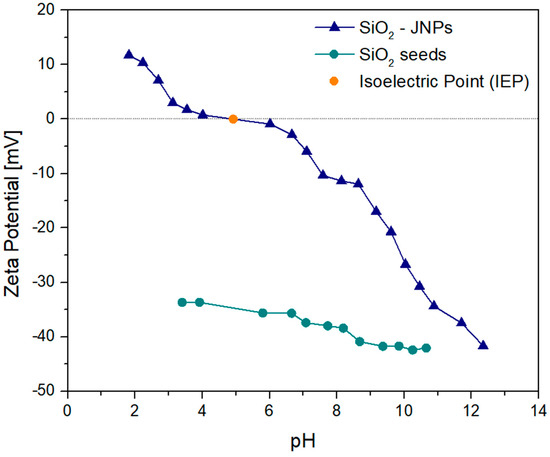
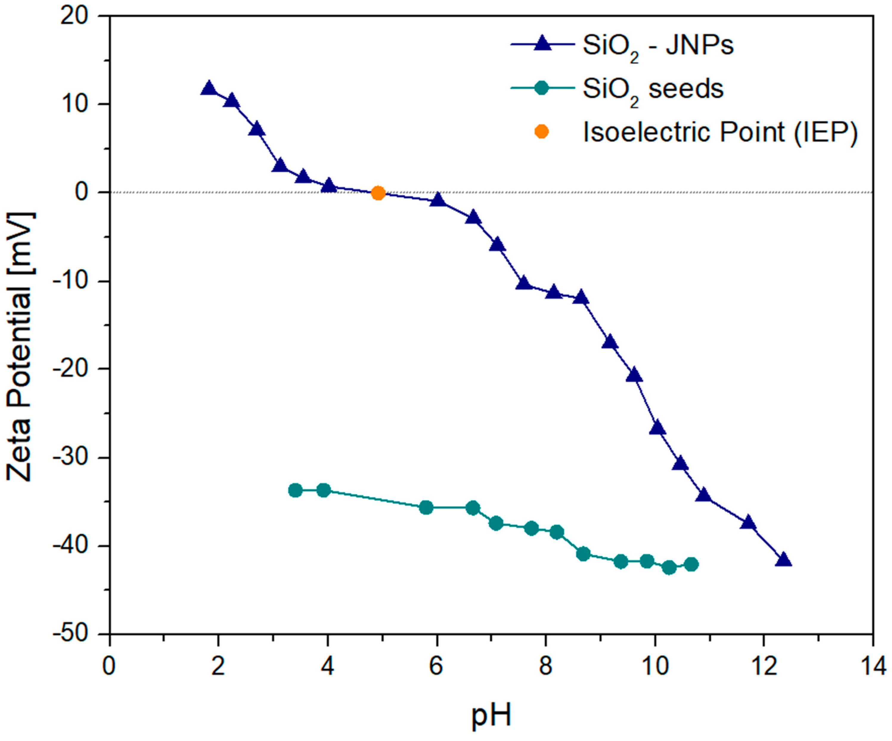
Zeta potential (ζ) measurements as a function of pH were performed to probe the acid–base behavior and surface charging properties of the nanoparticles. The ζ–pH behavior arises from the collective protonation–deprotonation of surface sites. In contrast, Janus-type structures—characterized by the spatial separation of hydrophilic and hydrophobic chemical domains—may exhibit nonlinear charging trends, slope discontinuities, or suppressed magnitude of ζ-values, reflecting the coexistence of chemically distinct hemispheres with different protonation–deprotonation equilibria. Although ζ-potential analysis alone cannot conclusively demonstrate Janus morphology, the presence of deviations from the behavior expected for fully homogeneous SiO2 NPs provides supporting evidence when interpreted in conjunction with the functionalization strategy, interfacial behavior, and emulsion tests. Therefore, the ζ–pH curves serve as a complementary approach to assess the asymmetric surface chemistry that underlies amphiphilicity in JNPs [57].
Figure 7 shows the ζ-potential vs. pH curves of the SiO2 JNPs, and SiO2 NPs without modification in deionized water. A clear variation in ζ-potential with pH was observed for both samples, with SiO2 JNPs demonstrating a more pronounced effect. The variation in SiO2 JNPs ζ-potential at elevated pH was steep, a phenomenon attributable to their larger surface area [58]. This increased surface area results in a greater number of functional groups available for deprotonation when compared to SiO2 NPs, due to further steps during the synthesis process associated with GPTMOS and PhTMOS monomers.
Figure 7.
Dependence of ζ-potential on pH for suspension of SiO2-based JNPs, and homogeneous SiO2 NPs.
On the one hand, the amorphous SiO2 NPs exhibited a negative surface charge over the pH range 3–11. It is also appreciable that the negative charge on the nanoparticles’ surface has a slight increase with an increasing pH, which was associated with an increasing silanol group formed on the silica surface. The silica surface attains a net negative charge primarily due to the deprotonation (H+) of silanol groups, resulting in a surface that exhibits an increasing negative charge (Figure 7) [59]. On the other hand, the SiO2-based JNPs showed a positive ζ-potential for pH < 4.9, and negative otherwise. At this pH, the JNPs exhibit the isoelectric point, confirming the absence of this phenomenon for SiO2 NPs, which are negatively charged in the studied pH range. The analysis of the curve indicated that the behavior of the SiO2 JNPs exhibited amphoteric properties [60]. This observation was further strengthened by additional visual evidence.
2.6. Janus Character Tests in Immiscible Liquid Systems for SiO2 JNPs
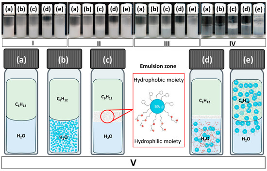
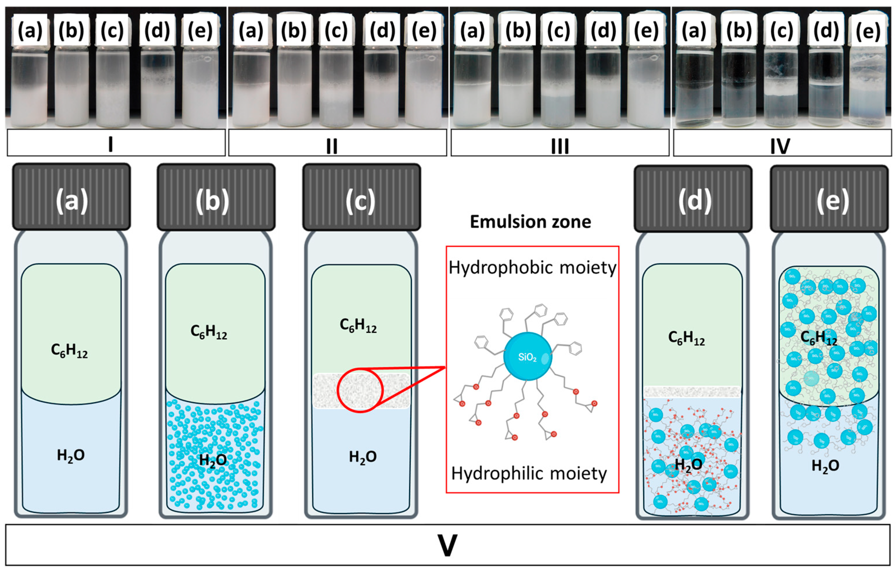
Determining the SiO2 JNPs’ Janus properties was not achievable through the SEM technique, in contrast to copper-based nanomaterials. Consequently, further analyses were conducted exclusively on SiO2 JNPs and their precursors (see Table 2) by interface tests (Figure 8 and Figure 9), as reported in ref. [2]. Janus character assessment involved examining the emulsion stabilization formed at the interface. Figure 8 presents a comparative analysis of various precursors and the resultant SiO2 JNPs. It is noteworthy that the liquid systems in a 1:1 mixture facilitate the assessment of interfacial separation between the two immiscible phases (water and cyclohexane, H2O:C6H12), a process that exhibits delays when considering the final SiO2 JNPs. This behavior provides insight into the potential for emulsion stabilization, suggesting applications of JNPs in the recovery of heavy oil from diverse media [15,16,17,18]. SiO2 JNPs exhibited distinct Janus behavior in a 1:1 H2O:C6H12 mixture, as illustrated in Figure 8(IVc,Vc).
Table 2.
Nomenclature and pertinent information concerning JNPs developed in this research.
Figure 8.
Interface test focusing on separation of two immiscible phases (1:1 H2O:C6H12 mixture) over time [(I): immediately after vigorous stirring and ultrasonication for 10 min; (II): after 30 min of settling; (III): after 24 h of settling; (IV): 20 days later; (V): schematic representation of case IV)]. Conditions studied included (a) without nanoparticles, (b) SiO2 NPs, (c) SiO2 JNPs, (d) GPTMOS-SiO2 NP, and (e) PhTMOS-SiO2 NP. In this depiction, the blue sphere illustrates the core of the SiO2 nanoparticles (NPs). The chemical structures with two red circles (oxygen atoms) depict GPTMOS units, while those with a benzene group represent PhTMOS units.
Figure 9.
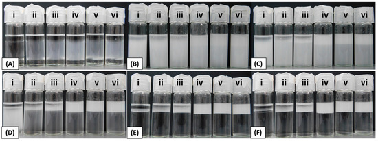
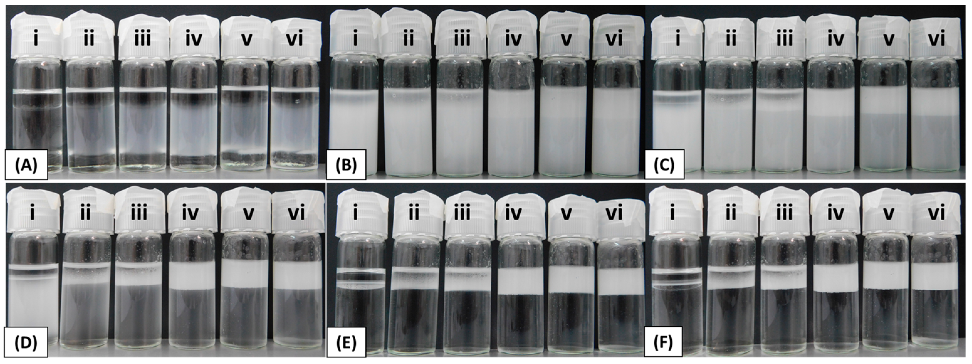
Emulsion stabilization tests in the 31.6:15.8:52.6 C12H26:PEG 400:H2O (v/v) ternary system using different amounts of SiO2 JNPs. Nanoparticle concentrations evaluated: (i) 0 g (control), (ii) 0.001 g, (iii) 0.005 g, (iv) 0.015 g, (v) 0.020 g, and (vi) 0.025 g. Images were recorded at the following times: (A) immediately after addition of dodecane to the PEG 400 solution; (B) after vigorous stirring and 10 min ultrasonication; (C) after 30 min; (D) after 4 h; (E) after 24 h; and (F) after 20 days. Long-term interface stabilization was observed for systems iv–vi.
Initially, a rapid separation of immiscible phases was observed in the absence of nanoparticles (Figure 8(Ia–Va)). A comparable phase separation behavior was also noted when using homogeneous SiO2 NPs, which lack amphiphilic properties (Figure 8(Ib–Vb)). These results suggest that the Janus character plays a critical role in stabilizing the emulsification process, likely due to preferential localization at the interface between the two immiscible phases (Figure 8(Ic–Vc)).
The schematic in Figure 8V depicts the behavior of nanoparticles in the immiscible 1:1 C6H12:H2O system, where the different nature of the nanoparticles is evident, and the resulting effects on the system are shown. Furthermore, PhTMOS-SiO2 NPs demonstrated a low reversion degree to the initial state, attributable to their hydrophobic characteristics due to the phenyl group, with a high affinity for the C6H12 phase (Figure 8(Ve)); meanwhile, GPTMOS-SiO2 NPs (Figure 8(Id–Vd)) demonstrated properties comparable to those of homogeneous SiO2 NPs (Figure 8(Ib–Vb)), attributable to their hydrophilic nature.
2.7. Discussion on Emulsion Stabilization in a Multicomponent System: Potential Application in EOR
The potential of SiO2 JNPs to stabilize emulsions was further evaluated using a ternary system composed of 31.6% dodecane (C12H26), 15.8% polyethylene glycol 400 (PEG 400), and 52.6% water (v/v). The influence of the nanoparticle concentration on interfacial stability was assessed, as shown in Figure 9. The amphiphilic nature of the JNPs significantly enhanced the stability of the oil–water interface, with long-term stabilization observed at higher nanoparticle loadings. After more than 20 days, systems iv, v, and vi exhibited a persistent and well-defined interface, indicating effective stabilization. Notably, a concentration of 0.015 g of JNPs was sufficient to achieve robust interfacial stability (system iv, panels D–F), suggesting that only small amounts of nanoparticles are required to impart long-term structural integrity to the emulsion.
While these results demonstrate a clear proof-of-concept application, it is important to note that the C12H26–PEG 400–H2O formulation has not been optimized in terms of phase composition. Further studies are being planned to determine the optimal ratios and to evaluate performance under conditions more closely aligned with industrial practice. The findings reveal the emerging potential of the developed SiO2 JNPs for EOR. However, achieving and maintaining colloidal stability under conditions that mimic reservoir environments—such as pH fluctuations and varying JNP concentrations—presents intricate challenges. These complexities highlight the need for a more comprehensive understanding of JNP interactions and behaviors under these specific conditions, which is essential for optimizing their application in EOR processes. This management is essential for the effective application of SiO2 JNPs in injection-based oil recovery processes.
3. Materials and Methods
3.1. Materials and Reagents
Trimethoxy [3-(methylamine) propyl] silane (APTMOS), (3-glycidyloxypropyl) trimethoxysilane (GPTMOS), trimethoxy (2-phenylethyl) silane (PhTMOS), and tetraethyl orthosilicate (TEOS) (all reactant grade, Sigma-Aldrich, GmbH, Albuch, Germany) were employed as silane monomers in the synthesis process. Ethanol, acetonitrile, and cyclohexane (all absolute grade for analysis, Merck, KGaA, Darmstadt, Germany) were employed as solvents for the sol–gel method. Synthesis was performed via basic hydrolysis by adding 25% NH4OH solution procured from Merck KGaA, Darmstadt, Germany. Copper nanoparticles with an average particle size of 60–80 nm, as reported by the supplier (Merck, KGaA, Darmstadt, Germany), were purchased as nano-powder. Scanning electron microscopy (SEM) of the powder further confirmed the particle size. CTAB (99% hexadecyltrimethylammonium bromide) from Aldrich-Sigma Chemicals Company (GmbH, Albuch, Germany) was used as a stabilizer agent in the masking method.
3.2. Characterization and Equipment
Morphological characterization was conducted using scanning electron microscopy (SEM, FEI Verios 460 L, Hillsboro, OR, USA), and size distributions were determined by “ImageJ” Version 1.54k software. Elemental mapping was achieved using a Tescan FE Mira II LMU (Brno, Czech Republic) instrument equipped with an energy-dispersive X-ray spectroscope (EDS) detector (Bruker AXS, Inc., AZtec Version 4.2 software package released by Oxford Instruments, Abingdon, UK). Surface chemical composition analysis was performed utilizing X-ray photoelectron spectroscopy (XPS) on a Kratos Axis Supra spectrometer (Manchester, UK) equipped with monochromatic Al Kα X-ray radiation, operating at an emission current of 15 mA and employing the hybrid lens mode. For XPS analysis, the passing energy was set at 80 eV for detailed spectra and 20 eV for broad spectra. The resulting XPS spectra were processed and analyzed using CasaXPS Version 2.3.25PR1.0 software, with a calibration reference based on the C 1s peak fixed at 284.7 eV. Spectra fitting was achieved by employing the Shirley algorithm for background subtraction and Gaussian–Lorentzian (30–70%) line shape modeling.
Furthermore, crystalline phase composition was investigated using an X-ray diffractometer (XRD) with a high-brightness source (Rigaku SmartLab 9 kW CF2). XRD scans were conducted in Bragg–Brentano geometry, employing Cu Κα radiation within a scan range spanning from 20° to 80°, with a scan speed of 4°·min−1. ζ-potential was measured with a LITESIZER 500 instrument from Anton Paar (Hampton Falls, NH, USA) by preparing a dispersion of 0.01 mg/mL of SiO2 JNPs in water and homogenizing with an ultrasonic probe; dependence on pH from 2 to 12 values was tested by employing NaOH/HCl buffer solutions with an automatic dosing system.
3.3. Experimental Procedures
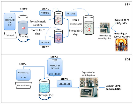
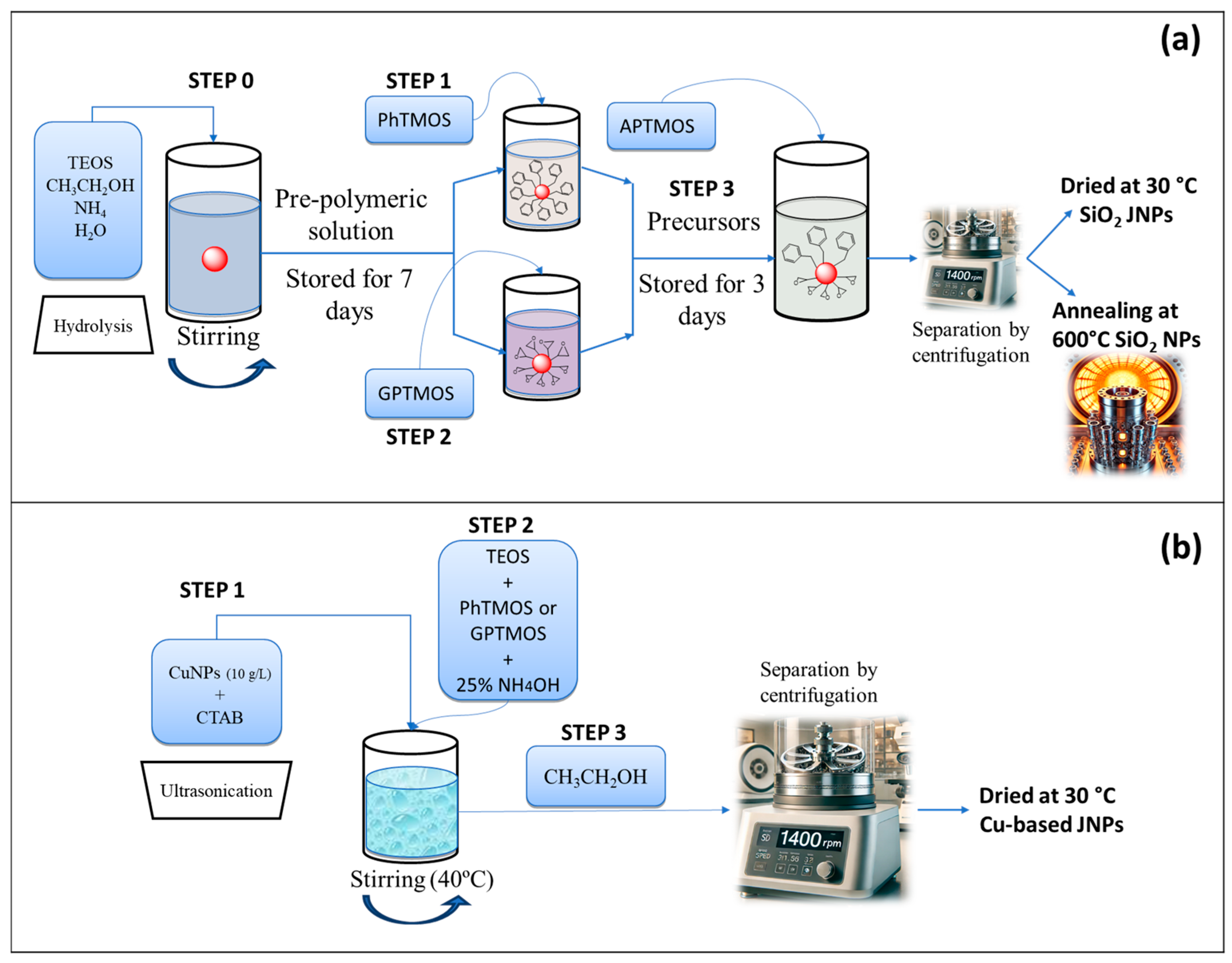
This work examines the efficacy of sol–gel and masking techniques in developing doubly functionalized silica (SiO2-JNPs) and copper–silica (GPTMOS-SiO2@Cu and PhTMOS-SiO2@Cu JNPs) structures, respectively (Scheme 3). A brief description of processes and a summary of developed nanomaterials are outlined in the next sections and Table 2.
Scheme 3.
Representation of employed self-assembled synthesis for obtaining (a) SiO2 JNPs and annealing-based post-treatment to remove amphiphilic character (SiO2 NPs), and (b) SiO2@Cu-based JNPs (GPTMOS-SiO2@Cu and PhTMOS-SiO2@Cu JNPs).
3.3.1. Preparation of Janus Silica Nanoparticles SiO2-JNPs
A silica–ethanol sol produced by a sol–gel method, as reported earlier [61], was applied to achieve SiO2-based JNPs (Scheme 3a). The specific procedure comprised the following steps:
Step 0 or initial: TEOS (silica precursor) was mixed with absolute ethanol (solvent), then 25% NH4OH solution and water were added together and magnetically stirred at 30 °C for two hours. After reaction, sol was allowed to age in a water bath at 25 °C for 7 days. Afterward, samples were refluxed in ethanol to remove ammonia in the sample, obtaining silica nano-seeds (around 70 nm) to be modified [61] (step 0, see Table 1).
Subsequently, a nano-silica dispersion with amphiphilic properties and a double-particle structure was produced by following these steps:
Step 1: Silica–ethanol sol (obtained in step 0) with a concentration of 3 wt. % was mixed with PhTMOS, continuously stirred at 30 °C for 2 h, and aged at 30 °C for 3 days. A lipophilically modified silica–ethanol sol was then acquired.
Step 2: Silica–ethanol sol with a concentration of 3 wt. % was mixed with GPTMOS at 30 °C for 2 h and aged at 30 °C for 3 days to attain a hydrophilic modified silica–ethanol sol.
Step 3: After 3 days, modified sols obtained in step 1 and step 2 were mixed and combined by adding APTMOS and continuously stirred at 30 °C for 3 h to develop the nano-silica dispersion with amphiphilic properties and a double-particle structure (SiO2-JNPs). Later, the suspension was repeatedly centrifugated at high velocity and washed with water and ethanol.
3.3.2. Preparation of SiO2@Cu-JNPs
SiO2@Cu-based JNPs were synthesized using a modified solvothermal method adapted from [62], where Fe3O4 nanoparticles were originally employed as seeds. In the present work, these seeds were replaced by Cu nano-seeds, followed by functionalization of the silica face to achieve GPTMOS-SiO2@Cu and PhTMOS-SiO2@Cu JNPs with epoxy and phenyl groups, respectively, a key novelty of this work.
Step 1: Initially, a copper nanoparticle dispersion with a concentration of 1 g∙L−1 was ultrasonicated. Subsequently, CTAB was incorporated into the dispersion and subjected to continuous ultrasonication for 30 min.
Step 2: SiO2 nanoparticles were prepared in situ with the addition of TEOS, and other silanes were used for functionalization (TEOS combined with GPTMOS or TEOS combined with PhTMOS). They were added along with a solution containing 25% NH4OH. The reaction was maintained under stirring for an additional 40 min at 40 °C.
Step 3: The reaction was concluded by ethanol addition. The resultant GPTMOS-SiO2@Cu and PhTMOS-SiO2@Cu JNPs were then subjected to repeated centrifugation and washing processes with both ethanol and water.
3.4. Determination of Amphiphilic Character of SiO2-Based JNPs and EOR Assessment
The Janus character of the SiO2@Cu-based nanoparticles was assessed using SEM images, in which functionalization was clearly observed in EDS elemental maps, and was corroborated by FTIR, XRD, and XPS measurements.
SiO2-based JNPs amphiphilic characteristics (Janus nature) were assessed for the samples listed in Table 2, utilizing a straightforward and cost-effective interface stability test [2]. This approach revealed the dual hydrophobic and hydrophilic JNPs’ properties by showing emulsion stabilization at the interface between two immiscible solvents. A 1:1 mixture of water (H2O) and hexane (C6H12) was used to compare the precursors of SiO2 JNPs with the final material. Subsequently, the resulting dispersions were subjected to vigorous stirring and ultrasonic treatment for 5 min for each mixture.
EOR assessment was performed by employing a heterogeneous mixture of dodecane (C12H26, 31.6% v/v), polyethylene glycol PEG 400 (15.8% v/v), and water (52.6% v/v). Briefly, PEG 400 was dissolved in a specified volume of water, to which varying quantities of SiO2 JNPs were systematically added (0.001, 0.005, 0.015, 0.020, and 0.025 g). Subsequently, dodecane was incorporated into the mixtures, and samples were subjected to ultrasonication at 6000 rpm for 5 min. Given the hydrophobicity of dodecane (a typical oil-like solvent), this test was also employed to assess the potential of JNPs for EOR, a widely proposed application.
After performing both approaches, emulsions were allowed to equilibrate, and phase separation phenomena for all samples were documented using a COOLPIX B500 Nikon camera at specified time intervals, assessing the extent to which they revert to the original state with both phases fully separated (reversion degree). Additionally, to illustrate the amphiphilic properties of developed SiO2-based JNPs, a specific portion of synthesized SiO2 JNPs underwent a post-treatment process involving annealing at 600 °C for 4 h in a conventional tube furnace. This procedure resulted in surface group degradation that conferred amphiphilic characteristics on these materials. Consequently, new SiO2 nanoparticles designated as SiO2 NPs, lacking the dual nature exhibited by original JNPs, were produced.
4. Conclusions
Amphiphilic silica@copper and silica-based nanomaterials with a median diameter of approximately 135 nm, as determined by SEM, were obtained using two sol–gel-based methods. Comprehensive chemical characterization using XRD, FTIR, and XPS confirmed the incorporation of functional epoxy and phenyl groups. Novel SiO2@Cu JNPs, PhTMOS-SiO2@Cu and GPTMOS-SiO2@Cu JNPs, were synthesized using an innovative masking technique. These nanoparticles measured 40–70 nm in diameter, as verified by SEM, which also provided direct visual evidence of their Janus morphology. The presence of functional epoxy and phenyl groups on the silica surface was corroborated through the same set of chemical characterization techniques. Nevertheless, determining the Janus character of nanomaterials is complex and requires multifaceted techniques such as dynamic light scattering (DLS) for ζ-potential measurements, as was the case for SiO2 JNPs. Consequently, the amphiphilic characteristics of these silica-based JNPs were further corroborated through interfacial and emulsion stabilization tests, which assessed the interfacial behavior of JNPs within biphasic and triphasic liquid systems. These methodologies are straightforward and cost-effective for evaluating Janus nature.
SiO2 JNPs demonstrated excellent emulsion-stabilizing capabilities, maintaining stability over 20 days in 1:1 H2O:C6H12 systems and in ternary mixtures containing 31.6%, 15.8%, and 52.6% of C12H6/PEG400/H2O, respectively. Furthermore, the methodologies established in this work not only increased the understanding of the synthesized JNPs but also enabled the reliable and cost-effective identification of their Janus character. The functionalization of the silica face not only enhances amphiphilicity but also highlights the potential for flexible design and architectures, holding the potential to significantly enhance the scalability and applicability of Janus nanomaterials across various disciplines, including EOR.
Supplementary Materials
The following supporting information can be downloaded at: https://www.mdpi.com/article/10.3390/inorganics14040101/s1, Figure S1: SEM images and EDS analyses, including Si and Cu determination, for the different JNPs developed in this work: (a) SiO2-based JNP, (b) nanoparticles of Cu (Sigma-Aldrich commercial reagent), (c) GPTMOS-SiO2@Cu JNP and (d) PhTMOS-SiO2@Cu JNP; Figure S2: XRD diffraction pattern of the SiO2 JNPs; Table S1. Result of XPS C 1s, Si2p, O1s multiplet peak fitting.
Author Contributions
Conceptualization, M.C. and Y.E.M.; investigation (material characterization and JNP synthesis, respectively), M.C. and Y.E.M.; data analysis, M.C. and Y.E.M.; methodlogy, M.C. and Y.E.M.; formal analysis, M.C. and Y.E.M.; manuscript writing, M.C. and Y.E.M.; validation, S.U. and S.V.; manuscript editing, S.U. and S.V.; visualization, S.U. and S.V.; manuscript review, S.U. and S.V.; funding adquisition, Y.E.M., S.U. and S.V. All authors have read and agreed to the published version of the manuscript.
Funding
This research was funded by the ANID/FONDECYT REGULAR 1231721 project and supported by “FONDECYT Postdoctoral” 2020 [grant number 3200786] (Chile); and partially funded by Spain’s Recovery and Resilience Plan and the European Union’s Next Generation EU program of CIIAE, the Ministry of Science, Innovation and Universities, and the Regional Government of Extremadura through the CIIAE Unit. S.V. thanks the support of the project TED2021-131058B-I00 funded by the MICIU/AEI/10.13039/501100011033 and the NextGenerationEU/PRTR under grant CNS2023-143859 Consolidación Investigadora.
Data Availability Statement
Data is contained within the article or Supplementary Materials.
Acknowledgments
The authors thank Universidad de Antofagasta (UA) for facilitating the Department of Chemistry (UA) and CELiMIN infrastructure (Chile); as well as CEITEC Nano Research Infrastructure for providing characterization facilities through the CzechNanoLab project LM2023051 funded by MEYS CR (Czech Republic).
Conflicts of Interest
The authors declare no conflicts of interest. The funders had no role in the design of the study; in the collection, analyses, or interpretation of data; in the writing of the manuscript; or in the decision to publish the results.
Abbreviations
The following abbreviations are used in this manuscript:
| NP | Nanoparticles |
| JNPs | Janus nanoparticles |
| SEM | Scanning electron microscopy |
| XRD | X-ray diffraction |
| FTIR | Fourier Transform Infrared Spectroscopy |
| XPS | X-ray photoelectron spectroscopy |
| EDS | Energy-dispersive X-ray spectroscope |
| CTAB | Hexadecyltrimethylammonium bromide |
| TEOS | Tetraethyl orthosilicate |
| APTMOS | Trimethoxy [3-(methylamine) propyl] silane |
| GPTMOS | (3-glycidyloxypropyl) trimethoxysilane |
| PhTMOS | Trimethoxy (2-phenylethyl) silane |
| GPTMOS-SiO2@Cu JNPs | JNPs based on GPTMOS-modified SiO2 NPs at Cu NPs |
| PhTMOS-SiO2@Cu JNPs | JNPs based on PhTMOS-modified SiO2 NPs at Cu NPs |
| ν | Stretching |
| νs | Symmetric stretching |
| νas | Asymmetric stretching |
| δ | Bending or deformation |
| δs | Symmetric bending/symmetric deformation |
| δas | Asymmetric bending/asymmetric deformation |
| γ | Rocking or out-of-plane bending |
References
- Lattuada, M.; Hatton, T.A. Synthesis, properties and applications of Janus nanoparticles. Nano Today 2011, 6, 286–308. [Google Scholar] [CrossRef] [Scilit]
- Milian, Y.E.; Claros, M.; Ushak, S.; Vallejos, S. Silica based Janus nanoparticles: Synthesis methods, characterization, and applications. Appl. Mater. Today 2023, 34, 101901. [Google Scholar] [CrossRef] [Scilit]
- Gerdroodbar, A.E.; Damircheli, R.; Eliseeva, S.N.; Salami-Kalajahi, M. Janus Structures in Energy Storage Systems: Advantages and Challenges. J. Electroanal. Chem. 2023, 948, 117831. [Google Scholar] [CrossRef] [Scilit]
- Agrawal, G.; Agrawal, R. Janus Nanoparticles: Recent Advances in Their Interfacial and Biomedical Applications. ACS Appl. Nano Mater. 2019, 2, 1738–1757. [Google Scholar] [CrossRef] [Scilit]
- Tang, C.; Zhang, C.; Liu, J.; Qu, X.; Li, J.; Yang, Z. Large scale synthesis of janus submicrometer sized colloids by seeded emulsion polymerization. Macromolecules 2010, 43, 5114–5120. [Google Scholar] [CrossRef] [Scilit]
- Zheng, X.; Cheng, W.; Chen, S.; Chen, J.; Chen, Q. Fabrication of anisotropic janus composite particles based on natural renewable urushiol. Polym. Eng. Sci. 2019, 59, 411–417. [Google Scholar]
- Hong, W.; Lv, C.; Sun, S.; Chen, G. Fabrication and study of the synergistic effect of Janus Ni2P/Ni5P4 embedded in N-doped carbon as efficient electrocatalysts for hydrogen evolution reaction. Catal. Sci. Technol. 2020, 10, 1023–1029. [Google Scholar] [CrossRef] [Scilit]
- Yang, J.; Wang, J.; Liu, Y.; Li, H.; Lin, Z. Stimuli-responsive Janus mesoporous nanosheets towards robust interfacial emulsification and catalysis. Mater. Horiz. 2020, 7, 3242–3249. [Google Scholar] [CrossRef] [Scilit]
- Sun, Z.; Wang, T.; Wang, J.; Xu, J.; Shen, T.; Zhang, T.; Zhang, B.; Gao, S.; Zhao, C.; Yang, M.; et al. Self-Propelled Janus Nanocatalytic Robots Guided by Magnetic Resonance Imaging for Enhanced Tumor Penetration and Therapy. J. Am. Chem. Soc. 2023, 145, 11019–11032. [Google Scholar] [CrossRef] [Scilit] [PubMed]
- Abid, N.; Khan, A.M.; Shujait, S.; Chaudhary, K.; Ikram, M.; Imran, M.; Haider, J.; Khan, M.; Khan, Q.; Maqbool, M. Synthesis of Nanomaterials Using Various Top-down and Bottom-up Approaches, Influencing Factors, Advantages, and Disadvantages: A Review. Adv. Colloid Interface Sci. 2021, 300, 102597. [Google Scholar] [CrossRef] [Scilit]
- Percebom, A.M.; Costa, L.H.M. Formation and Assembly of Amphiphilic Janus Nanoparticles Promoted by Polymer Interactions. Adv. Colloid Interface Sci. 2019, 269, 256–269. [Google Scholar] [CrossRef] [Scilit]
- Shankar, V.S.; Velmurugan, G.; Raja, D.E.; Manikandan, T.; Kumar, S.S.; Singh, J.; Nagaraj, M.; Kumar, A.J.P. A review on the development of silicon and silica based nano materials in the food industry. Silicon 2024, 16, 979–988. [Google Scholar] [CrossRef] [Scilit]
- Wu, D.; Zhu, J.; Xu, J.; Zhao, X.; Jiang, O.; Huang, X.; Nie, Z.; Chen, D. Recyclable Amphiphilic Magnetic-responsive Mixed-Shell Nanoparticles with High Interfacial Activity Comparable to Janus Particles for Oily Water Purification, Macromol. Rapid Commun. 2025, 46, 2400734. [Google Scholar] [CrossRef] [Scilit]
- Yao, M.; Liu, B.; Cong, Z.; Zhu, X.; Wei, L.; Qin, C.; Liang, C.; Huang, C.; Yao, S. Stabilized Janus micro/nano-paper with lignin nanoparticle bridges for efficient oil-water emulsion separation. J. Clean. Prod. 2025, 505, 145500. [Google Scholar] [CrossRef] [Scilit]
- Gao, H.; Hu, J.; Chi, M.; Fan, J.; Zhang, T.; Xie, W.; Ituen, E.; Sun, S.; Li, C.; Hu, S. Design and synthesis of temperature-responsive Janus nanoparticles with high salt tolerant for enhanced heavy oil recovery. Geoenergy Sci. Eng. 2025, 244, 213433. [Google Scholar] [CrossRef] [Scilit]
- Gao, F.; Liu, B.; Liu, Y.; Xing, L.; Zhang, Y. Preparation and Enhanced Oil Recovery Mechanisms of Janus-SiO2-Reinforced Polymer Gel Microspheres. Gels 2025, 11, 506. [Google Scholar] [CrossRef] [Scilit] [PubMed]
- Duan, M.; Xu, Y.; Fang, S.; Zhang, C.; Li, J.; Deng, M.; Hao, Y. Preparation of Janus Polymer Nanosheets and Corresponding Oil Displacement Properties at Ultralow Concentration. Langmuir 2025, 41, 6298–6310. [Google Scholar] [CrossRef] [Scilit] [PubMed]
- Chen, Q.; Ye, M.; Liu, T.; Wang, D.; Pu, W.; Fan, S.; Li, Z. Research on Enhanced Oil Recovery and In Situ Drag Reduction Mechanism of Janus Amphiphilic Carbon Nanotube Nanofluids. Langmuir 2025, 41, 18000–18016. [Google Scholar] [CrossRef] [Scilit] [PubMed]
- Saeedi Dehaghani, A.H.; Gharibshahi, R.; Mohammadi, M. Synthesis and performance analysis of novel SiO2 Janus nanoparticles for enhancing gas foam injection in oil reservoirs. Sci. Rep. 2025, 15, 2994. [Google Scholar] [CrossRef] [Scilit]
- Turpin, G.; Nguyen, D.; Sypkes, K.I.; Vega-Sanchez, C.; Davey, T.; Hawkett, B.S.; Neto, C. Encapsulation of Oil Droplets Using Film-Forming Janus Nanoparticles. Langmuir 2025, 41, 3166–3176. [Google Scholar] [CrossRef] [Scilit]
- Liu, H.; Long, Y.; Liang, F. Interfacial activity of Janus particle: Unity of molecular surfactant and homogeneous particle. Chem. Asian J. 2024, 19, e202301078. [Google Scholar] [CrossRef] [Scilit]
- Zhao, W.; Ding, Q.; Zhou, B.; Liu, J.; Shi, Y.; Liu, C.; Li, C.; Dong, B.; Qi, M.; Kim, J.S.; et al. Nitric oxide-actuated titanium dioxide Janus nanoparticles for enhanced multimodal disruption of infectious biofilms. Adv. Funct. Mater. 2024, 34, 2407626. [Google Scholar] [CrossRef] [Scilit]
- Zheng, J.; Zhao, F.; Pariente, E.; Xu, X.; Zhang, X.; Shabiti, S.; Ke, Y.; Hao, J.; Delville, J.; Delville, M.H.; et al. Tumor-Targeted Glutamine Metabolism Blocker Synergizes with TiO2-Au Janus Nanoparticles for Enhanced Sono-Metabolic Antitumor Therapy. Adv. Mater. 2025, 37, 2418800. [Google Scholar] [CrossRef] [Scilit] [PubMed]
- Bakina, O.V.; Glazkova, E.A.; Svarovskaya, N.V.; Rodkevich, N.G.; Lerner, M.I. «Janus»-like Cu-Fe bimetallic nanoparticles with high antibacterial activity. Mater. Lett. 2019, 242, 187–190. [Google Scholar] [CrossRef] [Scilit]
- Fomina, M.; Sizova, E.; Nechitailo, K. Antibacterial activity of CuO-Ag Janus like nanoparticles against recombinant strain Escherichia coli. Arch. Microbiol. 2023, 205, 205. [Google Scholar] [CrossRef] [Scilit]
- Tang, L.; Wu, W.; He, L.; Xu, T.; Dong, H.; Zhang, L.; Shi, L.; Sun, L. In situ observation of the solid solution-induced sublimation of CuAg Janus nanoparticles. J. Alloys Compd. 2021, 877, 160168. [Google Scholar] [CrossRef] [Scilit]
- Zheng, Y.; Zhang, J.; Ma, Z.; Zhang, G.; Zhang, H.; Fu, X.; Ma, Y.; Liu, F.; Liu, M.; Huang, H. Seeded growth of gold–copper Janus nanostructures as a tandem catalyst for efficient electroreduction of CO2 to C2+ products. Small 2022, 18, 2201695. [Google Scholar]
- Yang, Q.; Kong, H.; Tang, L.; Ma, Y.; Liu, F.; Liu, M.; Wang, Y.; Zhang, P.; Zheng, Y. Au–Cu Janus Nanostructures as NIR-II Photothermal Antibacterial Agents. ACS Appl. Nano Mater. 2024, 7, 20783–20792. [Google Scholar]
- Wang, Y.; Li, Z.; Hu, Y.; Liu, J.; Guo, M.; Wei, H.; Zheng, S.; Jiang, T.; Sun, X.; Ma, Z.; et al. Photothermal conversion-coordinated Fenton-like and photocatalytic reactions of Cu2-xSe-Au Janus nanoparticles for tri-combination antitumor therapy. Biomaterials 2020, 255, 120167. [Google Scholar] [CrossRef] [Scilit] [PubMed]
- Zhu, J.; Jiang, E.; Wang, X.; Pan, Z.; Xu, X.; Ma, S.; Shen, P.K.; Pan, L.; Eguchi, M.; Nanjundan, A.K.; et al. Gram-Scale production of Cu3P-Cu2O Janus nanoparticles into nitrogen and phosphorous doped porous carbon framework as bifunctional electrocatalysts for overall water splitting. Chem. Eng. J. 2022, 427, 130946. [Google Scholar] [CrossRef] [Scilit]
- Shimose, H.; Singh, M.; Ahuja, D.; Zhao, W.; Shan, S.; Nishino, S.; Miyata, M.; Higashimine, K.; Mott, D.; Koyano, M.; et al. Copper Sulfide–Zinc Sulfide Janus Nanoparticles and Their Seebeck Characteristics for Sustainable Thermoelectric Materials. J. Phys. Chem. C 2016, 120, 5869–5875. [Google Scholar] [CrossRef] [Scilit]
- Li, D.; Wang, T.; Li, L.; Zhang, L.; Wang, C.; Dong, X. Designed formation of Prussian Blue/CuS Janus nanostructure with enhanced NIR-I and NIR-II dual window response for tumor thermotherapy. J. Colloid Interface Sci. 2022, 613, 671–680. [Google Scholar] [CrossRef] [Scilit] [PubMed]
- Sanjarnia, P.; Nourmohammadi, J.; Hesaraki, S. Nanocomposite chitosan dressing incorporating polydopamine-copper Janus nanoparticle. Int. J. Biol. Macromol. 2023, 251, 126173. [Google Scholar]
- Yu, Y.; Lin, R.; Yu, H.; Liu, M.; Xing, E.; Wang, W.; Zhang, F.; Zhao, D.; Li, X. Versatile synthesis of metal-compound based mesoporous Janus nanoparticles. Nat. Commun. 2023, 14, 4249. [Google Scholar] [CrossRef] [Scilit]
- Lu, Y.; Xu, R.; Liu, W.; Song, X.; Cai, W.; Fang, Y.; Xue, W.; Yu, S. Copper peroxide nanodot-decorated gold nanostar/silica nanorod Janus nanostructure with NIR-II photothermal and acid-triggered hydroxyl radical generation properties for the effective treatment of wound infections. J. Mater. Chem. B 2024, 12, 5111–5127. [Google Scholar]
- Dey, U.K.; Demirci, S.; Ortega, R.; Rawah, T.; Chaudary, A.; Liu, F.; Yang, Z.; Huang, B.; Jiang, S. Beyond Surfactants: Janus Particles for Functional Interfaces and Coatings. Langmuir 2025, 41, 2980–2993. [Google Scholar] [CrossRef] [Scilit]
- Hou, J.; Wang, C.; Yu, L.; Wang, D.; Ji, J.; Xin, Y.; Li, X. Double-Cross-Linking of Polystyrene for the Fabrication of Snowman-Shaped Janus Nanoparticles for Emulsion Stabilization. ACS Appl. Nano Mater. 2025, 8, 2522–2535. [Google Scholar] [CrossRef] [Scilit]
- Wu, D.; Chew, J.W.; Honciuc, A. Polarity reversal in homologous series of surfactant-free janus nanoparticles: Toward the next generation of amphiphiles. Langmuir 2016, 32, 6376–6386. [Google Scholar] [CrossRef] [Scilit] [PubMed]
- Yin, T.; Yang, Z.; Dong, Z.; Lin, M.; Zhang, J. Physicochemical properties and potential applications of silica-based amphiphilic Janus nanosheets for enhanced oil recovery. Fuel 2019, 237, 344–351. [Google Scholar] [CrossRef] [Scilit]
- Malay, O.; Yilgor, I.; Menceloglu, Y.Z. Effects of solvent on TEOS hydrolysis kinetics and silica particle size under basic conditions. J. Sol-Gel Sci. Technol. 2013, 67, 351–361. [Google Scholar] [CrossRef] [Scilit]
- Rahman, I.A.; Padavettan, V. Synthesis of silica nanoparticles by sol-gel: Size-dependent properties, surface modification, and applications in silica-polymer nanocomposites—A review. J. Nanomater. 2012, 2012, 132424. [Google Scholar] [CrossRef] [Scilit]
- Milián, Y.E.; Reinaga, N.; Grágeda, M.; Ushak, S. Development of new inorganic shape stabilized phase change materials with LiNO3 and LiCl salts by sol-gel method. J. Solgel Sci. Technol. 2020, 94, 22. [Google Scholar] [CrossRef] [Scilit]
- Milian, Y.E.; Ushak, S. Influence of monomers and solvents in the direct sol-gel synthesis of LiNO3 shape stabilized phase change materials. Mater. Chem. Phys. 2021, 273, 125089. [Google Scholar] [CrossRef] [Scilit]
- Zhang, W.; Ji, X.; Yin, Y.; Wang, C. Temperature induced color changing cotton fabricated via grafting epoxy modified thermochromic capsules. Cellulose 2019, 26, 5745–5756. [Google Scholar] [CrossRef] [Scilit]
- Balaji, J.; Roh, S.H.; Edison, T.N.J.I.; Jung, H.Y.; Sethuraman, M.G. Sol-gel based hybrid silane coatings for enhanced corrosion protection of copper in aqueous sodium chloride. J. Mol. Liq. 2020, 302, 112551. [Google Scholar] [CrossRef] [Scilit]
- Zhang, D.; Xiao, J.; Guo, Q.; Yang, J. 3D-printed highly porous and reusable chitosan monoliths for Cu(II) removal. J. Mater. Sci. 2019, 54, 6728–6741. [Google Scholar] [CrossRef] [Scilit]
- Khomiakova, N.; Hanuš, J.; Kuzminova, A.; Kylián, O. Investigation of Wettability, Drying and Water Condensation on Polyimide (Kapton) Films Treated by Atmospheric Pressure Air Dielectric Barrier Discharge. Coatings 2020, 10, 619. [Google Scholar] [CrossRef] [Scilit]
- Sun, L.; Zhan, Z.; Wu, Z.; Zou, R.; Zhu, Y.; Yi, T.; Luo, J.; Niu, G.; Lei, F.; Wang, J.; et al. Optimizing cure temperature of polyimide flyer for exploding foil initiator applications. J. Appl. Polym. Sci. 2022, 139, e53072. [Google Scholar] [CrossRef] [Scilit]
- Tan, D.; Ma, Z.; Xu, B.; Dai, Y.; Ma, G.; He, M.; Jin, Z.; Qiu, J. Surface passivated silicon nanocrystals with stable luminescence synthesized by femtosecond laser ablation in solution. Phys. Chem. Chem. Phys. 2011, 13, 20255–20261. [Google Scholar] [CrossRef] [Scilit]
- Claros, M.; Gràcia, I.; Figueras, E.; Vallejos, S. Hydrothermal Synthesis and Annealing Effect on the Properties of Gas-Sensitive Copper Oxide Nanowires. Chemosensors 2022, 10, 353. [Google Scholar] [CrossRef] [Scilit]
- Biesinger, M.C. Advanced analysis of copper X-ray photoelectron spectra. Surf. Interface Anal. 2017, 49, 1325–1334. [Google Scholar]
- Vuong, N.M.; Chinh, N.D.; Huy, B.T.; Lee, Y.I. CuO-decorated ZnO hierarchical nanostructures as efficient and established sensing materials for H2S Gas Sensors. Sci. Rep. 2016, 6, 26736. [Google Scholar] [CrossRef] [Scilit]
- Claros, M.; Setka, M.; Jimenez, Y.P.; Vallejos, S. AACVD Synthesis and Characterization of Iron and Copper Oxides Modified ZnO Structured Films. Nanomaterials 2020, 10, 471. [Google Scholar] [CrossRef] [Scilit]
- Bamane, P.B.; Jagtap, R.N. Synthesis of the hydrophilic additive by grafting glycidyloxypropyl trimethoxysilane on hydrophilic nanosilica and its modification by using dimethyl propionic acid for self-cleaning coatings. Colloid Interface Sci. Commun. 2021, 43, 100444. [Google Scholar] [CrossRef] [Scilit]
- Milian, Y.E.; Ushak, S. Design of synthesis route for inorganic shape-stabilized phase change materials. Direct sol–gel process versus vacuum impregnation method. J. Solgel Sci. Technol. 2020, 94, 67–79. [Google Scholar] [CrossRef] [Scilit]
- Liu, T.; Du, G.; Wu, Y.; Liu, C.; Yang, H.; Ni, K.; Yin, C.; Ran, X.; Gao, W.; Yang, L. Activated wood surface and functionalized cellulose co-building strong chemical wood bonding performance. Carbohydr. Polym. 2023, 305, 120573. [Google Scholar] [CrossRef] [Scilit]
- Jia, H.; Dai, J.; Miao, J.; Wei, X.; Tang, H.; Huang, P.; Jia, H.; He, J.; Lv, K.; Liu, D. Potential application of novel amphiphilic Janus-SiO2 nanoparticles stabilized O/W/O emulsion for enhanced oil recovery. Colloids Surf. A Physicochem. Eng. Asp. 2021, 622, 126658. [Google Scholar]
- Liu, Z.; Bode, V.; Hadayati, P.; Onay, H.; Sudhölter, E.J. Understanding the stability mechanism of silica nanoparticles: The effect of cations and EOR chemicals. Fuel 2020, 280, 118650. [Google Scholar] [CrossRef] [Scilit]
- Lowe, B.M.; Skylaris, C.K.; Green, N.G. Acid-base dissociation mechanisms and energetics at the silica–water interface: An activationless process. J. Colloid Interface Sci. 2015, 451, 231–244. [Google Scholar]
- Liao, D.L.; Wu, G.S.; Liao, B.Q. Zeta potential of shape-controlled TiO2 nanoparticles with surfactants. Colloids Surf. A Physicochem. Eng. Asp. 2009, 348, 270–275. [Google Scholar]
- Luo, J.; Jiang, B.; He, L.; Wang, P.; Peng, B.; Yang, J.; Ding, B.; Li, Y.; Geng, X. U.S. Method and Apparatus for Treating Cancer Using Targeted Nanoparticles. U.S. Patent 10682619B2, 14 February 2019. [Google Scholar]
- Zhao, T.; Zhu, X.; Hung, C.T.; Wang, P.; Elzatahry, A.; Al-Khalaf, A.A.; Hozzein, W.N.; Zhang, F.; Li, X.; Zhao, D. Spatial Isolation of Carbon and Silica in a Single Janus Mesoporous Nanoparticle with Tunable Amphiphilicity. J. Am. Chem. Soc. 2018, 140, 10009–10015. [Google Scholar] [CrossRef] [Scilit] [PubMed]



Disclaimer/Publisher’s Note: The statements, opinions and data contained in all publications are solely those of the individual author(s) and contributor(s) and not of MDPI and/or the editor(s). MDPI and/or the editor(s) disclaim responsibility for any injury to people or property resulting from any ideas, methods, instructions or products referred to in the content. |
© 2026 by the authors. Licensee MDPI, Basel, Switzerland. This article is an open access article distributed under the terms and conditions of the Creative Commons Attribution (CC BY) license.