


Abstract
Tumor-mediated hypoxia resistance severely limits the therapeutic efficacy of conventional platinum-based drugs against oral cancer. Targeting carbonic anhydrase IX (CA IX), which is specifically overexpressed in hypoxic tumor cells, represents an effective strategy to reverse chemoresistance. In this work, a novel CA IX-targeting platinum(II) complex was synthesized using pyridoxal 4-sulfamoylphenylhydrazone as the ligand. Single-crystal X-ray diffraction reveals its stable square-planar coordination geometry in the solid state. Enzymatic assays revealed that the complex exhibited negligible inhibition toward the normal CA II isoform and displayed potent inhibitory activity against CA IX (Ki = 26.5 nM), superior to the ligand and the positive control compound U-104. Molecular docking suggests its stable binding to the active pocket of CA IX. In vitro assays exhibited notable hypoxia-selective cytotoxicity against oral cancer HSC-2 cells, with an IC50 value of 3.7 ± 0.2 μM under hypoxic conditions. Mechanistic investigations suggest that the complex may induce oral cancer cell death partly by elevating intracellular reactive oxygen species (ROS) levels and activating the mitochondrial-mediated apoptotic pathway. It should be noted that the solution-state behavior, aquation, and hydrolysis of this platinum complex have not been experimentally explored in the present study; thus, the biologically active platinum-containing species remains uncharacterized, and the observed bioactivity cannot be unequivocally assigned to the intact solid-state complex. This study provides new insights and experimental support for the development of hypoxia-responsive platinum-based agents targeting CA IX against oral cancer.
1. Introduction
Tumor hypoxia is a hallmark of the solid tumor microenvironment and serves as a critical driver of tumor proliferation, invasion, resistance to radiotherapy and chemotherapy, as well as poor clinical prognosis [1,2,3]. Under hypoxic conditions, tumor cells activate multiple stress signaling pathways to reshape metabolic patterns and apoptotic programs, thereby markedly reducing the sensitivity of tumor cells to conventional chemotherapeutic agents and ultimately leading to treatment failure and tumor recurrence [4,5,6]. Although traditional platinum-based drugs such as cisplatin possess broad-spectrum antitumor effects, their clinical applications are severely restricted by inherent drawbacks, including hypoxia-induced drug resistance, non-specific cytotoxicity, and poor tumor-targeting capability [7,8,9]. Therefore, the development of novel platinum candidates with tumor-targeting ability and hypoxia-responsive antitumor activity is urgently required to overcome hypoxia-associated chemotherapy resistance and improve the safety and efficacy of tumor-targeted therapy [10,11].
Carbonic anhydrase IX (CA IX) is a key functional protein specifically overexpressed in hypoxic tumor microenvironments. It facilitates tumor-cell proliferation, migration, and chemoresistance by regulating intracellular and extracellular acid-base homeostasis, thus emerging as a promising therapeutic target for tumor-targeted therapy [12,13,14]. Compared with the ubiquitously expressed CA II isoform in normal tissues, CA IX exhibits strict tumor-specific expression characteristics, making it an ideal target for tumor intervention and reduction of chemotherapy side effects [15,16,17]. Several sulfonamide-based CA inhibitors (e.g., U-104) have been reported to possess CA inhibitory potential; however, most of these candidates suffer from insufficient CA IX selectivity, lack of hypoxia-responsive properties, and weak in vitro antitumor potency, which greatly limits their therapeutic efficacy against hypoxic tumors [18,19]. Structural modification and functional optimization of small molecules based on target protein structural characteristics have proven to be an effective strategy for improving CA IX selectivity and endowing compounds with superior hypoxia-selective antitumor performance [20,21].
Metal coordination modification is a classic and effective strategy to optimize the targeting capacity and pharmacological activity of small-molecule drugs [22,23,24]. Platinum complexes exhibit unique advantages in antitumor drug development due to their distinctive coordination geometries, favorable biocompatibility, and facile structural tunability [25,26]. Accordingly, in this study, a novel platinum(II) complex was rationally designed and synthesized using pyridoxal 4-sulfamoylphenylhydrazone as the ligand. Structural optimization via metal coordination was performed to improve molecular spatial configuration, enhance CA IX recognition and binding affinity, and simultaneously introduce hypoxia-responsive antitumor properties, aiming to overcome the hypoxia resistance defects of traditional platinum drugs.
The crystal structure and spatial coordination configuration of the target complex were systematically characterized by single-crystal X-ray diffraction. Enzymatic kinetic assays were performed to evaluate the inhibitory activity of the complex against CA II, CA IX, and CA XI isoforms, with emphasis on its CA IX-targeting selectivity and biological safety. Molecular docking simulations were further applied to theoretically elucidate the binding mode between the complex and the active pocket of CA IX. Furthermore, the in vitro antiproliferative activity of the platinum complex against oral cancer cells was evaluated under both normoxic and hypoxic conditions, and the underlying molecular mechanisms involving oxidative stress and mitochondrial apoptosis pathways were systematically explored. This study aimed to construct CA IX-targeted and hypoxia-responsive platinum antitumor candidates, explore hypoxia-selective antitumor mechanisms, and provide novel design strategies and an experimental basis for the development of CA-targeted small-molecule antitumor agents.
2. Results and Discussion
2.1. Synthesis and Characterization
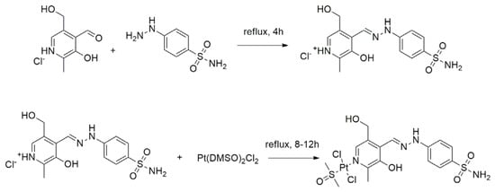
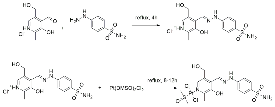
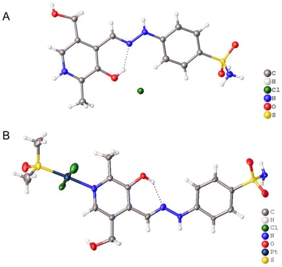
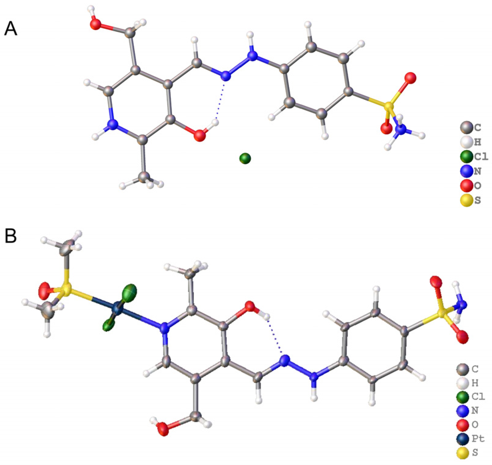
To clarify the spatial structures and configurational features of the ligand and target platinum complex (Scheme 1), single-crystal X-ray diffraction was employed for crystallographic analysis (Figure 1). Full crystallographic parameters, bond lengths, and bond angles are summarized in Tables S1–S5. Crystallographic results reveal that both the ligand and platinum complex crystallize in the monoclinic system with the same space group P21/n, free of significant defects or residual solvent, which is indicative of high-quality crystals. Complete diffraction datasets with sufficient independent reflections and good structure-fit quality were obtained. The final R1, wR2 factors and maximum difference peak/hole values fall within reasonable error ranges for crystallographic refinement, suggesting the reasonable reliability of the solved crystal structures.
Scheme 1.
Synthetic route for the ligand and Pt complex.
Figure 1.
(A) Crystal structure of the ligand; (B) Crystal structure of the Pt complex.
Further fine analysis of bond lengths and bond angles confirms the stability of molecular skeletons and the well-defined coordination geometry of the platinum complex. For the ligand, the S=O bond lengths of the sulfonyl group range from 1.437 Å to 1.438 Å; the S–N and S–C bond lengths are 1.619 Å and 1.764 Å, respectively. The C–C and C–N bond lengths of aromatic rings are consistent with typical values for organic small molecules. All bond angles are well-distributed, and no obvious spatial distortion or steric hindrance is observed, indicating a regular and stable solid-state conformation. In the platinum complex, the central Pt(II) ion adopts a typical four-coordinate square-planar geometry, coordinated to one nitrogen atom and one sulfur atom from the ligand, as well as two chloride anions. The key coordination-bond parameters are stable: Pt–N and Pt–S bond lengths are 2.072 Å and 2.225 Å, respectively, while Pt–Cl bond lengths vary from 2.287 Å to 2.306 Å, which are in good agreement with reported values for Pt(II) complexes. Bond angle measurements show that the trans Cl–Pt–Cl and S–Pt–N angles are 176.84° and 177.39°, close to the ideal 180° for square-planar geometry. The remaining adjacent coordination angles are uniformly distributed, and no evident spatial strain or configurational distortion exists within the central coordination plane. Moreover, no abnormal distortion occurs for the core skeleton bond lengths and angles after metal coordination, indicating that coordination modification does not disrupt the fundamental framework of the ligand. The Pt–N and Pt–L bond lengths of our complex fall within the range reported for analogous four-coordinate Pt(II) complexes [27,28,29]. The observed bond angles around the Pt center are consistent with the expected square-planar geometry, with minor deviations arising from the constraints imposed by the chelating ligand framework [27,28,29].
2.2. Evaluation of Carbonic Anhydrase Inhibitory Activity and In Vitro Antiproliferative Activity
The carbonic anhydrase isoenzyme inhibitory potency and in vitro antiproliferative performance of the ligand, as-synthesized platinum complex, and the positive control U-104 were systematically evaluated in this work. As summarized in Table 1, all tested compounds exhibited very weak inhibition against CA II, which is abundantly expressed in normal human tissues. The Ki values of the ligand, platinum complex, and U-104 were 3878 nM, 4963 nM, and 5891 nM, respectively. Hardly any interference with physiological CA II was observed. In contrast, all compounds exerted potent inhibition against the tumor-specific CA IX isoform with remarkable isoform-selective preference. Among them, the synthetic platinum complex displayed the most prominent CA IX-inhibitory capacity with a Ki value of 26.5 nM, outperforming the ligand (Ki = 30.4 nM) and positive control U-104 (Ki = 39.6 nM). It ranked as the most effective CA IX-targeting agent among all tested samples. The ligand took second place for CA IX inhibition, whereas U-104 showed relatively moderate potency, which formed a sharp contrast with the high-efficiency CA IX suppression of the platinum complex.
Table 1.
Inhibition constants Ki (nM) of tested compounds against carbonic anhydrase isoenzymes.
As shown in Table 2, the antiproliferative potency was highly correlated with CA IX-inhibitory activity. The ligand possessed moderate cytotoxicity against oral cancer HSC-2 cells, with an IC50 of 18.6 ± 0.4 μM under normoxia. Its antitumor effect was slightly improved under hypoxia, giving an IC50 value of 13.9 ± 0.3 μM. Benefiting from its CA IX-targeting ability, the platinum complex exhibited distinctly enhanced antitumor activity compared with the ligand, affording an IC50 of 12.3 ± 0.5 μM in normoxia. Notably, its cytotoxicity was drastically boosted under a tumor-mimicking hypoxic microenvironment, reaching an IC50 value as low as 3.7 ± 0.2 μM, which demonstrated outstanding hypoxia-selective antitumor property and revealed that potent CA IX inhibition contributes to improved killing efficiency toward hypoxic tumor cells. Furthermore, positive control U-104, clinical first-line drug cisplatin (DDP), as well as synthetic precursor Pt(DMSO)2Cl2, showed negligible growth-suppressive effects both in normoxia and hypoxia, with IC50 values higher than 20 μM within the tested concentration range. These results further highlight the unique advantages of our designed platinum complex in CA IX-targeting therapy and hypoxia-selective anti-oral cancer treatment.
Table 2.
IC50 (μM) against HSC-2 cells under normoxic and hypoxic conditions for tested compounds.
2.3. Molecular Docking Simulation Analysis
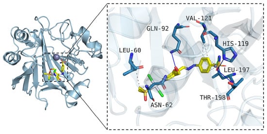
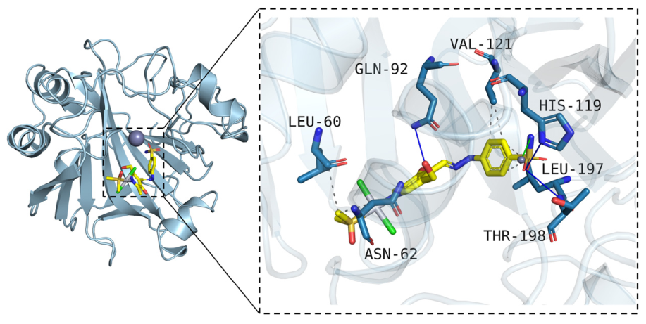
To elucidate the binding mechanism between the platinum complex and CA IX target at the molecular level, molecular docking simulations were performed using Vina 1.2.3 software to explore the interaction mode of the target compound within the active pocket of CA IX protein (PDB ID: 5OGN). The predicted binding mode is presented in Figure 2.
Figure 2.
Predicted binding mode of the platinum complex with CA IX protein (cyan: protein; yellow: ligand; blue lines: H-bonds; gray dotted lines: hydrophobic interactions; green lines: coordination bonds).
Docking results reveal that the platinum complex can stably embed into the active pocket of CA IX. Multiple non-covalent interactions, together with metal coordination interactions, synergistically stabilize the conformation of the protein–ligand complex. For hydrophobic interactions, the platinum complex forms close hydrophobic contacts with hydrophobic residues ASN-62, LEU-60, VAL-121, and LEU-197, constructing a stable hydrophobic microenvironment around the binding pocket and firmly locking the spatial binding pose of the compound. In terms of hydrogen bond interactions, intermolecular hydrogen bonds are established between the platinum complex and polar amino acid residues GLN-92, THR-198, and HIS-119, which adjust the binding orientation and further strengthen the stability of the protein–ligand complex. Moreover, the amino group of the sulfonamide moiety in the compound undergoes specific coordination with the Zn2+ ion located in the catalytic center of CA IX, enabling accurate anchoring toward the core active region of the target. In summary, the platinum complex achieves efficient and stable binding to CA IX through the synergistic effects of hydrophobic forces, hydrogen bonds, and Zn2+ coordination.
2.4. Intracellular ROS Generation
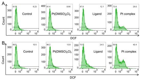
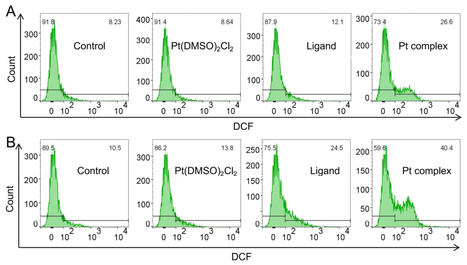
To explore the antitumor mechanism of the platinum complex, DCFH-DA fluorescent probe combined with flow cytometry was applied to quantitatively measure intracellular ROS accumulation in tumor cells under normoxic and hypoxic conditions (Figure 3).
Figure 3.
Detection of intracellular ROS levels by flow cytometry. (A) Normoxia; (B) Hypoxia.
ROS measurements revealed that cells in the blank control group and the platinum precursor Pt(DMSO)2Cl2-treated group maintained extremely low DCF fluorescence intensity; neither treatment triggered intracellular ROS accumulation. Under normoxia, the ligand only slightly provoked ROS generation with 12.1% ROS-positive cells. In contrast, the platinum complex remarkably induced ROS accumulation, raising the proportion of ROS-positive cells to 28.6%. The hypoxic microenvironment further amplified ROS-activating effects of the platinum complex. Under hypoxia, ROS levels in control and precursor groups remained almost unchanged; the percentage of ROS-positive cells for the parent-ligand group increased to 24.5%, while a prominent ROS burst was observed in the platinum complex-treated group with 40.4% positive cells. These data demonstrate that the platinum complex effectively induces ROS accumulation in tumor cells, and such effect can be markedly potentiated under hypoxia. Compared with the ligand and inactive platinum precursor, the platinum complex exhibits superior ROS-stimulating capacity, suggesting that ROS-mediated oxidative stress damage serves as an important pathway for tumor-cell killing.
2.5. Apoptosis-Related Protein Expression
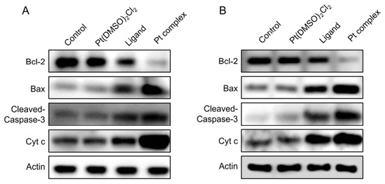
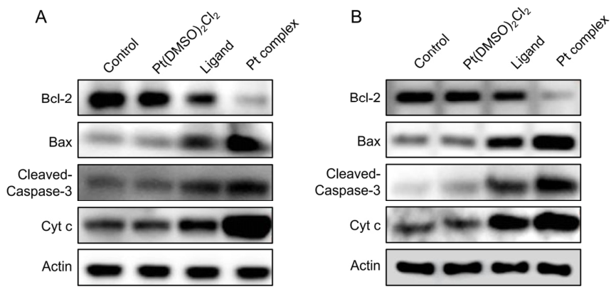
To clarify the molecular mechanism of platinum complex-triggered tumor-cell apoptosis, Western blotting was performed to detect differential expression of key proteins involved in the mitochondrial apoptosis pathway. Figure 4A represents normoxic conditions, and Figure 4B represents hypoxia; β-Actin was selected as the internal reference. Detected biomarkers included the anti-apoptotic protein Bcl-2, pro-apoptotic protein Bax, executioner apoptotic protein Cleaved-Caspase-3, and cytochrome c (Cyt c). The results are shown in Figure 4.
Figure 4.
Western blot analysis of proteins related to mitochondrial apoptosis pathway. (A) Normoxia; (B) Hypoxia.
No obvious difference in apoptosis-associated protein expression was observed between the blank group and Pt(DMSO)2Cl2 precursor group, suggesting that the platinum precursor exhibits negligible apoptosis-modulating effects under these experimental conditions. Compared with the control groups, both the ligand and platinum complex significantly down-regulated anti-apoptotic Bcl-2 while up-regulating Bax, Cleaved-Caspase-3, and Cyt c, and such regulatory effects were much stronger for the platinum complex. Comparative analysis of oxygen conditions indicated that hypoxia further enhanced apoptosis activation induced by the platinum complex. Upon hypoxic incubation, Bcl-2 expression was further decreased, whereas Bax, Cleaved-Caspase-3, and Cyt c were significantly up-regulated. These findings indicate that the platinum complex disturbs the balance of key mitochondrial apoptosis-related proteins, promotes mitochondrial cytochrome-C release, activates the Caspase cascade, and consequently induces tumor-cell apoptosis, and the hypoxic microenvironment can greatly strengthen this pro-apoptotic effect.
2.6. Cell Apoptosis
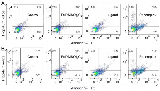
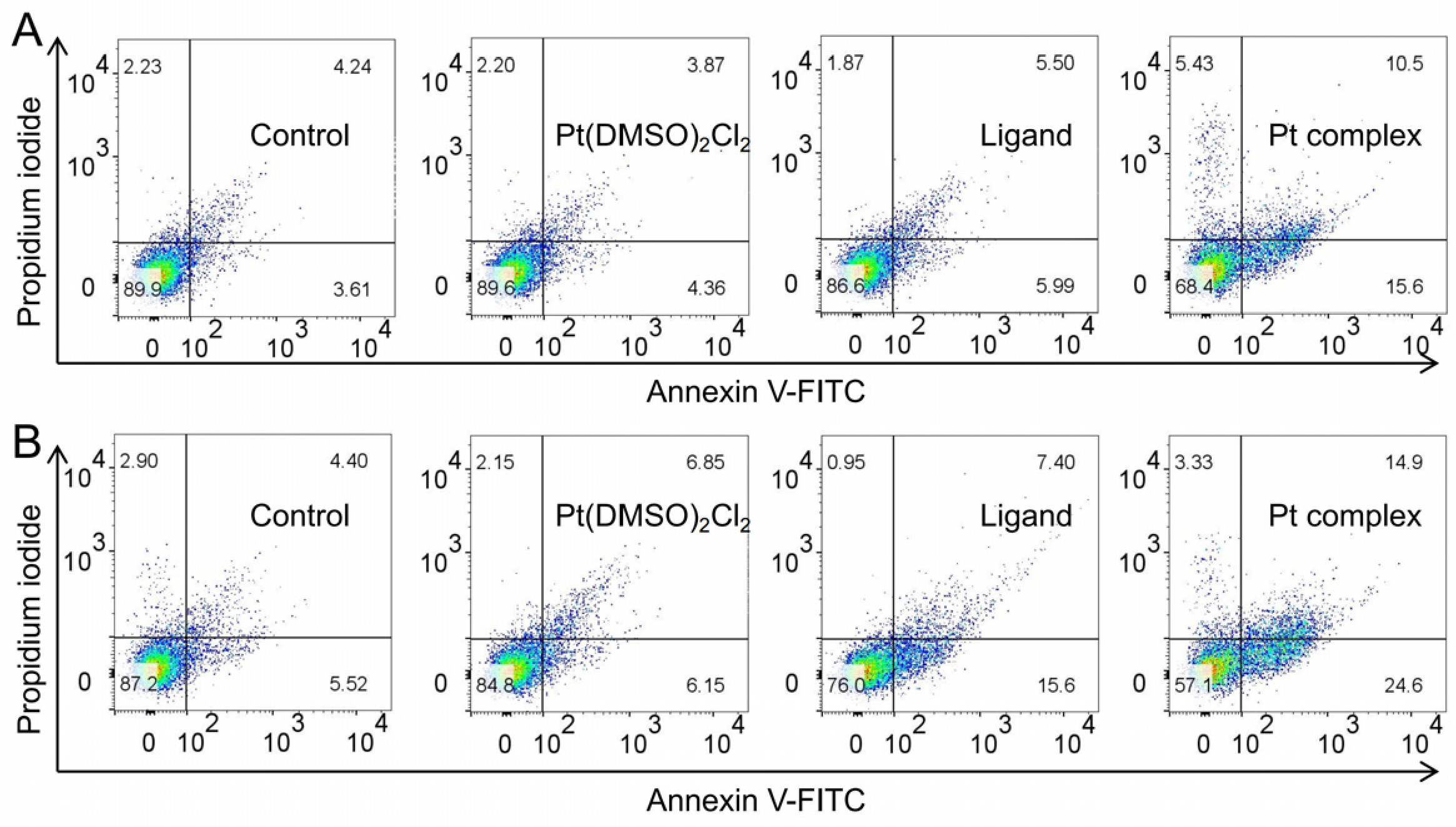
Annexin V-FITC/PI double-staining flow cytometry was used to quantify apoptosis induction by various compounds under different oxygen conditions. Figure 5A corresponds to normoxia and Figure 5B to hypoxia. The lower-right quadrant represents early-apoptotic cells, and the upper-right quadrant represents late-apoptotic/necrotic cells; the total apoptosis rate is the sum of the two fractions, as illustrated in Figure 5.
Figure 5.
(A) Dot plots of Annexin V-FITC/PI double staining; (B) Quantitative analysis of live, early-apoptotic, late-apoptotic, and necrotic cell percentages.
Quantitative apoptosis results were highly consistent with protein expression trends. Under normoxia, the total apoptosis rates of the blank group and platinum precursor group were merely 7.85% and 8.23%, showing negligible apoptosis. The ligand induced mild apoptosis with a total apoptosis rate of 11.49%, whereas the platinum complex markedly triggered tumor-cell apoptosis with a total apoptosis rate reaching 26.1%. Elevated apoptosis levels were observed for all experimental groups under hypoxia. The apoptosis rates of the blank and precursor groups barely increased; the total apoptosis rate of the parent-ligand group rose to 23.00%. Notably, the pro-apoptotic potency of the platinum complex was drastically augmented under hypoxia, achieving a total apoptosis rate up to 39.5%. Collectively, the platinum complex possesses notable in vitro capacity for inducing tumor-cell apoptosis, far exceeding that of the ligand and platinum precursor. The hypoxic microenvironment further enhances its pro-apoptotic activity through synergistic effects, which is highly consistent with its CA IX-inhibitory potency, ROS accumulation, and mitochondrial-pathway activation.
2.7. Discussion
Single-crystal X-ray diffraction confirms its stable square-planar coordination geometry in the solid state; coordination modification does not impair the integrity of parent-ligand scaffold, providing solid structural basis for CA-isoform recognition. Enzymatic assays reveal that this platinum complex scarcely inhibits the normal CA II isoform and exhibits stronger CA IX-suppressing activity than the ligand and positive control U-104, with a distinct tumor-targeting preference. Molecular docking theoretically illustrates that the platinum complex stably occupies the CA IX active pocket via hydrogen bonds, hydrophobic interactions, and specific Zn2+ coordination, which provides theoretical insights into its potent target-binding mode. Cellular experiments confirm that, benefiting from CA IX-inhibitory performance, the platinum complex effectively counteracts hypoxia-mediated chemoresistance in vitro and displays strengthened antiproliferative effects under hypoxia. The CA IX-inhibitory activity (Ki = 26.5 ± 1.9 nM) and in vitro antiproliferative potency against HSC-2 cells (IC50 = 3.7 ± 0.2 μM under hypoxia) of our complex are comparable to those of reported analogues. Molecular docking simulations predict that the sulfonamide moiety of the complex may coordinate with the Zn2+ ion in the CA IX active site, while additional hydrophobic and hydrogen bond interactions further stabilize binding [29,30,31]. Differences in isoform selectivity and hypoxia responsiveness among these complexes likely arise from variations in ligand structure and coordination environment. By contrast, cisplatin, the platinum precursor, and U-104 show no obvious hypoxia-selective antitumor activity, highlighting the unique merits of our structural design. Comparison of the 1H NMR spectra of the free ligand and Pt complex reveals small changes in chemical shift for selected protons upon complexation, supporting Pt–ligand interaction in DMSO-d6. However, these NMR data cannot exclude partial ligand dissociation, and the solid-state coordination geometry observed by single-crystal XRD may not be fully retained in solution, especially in aqueous environments. It should be noted that these biological assays were performed in aqueous media, where the platinum complex may undergo partial aquation; accordingly, the observed bioactivity should be attributed to the complex and its equilibrium solution species rather than exclusively to the intact chloro form. Consistent with this consideration, aquation and solution-phase speciation have not been experimentally characterized in the current work, and the true biologically active Pt-containing species remains unknown. Mechanistic studies indicate that the platinum complex elevates intracellular ROS levels to provoke oxidative stress injury, further activates the mitochondrial apoptosis pathway, down-regulates anti-apoptotic Bcl-2 and up-regulates pro-apoptotic Bax, Cleaved-Caspase-3, and Cyt c, and ultimately triggers tumor-cell apoptosis. All the above antitumor effects can be further potentiated under hypoxia. In summary, this work investigates metal coordination modification targeting CA IX to generate hypoxia-selective antitumor candidates, and the obtained results offer experimental insights for the design of new CA-targeting platinum-based anticancer agents.
3. Materials and Methods
3.1. Chemicals
Pyridoxal, 4-sulfamoylphenylhydrazine, Pt(DMSO)2Cl2 and other chemical reagents were of analytical grade and purchased from Aladdin Reagent Co., Ltd. (Shanghai, China). Recombinant human CA II, CA IX and CA XI proteins, DCFH-DA fluorescent probe, Annexin V-FITC/PI apoptosis detection kit, and primary antibodies against Bcl-2, Bax, Cleaved-Caspase-3, Cyt c and β-Actin were commercially obtained. Fetal bovine serum and medium for cell culture were supplied by Beyotime Biotechnology (Nanjing, China).
3.2. Synthesis of the L
The 4-sulfamoylphenylhydrazine (2.0 mmol) was dissolved in 20 mL of ethanol. Under continuous stirring, a solution of pyridoxal hydrochloride (2.0 mmol) in 10 mL ethanol was added dropwise. The mixture was heated under reflux for 4 h, during which a white precipitate gradually formed. After cooling to room temperature, the precipitate was collected by filtration, washed with cold ethanol, and dried to afford the target ligand L with a yield of 83.6%. Anal. Calcd (%) for C14H17ClN4O4S: C, 45.10; H, 4.60; N, 15.03; O, 17.17. Found: C, 45.11; H, 4.61; N, 15.02; O, 17.16. 1H NMR (400 MHz, DMSO-d6) δ 12.16 (s, 1H), 8.53 (s, 1H), 8.20 (s, 1H), 7.82–7.76 (m, 2H), 7.26–7.15 (m, 4H), 4.83 (s, 2H), 2.64 (s, 3H). 13C NMR (101 MHz, DMSO-d6) δ 151.30, 145.88, 141.44, 136.74, 136.26, 134.62, 131.13, 129.75, 128.20, 112.82, 59.52, 15.10. Calculated exact mass for C14H17ClN4O4S: m/z = 372.07; found: 337.10 [M-Cl−]+.
3.3. Synthesis of the Platinum Complex
Pt(DMSO)2Cl2 and ligand L were combined at a molar ratio of 1:1 and dissolved in mixed methanol-acetonitrile solvent. The mixture was heated under reflux in the dark for 8–12 h. After the reaction finished, insoluble impurities were removed by filtration. The filtrate was subjected to slow evaporation at room temperature. Block-shaped crystals suitable for single-crystal X-ray diffraction measurement were harvested after several days, with a yield of 72.9%. Anal. Calcd (%) for C16H22Cl2N4O5PtS2: C, 28.24; H, 3.26; N, 8.23; O, 11.76. Found: C, 28.25; H, 3.25; N, 8.24; O, 11.74. 1H NMR (400 MHz, DMSO-d6) δ 12.13 (s, 1H), 8.50 (d, J = 1.0 Hz, 1H), 8.20 (s, 1H), 7.81–7.76 (m, 2H), 7.33–7.14 (m, 4H), 4.83 (s, 2H), 2.63 (s, 3H), 2.55 (s, 6H). 13C NMR (101 MHz, DMSO-d6) δ 151.33, 145.76, 141.23, 136.72, 136.33, 134.45, 131.28, 129.43, 128.18, 112.81, 59.49, 40.87, 14.99. Calculated exact mass for C16H22Cl2N4O5PtS2: m/z = 679.01; found: 680.01 [M+H]+.
3.4. Crystallography Experimental
Single-crystal X-ray diffraction data of the target platinum (II) complex were acquired using a Bruker D8 VENTURE diffractometer (Bruker AXS, Madison, WI, USA) with Mo-Kα monochromatic radiation (λ = 0.71073 Å). Raw diffraction data were processed, including frame integration, scaling, and empirical absorption correction, utilizing the APEX4 software suite (Version 2021.4-1). The crystal structure was initially solved via direct methods with SHELXT-2018 and further refined against F2 by the full-matrix least-squares method implemented in SHELXL-2018, within the Olex2-1.5 graphical interface. All non-hydrogen atoms were refined anisotropically, while carbon-bound hydrogen atoms were placed at geometrically calculated positions and refined in a riding model manner.
3.5. Determination of Carbonic Anhydrase Inhibitory Activity
The inhibitory constants (Ki) of the tested compounds against CA isoforms were determined via an esterase hydrolysis assay. 4-nitrophenylacetic acid was used as the substrate. Serial concentrations of the compounds were prepared and incubated with recombinant CA II, CA IX, and CA XI proteins at room temperature. The hydrolysis rate of the substrate was monitored at 405 nm using a microplate reader. The Ki values (nM) of each compound were calculated by Dixon plot analysis. All experiments were performed in triplicate.
3.6. In Vitro Cell Proliferation Inhibition Assay
HSC-2 cells were purchased from Procell Life Science & Technology Co., Ltd., Wuhan, China. Tumor cells were seeded in 96-well plates and cultured under normoxic (21% O2) and hypoxic (1% O2) conditions. After cell adherence, gradient concentrations of the ligand, Pt complex, U-104, cisplatin (DDP), and Pt(DMSO)2Cl2 were added, and the cells were continuously incubated for 48 h. Subsequently, CCK-8 solution was added to each well, and the plates were incubated in the dark. The absorbance was measured at 450 nm. Cell viability was calculated, and the half-maximal inhibitory concentration (IC50, μM) was fitted using Origin software (OriginPro 2022b). All experiments were independently repeated three times.
3.7. Molecular Docking Simulation
The crystal structure of CA IX (PDB ID: 5OGN) was downloaded from the Protein Data Bank (PDB). AutoDock-Tools (MGLTools 1.5.6) was used for protein pretreatment, including water molecule removal, polar hydrogen addition, and Gasteiger charge assignment. Molecular docking was performed using Vina 1.2.3, and the grid box parameters were defined according to the active pocket of the CA IX crystal structure. The optimized three-dimensional conformation of the platinum complex obtained from single-crystal analysis was adopted as the ligand input structure. After docking calculation, the optimal binding conformation was selected to analyze hydrogen bonding, hydrophobic interactions, and Zn2+ coordination. The binding mode was visualized using PyMOL (Version 2.6).
3.8. Flow Cytometric Detection of Intracellular ROS Level
Tumor cells were seeded in 6-well plates and treated with 8 μM ligand, Pt(DMSO)2Cl2, and Pt complex for 48 h under normoxic and hypoxic conditions, respectively. After incubation, the culture medium was discarded, and the cells were rinsed with PBS. The DCFH-DA fluorescent probe was added, and the cells were incubated at 37 °C for 20 min in the dark. Subsequently, cells were digested with trypsin, resuspended with PBS, and detected by flow cytometry. The DCF fluorescence intensity and the proportion of ROS-positive cells were statistically analyzed. Each group was set with three biological replicates.
3.9. Annexin V-FITC/PI Apoptosis Detection
Tumor cells were seeded in 6-well plates and treated with 8 μM ligand, Pt(DMSO)2Cl2, and Pt complex for 48 h under normoxia and hypoxia. The supernatant and digested cells were collected and washed twice with pre-cooled PBS. Cell apoptosis was detected using an Annexin V-FITC/PI apoptosis kit. Briefly, cells were resuspended in binding buffer and stained sequentially with Annexin V-FITC and PI staining solutions. After incubation at room temperature for 15 min in the dark, cell apoptosis was analyzed by flow cytometry. The lower-right quadrant represented early-apoptotic cells, and the upper-right quadrant represented late-apoptotic/necrotic cells. The total apoptosis rate was defined as the sum of early and late-apoptotic cell proportions.
3.10. Western Blot Analysis
Tumor cells were seeded in 6-well plates and treated with 8 μM ligand, Pt(DMSO)2Cl2, and Pt complex for 48 h under normoxic and hypoxic environments. Cells were collected and lysed with RIPA lysis buffer to extract total cellular protein. The protein concentration was quantified using the BCA method. Equal amounts of protein were separated by SDS-PAGE and transferred onto PVDF membranes. The membranes were blocked with 5% skimmed milk at room temperature for 1 h and then incubated with primary antibodies against Bcl-2, Bax, Cleaved-Caspase-3, Cyt c, and β-Actin overnight at 4 °C. After washing with TBST, the membranes were incubated with corresponding secondary antibodies. Protein bands were visualized by ECL chemiluminescence. β-Actin was used as an internal reference for the normalization of protein loading.
3.11. Statistical Analysis
All results are presented as mean ± standard deviation (SD) from ≥3 independent experiments. Statistical comparisons were carried out using the Student’s t-test.
4. Conclusions
In this study, a novel CA IX-targeted platinum(II) complex was successfully designed and synthesized. Its structural characteristics, target selectivity, and antitumor mechanism were systematically elucidated through single-crystal diffraction, enzymatic screening, molecular simulation, cellular functional assays, and molecular mechanism verification. Structural characterization reveals that the obtained platinum complex possesses a stable and well-defined configuration. Metal coordination modification effectively retains the structural advantages of the ligand while significantly improving the CA IX recognition and targeting capability. Enzymatic assays demonstrated that the complex exhibits negligible inhibitory effects against the normal CA II isoform, which may confer favorable safety profiles toward normal CA-expressing tissues in vitro, and displays superior CA IX-inhibitory activity compared with the ligand and the positive control U-104, thereby achieving tumor-targeting selectivity. Cellular evaluations suggest that the platinum complex possesses prominent hypoxia-selective antitumor potency and efficiently reverses hypoxia-induced chemoresistance, with significantly enhanced anticancer activity under hypoxic conditions compared with conventional cisplatin and the synthetic precursor. Mechanistic studies revealed that the platinum complex induces intracellular ROS accumulation to trigger oxidative stress, further activates the mitochondrial apoptotic pathway, and disrupts the expression balance of apoptosis-related proteins, ultimately leading to efficient tumor-cell apoptosis. Note that aquation, hydrolysis, and solution-phase speciation of the platinum complex were not probed experimentally. The actual biologically active platinum-containing species is unknown, so the observed in vitro bioactivity cannot be unequivocally attributed to the intact solid-state Pt(II) complex. Moreover, the hypoxic tumor microenvironment synergistically amplifies its antitumor efficacy.
Supplementary Materials
The following supporting information can be downloaded at: https://www.mdpi.com/article/10.3390/inorganics14100250/s1, Table S1. Crystal data and structure refinement for Ligand and Pt complex; Table S2. Bond lengths for Ligand; Table S3. Bond angles for Ligand; Table S4. Bond lengths for Pt complex; Table S5. Bond angles for Pt complex; Figure S1. 1H NMR for Ligand; Figure S2. 13C NMR for Ligand; Figure S3. 1H NMR for Pt complex; Figure S4. 13C NMR for Pt complex.
Author Contributions
Conceptualization, J.Q.; methodology, Y.K.; software, Y.Z. and B.L.; validation, Y.K.; formal analysis, Y.Z.; investigation, Y.Z.; resources, J.Q.; data curation, X.B.; writing—original draft preparation, J.Q.; writing—review and editing, X.B.; visualization, Y.K.; supervision, J.Q.; project administration, J.Q.; funding acquisition, Y.K. All authors have read and agreed to the published version of the manuscript.
Funding
This work was supported by the Key Scientific Research Project Plan of Institutions of Higher Education in Henan Province (Grant No. 24B320013).
Institutional Review Board Statement
Not applicable.
Informed Consent Statement
Not applicable.
Data Availability Statement
The original contributions presented in this study are included in the article/Supplementary Materials. Further inquiries can be directed to the corresponding authors.
Conflicts of Interest
The authors declare no conflicts of interest.
Abbreviations
The following abbreviations are used in this manuscript:
| DDP | Cisplatin |
| CA | Carbonic anhydrase |
| ROS | Reactive oxygen species |
| PBS | Phosphate-buffered saline |
| CCK-8 | Cell counting kit-8 |
| PI | Propidium iodide |
| FITC | Fluorescein isothiocyanate |
| PDB | Protein Data Bank |
| Ki | Inhibition constant |
| IC50 | Half-maximal inhibitory concentration |
References
- Zhang, X.; Li, W.; Zheng, C.; Ding, L.; Wang, J.; Liang, Y.; Li, H.; Qi, W.; Ke, J.; Sun, Y.; et al. Overcoming Tumor Hypoxia and Adenosine-mediated Immunosuppression for Boosting Photodynamic Therapy and Systemic Antitumor Immune Responses. Small 2026, e74971. [Google Scholar] [CrossRef] [Scilit]
- Qin, H.; Yang, S.; He, J.; Zhao, M.; Zhao, L.; Wang, J.; Jiang, X.; Chen, X.; Xu, X. Hypoxia-driven tumor immune escape: Mechanisms and therapeutic opportunities. Front. Immunol. 2026, 17, 1893099. [Google Scholar] [CrossRef] [Scilit]
- Ma, G.; Zhang, N.; Shi, H.; Li, Y.; Wang, H.; Tang, C.; Wang, S.; Chen, H.; Wang, Q.; Wang, Z.; et al. Algae-Integrated Optoelectronic Nanoplatform for Tumor Hypoxia Relief and Enhanced Photodynamic Therapy. Adv. Healthc. Mater. 2026, 15, e71583. [Google Scholar] [CrossRef] [Scilit]
- Li, W.; Li, H.; Lin, C.; Jiang, G.; Yang, L.; Zhang, M.; Nie, H.; Gao, W.; Akhtar, M.; Prince, S.; et al. Axially Chlorinated Carbon Nanodots for Hypoxia-Tolerant Neutrophil-Mimicking Photoenzymatic Cascade Therapy against Postoperative Tumor Recurrence. ACS Nano 2025, 19, 25501–25514. [Google Scholar] [CrossRef] [Scilit]
- Tawk, B.; Rein, K.; Schwager, C.; Knoll, M.; Wirkner, U.; Horner-Rieber, J.; Liermann, J.; Kurth, I.; Balermpas, P.; Rodel, C.; et al. DNA-Methylome-Based Tumor Hypoxia Classifier Identifies HPV-Negative Head and Neck Cancer Patients at Risk for Locoregional Recurrence after Primary Radiochemotherapy. Clin. Cancer Res. 2023, 29, 3051–3064. [Google Scholar] [CrossRef] [Scilit]
- Fang, Y.; Luo, X.; Xu, Y.; Liu, Z.; Mintz, R.L.; Yu, H.; Yu, X.; Li, K.; Ju, E.; Wang, H.; et al. Sandwich-Structured Implants to Obstruct Multipath Energy Supply and Trigger Self-Enhanced Hypoxia-Initiated Chemotherapy Against Postsurgical Tumor Recurrence and Metastasis. Adv. Sci. 2023, 10, e2300899. [Google Scholar] [CrossRef] [Scilit]
- Szefler, B.; Szupryczynski, K.; Czelen, P. Platinum Meets Pyridine: Affinity Studies of Pyridinecarboxylic Acids and Nicotinamide for Platinum-Based Drugs. Int. J. Mol. Sci. 2025, 26, 11875. [Google Scholar] [CrossRef] [Scilit]
- Shao, C.; Li, M.; Liu, L.; Bai, D.; Yang, J.; Liu, C.; Cai, L.; Wang, X.; Peng, J.; Guo, Z.; et al. Pt-seq unveils the genomic binding pattern of platinum-based drugs. Sci. Adv. 2025, 11, eadx6809. [Google Scholar] [CrossRef] [Scilit]
- Kee, J.X.; Yau, J.N.N.; Kumar Muthuramalingam, R.P.; Wang, X.; Chng, W.H.; Lopez-Sanchez, A.; Tay, K.K.W.; Deng, L.W.; Gibson, D.; Bertrand, H.C.; et al. Colorectal Cancer at the Crossroads: The Good, the Bad, and the Future of Platinum-Based Drugs. Chem. Rev. 2025, 125, 10248–10341. [Google Scholar] [CrossRef] [Scilit]
- Fang, R.; Li, Y.; Jin, J.; Yang, F.; Chen, J.; Zhang, J. Development of Anticancer Ferric Complex Based on Human Serum Albumin Nanoparticles That Generate Oxygen in Cells to Overcome Hypoxia-Induced Resistance in Metal Chemotherapy. J. Med. Chem. 2024, 67, 1184–1196. [Google Scholar] [CrossRef] [Scilit]
- Chang, P.; Guo, Y.; Chen, D.; Li, K.; Wang, W.; Yang, Z.; Ma, J.; Zeng, Y.; Zhan, W.; Zhan, Y. High-temperature PTT/CDT coordination nanoplatform realizing exacerbated hypoxia for enhancing hypoxia-activated chemotherapy to overcome tumor drug resistance. J. Nanobiotechnol. 2024, 22, 374. [Google Scholar] [CrossRef] [Scilit]
- Liu, W.; Chen, Y.; Li, Z.; Cui, M. Preclinical evaluation of (68)Ga-labeled acetazolamide derivatives as radiotracers targeting carbonic anhydrase IX in clear cell renal cell carcinoma. Eur. J. Med. Chem. 2026, 305, 118577. [Google Scholar] [CrossRef] [Scilit]
- Li, S.; Chen, Y.; Li, J.; Zhou, X.; Miao, W.; Shu, H.; Zhao, W.; Wang, Y.; Wu, T.; Luo, J.; et al. Development and preclinical evaluation of NYM074 as a novel Theranostic ligand targeting carbonic anhydrase IX in renal cell carcinoma. Bioorg. Chem. 2026, 176, 109890. [Google Scholar] [CrossRef] [Scilit]
- Nerella, S.G.; Shaik, M.G.; Singh, P.; Arifuddin, M.; Ullah, Q.; Supuran, C.T. Antibody-drug conjugates and radioconjugates targeting carbonic anhydrase IX and XII in hypoxic tumors: Bench to clinical applications. Bioorg. Chem. 2025, 159, 108408. [Google Scholar] [CrossRef] [Scilit]
- Liu, T.; Xu, H.; Chen, X.; Cui, K.; Zhou, J.; Cai, L.; Wang, Y.; Zhang, J.; Yang, X.; Gong, K. Development of a Novel 68Ga-Labeled Radiotracer Targeting Carbonic Anhydrase IX for the Diagnosis of Clear Cell Renal Cell Carcinoma. Mol. Pharm. 2026, 23, 4123–4132. [Google Scholar] [CrossRef] [Scilit]
- Vaskevicius, A.; Zvirblis, M.; Kurtenoka, M.; Leitans, J.; Manakova, E.; Paketuryte-Latve, V.; Kvietkauskaite, A.; Kazaks, A.; Eimonta, V.; Cerepenkaite, K.; et al. Di-meta-Substituted Fluorinated Benzenesulfonamides as Potent and Selective Anticancer Inhibitors of Carbonic Anhydrase IX and XII. J. Med. Chem. 2025, 68, 18389–18406. [Google Scholar] [CrossRef] [Scilit]
- Nencetti, S.; Cuffaro, D.; Ciccone, L.; Nocentini, A.; Di Stefano, M.; Poli, G.; Macchia, M.; Tuccinardi, T.; Nuti, E.; Supuran, C.T.; et al. A series of benzensulfonamide derivatives as new potent carbonic anhydrase IX and XII inhibitors. Future Med. Chem. 2025, 17, 271–285. [Google Scholar] [CrossRef] [Scilit]
- Khasawneh, H.E.N.; Ali, E.I.; Elmagzoub, R.M.; Al-Aouadi, R.F.A.; Almagharbeh, W.T.; Alotaibi, G.; Brase, S.; Alkhammash, A. A novel thiazole-sulfonamide hybrid molecule as a promising dual tubulin/carbonic anhydrase IX inhibitor with anticancer activity. Front. Chem. 2025, 13, 1606848. [Google Scholar] [CrossRef] [Scilit]
- He, W.; Chen, C.; Cai, R.; Zheng, J.; Yao, M.; Shim, J.S.; Kwok, H.F.; Yao, X.; Fang, L.; Chen, L. Bifunctional compounds for targeted degradation of carbonic anhydrase IX through integrin-facilitated lysosome degradation. J. Biol. Chem. 2025, 301, 108482. [Google Scholar] [CrossRef] [Scilit]
- Khanfar, M.A.; Saleh, M. Design and Synthesis of 3-(Phenylsulfonamido)benzamide Derivatives as Potent Carbonic Anhydrase IX Inhibitors: Biological Evaluations and Molecular Modeling Studies. Med. Chem. 2025, 21, 160–167. [Google Scholar] [CrossRef] [Scilit]
- Elshamsy, A.M.; Mustafa, M.; Nocentini, A.; Massardi, M.L.; Ali, T.F.S.; Rabea, S.M.; Tuzun, B.; Smietana, M.; Kapancik, S.; Abdel-Aziz, M.; et al. Design, Synthesis, and In Vitro Anticancer Evaluation of Thiazole-Based Chalcones Linked to Sulfanilamide as Tumor-Associated Carbonic Anhydrase IX and XII Inhibitors. J. Med. Chem. 2025, 68, 15151–15164. [Google Scholar] [CrossRef] [Scilit]
- Njardarson, J.T.; Tomat, E. Bidentate Metal-Binding Motifs in US FDA Approved Small Molecule Drugs. J. Med. Chem. 2026, 69, 17714–17735. [Google Scholar] [CrossRef] [Scilit]
- Dias, I.; Bon, L.; Banas, A.; Chavarria, D.; Borges, F.; Guerreiro-Oliveira, C.; Cardoso, S.M.; Sanna, D.; Garribba, E.; Chaves, S.; et al. Exploiting the potential of rivastigmine-melatonin derivatives as multitarget metal-modulating drugs for neurodegenerative diseases. J. Inorg. Biochem. 2025, 262, 112734. [Google Scholar] [CrossRef] [Scilit]
- Castro-Ramirez, R.; Barba-Behrens, N. A new therapeutic perspective on metal-based drugs. Dalton Trans. 2025, 54, 15964–15979. [Google Scholar] [CrossRef] [Scilit]
- Jin, S.; Feng, C.; Wang, X. DNA or not DNA—That is the question determining the design of platinum anticancer drugs. Eur. J. Med. Chem. 2025, 282, 117077. [Google Scholar] [CrossRef] [Scilit]
- Wei, T.; Chen, L.; Shi, P.; Wang, C.; Peng, Y.; Yang, J.; Liao, X.; Yang, B.; Gao, C. Platinum (IV) drugs with cannabidiol inducing mitochondrial dysfunction and synergistically enhancing anti-tumor effects. J. Inorg. Biochem. 2024, 254, 112515. [Google Scholar] [CrossRef] [Scilit]
- Ming, J.; Bhatti, M.Z.; Ali, A.; Zhang, Z.; Wang, N.; Mohyuddin, A.; Chen, J.; Zhang, Y.; Rahman, F.U. Vitamin B6 based Pt(II) complexes: Biomolecule derived potential cytotoxic agents for thyroid cancer. Metallomics 2022, 14, mfac053. [Google Scholar] [CrossRef] [Scilit]
- Poma, A.; Forni, A.; Baldoli, C.; Mussini, P.R.; Bossi, A. Cyclometalated Pt(ii) complexes with a bidentate Schiff-base ligand displaying unexpected cis/trans isomerism: Synthesis, structures and electronic properties. Dalton Trans. 2017, 46, 12500–12506. [Google Scholar] [CrossRef] [Scilit]
- Qi, J.; Zheng, Y.; Li, B.; Wei, L.; Li, J.; Xu, X.; Zhao, S.; Zheng, X.; Wang, Y. Mechanism of vitamin B6 benzoyl hydrazone platinum(II) complexes overcomes multidrug resistance in lung cancer. Eur. J. Med. Chem. 2022, 237, 114415. [Google Scholar] [CrossRef] [Scilit]
- Razi, S.S.; Koo, Y.H.; Kim, W.; Yang, W.; Wang, Z.; Gobeze, H.; D’Souza, F.; Zhao, J.; Kim, D. Ping-Pong Energy Transfer in a Boron Dipyrromethane Containing Pt(II)-Schiff Base Complex: Synthesis, Photophysical Studies, and Anti-Stokes Shift Increase in Triplet-Triplet Annihilation Upconversion. Inorg. Chem. 2018, 57, 4877–4890. [Google Scholar] [CrossRef] [Scilit]
- Deng, J.; Yu, P.; Zhang, Z.; Zhang, J.; Zhewen, S.; Cai, M.; Yuan, H.; Liang, H.; Yang, F. Novel Pt(ii) complexes with modified aroyl-hydrazone Schiff-base ligands: Synthesis, cytotoxicity and action mechanism. Metallomics 2019, 11, 1847–1863. [Google Scholar] [CrossRef] [Scilit]

Disclaimer/Publisher’s Note: The statements, opinions and data contained in all publications are solely those of the individual author(s) and contributor(s) and not of MDPI and/or the editor(s). MDPI and/or the editor(s) disclaim responsibility for any injury to people or property resulting from any ideas, methods, instructions or products referred to in the content. |
© 2026 by the authors. Licensee MDPI, Basel, Switzerland. This article is an open access article distributed under the terms and conditions of the Creative Commons Attribution (CC BY) license.