


Abstract
Gold-sputtered tapered optical fiber (Au-TOF) sensors were developed for high-sensitivity refractive-index (RI) detection using aqueous glucose standards spanning 1.33–1.41 RIU (0–50% w/v). The sensors were fabricated using a reproducible workflow combining flame-brushing tapering, plasma surface preparation, and rotational magnetron sputtering, enabling azimuthally uniform gold coatings without the use of thiol or sulfur linker chemistries. Two sputtering durations (24 s and 30 s) were investigated to examine thickness-dependent plasmonic coupling and sensing performance. Optical measurements were conducted using a broadband supercontinuum source and compact spectrometer within a 20 µL microfluidic sensing chamber. Increasing glucose concentration produced a monotonic decrease in transmission intensity and a systematic red shift of the resonance minimum, consistent with enhanced evanescent-field interaction at the gold–dielectric interface. Across four independent trials, the 24 s Au-TOF sensor exhibited sensitivities up to 985 nm/RIU, while the 30 s device achieved sensitivities up to 1590 nm/RIU with strong linearity (R2 = 0.99). The enhanced sensitivity observed for the longer sputtering duration is attributed to improved gold film continuity and stronger plasmonic coupling. These results demonstrate a scalable, linker-free fabrication strategy for plasmonically enhanced tapered fiber sensors and establish the Au-TOF platform as a promising approach for label-free optical biosensing in compact microfluidic environments.
1. Introduction
Optical fiber biosensors exploiting plasmonic phenomena have emerged as powerful platforms for biomedical diagnostics due to their capability for label-free, real-time detection with high sensitivity and minimal sample consumption [1,2,3]. Compared with conventional planar plasmonic sensors, optical fiber-based platforms offer several intrinsic advantages, including compact size, mechanical flexibility, chemical inertness, and compatibility with remote and in situ interrogation [4,5,6]. These characteristics make fiber-optic sensors particularly attractive for clinical diagnostics, environmental monitoring, and point-of-care testing applications where portable and low-volume sensing platforms are required. As a result, numerous optical fiber sensing geometries have been developed, including unclad fibers, fiber Bragg gratings, interferometric structures, microstructured fibers, and tapered optical fibers (TOFs), each exploiting distinct optical interaction mechanisms to transduce biochemical events into measurable spectral responses [1,6,7,8,9].
Among these configurations, sensing performance is fundamentally determined by the strength of interaction between the guided optical field and the surrounding medium. In conventional optical fibers, strong modal confinement within the core restricts evanescent-field penetration into the external environment, thereby limiting sensitivity to surface refractive-index variations [6,10]. To overcome this limitation, several strategies have been proposed to enhance evanescent-field exposure, including cladding removal, surface micro- and nanostructuring, and geometric modification of the fiber waveguide [1,11,12,13]. In particular, tapered optical fibers have received considerable attention because the gradual reduction in the fiber diameter weakens modal confinement and increases the proportion of optical power propagating within the evanescent field. This enhanced light–matter interaction enables highly sensitive refractive-index detection and provides a favorable platform for optical biosensing applications [14,15,16].
Beyond geometric optimization, plasmonic surface functionalization has been widely employed to further enhance evanescent-field interactions. Noble metal coatings, particularly gold, enable strong coupling between guided optical modes and collective electron oscillations at the metal–dielectric interface, generating surface plasmon resonance (SPR) or localized surface plasmon resonance (LSPR) effects [6,17,18,19]. These plasmonic resonances produce strong electromagnetic field confinement at the sensor interface and exhibit pronounced sensitivity to local refractive-index changes. Gold is therefore widely used in optical fiber plasmonic biosensors because of its excellent chemical stability, biocompatibility, and compatibility with established biomolecular immobilization strategies [1,20,21].
Various approaches have been explored to introduce plasmonic gold structures onto optical fiber surfaces, including self-assembled monolayers, layer-by-layer assembly, dip coating, electroless plating, and chemical immobilization of gold nanoparticles [22,23,24,25]. Although these techniques can produce plasmonically active sensing surfaces with high sensitivity, they often rely on multi-step solution-based fabrication processes requiring precise chemical control. Such procedures may lead to variability in surface coverage, nanoparticle aggregation, and device-to-device reproducibility, particularly when applied to mechanically fragile tapered fiber geometries [24,25,26]. These limitations present challenges for scalable fabrication and translation of optical fiber biosensors toward robust point-of-care sensing platforms.
Physical vapor deposition methods, such as magnetron sputtering, offer an attractive alternative for plasmonic surface fabrication by enabling direct deposition of metallic films onto optical fiber surfaces without the need for linker molecules or solution-based chemistry [27,28,29]. Sputtering provides precise control over film thickness, improved surface uniformity, and reduced fabrication complexity, making it well suited for reproducible plasmonic coating of tapered optical fibers [30]. Despite these advantages, relatively few studies have systematically investigated sputter-deposited gold coatings on tapered optical fibers, particularly with respect to the influence of deposition parameters on plasmonic coupling behavior and sensing performance [29,30]. Addressing this gap is important for establishing scalable and reproducible fabrication strategies for high-performance fiber-optic biosensors.
In this work, we present a reproducible, low-cost, and scalable fabrication strategy for gold-coated tapered optical fiber (Au-TOF) sensors that addresses key limitations in conventional plasmonic fiber fabrication, including complex surface chemistry, limited reproducibility, and poor mechanical robustness. The proposed approach integrates an in-house flame-brushing tapering system with controlled magnetron sputtering, enabling precise modulation of gold layer thickness and plasmonic coupling through short-duration deposition (24 s and 30 s). In contrast to conventional chemically functionalized platforms, a linker-free fabrication strategy is employed, significantly simplifying surface preparation while improving reproducibility and reducing processing variability. A custom-designed 3D-printed microfluidic mounting platform is further introduced to enhance mechanical stability, facilitate handling, and enable seamless integration into sensing systems, an aspect often overlooked in tapered fiber sensor design.
The fabricated Au-TOF sensors were experimentally evaluated through bulk refractive-index sensing of aqueous glucose solutions spanning refractive indices from 1.33 to 1.41 (0–50% glucose concentration). Both devices demonstrated strong reproducibility across four independent trials. The 24 s Au-TOF sensor exhibited an average sensitivity of 908.35 ± 53.66 nm/RIU (R2 = 0.957), whereas the 30 s device achieved a substantially higher average sensitivity of 1366.41 ± 158.23 (R2 = 0.962), confirming the critical role of sputtering duration in tuning plasmonic response. This work introduces a practical and fabrication-efficient Au-TOF sensing platform that balances sensitivity, reproducibility, and system-level integration. While the sensitivity is modest compared to state-of-the-art laboratory-optimized systems, the proposed approach prioritizes scalability, robustness, and ease of deployment, representing a meaningful step toward real-world optical biosensing applications.
2. Materials and Methods
2.1. Chemical Reagents and Materials
Reagent-grade alcohols, including ethanol, methanol, and isopropyl alcohol, were obtained from VWR International (Radnor, PA, USA). D-(+)-Glucose was purchased from Sigma-Aldrich (St. Louis, MO, USA) and used as received without further purification. Optical adhesive was obtained from Dymax Corporation (Torrington, CT, USA). Polylactic acid (PLA) filament used for fabrication of custom sensor mounts was purchased from Hatchbox (Pomona, CA, USA). Ultrapure deionized (DI) water with a resistivity of 18.2 MΩ·cm−1 was used for the preparation of all aqueous solutions.
2.2. Tapered Optical Fiber Fabrication
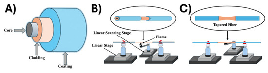
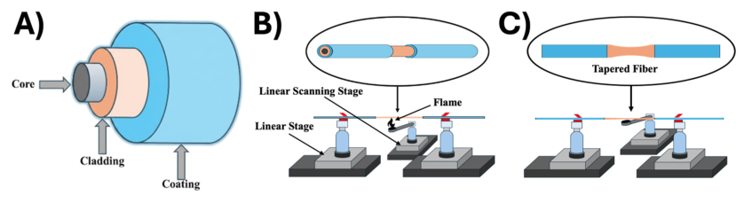
A standard single-mode optical fiber (Thorlabs 1060XP, operating range 980–1600 nm, Ann Arbor, MI, USA) was used as the substrate for sensor fabrication (Figure 1A). The fiber possesses a 5.8 µm core diameter, 125 ± 1.5 µm cladding diameter, and 245 ± 10 µm polymer coating diameter. The total fiber length used in this study was 38.1 cm. Fiber tapering was performed using an in-house flame-brushing system consisting of three programmable Aerotech PRO115L linear translation stages controlled by an Aerotech A3200 (Pittsburgh, PA, USA) motion controller. One stage was configured to translate the flame laterally along the fiber axis, while two opposing stages symmetrically pulled the fiber to produce controlled taper elongation. Prior to tapering, approximately 7.62 cm of the polymer coating was mechanically removed from the central region of the optical fiber to expose the silica cladding. The exposed region was cleaned using ethanol applied with a lint-free Kimwipe and allowed to air dry. The prepared fiber was then mounted onto the Aerotech three-axis translational stage system using precision fiber holders (Newport 561 Series, Irvine, CA, USA, compatible with 125 µm fibers) (Figure 1B).
Figure 1.
(A) Geometrical structure of a standard optical fiber showing core, cladding, and protective coating. (B,C) Fabrication of the tapered optical fiber (TOF) sensor using the flame-brushing technique, where the stripped fiber is heated and symmetrically pulled to form the tapered sensing region.
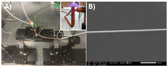
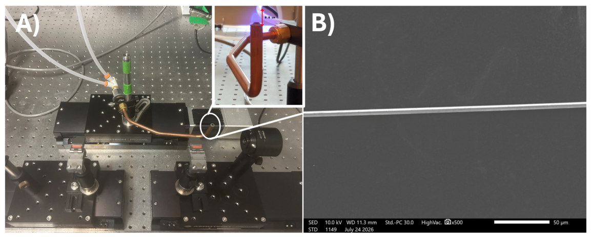
Taper fabrication was performed using the flame-brushing technique (Figure 2B). A propane–air mixture was delivered through a dual thermal mass flow control system (Brooks Instrument GF Series with 0254 Master Controller, Hatfield, PA, USA) with relative flow rates of 96% air and 4% propane. This configuration generated a stable laminar blue flame with an estimated temperature of approximately 1212 °C, sufficient to locally soften the silica fiber during pulling. During taper formation, the flame scanning stage oscillated 0.5 cm along the fiber axis at a velocity of 0.3 mm s−1, while the opposing pulling stages applied a total elongation of 3.46 cm over 6.3 min. This coordinated motion produced a smooth and symmetric tapered waist as shown in Figure 1C. The fabricated TOF was tapered over a 3 cm region from 125 µm to a waist diameter of ca. 8.5 µm, which was visually confirmed by scanning electron microscopy (Figure 2B). Moreover, tapering is essential for enhancing sensing performance, as the reduced fiber diameter increases evanescent field penetration into the surrounding medium. This enhances interaction with the gold coating and improves SPR coupling, thereby increasing sensitivity to refractive index changes.
Figure 2.
(A) Experimental set-up for the fabrication of TOF sensor using the flame-brushing technique.The red arrow indicates the localized flame-heating region where the stripped silica fiber is softened and drawn during taper formation. (B) Scanning electron microscopy micrograph of the as-fabricated TOF.
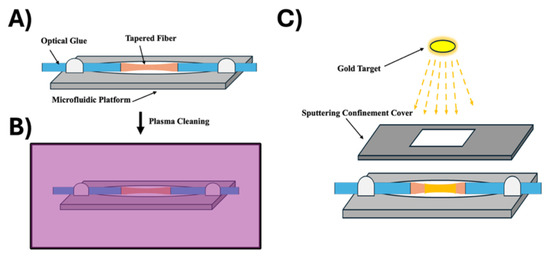
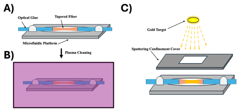
Following taper fabrication, the TOF was transferred to a custom 3D-printed mounting platform fabricated from PLA and secured at both ends using optical adhesive to ensure mechanical stability during subsequent processing (Figure 3A). Prior to gold deposition, the mounted TOF was then plasma-cleaned using low-power oxygen plasma to remove surface contaminants and improve film adhesion (Figure 3B). The plasma parameters were carefully controlled (low power and short exposure time) to avoid altering the fiber geometry or optical properties, ensuring that only surface contaminants were removed without affecting the tapered structure.
Figure 3.
(A) TOF mounted on a custom 3D-printed platform. (B) Plasma cleaning of the TOF surface prior to metallization. (C) Gold deposition on the tapered region by magnetron sputtering.
2.3. Gold Sputter Deposition
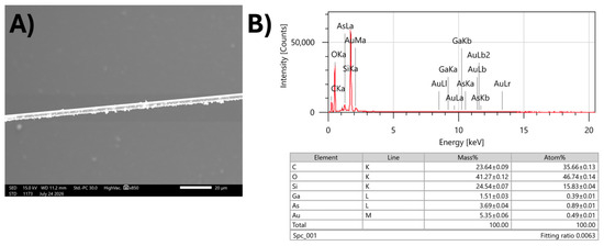
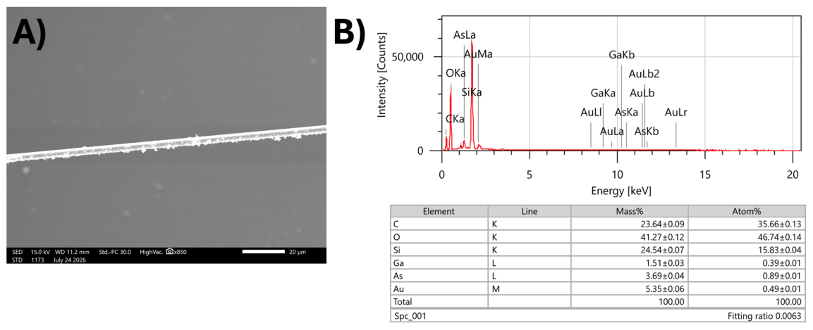
Gold metallization was performed using magnetron sputtering with the discharge current maintained at 10 mA and chamber pressure set to 6 mbar (Figure 3C). To obtain gold coverage around the cylindrical tapered region, a multi-step sputtering and rotation protocol was implemented. For the 24 s Au-coated TOF, sputtering was first conducted for 10 s, followed by a 180° rotation of the fiber and an additional 10 s sputtering step. The fiber was then rotated twice more by 180°, with 2 s sputtering per orientation, resulting in a total deposition time of 24 s. A second device with a 30 s gold coating was fabricated using an analogous procedure. Two initial sputtering steps of 10 s were performed with a 180° rotation between each deposition. The fiber was subsequently rotated twice more by 180°, with 5 s sputtering per orientation, yielding a total deposition time of 30 s. This rotational sputtering protocol minimized shadowing effects and promoted gold nanoparticle coverage along the tapered optical fiber surface, as confirmed by SEM imaging (Figure 4A). Energy-dispersive X-ray spectroscopy (EDS) further verified successful gold metallization through the characteristic Au Mα peak and elemental analysis, while the presence of Si and O originated from the silica optical fiber substrate (Figure 4B). The estimated gold film thickness corresponding to the sputtering durations used in this study was approximately 2.4 nm for the 24 s deposition and 3.0 nm for the 30 s deposition, using a linear deposition rate of 0.2 nm/sec under the applied sputtering conditions.
Figure 4.
SEM and EDS characterization of the Au-coated tapered optical fiber. (A) SEM image showing gold deposition along the TOF following the rotational sputtering process. (B) EDS spectrum and elemental analysis confirming successful gold metallization.
2.4. Optical Sensing Measurements
The Au-TOF sensor was mounted on a custom 3D-printed platform and positioned within a fiberglass microfluidic chamber designed to selectively expose only the tapered waist region to the test solution. The chamber geometry allowed controlled immersion of the sensing region while isolating the untapered fiber sections. Approximately 20 µL of analyte solution was introduced into the chamber to fully cover the tapered region during each measurement cycle. Optical excitation was provided by a supercontinuum light source (NKT Photonics SuperK Compact, spectral range 450–2400 nm, Birkerød, Denmark.). This broadband, high-intensity output enables simultaneous excitation across a wide wavelength range, facilitating accurate identification and tracking of the SPR dip in the transmission spectrum. This is particularly advantageous for wavelength-interrogation sensing, where resonance shifts occur in response to changes in the external refractive index. The high optical power also improves the signal-to-noise ratio, enabling precise detection of small spectral shifts.
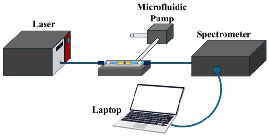
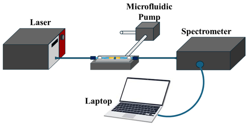
The broadband light was coupled into the input end of the optical fiber and propagated through the tapered sensing region. The transmitted spectrum was collected from the output end of the fiber and analyzed using a compact spectrometer (Thorlabs CCS200, Ann Arbor, MI, USA) for spectral acquisition and subsequent signal processing. Following each measurement, the test solution was removed from the microfluidic chamber using a peristaltic microfluidic pump (Cole-Parmer Masterflex, Vernon Hills, IL, USA) to enable controlled exchange between analyte concentrations (Figure 5). This procedure ensured consistent fluid replacement and minimized cross-contamination between sequential measurements.
Figure 5.
Experimental setup for optical sensing measurements, including a supercontinuum broadband light source, the Au-sputtered tapered optical fiber (Au-TOF) sensor, and a spectrometer for transmission spectrum acquisition.
3. Results
The refractive-index sensing performance of the Au-TOF sensors was evaluated using aqueous glucose solutions with concentrations ranging from 0–50% (w/v). Two sputtering durations, 24 s and 30 s, were investigated to assess the influence of gold film deposition time on plasmonic coupling and sensing performance. Each device was characterized through four independent experimental trials to evaluate measurement reproducibility. Glucose concentration was increased incrementally in 10% intervals, beginning with DI water as the baseline reference. For each measurement, approximately 20 μL of analyte solution was introduced into the microfluidic sensing chamber to fully immerse the tapered region. Following each measurement cycle, the chamber was rinsed thoroughly with DI water to prevent residual analyte carryover between concentration steps.
3.1. Spectral Response and Signal Processing
The sensing mechanism of the Au-TOF is governed by the interaction between the guided optical mode, the evanescent field extending into the surrounding medium, and SPR at the metal–dielectric interface. When light propagates through the tapered region of the optical fiber un der total internal reflection, a portion of the electromagnetic field extends beyond the fiber surface into the surrounding medium as an evanescent field. This field decays exponentially with distance from the interface according to [18]:
where dp represents the penetration depth given by:
E(z) = E0e−z/dp
Here, neff is the effective refractive index of the guided mode and next is the refractive index of the surrounding medium. This expression defines the interaction range of the evanescent field, which typically extends on the order of hundreds of nanometers. In TOFs, the reduced diameter weakens modal confinement, increasing the penetration depth and enhancing interaction between the guided light and the external environment. The introduction of a thin gold layer onto the tapered region enables excitation of surface plasmon resonance through coupling with the evanescent field. This coupling occurs when the propagation constant of the guided optical mode matches that of the surface plasmon wave at the gold–dielectric interface. The phase-matching condition can be expressed as [17]:
where the propagation constant of the guided mode is:
and the surface plasmon wavevector is given by:
keff = ksp
Here, and are the permittivities of the metal and surrounding dielectric, respectively, and k0 = . When this phase-matching condition is satisfied, efficient energy transfer occurs from the guided optical mode to the surface plasmon mode, resulting in attenuation at a specific wavelength. This attenuation is observed experimentally as a reduction in transmission intensity and a red shift of the resonance wavelength with increasing analyte concentration.
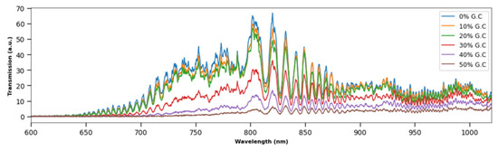
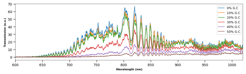
Consequently, increasing glucose concentration resulted in a monotonic decrease in transmitted optical intensity across the plasmonic spectral region (Figure 6). This behavior arises from the increase in refractive index, which perturbs the SPR condition and enhances coupling between the guided mode and surface plasmon modes. The resulting plasmon excitation introduces additional optical loss due to resistive damping in the gold layer, leading to reduced transmitted intensity. The lowest transmission was consistently observed at 50% glucose concentration, confirming the strong refractive-index sensitivity of the Au-TOF sensor.
Figure 6.
Raw transmission spectra of the Au-TOF sensor measured with glucose solutions ranging from 0–50% (w/v). Increasing glucose concentration produces a progressive decrease in transmission intensity, with the lowest signal observed at 50% glucose, indicating sensitivity of the sensor to refractive-index changes.
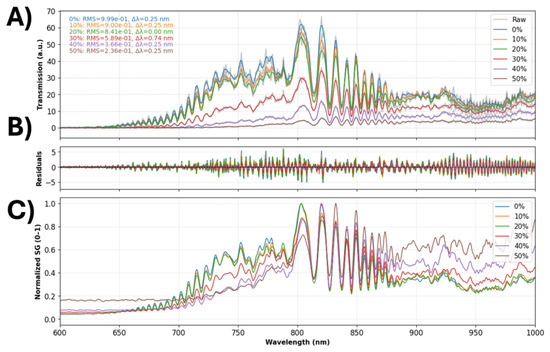
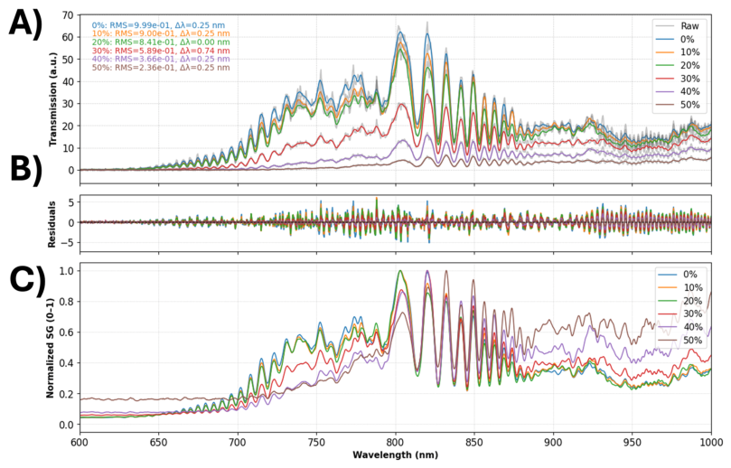
Because the raw transmission spectra contain both the SPR envelope and high-frequency interference fringes generated by modal interactions within the tapered region, signal processing was required to isolate the underlying plasmonic response. A Savitzky–Golay smoothing filter was first applied to suppress high-frequency noise while preserving the overall spectral shape. The signal-processing workflow is illustrated in Figure 7, where Figure 7A compares the raw spectrum (gray) with the processed signal, Figure 7B highlights the removed noise component, and Figure 7C presents the normalized spectrum used for analysis. The applied filtering approach effectively suppressed high-frequency noise while preserving the resonance features required for quantitative sensing. Additionally, normalization facilitated comparison between measurements obtained at different glucose concentrations, ensuring that spectral variations were primarily attributed to refractive index-induced plasmonic interactions rather than baseline fluctuations from the light source or detector response (Figure 7).
Figure 7.
Signal-processing workflow applied to the Au-TOF transmission spectra. (A) Raw spectral signal (gray) with the processed spectrum overlay after filtering. (B) High-frequency noise component removed during processing. (C) Normalized spectrum used for subsequent analysis.
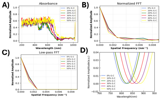
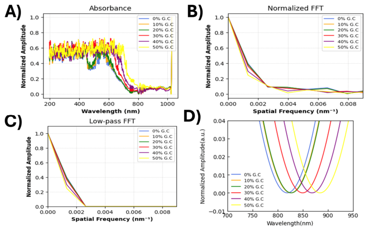
The wavelength offset (Δλ) between the minima of the raw and smoothed spectra was calculated as a diagnostic parameter to confirm that the filtering process did not introduce artificial spectral shifts. The negligible Δλ values confirmed that the smoothing procedure reduced noise without altering the intrinsic spectral response. To further isolate the plasmonic signal, the processed spectra (Figure 8A) were transformed into the Fourier domain, where modal interference fringes appear as distinct high-frequency components (Figure 8B). A frequency-domain low-pass filter was then applied to remove these fringe contributions (Figure 8C). The filtered spectra were reconstructed using inverse fast Fourier transform (IFFT), yielding a smooth SPR envelope suitable for resonance tracking (Figure 8D). The resulting absorbance spectra show a clear red shift of the plasmonic resonance with increasing glucose concentration, confirming the effectiveness of the signal processing approach in isolating the SPR response from interferometric artifacts.
Figure 8.
Signal-processing procedure used to extract the plasmonic resonance shift from the Au-TOF spectra. (A) Absorbance spectra for glucose concentrations ranging from 0–50% (w/v). (B) Fourier transform of the spectral signal, converting the wavelength-domain data into the spatial-frequency domain. (C) Application of a low-pass filter to remove high-frequency interference components. (D) Reconstructed spectrum obtained by inverse Fourier transform, revealing the plasmonic resonance red shift with increasing glucose concentration.
3.2. Refractive Index Sensing Performance of Au-TOF Sensors
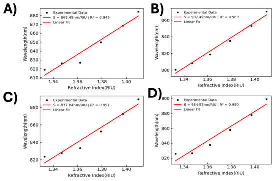
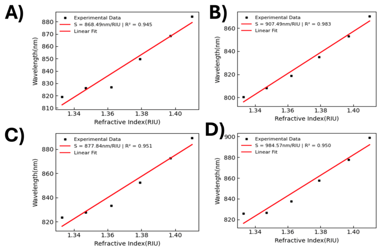
The refractive index response of the Au-TOF sensor was evaluated using glucose solutions spanning a refractive-index range of 1.333–1.40 RIU. The processed spectra exhibited a clear and consistent red shift of the resonance minimum with increasing glucose concentration across all four independent trials. The refractive indices corresponding to each glucose concentration were determined using established calibration data for aqueous glucose solutions. This behavior is characteristic of plasmonic sensors, where increases in the surrounding dielectric constant shift the SPR condition toward longer wavelengths. For each trial, the resonance wavelength was extracted from the transmission minimum and plotted as a function of refractive index. Linear regression (Figure 9) yielded sensitivities of 863.49, 907.49, 877.84, and 984.57 nm/RIU across four independent trials, corresponding to an average sensitivity of 908.35 ± 53.66 nm/RIU (mean ± SD, n = 4) with an average R2 = 0.957 (Figure 9A), demonstrating reproducible plasmonic sensing performance. The sensitivity is calculated using:
Figure 9.
Refractive index calibration curves for the 24 s Au-TOF sensor obtained from four independent experimental trials. Extracted sensitivities were (A) 868.49; (B) 907.49; (C) 877.84; and (D) 984.57 nm/RIU with corresponding R2 values of 0.945–0.983.
The linear relationship between resonance wavelength shift and refractive index can be approximated as:
This behavior arises from SPR coupling at the gold–dielectric interface. Over the measured range, this response is approximately linear, consistent with prior SPR-based fiber sensor studies [6,17,18,19]. The coefficient of variation (~5.91%) further confirms stable and repeatable plasmonic coupling across independent measurements. Although minor variations in sensitivity were observed between trials, the magnitude and linearity of the response remained consistent.
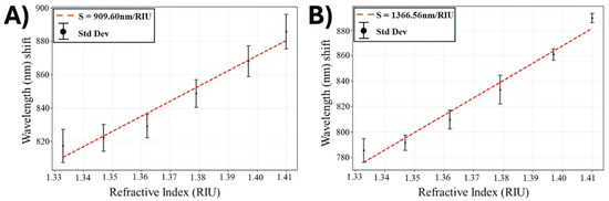
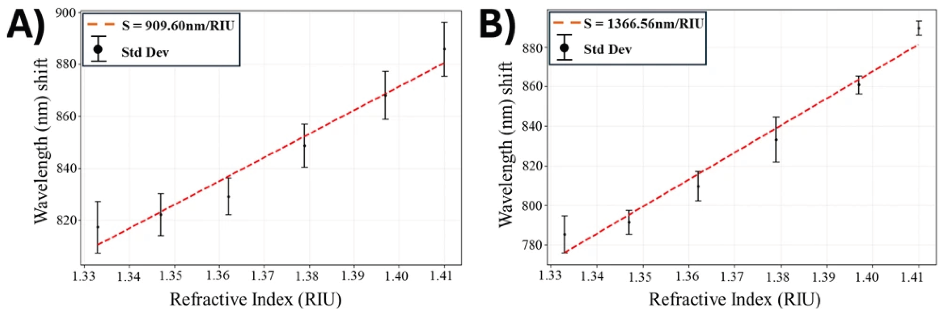
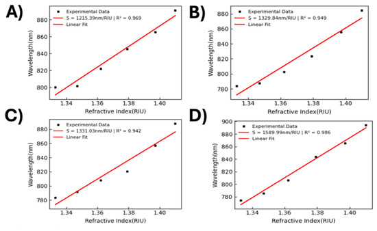
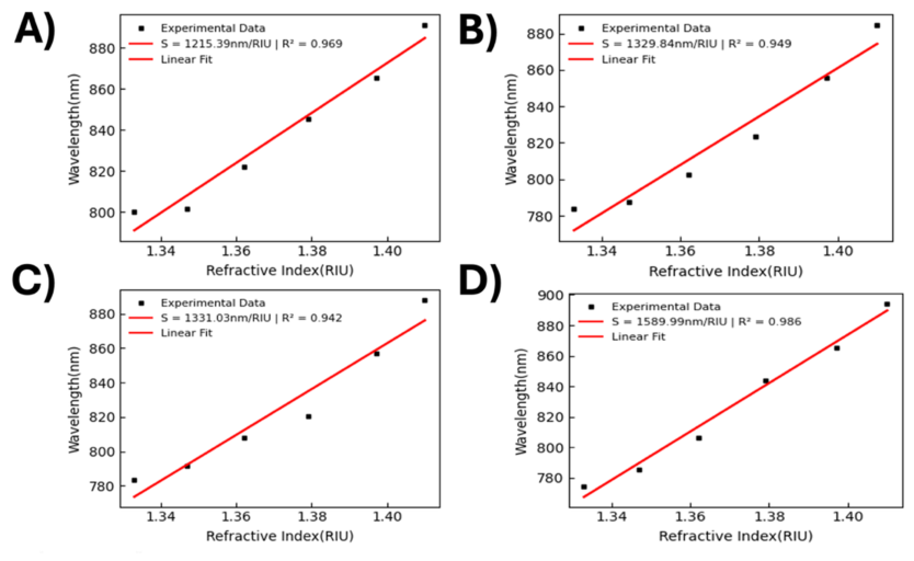
Figure 10 compares the refractive-index sensitivities for sensors fabricated with 24 s and 30 s gold sputtering durations. The 30 s device exhibited a more pronounced red shift, indicating stronger plasmonic coupling. For the 30 s Au-TOF sensor, linear regression (Figure 11) yielded sensitivities of 1215.39, 1329.84, 1331.03, and 1589.99 nm/RIU, corresponding to an average sensitivity of 1366.41 ± 158.23 nm/RIU (mean ± SD, n = 4) with an average R2 = 0.962 for the 30 s Au-TOF device (Figure 9B). The corresponding coefficient of variation (11.58%) indicates slightly higher variability compared to the 24 s device, but still demonstrates reproducible sensing performance. The maximum sensitivity of 1589.99 nm/RIU represents approximately a 1.5× enhancement relative to the 24 s device, confirming that increased sputtering duration improves plasmonic coupling strength.
Figure 10.
Linear regression comparison of refractive-index sensitivities for Au-TOF sensors fabricated with (A) 24 s and (B) 30 s gold sputtering durations. The 24 s device exhibited an average sensitivity of 908.35 ± 53.66 nm/RIU, while the 30 s device achieved 1366.41 ± 158.23 nm/RIU (mean ± SD, n = 4). Error bars represent standard deviation across four independent trials.
Figure 11.
Refractive index calibration curves for the 30 s Au-TOF sensor obtained from four independent experimental trials. Extracted sensitivities were (A) 1215.39; (B) 1329.84; (C) 1331.03; and (D) 1589.99 nm/RIU with corresponding R2 values of 0.942–0.986.
Although strong linearity was observed, the measured R2 values are slightly below the ideal values typically reported for optimized plasmonic systems. This deviation can be attributed to two primary factors. First, the relationship between resonance wavelength and refractive index is inherently nonlinear due to the dependence of dielectric permittivity Ed = n2. As a result, the linear calibration model represents an approximation that is valid only over a limited refractive index range [17,18,19]. Second, the sputtered gold films lie in the ultrathin regime, where variations in film continuity, thickness, and nanoscale morphology can introduce localized differences in plasmonic coupling strength. These structural variations can slightly perturb the resonance condition, leading to minor deviations from ideal linear behavior, particularly at lower concentrations (0–30% w/v glucose).
3.3. Influence of Gold Sputtering Duration on Plasmonic Coupling
The enhanced sensitivity observed for the 30 s Au-TOF sensor can be attributed to the increased gold thickness and improved film continuity produced by the longer sputtering duration. The estimated gold thicknesses of approximately 3 nm (30 s) and 2.4 nm (24 s) influence plasmonic coupling between the evanescent optical field and the metal-dielectric interface. A thicker and more continuous gold layer strengthens electromagnetic coupling with the guided optical mode, enhancing surface plasmon excitation and amplifying the spectral response to refractive-index perturbations. In tapered fiber geometries, where a significant portion of the optical mode propagates within the evanescent field, small variations in metal film properties can strongly influence plasmonic coupling strength [6]. These results suggest that the 30 s sputtering condition produces a gold layer closer to the optimal thickness regime for efficient plasmon excitation in the present device architecture.
The spectral response remained highly reproducible across four independent experimental trials, indicating stable optical coupling conditions and robust device performance during repeated fluid exposure and measurement cycles. Although the 24 s device exhibited slightly lower variability, the 30 s sensor achieved substantially higher sensitivity while maintaining strong linearity (R2 ≈ 0.94–0.99). Minor increases in sensitivity observed in later trials may reflect subtle changes in the local surface environment during repeated exposure and rinsing cycles, which can slightly modify the effective refractive index experienced by the evanescent field. Overall, these results demonstrate that gold sputtering duration is a critical fabrication parameter governing plasmonic sensing performance in Au-TOF devices and confirm the potential of this fabrication strategy as a scalable platform for future surface-functionalized optical fiber biosensors.
To contextualize device performance, Table 1 compares the present Au-TOF platform with recent gold-based fiber-optic RI sensors. The 30 s Au-TOF reached a maximum sensitivity of 1589.99 nm/RIU, exceeding the 24 s device and confirming that sputtering duration is an effective tuning parameter for plasmonic coupling. Although the sensitivity remains below that reported for more structurally elaborate AuNP-coated, photonic crystal fiber (PCF)-based, or hollow-core SPR platforms, the present sensor offers notable practical advantages, including direct linker-free metallization, azimuthally uniform coating, simple tapered-fiber fabrication, and compatibility with a low-volume microfluidic format. This tradeoff is important for translation, since highly optimized specialty-fiber designs often achieve higher sensitivity at the expense of fabrication simplicity and reproducibility.
Table 1.
Performance comparison of Au-based fiber-plasmonic sensors.
The presented Au-TOF platform demonstrates a scalable, linker-free fabrication strategy capable of achieving high refractive-index sensitivity while maintaining good measurement reproducibility. Compared with chemically functionalized plasmonic fiber sensors, the direct sputtering approach reduces fabrication complexity and improves device-to-device consistency, which is advantageous for practical deployment. The demonstrated Au-TOF platform offers strong potential for practical sensing applications due to its simple fabrication, reproducibility, and compatibility with microfluidic systems. Potential applications include label-free biochemical sensing, point-of-care diagnostics, and environmental monitoring. The linker-free gold deposition approach further enhances scalability and device consistency for real-world deployment.
4. Conclusions
In this work, Au-TOF sensors were fabricated and systematically evaluated for refractive-index sensing using aqueous glucose solutions spanning 1.33–1.41 RIU (0–50% w/v glucose). The fabrication process combined in-house flame-brushing tapering, plasma surface preparation, and rotational magnetron sputtering to produce azimuthally uniform gold coatings on the tapered fiber waist without relying on thiol- or sulfur-based linker chemistries. This approach provides a simplified and reproducible route for forming plasmonically active optical fiber sensors. Two sputtering durations were investigated to examine the influence of gold deposition time on plasmonic coupling and sensing performance. Across four independent trials, the 24 s Au-TOF sensors exhibited sensitivities ranging from 863–985 nm/RIU with strong linearity (R2 up to 0.98) and an average sensitivity of 908.35 ± 53.66 nm/RIU, demonstrating stable and reproducible refractive-index sensing. Increasing the sputtering duration to 30 s significantly enhanced device performance, yielding sensitivities from 1215–1589 nm/RIU with R2 values up to 0.99 and an average sensitivity of 1366.41 ± 158.23 nm/RIU. The increased sensitivity observed for the 30 s device is attributed to improved gold film continuity and increased metal thickness, which enhance evanescent-field interaction and strengthen surface plasmon excitation at the gold–dielectric interface. Although the present study investigates two sputtering durations (24 s and 30 s), a broader investigation across additional thicknesses is required to identify the optimal plasmonic coupling regime and potential saturation behavior. This will be explored in future work along with optimizing gold film thickness, improving resonance stability, and integrating selective surface functionalization layers to enable detection of biochemical targets in complex media. The combination of tapered fiber geometry, plasmonic enhancement, and microfluidic compatibility positions the Au-TOF architecture as a promising platform for compact optical biosensors and point-of-care diagnostic systems.
Author Contributions
Conceptualization, E.U. and G.S.; methodology, E.U.; validation, E.U. and G.S.; formal analysis, E.U.; investigation, E.U.; resources, G.S.; data curation, E.U.; writing—original draft preparation, E.U.; writing—review and editing, G.S.; visualization, E.U.; supervision, G.S. All authors have read and agreed to the published version of the manuscript.
Funding
This research received no external funding.
Data Availability Statement
Data is contained within the article.
Conflicts of Interest
The authors declare no conflicts of interest.
References
- Liyanage, T.; Lai, M.; Slaughter, G. Label-free tapered optical fiber plasmonic biosensor. Anal. Chim. Acta 2021, 1169, 338629. [Google Scholar] [CrossRef] [Scilit] [PubMed]
- Anker, J.N.; Hall, W.P.; Lyandres, O.; Shah, N.C.; Zhao, J.; Van Duyne, R.P. Biosensing with plasmonic nanosensors. Nat. Mater. 2008, 7, 442–453. [Google Scholar] [CrossRef] [Scilit] [PubMed]
- Lai, M.; Slaughter, G. Label-free MicroRNA optical biosensors. Nanomaterials 2019, 9, 1573. [Google Scholar] [CrossRef] [Scilit] [PubMed]
- Wolfbeis, O.S. Fiber-optic chemical sensors and biosensors. Anal. Chem. 2008, 80, 4269–4283. [Google Scholar] [CrossRef] [Scilit] [PubMed]
- Leung, A.; Shankar, P.M.; Mutharasan, R. A review of fiber-optic biosensors. Sens. Actuators B 2007, 125, 688–703. [Google Scholar] [CrossRef] [Scilit]
- Liyanage, T.; Alharbi, B.; Quan, L.; Esquela-Kerscher, A.; Slaughter, G. Plasmonic-based biosensor for the early diagnosis of prostate cancer. ACS Omega 2022, 7, 2411–2418. [Google Scholar] [CrossRef] [Scilit] [PubMed]
- Gandhi, M.S.A.; Chu, S.; Senthilnathan, K.; Babu, P.R.; Nakkeeran, K.; Li, Q. Recent Advances in Plasmonic Sensor-Based Fiber Optic Probes for Biological Applications. Appl. Sci. 2019, 9, 949. [Google Scholar]
- Monzón-Hernández, D.; Villatoro, J. High-resolution refractive index sensing with cladded multimode tapered fibers. Appl. Opt. 2006, 45, 2273–2278. [Google Scholar]
- Bhatia, P.; Gupta, B.D. Surface plasmon resonance based fiber optic refractive index sensor. Sens. Actuators B 2006, 114, 576–583. [Google Scholar]
- Snyder, A.W.; Love, J.D. Optical Waveguide Theory; Chapman & Hall: Boca Raton, FL, USA, 1983. [Google Scholar]
- Villatoro, J.; Monzón-Hernández, D. Fast detection of hydrogen with nano fiber tapers coated with palladium nanoparticles. Opt. Express 2005, 13, 5087–5092. [Google Scholar] [CrossRef] [Scilit] [PubMed]
- Tian, Z.; Yam, S.S.H.; Barnes, J.A. Refractive index sensing with Mach–Zehnder interferometer based on thin-core fiber. Opt. Lett. 2008, 33, 122–124. [Google Scholar]
- Xu, Y.; Lin, J.; Gao, S.F.; Liang, D.K.; Li, C.Z. Optical fiber evanescent-wave biosensors. Sensors 2014, 14, 151–170. [Google Scholar] [CrossRef] [Scilit] [PubMed]
- Birks, T.A.; Li, Y.W. The shape of fiber tapers. J. Light. Technol. 1992, 10, 432–438. [Google Scholar] [CrossRef] [Scilit]
- Ujah, E.; Lai, M.; Slaughter, F. Ultrasensitive tapered optical fiber refractive index glucose sensor. Sci. Rep. 2023, 13, 4495. [Google Scholar] [CrossRef] [Scilit] [PubMed]
- Tong, L.; Gattass, R.R.; Ashcom, J.B.; He, S.; Lou, J.; Shen, M.; Maxwell, I.; Mazur, E. Subwavelength-diameter silica wires for low-loss optical wave guiding. Nature 2003, 426, 816–819. [Google Scholar] [CrossRef] [Scilit] [PubMed]
- Raether, H. Surface Plasmons on Smooth and Rough Surfaces and on Gratings; Springer: Berlin/Heidelberg, Germany, 1988. [Google Scholar]
- Homola, J.; Yee, S.S.; Gauglitz, G. Surface plasmon resonance sensors. Sens. Actuators B 1999, 54, 3–15. [Google Scholar] [CrossRef] [Scilit]
- Maier, S.A. Plasmonics: Fundamentals and Applications; Springer: Berlin/Heidelberg, Germany, 2007. [Google Scholar]
- Mayer, K.M.; Hafner, J.H. Localized surface plasmon resonance sensors. Chem. Rev. 2011, 111, 3828–3857. [Google Scholar] [CrossRef] [Scilit] [PubMed]
- Willets, K.A.; Van Duyne, R.P. Localized surface plasmon resonance spectroscopy. Annu. Rev. Phys. Chem. 2007, 58, 267–297. [Google Scholar] [CrossRef] [Scilit] [PubMed]
- Slavík, R.; Homola, J. SPR sensor based on a polymer optical fiber. Sens. Actuators B 2007, 123, 10–17. [Google Scholar]
- Kim, S.M.; Kang, M.J.; Cho, K.H.; Pyun, J.C.; Lee, S.D.; Lee, S.K. Fiber-optic LSPR sensors fabricated by gold nanoparticle immobilization. Biosens. Bioelectron. 2011, 26, 3615–3620. [Google Scholar]
- Guo, T.; Albert, J.; Cheng, X.M.; Ding, J.F.; Jiang, Y.Y.; Yuan, W.J.; Kang, M.Y. Gold nanoparticle-based fiber optic SPR sensors. Opt. Express 2010, 18, 27672–27679. [Google Scholar]
- Piliarik, M.; Homola, J. Surface plasmon resonance sensors: Approaching their limits? Opt. Express 2009, 17, 16505–16517. [Google Scholar] [CrossRef] [Scilit] [PubMed]
- Sharma, A.K.; Gupta, B.D.; Verma, R. Fiber-optic SPR sensors: Fabrication challenges and performance limitations. Opt. Laser Technol. 2018, 101, 118–133. [Google Scholar]
- Verma, R.; Gupta, B.D. Gold-sputtered optical fiber SPR sensor for refractive index sensing. Appl. Opt. 2015, 54, 5456–5462. [Google Scholar]
- Kaur, P.; Gupta, B.D. SPR based fiber optic sensors using sputtered gold films. Opt. Fiber Technol. 2014, 20, 260–264. [Google Scholar]
- Cennamo, N.; Massarotti, D.; Conte, L.; Zeni, L. Low-cost SPR sensors based on sputtered gold films on optical fibers. Sensors 2013, 13, 14676–14693. [Google Scholar] [PubMed]
- Sharme, R.K.; Quijada, M.; Terrones, M.; Rana, M.M. Thin conducting films: Preparation methods, optical and electrical properties. Materials 2024, 17, 4559. [Google Scholar] [CrossRef] [Scilit] [PubMed]
- Fakhri, M.A.; Salim, E.T.; Tariq, S.M.; Ibrahim, R.K.; Alsultany, F.H.; Alwahib, A.A.; Alhasan, S.F.; Gopinath, S.C.; Salim, Z.T.; Hashim, U. A gold nanoparticles coated unclad single mode fiber-optic sensor based on localized surface plasmon resonance. Sci. Rep. 2023, 13, 5680. [Google Scholar] [CrossRef] [Scilit] [PubMed]
- Li, K.; Yin, Z.; Li, S.; Jing, X. Experimental study on ultra-high sensitivity gold-based SPR sensor for refractive index and temperature measurement. Photonics Nanostructures-Fundam. Appl. 2024, 60, 101262. [Google Scholar] [CrossRef] [Scilit]
- Divya, J.; Selvendran, S. Surface plasmon resonance-based gold-coated hollow-core negative curvature optical fiber sensor. Biosensors 2023, 13, 148. [Google Scholar] [CrossRef] [Scilit] [PubMed]
- Liu, Z.; Ji, X.; Qin, Y.; Zhang, Y.; Mou, J.; Deng, Y.; Liu, W.; Zhang, Y.; Yuan, L. Refractive index SPR sensor with high sensitivity and wide detection range using tapered silica fiber and photopolymer coating. Opt. Express 2023, 31, 31768–31779. [Google Scholar] [CrossRef] [Scilit] [PubMed]

Disclaimer/Publisher’s Note: The statements, opinions and data contained in all publications are solely those of the individual author(s) and contributor(s) and not of MDPI and/or the editor(s). MDPI and/or the editor(s) disclaim responsibility for any injury to people or property resulting from any ideas, methods, instructions or products referred to in the content. |
© 2026 by the authors. Licensee MDPI, Basel, Switzerland. This article is an open access article distributed under the terms and conditions of the Creative Commons Attribution (CC BY) license.