


Abstract
Accurate and non-invasive monitoring of glucose levels remains a critical challenge in diabetes management, motivating the development of highly sensitive optical biosensors. In this work, a surface plasmon resonance-based biosensor operating in the Kretschmann configuration is proposed and numerically investigated for glucose detection. The sensor architecture consists of a BK7 prism/TiO2/Ag/graphene multilayer, and the effect of incorporating a hexagonal boron nitride (h-BN) interlayer with varying thicknesses is systematically analyzed to enhance sensing performance. Electromagnetic simulations were performed using the finite-difference time-domain method in Lumerical FDTD Solutions at a wavelength of 633 nm. Key performance parameters, including angular sensitivity, full width at half maximum, detection accuracy, figure of merit, signal-to-noise ratio, and limit of detection, were evaluated for glucose concentrations corresponding to refractive indices ranging from 1.3282 to 1.3767 RIU. The conventional BK7/TiO2/Ag/TiO2/Graphene/Sensing Medium (SM) configuration achieved a sensitivity of 167.48 deg/RIU. By introducing an h-BN layer, significant performance enhancement was observed. The optimized structure with an 8 nm h-BN layer exhibited a maximum angular sensitivity of 205.35 deg/RIU, representing an improvement of approximately 22.6% over the reference design, while maintaining a low detection limit of 2.43 × 10⁻⁴ RIU. The results further reveal that h-BN thickness plays a crucial role in balancing sensitivity and resonance quality, where excessive thickness broadens the resonance curve and degrades detection accuracy. The proposed graphene-h-BN-assisted SPR platform demonstrates high potential for high-performance, non-invasive glucose monitoring and provides practical design guidelines for next-generation plasmonic biosensors.
1. Introduction
According to the International Diabetes Federation, the number of individuals with diabetes globally was estimated at 536.6 million in 2021 and is projected to increase to 783.2 million by 2045 [1]. Diabetes is a chronic multisystem illness that affects several organs and can lead to cardiovascular disease, neurological diseases, kidney disease, eye issues, oral injuries, stroke, heart attack, and cognitive dysfunction [1,2]. According to the World Health Organization, deaths attributable to diabetes are projected to increase by 2030, so vigilant monitoring and early detection of this condition are particularly important [3]. In normal conditions, the body regulates blood glucose levels through the hormone insulin. Diabetes, a metabolic disorder, occurs when the body fails to regulate glucose levels derived from food and beverages. The condition is classified into two types: type 1 diabetes, which is an autoimmune disease in which insulin synthesis is virtually nonexistent. Type 2 diabetes is a metabolic disorder in which insulin resistance is the primary cause, and the body is unable to use insulin effectively; in severe stages of the disease, insulin secretion may be diminished [4]. Prediabetes is a condition in which glucose levels are above normal but below the diabetes threshold, increasing the chance of developing type 2 diabetes. Because there is no definite treatment for diabetes, patients must maintain a healthy diet, engage in regular physical activity, take their medicine, and receive home care. The majority of traditional glucose measuring techniques use invasive skin punctures for blood collection, which can result in pain, psychological anguish, scarring, and even infection. Patients with diabetes who need regular measurements find these issues frustrating. Noninvasive techniques, on the other hand, have gained popularity since they are non-invasive, cause less pain, respond quickly, and are simple to use. These techniques use various biological fluids, including tears, saliva, urine, and perspiration, to estimate glucose levels [5].
Surface plasmons (SPs), defined as the coupled collective oscillations of conduction electrons and electromagnetic fields at the interface between a metal and a dielectric, have emerged as a cornerstone of modern photonics and nanoscale technologies [6,7]. Due to their unparalleled capability for sub-wavelength light confinement beyond the classical diffraction limit and their generation of highly localized field enhancements, these surface waves provide an ideal platform for developing high-speed, miniaturized all-optical devices. Today, controlling and manipulating these plasmons within nanostructures has paved new pathways for designing high-speed optical switches [8,9], ultra-compact waveguides [10,11], high-sensitivity sensors [12,13], and advanced decoders [14,15], effectively pushing the boundaries of light-matter interactions at the nanoscale.
Recent developments in biosensors, particularly surface plasmon resonance (SPR) sensors, have made non-invasive glucose monitoring possible. Because of their remarkable sensitivity to changes in refractive index (RI) and label-free, real-time measurement capabilities, these sensors have found extensive use in biotechnology [16]. SPR-based devices can detect changes in the refractive index of the environment caused by biological interactions. To date, this technology has been applied to detecting cancer [17], COVID-19 [18], very early pregnancy [19], milk fat [20], and organic pollutants [21], chemicals [22], gases [23], and pathogens in water [24]. SPR has been utilized to identify cementum, dentin, and enamel layers even in dentistry. The SPR sensor measures changes in refractive index and transforms them into an optical signal that can be analyzed. With its rapid and precise detection capabilities, this technology has emerged as a potent choice for diagnosing numerous serious illnesses [25]. The fundamental idea behind SPR is the excitation of surface plasmons, which are free-electron oscillations on metal surfaces. When TM light hits a metal layer, it generates surface plasmon polaritons (SPPs) at the metal-dielectric interface. This phenomenon shows small changes in RI [26,27,28].
Based on the optical structure, SPR sensors may be categorized into four categories, i.e., prism-based, waveguide-coupled [29], grating-coupled [30], and fiber-optic-based [9,10,11]. Of these, prism-based sensors are most commonly employed, particularly in point-of-care (POC) applications, as they require minimal sample pretreatment, offer very high sensitivity down to the femtomolar range, and are compact in design [31]. Among them, two prominent configurations are the Kretschmann and Otto configurations. The Kretschmann configuration used in this work is better suited for accurate and immediate applications because of its higher light-coupling efficiency, higher sensitivity, sharper resonance peak, lower noise, and greater stability than the Otto configuration [32]. The sensitivity of the Kretschmann configuration strongly depends on the choice of metal. The most commonly used metals are gold (Au), silver (Ag), and copper (Cu) [33,34]. Silver was selected as the plasmonic material instead of other noble metals, such as gold and copper, due to its superior plasmonic properties in the visible wavelength range [35]. Ag exhibits a highly negative real part of the dielectric permittivity and relatively low optical losses at 633 nm, enabling efficient coupling between incident photons and surface plasmon polaritons. This results in sharper resonance characteristics, stronger electromagnetic field confinement, and higher refractive-index sensitivity. Although Au provides excellent chemical stability and resistance to oxidation, it generally exhibits higher optical losses and broader SPRs in the visible region compared with Ag, which can reduce sensing accuracy. Copper, despite its favorable optical properties, suffers from severe oxidation and long-term instability [36], limiting its practical application in biosensing systems. Therefore, Ag provides an appropriate balance between strong plasmonic excitation, high sensitivity, narrow resonance linewidth, and acceptable stability, making it a suitable choice for the proposed SPR biosensor [13,14]. The use of 2D materials such as graphene, MoS2, WS2, and WSe2 has significantly enhanced SPR performance [37,38]. Graphene, with its two-dimensional honeycomb structure, high optical absorption, and strong π–π interactions with biomolecules [39], not only prevents metal oxidation but also increases the sensor’s sensitivity [15,16,20,40].
As a two-dimensional hexagonal lattice of carbon atoms, graphene finds widespread application in fields such as biosensing, optical switching, plasmonic sensing, and broadband polarization conversion [41,42]. However, a monolayer of graphene has a light absorption efficiency of just 2.3%, which is insufficient to improve SPR sensitivity [43]. Two-dimensional materials have recently been widely applied in SPR-based sensors owing to their outstanding optical and electrical properties [44]. These materials are recognized as suitable for improving the performance of SPR-based sensors [15].
Bornacelli et al. [45] investigated plasmon-enhanced multiphoton-excited photoluminescence in Au, Ag, and Pt nanoclusters. They showed that Au and Ag nanostructures exhibited nearly fourfold stronger multiphoton photoluminescence than Pt due to their stronger localized surface plasmon resonance and enhanced near-field electromagnetic coupling. They demonstrated the significant role of plasmon-mediated light-matter interactions in boosting nonlinear optical responses and suggested that hybrid plasmonic nanocomposites are promising candidates for infrared sensing.
Composite layered architectures have been shown to boost SPR efficiency significantly. Wang et al. [46] presented a prism-based SPR sensor with an Ag/Au/WS2/graphene structure, reporting a sensitivity of up to 182.5 deg/RIU and a full width at half maximum (FWHM) of 5.4 degrees. Jining Lin et al. [47] proposed an SPR sensor based on a hybrid structure of 2D materials (graphene, MoS2, WS2, and WSe2). They showed that, by optimizing the number of layers of 2D materials, the thickness of the Au, Ag, or Cu metal, and the wavelength of the incident light, the sensitivity of the sensor in the visible region reaches maximum values of 194.159 deg/RIU and 155 deg/RIU for Au, Ag, and Cu, with the highest sensitivity belonging to the silver-based structure. MD Mahabubur Rahman et al. [48] proposed two numerical SPR biosensors with Ag/PtSe2/WS2 and Au/PtSe2/WS2 structures, which offered ultrahigh sensitivities of 194 (deg/RIU) and 187 (deg/RIU), and the Performance Factor and quality factor with optimized layer thicknesses. An Au/MoS2/h-BN/graphene-based layered SPR sensor for urine glucose detection was proposed by Mudgal et al. [49] and numerically optimized. The results demonstrate that the addition of an h-BN coating layer improved the quality and accuracy of detection and increased the sensor’s sensitivity to 194.12 (deg/RIU). A CaF2-Ag-Al-MoS2 (TMDC-based) SPR biosensor for non-invasive urine glucose detection was presented by Biswajit D. et al. [50]. It has a wide linear response, excellent accuracy, and high sensitivity (deg/RIU) of 455.83, enabling detection of glucose levels across both the non-diabetic and diabetic ranges. Imman A. Al-Thawaib et al. investigated a non-invasive multilayer SPR sensor based on WS2-graphene-carbon nanotubes in [51] to measure the amount of glucose in urine. The sensor’s sensitivity was 400 (deg/RIU), and its figure of merit (FOM) value was 59.7 (RIU−1). Fast and accurate glucose monitoring is made possible by its numerical simulation design.
Despite significant advances in SPR-based glucose biosensors, several challenges remain unresolved. Many previously reported designs achieve high sensitivity at the expense of broadened resonance curves, reduced detection accuracy (DA), increased optical losses, or the use of complex multilayer architectures that may hinder practical implementation. Furthermore, the influence of hexagonal boron nitride thickness on the plasmonic response and sensing performance has not yet been systematically investigated in graphene-assisted SPR glucose sensors. Therefore, there is a need for optimized sensor architectures that simultaneously enhance sensitivity, maintain resonance quality, and preserve fabrication feasibility. Motivated by these challenges, this work proposes and numerically investigates two graphene-based SPR biosensor configurations, namely BK7/TiO2/Ag/TiO2/Graphene/SM and BK7/TiO2/Ag/h-BN/Graphene/SM, operating in the Kretschmann configuration. A comprehensive parametric study is performed to evaluate the effects of h-BN incorporation and thickness optimization on the resonance characteristics and sensing performance. The proposed design demonstrates that the synergistic integration of graphene and h-BN can significantly improve plasmonic coupling and glucose-sensing capability while maintaining favorable resonance characteristics. The obtained results establish an optimized platform for non-invasive glucose monitoring and provide practical design guidelines for the development of high-performance SPR biosensors based on two-dimensional materials.
2. Proposed Structure for SPR-Based Sensor
Among the various approaches for exciting surface plasmons, prism coupling based on the Kretschmann configuration is the most widely adopted due to its simplicity, high efficiency, and precise control of the optical coupling conditions [52,53]. The resonance condition for the Kretschmann configuration can therefore be expressed as follows [26,27]:
where Klight and βSP are the propagation constant of the incident light and the wave vector of the SP wave, λ denotes the wavelength of the incident optical beam, np is the refractive index of the coupling prism, and θinc represents the incident angle. The quantities εM and εD correspond to the real parts of the dielectric permittivities of the metal film and dielectric SM, respectively. Based on the phase-matching condition between the incident wave and the surface plasmon polariton mode, the SPR angle can be obtained from the following expression [26,27]:
The proposed SPR biosensor is indeed dependent on the polarization state of the incident light. In the Kretschmann configuration, only transverse magnetic polarization can excite surface plasmon polaritons at the metal-dielectric interface because the electric field component normal to the interface is required to couple to the free-electron oscillations in the metallic layer. In contrast, transverse electric polarization does not support SPR excitation because its electric field is entirely parallel to the interface and cannot satisfy the momentum matching condition required for coupling with the surface plasmon mode. Therefore, the incident light in all simulations was considered to be TM-polarized, which represents the appropriate polarization state for efficient SPR excitation in Ag-based multilayer structures.
The excitation of surface plasmon polaritons at the metal-dielectric interface produces a distinct resonance dip in the angular reflectance spectrum, which forms the basis of surface plasmon resonance sensing. The sensing principle relies on monitoring the shift in the resonance angle resulting from variations in the refractive index of the analyte adjacent to the metallic layer. Figure 1a,b illustrate the proposed SPR biosensor designed for blood glucose detection. The sensor architecture comprises six functional layers. A BK7 prism is used as the coupling substrate, followed by a thin titanium dioxide layer that serves as an adhesion and optical matching layer between the prism and the metallic film. Silver is utilized as the plasmonic metal due to its superior plasmonic characteristics in the visible spectrum. The fourth layer consists of either TiO2 or hexagonal boron nitride, depending on the investigated configuration. The remaining layers include graphene and the SM containing the glucose solution. Accordingly, two sensor configurations were analyzed: BK7/TiO2/Ag/TiO2/Graphene/SM and BK7/TiO2/Ag/h-BN/Graphene/SM. Both structures are based on the Kretschmann configuration, and all simulations were conducted within the visible wavelength range.
Figure 1.
Schematic illustrations of the proposed SPR biosensors based on the Kretschmann configuration for glucose sensing. (a) BK7/TiO2/Ag/TiO2/Graphene/SM structure, where TiO2 acts as both an adhesion and protective layer for the Ag film. (b) BK7/TiO2/Ag/h-BN/Graphene/SM structure incorporating an h-BN layer with thicknesses ranging from a monolayer (0.33 nm) to 10 nm.
In the proposed multilayer structure, the coupling between Ag, TiO2, and graphene occurs through modification of the local dielectric environment and redistribution of the electromagnetic field at the metal-dielectric interface. The Ag layer acts as the primary plasmonic component, where the interaction between the incident TM-polarized electromagnetic wave and free electrons in Ag generates the SPR mode. The adjacent TiO2 layer, due to its high refractive index and dielectric properties, modifies the effective refractive index near the Ag interface, enhances electromagnetic field confinement, and improves the stability of the plasmonic structure. The graphene layer further influences the SPR response through its two-dimensional electronic properties, high surface area, and strong interaction with the evanescent electromagnetic field generated at the Ag/dielectric interface. The presence of graphene increases the interaction between the enhanced near field and glucose molecules, providing additional adsorption sites and improving the sensing capability.
The BK7 prism is used as the coupling substrate due to its suitable refractive index, high optical transparency, and low optical losses, which enable efficient excitation of surface plasmon polaritons in the Kretschmann configuration. The first TiO2 layer between BK7 and Ag acts as an adhesion and optical matching layer, improving the attachment of the silver film to the prism surface and enhancing electromagnetic coupling. The Ag layer is used as the plasmonic active metal because of its strong ability to support SPP excitation in the wavelength region, providing high sensitivity to refractive index variations near the sensing interface. Silver may be degraded due to oxidation, so an additional ultrathin TiO2 layer (in the reference structure) is introduced above the Ag film to protect the metal surface, reduce oxidation effects, and modify the local dielectric environment for improved plasmonic interaction. In the enhanced configuration, this upper TiO2 layer is replaced by an h-BN layer to investigate its influence on the plasmonic response. The h-BN layer provides a chemically stable dielectric interface and modifies the electromagnetic field distribution near the sensing region. Furthermore, graphene is used as the final functional 2D material due to its excellent adsorption capability, high surface area, chemical stability, and strong interaction with biomolecules, which increases the interaction between the evanescent field and glucose molecules. Finally, the sensing medium is essential because the SPR response originates from changes in its refractive index caused by different glucose concentrations. Therefore, each layer contributes independently to the optical coupling, plasmon excitation, protection, and analyte interaction mechanisms. Removing any of these functional layers would significantly alter the phase-matching condition, electromagnetic field confinement, and overall sensing performance.
The BK7 prism does not require a defined thickness in the simulation because it acts as the optical coupling substrate, and its refractive index is the dominant parameter determining the phase-matching condition. The thickness of the Ag layer was selected as 50 nm after performing a systematic optimization process. In SPR sensors, the metal thickness strongly affects the coupling efficiency between the incident electromagnetic wave and the surface plasmon mode. A very thin Ag layer leads to insufficient plasmon excitation due to incomplete interaction with the evanescent field. In contrast, an excessively thick Ag layer reduces optical tunneling through the metal film and weakens coupling efficiency. Therefore, the optimized thickness of 50 nm provides a suitable balance between low reflectance, strong SPR excitation, and high angular sensitivity. The TiO2 layer thickness was selected as 5 nm because ultrathin TiO2 films provide effective adhesion between the BK7 prism and the Ag layer while maintaining efficient electromagnetic coupling. Increasing the TiO2 thickness excessively would increase the separation between the prism and the plasmonic interface, reducing the penetration of the evanescent field and degrading the SPR response. Therefore, a 5 nm thickness was chosen to achieve improved stability without significantly affecting plasmon excitation. A monolayer graphene thickness of approximately 0.34 nm was considered based on the experimentally reported thickness of a single graphene sheet. Graphene was maintained as a monolayer because it provides sufficient biomolecular adsorption capability and field interaction while minimizing additional optical losses caused by multiple graphene layers. For the h-BN-assisted structure, the thickness was systematically varied from 1 nm to 10 nm to investigate its influence on plasmonic coupling, electromagnetic field distribution, and sensing performance. Unlike graphene, the h-BN thickness was treated as an optimization parameter because it acts as an intermediate dielectric layer that modifies the effective refractive index and controls the distance between the plasmonic metal surface and the sensing medium. The optimum thickness was determined based on the highest sensitivity and acceptable resonance quality. The refractive index of BK7 is obtained from Equation (4), yielding a value of 1.5151 at a wavelength of 633 nm.
The refractive index of TiO2, evaluated using Equation (5), is found to be 1.99 at a wavelength of 633 nm [29,30].
The refractive index of silver is determined using the Drude-Lorentz model, as expressed in Equation (6) [31]. In this model, the collision wavelength (λC) of silver is 1.614 × 10⁻⁵ m, and the plasma wavelength (λP) is 1.4541 × 10⁻⁷ m. Accordingly, the refractive index of Ag at 633 nm is calculated to be 0.056206 + 4.2776i.
Within a concentration range of 0 to 30 wt%, the refractive index of the glucose solution varies from 1.3282 to 1.3767 [32]. Table 1 summarizes the refractive indices of the different layers used in the proposed sensor configuration.
Table 1.
Thickness and refractive index values for the proposed SPR biosensors operating at a wavelength of 633 nm.
The electromagnetic behavior of the proposed sensor is investigated using the commercial simulation platform Lumerical FDTD Solutions, which is based on the finite-difference time-domain technique. This numerical method, implemented via the Yee grid algorithm, provides a rigorous and time-domain solution of Maxwell’s equations, making it particularly suitable for accurately modeling light–matter interactions at the nanoscale, especially in multilayer plasmonic configurations. To ensure physically reliable and reflection-free computational boundaries, perfectly matched layers are applied along the simulation domain edges, while Bloch boundary conditions are used where periodicity is required. These boundary settings are essential to eliminate non-physical reflections and to correctly model the propagation of electromagnetic waves in open-space plasmonic systems. The incident angle is systematically varied between 55° and 85° in order to fully capture the angular response of the SPR structure and identify the optimal resonance conditions. For numerical stability and accuracy, an auto non-uniform meshing scheme is employed, allowing finer spatial resolution in regions with strong field gradients, particularly near metal-dielectric interfaces, while coarser meshes are used elsewhere to optimize computational efficiency. All remaining simulation parameters are retained at their default values to ensure consistency and reproducibility. The system temperature is set to 300 K to represent standard room-temperature operation, and the total simulation time is chosen as 1000 fs to guarantee that the electromagnetic fields reach a steady-state condition and that transient effects fully decay. The surrounding medium is assumed to be air with a refractive index of 1.0, providing a standard reference environment for evaluating sensor performance.
Equations (7)–(12) are used to evaluate the key performance parameters of the sensor, including the sensitivity (S), FWHM, FOM, DA, signal-to-noise ratio (SNR), and limit of detection (LOD) [31,38,56].
FWHM is the distance or range over which a spectral or angular peak is measured at half of its maximum value. A tiny FWHM indicates a sharp, narrow resonance peak in SPR sensors. The lower the FWHM, the more noticeable the change in the sensor medium’s refractive index will be, leading to a more pronounced shift in the resonance angle or wavelength and making the sensor more adept at detecting minute variations in the analyte’s refractive index. The FOM indicates a direct relationship between the sensor sensitivity and the resonant peak width. The higher the FOM, the better the sensor’s performance, since, in addition to high sensitivity, the resonance peak is narrow and sharp, and slight changes in the refractive index of the sensor medium result in a clear change in the resonance angle or wavelength. It is a quantitative, practical index for evaluating and optimizing the performance of optical and biosensors. DA is a measurement that reflects how well a sensor can reliably detect minute variations in the concentration of an analyte or the refractive index. In general, it is ideal for an SPR sensor to have greater values of angular sensitivity, DA, quality factor, and SNR, while having lower values of LOD and FWHM. This is because these values suggest increased sensitivity, precision, and accuracy in detecting changes in refractive index.
3. Results and Discussion
A systematic analysis is performed to enhance the optical response and sensitivity of the proposed sensor. Initially, the configuration without the h-BN layer (Prism/TiO2/Ag/TiO2/Graphene/SM) is examined, followed by an investigation of the structure in which the TiO2 layer is replaced by h-BN (Prism/TiO2/Ag/h-BN/Graphene/SM), as illustrated in Figure 1b. The thickness of the silver layer is optimized by evaluating both the minimum reflectance (Rmin) and the sensitivity. For this purpose, the Ag thickness is systematically varied while keeping the remaining layer thicknesses constant, and the corresponding values of sensitivity and Rmin are calculated for each case. The lowest Rmin corresponds to the most efficient coupling of incident optical energy into surface plasmon waves (SPWs), thereby strengthening the interaction between analyte molecules in the sensing medium and the evanescent field [57]. This optimization leads to improved DA, an enhanced FOM, and a reduced FWHM. Furthermore, higher sensitivity reflects the sensor’s ability to detect extremely small refractive index variations induced by analyte binding at the surface. The performance of the proposed sensor is evaluated using the angular interrogation method, which provides high resolution due to the sharp resonance dips generated during plasmon excitation.
3.1. Investigating Optimal Thickness of Ag and h-BN Layers
A detailed study is carried out to improve the performance of the proposed structure. Figure 2 presents the reflectance spectra for different silver layer thicknesses as a function of the incident angle. The minimum reflectance under resonance conditions is achieved by optimizing the thickness of the Ag layer, while maintaining the TiO2 layers at a fixed thickness of 5 nm. The fifth layer, consisting of a two-dimensional material, is excluded from the optimization process. In this analysis, the sensing medium is assumed to have a refractive index of n= 1.3282 RIU, and the Ag thickness is varied within the range of 45–50 nm.
Figure 2.
Reflectance response as a function of incident angle for optimization of the silver layer thickness.
Table 2 summarizes the corresponding minimum reflectance (Rmin) and angular sensitivity values obtained for a refractive index variation of Δn = 0.0485. The resonance angle is found to be 69.63°. Based on the obtained results, a thickness of 50 nm is selected as the optimal value for the Ag layer due to its low reflectance (Rmin= 0.045) and satisfactory angular sensitivity. This optimized thickness effectively supports surface plasmon excitation by enhancing the coupling between the incident electromagnetic wave and surface plasmons at the metal-dielectric interface.
Table 2.
Minimum reflectance and angular sensitivity for different silver layer thicknesses.
After determining the optimal thickness of the silver layer, the effect of the h-BN layer thickness was investigated in the second proposed configuration, Prism/TiO2/Ag/h-BN/Graphene/SM. To systematically evaluate this influence, an h-BN monolayer with thicknesses varying from 1 to 10 nm was considered, while the thicknesses of the TiO2, Ag, and graphene layers were kept constant. Figure 3 presents the reflectance curves as a function of the incident angle for different h-BN thicknesses. In this simulation, a BK7 prism with a refractive index of n = 1.5151 and a sensing medium with n = 1.3282 were used. Variations in layer thickness lead to noticeable changes in the reflectance profile, which in turn affect the surface plasmon wavelength and penetration depth. These changes also influence the FWHM, while improved performance in terms of minimum reflectance is achieved at an optimal layer thickness.
Figure 3.
Reflectance as a function of the incident angle for biosensor configurations BK7/TiO2/Ag/h-BN/Graphene/SM and BK7/TiO2/Ag/TiO2/Graphene/SM at a refractive index of n = 1.3282 RIU.
When incident light is reflected from a glass sensor chip coated with multiple thin metallic layers, total internal reflection is established under specific conditions. In the proposed BK7/TiO2/Ag/TiO2/Graphene (or h-BN)/SM configuration, free electrons at the metal-dielectric interface are essential for the excitation of surface plasmons. As the incident electromagnetic wave interacts with the delocalized electrons in the metallic film, part of the optical energy is transferred at a specific resonance angle rather than being fully reflected, resulting in a pronounced dip in the reflected intensity. This mechanism leads to the excitation of surface plasmon polaritons, which are highly sensitive to refractive index variations in the vicinity of the sensor surface. Consequently, when blood is used as the analyte, the SPR reflectance curve exhibits a noticeable decrease in intensity.
The non-monotonic variations observed around h-BN thicknesses of 3–4 nm and 8–9 nm originate from changes in the optical interference and phase-matching conditions within the multilayer SPR structure. As the h-BN thickness increases, the optical phase accumulated across the dielectric spacer modifies the effective propagation constant of the SPP mode and the coupling efficiency between the incident TM wave and the plasmon resonance. At specific thicknesses, small changes in the spacer thickness shift the structure between different coupling regimes, resulting in abrupt variations in resonance depth and linewidth. Furthermore, at larger thicknesses (approximately 9 nm and above), the increased separation between the metal surface and the sensing medium weakens the overlap of the evanescent plasmonic field with the analyte, leading to reduced coupling efficiency, increased reflectance, and broader resonance curves. Figure 3 demonstrates the variation in reflectance with respect to the incident angle for blood without glucose (C = 0 mg/dL), using a refractive index of 1.3282 for different h-BN thicknesses in the BK7/TiO2/Ag/h-BN/Graphene/SM and BK7/TiO2/Ag/TiO2/Graphene/SM structures.
In SPR-based sensing, the optical response is strongly influenced by the wavelength of the incident radiation and the resulting resonance characteristics. In this work, a fixed excitation wavelength of 633 nm is used, where the Ag and TiO2 layers play the dominant role in supporting plasmon excitation at the metal-dielectric interface. The interaction between the incident electromagnetic field and the electron plasma in the silver layer leads to the formation of SPPs. The resonance condition is strongly dependent on the incident angle, which is varied between 55° and 85° to fully capture the angular response of the system. The FWHM of the SPR curve is also wavelength dependent, where longer wavelengths typically result in narrower resonance dips, while shorter wavelengths broaden the spectral response [58]. The evanescent field generated at the interface decays exponentially into the sensing medium, and its penetration depth and intensity are governed by both the incident angle and refractive index of the surrounding medium [41,59]. As the incident angle changes, the coupling efficiency between the incident light and surface plasmons is modified, leading to variations in resonance conditions. For different analytes with varying refractive indices, SPR excitation occurs at distinct resonance angles. At resonance, the majority of the incident optical energy is absorbed by surface plasmon polaritons in the silver layer, producing a sharp minimum in reflectance [42,60]. These shifts in resonance angle and reflectance intensity form the basis for detecting changes in glucose concentration, enabling accurate biosensing through precise monitoring of the SPR response.
Table 3 presents the Rmin and FWHM values for different h-BN thicknesses, considering a refractive index of 1.3282 and a fixed silver layer thickness of 50 nm. As the h-BN thickness increases, Rmin initially decreases, indicating enhanced coupling between the incident light and the surface plasmon wave. However, beyond a certain thickness (approximately 9–10 nm), Rmin rises sharply. This behavior is attributed to coupling deterioration, where the increased h-BN thickness enlarges the separation between the incident electromagnetic wave and the plasmonic surface, thereby hindering efficient energy transfer to the surface plasmon wave. Consequently, this regime leads to reduced sensor sensitivity and degraded detection performance. The results for FWHM indicate that the monolayer and 2 nm h-BN configurations exhibit the lowest value, while a progressive increase in FWHM is observed with increasing h-BN thickness. The maximum FWHM is obtained at a thickness of 4 nm.
Table 3.
Rmin and FWHM for different h-BN layer thicknesses in the analyte medium with a refractive index of 1.3282.
Figure 4 presents the reflectance as a function of the incident angle for an aqueous glucose solution with concentrations ranging from 0 to 30% (n = 1.3282–1.3767) in the BK7/TiO2/Ag/TiO2/Graphene/SM configuration. The figure shows the corresponding reflectance curves for different refractive index values. The minimum resonance angle of the proposed SPR sensor is 70.4545°, with a minimum reflectance of 0.05, which occurs at the baseline condition of 0 mM glucose (n = 1.3282), representing the lowest sensor response. This reference curve is used to assess the capability of the SPR-based sensor for glucose monitoring in blood samples. The refractive index of the glucose solution at room temperature within the 0–30 wt% range is determined experimentally using the following relation [32,61].
where c represents the weight percentage of the glucose solution. As indicated by the above relation, the refractive index of the glucose solution increases linearly with concentration. Consequently, higher glucose concentrations lead to an increased refractive index, which causes the resonance angle to shift toward larger values. This change in concentration modifies the refractive index of the sensing medium and, in turn, alters the phase-matching condition between the incident light and the surface plasmon resonance. As a result, the angle at which maximum coupling and energy transfer occur (i.e., the resonance angle) is correspondingly shifted. The estimated refractive indices for different glucose concentrations in aqueous solution (c = 0, 5, 10, 15, 20, 25, and 30 wt%) are 1.3282, 1.3367, 1.3447, 1.3527, 1.3607, 1.3687, and 1.3767, respectively. As shown in Figure 4, the refractive index increases from 1.3282 to 1.3767 (Δn = 0.0485) as the glucose concentration rises from 0 to 30 wt% at room temperature.
Figure 4.
Reflectance versus the incident angle for the BK7/TiO2/Ag/TiO2/Graphene/SM configuration at different glucose concentrations.
Figure 5 shows the variation in refractive index versus SPR angle for the analyte solution in the proposed BK7/TiO2/Ag/TiO2/Graphene/SM sensor. The resonance angle is extracted from the minimum points of the reflectance curves, revealing an almost linear relationship between the analyte refractive index and the corresponding SPR angle over the investigated range.
Figure 5.
Relationship between the refractive index of the sensing layer and the SPR angle in the proposed BK7/TiO2/Ag/TiO2/Graphene/SM sensor.
Table 4 summarizes the values of sensitivity, DA, FOM, SNR, LOD, and FWHM for the BK7/TiO2/Ag/TiO2/Graphene/SM configuration. As the refractive index of the analyte (or glucose concentration) increases, the resonance angle shifts to higher values. Specifically, when the refractive index increases from 1.3282 to 1.3767 (Δn = 0.0485), the resonance angle changes from 70.4545° to 78.6977° (ΔθSPR = 8.2425°). The calculated sensitivity of the proposed structure is 168.9, demonstrating strong capability for detecting small refractive index variations. At a refractive index of 1.3282, the FWHM is 2.2323°. As will be shown in Table 5, the FWHM increases with increasing refractive index, while the DA decreases due to its inverse dependence on FWHM. This behavior can be attributed to changes in phase-matching conditions between the incident electromagnetic wave and surface plasmons as the refractive index of the sensing medium increases. These variations reduce coupling efficiency, leading to broader resonance dips. Consequently, increased optical losses and reduced resonance sharpness result in lower DA, reduced FOM, and degraded SNR. For refractive indices of 1.3282, 1.3367, 1.3447, 1.3527, 1.3607, 1.3687, and 1.3767, corresponding to glucose concentrations from 0 to 30 wt%, the resonance angles are 70.4545°, 71.8283°, 72.8586°, 74.2323°, 75.6061°, 76.9798°, and 78.6977°, respectively. The BK7/TiO2/Ag/TiO2/Graphene/SM configuration is treated as the reference structure in this study. To further evaluate performance variations, the effects of h-BN thickness and the inclusion of the fourth TiO2 layer on parameters such as S, DA, FOM, SNR, LOD, and FWHM are also investigated.
Table 4.
Parameters S, DA, FOM, SNR, LOD, and FWHM for the BK7/TiO2/Ag/TiO2/Graphene/SM structure.
Table 5.
Parameters S, FOM, SNR, LOD, and FWHM for the sensor with the structure BK7/TiO2/Ag/h-BN (monolayer)/Graphene/SM.
All observed variations in performance characteristics are caused by a change in the analyte medium’s refractive index, which influences the dispersion relation and phase matching condition between the incoming wave and the surface plasmons. As the refractive index increases, the effective wave vector at the metal-dielectric boundary changes, and the optical coupling deviates from the ideal condition; as a result, the resonance width increases, while the evanescent field becomes more confined. These two effects immediately reduce the sharpness of the resonance, DA, FOM, and SNR. Still, angular sensitivity continues to rise due to a bigger shift in the resonance state. In conclusion, the changes in the sensor performance characteristics observed are a consequence of the change in the refractive index of the analyte medium and its direct impact on the propagation conditions of surface plasmon waves at the metal-dielectric interface. As the refractive index grows, the propagation constant of SPP modes increases, and the phase matching condition between the incident wave and the surface plasmon shifts further from the original optimal position. This mismatch reduces energy-coupling efficiency and increases resonant losses within the multilayer structure, as evidenced by a widening of the resonance width in the reflection curve. However, an increase in the resonance bandwidth reduces the response slope and degrades signal resolution; therefore, metrics such as DA, FOM, and SNR decrease. Instead, because of the more sensitive resonance condition to the change in the refractive index, the amount of the shift in the resonance angle rises, and the angular sensitivity keeps its upward trend. Thus, the behavior observed in the simulation results may be related to changes in the dispersion of SPP modes and the progressive weakening of the local field due to changes in the dielectric medium.
3.2. Investigation of h-BN-Based Sensor
Hexagonal boron nitride is a promising two-dimensional nanomaterial that can enhance the surface plasmon resonance response of silver films, improve sensor sensitivity, and lower the detection limit [28]. The objective of this work is to investigate a prism-based SPR biosensor incorporating h-BN as a potential non-invasive platform for glucose sensing and diabetes monitoring. Owing to its excellent optoelectronic characteristics, including strong ultraviolet light absorption and low absorption in the visible spectrum, h-BN has attracted considerable attention for advanced sensing and optoelectronic applications [33]. The reference BK7/TiO2/Ag/TiO2/Graphene/SM sensor achieves a sensitivity of 168.9°/RIU. Building upon this baseline structure, the present section examines the performance of an enhanced configuration in which the TiO2 layer adjacent to the graphene layer is replaced with an h-BN layer. The influence of h-BN is evaluated in light of its favorable optical and electronic properties and its ability to modify plasmonic coupling conditions. The proposed sensor is designed to detect glucose concentrations in aqueous solutions under laboratory conditions, thereby emulating refractive index variations corresponding to different blood glucose levels. To identify the optimal configuration and maximize sensing performance, a systematic analysis of different h-BN layer thicknesses is carried out, with particular emphasis on their effects on sensitivity and other key sensing parameters.
The first h-BN configuration investigated consists of an h-BN monolayer with a thickness of 0.33 nm. After replacing the TiO2 layer in the BK7/TiO2/Ag/TiO2/Graphene/SM structure with a monolayer h-BN film, the key performance parameters, including sensitivity, DA, FOM, SNR, LOD, and FWHM, were evaluated. The resulting BK7/TiO2/Ag/h-BN (monolayer)/Graphene/SM configuration achieved a sensitivity of 149.18°/RIU, which is lower than that of the reference BK7/TiO2/Ag/TiO2/Graphene/SM structure. Since higher sensitivity corresponds to a greater resonance-angle shift for a given refractive index variation, a reduction in sensitivity indicates a decreased capability of the sensor to detect small changes in the refractive index of the analyte.
Figure 6 presents the reflectance curves of the BK7/TiO2/Ag/h-BN/Graphene/SM sensor for glucose solutions with concentrations ranging from 0 to 30 wt%. For the glucose-free solution (0 wt%), corresponding to a refractive index of 1.3282, the sensor exhibits a resonance dip at an angle of 69.4242°. As the glucose concentration increases, the refractive index of the sensing medium rises accordingly, resulting in a progressive shift in the resonance angle toward larger values. The resonance angles corresponding to refractive indices of 1.3282, 1.3367, 1.3447, 1.3527, 1.3607, 1.3687, and 1.3767 are 69.4242°, 70.4545°, 71.4848°, 72.5152°, 73.8889°, 75.2626°, and 76.6364°, respectively. This systematic angular shift confirms the sensor’s ability to detect refractive index variations associated with changes in glucose concentration.
Figure 6.
Reflectance intensity in terms of the incident angle for the proposed BK7/TiO2/Ag/h-BN/Graphene/SM sensor at different glucose concentrations.
Table 5 summarizes the S, FOM, SNR, LOD, and FWHM of the BK7/TiO2/Ag/h-BN (monolayer)/Graphene/SM sensor for glucose concentrations ranging from 0 to 30 wt%. The results indicate that the monolayer h-BN configuration achieves a sensitivity of 149.18°/RIU, which is lower than that of the reference BK7/TiO2/Ag/TiO2/Graphene/SM sensor (167.48°/RIU). This reduction in sensitivity is associated with a smaller resonance-angle shift in response to refractive index variations. Despite the decrease in angular sensitivity, the monolayer h-BN structure exhibits a narrower and sharper resonance dip, resulting in a lower FWHM compared with the reference sensor. This behavior can be attributed to modifications in the phase-matching condition and a reduced interaction between the evanescent field and the analyte medium. The introduction of the h-BN layer changes the effective dielectric environment at the metal-dielectric interface, thereby reducing the magnitude of the resonance-angle shift induced by refractive index variations. At the same time, the resonance width decreases due to lower plasmonic losses and improved electromagnetic field confinement [39,62]. Furthermore, the h-BN layer influences the distribution of the electromagnetic field near the metal-dielectric interface. Although the resulting resonance-angle shift becomes smaller, the resonance curve becomes sharper and more stable because of enhanced plasmon confinement and reduced optical damping. Consequently, the h-BN-based sensor exhibits a trade-off between sensitivity and resonance quality, where the reduction in angular sensitivity is partially compensated by improvements in resonance sharpness, DA, and overall sensing performance.
Following the analysis of the monolayer h-BN configuration, the thickness of the h-BN layer was increased to 1 nm, and its effect on sensor performance was evaluated. For this structure, the minimum reflectance, resonance angle, and FWHM were found to be 0.088305, 69.0808°, and 1.5455°, respectively. The corresponding angular sensitivity was calculated as 150.9113°/RIU. Although this sensitivity is still lower than that of the reference BK7/TiO2/Ag/TiO2/Graphene/SM sensor, it is slightly higher than that obtained for the monolayer h-BN configuration. Moreover, the DA is improved compared with the monolayer case, primarily due to the narrower resonance width. These observations suggest that increasing the h-BN thickness can enhance certain sensing characteristics. Therefore, the effects of further increases in h-BN thickness were systematically investigated to identify the optimal configuration and achieve improved overall sensor performance.
In the case of a 2 nm h-BN layer, the analysis was carried out for aqueous glucose solutions with concentrations ranging from 0 to 30 wt%, corresponding to different refractive index values. The resonance dips for refractive indices of 1.3282, 1.3367, 1.3447, 1.3527, 1.3607, 1.3687, and 1.3767 occur at incident angles of 69.0808°, 70.1111°, 71.1414°, 72.1717°, 73.5455°, 74.9192°, and 76.2929°, respectively. As the refractive index increases with glucose concentration, the resonance angle systematically shifts toward higher values, confirming the sensor’s ability to detect refractive index variations associated with changes in glucose levels.
Table 6 summarizes the sensitivity, DA, FOM, SNR, LOD, and FWHM of the BK7/TiO2/Ag/h-BN (2 nm)/Graphene/SM sensor for glucose concentrations ranging from 0 to 30 wt%. The proposed BK7/TiO2/Ag/h-BN (2 nm)/Graphene/SM configuration exhibits an angular sensitivity of 151.0598°/RIU, which remains lower than that of the reference BK7/TiO2/Ag/TiO2/Graphene/SM sensor. However, the corresponding FWHM is narrower than that of the base structure, indicating an improvement in resonance sharpness. The reduced sensitivity observed for ultrathin h-BN layers (up to 2 nm) can be attributed to modifications in the effective dielectric environment at the metal-dielectric interface and the resulting changes in the phase-matching condition between the incident electromagnetic wave and the surface plasmon mode. In this thickness regime, the h-BN layer limits the penetration of the evanescent field into the sensing medium and weakens its interaction with the analyte, thereby reducing the angular response to refractive index variations. As the h-BN thickness increases to 2 nm, a partial redistribution of the electromagnetic field occurs within the multilayer structure, accompanied by a readjustment of the plasmonic coupling conditions. This effect enhances the interaction between the confined field and the sensing medium, leading to a slight increase in sensitivity compared with the monolayer h-BN configuration. Nevertheless, despite this improvement, the overall sensing performance remains inferior to that of the reference BK7/TiO2/Ag/TiO2/Graphene/SM structure in terms of angular sensitivity. The results indicate that while thin h-BN layers can improve resonance quality through FWHM reduction, further optimization of the h-BN thickness is necessary to achieve a favorable balance between sensitivity and resonance sharpness.
Table 6.
Performance parameters for the sensor with the structure BK7/TiO2/Ag/h-BN (2 nm)/Graphene/SM.
In the case of a 4 and 8 nm h-BN layer, the analysis was performed for glucose solutions with refractive indices ranging from 1.3282 to 1.3767. For an h-BN thickness of 3 nm, the resonance dips corresponding to refractive indices of 1.3282, 1.3367, 1.3447, 1.3527, 1.3607, 1.3687, and 1.3767 occur at incident angles of 68.8268°, 69.7677°, 70.7980°, 72.1717°, 73.2020°, 74.5758°, and 75.9495°, respectively. When the h-BN thickness is increased to 4 nm, the resonance angles shift to 72.1717°, 73.5455°, 74.5758°, 76.2929°, 77.6667°, 79.7273°, and 81.7879°, respectively. These results demonstrate that increasing the h-BN thickness significantly modifies the resonance conditions and the angular response of the sensor, highlighting the important role of the h-BN layer in controlling plasmonic coupling and sensing performance.
The effect of increasing h-BN thickness on the SPR response was further analyzed using glucose solutions at different concentrations. For the analyte with 0% glucose concentration (n = 1.3282), resonance dips were observed at h-BN thicknesses of 3, 4, 5, 6, 7, 8, 9, and 10 nm, corresponding to incident angles of 68.8268°, 72.0263°, 71.1414°, 71.4848°, 71.8283°, 72.5151°, 73.5455°, and 74.5758°, respectively. In contrast, for a 30% glucose solution (n = 1.3767), resonance dips were obtained at thicknesses of 5–10 nm, with corresponding angles of 79.3838°, 80.4141°, 81.1010°, 82.1313°, 82.4747°, and 82.8181°, respectively. Table 7 further presents the performance parameters, S, DA, FOM, SNR, LOD, and FWHM, for both the BK7/TiO2/Ag/TiO2/Graphene/SM and BK7/TiO2/Ag/h-BN/Graphene/SM configurations across different h-BN thicknesses and glucose concentrations (0–30 wt%). The BK7/TiO2/Ag/h-BN (4 nm)/Graphene/SM sensor achieves a sensitivity of 196.35°/RIU, which is significantly higher than that of configurations with thinner h-BN layers. However, the base sensor (without h-BN) and the h-BN-based configurations at lower thicknesses exhibit comparatively narrower FWHM values than the 4 nm case. Overall, the results indicate that an h-BN thickness of 8 nm yields the highest sensitivity among the investigated structures. This optimal configuration demonstrates strong angular response, making it highly suitable for ultra-sensitive biosensing applications where even minimal changes in analyte concentration produce pronounced resonance-angle shifts.
Table 7.
Performance parameters of the proposed sensors BK7/TiO2/Ag/TiO2/Graphene/SM and BK7/TiO2/Ag/h-BN/Graphene/SM with refractive index 1.3282.
Table 8 summarizes the performance parameters of the SPR biosensor with the BK7/TiO2/Ag/h-BN (4 nm)/Graphene/SM configuration across different glucose concentrations. The results show that a 4 nm h-BN layer achieves an angular sensitivity of 196.35°/RIU, which is significantly higher than that of both the baseline structure and configurations with thinner h-BN layers. This improvement in sensitivity suggests enhanced plasmonic coupling and stronger interaction between the evanescent field and the sensing medium at this optimal thickness. The FWHM increases from 4.1212° to 7.0404° as the refractive index of the sensing medium rises from 1.3282 to 1.3767, indicating a progressive broadening of the resonance curve and higher plasmonic losses at elevated analyte concentrations. As a result, the DA decreases from 0.24265 to 0.14204, while the FOM declines from 47.64 to 27.88, reflecting a reduction in resonance sharpness and overall signal quality due to the widened resonance dip. In addition, the SNR drops from 2.3335 to 1.36586, confirming a deterioration in measurement reliability with increasing refractive index. Despite these degradations, the sensor maintains a low LOD of 0.00025, demonstrating its capability to resolve very small refractive index variations.
Table 8.
Performance parameters of the BK7/TiO2/Ag/h-BN (4 nm)/Graphene/SM structure.
Table 9 summarizes the performance parameters of the SPR biosensor based on the BK7/TiO2/Ag/h-BN (8 nm)/Graphene/SM configuration for different glucose concentrations. The highest angular sensitivity of 205.35°/RIU is achieved at an h-BN thickness of 8 nm, which exceeds both the baseline structure and all other investigated thicknesses. This improvement indicates stronger interaction between the evanescent field and the sensing medium, along with more favorable plasmonic coupling conditions at this optimal thickness. In addition, as the refractive index of the sensing medium increases from 1.3282 to 1.3767, the FWHM rises from 2.9192° to 4.63635°, reflecting a gradual broadening of the resonance dip and an increase in plasmonic losses at higher analyte concentrations. Conversely, the DA and FOM parameters exhibit a declining trend: the DA value has decreased from 0.3425 to 0.2156, and the FOM value has decreased from 68.62 to 43.33, attributable to the increased resonance width at elevated concentrations. The reduction in SNR from 3.411 to 2.148 signifies a decline in signal quality at elevated refractive indices. The low LOD of 0.00024288 indicates the structure’s exceptional ability to detect minute variations in the refractive index.
Table 9.
Performance parameters of the BK7/TiO2/Ag/h-BN (8 nm)/Graphene/SM structure.
As the h-BN thickness increases, the sensor response enters a regime governed by electromagnetic field redistribution and modified plasmonic coupling conditions. At a thickness of 4 nm, a pronounced improvement in angular sensitivity is obtained, reaching 196.35°/RIU, which suggests enhanced interaction between the evanescent field and the sensing medium and more efficient energy transfer to the surface plasmon mode. However, the corresponding increase in FWHM at this thickness indicates that the improved sensitivity is accompanied by higher resonant losses and a broadened resonance dip. In other words, although the resonance angle shift becomes more significant, the sharpness and precision of the resonance minimum are reduced, leading to lower spectral resolution.
At h-BN thicknesses of 5 and 6 nm, the sensitivity shows a noticeable reduction compared with the 4 nm case, which can be attributed to the increased separation of the plasmonic field from the analyte region and the consequent weakening of coupling between the surface plasmon mode and the sensing medium. At the same time, the decrease in FWHM at these thicknesses indicates reduced damping and an improvement in resonance sharpness, reflecting better spectral quality. At an h-BN thickness of 8 nm, the maximum angular sensitivity of 205.35°/RIU is achieved. This suggests that, at this optimal thickness, the phase-matching condition and electromagnetic field distribution at the metal-dielectric interface are optimally tuned, enabling stronger penetration of the evanescent field into the analyte medium. In addition, the FWHM at 8 nm is lower than that observed at 4 nm, confirming an improvement in resonance quality alongside enhanced sensitivity. These results demonstrate that the h-BN layer thickness plays a critical role in balancing angular sensitivity, resonant losses, and plasmonic response quality. Proper optimization of this parameter significantly enhances the overall performance of the SPR biosensor.
Figure 7 illustrates the electric field intensity distribution across the cross-section of the proposed SPR sensor. The field is highly localized near the Ag/h-BN/graphene interface, where surface plasmon polaritons are excited under the resonance condition. The strongest field enhancement is observed as a narrow red-colored region around the metal-dielectric interface, indicating that the incident TM-polarized wave is efficiently coupled into the plasmonic mode. Such strong localization is a characteristic feature of SPR and confirms that electromagnetic energy is concentrated at the sensing interface rather than propagating into the surrounding layers. The field intensity decays rapidly away from the interface in both directions, forming an exponentially decaying evanescent field. This behavior is expected for SPP modes and demonstrates that the electromagnetic energy is tightly confined to the vicinity of the metal surface. On the analyte side, the evanescent field extends only a short distance into the sensing medium, allowing the sensor to respond efficiently to refractive-index changes caused by variations in glucose concentration. At the same time, the rapid decay inside the metallic layer reflects the absorption losses of silver, while the relatively smooth decay into the dielectric layers indicates efficient confinement with limited radiation leakage.
Figure 7.
The electric field distribution for h-BN 8 nm in a resonance condition.
The comparison presented in Table 10 demonstrates that the proposed structure exhibits strong performance relative to previously reported SPR sensors based on 2D materials. While some earlier designs have achieved higher sensitivity through complex configurations involving materials such as MXene, MoS2, and metal–semiconductor multilayers, these improvements are often accompanied by broader resonance widths and reduced performance metrics, including lower FOM and DA. In contrast, the proposed TiO2/Ag/h-BN/Graphene sensor achieves a high sensitivity of 205.35°/RIU while maintaining a relatively narrow resonance width (FWHM = 2.7399). This results in a significant enhancement of the FOM = 73.33 and improved DA compared with many reported structures. The proposed design offers a well-balanced trade-off between sensitivity and signal quality, indicating that the carefully engineered layer configuration and the combined use of graphene and h-BN effectively strengthen the plasmonic response while mitigating the typical sensitivity–resolution compromise in SPR sensors.
Table 10.
Comparative performance analysis of the proposed biosensor with reported SPR sensors based on 2D materials.
The results presented in Table 10 demonstrate that an h-BN thickness of 8 nm provides an optimal balance between high sensitivity, strong resonance characteristics, and acceptable DA, making it the most suitable configuration for enhancing SPR biosensor performance. In contrast, both DA and FOM show a decreasing trend, with DA reducing from 0.3425 to 0.2156 and FOM declining from 68.62 to 43.33. This deterioration is attributed to the broadening of the resonance width at higher analyte concentrations. Similarly, SNR decreases from 3.411 to 2.148, indicating a noticeable reduction in signal quality as the refractive index increases. Nevertheless, the very low LOD confirms the high capability of the proposed structure to resolve minute refractive index changes. These results confirm that an 8 nm h-BN layer provides the best trade-off among sensitivity, resonance sharpness, and detection performance, and can be considered the optimal thickness for the proposed SPR biosensor.
4. Practical Fabrication Feasibility
The materials commonly used in SPR sensor fabrication, including BK7 prisms, silver films, and titanium dioxide layers, are well established and can be reliably deposited using standard techniques such as thermal evaporation, sputtering, and chemical/physical vapor deposition (CVD/PVD) [70,71,72,73]. In recent years, two-dimensional materials such as graphene and hexagonal boron nitride have also been widely synthesized through methods including chemical vapor deposition, mechanical exfoliation, and layer transfer processes, followed by integration onto metallic or dielectric substrates [74,75,76,77,78,79]. Figure 8 illustrates the step-by-step fabrication process of the proposed SPR sensor, showing the sequential deposition of the multilayer structure. From a fabrication compatibility standpoint, the proposed design is based on the Kretschmann configuration, which is one of the most widely adopted architectures in SPR sensing. This configuration eliminates the need for complex optical systems or advanced microfabrication facilities and instead relies on simple angular interrogation using a standard prism setup. Moreover, the nanometric thicknesses of the functional layers are fully compatible with conventional thin-film deposition technologies, enabling precise thickness control and reproducibility. In terms of fabrication reliability and operational stability, the inclusion of a TiO2 interlayer plays a dual role by improving optical coupling efficiency and enhancing the adhesion of the silver film while also protecting it from oxidation. This significantly improves the long-term stability and durability of the sensor. In addition, the incorporation of chemically stable 2D materials such as graphene and h-BN further enhances the structural robustness, improves chemical resistance, and reduces intrinsic optical losses within the multilayer stack. Overall, the proposed design is highly compatible with existing fabrication technologies while offering improved stability, performance, and practical feasibility for experimental realization.
Figure 8.
Schematic illustration of the proposed fabrication steps for the two SPR-based biosensor configurations: BK7/TiO2/Ag/TiO2/Graphene/SM and BK7/TiO2/Ag/h-BN/Graphene/SM.
The present sensor primarily detects changes in the local dielectric environment caused by glucose adsorption. Therefore, for practical glucose-specific detection, the graphene/h-BN sensing interface should be functionalized with selective recognition elements, such as glucose oxidase (GOx), aptamers, antibodies, or molecularly imprinted polymers. These recognition layers provide molecular specificity by selectively interacting with glucose molecules while minimizing interference from other carbohydrates.
Although the proposed TiO2/Ag/h-BN/Graphene-based SPR biosensor exhibits improved sensitivity due to enhanced electromagnetic field confinement and strong light–matter interaction, some challenges may be possible for practical implementation. SPR sensors are highly sensitive to environmental factors, particularly temperature fluctuations, which can induce resonance-angle shifts through thermo-optic effects. The fabrication of uniform ultrathin h-BN and graphene layers with controlled thickness and reproducibility also remains challenging for experimental realization. In addition, long-term stability issues, including surface contamination and nonspecific adsorption, should be addressed through appropriate surface functionalization and antifouling strategies.
5. Conclusions
This paper presents the design and numerical investigation of a surface plasmon resonance biosensor utilizing the Kretschmann configuration for non-invasive glucose monitoring. The proposed configuration comprised a BK7 prism, TiO2, silver, graphene, and a sensing layer. It was evaluated in two distinct designs, one incorporating a h-BN layer and the other without it. Numerical simulations were conducted utilizing the FDTD approach, and critical performance metrics, including angular sensitivity, FWHM, FOM, DA, SNR, and LOD, were assessed. The results demonstrated that the introduction of 2D materials, particularly h-BN combined with graphene, effectively improved light-matter interaction at the metal-dielectric interface and greatly enhanced the sensor’s sensitivity. The optimized structure showed the maximum angular sensitivity of 205.35 deg/RIU. However, the analysis showed an increase in the resonance width and a relative degradation of the response quality with increasing h-BN thickness, suggesting a fundamental trade-off between sensitivity and resonance resolution in the design of this class of sensors. This study’s results indicate that constructing multilayer structures with 2D materials can significantly improve the performance of SPR sensors, offering a viable pathway to advance non-invasive, rapid, and precise glucose monitoring systems. This methodology can significantly contribute to the development of a new generation of biosensors for clinical and POC applications. The proposed framework is ideal for scalability and practical use, with the potential for expansion to point-of-care sensors and non-invasive glucose monitoring devices. Consequently, the findings of this research are not only theoretically valid but also capable of being constructed and implemented in a laboratory setting from both operational and experimental perspectives.
Author Contributions
Conceptualization, M.A.; methodology, M.A. and M.S.; software, M.A. and M.J.M.; validation, S.S. and M.S.; formal analysis, MA. and M.J.M.; investigation, M.S. and S.S.; resources, M.A. and M.J.M.; data curation, M.S.; writing—original draft preparation, M.A. and M.J.M.; writing—review and editing, M.S. and S.S.; visualization, M.A.; supervision, M.S.; project administration, M.S.; funding acquisition, M.S. All authors have read and agreed to the published version of the manuscript.
Funding
This work was supported by Shahid Chamran University of Ahvaz, grant number SCU.EE1405.672.
Data Availability Statement
All data generated or analyzed during this study are included in this published article.
Conflicts of Interest
The authors declare no conflicts of interest.
Abbreviations
The following abbreviations are used in this manuscript:
| h-BN | Hexagonal Boron Nitride |
| SM | Sensing Medium |
| SPR | Surface Plasmon Resonance |
| RI | Refractive Index |
| SP | Surface Plasmon |
| SPPs | Surface Plasmon Polaritons |
| POC | Point-of-Care |
| S | Sensitivity |
| FWHM | Full Width at Half Maximum |
| FOM | Figure of Merit |
| DA | Detection Accuracy |
| SNR | Signal-to-Noise Ratio |
| LOD | Limit of Detection |
| SPWs | Surface Plasmon Waves |
References
- Varma, R.; Bressler, N.M.; Doan, Q.V.; Gleeson, M.; Danese, M.; Bower, J.K.; Selvin, E.; Dolan, C.; Fine, J.; Colman, S. Prevalence of and risk factors for diabetic macular edema in the United States. JAMA Ophthalmol. 2014, 132, 1334–1340. [Google Scholar] [CrossRef] [Scilit] [PubMed]
- Almawgani, A.H.; Taya, S.A.; Daher, M.G.; Colak, I.; Wu, F.; Patel, S.K. Detection of glucose concentration using a surface plasmon resonance biosensor based on barium titanate layers and molybdenum disulphide sheets. Phys. Scr. 2022, 97, 065501. [Google Scholar] [CrossRef] [Scilit]
- IHME. Global Burden of Disease Collaborative Network; Institute for Health Metrics and Evaluation (IHME): Seattle, WA, USA, 2020. [Google Scholar]
- Haxha, S.; Jhoja, J. Optical based noninvasive glucose monitoring sensor prototype. IEEE Photon. J. 2016, 8, 6805911. [Google Scholar] [CrossRef] [Scilit]
- Lee, H.; Hong, Y.J.; Baik, S.; Hyeon, T.; Kim, D.H. Enzyme-based glucose sensor: From invasive to wearable device. Adv. Healthc. Mater. 2018, 7, 1701150. [Google Scholar] [CrossRef] [Scilit] [PubMed]
- Maleki, M.; Soroosh, M.; Akbarizadeh, G. A subwavelength graphene surface plasmon polariton-based decoder. Diam. Relat. Mater. 2023, 134, 109780. [Google Scholar] [CrossRef] [Scilit]
- Maleki, M.J.; Soroosh, M.; Haddadan, F. Graphene-Based Optical Waveguides for Surface Plasmon Polariton Transmission. In Optical Waveguide Technology and and Applications; IntechOpen: London, UK, 2024. [Google Scholar] [CrossRef] [Scilit]
- Maleki, M.J.; Soroosh, M.; Akbarizadeh, G. Design and simulation of compact subwavelength electro-optic switch based on graphene surface plasmon polaritons. Q. J. Optoelectron. 2023, 6, 41–50. [Google Scholar] [CrossRef]
- Maleki, M.J.; Soroosh, M.; Rajasekar, R. Design of a Compact All-Optical Plasmonic Switch Based on a Nonlinear Resonator for Use in the Third Telecommunication Window. Q. J. Optoelectron. 2024, 7, 55–63. [Google Scholar] [CrossRef]
- Aziz, A.; Aziz, A. A novel plasmonic waveguide for the dual-band transmission of spoof surface plasmon polaritons. Eur. Phys. J. Plus 2022, 137, 605. [Google Scholar] [CrossRef] [Scilit]
- Maleki, M.; Soroosh, M. A low-loss subwavelength plasmonic waveguide for surface plasmon polariton transmission in optical circuits. Opt. Quantum Electron. 2023, 55, 1266. [Google Scholar] [CrossRef] [Scilit]
- Karki, B.; Pal, A.; Sarkar, P.; Yadav, R.B.; Muduli, A.; Trabelsi, Y. ZnO-silicon enhanced surface plasmon resonance sensor for chemical sensing. Silicon 2024, 16, 3861–3872. [Google Scholar] [CrossRef] [Scilit]
- Patel, S.K.; Wekalao, J.; Alsalman, O.; Surve, J.; Parmar, J.; Taya, S.A. Development of surface plasmon resonance sensor with enhanced sensitivity for low refractive index detection. Opt. Quantum Electron. 2023, 55, 1001. [Google Scholar] [CrossRef] [Scilit]
- Maleki, M.J.; Soroosh, M.; Akbarizadeh, G. Design and Simulation of Compact Plasmonic Decoder with High Contrast Ratio for Propagation of Graphene Surface Plasmon Polaritons. Q. J. Optoelectron. 2023, 6, 11–18. [Google Scholar] [CrossRef]
- Maleki, M.; Soroosh, M.; Akbarizadeh, G. A compact low-loss 2-to-4 plasmonic decoder based on suspended graphene for surface plasmon polariton transmission. Diam. Relat. Mater. 2024, 144, 110983. [Google Scholar] [CrossRef] [Scilit]
- Nguyen, H.H.; Park, J.; Kang, S.; Kim, M. Surface plasmon resonance: A versatile technique for biosensor applications. Sensors 2015, 15, 10481–10510. [Google Scholar] [CrossRef] [Scilit] [PubMed]
- Vasanthi, G.; Ramachandran, B.; Vasimalla, Y.; Kumar, S. High-performance boron nitride-integrated SPR biosensor for colorectal cancer detection: Design and optimization. Plasmonics 2025, 20, 7937–7950. [Google Scholar] [CrossRef] [Scilit]
- Akib, T.B.A.; Mostufa, S.; Rana, M.M.; Hossain, M.B.; Islam, M.R. A performance comparison of heterostructure surface plasmon resonance biosensor for the diagnosis of novel coronavirus SARS-CoV-2. Opt. Quantum Electron. 2023, 55, 448. [Google Scholar] [CrossRef] [Scilit] [PubMed]
- Pal, A.; Trabelsi, Y.; Sarkar, P.; Yadav, R.B.; Sharma, M.; Uniyal, A.; Kumar, R.; Buduma, P. Plasmonic pregnancy detector: Enhancing sensitivity with SPR sensor. Opt. Quantum Electron. 2024, 56, 1404. [Google Scholar] [CrossRef] [Scilit]
- Karki, B.; Trabelsi, Y.; Uniyal, A.; Pal, A.; Bharos Yadav, R. Detection of fat concentration milk using TMDC-based surface plasmon resonance sensor. Mod. Phys. Lett. B 2024, 38, 2450253. [Google Scholar] [CrossRef] [Scilit]
- Nazir, A.; Huo, P.; Wang, H.; Weiqiang, Z.; Wan, Y. A review on plasmonic-based heterojunction photocatalysts for degradation of organic pollutants in wastewater. J. Mater. Sci. 2023, 58, 6474–6515. [Google Scholar] [CrossRef] [Scilit] [PubMed]
- Tan, J.; Dai, Z.; Zhou, K.; Zhang, L.; Zhou, X.; Tan, Y. Ultrasensitive screening of endocrine-disrupting chemicals using a surface plasmon resonance biosensor with polarization-compensated laser heterodyne feedback. Anal. Chem. 2023, 95, 8687–8695. [Google Scholar] [CrossRef] [Scilit] [PubMed]
- Amoosoltani, N.; Zarifkar, A.; Farmani, A. Particle swarm optimization and finite-difference time-domain (PSO/FDTD) algorithms for a surface plasmon resonance-based gas sensor. J. Comput. Electron. 2019, 18, 1354–1364. [Google Scholar] [CrossRef] [Scilit]
- Panda, A.; Pukhrambam, P.D. Modeling of high-performance SPR refractive index sensor employing novel 2D materials for detection of malaria pathogens. IEEE Trans. NanoBiosci. 2021, 21, 312–319. [Google Scholar] [CrossRef] [Scilit] [PubMed]
- Panda, A.; Daher, M.G.; Pukhrambam, P.D.; Wu, F. Study of titanium nitride (TiN) as a novel plasmonic material for realization of Tamm-plasmon-polariton-based blood plasma sensor. Opt. Quantum Electron. 2022, 54, 796. [Google Scholar] [CrossRef] [Scilit]
- Nisha, A.; Maheswari, P.; Anbarasan, P.; Rajesh, K.; Jaroszewicz, Z. Sensitivity enhancement of surface plasmon resonance sensor with 2D material covered noble and magnetic material (Ni). Opt. Quantum Electron. 2019, 51, 19. [Google Scholar] [CrossRef] [Scilit]
- Dey, B.; Islam, M.S.; Park, J. Numerical design of high-performance WS2/metal/WS2/graphene heterostructure based surface plasmon resonance refractive index sensor. Results Phys. 2021, 23, 104021. [Google Scholar] [CrossRef] [Scilit]
- Sani, M.H.; Ghanbari, A.; Saghaei, H. High-sensitivity biosensor for simultaneous detection of cancer and diabetes using photonic crystal microstructure. Opt. Quantum Electron. 2022, 54, 2. [Google Scholar] [CrossRef] [Scilit]
- Madani, S.; Bahrami, M.; Rostami, A. Modulation instability and highly sensitive optical fiber biosensor. Opt. Contin. 2022, 1, 816–825. [Google Scholar] [CrossRef] [Scilit]
- Joseph, S.; Sarkar, S.; Joseph, J. Grating Coupled Surface Plasmon Sensor with Large Penetration Depth. In 2020 5th IEEE International Conference on Emerging Electronics (ICEE); IEEE: New York, NY, USA, 2020; pp. 1–4. [Google Scholar] [CrossRef] [Scilit]
- Raether, H. Surface plasmons on smooth surfaces. In Surface Plasmons on Smooth and Rough Surfaces and on Gratings; Springer Tracts in Modern Physics; Springer: Berlin/Heidelberg, Germany, 2006; Volume 111. [Google Scholar] [CrossRef] [Scilit]
- Fu, H.; Zhang, S.; Chen, H.; Weng, J. Graphene enhances the sensitivity of fiber-optic surface plasmon resonance biosensor. IEEE Sens. J. 2015, 15, 5478–5482. [Google Scholar] [CrossRef] [Scilit]
- Yesudasu, V.; Pradhan, H.S.; Pandya, R.J. Recent progress in surface plasmon resonance based sensors: A comprehensive review. Heliyon 2021, 7, e06321. [Google Scholar] [CrossRef] [Scilit] [PubMed]
- Karki, B.; Sarkar, P.; Mahmoud, K.; Alsubaie, A.S.; Sharma, M. Detection of organic material using tungsten ditelluride based surface plasmon resonance sensor. Plasmonics 2025, 20, 1157–1167. [Google Scholar] [CrossRef] [Scilit]
- Yi, R.-m.; Zhang, Z.; Liu, C.-x.; Qi, Z.-m. Gold-silver alloy film based surface plasmon resonance sensor for biomarker detection. Mater. Sci. Eng. C 2020, 116, 111126. [Google Scholar] [CrossRef] [Scilit] [PubMed]
- Jha, R.; Sharma, A.K. Chalcogenide glass prism based SPR sensor with Ag–Au bimetallic nanoparticle alloy in infrared wavelength region. J. Opt. A Pure Appl. Opt. 2009, 11, 045502. [Google Scholar] [CrossRef] [Scilit]
- Kukkar, M.; Sharma, A.; Kumar, P.; Kim, K.-H.; Deep, A. Application of MoS2 modified screen-printed electrodes for highly sensitive detection of bovine serum albumin. Anal. Chim. Acta 2016, 939, 101–107. [Google Scholar] [CrossRef] [Scilit] [PubMed]
- Bonaccorso, F.; Sun, Z.; Hasan, T.; Ferrari, A.C. Graphene photonics and optoelectronics. Nat. Photonics 2010, 4, 611–622. [Google Scholar] [CrossRef] [Scilit]
- Rahman, M.S.; Anower, M.S.; Hasan, M.R.; Hossain, M.B.; Haque, M.I. Design and numerical analysis of highly sensitive Au-MoS2-graphene based hybrid surface plasmon resonance biosensor. Opt. Commun. 2017, 396, 36–43. [Google Scholar] [CrossRef] [Scilit]
- Wong, L.W.; Yang, K.; Han, W.; Zheng, X.; Wong, H.Y.; Tsang, C.S.; Lee, C.-S.; Lau, S.P.; Ly, T.H.; Yang, M. Deciphering the ultra-high plasticity in metal monochalcogenides. Nat. Mater. 2024, 23, 196–204. [Google Scholar] [CrossRef] [Scilit] [PubMed]
- Ansari, G.; Pal, A.; Srivastava, A.K.; Verma, G. Detection of hemoglobin concentration in human blood samples using a zinc oxide nanowire and graphene layer heterostructure based refractive index biosensor. Opt. Laser Technol. 2023, 164, 109495. [Google Scholar] [CrossRef] [Scilit]
- Maleki, M.J.; Soroosh, M. A Novel High-Performance 2-to-4 Decoder Design Utilizing a Plasmonic Well and Suspended Graphene Nanoribbon. Crystals 2025, 15, 988. [Google Scholar] [CrossRef] [Scilit]
- Maleki, M.J.; Soroosh, M.; AL-Shammri, F.K.; Alkhayer, A.G.; Mondal, H. Design and simulation of a compact subwavelength graphene-based switch for surface plasmon polariton transmission in integrated optoelectronic circuits. Plasmonics 2025, 20, 3605–3617. [Google Scholar] [CrossRef] [Scilit]
- Mehaney, A.; El-Sherbeeny, A.M.; Wekalao, J.; Abukhadra, M.R.; Elsayed, H.A.; Rajakannu, A. Machine learning-enhanced surface plasmon resonance biosensor with multilayer BK7/Ag/WS2/graphene architecture for ultrasensitive tuberculosis biomarker detection. Microchem. J. 2025, 218, 115397. [Google Scholar] [CrossRef] [Scilit]
- Bornacelli, J.; Torres-Torres, C.; Crespo-Sosa, A.; Reyes-Esqueda, J.; Oliver, A. Plasmon-enhanced multi-photon excited photoluminescence of Au, Ag, and Pt nanoclusters. Nanotechnology 2024, 35, 175705. [Google Scholar] [CrossRef] [Scilit] [PubMed]
- Wang, M.; Huo, Y.; Jiang, S.; Zhang, C.; Yang, C.; Ning, T.; Liu, X.; Li, C.; Zhang, W.; Man, B. Theoretical design of a surface plasmon resonance sensor with high sensitivity and high resolution based on graphene–WS2 hybrid nanostructures and Au–Ag bimetallic film. RSC Adv. 2017, 7, 47177–47182. [Google Scholar] [CrossRef] [Scilit]
- Lin, Z.; Chen, S.; Lin, C. Sensitivity improvement of a surface plasmon resonance sensor based on two-dimensional materials hybrid structure in visible region: A theoretical study. Sensors 2020, 20, 2445. [Google Scholar] [CrossRef] [Scilit] [PubMed]
- Rahman, M.M.; Rana, M.M.; Rahman, M.S.; Anower, M.; Mollah, M.A.; Paul, A.K. Sensitivity enhancement of SPR biosensors employing heterostructure of PtSe2 and 2D materials. Opt. Mater. 2020, 107, 110123. [Google Scholar] [CrossRef] [Scilit]
- Mudgal, N.; Saharia, A.; Agarwal, A.; Ali, J.; Yupapin, P.; Singh, G. Modeling of highly sensitive surface plasmon resonance (SPR) sensor for urine glucose detection. Opt. Quantum Electron. 2020, 52, 307. [Google Scholar] [CrossRef] [Scilit]
- Dey, B.; Rahman, M.T.; Saha, A. Enhanced urine glucose sensing using two-dimensional TMDCs-based SPR biosensor. Sci. Rep. 2026, 16, 11250. [Google Scholar] [CrossRef] [Scilit] [PubMed]
- Althuwayb, A.A.; Rajan, M.M.; Alzaid, M.; Kumar, U.A.; Hassan, H.M. Enhanced plasmonic response in a WS2–graphene–CNT-assisted Kretschmann SPR biosensor for highly sensitive urine glucose quantification in clinical diagnostics. Microchem. J. 2026, 221, 117030. [Google Scholar] [CrossRef] [Scilit]
- Maier, S.A. Plasmonics: Fundamentals and Applications; Springer: Berlin/Heidelberg, Germany, 2007; Volume 1. [Google Scholar] [CrossRef] [Scilit]
- Kretschmann, E.; Raether, H. Radiative decay of non radiative surface plasmons excited by light. Z. Naturforschung A 1968, 23, 2135–2136. [Google Scholar] [CrossRef] [Scilit]
- Rah, Y.; Jin, Y.; Kim, S.; Yu, K. Optical analysis of the refractive index and birefringence of hexagonal boron nitride from the visible to near-infrared. Opt. Lett. 2019, 44, 3797–3800. [Google Scholar] [CrossRef] [Scilit] [PubMed]
- Islam, N.; Arif, M.F.H.; Yousuf, M.A.; Asaduzzaman, S. Highly sensitive open channel based PCF-SPR sensor for analyte refractive index sensing. Results Phys. 2023, 46, 106266. [Google Scholar] [CrossRef] [Scilit]
- Emon, W.; Nayan, M.F.; Salehin, S.; Chaki, A.; Tuba, S.; Ishtiak, K.M. Highly sensitive ZnO/Ag/BaTiO3/MoS2 hybrid structure-based surface plasmon biosensor for the detection of Mycobacterium tuberculosis bacteria. Opt. Contin. 2024, 3, 495–512. [Google Scholar] [CrossRef] [Scilit]
- Ekgasit, S.; Thammacharoen, C.; Knoll, W. Surface plasmon resonance spectroscopy based on evanescent field treatment. Anal. Chem. 2004, 76, 561–568. [Google Scholar] [CrossRef] [Scilit] [PubMed]
- Kushwaha, A.S.; Kumar, A.; Kumar, R.; Srivastava, S. A study of surface plasmon resonance (SPR) based biosensor with improved sensitivity. Photonics Nanostructures-Fundam. Appl. 2018, 31, 99–106. [Google Scholar] [CrossRef] [Scilit]
- Syubaev, S.; Zhizhchenko, A.Y.; Pavlov, D.; Gurbatov, S.; Pustovalov, E.; Porfirev, A.; Khonina, S.; Kulinich, S.; Rayappan, J.B.B.; Kudryashov, S. Plasmonic nanolenses produced by cylindrical vector beam printing for sensing applications. Sci. Rep. 2019, 9, 19750. [Google Scholar] [CrossRef] [Scilit] [PubMed]
- Menon, P.S.; Mulyanti, B.; Jamil, N.A.; Wulandari, C.; Nugroho, H.S.; Mei, G.S.; Abidin, N.F.Z.; Hasanah, L.; Pawinanto, R.E.; Berhanuddin, D.D. Refractive index and sensing of glucose molarities determined using Au-Cr K-SPR at 670/785 nm wavelength. Sains Malays. 2019, 48, 1259–1265. [Google Scholar] [CrossRef] [Scilit]
- Snyder, C.F.; Hattenburg, A.T. Refractive Indices and Densities of Aqueous Solutions of Invert Sugar; U.S. Department of Commerce: Washington, DC, USA, 1963. [CrossRef] [Scilit]
- Nur, J.N.; Hasib, M.H.H.; Asrafy, F.; Shushama, K.N.; Inum, R.; Rana, M.M. Improvement of the performance parameters of the surface plasmon resonance biosensor using Al2O3 and WS2. Opt. Quantum Electron. 2019, 51, 174. [Google Scholar] [CrossRef] [Scilit]
- Karki, B.; Vasudevan, B.; Uniyal, A.; Pal, A.; Srivastava, V. Hemoglobin detection in blood samples using a graphene-based surface plasmon resonance biosensor. Optik 2022, 270, 169947. [Google Scholar] [CrossRef] [Scilit]
- Jha, A. A novel design for glucose detection in urine samples using SPR sensor. Res. Sq. 2022. [Google Scholar] [CrossRef] [Scilit]
- Singh, Y.; Paswan, M.K.; Raghuwanshi, S.K. Sensitivity enhancement of SPR sensor with the black phosphorus and graphene with bi-layer of gold for chemical sensing. Plasmonics 2021, 16, 1781–1790. [Google Scholar] [CrossRef] [Scilit]
- Pandey, A.K. Graphene–Ti3C2Tx MXene hybrid nanostructure: A promising material for sensitivity enhancement in plasmonic sensor. Appl. Phys. A 2021, 127, 78. [Google Scholar] [CrossRef] [Scilit]
- Kumar, A.; Kumar, A.; Srivastava, S. A study on surface plasmon resonance biosensor for the detection of CEA biomarker using 2D materials graphene, Mxene and MoS2. Optik 2022, 258, 168885. [Google Scholar] [CrossRef] [Scilit]
- Kumar, R.; Kushwaha, A.S.; Srivastava, M.; Mishra, H.; Srivastava, S. Enhancement in sensitivity of graphene-based zinc oxide assisted bimetallic surface plasmon resonance (SPR) biosensor. Appl. Phys. A 2018, 124, 235. [Google Scholar] [CrossRef] [Scilit]
- Panda, A.; Pukhrambam, P.D.; Keiser, G. Performance analysis of graphene-based surface plasmon resonance biosensor for blood glucose and gas detection. Appl. Phys. A 2020, 126, 153. [Google Scholar] [CrossRef] [Scilit]
- Komaraiah, D.; Radha, E.; Sivakumar, J.; Reddy, M.R.; Sayanna, R. Photoluminescence and photocatalytic activity of spin coated Ag+ doped anatase TiO2 thin films. Opt. Mater. 2020, 108, 110401. [Google Scholar] [CrossRef] [Scilit]
- Yu, J.; Xiong, J.; Cheng, B.; Liu, S. Fabrication and characterization of Ag–TiO2 multiphase nanocomposite thin films with enhanced photocatalytic activity. Appl. Catal. B Environ. 2005, 60, 211–221. [Google Scholar] [CrossRef] [Scilit]
- El-Gohary, S.H.; Choi, M.; Kim, Y.L.; Byun, K.M. Dispersion curve engineering of TiO2/silver hybrid substrates for enhanced surface plasmon resonance detection. Sensors 2016, 16, 1442. [Google Scholar] [CrossRef] [Scilit] [PubMed]
- Amrani, R.; Lekoui, F.; Garoudja, E.; Zenati, Y.; Hamri, A.; Filali, W.; Meziane, L.; Kezzoula, F.; Oussalah, S.; Hassani, S. Structural and optical properties of highly Ag-doped TiO2 thin films prepared by flash thermal evaporation. Phys. Scr. 2024, 99, 065914. [Google Scholar] [CrossRef] [Scilit]
- Kelber, J.; Mi, Z.; Gaddam, S.; Yuan, C.; Foyle, S.; Kong, L.; Dowben, P. Direct growth of graphene on nitride and oxide substrates. In 2012 IEEE 11th International Conference on Solid-State and Integrated Circuit Technology; IEEE: New York, NY, USA, 2012; pp. 1–4. [Google Scholar] [CrossRef] [Scilit]
- Zhang, K.; Feng, Y.; Wang, F.; Yang, Z.; Wang, J. Two dimensional hexagonal boron nitride (2D-hBN): Synthesis, properties and applications. J. Mater. Chem. C 2017, 5, 11992–12022. [Google Scholar] [CrossRef] [Scilit]
- Alam, T.; Wang, B.; Pulavarthy, R.; Haque, M.A.; Muratore, C.; Glavin, N.; Roy, A.K.; Voevodin, A.A. Domain engineering of physical vapor deposited two-dimensional materials. Appl. Phys. Lett. 2014, 105, 213110. [Google Scholar] [CrossRef] [Scilit]
- Wang, L.; Chen, Z.; Dean, C.R.; Taniguchi, T.; Watanabe, K.; Brus, L.E.; Hone, J. Negligible environmental sensitivity of graphene in a hexagonal boron nitride/graphene/h-BN sandwich structure. ACS Nano 2012, 6, 9314–9319. [Google Scholar] [CrossRef] [Scilit] [PubMed]
- Song, X.; Sun, J.; Qi, Y.; Gao, T.; Zhang, Y.; Liu, Z. Graphene/h-BN heterostructures: Recent advances in controllable preparation and functional applications. Adv. Energy Mater. 2016, 6, 1600541. [Google Scholar] [CrossRef] [Scilit]
- Jung, S.; Park, M.; Park, J.; Jeong, T.-Y.; Kim, H.-J.; Watanabe, K.; Taniguchi, T.; Ha, D.H.; Hwang, C.; Kim, Y.-S. Vibrational properties of h-BN and h-BN-graphene heterostructures probed by inelastic electron tunneling spectroscopy. Sci. Rep. 2015, 5, 16642. [Google Scholar] [CrossRef] [Scilit] [PubMed]
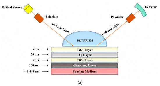
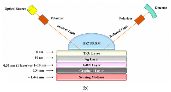
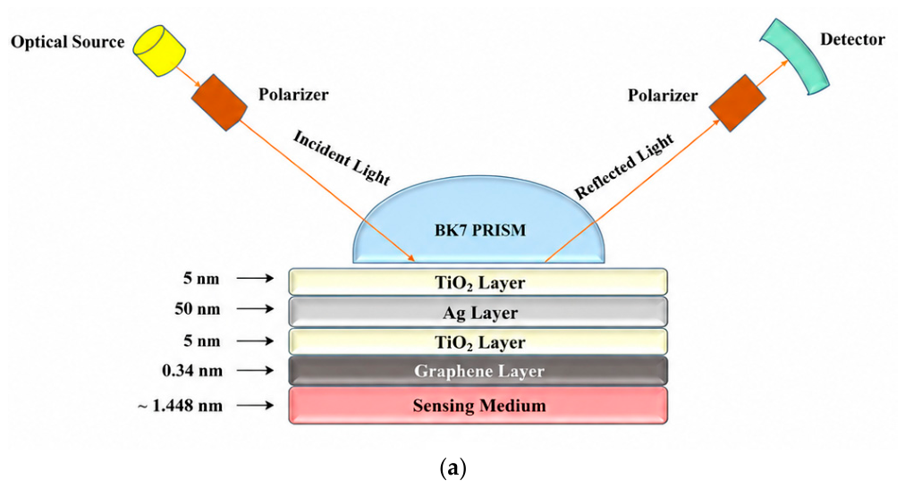
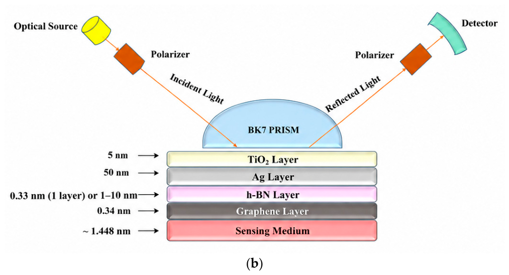
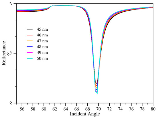
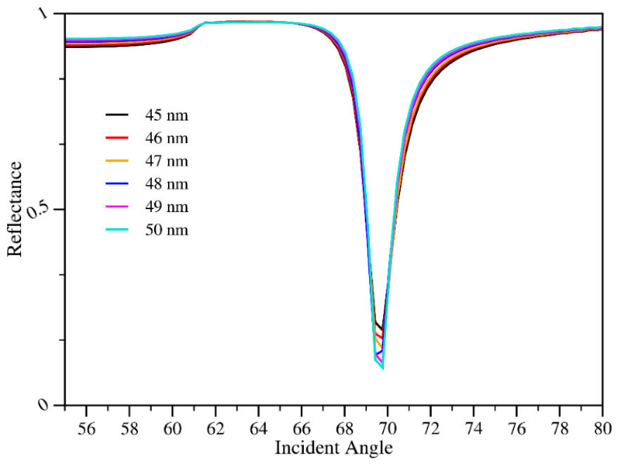
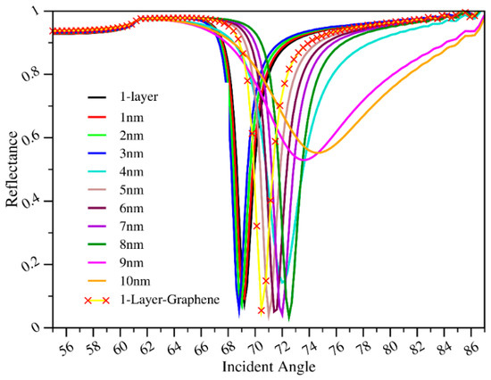
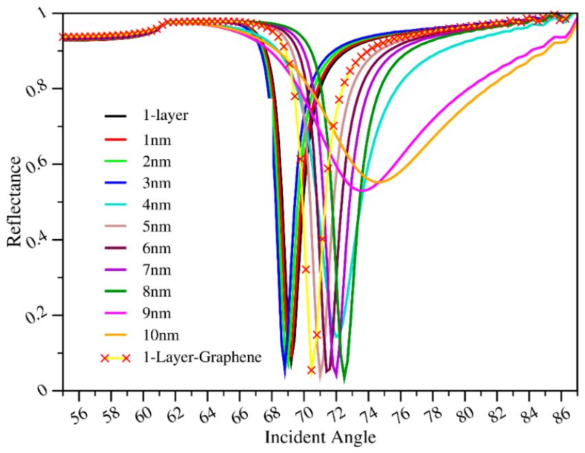
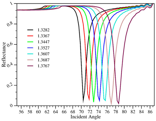
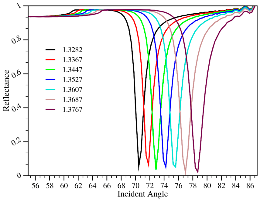
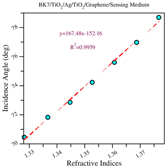
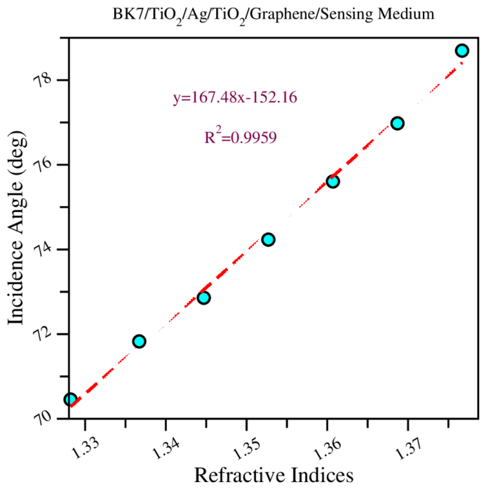
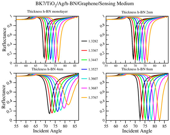
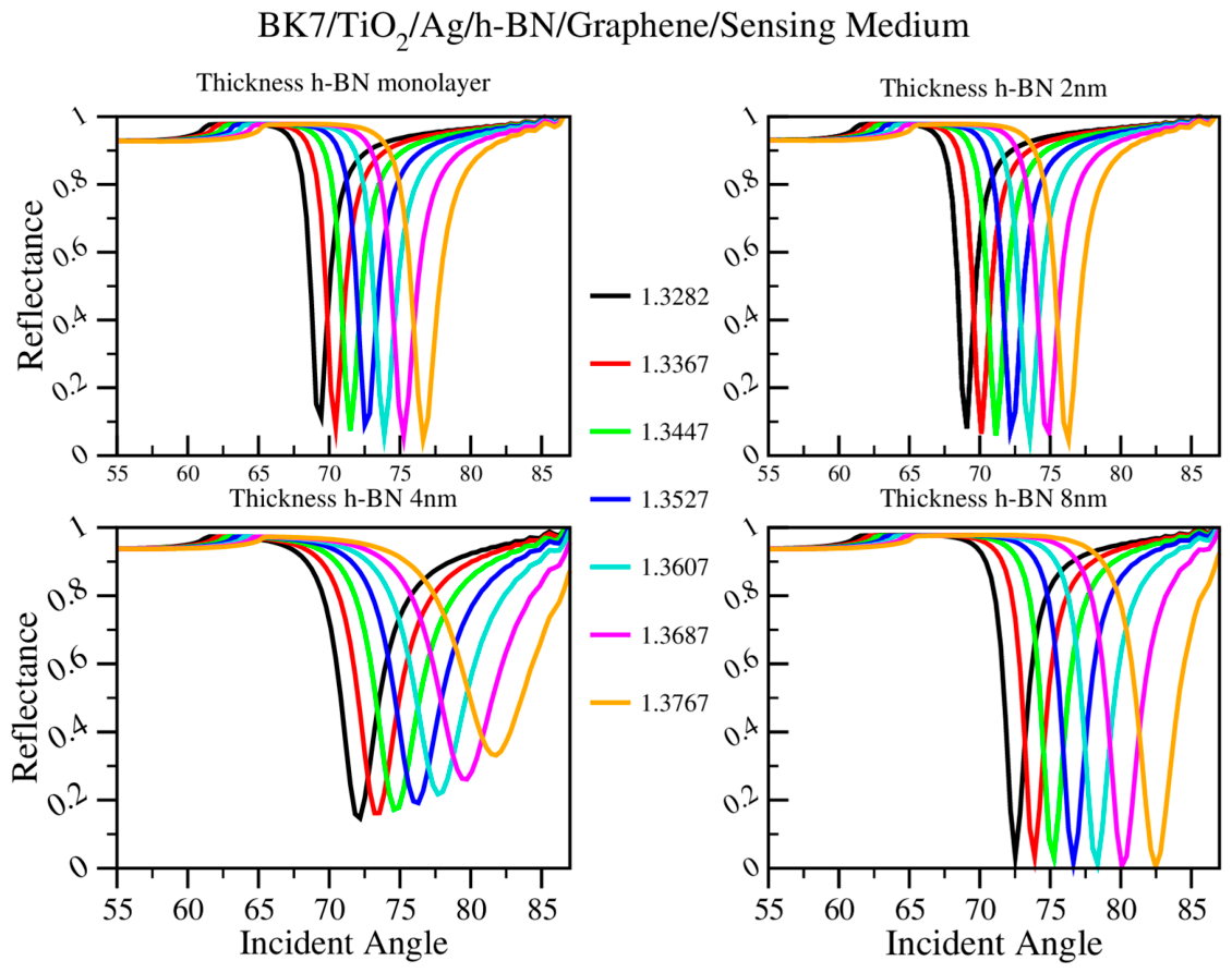
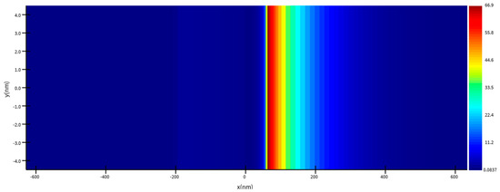
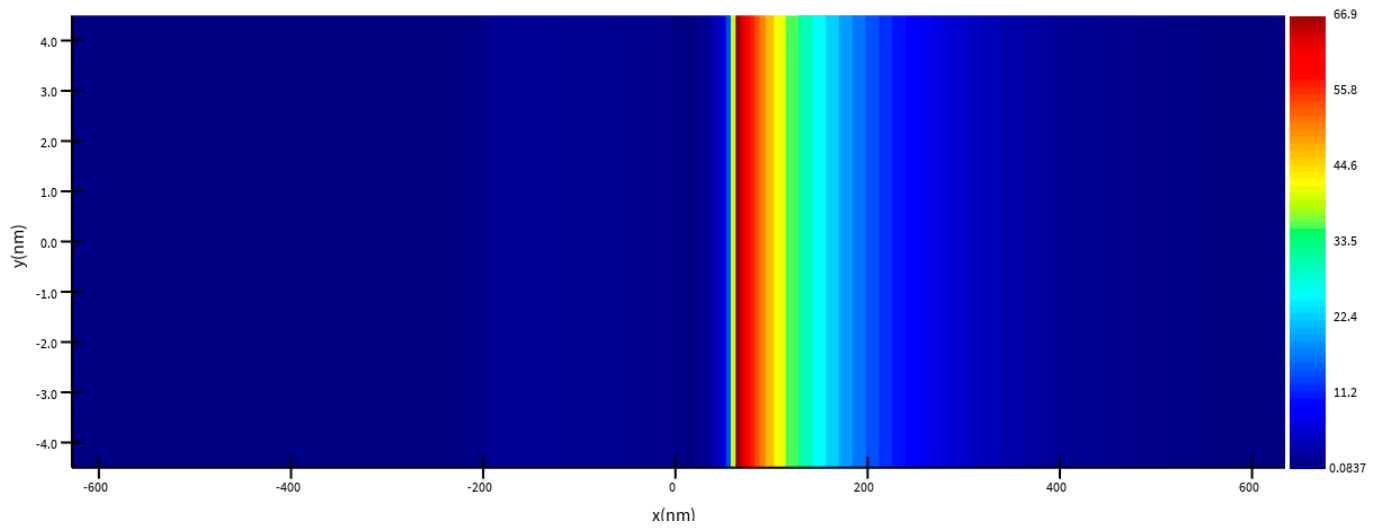
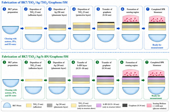
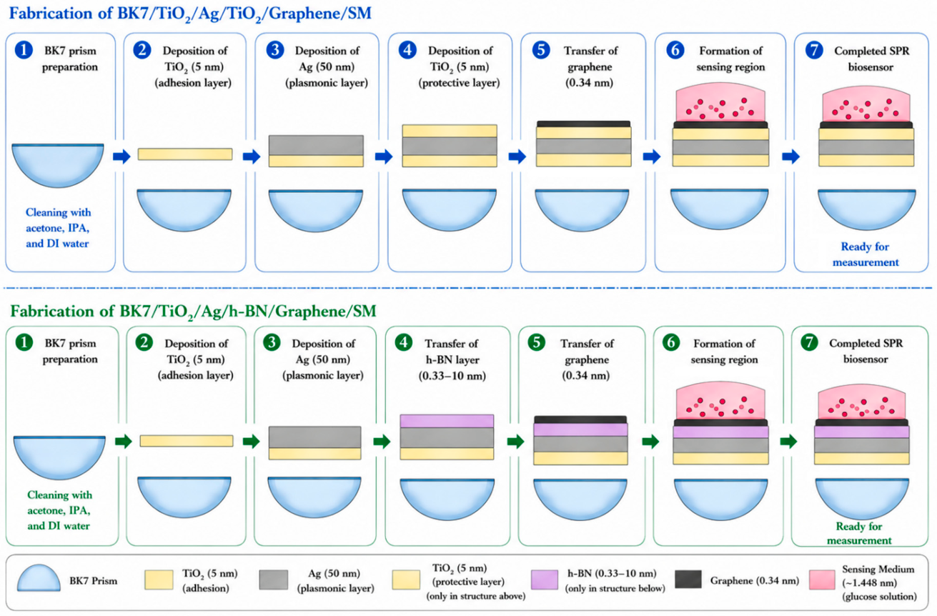

Disclaimer/Publisher’s Note: The statements, opinions and data contained in all publications are solely those of the individual author(s) and contributor(s) and not of MDPI and/or the editor(s). MDPI and/or the editor(s) disclaim responsibility for any injury to people or property resulting from any ideas, methods, instructions or products referred to in the content. |
© 2026 by the authors. Licensee MDPI, Basel, Switzerland. This article is an open access article distributed under the terms and conditions of the Creative Commons Attribution (CC BY) license.