


Abstract
Optical fiber tweezers enable the transmission of light over extended distances to trap micro- and nano-objects, offering the advantages of a compact design, flexible operation, and easy integration with endoscopic systems. These attributes have gained significant attention for manipulating and analyzing biological samples. Compared to traditional flat-cleaved optical fibers, micro-nanoscale structures on the fiber end-face allow for enhanced control over the emitted wavefront, focal field, and optical forces, thereby facilitating stable three-dimensional trapping, dynamic modulation, and signal analysis of samples. Recent advancements in micro-nano fabrication techniques have transformed the fiber end-face from a simple light-emitting interface into a sophisticated miniature multifunctional platform, which integrates capabilities such as focusing, near-field enhancement, multi-potential well creation, transmission mode regulation, and signal detection. This paper provides a review of recent progress in optical fiber tweezers based on various fiber end-face structures, introduces their structural characteristics and applications in particle manipulation, biological analysis, and microfluidic systems, and offers perspectives on future developments.
1. Introduction
Optical tweezers harness the momentum transfer between light and matter to trap micro- and nano-scale objects. As early as 1970, Ashkin reported that laser radiation pressure could accelerate micrometer-sized particles and developed a stable optical trap using counter-propagating light beams [1]. In 1986, Ashkin and colleagues created a single-beam optical trap based on an optical gradient force generated from a strong focal spot [2], and subsequently demonstrated its application in the manipulation of biological specimens such as viruses and living bacteria [3]. Due to advantages such as non-contact operation, high positioning precision, and compatibility with displacement and force measurements, optical tweezers have gradually become essential tools for manipulating microparticles, analyzing biological samples, and measuring weak forces [4,5,6].
Conventional optical tweezers mainly rely on optical microscopy systems, including multiple large-sized components for beam expansion, modulation, imaging, and focusing with a high numerical aperture (NA). Consequently, enhancing the system’s compactness and functional integration while still ensuring effective optical confinement for samples has become a key challenge for the advancement of optical tweezers technology.
Benefiting from the compact size and long-distance light transmission capability of optical fibers, optical fiber tweezers provide a compact configuration and enable remote optical manipulation of samples. In 1993, Constable and colleagues demonstrated dual-optical-fiber tweezers for trapping dielectric microspheres and yeast cells through balancing the scattering forces of counter-propagating beams from two flat-ended single-mode optical fibers [7]. Later, hemispherical lens fiber tips were used to improve the focusing ability, facilitating the trapping and manipulation of biological cells [8,9]. Advances in fiber-end lens fabrication and thermal tapering methods have also allowed a single optical fiber to generate a localized high-gradient optical focal field near its end-face, which can be utilized for trapping, moving, and arranging particles and biological samples [7,8,9,10,11].
In recent years, with advancements in micro-nano fabrication technologies, researchers have continuously expanded the structural design of fiber tips. By engineering the geometric shape, material distribution, and its interaction with guided modes inside the fiber, optical fiber tweezers can more precisely control the emitted light field, trapping position, and manipulation functions. As a result, the research focus of optical fiber tweezers has gradually shifted from merely enhancing the fiber tip’s focusing ability to the integrated design of functions such as light field control, particle trapping and transport, and sample signal detection, laying a new technical foundation for developing compact, multifunctional fiber manipulation platforms.
Several previous reviews have summarized optical fiber tweezers from different perspectives. Ribeiro et al. discussed optical fiber tweezers in terms of fabrication methods, fiber-tip structures, and trapping functions [12]; Xin and Li reviewed fiber-based optical trapping and manipulation according to photothermal trapping, evanescent-field trapping, dual-fiber tweezers, single-fiber tweezers, and particle or cell assembly [13]; Zhang et al. focused on optical-fiber-based tools for living cell manipulation [14]; and Zhao et al. provided a broad overview of optical fiber tweezers for trapping, manipulation, assembly, and sensing [15]. More recent reviews have further summarized optical fiber tweezers from preparation-to-application routes or focused on hollow-core-fiber optical traps [16,17]. Overall, these reviews mainly organize the field according to overall technical routes, fabrication methods, fiber configurations, trapping mechanisms, or application scenarios. In contrast, a systematic review that takes fiber end-face micro-nanostructures as the primary classification dimension remains limited. With the development of micro-nano fabrication techniques, diverse structures can now be formed on or near fiber end-faces, further highlighting the decisive role of end-face geometry in light-field modulation, trapping performance, and functional integration. Therefore, a review organized around fiber end-face micro-nanostructures is necessary to clarify the relationship between structural design and optical manipulation function.
In this paper, we review recent studies on optical fiber tweezers that utilize micro-nano structures at the fiber end-face. As illustrated in Figure 1, we classify the micro-nano structures fabricated at the fiber end-face into six primary categories: planar micro-nano structures, tip-type structures, end-face polished structures, microcavity-coupled structures, microsphere-integrated structures, and polymer microstructures. This classification is mainly based on the geometric morphology and construction form of the dominant structure at the fiber end-face. When a device contains more than one structural feature, it is assigned to the category that best matches its most representative end-face shape or integrated component. The details of optical fiber tweezers based on the six types of structures are introduced in Section 2, and finally in Section 3 we provide a summary of the above techniques and an outlook on the future development trends.
Figure 1.
Schematic diagram of research advancements in optical fiber tweezers categorized by six primary types of structures on the fiber end-face, including planar micro-nano structures [18], tip-type structures [19], end-face polished structures [20], microcavity-coupled structures [21], microsphere-integrated structures [22], and polymer microstructures [23].
2. Research Progress on Optical Fiber Tweezers with Different End-Face Structures
2.1. Planar Micro-Nanostructured Optical Fiber Tweezers
Through organizing micro-nanoscale functional structure units in a two-dimensional arrangement on the fiber end-face, optical fiber tweezers enable the precise manipulation of focused light fields and samples.
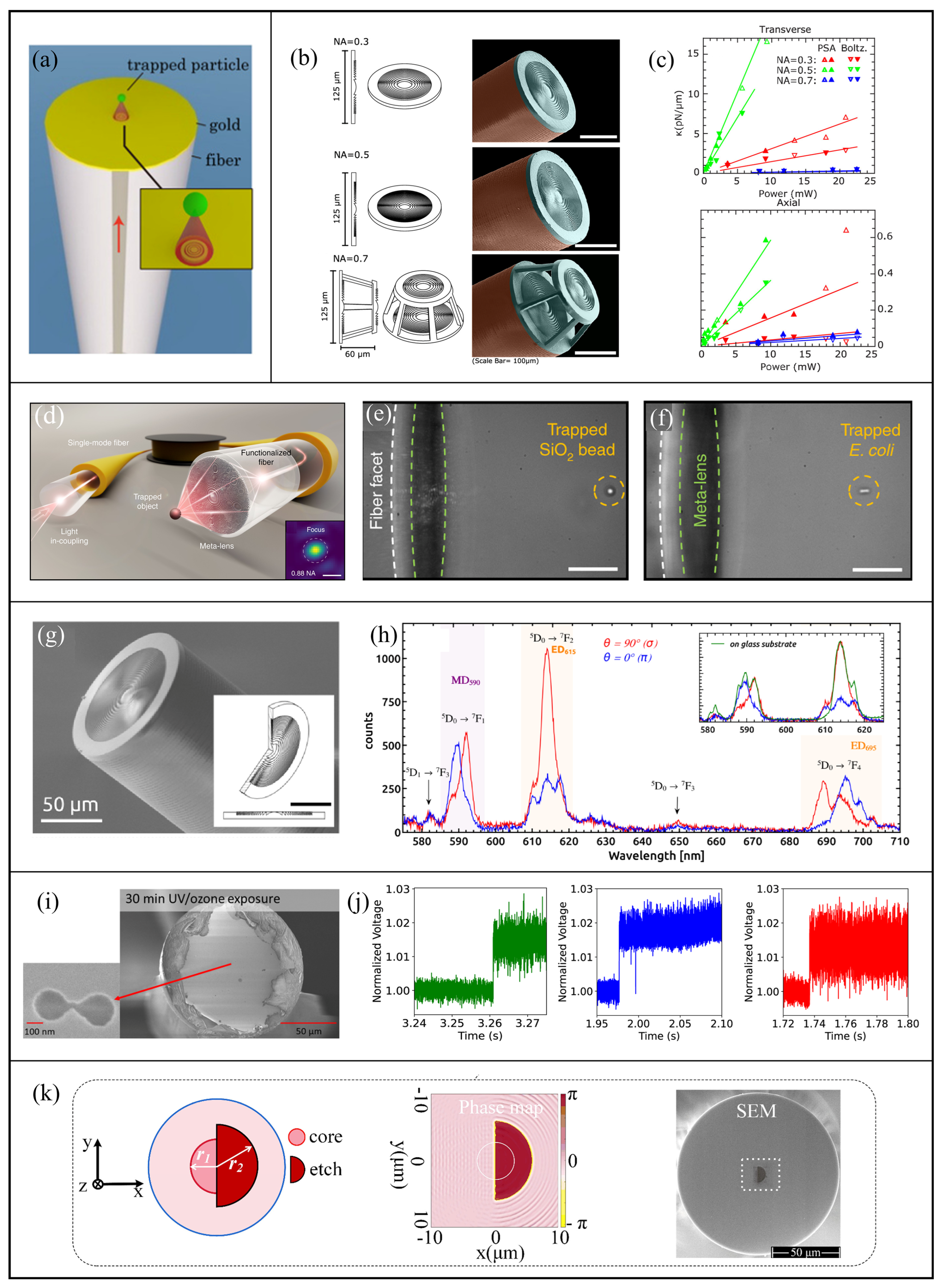
In 2013, Liu et al. fabricated concentric nanoscale ring slits on the end-face of an optical fiber coated with a thin metallic film, as illustrated in Figure 2a [24]. By exploiting surface plasmon excitation and reradiation induced by these ring slits, they generated a subwavelength focal spot exhibiting a full width at half maximum (FWHM) of ~450 nm and a working distance near 3.1 μm, and achieved three-dimensional (3D) trapping of polystyrene (PS) microspheres and Escherichia coli at an end-face power of 0.7–0.8 mW. Subsequently, in 2016, Saleh et al. incorporated annular gratings surrounding a coaxial plasmonic aperture [25] to enhance the maximum transmission efficiencies of gold- and silver-based structures from 2.5% and 1.48% to 19.6% and 16.7%, respectively, for stronger surface plasmon excitation. Correspondingly, the trapping potential well depths for dielectric particles with sizes of 30 nm and 10 nm increased to approximately 20 kBT and 11 kBT, respectively. These findings demonstrate the viability of employing planar metallic plasmonic structures on the fiber end-face for subwavelength, non-contact optical trapping, and their performance can be improved by the design of plasmonic coupling structures.
Beyond metallic plasmonic structures, researchers have also employed planar dielectric diffraction structures to realize focused optical traps. Dielectric structures circumvent the ohmic losses inherent to metals and offer expanded design possibilities for wavefront modulation. In 2017, Ribeiro et al. fabricated dielectric Fresnel zone plates on a fiber end-face, achieving two-dimensional trapping, manipulation, and detection of 8 μm polymethyl methacrylate microspheres and yeast cells [26]. Subsequently, to achieve 3D trapping, Asadollahbaik et al. [27] in 2020 fabricated diffractive Fresnel lenses with NAs = 0.3, 0.5, and 0.7 on the end-faces of two fibers positioned opposite each other, as illustrated in Figure 2b. The overlapping focal region of the two fibers forms a stable 3D trap for PS microspheres with diameters of 1 μm and 500 nm, based on the combination of a lateral optical gradient force and opposing axial optical scattering forces. Figure 2c presents the trap stiffness under different NA values, where the case of NA = 0.5 exhibited the optimal results.
Advancing towards 3D trapping using a single fiber, Plidschun et al. in 2021 integrated a high-NA focusing structure within a single optical fiber, as depicted in Figure 2d [18]. To enlarge the effective aperture of the end-face structure, they spliced a multimode fiber segment of 700–750 μm in length to the single-mode fiber, expanding the output beam diameter from ~4 μm to ~95 μm. Subsequently, a focusing phase structure with a diameter of 90 μm was fabricated to achieve an NA of 0.88–0.90 in water, producing near-diffraction-limited focal spots and enabling stable 3D trapping of 2 μm silica microspheres and Escherichia coli bacteria, as shown in Figure 2e and Figure 2f, respectively. This approach obviated the need for precise coaxial alignment and focal region overlap of two fibers by generating a tightly focused optical field from a single fiber, thereby enhancing suitability for remote operation in constrained environments.
In addition to trapping samples, optical fiber tweezers can also be integrated with spectroscopic measurements for sample analysis. In 2022, Kumar et al. employed a Fresnel lens on a fiber end-face with an NA of ~0.5 to trap NaYF4:Eu3+ nanorods, as shown in Figure 2g [28]. Their experiments not only achieved the trapping of individual nanorods and small clusters, but also obtained polarization-resolved emission spectra of the nanorods for the detection of crystallographic axis orientation, as presented in Figure 2h. In 2024, Kaur et al. integrated dual nanopores near the core of a single-mode fiber, as depicted in Figure 2i [29], and utilized an avalanche photodiode to record transmission signal variation induced by proteins passing through the dual nanopores. The system successfully trapped and analyzed various samples such as cytochrome c, carbonic anhydrase, and bovine serum albumin, as shown in Figure 2j. These works endow optical fiber tweezers with integrated functionalities such as focused field trapping, photoelectric signal acquisition, and protein conformational analysis within a single experimental platform.
Furthermore, structures on fiber end-faces have been utilized for high-precision displacement measurements of trapped samples. In 2025, Li et al. deposited Ta2O5 film on fiber end-faces to fabricate semicircular regions, as illustrated in Figure 2k, generating a phase difference of π between two parts of the end-face for particle displacement sensing [30]. By combining this fiber with another single-mode fiber to form dual-fiber tweezers, they achieved displacement detection sensitivity on the order of 0.1 pm·Hz−1/2 while trapping a 5 μm silica microsphere, providing a novel approach for integrating trapping and precision sensing within a single fiber.
Planar micro-nanostructured optical fiber tweezers can also generate focused vortex beams for particle rotation and fiber-based optical spanners [31], as well as enable multi-site trapping capabilities [32]. Among these structures, Fresnel-lens-based dual-fiber tweezers can provide high trap stiffness and a relatively large trapping region, while high-NA meta-fibers enable 3D trapping with a single fiber but require mode-field expansion and precise micro-nanofabrication. Fresnel lenses, double nanoholes, and structured-light end-face structures also extend optical trapping to spectroscopic characterization, single-protein analysis, and high-sensitivity displacement detection, respectively. Due to these various planar structural designs, such optical fiber tweezers not only realize tight focusing, localized field enhancement, and output wavefront control within the limited fiber end-face, but also transform from mere particle trapping tools into integrated platforms combining sample manipulation, positioning, and information readout functionalities [33,34,35,36].
Figure 2.
(a) The optical fiber end-face structure of a surface plasmon polariton lens [24], (b) Fresnel lens structures on fiber end-face with different NAs [27], (c) transverse and axial trap stiffness corresponding to different NAs [27], (d) ultra-high NA optical fiber tweezer configuration [18], (e) result of silica microsphere trapping [18], (f) result of Escherichia coli trapping [18], (g) Fresnel lens structure on fiber end-face [28], (h) emission spectra of trapped NaYF4:Eu3+ nanorods [28], (i) dual nanopore structure on fiber end-face [29], (j) capture signals of cytochrome c, carbonic anhydrase, and bovine serum albumin [29], (k) schematic diagram of a semicircular etched structure on fiber end-face [30].
2.2. Tip-Type Optical Fiber Tweezers
Tip-type optical fiber tweezers are typically fabricated by shaping the fiber tip into conical, parabolic, or semi-ellipsoidal geometries using techniques such as flame pulling, chemical etching, mechanical polishing, or arc fusion. This category is characterized by a protruding or tapered terminal morphology, including conical, parabolic, semi-ellipsoidal, elongated tapered, or locally protruded fiber ends.
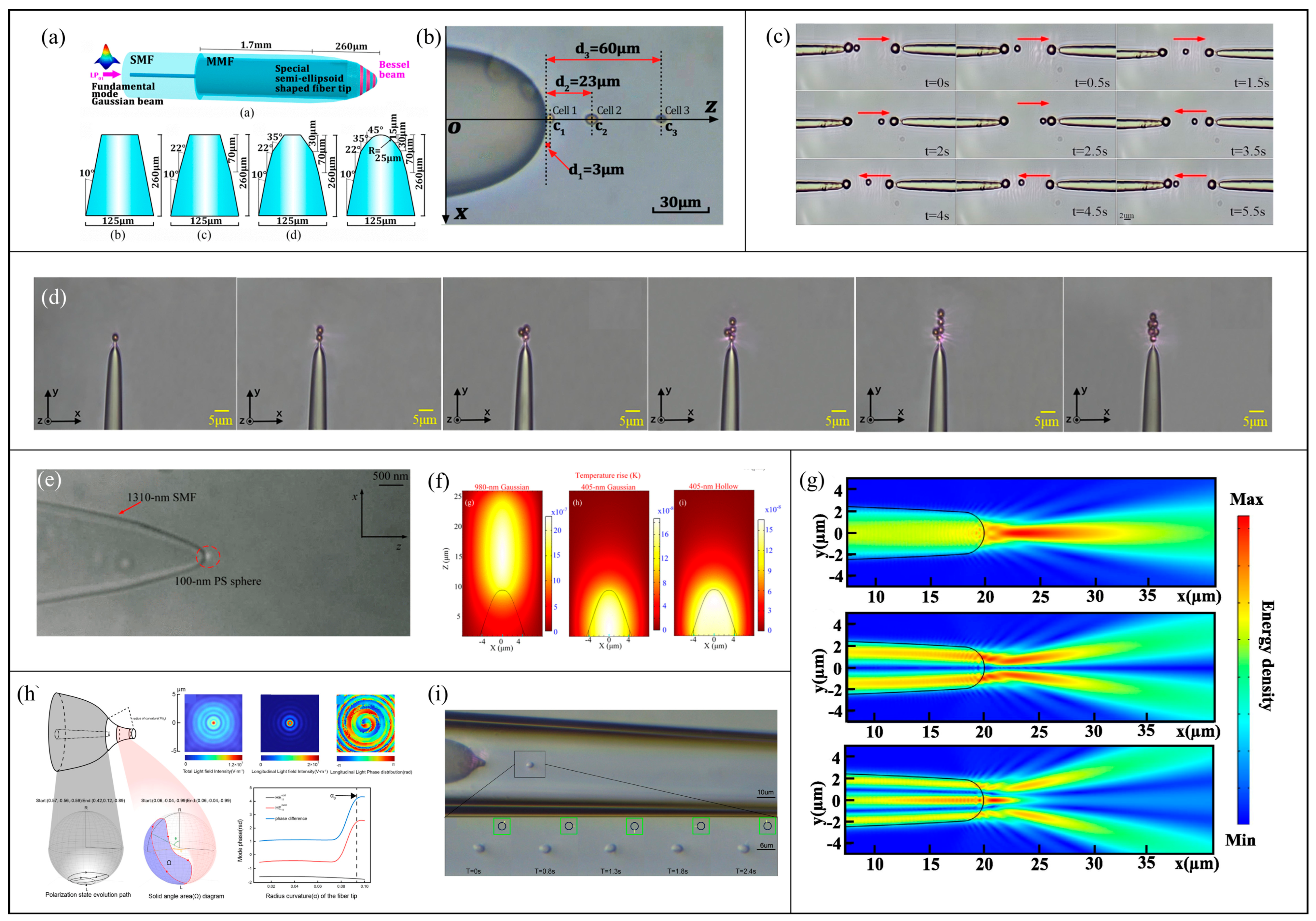
In 2017, Zhang et al. coaxially fused a single-mode fiber with a step-index multimode fiber and processed the multimode fiber tip into a semi-ellipsoidal taper, as illustrated in Figure 3a [37]. The concentric ring-shaped Bessel optical field generated by multimode interference converges at the tapered tip, creating multiple axially extended trapping regions to achieve the stable trapping of three yeast cells at distances of approximately 3, 23, and 60 μm from the tip, as shown in Figure 3b.
Subsequent investigations explored the use of multiple tapered tips to expand the particle trapping and manipulation region. In 2023, Gao et al. integrated two parabolic-profile tapered probes aligned coaxially in opposite directions to enable dual operational modes: single-fiber gradient force trapping and dual-fiber scattering force trapping [38]. As depicted in Figure 3c, each probe initially trapped a 3 μm PS microsphere near its tip, and then a 1 μm microsphere was confined between them by balancing the scattering forces of counter-propagating beams. By adjusting the input power to the left and right arms, the central particle could be moved bidirectionally between the two end particles.
Tapered fiber tip structures can generate stronger and smaller focal fields, facilitating the trapping of smaller and more complex particles. In 2024, Liu et al. fabricated a conical structure with a localized protrusion at the end of a single-mode fiber, as shown in Figure 3d [19]. The resulting intense focused spot not only enabled the lateral movement of a single magnetic microsphere, but also allowed the sequential trapping and alignment of one to six magnetic particles, demonstrating its manipulation adaptability to heterogeneous composite particles. In the same year, Zhang et al. cascaded a 405 nm single-mode fiber with a 1310 nm single-mode fiber to further suppress the optical trap size and thermal disturbances during nanoparticle trapping [39]. The resultant optical trap exhibited a lateral FWHM of 144 nm (about 0.36λ) and an axial FWHM of 447 nm, and achieved stable trapping of 100 nm diameter PS particles, as illustrated in Figure 3e. They also demonstrated in Figure 3f that the hollow mode of light at 405 nm wavelength can produce a more compact and uniform thermal field near the tip, thereby enhancing nanoscale confinement while reducing thermal perturbations.
Combined modulation of guided modes within the fiber and the focusing of the tip structures enable diverse dynamic trapping effects. In 2025, Bi et al. excited multiple modes at 980 nm within a multimode fiber, which were then filtered through a coaxially fused few-mode fiber segment to select the LP01, LP11, and LP21 modes [40]. By selectively focusing these modes, yeast cells could be stably trapped either in front of the tip or confined on the taper’s side surface. In the same year, they further employed the differential sensitivity of these modes to fiber bending loss to dynamically switch the trapping functions [41]. By controlling the fiber bending degree, the dominant mode was sequentially shifted among the LP21, LP11, and LP01 modes, with the corresponding focal fields shown in Figure 3g. This approach enabled sequential cell trapping, axial position adjustment, and long-distance transport without moving the probe or altering the light source. Also in 2025, Chen et al. coupled both 650 nm and 980 nm light into the same tapered probe [42], where the 980 nm light supported the LP01 and LP11 modes and the 650 nm light excited the LP21 and LP02 modes, resulting in opposite trapping and repulsion responses for yeast and SiO2 particles.
In 2026, Geng et al. extended the functionality of tip-type fiber tweezers from particle transport to cellular rotation control [43]. Using 980 nm near-infrared light and fabricating a non-adiabatic tapered fiber tip, as shown in Figure 3h, the fiber with a rapidly decreasing radius induced local polarization rotation, ultimately generating transverse spin angular momentum transferred to the trapped cells. Experimental results in Figure 3i demonstrated that yeast cells could be stably confined and continuously rotated over 0–2.4 s, confirming optical torque-driven sustained rotation.
Tip-type optical fiber tweezers, through the adjustment of tip curvature, internal fiber modes, and emitted optical fields, can form long-working-distance traps, multiple potential wells, and subwavelength trapping regions at the fiber end [14,44]. Semi-ellipsoidal and parabolic Bessel probes offer advantages in extending the axial interaction range and enhancing the localized optical field, although their performance is affected by the tip geometry and fabrication accuracy. Locally protruded conical tips are suitable for manipulating composite particles, while mode-division multiplexing, mode selection, and wavelength switching provide greater tunability of the trapping position and particle response but require more precise control of the excitation and input conditions. Non-adiabatic tapered fibers can further enable particle rotation through polarization control, thereby extending the manipulation capabilities of tip-type optical fiber tweezers [45,46,47].
Figure 3.
(a) A semi-ellipsoidal optical fiber tweezer tip [37], (b) its stable trapping of three yeast cells at different distances [37], (c) experimental images depicting the dual-mode trapping and transmission process of particles [38], (d) sequential capture and alignment of multiple magnetic particles [19], (e) trapping of PS particles with a diameter of 100 nm [39], (f) temperature fields under different wavelengths and modes [39], (g) output optical fields corresponding to LP01, LP11, and LP21 modes [41], (h) the mechanism and characteristics of transverse spin angular momentum formation in a non-adiabatic tapered fiber tip [43], (i) a series of continuous images showing the stable rotation of a yeast cell within 0 to 2.4 s [43].
2.3. End-Face Polished Reflective Optical Fiber Tweezers
End-face-polished reflective optical fiber tweezers achieve particle trapping and manipulation by fabricating inclined, pyramidal, or truncated conical surfaces on the fiber end-faces to reflect and alter the propagation direction of the emitted light. Morphologically, this category mainly includes fiber ends with multi-faceted ends or curved reflective end-face profiles.
In 2014, Gao and Yuan designed an asymmetrical truncated pyramidal structure with a slight rotational offset relative to the core positions on a four-core fiber [48], thereby generating four light beams to form vortex-type optical trapping and rotational driving of nonspherical particles. Building upon this, Zhao et al. [49] introduced distinct azimuthal compensation angles on the four polished surfaces, dividing the four beams into two groups that created dual optical traps. By alternately activating these two groups of beams, particles could be controllably shuttled between the two trapping potential wells.
Introducing varied polishing angles on multicore fiber end-faces allows multiple localized light gradient fields to extend the effective range of the optical traps. For instance, as illustrated in Figure 4a, Tang et al. in 2020 fabricated three reflective surfaces with inclination angles of 23°, 19°, and 16° on the end of a seven-core fiber, producing three optical traps at different axial positions [50]. These traps interconnected to form a continuous active pulling region spanning 23.5 to 65.5 μm, enabling yeast cells to be transported from 61 μm to 21 μm away from the fiber tip, as shown in Figure 4b.
Figure 4.
(a) The axial dual-beam formation and corresponding optical force calculations resulting from three sets of reflective surfaces with different inclination angles at the end-face of a seven-core optical fiber [50], (b) the directed movement of a yeast cell toward the probe within the active traction region [50], (c) the generation and convergence of hollow quasi-Bessel beams based on optical fibers [51], (d) experimental results of the focal light field outside the fiber end-face [51], (e) the lateral and axial displacement of a 32 μm PS microsphere following the probe movement [51], (f) multiple total internal reflections within a double-conical probe and the optical force equilibrium near the top end-face [20], (g) the gradient force induced by the evanescent field on a 100 nm particle [20], (h) experimental and numerical optical field results of photon hooks generated by asymmetric truncated conical end-faces [52], (i) the ability of photon hooks to selectively trap and manipulate an individual red blood cell [52], (j) comparative analysis of optical fields and potential wells formed by inclined planes, micro-prisms, and crab-claw-shaped curved surfaces [53], (k) The trapping of 3 μm PS microspheres and 6 μm yeast cells using crab-claw-shaped fiber tweezers [53].
Beyond directly modifying the focal positions of multicore beams, polished end-faces can synergize with in-fiber modes to enlarge the effective interaction area between the optical field and particles. In 2021, Zhang et al. [51] coaxially fused an annular-core fiber with a step-index multimode fiber, converting the input fundamental mode into a hollow Bessel-like beam focused by a truncated conical reflective surface at the fiber end, as illustrated in Figure 4c. The experimental convergence angle was 76°, as shown in Figure 4d. This configuration enabled the stable trapping of 32 μm PS microspheres and lateral displacements of 107.2 μm and 228 μm, as well as an axial displacement of 272 μm, as depicted in Figure 4e.
The polished structures can also confine the optical field near the end-face for near-field trapping of nanoparticles. In 2022, Liu et al. sequentially fabricated dual conical profiles with 18° and 54° angles on a dual-core fiber end, causing each light path to undergo three total internal reflections to generate an evanescent field at the top end-face [20], as shown in Figure 4f. The high intensity gradient of such an evanescent field exerted an axial trapping force of 2.77 pN on 100 nm PS spheres, forming a stable trapping potential well with a depth of 57.06 kBT, as shown in Figure 4g.
Asymmetry in the polished fiber end-face can also cause the emitted light to deviate from linear propagation, generating curved trapping fields. In 2025, Chen et al. fabricated an inclined polishing on the tapered fiber tip [52], producing a curved “photon hook” optical field, as illustrated in Figure 4h. This curved field not only altered the trapping position but, more importantly, enhanced spatial selectivity in dense samples. As depicted in Figure 4i, the photon hook field selectively trapped a target red blood cell (cell A) in the middle of a cell chain and moved it about 12.7 μm, demonstrating the capability of selective trapping and transfer of a target cell even in the presence of surrounding cells. In the same year, Liu et al. fabricated two crab-claw-shaped curved surfaces on the end of a seven-core fiber, which further compressed the axial FWHM of the focal spot to 2.73 μm and increased the corresponding potential well depth to 204.30 kBT/mW, significantly outperforming the other two structures of inclined plane and micro-prism [53], as shown in Figure 4j. Experimentally, this crab-claw structure achieved trapping of 3 μm PS microspheres and 6 μm yeast cells, as illustrated in Figure 4k. Moreover, fabricating multiple crab-claw units on the fiber end-face enables the trapping of multiple particles simultaneously.
Through the fabrication of multiple inclined reflective surfaces at the fiber tip, the end-face-polished optical fiber tweezers can reconstruct the external optical field with simple and compact probe structures and realize enhanced properties such as extended pulling regions, localized near-field potential wells, curved trapping, and multi-particle trapping [54,55,56,57,58,59], significantly broadening the capability to trap particles across various sizes and spatial ranges. Among these polished reflective end-face schemes, multi-angle reflective surfaces help extend the axial manipulation range and adjust the trapping position but require high geometric accuracy of the end face. Hollow Bessel-like fields can be used for trapping relatively large particles, whereas near-field evanescent-field structures help reduce thermal disturbance and enhance localized confinement, although their effective trapping range is limited. Asymmetric photonic-hook and crab-pincer-shaped reflective structures further extend the capabilities for selective manipulation and localized trapping but place higher demands on end-face fabrication accuracy. Different reflective end-face structures therefore exhibit distinct characteristics in manipulation range, particle size, and local confinement.
2.4. Microcavity-Coupled Optical Fiber Tweezers
Microcavity-coupled optical fiber tweezers are engineered by introducing microcavity structures such as hollow-core fiber segments, capillary microtubes, or microfluidic channels near the fiber end-face, thereby modulating the optical fields and fluid dynamics to facilitate the trapping, transport, storage, and release of microparticles and cells. For example, in 2018, Zhang et al. [60] exploited the intrinsic central channel of a hollow annular-core fiber as a microfluidic conduit, wherein the annular fiber core and central channel respectively performed light guidance and microfluidic transport functions, thereby enabling 3D trapping, suction, temporary storage, and the subsequent release of microspheres through the interplay of optical trapping forces and fluid viscous resistance.
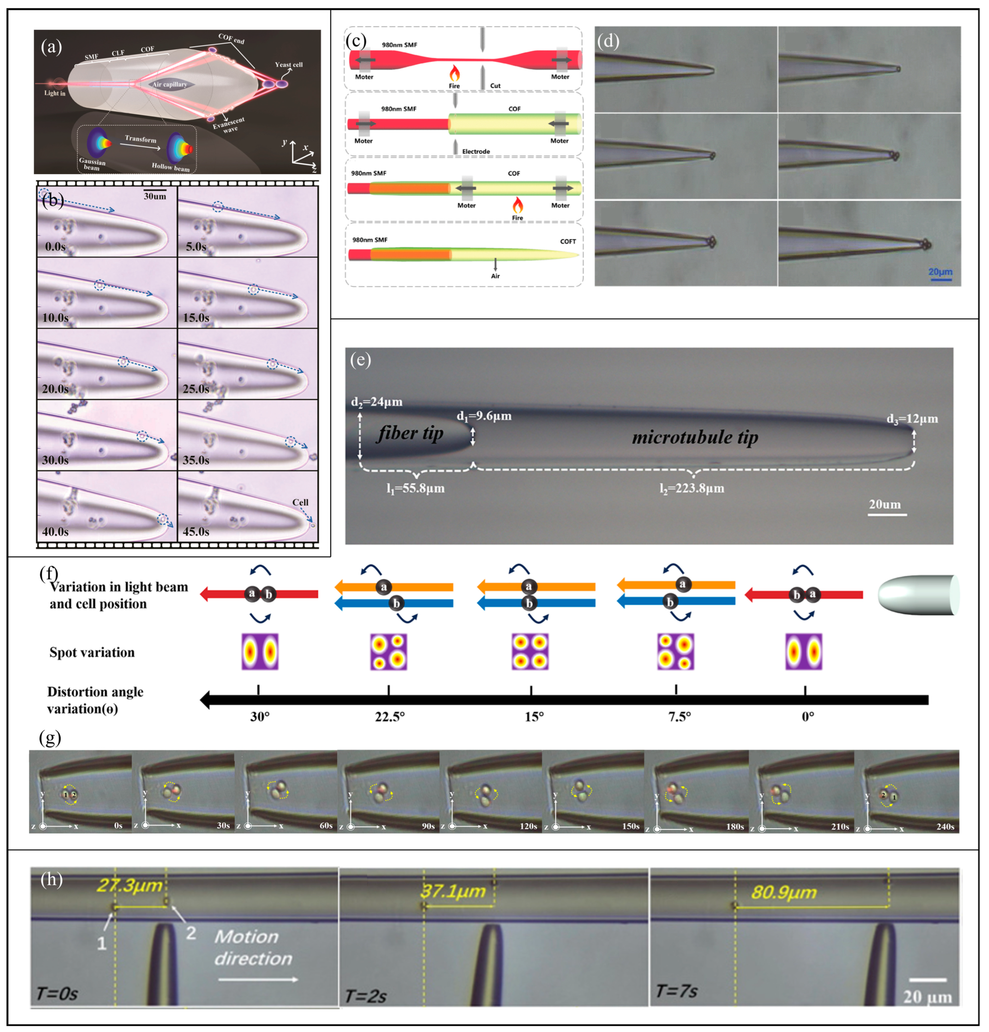
Microcavities within fibers are frequently employed to modulate optical modes, thereby generating specialized optical tweezing effects. In 2022, Deng et al. proposed capillary optical fiber tweezers formed by cascading a single-mode fiber, a coreless transition segment, and a capillary fiber, as illustrated in Figure 5a [21]. This configuration splits the light via an internal tapered air cavity and then utilizes total internal reflection at the fiber surface to produce an evanescent field that guides cells along the surface, and finally accomplishes cell trapping at the fiber tip. Experimental results in Figure 5b demonstrate cell transport distances exceeding 150 μm and multiple potential wells at the fiber tip sequentially capturing several cells. In 2024, Li et al. fabricated a flat-head tapered structure with a central air hole at the fiber end [61], enabling size-selective trapping of microspheres where the 10 μm microspheres were primarily propelled axially and 2 μm microspheres were stably trapped.
The integration of tapered optical fibers within capillary microtubes to form microcavities has been demonstrated to enable diverse trapping functionalities. In 2024, Wang et al. embedded a tapered single-mode fiber into a capillary microtube, fabricating a microcavity probe of 150 μm in length and 3 μm diameter [62], as illustrated in Figure 5c. This microcavity redistributes the optical field energy to generate both LP21 and LP01 modes, allowing the formation of distinct potential wells at the center and the periphery of the probe tip. Experimentally, this configuration enabled the annular trapping of at least five PS microspheres and lateral displacements of the microspheres by moving the probe, as shown in Figure 5d. In 2025, Ji et al. developed a probe structure composed of a flat-ended tapered single-mode fiber embedded within a hollow capillary microtube, as depicted in Figure 5e [63]. By employing a fiber twisting apparatus, they realized reversible mode conversion between the LP11 and LP21 modes in the probe, as presented in Figure 5f, resulting in a pair of cells completing a 180° rotation within 240 s, as illustrated in Figure 5g. Liu et al. in 2025 also nested a tapered optical fiber within a capillary microtube to modulate both axial optical forces and capillary forces, and successfully trapped yeast cells, PS, silica, and magnetic microparticles in water and yeast cells in a high-refractive-index triethylene glycol environment [64], extending the range of applicable environments. In 2025, Jiang et al. investigated the influence of capillary microcavity parameters on optical field modulation and particle size sorting [65]. Experimentally, both 3 μm and 5 μm silica particles could be trapped within the microcavity at lower power levels, but increasing the power caused only larger particles to be repelled, thereby enabling size-selective trapping of microparticles.
The function of the microcavity can even be extended from the internal fiber channels to external microfluidic structures. In 2023, Jiang et al. positioned an optical fiber probe outside a capillary microtube and utilized the curved tube wall of the capillary microtube to further compress the light beam, finally achieving the fully non-invasive manipulation of Chlorella cells within the tube, as illustrated in Figure 5h [66].
Figure 5.
(a) The air cavity structure, optical evanescent field propagation, and multifocal formation mechanism of capillary optical fiber tweezers [21], (b) the process of transporting a yeast cell over a long distance and subsequently trapping it at the fiber tip [21], (c) the fabrication process of hollow conical optical fiber probes [62], (d) the sequential trapping process of five PS microspheres [62], (e) the cascaded microcavity optical tweezers probe structure [63], (f) mode conversion between LP11 and LP21 modes induced by fiber twisting and the corresponding changes in cell positioning [63], (g) positional changes in two cells over a duration of 240 s [63], (h) non-invasive manipulation of Chlorella cells within a microtube [66].
The microcavity-coupled optical fiber tweezers incorporate hollow channels, air microcavities, or capillary boundaries to synergistically regulate the optical field distribution and fluid dynamics at the fiber tip to control microparticle motion [67,68,69,70,71], offering advantages such as straightforward structural fabrication, high design flexibility, and multifunctional capabilities. Introducing cavity structures enables fiber probes to combine optical-field control with particle transport and sample isolation. Hollow-core and suspended-core structures facilitate trapping, storage, and transfer within a single fiber, while cascaded capillary structures can combine surface guiding and mode control to realize multi-trap confinement, particle rotation, and selective manipulation. External capillary microtubes further avoid direct contact between the fiber and the sample, offering advantages for the isolated manipulation of biological specimens. These functions are mainly influenced by the cavity dimensions, particle size, and optical coupling conditions.
2.5. Microsphere-Integrated Optical Fiber Tweezers
By affixing high-refractive-index dielectric microspheres onto the fiber end-face, the microsphere-integrated optical fiber tweezers enable stronger focusing of light through the microsphere, resulting in a reduced focal spot size and a strengthened trapping potential. This category is identified by the presence of one or multiple microspheres attached to, assembled on, or integrated with the fiber end-face. Hybrid devices are included here when the microsphere is the most recognizable added component of the end-face structure.
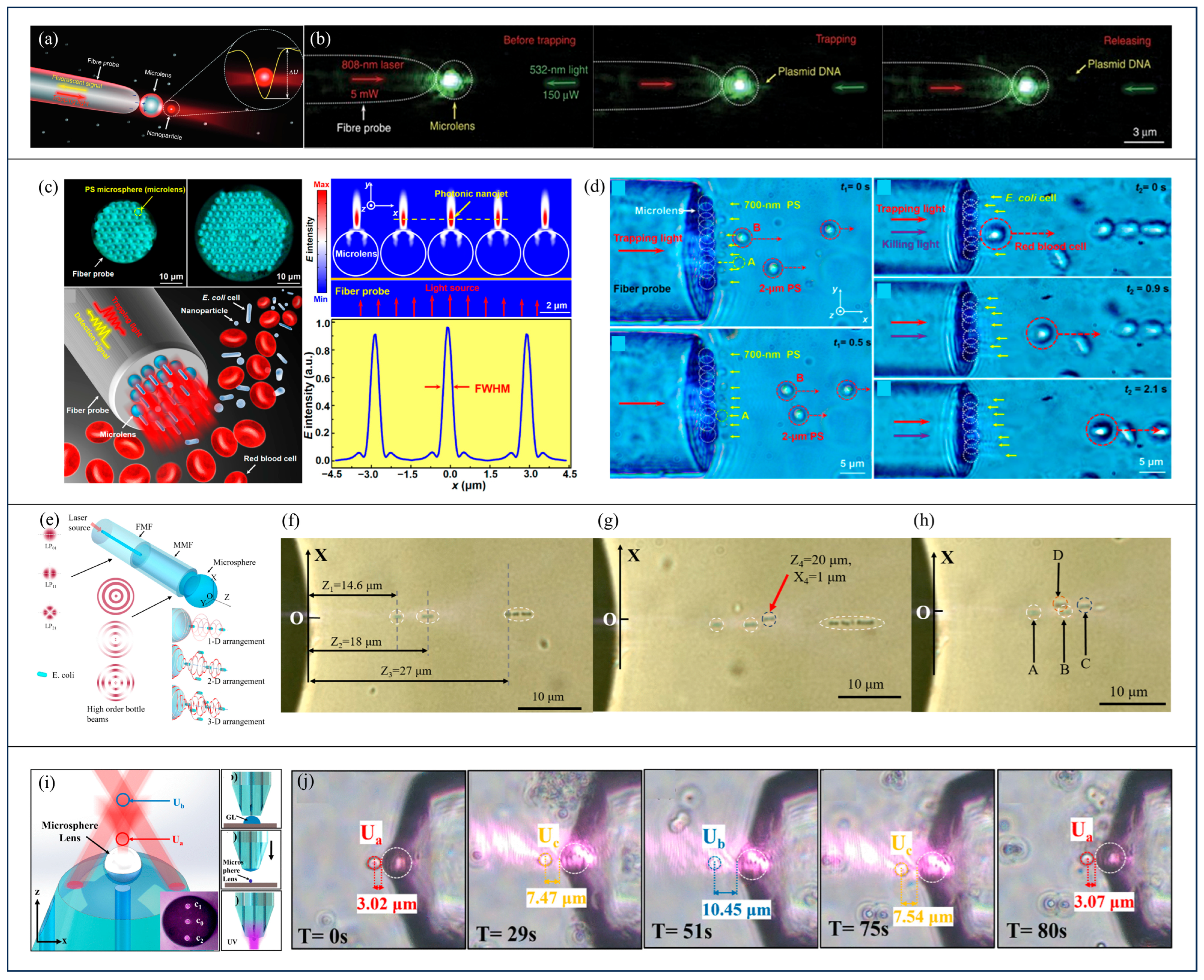
In 2016, Li et al. investigated a microsphere-integrated optical fiber tweezer system [22], comprising a parabolic single-mode optical fiber with a 3 μm dielectric microsphere attached at the fiber tip, as illustrated in Figure 6a. They revealed that the FWHM of the focused photonic nanojet field was reduced from 840 nm for a bare fiber to 385 nm and 208 nm upon the addition of PS and TiO2 microspheres, indicating stronger local optical confinement, thereby achieving stable trapping and 3D manipulation of 85 nm fluorescent particles and DNA molecules, as depicted in Figure 6b. Moreover, the PS and TiO2 microspheres enhanced the fluorescence intensity of a single nanoparticle by approximately 20-fold and 30-fold, respectively, while the backscattering signal was used for real-time monitoring of the trapping process. The significance of this work lies not only in achieving a subwavelength localized optical field, but also in integrating localized trapping, 3D manipulation, and enhanced detection of nanoscale samples within a single fiber probe. Subsequently, they further developed a two-dimensional microsphere array on a fiber end-face [72], as shown in Figure 6c. Each microsphere generated a focused optical field with an FWHM of 360 nm, enabling the formation of over a hundred 3D trapping potential wells. The array enabled the simultaneous trapping of multiple 190 nm PS nanoparticles and E. coli cells, while backscattering signals provided real-time detection with single nanoparticle resolution. The microsphere size and refractive index jointly influenced the optical trapping forces, imparting selective trapping capabilities dependent on particle size and morphology. As demonstrated in Figure 6d, 700 nm particles were attracted but 2 μm particles were repelled. In mixed blood samples, elongated Escherichia coli bacteria were selectively trapped, while larger red blood cells were excluded. These two studies by Li et al. represent a progression from single microsphere trapping and manipulation to parallel trapping, detection, and selective separation using a microsphere array. Subsequent studies further extended photonic-nanojet-based microsphere structures toward more complex biological manipulation. In 2024, Xiong et al. used a TiO2 microsphere attached to a tapered optical fiber probe to generate a photonic nanojet for localized stimulation of the photoreceptor of a microalga, enabling controllable deformation and navigation of a soft microalga robot [73]. In 2026, He et al. further integrated large-scale TiO2 microlens arrays onto flexible substrates, generating up to 1000 subwavelength focal spots for high-throughput trapping and sorting of bioparticles, including exosomes, bacteria, and cells [74].
The fusion of single-mode and multimode optical fibers, combined with microsphere structures, allows for tunable focusing field distributions and the trapping of low-refractive-index samples. In 2018, Zhang et al. [75] coaxially fused a single-mode fiber with a multimode fiber and a glass microsphere at the fiber end-face, generated two axial dark-field regions, enabling 3D confinement of low-refractive-index yeast cells within a high-refractive-index triethylene glycol medium. In 2019, Tang et al. employed a similar configuration [76] to produce a focal spot with axial and lateral FWHMs of 1.2 μm and 350 nm, respectively, and achieved stable trapping of 200 nm PS particles at an incident power of 0.7 mW, with the trapping status monitored via backward scattering signal variation. In 2020, Liu et al. utilized a comparable structure to generate sequential bright focal points and dark fields along the axial direction [77], enabling the trapping of high-refractive-index PS particles at bright foci and low-refractive-index yeast cells at dark-field traps. They further combined annular fiber cores with microspheres [78], exploiting photophoretic forces in pure glycerol environments to achieve stable trapping of strongly absorbing particles.
Microspheres can also be integrated with various fiber structures to focus different propagation modes, yielding diverse manipulation effects. An optical bottle beam generally features a localized low-intensity region surrounded by higher-intensity light [79]. Such a dark region is favorable for trapping low-refractive-index particles, which tend to be repelled from high-intensity regions, whereas high-refractive-index particles are generally confined at bright intensity maxima [80]. Therefore, the dark-field traps and bright-dark composite potential wells discussed above are related to optical bottle beams through the spatial redistribution of optical intensity, although their specific field distributions and trapping positions may differ. In 2022, Zhang et al. fabricated a structure sequentially composed of a single-mode fiber, a few-mode fiber, a multimode fiber with a length of 1850 μm, and a high-refractive-index microsphere at the fiber end-face [81], as illustrated in Figure 6e. In this work, multimode interference generated an optical bottle beam with a central spot and concentric rings, whose intensity distribution is similar to that of a high-order Bessel beam; after focusing by the end-face microsphere, several bright focal regions were formed for particle trapping. By adjusting the lateral fusion offset between the single-mode fiber and the few-mode fiber, the excitation ratios of the LP01, LP11, and LP21 modes can be controlled. This enables the manipulation of Escherichia coli into one-dimensional chain structures (Figure 6f), two-dimensional planar arrangements (Figure 6g), and three-dimensional spatial configurations (Figure 6h), thereby endowing a single optical fiber with the capability for multi-particle spatial assembly. In 2026, Zhang et al. employed a three-core fiber combined with an end-face microsphere and a polished reflective surface to construct a tunable dual trap system at the fiber tip [82]. As depicted in Figure 6i, a glass microsphere fixed at the end of the central core generates a proximal potential well Ua, while the two side-core beams form a second potential well Ub outside the fiber. Experimentally, both potential wells successfully trapped yeast cells, and further enabled reversible axial transport of yeast cells between the two potential wells (Figure 6j).
The optical fiber tweezers integrated with microspheres leverage the strong focusing capability of microspheres to generate various optical fields such as photonic nanojets, Bessel-like convergent fields, bright-dark composite potential wells, and multi-channel tunable potential wells. Single microspheres and microsphere arrays can support the trapping, detection, and parallel manipulation of nanoparticles and biomolecules, although their performance depends on microsphere positioning and the end-face field distribution. When combined with Bessel beams, annular cores, or multicore structures, these probes can further manipulate particles with different refractive indices or absorption properties and enable multidimensional arrangement and dynamic transport. Consequently, these configurations enhance trapping efficiency and expand manipulation functionalities, facilitating the trapping, detection, arrangement, and transport of nanoparticles, biomolecules, cells, and microparticles with varying refractive indices [83,84].
Figure 6.
(a) Schematic illustration of a single dielectric microsphere attached to the tip of an optical fiber probe to generate a photonic nanojet for the trapping of nanoparticles [22], (b) the process of trapping, holding, and releasing a single DNA molecule [22], (c) a two-dimensional PS microsphere array on the fiber end-face, the corresponding electric field distribution and FWHM of the parallel photonic nanojet array [72], (d) selective trapping of nanoparticles and cells [72], (e) optical bottle beams based on LP01, LP11, and LP21 modes and the principle of multidimensional particle arrangement [81], (f) one-dimensional axial arrangement of Escherichia coli [81], (g) two-dimensional planar arrangement of multiple particles [81], (h) experimental demonstration of three-dimensional spatial arrangement of multiple particles by single-fiber optical tweezer (Cells A, B and C are trapped within the same imaging plane, whereas cell D locates at a different depth plane) [81], (i) dual potential well structure of a three-core optical fiber [82], (j) reversible axial transport of a single yeast cell between dual potential wells under power regulation [82].
2.6. Polymer Structured Optical Fiber Tweezers
Polymer-structured optical fiber tweezers are primarily fabricated at the fiber end-face using polymer additive manufacturing techniques such as self-written photopolymerization, waveguide photopolymerization, dual photopolymerization, and two-photon polymerization [85,86,87,88]. These methods enable the formation of high-refractive-index microtips, microlenses, or complex 3D end-face structures, which shape the output optical field for particle trapping, manipulation, and signal acquisition. This category is identified by these complex 3D end-face structures when the polymer-formed morphology is the main structural feature of the fiber end-face.
In 2009, Valkai et al. fabricated polymer waveguide tips on single-mode fiber end-faces and used two such fibers with polymer tips facing each other to trap 1 μm microspheres [89]. Building upon this foundation, Ribeiro et al. in 2015 developed a method for fabricating polymer microlenses by inducing photopolymerization at 405 nm within droplets on the fiber end-face, and further controlled the polymer tip profile and output optical fields by exciting different fiber modes such as LP01, LP02, and LP21 [90].
Polymer microlenses can be used not only for sample trapping but also for the subsequent detection and identification of the trapped samples. In 2018, Paiva et al. fabricated a polymer microlens on the end-face of a 980 nm single-mode fiber as shown in Figure 7a [91]. The lensed fiber was used to trap PS microspheres, polymethyl methacrylate microspheres, and yeast cells, and the back-scattered signal from each trapped sample was further collected for label-free identification, as shown in Figure 7b. After signal acquisition, time-domain and frequency-domain features were extracted from the back-scattered signals, and linear discriminant analysis (LDA) was used to reduce these signal-derived features into a single discriminant feature for distinguishing different sample types. In the same year, Rodrigues et al. first spliced a short segment of multimode fiber onto the end of a single-mode fiber to enhance the light coupling to the polymer microlens [92], as shown in Figure 7c. The experimental results in Figure 7d demonstrate that this structure can improve the cell trapping efficiency by up to about 10 times compared to previous single-mode polymer microlenses, while retaining the ability to distinguish trapped samples based on backscattering signals. In 2019, Paiva et al. applied similar polymer microlens probes for the concentration detection of 100 nm PS nanoparticle suspensions through scattering signals [23], as presented in Figure 7e. Detection accuracy reached 100% at concentrations above 3.89 μg/mL and maintained 90% classification performance at 1.22 × 10⁻³ μg/mL. These advancements extended the functional scope of polymer structured fiber tweezers from particle and cell manipulation to the detection of biological nanoscale samples.
Figure 7.
(a) Schematic illustration of the polymer microlens structure on the fiber end-face [91], (b) stable capture of PS microspheres, polymethyl methacrylate microspheres, and yeast cells [91], (c) geometric configurations of polymer microlenses for composite multimode and single-mode fibers [92], (d) comparison of the maximum lateral trapping forces exerted by this structure on yeast cells [92], (e) morphology of the polymer microlens end-face used for detecting 100 nm nanoparticle suspensions and the corresponding light spot pattern upon immersion in water [23], (f) morphology of a polymer probe fabricated via Bessel beam photopolymerization and its output convergent light fields [93], (g) distribution of superimposed cured light fields and comparative lens-shaped polymer tip structures with and without air cavities [94], (h) experimental images depicting axial and lateral movement of a trapped yeast cell [94], (i) scanning electron microscopy (SEM) image of a polymer microlens on a multimode fiber end-face [95], (j) experimental trapping of yeast cells under two different wavelengths [95].
Recent research has further developed the preparation methods for various polymer structures. In 2022, Liu et al. proposed an all-fiber Bessel beam-assisted photopolymerization method [93]. By coaxially splicing single-mode and multimode fibers, a Bessel-like light field was excited, whose central bright spot forms a convergent polymer tip and the surrounding concentric light rings form an enhanced structure around the tip, as shown in Figure 7f. Subsequent experiments demonstrated its capability of stable trapping and manipulation of yeast cells. In 2024, Liu et al. further proposed a dual photopolymerization method to fabricate a polymer tip lens [94]. They first formed a hemispherical polymer structure on the fiber end-face by exposure to 532 nm light, and then used 405 nm light to decompose and create a cone-shaped air channel within it, finally the air channel induced the growth of a smaller and finer polymer tip lens at the fiber end-face under a second 532 nm light exposure, as shown in Figure 7g. The polymer tip lens was able to trap yeast cells and manipulate their axial and lateral movement, as shown in Figure 7h. In 2026, Shen et al. used two-photon polymerization to fabricate polymer microlens structures with a top concave cavity on a multimode fiber system [95], as shown in Figure 7i. Utilizing both sidewall reflection and the concave cavity to converge the output light field, such a polymer microlens enables stable 3D trapping and manipulation of yeast cells for both 532 nm continuous-wave light and 800 nm femtosecond pulses, as demonstrated in Figure 7j. Additionally, by adjusting the off-axis coupling condition of the incident light to excite different propagation modes within the multimode fiber, the output focal position can be changed, thereby achieving dynamic axial control of the trapped cells.
Due to the strong fabrication capability for generating various 3D polymer structures, the polymer structured optical fiber tweezers facilitate the integration of end-face optical field shaping, microparticle and cell trapping, and sample properties and concentration detection within a single fiber system. This integration offers a scalable solution for low-cost, miniaturized, and biocompatible fiber-based micro-nanoscale manipulation platforms. Self-written tips and polymer microlenses have relatively simple structures and can be used for particle trapping and backscattering-based detection, while Bessel-beam-assisted photopolymerization and dual photopolymerization further extend three-dimensional trapping capabilities. Two-photon printing provides greater freedom in three-dimensional structural design and supports multiwavelength operation and dynamic optical-field control, but places higher demands on fabrication accuracy and mode control.
3. Conclusions
Conventional optical tweezers rely on high-NA objectives to achieve strongly focused light fields and have played a significant role in microparticle manipulation, single-molecule mechanics, and cellular biology [96,97,98]. However, their applicability and integration remain limited in thick samples, enclosed cavities, microfluidic channels, and localized tissue environments due to constraints in free-space optical access. Optical fiber tweezers based on end-face micro-nanostructures represent an advancement toward miniaturization and remote manipulation within optical tweezing technology. These fiber-based tweezers retain the compactness, flexibility, and insertability inherent to optical fibers while addressing the shortcomings of conventional fibers in terms of insufficient focusing intensity and limited light field modulation capability by incorporating micro-nanostructures at the fiber end. Consequently, a single optical fiber can achieve enhanced light field shaping and particle control.
The six types of end-face structures reviewed above exhibit distinct characteristics in terms of optical field control, manipulation capability, and functional integration. Their representative capabilities, demonstrated functions, and main pros and cons are summarized and compared in Table 1.
Table 1.
Comparison of six types of optical fiber tweezers based on end-face micro- and nanostructures.
From a practical perspective, the value of an optical tweezing system is determined by multiple factors, such as trap stability, thermal disturbance levels, sample adaptability, detection signal-to-noise ratio, and long-term operational stability. For end-face micro-nanostructured optical fiber tweezers, these factors are also closely related to fabrication reproducibility and optical power. Increasing structural complexity can improve light-field confinement and functionality, but tighter fabrication tolerances are generally required, and deviations in structural dimensions or alignment may affect the focal field and trapping stability. Particularly in the manipulation of nanoparticles and biological specimens, although some optical tweezing systems, such as plasmonic tweezers, can provide strong localized fields, they may introduce issues related to absorption-induced heating and microfluidic flow perturbations [99,100,101]. Increasing optical power can strengthen optical confinement, but the associated temperature rise and thermally induced fluid motion may counteract stable trapping and reduce sample viability. Research in biophotonics and single-cell manipulation imposes more stringent requirements on optical tweezers, necessitating not only effective target trapping but also minimal mechanical contact, photothermal damage, and environmental perturbations, alongside the capability for synchronous detection and analysis of cellular states, molecular interactions, or microenvironmental changes [102]. Material compatibility, surface interactions, and prolonged optical exposure should also be considered when these probes are used with living biological samples. Therefore, the development focus of end-face micro-nanostructured optical fiber tweezers has shifted beyond mere particle and cell trapping. Current efforts emphasize the exploration of diverse novel end-face micro-nanostructures that enable the fiber tip not only to emit light but also to participate in focusing, mode selection, near-field signal enhancement, and collection, ultimately advancing toward the construction of a multifunctional, compact platform.
Future development of end-face micro-nanostructured optical fiber tweezers primarily centers on three aspects: structure design and fabrication, dynamic manipulation of samples, and multidimensional signal detection. First, the design of end-face structures must become more diverse, with improvements in fabrication precision and stability. In particular, high-NA planar structures, nanopore configurations, and multi-channel end-face architectures impose stringent requirements on end-face positioning, material shrinkage, and long-term immersion stability, all of which critically affect actual trapping performance. In addition to pursuing more complex structures, improving fabrication reproducibility and simplifying preparation procedures will be important for practical implementation. Second, the functionality of optical tweezers needs to evolve from static trapping toward dynamic manipulation of samples. Techniques such as multicore fibers, mode multiplexing, wavelength multiplexing, and polarization control can provide greater degrees of freedom for further sample translation, switching, sorting, and rotation. Stable manipulation under fiber bending, sample flow, and other environmental disturbances will also need to be maintained. Third, there remains considerable potential to integrate optical tweezing with multidimensional sample information detection. Optical fibers are inherently well-suited for light signal transmission and collection, especially multimode fibers. Once the end-face structures can simultaneously accomplish sample trapping and the enhancement/reception of spectral signals such as fluorescence or Raman scattering signals, this could enable the development of compact platforms for single-particle identification, single-cell state analysis, drug screening and biological sensing [103,104]. For further translation toward endoscopic or in vivo applications, additional challenges include miniaturized probe packaging, sterilization, stable optical performance during fiber bending, real-time localization and imaging feedback, and control of optical exposure and temperature in complex physiological environments. These requirements place higher demands on both the robustness of the end-face structure and the long-term biocompatibility of the entire probe [105]. End-face micro-nanostructured optical fiber tweezers should not be regarded merely as miniaturized substitutes for traditional optical tweezers, but rather as insertable, integrable, and quantitatively characterizable tools for micro-nanoscale sample manipulation and detection. They are poised to play increasingly important roles in nanoparticle detection, live-cell operation, and miniaturized biological analysis [106,107,108,109,110].
Author Contributions
Conceptualization, C.M., Y.Z. and C.Z.; writing—original draft preparation, X.T. and Y.S.; writing—review and editing, C.M. and X.Y.; supervision, C.M. All authors have read and agreed to the published version of the manuscript.
Funding
This work was partially supported by National Natural Science Foundation of China (62575184, 62375177, and 12534017); Shenzhen Science and Technology Program (JCYJ20210324120403011, RCJC20210609103232046); Shenzhen Medical Research Fund (D250403001); Research Team Cultivation Program of Shenzhen University (2023QNT014); Shenzhen University 2035 Initiative (2023B004).
Data Availability Statement
No new data were created or analyzed in this study. Data sharing is not applicable to this article.
Conflicts of Interest
The authors declare no conflicts of interest.
References
- Ashkin, A. Acceleration and Trapping of Particles by Radiation Pressure. Phys. Rev. Lett. 1970, 24, 156–159. [Google Scholar] [CrossRef] [Scilit]
- Ashkin, A.; Dziedzic, J.M.; Bjorkholm, J.E.; Chu, S. Observation of a Single-Beam Gradient Force Optical Trap for Dielectric Particles. Opt. Lett. 1986, 11, 288–290. [Google Scholar] [CrossRef] [Scilit] [PubMed]
- Ashkin, A.; Dziedzic, J.M. Optical Trapping and Manipulation of Viruses and Bacteria. Science 1987, 235, 1517–1520. [Google Scholar] [CrossRef] [Scilit] [PubMed]
- Dufresne, E.R.; Grier, D.G. Optical Tweezer Arrays and Optical Substrates Created with Diffractive Optics. Rev. Sci. Instrum. 1998, 69, 1974–1977. [Google Scholar] [CrossRef] [Scilit]
- Righini, M.; Ghenuche, P.; Cherukulappurath, S.; Myroshnychenko, V.; García De Abajo, F.J.; Quidant, R. Nano-Optical Trapping of Rayleigh Particles and Escherichia coli Bacteria with Resonant Optical Antennas. Nano Lett. 2009, 9, 3387–3391. [Google Scholar] [CrossRef] [Scilit] [PubMed]
- Pang, Y.; Gordon, R. Optical Trapping of a Single Protein. Nano Lett. 2012, 12, 402–406. [Google Scholar] [CrossRef] [Scilit] [PubMed]
- Constable, A.; Kim, J.; Mervis, J.; Zarinetchi, F.; Prentiss, M. Demonstration of a Fiber-Optical Light-Force Trap. Opt. Lett. 1993, 18, 1867–1869. [Google Scholar] [CrossRef] [Scilit] [PubMed]
- Lyons, E.R.; Sonek, G.J. Confinement and Bistability in a Tapered Hemispherically Lensed Optical Fiber Trap. Appl. Phys. Lett. 1995, 66, 1584–1586. [Google Scholar] [CrossRef] [Scilit]
- Taguchi, K.; Ueno, H.; Hiramatsu, T.; Ikeda, M. Optical Trapping of Dielectric Particle and Biological Cell Using Optical Fibre. Electron. Lett. 1997, 33, 413–414. [Google Scholar] [CrossRef] [Scilit]
- Hu, Z.; Wang, J.; Liang, J. Manipulation and Arrangement of Biological and Dielectric Particles by a Lensed Fiber Probe. Opt. Express 2004, 12, 4123–4128. [Google Scholar] [CrossRef] [Scilit] [PubMed]
- Liu, Z.; Guo, C.; Yang, J.; Yuan, L. Tapered Fiber Optical Tweezers for Microscopic Particle Trapping: Fabrication and Application. Opt. Express 2006, 14, 12510. [Google Scholar] [CrossRef] [Scilit] [PubMed]
- Rodrigues Ribeiro, R.S.; Soppera, O.; Oliva, A.G.; Guerreiro, A.; Jorge, P.A.S. New Trends on Optical Fiber Tweezers. J. Light. Technol. 2015, 33, 3394–3405. [Google Scholar] [CrossRef] [Scilit]
- Xin, H.; Li, B. Fiber-Based Optical Trapping and Manipulation. Front. Optoelectron. 2019, 12, 97–110. [Google Scholar] [CrossRef] [Scilit]
- Zhang, X.; Yang, S.; Yuan, L. Optical-Fiber-Based Powerful Tools for Living Cell Manipulation [Invited]. Chin. Opt. Lett. 2019, 17, 090603. [Google Scholar] [CrossRef] [Scilit]
- Zhao, X.; Zhao, N.; Shi, Y.; Xin, H.; Li, B. Optical Fiber Tweezers: A Versatile Tool for Optical Trapping and Manipulation. Micromachines 2020, 11, 114. [Google Scholar] [CrossRef] [Scilit] [PubMed]
- Zhang, Y.; Liu, J.; Hu, F.; Wang, Z.; Liu, Z.; Qin, Y.; Zhang, Y.; Zhang, J.; Yang, X.; Yuan, L. Optical Fiber Tweezers: From Fabrication to Applications. Opt. Laser Technol. 2024, 175, 110681, Corrigendum in Opt. Laser Technol. 2024, 179, 111301. https://doi.org/10.1016/j.optlastec.2024.111301. [Google Scholar] [CrossRef] [Scilit]
- Wang, R.; Li, W.; Xia, Z.; Deng, H.; Zhang, Y.; Fu, R.; Zhang, S.; Euser, T.G.; Yuan, L.; Song, N.; et al. Optical Trapping of Mesoscale Particles and Atoms in Hollow-Core Optical Fibers: Principle and Applications. Light Sci. Appl. 2025, 14, 146. [Google Scholar] [CrossRef] [Scilit] [PubMed]
- Plidschun, M.; Ren, H.; Kim, J.; Förster, R.; Maier, S.A.; Schmidt, M.A. Ultrahigh Numerical Aperture Meta-Fibre for Flexible Optical Trapping. Light Sci. Appl. 2021, 10, 57. [Google Scholar] [CrossRef] [Scilit] [PubMed]
- Liu, X.; Jiang, C.; Dong, T.; Wang, Y.; Cong, Z.; Wang, X. Non-Uniform Magnetic Particle Capture Based on Single-Fiber Optical Tweezers. Opt. Laser Technol. 2024, 179, 111268. [Google Scholar] [CrossRef] [Scilit]
- Liu, Z.; Hu, F.; Zhang, Y.; Zhang, Y.; Zhang, K.; Su, W.; Zhang, J.; Yang, X.; Yuan, L. All-Fiber Near-Field Optical Tweezer: Reducing Thermal Disturbance. J. Light. Technol. 2022, 40, 2511–2515. [Google Scholar] [CrossRef] [Scilit]
- Deng, H.; Chen, D.; Wang, R.; Li, F.; Luo, Z.; Deng, S.; Yin, J.; Yu, L.; Zhang, W.; Yuan, L. Fiber-Integrated Optical Tweezers for Ballistic Transport and Trapping Yeast Cells. Nanoscale 2022, 14, 6941–6948. [Google Scholar] [CrossRef] [Scilit] [PubMed]
- Li, Y.-C.; Xin, H.-B.; Lei, H.-X.; Liu, L.-L.; Li, Y.-Z.; Zhang, Y.; Li, B.-J. Manipulation and Detection of Single Nanoparticles and Biomolecules by a Photonic Nanojet. Light Sci. Appl. 2016, 5, e16176. [Google Scholar] [CrossRef] [Scilit] [PubMed]
- Paiva, J.S.; Jorge, P.A.S.; Ribeiro, R.S.R.; Sampaio, P.; Rosa, C.C.; Cunha, J.P.S. Optical Fiber-Based Sensing Method for Nanoparticle Detection through Supervised Back-Scattering Analysis: A Potential Contributor for Biomedicine. Int. J. Nanomed. 2019, 14, 2349–2369. [Google Scholar] [CrossRef] [Scilit] [PubMed]
- Liu, Y.; Stief, F.; Yu, M. Subwavelength Optical Trapping with a Fiber-Based Surface Plasmonic Lens. Opt. Lett. 2013, 38, 721–723. [Google Scholar] [CrossRef] [Scilit] [PubMed]
- Saleh, A.A.E.; Sheikhoelislami, S.; Gastelum, S.; Dionne, J.A. Grating-Flanked Plasmonic Coaxial Apertures for Efficient Fiber Optical Tweezers. Opt. Express 2016, 24, 20593–20603. [Google Scholar] [CrossRef] [Scilit] [PubMed]
- Rodrigues Ribeiro, R.S.; Dahal, P.; Guerreiro, A.; Jorge, P.A.S.; Viegas, J. Fabrication of Fresnel Plates on Optical Fibres by FIB Milling for Optical Trapping, Manipulation and Detection of Single Cells. Sci. Rep. 2017, 7, 4485. [Google Scholar] [CrossRef] [Scilit] [PubMed]
- Asadollahbaik, A.; Thiele, S.; Weber, K.; Kumar, A.; Drozella, J.; Sterl, F.; Herkommer, A.M.; Giessen, H.; Fick, J. Highly Efficient Dual-Fiber Optical Trapping with 3D Printed Diffractive Fresnel Lenses. ACS Photonics 2020, 7, 88–97. [Google Scholar] [CrossRef] [Scilit]
- Kumar, A.; Asadollahbaik, A.; Kim, J.; Lahlil, K.; Thiele, S.; Herkommer, A.M.; Chormaic, S.N.; Kim, J.; Gacoin, T.; Giessen, H.; et al. Emission Spectroscopy of NaYF4: Eu Nanorods Optically Trapped by Fresnel Lens Fibers. Photonics Res. 2022, 10, 332–339. [Google Scholar] [CrossRef] [Scilit]
- Kaur, V.; Wright, D.; Mathew, S.; Peters, M.; Zacharopoulou, M.; Hua Yang, S.; Itzhaki, L.S.; Bahar, I.; Gordon, R. Portable Fiber-Based Double Nanohole Optical Tweezer for Trapping Small Proteins. J. Phys. Photonics 2024, 6, 035023. [Google Scholar] [CrossRef] [Scilit]
- Li, G.; Xiong, W.; Feng, H.; Feng, Z.; Kuang, T.; Lu, Z.; Han, X.; He, X.; Chen, X.; Yang, J.; et al. Dual-Fiber Optical Tweezers Integrating High-Sensitivity Structured-Light Displacement Measurement System on Fiber End-Face. Sci. Rep. 2025, 15, 9221. [Google Scholar] [CrossRef] [Scilit] [PubMed]
- Yu, J.; Wang, Y.-P.; Yang, W.; Bai, Z.-Y.; Xie, Z.; Liu, Z.; Liao, C.-R. All-Fiber Focused Beam Generator Integrated on an Optical Fiber Tip. Appl. Phys. Lett. 2020, 116, 241102. [Google Scholar] [CrossRef] [Scilit]
- Liu, J.; Guan, C.; Chen, H.; Liu, B.; Cheng, T.; Yang, J.; Shi, J.; Yuan, L. Dual-Core Optical Fiber Tweezers Based on All-Dielectric Metasurface. Opt. Commun. 2023, 531, 129232. [Google Scholar] [CrossRef] [Scilit]
- Guan, C.; Ding, M.; Shi, J.; Hua, P.; Wang, P.; Yuan, L.; Brambilla, G. Experimental Observation and Analysis of All-Fiber Plasmonic Double Airy Beams. Opt. Express 2014, 22, 18365. [Google Scholar] [CrossRef] [Scilit] [PubMed]
- Zhao, Z.; Song, N.; Long, C.; Wang, G.; Zhang, W.; Yuan, L.; Deng, H. Multi-Functional Optical Fiber Tweezers Based on Annular Aperture Array Metasurface. In Proceedings of the 2023 Cross Strait Radio Science and Wireless Technology Conference (CSRSWTC), Guilin, China, 10–13 November 2023; pp. 1–3. [Google Scholar]
- Wang, Y.; Li, M.; Zhu, H.; Min, Q.; Lou, Y.; Wu, D.; Ma, J.; Yang, Z.; Zhao, M.; Pang, Y. Fresnel Lens Three-Dimensionally Printed on the Facet of a Single Mode Fiber for Trapping, Manipulation, and Spectrum. Opt. Lett. 2024, 49, 3259. [Google Scholar] [CrossRef] [Scilit] [PubMed]
- Asadollahbaik, A.; Kumar, A.; Heymann, M.; Giessen, H.; Fick, J. Fresnel Lens Optical Fiber Tweezers to Evaluate the Vitality of Single Algae Cells. Opt. Lett. 2022, 47, 170. [Google Scholar] [CrossRef] [Scilit] [PubMed]
- Zhang, Y.; Tang, X.; Zhang, Y.; Liu, Z.; Zhao, E.; Yang, X.; Zhang, J.; Yang, J.; Yuan, L. Multiple Particles 3-D Trap Based on All-Fiber Bessel Optical Probe. J. Light. Technol. 2017, 35, 3849–3853. [Google Scholar] [CrossRef] [Scilit]
- Gao, B.; Zhong, H.; Yan, B.; Yue, L.; Dang, Y.; Chen, P.; Jiang, C.; Wang, Z. Combined Single/Dual Fiber Optical Trapping for Flexible Particle Manipulation. Opt. Lasers Eng. 2023, 161, 107373. [Google Scholar] [CrossRef] [Scilit]
- Zhang, Y.; Su, W.; Qin, Y.; Jin, W.; Liu, J.; Zhang, M.; Wang, Z.; Deng, Y.; Liu, Z. Minimum-Thermal-Disturbance Nanoparticle Fiber Optical Tweezers Using High-Order Bessel-Like Hollow Beam. J. Light. Technol. 2024, 42, 7336–7341. [Google Scholar] [CrossRef] [Scilit]
- Bi, B.; Zhao, C.; Jiang, C.; Dai, P.; Jiang, H.; Chen, P.; Wang, X.; Sun, Y.; Liu, X. Utilizing Mode-Division Multiplexing for Multicell Trapping. IEEE Sens. J. 2025, 25, 15130–15135. [Google Scholar] [CrossRef] [Scilit]
- Bi, B.; Zhao, C.; Li, R.; Jiang, C.; Dong, T.; Liu, X.; Sun, Y.; Cong, Z. Multifunctional Single-Fiber Optical Tweezers System Based on Mode Selection. Opt. Laser Technol. 2025, 187, 112805. [Google Scholar] [CrossRef] [Scilit]
- Chen, P.; Zhou, L.; Li, L.; Dang, Y.; Jiang, C. Selective Manipulation of Particles for Multifunctional Optical Fiber Tweezers with Wavelength Division Multiplexing Technology. Opt. Lasers Eng. 2025, 184, 108661. [Google Scholar] [CrossRef] [Scilit]
- Geng, D.; Zhang, M.; Jiang, C.; Ji, Z.; Shi, Q.; Liu, X.; Dong, T.; Yao, L.; Chen, P.; Wang, X. Transverse Spin Angular Momentum from Pancharatnam–Berry Phase in a Non-Adiabatic Tapered Fiber Drives Yeast Rotation. Opt. Laser Technol. 2026, 194, 114451. [Google Scholar] [CrossRef] [Scilit]
- Ramser, K.; Hanstorp, D. Optical Manipulation for Single-cell Studies. J. Biophotonics 2010, 3, 187–206. [Google Scholar] [CrossRef] [Scilit] [PubMed]
- Habaza, M.; Gilboa, B.; Roichman, Y.; Shaked, N.T. Tomographic Phase Microscopy with 180° Rotation of Live Cells in Suspension by Holographic Optical Tweezers. Opt. Lett. 2015, 40, 1881. [Google Scholar] [CrossRef] [Scilit] [PubMed]
- Wang, Y.; Lu, Y.; Sun, Y.; Dong, T.; Zhou, Y.; Li, D.; Yan, L.; Sun, J.; Jiang, C.L. Compact Single Fiber Optical Tweezer–Micropipette System for Completely Noninvasive Cell Sorting. Appl. Phys. Lett. 2023, 122, 233701. [Google Scholar] [CrossRef] [Scilit]
- Abujetas, D.R.; Marqués, M.I.; Sánchez-Gil, J.A. Modulated Flipping Torque, Spin-Induced Radiation Pressure, and Chiral Sorting Exerted by Guided Light. Opt. Express 2021, 29, 16969. [Google Scholar] [CrossRef] [Scilit] [PubMed]
- Gao, S.; Yuan, L. Rotation Function Development of Four-Core Fiber Optic Tweezers. Chin. Opt. Lett. 2014, 12, S20601. [Google Scholar] [CrossRef] [Scilit]
- Zhao, H.; Farrell, G.; Wang, P.; Yuan, L. Investigation of Particle Harmonic Oscillation Using Four-Core Fiber Integrated Twin-Tweezers. IEEE Photonics Technol. Lett. 2016, 28, 461–464. [Google Scholar] [CrossRef] [Scilit]
- Tang, X.; Zhang, Y.; Zhang, Y.; Liu, Z.; Yang, X.; Zhang, J.; Yang, J.; Yuan, L. All-Fiber Active Tractor Beam Generator and Its Application. J. Light. Technol. 2020, 38, 1420–1426. [Google Scholar] [CrossRef] [Scilit]
- Zhang, Y.; Dong, W.; Zhang, Y.; Tang, X.; Xiao, G.; Liu, Z.; Zhang, J.; Yang, J.; Yuan, L. All-Fiber Hollow Bessel-Like Beam for Large-Size Particle Trap. J. Light. Technol. 2021, 39, 3291–3296. [Google Scholar] [CrossRef] [Scilit]
- Chen, W.-Y.; Hung, T.-Y.; Hsieh, Y.-K.; Po-Hung Li, L.; Chen, Y.-B.; Minin, O.V.; Minin, I.V.; Liu, C.-Y. Numerical and Experimental Demonstrations of Optical Trapping and Manipulation of a Selective Red Blood Cell Using a Photonic Hook Based on Broken Symmetry Tapered Fiber Probe. Opt. Laser Technol. 2025, 180, 111520. [Google Scholar] [CrossRef] [Scilit]
- Liu, J.; Zhang, Y.; Ren, Z.; Jin, X.; Li, S.; Su, W.; Liu, Z.; Qin, Y.; Yuan, L. Crab-Pincer Shaped Non-Contact Optical Fiber Tweezers. J. Light. Technol. 2025, 43, 9699–9705. [Google Scholar] [CrossRef] [Scilit]
- Zhang, Y.; Liu, Z.; Yang, J.; Yuan, L. A Real Non-Contact Remote Trapping Single Fiber Tweezers. In Proceedings of the 2013 International Conference on Optoelectronics and Microelectronics (ICOM), Harbin, China, 7–9 September 2013; pp. 219–223. [Google Scholar]
- Deng, H.; Yuan, L. Highly Focused Conical Optical Field for Pico-Newton Scale Force Sensing. In Proceedings of the OFS2014 23rd International Conference on Optical Fibre Sensors; López-Higuera, J.M., Jones, J.D.C., López-Amo, M., Santos, J.L., Eds.; SPIE Digital Library: Bellingham, WA, USA, 2014. [Google Scholar]
- Zhang, X.; Yuan, T.; Yuan, Y.; Yang, J.; Yuan, L. Twin-Core Fiber End Polish Technique for Particle Trapping. In Proceedings of the Fifth Asia Pacific Optical Sensors Conference; Lee, B., Lee, S.-B., Rao, Y., Eds.; SPIE Digital Library: Bellingham, WA, USA, 2015. [Google Scholar]
- Zhang, K.; Wang, H.; Li, H.; Yuan, T.; Zhang, X.; Zhang, Y.; Yuan, L. Interaction between Particles and Multiple Optical Potential Wells: Trapping, Transition, and Transfer. Opt. Lett. 2025, 50, 4206. [Google Scholar] [CrossRef] [Scilit] [PubMed]
- Zhang, K.; Li, H.; Li, Y.; Yuan, T.; Zhang, X.; Yuan, L. Particle Manipulation Based on Multiple Optical Potential Wells Using Optical Fiber Tweezers. IEEE Photonics Technol. Lett. 2026, 38, 723–726. [Google Scholar] [CrossRef] [Scilit]
- Liu, Z.; Chen, Y.; Zhao, L.; Zhang, Y.; Wei, Y.; Zhu, Z.; Yang, J.; Yuan, L. Single Fiber Optical Trapping of a Liquid Droplet and Its Application in Microresonator. Opt. Commun. 2016, 381, 371–376. [Google Scholar] [CrossRef] [Scilit]
- Zhang, Y.; Li, Y.; Zhang, Y.; Hu, C.; Liu, Z.; Yang, X.; Zhang, J.; Yang, J.; Yuan, L. HACF-Based Optical Tweezers Available for Living Cells Manipulating and Sterile Transporting. Opt. Commun. 2018, 427, 563–566. [Google Scholar] [CrossRef] [Scilit]
- Li, H.; Xing, S.; Zhou, Y.; Zhu, Y.; Shen, F.; Zhu, L. Flat-Head Taper Single-Hole Dual-Core Suspended-Core Fiber Optical Tweezers. Opt. Fiber Technol. 2024, 86, 103835. [Google Scholar] [CrossRef] [Scilit]
- Wang, T.; Yao, L.; Gao, B.; Jiang, C.; Wang, Y.; Qan, Z.; Sun, J.; Dong, T.; Sun, Y.; Dai, P. Fiber-Optic Probes for Ring-Shaped Multiparticle Capture. IEEE Sens. J. 2024, 24, 6129–6135. [Google Scholar] [CrossRef] [Scilit]
- Ji, Z.; Jiang, C.; Chen, P.; Yao, L.; Zhang, M.; Shi, Q.; Zhao, C.; Wang, X.; Sun, Y.; Dong, T. Radial Rotation of Cell-Pair under Beam Mode Coupling Effect of Microcavity Cascaded Single Fiber Optical Tweezers. Nanophotonics 2025, 14, 1405–1414. [Google Scholar] [CrossRef] [Scilit] [PubMed]
- Liu, X.; Yao, L.; Jiang, C.; Zhang, M.; Ji, Z.; Shi, Q. Capture of High and Low Refractive Index Particles Based on Cascaded Optical Tweezers. Acta Opt. Sin. 2025, 45, 0517001. [Google Scholar] [CrossRef] [Scilit]
- Jiang, C.; Ji, Z.; Chen, P.; Liu, X.; Song, Z.; Zhang, J.; Zhao, C.; Wang, X.; Sun, Y.; Dong, T. Mechanisms of Light Field Modulation in Parameter-Tuned Cascaded Microcavity Optical Tweezers and Particle Sorting Study. Opt. Lasers Eng. 2025, 194, 109181. [Google Scholar] [CrossRef] [Scilit]
- Jiang, C.; Wang, Y.; Dong, T.; Li, D.; Yan, B.; Chen, P.; Shao, K.; Wang, X.; Wang, Z. Completely Non-Invasive Cell Manipulation in Lens-Integrated Microfluidic Devices by Single-Fiber Optical Tweezers. Opt. Lett. 2023, 48, 2130. [Google Scholar] [CrossRef] [Scilit] [PubMed]
- Gong, Y.; Zhang, C.; Liu, Q.-F.; Wu, Y.; Wu, H.; Rao, Y.; Peng, G.-D. Optofluidic Tunable Manipulation of Microparticles by Integrating Graded-Index Fiber Taper with a Microcavity. Opt. Express 2015, 23, 3762. [Google Scholar] [CrossRef] [Scilit] [PubMed]
- Ni, X.; Wang, M.; Wang, R.; Huang, Y.; Wang, Y.; Guo, D. Optical Manipulation of Microparticles with a Fiber Tip Containing a Hollow Cavity. Opt. Quantum Electron. 2018, 50, 238. [Google Scholar] [CrossRef] [Scilit]
- Jiang, J.; Shi, Q.; Chen, W.; Liu, X.; Yao, L.; Ji, Z.; Zhang, M.; Wang, X.; Chen, P.; Dong, T.; et al. Collective Transport of Particles and Cells Enabled by Wavelength-Division Multiplexing in Microcavity Cascade Optical Tweezers. Opt. Lett. 2025, 50, 2167. [Google Scholar] [CrossRef] [Scilit] [PubMed]
- Ji, Z.; Lin, Y.; Jiang, C.; Chen, P.; Zhang, M.; Liu, X.; Shi, Q.; Sun, Y.; Dong, T.; Zhang, J. Optical Rotation and Cell Control Based on the LP21 Mode and a Tapered Microcavity Optical Waveguide. Opt. Lett. 2025, 50, 5194. [Google Scholar] [CrossRef] [Scilit] [PubMed]
- Wang, D.; Ji, Z.; Jiang, C.; Chen, P.; Liu, X.; Chen, W.; Zhu, Y.; Zhao, C.; Wang, X.; Sun, Y.; et al. Fiber-Integrated Generation of Optical-Bottle Beams Using Microcavity-Cascade Optical Tweezers for Particle Manipulation. Phys. Rev. A 2026, 113, 023501. [Google Scholar] [CrossRef] [Scilit]
- Li, Y.; Xin, H.; Liu, X.; Zhang, Y.; Lei, H.; Li, B. Trapping and Detection of Nanoparticles and Cells Using a Parallel Photonic Nanojet Array. ACS Nano 2016, 10, 5800–5808. [Google Scholar] [CrossRef] [Scilit] [PubMed]
- Xiong, J.; He, Z.; Zhu, G.; Li, X.; Shi, Y.; Pan, T.; Zhong, S.; Wang, H.; Su, Z.; Ye, L.; et al. Photonic Nanojet-Regulated Soft Microalga-Robot with Controllable Deformation and Navigation Capability. PhotoniX 2024, 5, 43. [Google Scholar] [CrossRef] [Scilit]
- He, Z.; Xiong, J.; Shi, Y.; Pan, T.; Chen, S.; Zhang, X.; Chen, Y.; Wang, X.; Li, B.; Xin, H. Flexible, Stretchable, on-Chip Optical Tweezers for High-Throughput Bioparticle Manipulation. Light Sci. Appl. 2026, 15, 102. [Google Scholar] [CrossRef] [Scilit] [PubMed]
- Zhang, Y.; Tang, X.; Zhang, Y.; Su, W.; Liu, Z.; Yang, X.; Zhang, J.; Yang, J.; Oh, K.; Yuan, L. 3-Dimensional Dark Traps for Low Refractive Index Bio-Cells Using a Single Optical Fiber Bessel Beam. Opt. Lett. 2018, 43, 2784. [Google Scholar] [CrossRef] [Scilit] [PubMed]
- Tang, X.; Zhang, Y.; Su, W.; Zhang, Y.; Liu, Z.; Yang, X.; Zhang, J.; Yang, J.; Yuan, L. Super-Low-Power Optical Trapping of a Single Nanoparticle. Opt. Lett. 2019, 44, 5165. [Google Scholar] [CrossRef] [Scilit] [PubMed]
- Liu, Z.; Tang, X.; Zhang, Y.; Zhang, Y.; Ma, L.; Zhang, M.; Yang, X.; Zhang, J.; Yang, J.; Yuan, L. Simultaneous Trapping of Low-Index and High-Index Microparticles Using a Single Optical Fiber Bessel Beam. Opt. Lasers Eng. 2020, 131, 106119. [Google Scholar] [CrossRef] [Scilit]
- Liu, Z.; Zhang, Z.; Zhang, Y.; Zhang, Y.; Tang, X.; Liu, K.; Yang, X.; Zhang, J.; Yang, J.; Yuan, L. Absorbing Particle 3D Trap Based on Annular Core Fiber Tweezers. Opt. Commun. 2019, 437, 399–402. [Google Scholar] [CrossRef] [Scilit]
- Arlt, J.; Padgett, M.J. Generation of a Beam with a Dark Focus Surrounded by Regions of Higher Intensity: The Optical Bottle Beam. Opt. Lett. 2000, 25, 191. [Google Scholar] [CrossRef] [Scilit] [PubMed]
- Gahagan, K.T.; Swartzlander, G.A. Simultaneous Trapping of Low-Index and High-Index Microparticles Observed with an Optical-Vortex Trap. Josa B 1999, 16, 533–537. [Google Scholar] [CrossRef] [Scilit]
- Zhang, Y.; Wang, C.; Zhang, Y.; Tang, X.; Zhou, Y.; Liu, Z.; Zhang, J.; Yuan, L. Single Fiber Optical Tweezer for Particles Multi-Dimensional Arrangement. J. Light. Technol. 2022, 40, 1144–1149. [Google Scholar] [CrossRef] [Scilit]
- Zhang, K.; Li, H.; Yuan, T.; Li, Y.; Zhang, X.; Yuan, L. Particle Trapping and Optical Conveyor Belt Based on a Triple-Core Fiber. IEEE Sens. J. 2026, 26, 19754–19759. [Google Scholar] [CrossRef] [Scilit]
- Liu, Z.; Wang, L.; Zhang, Y.; Liu, C.; Wu, J.; Zhang, Y.; Yang, X.; Zhang, J.; Yang, J.; Yuan, L. Optical Funnel for Living Cells Trap. Opt. Commun. 2019, 431, 196–198. [Google Scholar] [CrossRef] [Scilit]
- Xu, Y.; Tang, X.; Su, W.; Zhang, Y.; Zhang, W.; Zhang, Y.; Liu, Z. Optical Trapping and Manipulation of Hexogen Particles. Opt. Commun. 2021, 485, 126734. [Google Scholar] [CrossRef] [Scilit]
- Bachelot, R.; Ecoffet, C.; Deloeil, D.; Royer, P.; Lougnot, D.-J. Integration of Micrometer-Sized Polymer Elements at the End of Optical Fibers by Free-Radical Photopolymerization. Appl. Opt. 2001, 40, 5860. [Google Scholar] [CrossRef] [Scilit] [PubMed]
- Shoji, S.; Kawata, S.; Sukhorukov, A.A.; Kivshar, Y.S. Self-Written Waveguides in Photopolymerizable Resins. Opt. Lett. 2002, 27, 185. [Google Scholar] [CrossRef] [Scilit] [PubMed]
- Cojoc, G.; Liberale, C.; Candeloro, P.; Gentile, F.; Das, G.; De Angelis, F.; Di Fabrizio, E. Optical Micro-Structures Fabricated on Top of Optical Fibers by Means of Two-Photon Photopolymerization. Microelectron. Eng. 2010, 87, 876–879. [Google Scholar] [CrossRef] [Scilit]
- Gissibl, T.; Thiele, S.; Herkommer, A.; Giessen, H. Sub-Micrometre Accurate Free-Form Optics by Three-Dimensional Printing on Single-Mode Fibres. Nat. Commun. 2016, 7, 11763. [Google Scholar] [CrossRef] [Scilit] [PubMed]
- Valkai, S.; Oroszi, L.; Ormos, P. Optical Tweezers with Tips Grown at the End of Fibers by Photopolymerization. Appl. Opt. 2009, 48, 2880. [Google Scholar] [CrossRef] [Scilit] [PubMed]
- Ribeiro, R.S.R.; Queirós, R.; Soppera, O.; Guerreiro, A.; Jorge, P.A.S. Optical Fiber Tweezers Fabricated by Guided Wave Photo-Polymerization. Photonics 2015, 2, 634–645. [Google Scholar] [CrossRef] [Scilit]
- Paiva, J.S.; Ribeiro, R.S.R.; Cunha, J.P.S.; Rosa, C.C.; Jorge, P.A.S. Single Particle Differentiation through 2D Optical Fiber Trapping and Back-Scattered Signal Statistical Analysis: An Exploratory Approach. Sensors 2018, 18, 710. [Google Scholar] [CrossRef] [Scilit] [PubMed]
- Rodrigues, S.M.; Paiva, J.S.; Ribeiro, R.S.R.; Soppera, O.; Cunha, J.P.S.; Jorge, P.A.S. Fabrication of Multimode-Single Mode Polymer Fiber Tweezers for Single Cell Trapping and Identification with Improved Performance. Sensors 2018, 18, 2746. [Google Scholar] [CrossRef] [Scilit] [PubMed]
- Liu, Z.; Sha, C.; Zhang, Y.; Zhang, Y.; Tang, X.; Zhang, K.; Zhang, J.; Yang, X.; Yuan, L. Improved Photopolymerization for Fabricating Fiber Optical Tweezers. Opt. Commun. 2022, 508, 127801. [Google Scholar] [CrossRef] [Scilit]
- Liu, Z.; Wang, Z.; Qin, Y.; Zhang, Y.; Sha, C.; Mou, J.; Li, S.; Zhang, Y.; Yuan, L. Fabrication Method for Optical Tweezers Using a Single-Mode Fiber and Dual-Photopolymerization. J. Light. Technol. 2024, 42, 326–331. [Google Scholar] [CrossRef] [Scilit]
- Shen, Y.; Zhang, C.; Zhang, Y.; Pan, J.; Chen, S.; Xu, H.; Chen, Y.; Jin, Q.; Yuan, X.; Min, C. Multi-Mode Fiber Enabled Multi-Wavelength Optical Trapping and Dynamic Manipulation. Opt. Laser Technol. 2026, 204, 116402. [Google Scholar] [CrossRef] [Scilit]
- Neuman, K.C.; Block, S.M. Optical Trapping. Rev. Sci. Instrum. 2004, 75, 2787–2809. [Google Scholar] [CrossRef] [Scilit] [PubMed]
- Grier, D.G. A Revolution in Optical Manipulation. Nature 2003, 424, 810–816. [Google Scholar] [CrossRef] [Scilit] [PubMed]
- Wang, M.D.; Yin, H.; Landick, R.; Gelles, J.; Block, S.M. Stretching DNA with Optical Tweezers. Biophys. J. 1997, 72, 1335–1346. [Google Scholar] [CrossRef] [Scilit] [PubMed]
- Quidant, R.; Girard, C. Surface-plasmon-based Optical Manipulation. Laser Photonics Rev. 2008, 2, 47–57. [Google Scholar] [CrossRef] [Scilit]
- Juan, M.L.; Righini, M.; Quidant, R. Plasmon Nano-Optical Tweezers. Nat. Photonics 2011, 5, 349–356. [Google Scholar] [CrossRef] [Scilit]
- Erickson, D.; Serey, X.; Chen, Y.-F.; Mandal, S. Nanomanipulation Using near Field Photonics. Lab. Chip 2011, 11, 995. [Google Scholar] [CrossRef] [Scilit] [PubMed]
- Stevenson, D.J.; Gunn-Moore, F.; Dholakia, K. Light Forces the Pace: Optical Manipulation for Biophotonics. J. Biomed. Opt. 2010, 15, 041503. [Google Scholar] [CrossRef] [Scilit] [PubMed]
- Zhong, M.-C.; Wei, X.-B.; Zhou, J.-H.; Wang, Z.-Q.; Li, Y.-M. Trapping Red Blood Cells in Living Animals Using Optical Tweezers. Nat. Commun. 2013, 4, 1768. [Google Scholar] [CrossRef] [Scilit] [PubMed]
- Chen, Y.-F.; Jiang, L.; Mancuso, M.; Jain, A.; Oncescu, V.; Erickson, D. Optofluidic Opportunities in Global Health, Food, Water and Energy. Nanoscale 2012, 4, 4839. [Google Scholar] [CrossRef] [Scilit] [PubMed]
- Pan, T.; Bi, A.; Li, B.; Xin, H. Optical Manipulation-Based Cell Modulation and Bio-Microrobot. J. Innov. Opt. Health Sci. 2026, 19, 2630012. [Google Scholar] [CrossRef] [Scilit]
- Liberale, C.; Minzioni, P.; Bragheri, F.; De Angelis, F.; Di Fabrizio, E.; Cristiani, I. Miniaturized All-Fibre Probe for Three-Dimensional Optical Trapping and Manipulation. Nat. Photonics 2007, 1, 723–727. [Google Scholar] [CrossRef] [Scilit]
- Xin, H.; Li, Y.; Li, L.; Xu, R.; Li, B. Optofluidic Manipulation of Escherichia coli in a Microfluidic Channel Using an Abruptly Tapered Optical Fiber. Appl. Phys. Lett. 2013, 103, 033703. [Google Scholar] [CrossRef] [Scilit]
- Xin, H.; Li, Y.; Xu, D.; Zhang, Y.; Chen, C.; Li, B. Single Upconversion Nanoparticle–Bacterium Cotrapping for Single-Bacterium Labeling and Analysis. Small 2017, 13, 1603418. [Google Scholar] [CrossRef] [Scilit] [PubMed]
- Li, Y.; Xin, H.; Zhang, Y.; Lei, H.; Zhang, T.; Ye, H.; Saenz, J.J.; Qiu, C.-W.; Li, B. Living Nanospear for Near-Field Optical Probing. ACS Nano 2018, 12, 10703–10711. [Google Scholar] [CrossRef] [Scilit] [PubMed]
- Faez, S.; Lahini, Y.; Weidlich, S.; Garmann, R.F.; Wondraczek, K.; Zeisberger, M.; Schmidt, M.A.; Orrit, M.; Manoharan, V.N. Fast, Label-Free Tracking of Single Viruses and Weakly Scattering Nanoparticles in a Nanofluidic Optical Fiber. ACS Nano 2015, 9, 12349–12357. [Google Scholar] [CrossRef] [Scilit] [PubMed]
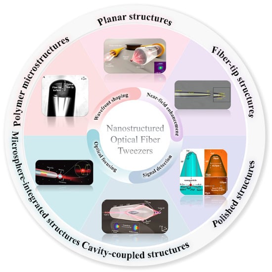
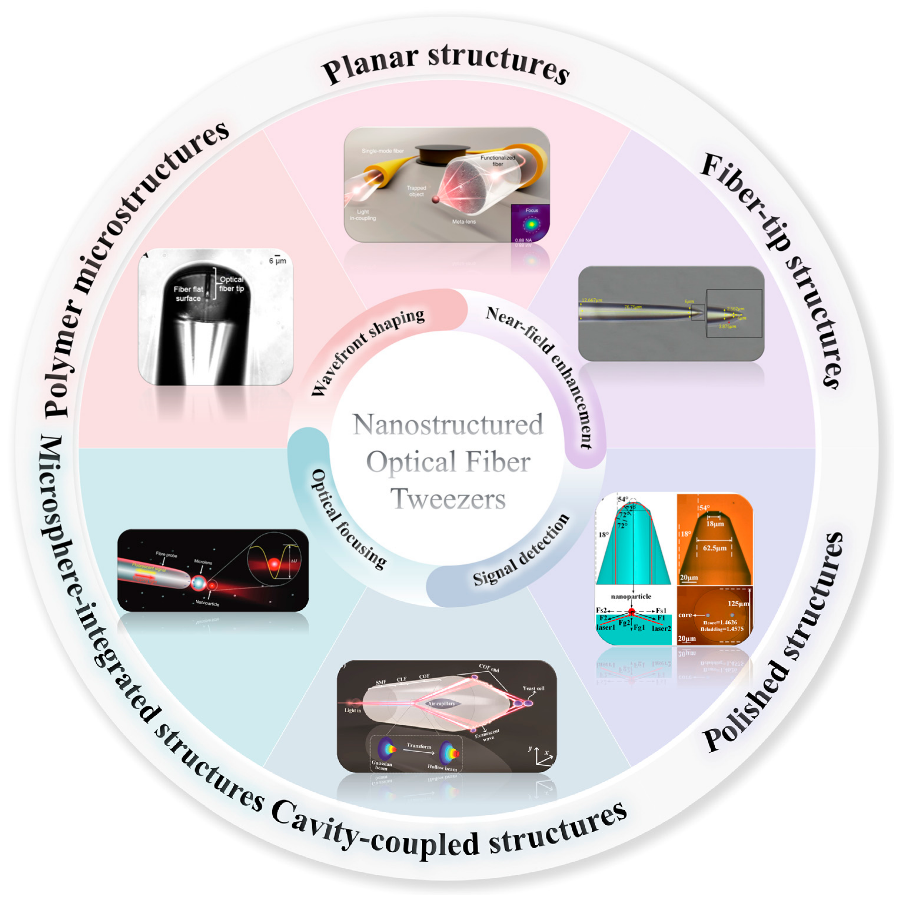
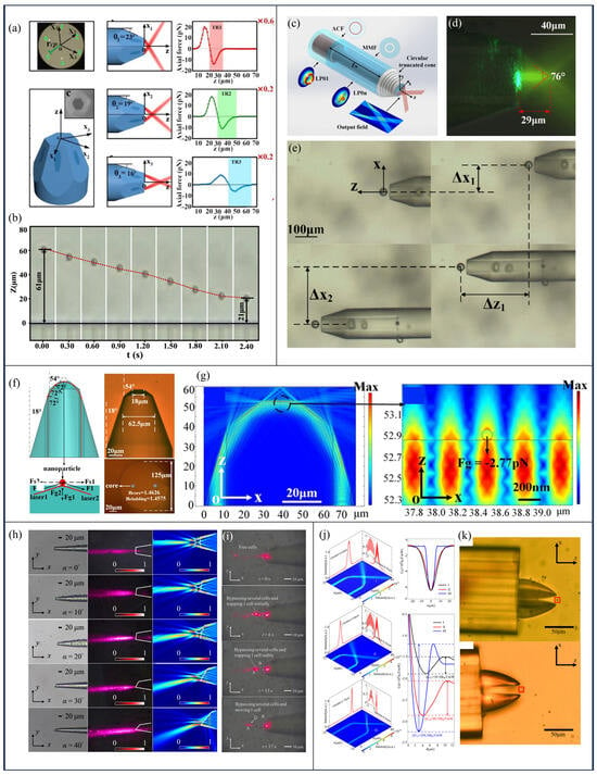
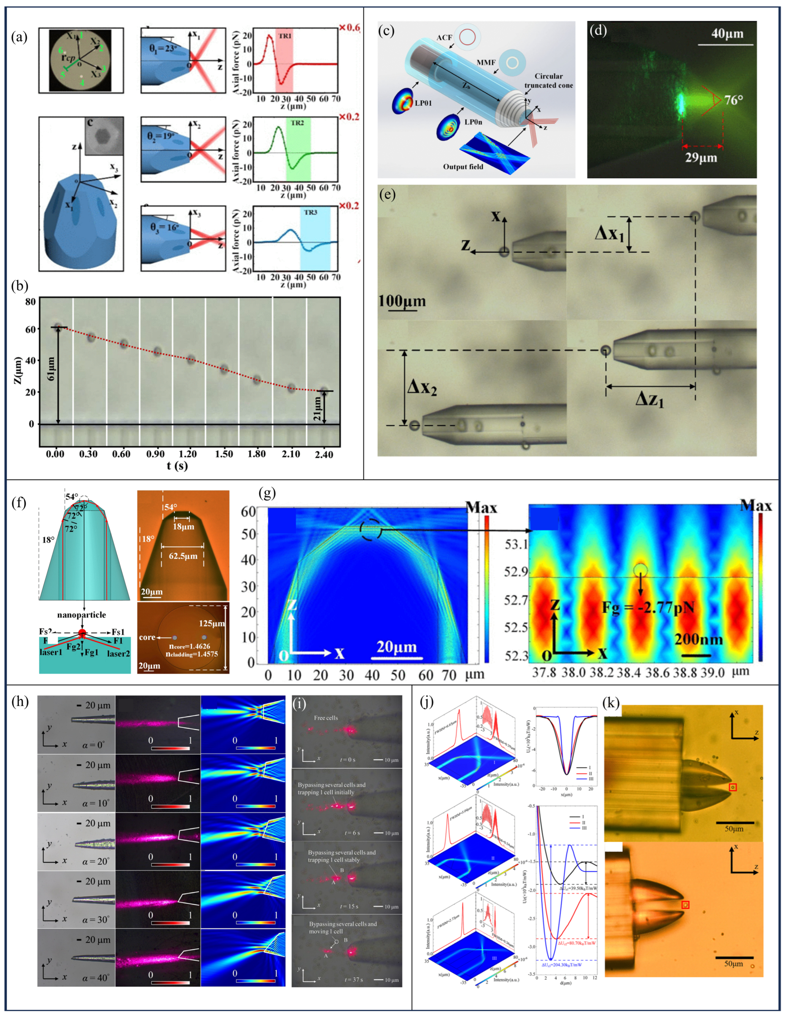
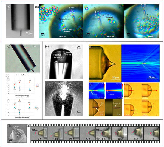
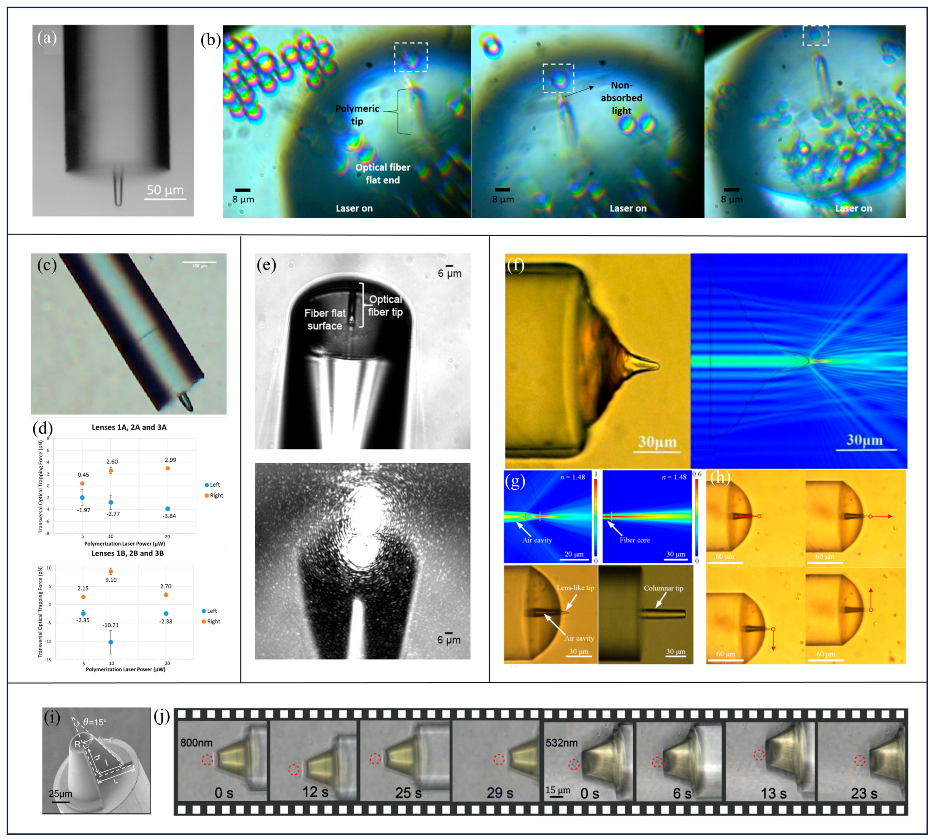

Disclaimer/Publisher’s Note: The statements, opinions and data contained in all publications are solely those of the individual author(s) and contributor(s) and not of MDPI and/or the editor(s). MDPI and/or the editor(s) disclaim responsibility for any injury to people or property resulting from any ideas, methods, instructions or products referred to in the content. |
© 2026 by the authors. Licensee MDPI, Basel, Switzerland. This article is an open access article distributed under the terms and conditions of the Creative Commons Attribution (CC BY) license.