


Abstract
Phycocyanin as water-soluble, accessory pigment is commonly extracted from Limnospira platensis (L. platensis). The pigment is often used as natural food colourant and is a potential antioxidant. Usually, the pigment is extracted with several freeze–thawing cycles with subsequent purification steps. The cell disintegration with physical plasma (here spark discharges) is already described for microalgae and was tested for the extraction of phycocyanin from L. platensis. Moreover, calcium chloride was examined as a non-toxic and inexpensive precipitator in the purification step. Samples were treated with water as solvent alone or precipitated with calcium chloride. Also, samples were divided for immediate analysis after extraction and 24 h incubation after the respective cell wall rupture method with subsequent analysis. The extracts were tested for C-phycocyanin (C-PC) concentration, content, selectivity, and radical scavenging activity. Spark discharges were administered with 4 Hz, 100 ns high-voltage pulses of 35 kV, resulting in a pulse energy of ca. 0.2 J/pulse. For freeze–thawing, a maximum yield of 2.75 mg/mL was obtained, for spark discharges, max. 0.24 mg/mL. However, despite a lower concentration, a remarkable purity was found for plasma-treated samples. In addition, radical scavenging activity was not influenced by the spark discharge exposure.
1. Introduction
Phycocyanin is a protein-based, water-soluble blue pigment that can be found in cyanobacteria, red algae and some cryptophytes. It plays a significant role as an accessory pigment during the photosynthesis process, but is also known for its high antioxidative capacity [1]. The pigment can be used widely for colouring, but due to its antioxidative potential, it can also be used in nutraceuticals, cosmetics, and pharmaceutical products because of its potentially beneficial biological properties, i.e., radical scavenging, immune modulating, and lipid peroxidase activities. Therefore, it is interesting for therapies regarding inflammation/wound healing, cancer, antimicrobial activity, neurodegeneration, diabetes, or hyperpigmentation [2,3].
For the extraction of phycocyanin, numerous standard extraction techniques exist, which are, nevertheless, associated with several drawbacks. Typically, the pigment is won by the application of several freeze–thaw cycles, as the cell wall of the microorganism is weakened by the ice crystal stress, and the freezing of water leads to an expansion of cell volume, whereas thawing induces a contraction. When repeating the cycles, the cell walls are gradually weakened and might be ruptured, eventually [4]. Freeze–thawing is simple in conduction, but it takes many hours for the process, and normally, more than one cycle is necessary. However, the technique is quite effective as the multi-layered cell wall of Limnospira is comparable to Gram-negative bacteria and thus, easily penetrable [5]. In contrast, if phycocyanin needs to be extracted from microalgae, freeze–thawing is rather ineffective [6]. In addition, proteomic analysis in the study of Sommer et al. demonstrated that freeze–thawing generates surprisingly high amounts of modifications on the protein complex of phycocyanin. Also, the repetition of the freezing and thawing cycle is time- and energy-intensive and is thus not widely accepted on an industrial scale [7].
The utilisation of ultrasound for the extraction of phycocyanin is also an appropriate method, which uses cavitation bubbles that are responsible for strong shear forces. However, ultrasound effects strongly depend on the probe, duration and whether the operation mode was pulsed [4]. Heat development is also a problem, especially with regard to the heat sensitive pigment and associated proteins. For instance, intermittent treatment decreases the extraction yield enormously, as was demonstrated by Sommer et al. [6]. However, cooling of the system might be possible, but would increase the costs of the whole procedure and thus decrease the economic viability of this extraction method. In addition, Sommer et al. could also prove, that, as was shown for freeze–thawing, ultrasound-assisted extraction causes high rates of modifications on the protein level of phycocyanin.
To meet the aforementioned drawbacks of heat development, modification of proteins and insufficient cell wall rupture, a novel technique for the extraction of bioactive compounds from microalgae has been developed. For cell wall disintegration, a physical plasma is ignited directly in the microalgal suspension. As plasma can be generated in many different ways, so-called spark discharges have proven to be the most effective type of plasma for cell wall rupture [8]. Physical plasmas are well-known for their unique characteristics of ionised gases, e.g., formation of reactive species (especially oxygen and nitrogen species), UV light emission, high electric fields, and shockwaves [9]. Spark discharges as the most effective plasma for the extraction of microalgae are especially known for the generation of powerful shockwaves, which have a massive impact on cell walls, without considerable effects of other plasma characteristics on the extractive itself [10,11]. Therefore, in this study we examined whether spark discharge-assisted extraction also has an effect on the extractability of phycocyanin from cyanobacteria and could serve as an alternative technology to the commonly used processes so far. Therefore, C-phycocyanin from Limnospira platensis was extracted via freeze–thawing, shaking incubation and spark discharge plasma as a proof-of-concept study. The extracted phycocyanin was detected via UV/ vis-measurements, fluorescence detection and the antioxidative capacity was determined with an ABTS (2,2′-azino-bis(3-ethylbenzothiazoline-6-sulfonic acid) test kit.
2. Material and Methods
2.1. Cultivation and Sample Preparation
Limnospira platensis (UTEX 2340) was cultivated with a commercial use culture media in novel foil photobioreactors (algabag™, algatec GmbH, Berlin, Germany). The culture media was prepared as shown in Table 1. The fertiliser solution, which was added to the culture media, is described in Table 2.
Table 1.
Chemical components of the used culture media used for the cultivation of L. platensis (UTEX 2340) at the algatec GmbH. * describes the addition of a fertiliser solution, which components are described in detail in Table 2.
Table 2.
Chemical components of the fertiliser solution, used in the CM II culture media.
Cultivation of L. platensis was carried out in two 25 L capacity algabags™ for 35 days before the harvest of biomass. Both algabags™ were wrapped in insulating foil for thermoregulation and supplied with constant light via a 3 m LED-string (24:00 h). For further analysis, 10 mL of each culture was taken twice a week for the whole cultivation period.
For the photometric measurements of the optical density, 2 mL culture was screened for their absorbance between 450 nm and 700 nm using a S50 UV/Vis Biochrom Libra spectrophotometer ( Biochrom Limited, 22 Cambridge Science Park, Cambridge, CB4 0FJ, England) to determine absorption peaks. Absorbance values at 680 nm (absorption maximum of the Qy band of the bulk chlorophyll a in the photosystem I) were then further used [12]. For each sample, distilled water was utilised as a blank. To determine the maximum specific growth rate [μmax] over a period from day 0 to day 34, a sigmoidal fit was applied to the data.
The harvested cells were centrifuged to achieve a thick cell slurry. The cell mass was split into 4 beakers (3 treatments (n = 3, biological replicates + 1 control (n = 1)) with 20 g cell slurry each. Afterwards, the slurry was resuspended in deionised water up to 200 mL each. The resuspended samples were exposed to spark discharges for 30 min (description in Section 2.4, electrical setup). Control samples underwent the same handling procedures without spark exposure. After treatment, the samples were split in 4x 50 mL testing tubes. Two tubes were transferred to the fridge (4 °C) for 24 h for incubation. The two other tubes (one containing 0.5 g CaCl2, the second one without any additives) were centrifuged at 4000× g for 5 min (Hettich centrifuge Rotofix 32, type 1206,) Andreas Hettich GmbH, Tutlingen, Germany. After spark discharge exposure, the samples were centrifuged and CaCl2 was added to the supernatant, with 0.5 g in each 50 mL tube. Calcium chloride was added after the treatment to keep the conductivity low during spark discharge treatment. Currently, the setup can only handle low conductivities, and possible (but unlikely) interferences between sparks and CaCl2 need to be avoided. After the salt-bound residual was concentrated at the bottom of the tube, the samples were centrifuged once more with the same centrifuge settings. From these two tubes, 10 mL was taken for photometric measurement (Thermo Fisher, Evolution 300 Thermo Fisher Scientific, Waltham, MA USA 02451). The remaining supernatant was then transferred to the freezer (−20 °C), prior to lyophilisation of the samples. The other two samples were stored at 4° C after spark discharge exposure for 24 h incubation.
2.2. Freeze–Thawing Extraction
After dry matter content measurement, the wet biomass was diluted with calcium chloride solution (1%) to a new dry matter content of 2.5%. The CaCl2 concentration was based on previous publications [13,14] and on preliminary experiments (not published). Half of the cell suspension was then distributed among three micro reaction tubes (biological triplicates) and frozen at −80 °C overnight, while the other half was used for the shaking incubation extraction. The following day, the suspensions were thawed at room temperature for 4 h and frozen at −80 °C overnight again. After an additional thawing, the samples were centrifuged at 13,000 rpm for 30 min at 4 °C and the supernatants were collected for further analysis.
2.3. Shaking Incubation Extraction
The other half of the cell suspension mentioned before (2.5% dry matter content adjusted with 1%-calcium chloride solution) was distributed among three micro reaction tubes (biological triplicates) and stored overnight at room temperature under constant shaking (250 rpm) and exclusion of light. After incubation, the samples were centrifuged at 13,000 rpm for 30 min at 4 °C and the supernatants were collected for further analysis.
2.4. Electrical Setup Spark Discharges
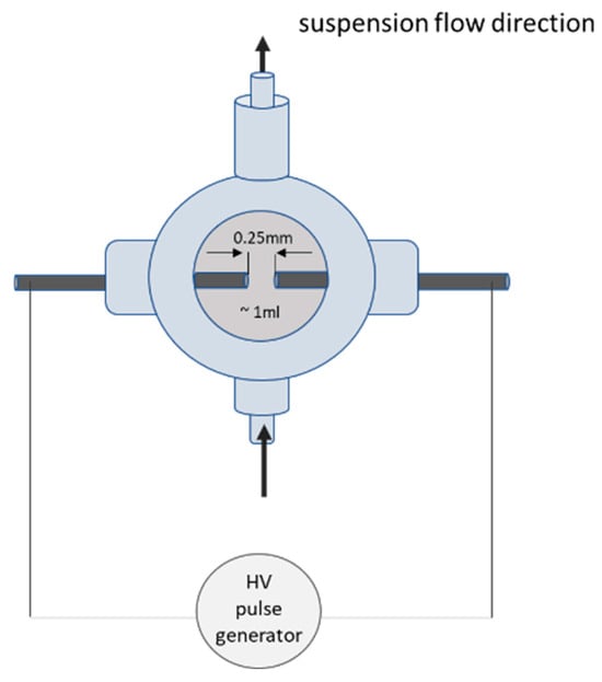
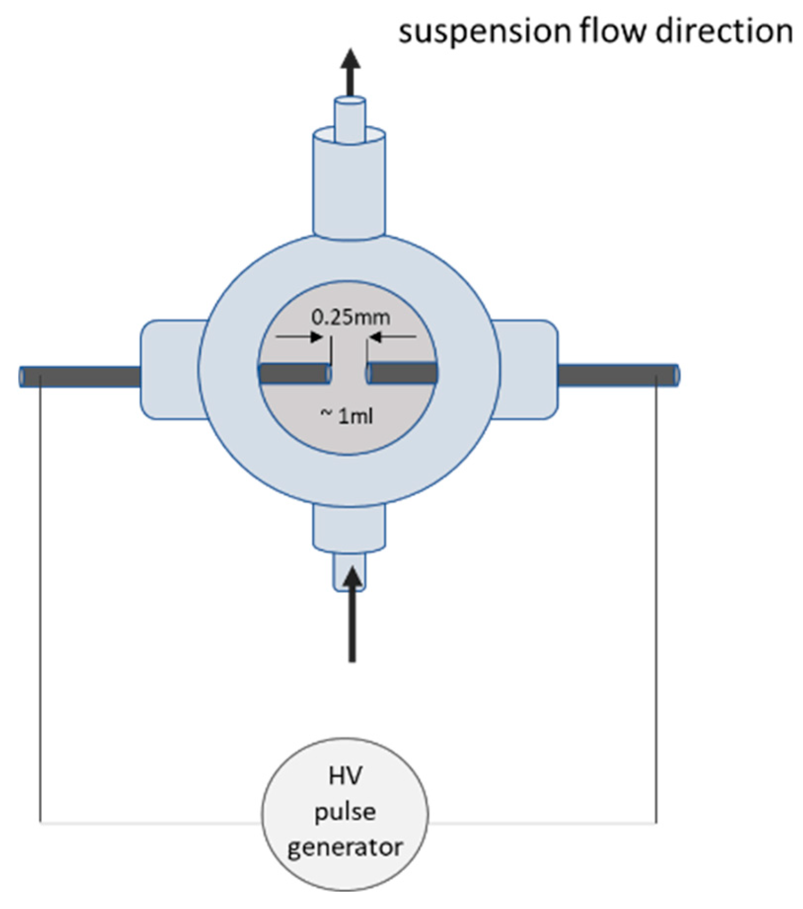
The electrical setup was already successfully established for the treatment of microalgae and slightly adapted regarding operation parameters for the disintegration of cyanobacteria in this work [6]. The ignition of spark discharged was instigated in two plasma reactors, between two tungsten rods each, with an electrode diameter of 1 mm. The electrode gap distance was adjusted to 0.25 mm and the reactor volume was about 1 mL. The electrode distance was adjusted every five minutes via semi-automatic step-motors to ensure constant plasma parameters. Sparks were applied for 30 min with a frequency of 4 Hz. Ignition was realised with the electrical breakdown of 100 ns high-voltage pulses of 35 kV, resulting in a pulse energy of ca. 0.2 J/pulse, which were applied from two custom-build Blumlein pulse forming networks for each plasma reactor. Moreover, the cyanobacteria suspension was recirculated through the plasma chambers with an inner volume of 1 mL and a flow rate set to 500 mL/min (peristaltic pump BT300-2J, Longer Precision Pump Co., Ltd., i, Baoding, China). With the set parameters of the system, the sample volume was passed through the entire system 150 times, with 14,400 pulses administered in 30 min.
A schematic overview of the system is given in Figure 1 for one of the two reactors. During the treatment time, the temperature never exceeded the temperature of 25 °C.
Figure 1.
Schematic sketch of one of two plasma reactors as described in Section 2.4. Each pulse generator is operated as a 100 ns Blumlein line pulse forming network.
2.5. C-PC Analysis
The analysis of the C-PC was carried out using a Genesys 50 UV/VIS spectrophotometer (Thermo-Fisher Scientific Inc., Waltham, MA, USA). The absorbance spectrum of each sample was measured from 250 to 800 nm. If necessary, the extracts were diluted with deionised water to ensure that the absorbance values fell within the linear measuring range. The phycocyanin concentration, purity and content were calculated using the following adapted formulas originally proposed by [13,15].
where CC-PC is the concentration of phycocyanin in the extract; A280, A620, A650 and A680, are the absorptions of the final extract at the respective wavelengths of 280 nm, 620 nm, 650 nm or 680 nm; purityC-PC is the purity of the phycocyanin in the extract, calculated as the ratio of absorptions at 620 nm and 280 nm, and the phycocyanin content is expressed as percentage (w/w), representing the mass of phycocyanin in grams, per 100 g of dry matter (mDM), with Vextract as the volume of the extract. Additionally, the selectivity of the extraction methods (selectivity C-PC) was expressed as the ratio of absorbance at 620 nm to the absorbance at 680 nm. All measurements were carried out in technical triplicates.
2.6. ABTS Test for Antioxidative Capacity
The assay for the measurement of the antioxidant capacity was conducted according to the method first proposed by Re et al. with minor adjustments [16]: In short, 7 mmol/L ABTS solution (2,2′-azino-bis(3-ethylbenzothiazoline-6-sulfonic acid)) was mixed 1:1 with 2.45 mmol/L potassium persulfate solution and incubated at 4 °C overnight. When the actual experiment was carried out, the activated ABTS solution was diluted with deionised water until the absorbance at 734 nm fell within the linear range of the photometer. Then, 360 µm of ABTS·solutions was mixed with 40 µL of control (deionised water, calcium chloride) or C-PC solutions and incubated for 6 min. Subsequently, the mixture was measured in the spectrophotometer, recording the absorbance between 400 and 800 nm. The absorbance at 734 nm was used for further calculations. The scavenging of ABTS·radicals by antioxidants leads to a decrease in absorbance and can therefore be measured photometrically. Radical scavenging activity (RSA) was calculated by applying Equation (5), with A734 as the absorption measured at 734 nm:
All measurements were conducted as technical triplicates.
3. Results
3.1. C-PC Concentration, Content, Purity and pH
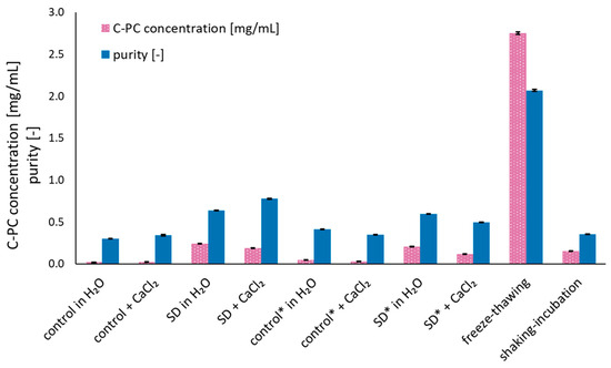
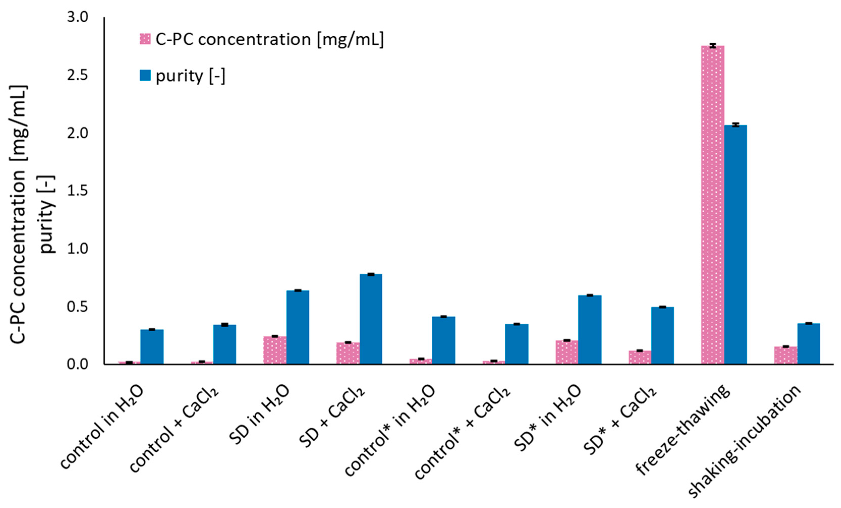
Phycocyanin concentration and purity were determined for all extraction methods and are displayed in Figure 2 (pink bars = concentration, blue bars = purity). The C-PC concentration of the final extract was by far the highest (2.75 ± 0.02 mg/mL) for the freeze–thawing method. Any other approaches showed much lower C-PC concentrations: 0.24 ± 0.004 mg/mL, 0.21 ± 0.002, 0.18 ± 0.002, and 0.12 ± 0.002 for extracts obtained by spark discharge extraction in deionized water (no incubation, 24 h incubation, as described in Section 2.1.) and in CaCl2 solution (no incubation and 24 h incubation, as described in Section 2.1.), respectively. The control samples all had C-PC concentrations of 0.05 mg/mL or less. With shaking-incubation extraction, a final concentration of 0.16 ± 0.001 mg/mL was achieved.
Figure 2.
C-PC concentration and purity of the extracts obtained by different methods. SD = spark discharge method; FT = Freeze–thawing method; SI = shaking incubation method; * = 24 h of incubation after SD. Shown are the mean values with standard deviation as error bars (n = 3).
The purity was also highest for the extract obtained by freeze–thawing (2.07 ± 0.011). When spark discharge disruption was applied, the purity was 0.64 ± 0.002 and 0.60 ± 0.002 with water as solvent and 0.78 ± 0.003 and 0.50 ± 0.002 precipitated with CaCl2. The purity for extracts in the spark discharge controls was between 0.30 and 0.41 and for shaking incubation a purity of 0.35 ± 0.003 was found.
When the amount of C-PC is set into relation to the inserted dry biomass, the disruption efficacy can be quantified as C-PC content (blue bars, Figure 2). The highest result was achieved by freeze–thawing with a C-PC content of 10.73 ± 0.06%. When spark discharge disruption was used, the C-PC content was found to be 1.42 ± 0.02% and 1.22 ± 0.01% for water as solvent and 1.12 ± 0.01% and 0.69 ± 0.01% precipitated with CaCl2. For all four spark discharge controls, the C-PC content was below 0.3%. Extracts obtained by shaking incubation had a C-PC content of 0.61 ± 0.01%.
The pH values in all approaches were found to be between 5.4 and 6.7. In the spark discharge approaches, pH ranged from 6.1 ± 0.09 (in CaCl2, no incubation) to 6.66 ± 0.08 (in H2O, 24 h incubation). The average pH of all spark discharge samples was 6.32 ± 0.25 and for the control 6.33 ± 0.22. The pH values of the extracts obtained by shaking-incubation and freeze–thawing were 5.51 ± 0.05 and 5.43 ± 0.04, respectively. Hence, a considerable effect on the pH value could not be detected for any method applied.
3.2. Selectivity
Selectivity describes the ratio of C-phycocyanin to chlorophyll, i.e., the higher the selectivity, the less chlorophyll impurities were detected. The results are depicted in Figure 3.
Figure 3.
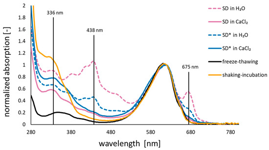
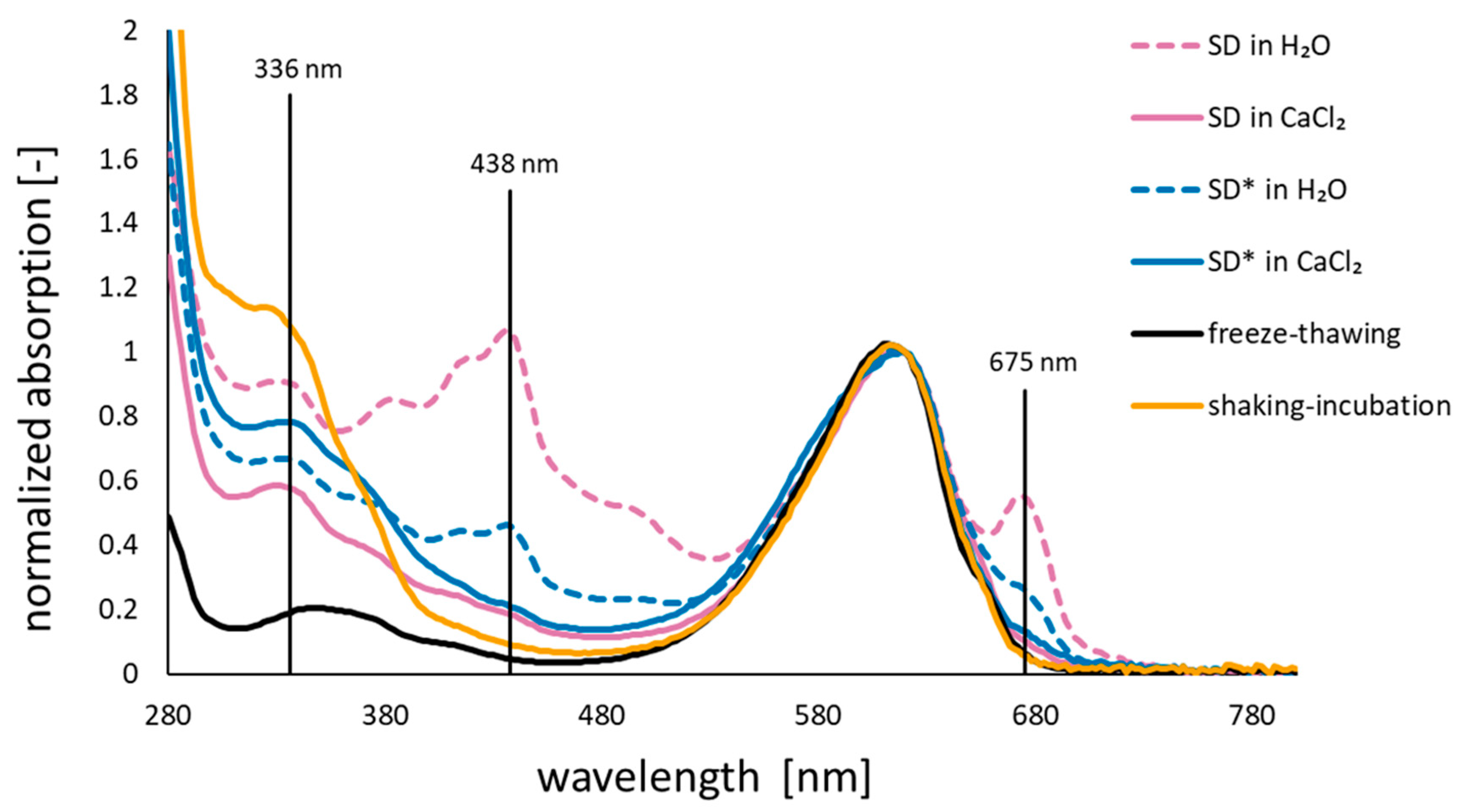
Normalised mean (n = 3) absorption spectra of the C-PC extracts (without control samples). black: freeze–thawed; orange: shaking-incubated; red violet: spark discharge (SD) without subsequent incubation; dark blue: SD with subsequent incubation for 24 h (additionally marked with asterisk in legend); broken lines: with water as solvent; continuous lines: with calcium chloride treatment.
In the normalised absorption spectrum, where the absorption at 620 nm is set to 1, freeze–thawed extracts showed the lowest absorption between 300 and 500 nm. The extracts obtained by shaking-incubation also had low absorption values between 400 and 500 nm, but showed a shoulder at around 336 nm. Low absorption between 400 and 500 nm as well as a smaller shoulder at 340 nm were found for spark discharge extraction with added CaCl2. Compared to all the other approaches, spark discharge extraction with deionised H2O as the solvent and untreated controls showed clear peaks at 438 and 336 nm.
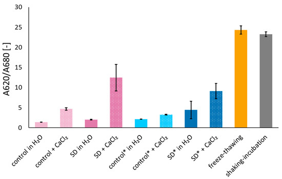
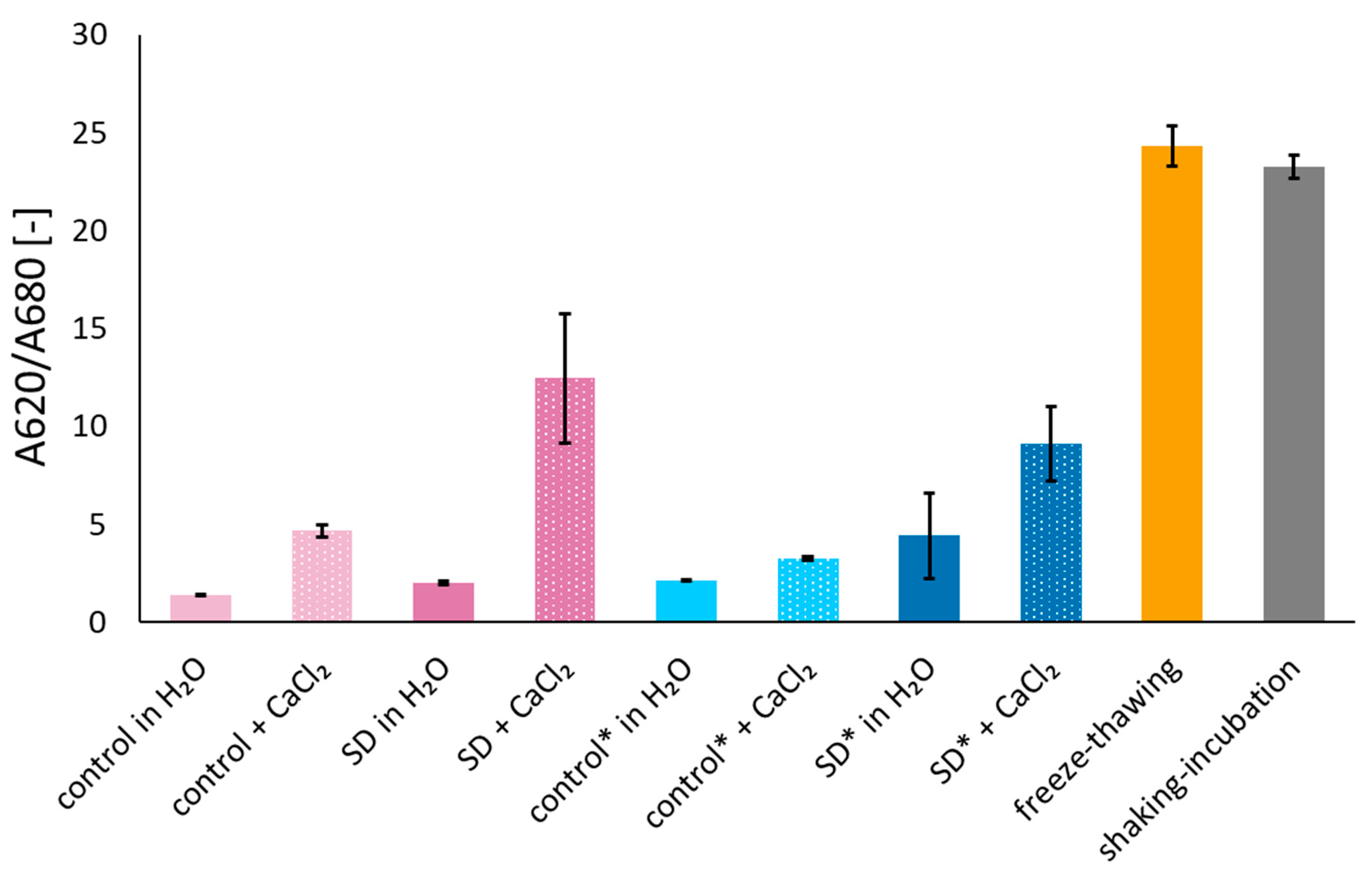
The ratios between the absorption at 620 nm and 680 nm was used to display the selectivity of the method (see Figure 4). The selectivity was highest for freeze–thawed extracts (24.3) and shaking-incubation extracts (23.3). The extracts obtained by the spark discharge method with CaCl2 with no and with 24 h of incubation had values of 12.5 and 9.1, respectively. All other approaches had values of 4.4 or lower for the selectivity.
Figure 4.
Selectivity values of the extracts obtained by the applied methods, described in Section 2. Values are displayed as the relation between absorptions at 620 and 680 nm. SD = spark discharge extraction; * = samples were incubated for 24 h, subsequently to spark discharge extraction. Shown are the mean values with standard deviation as error bars (n = 3).
3.3. Radical Scavenging Activity (Antioxidant Capacity, RSA)
The radical scavenging activity (RSA) was detected to observe if spark discharge treatment has a negative effect on the antioxidative capacity of phycocyanin. Some physical plasmas, especially those which are ignited in air or on the surface of a liquid, are known to produce strong radicals, which may affect the RSA of phycocyanin [17]. However, for spark discharges, the energy is mainly dissipated in the generation of shockwaves and not radical formation [11]. Nevertheless, even a low concentration might have had an effect on the RSA.
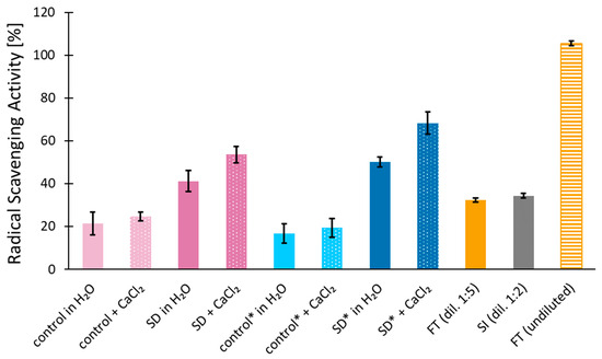
The results for the detected RSA of all extraction methods are found in Figure 5. For the non-diluted freeze–thawing extract, the RSA was around 105.5 ± 1.0%. When these samples were diluted 1:5, the RSA was 32.4 ± 1.0%. The extracts obtained by shaking incubation had a comparable RSA of 34.4 ± 1.1%, but were diluted 1:2 prior to the ABTS assay. The RSA of the extracts obtained by spark discharge technique was 41.2 ± 5.0% and 53.6 ± 3.7% in water and in the presence of CaCl2, respectively (control: 21.4 ± 5.4% and 24.8 ± 2.1%). When the cell suspension treated with spark discharge technique was incubated for 24 h prior to further extraction steps, the RSA was 50.3 ± 2.3% for deionised water and 68.2 ± 5.2% in the presence of CaCl2 (control: 16.7 ± 4.6% and 19.4 ± 4.3%).
Figure 5.
Radical scavenging activity of C-PC extracts obtained by different extraction methods. SD = spark discharge method; FT = freeze–thawing method; SI = shaking incubation method; * = 24 h of incubation after SD. Shown are the mean values with standard deviation as error bars (n = 3).
4. Discussion
The extractability of C-phycocyanin from vital L. platensis was studied by applying a physical plasma (in this study, spark discharges) and the results were compared with a reference extraction method. The extract was examined regarding, yield, purity, selectivity, and antioxidant capacity. It could be demonstrated that spark discharges are not only suitable for the extraction of microalgae, as was shown in our previous studies, but also for other microorganisms and are an interesting alternative method for the extraction of phycocyanin [6,18]. Moreover, in this study we could prove that the plasma has no negative effect on the extractive, e.g., the antioxidative potential of strong radical scavengers, such as phycocyanin. This indicates that the health-promoting, anti-inflammatory properties and antidiabetic effects of phycocyanin can be preserved by this extraction process [19,20].
4.1. C-PC Concentration
The C-PC concentration (Figure 2) in extracts obtained by the spark discharge methods was found to be between 0.12 and 0.24 mg mL−1., whereat the untreated control had concentrations ten-fold lower. Therefore, an effect of the spark discharge method on the extraction effectivity is apparent. In shaking-incubation extracts, a C-PC concentration of 0.16 mg mL−1 was extracted. Similar results have been obtained by Noore et al., where phycobiliproteins from Porphyridium purpureum were extracted with spark discharges and a plasma jet. For the spark discharges, a two-fold increase after 9 min treatment could be detected, including phycocyanin [21]. However, the authors used dried and re-hydrated samples, which was circumvented in this work, as vital cells were treated. Drying and rehydration does increase the economic costs and would not be a profound alternative. Nonetheless, the plasma system can still be approved to further increase the extraction yield, e.g., by adjusting the treatment time, the spark frequency, or the biomass density.
Freeze–thawing is often applied to for the extraction of phycocyanin from cyanobacteria, as it is simple in execution [22]. Here, the concentration of C-PC was 2.75 mg mL−1 and thus ten-fold higher than with spark discharges. This concentration is lower than reported in previous works yielding C-PC concentrations of around 5 mg mL−1 using the same extraction method [1,23]. A reason it could be different the cultivation conditions of the cyanobacteria, as it is known to influence the cell’s C-PC content [24,25]. Despite the simple conduction, freeze–thawing influences the molecular level of phycocyanin. In a previous work, the proteome of phycocyanin from Cyanidium caldarium was studied after spark discharge treatment, sonication and freeze–thawing. It could be clearly shown that freeze–thawing had a massive impact on molecular level. The number of modifications was highest for this technique, followed by sonication. Only minor modifications were detected for spark discharges [6].
Although the C-PC yield is lower for the freeze–thawing samples, spark discharges are able to disintegrate the cell walls from cyanobacteria and can be considered an interesting alternative. For instance, the processing time in comparison to freeze-–thawing is much lower (30 min vs. several hours). The implementation in industrial processes is well conductable because the system is invented as a continuous flow setup, where the biomass can be harvested and provided to the plasma system without interruption in the processing chain as would be for freeze–thawing, which is a batch mode system. Indeed, the spark parameter can still be optimised to gain higher yields and is part of our research. That is why the spark discharges can be considered an effective method and advantageous for pigment extraction from cyanobacteria. Also, the amount of energy, which is necessary for multiple freezing and thawing of cells, is massive and should be taken into consideration when comparing extraction techniques. We calculated the energy efficiency for the spark discharges with 60 kJ/g C-PC under the given plasma parameters for both plasma reactors together. For freeze–thawing, an energy consumption is not always easy to determine as it also strongly depends on the freezing devices. Therefore, we calculated CO2 equivalents (similar for life cycle assessment calculations from DIN ISO 14040/44 [26]) for both methods, assuming 1 m3 solution would have been treated. The CO2 equivalent amount for freeze–thawing would be 80.724 g CO2 for two freezing cycles and 477 g CO2 equivalent for spark discharge treatment. The reduction in CO2 equivalents by a factor of 170 with the spark discharge treatment underlines the advantages of the plasma-driven technology as environmentally friendly method.
With regard to scaling, the system was designed in continuous flow mode, which allows a direct integration into the downstreaming cascade. A batch mode would mean an interruption of the process chain. In the energy supply and reactor arrangement, the system can be adapted from the scaled pulsed electric fields technology. Those systems are already in industrial scale and the energy supply can be compared to that from the plasma system. It is conceivable to stack the plasma reactors in parallel or in a row, depending on the surrounding environment and space available. Regarding the electrode design, the most challenging part is the automation of the electrode gap distance control. However, the electrodes can be adjusted by step motors, which allow for semi-automation of gap distance control. Now, inductive sensors are tested and seem to be a very promising approach for scaling.
4.2. C-PC Content
The C-PC content can be obtained by setting the extracts’ C-PC concentration in relation to the dry biomass used for the extraction (Figure 2, pink bars). This measurement mostly corresponds to the determined concentration. Consequently, the freeze–thawing extract had the highest C-PC content of 10% C-PC per g of dry biomass, compared to 0.69 and 1.42% from spark discharge. Interestingly, the C-PC contents of the spark discharge extraction method were higher, when deionised water was used without CaCl2, compared to samples with added CaCl2. Also, the abundance of unwanted chlorophyll a in the extracts was also higher. This has also been observed by another publication, where water yielded higher C-PC contents, but also higher chlorophyll contents compared to CaCl2 [27]. Interestingly, the C-PC contents of extracts incubated for 24 h after spark discharge application are constantly lower compared to extracts that were not incubated. This might be the result of microbial [28,29,30] or chemical degradation processes occurring during the incubation [31,32]. Plasmas in general are known for the generation of reactive species (nitrogen and oxygen species = RONS), which may be responsible for the detected effects. However, the applied plasma setup was previously described with very low RONS yields, with little to no effect on molecular level, but it cannot be excluded that these small amounts may decrease the observed loss in content [18]. The C-PC content after shaking-incubation provided 0.69% of C-PC per g of dry biomass. This is less than found in the literature. For instance, 11.6%, 9.93%, and 4.60% were achieved by others [14,33,34]. This might be the consequence of variations within the experimental design parameters of the mentioned works compared to this work.
4.3. Extract Purity
The highest purity was observed in freeze–thawing extracts with, as shown in Figure 2 (blue bars). The purity for extracts obtained by the spark discharge method ranged between 0.50 and 0.78, with CaCl2 and no incubation, yielding the purest C-PC extracts. Shaking incubation led to a purity of 0.35 and is therefore comparable to the untreated controls (0.30–0.41). When no incubation was applied to the spark discharge samples, the purity was higher for extraction in CaCl2, highlighting the advantage by the use of CaCl2. Higher purity values by the use of CaCl2 compared to water has also been observed by Lin et al. [35]. The increased purity can be the result of the precipitation of chlorophyll a-associated and other proteins diminishing the purity [36]. Contrarily, when the cell suspension was incubated for 24 h subsequent to spark discharge exposure, deionized water as solvent obtained higher purities. For industrial purposes, the purity of C-PC extracts is used for assigning the extracts to different applications. Purity values of ≥0.7 are considered food grade, purities up to 3.9 are categorised as reactive grade. Purities of more than 4 are analytical grade [37]. Therefore, only the freeze–thawing and the spark discharge extraction with CaCl2 and without incubation could obtain C-PC purities in food-grade quality in this work. Also, an incubation, which is necessary, e.g., after pulsed electric fields treatment to increase osmotic imbalance between inner cell and surrounding medium, is unnecessary for plasma treatment [38,39]. The lack of incubation time is beneficial for the plasma treatment, as it decreases costs during the downstreaming process in general and all together raises the economic viability of marine organisms as a natural resource.
4.4. Selectivity
The selectivity expressed as the absorbance ratio between 620 and 680 nm was the highest for the freeze–thawing extraction and shaking-incubation (24.32 and 23.25, respectively; see Figure 4) and corresponds to previous work, using the same method [1]. The extracts obtained by spark discharge had selectivities of 2.02 and 4.41 in water without and with incubation for 24 h, respectively, compared to 12.46 and 9.11 samples precipitated with CaCl2 without and with subsequent incubation, respectively (Figure 4). Thus, for plasma-treated samples precipitated with CaCl2, the selectivity was consistently higher compared to the water samples. Higher selectivity with CaCl2 was also observed in the control samples. This underlines the advantageous effect of CaCl2 for C-PC extraction and exclusion of chlorophyll a from the final extract in this work. Similar observations have already been reported in the literature, where calcium chloride solutions helped with obtaining extracts of deep blue colour, compared to more greenish extracts when water or phosphate buffers were used [27,36]. It was explained either by the ionic strength of CaCl2 solutions similar to those occurring in cells or by CaCl2 altering cell membrane permeability, while leaving the cell structure intact [14,27]. Alternatively, it was found that a CaCl2 precipitation step subsequent to extraction would enhance C-PC purity by salting out chlorophyll a-associated protein complexes [36]. In summary, it can be confirmed that CaCl2 is a non-toxic precipitant that can improve the purity of phycocyanin extracts and can be recommended for improving extraction results.
When water was used as solvent alone, incubation of the cell suspension for 24 h subsequent to spark discharge treatment increased the selectivity. When CaCl2 was added after treatment, additional incubation decreased the selectivity. These observations also applied to the control samples and might be connected to the hypothesis that microbial or chemical degradation pathways are responsible for the detected effect. However, a satisfying explanation cannot be given here and further analyses are necessary.
4.5. Radical Scavenging Activity (RSA)
The literature has shown that a vast variety of techniques for the detection of radical scavenging activity (RSA) exists and reliable, standardised methods are necessary [40]. Still, comparison of the results from antioxidant capacity measurements is difficult due to the lack of a standardised method. Two antioxidant test systems are commonly used in the context of C-PC, namely the ABTS and the DPPH methods [40]. Many publications report the application of the ABTS method, introduced by Re et al. [16]. An advantage of this assay is its suitability for a wide range of compounds, including pure substances, mixtures, and biological fluids. Also, it allows for the quantification of antioxidant capacity in diverse media and provides insights into the antioxidant activity of compounds in both hydrophilic and lipophilic media [40]. Still, adjustments to this method are not unusual concerning, for instance, the sample preparation, the concentration of the test solutions, the solvent, etc. Recent publications apparently prefer to display the results as radical scavenging activity (RSA) [%] [41,42,43]. As a consequence, the results of this work are not fully comparable to the literature, but they can be compared to each other. It might be helpful to use various methods for the examination of the antioxidant capacity in order to exploit different radical scavenging reaction mechanisms [43,44]. It has been reported that the RSA values of C-PC extracts were significantly lower in the ABTS assay compared to the DPPH assay [42]. Other authors prefer the ABTS assay over the DPPH assay because ABTS radicals interact with a generally broader range of potential antioxidant agents, e.g., flavanones and dihydrochalcones, and are therefore less specific when complex matrices are tested. Moreover, the ABTS assay is presumably more precise in examining the antioxidant capacity of hydrophilic and high-pigmented matrices compared to non-water-soluble DPPH assays [43,45,46,47]. Thus, the ABTS assay was administered in this study for the determination of RSA.
One important reason for detecting RSA in this work is that physical plasmas generate reactive species, especially reactive oxygen and nitrogen species (RONS), next to UV light emission, shockwaves, electric fields and temperature [48,49]. The amount and type strongly depend on the medium (such as air, water, culture medium, etc.) it is ignited and on the type of plasma source (e.g., plasma jet, corona discharge, dielectric barrier discharge, spark discharge) [50,51,52]. Although the main energy of the plasma channel from a spark discharge is dissipated in shockwaves, a small amount of energy is dissipated in the formation of RONS [53]. In water, the main species are reactive oxygen species (ROS) that may have a significant influence on the RSA of C-PC. A decrease in RSA could indicate a negative impact of generated RONS, i.e., it is consumed by the plasma-generated ROS. As shown in Figure 5, the RSAs of the extracts obtained via the spark discharge method were 41.2% (water) and 53.6% (CaCl2). When subsequently incubated for 24 h, RSAs were 50.3% (water) and 68.2% (CaCl2). These results imply that the plasma-derived ROS have little to no effect on the RSA of phycocyanin, otherwise a steep decrease in RSA would have been detected. This is in accordance with previous findings, where it could be demonstrated that only small amount of ROS is generated in the applied setup, barely modifying proteins by ROS [8,18]. Hence, the extraction success is not influenced by ROS, and the main effect of cell wall rupture is of mechanical nature, i.e., shock waves with several hundreds of Megapascal pressure [11]. Remarkably, RSA is higher in samples in spark discharge extracts incubated for 24 h. This might be the consequence of increased release of other antioxidant cell components over the incubation time. Another possible explanation is given by the demonstration of increased antioxidant capacity of C-PC via enzymatically simulated digestion [54]. Further research is needed to answer this question and to help to understand the effects of plasma on the extractive. The extracts obtained by freeze–thawing (0.55 to 2.75 mg mL−1 C-PC) exhibited an RSA of 32 to 100% and therefore complete radical depletion. Other works have demonstrated comparably high RSA values for C-PC in the range of 0.2–5 mg mL−1 [35,41,42,43,55]. Lower RSA (13–60%) has been found for C-PC concentrations of less than 0.1 mg mL−1 [47].
Notably, the RSA of C-PC in CaCl2 was consistently higher in all samples compared to C-PC in water, even though the C-PC concentration was lower. This demonstrates that other factors play a crucial role in the antioxidant capacity of C-PC and underlines the advantage of using CaCl2 as non-toxic and inexpensive precipitator. In this work, calcium chloride solution was also included in the ABTS assay as a control sample and did not show any antioxidative property. Thus, an antioxidative agents’ direct effect is unlikely. Indirect increase in antioxidant capacity, however, has indeed been reported: When CaCl2 was added to the solvent prior to C-PC extraction, higher phenolic contents in the final extract were found [56]. In germinated brown rice, the addition of CaCl2 led to increased formation of antioxidative compounds by enhanced activity of Ca-dependent enzymes [57]. Nevertheless, chloride salts are known to reduce the solubility of phenolic compounds [58]. More research is needed to examine the effect of CaCl2 on the antioxidant capacity of C-PC extracts and cannot be fully excluded from the results in this work.
A dependency of C-PC concentration and antioxidant capacity has already been reported. In those publications, higher C-PC concentrations lead to elevated RSA values. In most of them, this relationship follows an asymptotic pattern with the asymptote tending to reach an RSA between 80 and 100% [35,41,42,43]. A study that plotted several other publications in one single graph confirmed this saturating trend [59]. Another study found a linear relationship between C-PC concentration and RSA, notably using substantially lower C-PC concentrations (<0.1 mg/mL) [47]. In our work, we discovered a similar pattern as the highly concentrated freeze–thawing extract exhibited maximum RSA, while lower concentrated plasma-discharge extracts showed lower RSA.
4.6. Other Cell Compounds Contributing to RSA
The antioxidative effect of C-PC itself is assumed to be contributed by both the abundance of certain amino acids within the apoprotein and the chromophore phycocyanobilin [60]. In particular, the amino acid cysteine has been found to show consistent antioxidant effects independent of the test system applied [61]. This has been explained by the free sulfhydryl group (-SH) in the residue, which can act as both a hydrogen donor as well as an electron donor [62,63]. The amino acids tryptophan, tyrosine, lysine, histidine, arginine, valine, phenylalanine and methionine have also been reported to show radical scavenging activities [61,64,65]. A comparison of amino acid composition reveals that especially cysteine, tyrosine, and arginine are found more frequently in phycocyanin compared to other proteins or protein extracts from algae, while histidine, valine, lysine and tryptophane are less abundant in C-PC [66,67]. This also means that other proteins can contribute considerably to the overall antioxidant capacity of the extracts [61,68]. Still, phycocyanin is considered the main component for antioxidant capacity in L. platensis extracts, which is further supported by our experiments, where higher RSA was associated with a higher purity [68]. These things considered, it might be challenging to distinguish between the antioxidant effect of C-PC and allophycocyanin (APC), since C-PC extracts usually also contain APC. In a publication aiming to separate both phycobiliproteins and assess their antioxidant capacity independent from each other, it was found that C-PC had 25% higher scavenging activity [59].
Besides the phycobiliproteins, other potentially antioxidant components have also been described to occur in L. platensis biomass. Specifically, the other main pigment in this organism, chlorophyll a, is of relevance in this context [69]. However, its total antioxidant capacity is supposed to be rather low compared to other antioxidants like Trolox® [70] or carotenoids [44]. One experiment demonstrated that the removal of chlorophylls from olive leaves and green tea extracts did not alter their antioxidant activity [71]. It was proposed that the chlorophyll radical scavenging mechanism is not compatible to the ABTS test system. In addition, the total abundance of chlorophyll a in L. platensis was reported to be relatively low (<1% of dry matter) when compared to C-PC (≤20%) [72,73]. This is further supported by the normalised absorption spectrums in Figure 5, which shows a positive correlation between the exclusion of chlorophyll and RSA. Therefore, the contribution of chlorophyll a to the RSA obtained in this work in all of the samples is negligible.
L. platensis also contains carotenoids, mainly β-carotene, zeaxanthin, lutein, and cryptoxanthin, which are well known lipophilic antioxidants, but their abundance in the biomass is relatively low compared to C-PC [69,74]. Moreover, it requires the use of non-polar solvents to extract significant amounts of carotenoids from the biomass [69,73]. Therefore, their contribution to the total antioxidant capacity of C-PC extracts is negligible as all experiments were conducted in water. Flavonoids are hardly found in hydrophilic C-PC extracts, but rather in lipophilic carotenoid extracts, where their contribution to the total antioxidant capacity is considered rather low. Contrarily, phenols can indeed be found in C-PC extracts and partly contribute to their antioxidant capacity, although C-PC is still the major antioxidant reagent in the C-PC extracts [73]. It is also known that L. platensis contains the well-established antioxidants tocopherol and superoxide dismutase; however, both are present in such small amounts that their contribution to the ABTS assay can be considered negligible [72,75].
5. Conclusions
The scope of this work was to determine whether C-phycocyanin is extractable with spark discharges and whether it has an influence on the radical scavenging activity of the pigment. In addition, calcium chloride was tested as a non-toxic and cheap precipitator for the purification of phycocyanin. It is clearly shown that phycocyanin can be well extracted with that technique and is thus an alternative method for cell wall disintegration of cyanobacteria. As this is a proof-of-principle study, the extraction can still be increased by adjusting the treatment parameters accordingly. For instance, treatment time, spark frequency, or biomass density are parameters that can be adjusted. A major advantage of the technique is the direct treatment in the cell suspension with vital cells. Hence, the cells are in direct contact with the plasma and energy is only necessary for the ignition of the plasma. Freezing and thawing consumes a high amount of energy, which can be avoided by the application of spark discharges. Scaling of the system and in-depth analysis on a molecular level is a topic of current research.
Concurrently, calcium chloride could be an interesting substance to precipitate pigments as a purification step. Further research on the mechanism, influence on the RSA and applicability on other compound compositions (other than C-PC-chlorophyll) is needed and highly recommended. Also, the impact on stability, shelf-life and compatibility, for example, in food matrices, need to be studied for a more profound statement.
Author Contributions
Conceptualisation, J.K., M.B. and K.Z.; methodology, J.K. and M.B.; investigation, J.K., A.B., M.B. and K.Z.; resources, A.B.; data curation, J.K., A.B., M.B. and K.Z.; Writing—original draft, J.K. and K.Z.; Writing—review and editing, J.K., M.B., A.N. and K.Z.; Supervision, A.N.; Project administration, K.Z. All authors have read and agreed to the published version of the manuscript.
Funding
This research was funded by Bundesministerium für Forschung, Technologie und Raumfahrt grant number 03WIR2222.
Data Availability Statement
The original contributions presented in this study are included in the article. Further inquiries can be directed to the corresponding author(s).
Conflicts of Interest
Albert Beyer was employed at algatec GmbH during the experimental part of the manuscript. No money was paid for the experiments or for writing. The remaining authors declare that the research was conducted in the absence of any commercial or financial relationships that could be construed as a potential conflict of interest.
References
- Kuhnholz, J.; Glockow, T.; Siebecke, V.; Le, A.T.; Tran, L.D.; Noke, A. Comparison of different methods for extraction of phycocyanin from the cyanobacterium Arthrospira maxima (Spirulina). J. Appl. Phycol. 2024, 36, 1725–1735. [Google Scholar] [CrossRef] [Scilit]
- de Morais, M.G.; da Fontoura Prates, D.; Moreira, J.B.; Duarte, J.H.; Costa, J.A.V. Phycocyanin from microalgae: Properties, extraction and purification, with some recent applications. Ind. Biotechnol. 2018, 14, 30–37. [Google Scholar] [CrossRef] [Scilit]
- Yuan, B.; Li, Z.; Shan, H.; Dashnyam, B.; Xu, X.; McClements, D.J.; Zhang, B.; Tan, M.; Wang, Z.; Cao, C. A review of recent strategies to improve the physical stability of phycocyanin. Curr. Res. Food Sci. 2022, 5, 2329–2337. [Google Scholar] [CrossRef] [Scilit] [PubMed]
- Fabre, J.-F.; Niangoran, N.G.U.F.; Gaignard, C.; Buso, D.; Mouloungui, Z.; Valentin, R. Extraction, purification and stability of C-phycocyanin from Arthrospira platensis. Eur. Food Res. Technol. 2022, 248, 1583–1599. [Google Scholar] [CrossRef] [Scilit]
- Sena, L.; Rojas, D.; Montiel, E.; González, H.; Moret, J.; Naranjo, L. A strategy to obtain axenic cultures of Arthrospira spp. cyanobacteria. World J. Microbiol. Biotechnol. 2011, 27, 1045–1053. [Google Scholar] [CrossRef] [Scilit] [PubMed][Green Version]
- Sommer, M.-C.; Balazinski, M.; Rataj, R.; Wenske, S.; Kolb, J.F.; Zocher, K. Assessment of Phycocyanin Extraction from Cyanidium caldarium by Spark Discharges, Compared to Freeze-Thaw Cycles, Sonication, and Pulsed Electric Fields. Microorganisms 2021, 9, 1452. [Google Scholar] [CrossRef] [Scilit] [PubMed]
- Najafian, N.; Arabi, A.; Ejhieh, N.; Reza, A. Intensified phycobiliprotein extraction from Spirulina platensis by freezing and ultrasound methods. Iran. J. Chem. Chem. Eng. Res. Artic. 2023, 42, 601–617. [Google Scholar]
- Zocher, K.; Banaschik, R.; Schulze, C.; Schulz, T.; Kredl, J.; Miron, C.; Schmidt, M.; Mundt, S.; Frey, W.; Kolb, J.F. Comparison of Extraction of Valuable Compounds from Microalgae by Atmospheric Pressure Plasmas and Pulsed Electric Fields. Plasma Med. 2016, 6, 273–302. [Google Scholar] [CrossRef] [Scilit]
- Akiyama, H. Streamer discharges in liquids and their applications. IEEE Trans. Dielectr. Electr. Insul. 2000, 7, 646–653. [Google Scholar] [CrossRef] [Scilit]
- Boussetta, N.; Lesaint, O.; Vorobiev, E. A study of mechanisms involved during the extraction of polyphenols from grape seeds by pulsed electrical discharges. Innov. Food Sci. Emerg. Technol. 2013, 19, 124–132. [Google Scholar] [CrossRef] [Scilit]
- Zocher, K.; Rataj, R.; Steuer, A.; Weltmann, K.D.; Kolb, J.F. Mechanism of microalgae disintegration by spark discharge treatment for compound extraction. J. Phys. D-Appl. Phys. 2020, 53, 215402. [Google Scholar] [CrossRef] [Scilit]
- Koehne, B.; Trissl, H.-W. The cyanobacterium Spirulina platensis contains a long wavelength-absorbing pigment C 738 (F76077K) at room temperature. Biochemistry 1998, 37, 5494–5500. [Google Scholar] [CrossRef] [Scilit] [PubMed]
- Abalde, J.; Betancourt, L.; Torres, E.; Cid, A.; Barwell, C. Purification and characterization of phycocyanin from the marine cyanobacterium Synechococcus sp. IO9201. Plant Sci. 1998, 136, 109–120. [Google Scholar] [CrossRef] [Scilit]
- Silveira, S.T.; Burkert, J.D.M.; Costa, J.A.V.; Burkert, C.A.V.; Kalil, S.J. Optimization of phycocyanin extraction from Spirulina platensis using factorial design. Bioresour. Technol. 2007, 98, 1629–1634. [Google Scholar] [CrossRef] [Scilit] [PubMed]
- Bennett, A.; Bogorad, L. Complementary chromatic adaptation in a filamentous blue-green alga. J. Cell Biol. 1973, 58, 419–435. [Google Scholar] [CrossRef] [Scilit] [PubMed]
- Re, R.; Pellegrini, N.; Proteggente, A.; Pannala, A.; Yang, M.; Rice-Evans, C. Antioxidant activity applying an improved ABTS radical cation decolorization assay. Free Radic. Biol. Med. 1999, 26, 1231–1237. [Google Scholar] [CrossRef] [Scilit] [PubMed]
- Lukes, P.; Dolezalova, E.; Sisrova, I.; Clupek, M. Aqueous-phase chemistry and bactericidal effects from an air discharge plasma in contact with water: Evidence for the formation of peroxynitrite through a pseudo-second-order post-discharge reaction of H2O2 and HNO2. Plasma Sources Sci. Technol. 2014, 23, 015019. [Google Scholar] [CrossRef] [Scilit]
- Zocher, K.; Lackmann, J.W.; Volzke, J.; Steil, L.; Lalk, M.; Weltmann, K.D.; Wende, K.; Kolb, J.F. Profiling microalgal protein extraction by microwave burst heating in comparison to spark plasma exposures. Algal Res. 2019, 39, 101416. [Google Scholar] [CrossRef] [Scilit]
- Liu, R.; Li, Q.S. Phycocyanin: Anti-inflammatory effect and mechanism. Biomed. Pharmacother. 2022, 153, 113362. [Google Scholar] [CrossRef] [Scilit] [PubMed]
- Kobra Ziyaei, F.A.; Mokhtari, M.; Daneshmehr, M.A.; Ataie, Z. Phycocyanin as a nature-inspired antidiabetic agent: A systematic review. Phytomedicine 2023, 119, 154964. [Google Scholar] [CrossRef] [Scilit] [PubMed]
- Noore, S.; Tiwari, B.K.; Jambrak, A.R.; Dukić, J.; Wanigasekara, J.; Curtin, J.F.; Fuentes-Grunewald, C.; O’Donnell, C. Extraction yield and biological activity of phycobiliproteins from Porphyridium purpureum using atmospheric cold plasma discharge and jet systems. LWT 2023, 187, 115204. [Google Scholar] [CrossRef] [Scilit]
- Chittapun, S.; Jonjaroen, V.; Khumrangsee, K.; Charoenrat, T. C-phycocyanin extraction from two freshwater cyanobacteria by freeze thaw and pulsed electric field techniques to improve extraction efficiency and purity. Algal Res. 2020, 46, 101789. [Google Scholar] [CrossRef] [Scilit]
- Tran, L.-D.; Kuhnholz, J.; Siebecke, V.; Noke, A. Augmentation of the stability of phycocyanin from Arthrospira maxima (Spirulina) by the addition of food preservatives and influence of pH on extracts obtained with different extraction procedures. J. Appl. Phycol. 2025, 37, 1933–1949. [Google Scholar] [CrossRef] [Scilit]
- Oliveira, M.D.; Monteiro, M.P.C.; Robbs, P.G.; Leite, S.G.F. Growth and chemical composition of Spirulina maxima and Spirulina platensis biomass at different temperatures. Aquac. Int. 1999, 7, 261–275. [Google Scholar] [CrossRef] [Scilit]
- Marrez, D.; Naguib, M.M.; Sultan, Y.Y.; Daw, Z.Y.; Higazy, A.M. Evaluation of chemical composition for Spirulina platensis in different culture media. Res. J. Pharm. Biol. Chem. Sci. 2014, 5, 1161. [Google Scholar]
- German Version EN ISO 14040:2006 + A1:2020; Environmental Management—Life Cycle Assessment—Principles and Framework (ISO 14040:2006 + Amd 1:2020). Bundesumweltamt: Berlin, Germany, 2021; ICS 13.020.10, 13.020.60. [CrossRef] [Scilit]
- İlter, I.; Akyıl, S.; Demirel, Z.; Koç, M.; Conk-Dalay, M.; Kaymak-Ertekin, F. Optimization of phycocyanin extraction from Spirulina platensis using different techniques. J. Food Compos. Anal. 2018, 70, 78–88. [Google Scholar] [CrossRef] [Scilit]
- Doke, J.M. An improved and efficient method for the extraction of phycocyanin from Spirulina sp. Int. J. Food Eng. 2005, 1, 1. [Google Scholar] [CrossRef] [Scilit]
- Chaiklahan, R.; Chirasuwan, N.; Bunnag, B. Stability of phycocyanin extracted from Spirulina sp.: Influence of temperature, pH and preservatives. Process Biochem. 2012, 47, 659–664. [Google Scholar] [CrossRef] [Scilit]
- Hu, Z.; Liu, X.; Xie, L.; Zhou, W.; Qian, J. Optimization of the extraction, purification, and preservation conditions to enhance the stability of phycocyanin derived from Arthrospira. Sep. Purif. Technol. 2025, 378, 134791. [Google Scholar] [CrossRef] [Scilit]
- Huo, Y.; Hou, X.; Yu, Y.; Wen, X.; Ding, Y.; Li, Y.; Wang, Z. Improving the thermal and oxidative stability of food-grade phycocyanin from Arthrospira platensis by addition of saccharides and sugar alcohols. Foods 2022, 11, 1752. [Google Scholar] [CrossRef] [Scilit] [PubMed]
- Ibtissam, L.; Fadia, A.; Ouissal, H.; Zineb, M.; Anass, W.; Laila, R.; Imane, W. Efficiency of Natural Preservatives on Stability and Shelf-Life Prolongation of Phycobiliproteins. J. Aquat. Food Product. Technol. 2025, 34, 428–443. [Google Scholar] [CrossRef] [Scilit]
- Minchev, I.; Petkova, N.; Milkova-Tomova, I. Ultrasound-assisted extraction of chlorophylls and phycocyanin from Spirulina platensis. Biointerface Res. Appl. Chem. 2020, 11, 9296–9304. [Google Scholar] [CrossRef] [Scilit]
- Nisticò, D.M.; Piro, A.; Oliva, D.; Osso, V.; Mazzuca, S.; Fagà, F.A.; Morelli, R.; Conidi, C.; Figoli, A.; Cassano, A. A Combination of Aqueous Extraction and Ultrafiltration for the Purification of Phycocyanin from Arthrospira maxima. Microorganisms 2022, 10, 308. [Google Scholar] [CrossRef] [Scilit] [PubMed]
- Lin, J.; Pang, Y.; Huo, Y.; Jiang, J.; Zhou, B.; Shang, C. Extraction, purification and characterization of Spirulina phycocyanin. Algal Res. 2025, 85, 103861. [Google Scholar] [CrossRef] [Scilit]
- Zeng, W.; Luo, J.; Grimi, N. Maximizing C-phycocyanin purification efficiency from Spirulina: A synergistic strategy combining CaCl2 precipitation and membrane diafiltration. Sep. Purif. Technol. 2025, 361, 131575. [Google Scholar] [CrossRef] [Scilit]
- Rito-Palomares, M.; Nunez, L.; Amador, D. Practical application of aqueous two-phase systems for the development of a prototype process for c-phycocyanin recovery from Spirulina maxima. J. Chem. Technol. Biotechnol. 2001, 76, 1273–1280. [Google Scholar] [CrossRef] [Scilit]
- Luengo, E.; Martínez, J.M.; Coustets, M.; Álvarez, I.; Teissié, J.; Rols, M.P.; Raso, J. A Comparative Study on the Effects of Millisecond-and Microsecond-Pulsed Electric Field Treatments on the Permeabilization and Extraction of Pigments from Chlorella vulgaris. J. Membr. Biol. 2015, 248, 883–891. [Google Scholar] [CrossRef] [Scilit] [PubMed]
- Martínez, J.M.; Delso, C.; Álvarez, I.; Raso, J. Pulsed electric field permeabilization and extraction of phycoerythrin from Porphyridium cruentum. Algal Res. 2019, 37, 51–56. [Google Scholar] [CrossRef] [Scilit]
- Gulcin, İ. Antioxidants: A comprehensive review. Arch. Toxicol. 2025, 99, 1893–1997. [Google Scholar] [CrossRef] [Scilit] [PubMed]
- Aoki, J.; Sasaki, D.; Asayama, M. Development of a method for phycocyanin recovery from filamentous cyanobacteria and evaluation of its stability and antioxidant capacity. BMC Biotechnol. 2021, 21, 40. [Google Scholar] [CrossRef] [Scilit] [PubMed]
- Chen, Q.; Li, S.; Xiong, H.; Zhao, Q. Effect of different extraction methods on physicochemical characteristics and antioxidant activity of C-phycocyanin from dry biomass of arthrospira platensis. Foods 2022, 11, 1296. [Google Scholar] [CrossRef] [Scilit] [PubMed]
- Bougatef, H.; Hadrich, F.; Gazbar, M.; Sila, A.; Chamkha, M.; Bougatef, A. Development of a novel method for the extraction of phycocyanin pigment from Spirulina platensis and assessment of its antioxidant, antimicrobial, and anticancer activities. Biomass Convers. Biorefinery 2025, 15, 8001–8013. [Google Scholar] [CrossRef] [Scilit]
- Sözgen Başkan, K.; Tutem, E.; Ozer, N.; Apak, R. Spectrophotometric and chromatographic assessment of contributions of carotenoids and chlorophylls to the total antioxidant capacities of plant foods. J. Agric. Food Chem. 2013, 61, 11371–11381. [Google Scholar] [CrossRef] [Scilit] [PubMed]
- Floegel, A.; Kim, D.O.; Chung, S.J.; Koo, S.I.; Chun, O.K. Comparison of ABTS/DPPH assays to measure antioxidant capacity in popular antioxidant-rich US foods. J. Food Compos. Anal. 2011, 24, 1043–1048. [Google Scholar] [CrossRef] [Scilit]
- Platzer, M.; Kiese, S.; Herfellner, T.; Schweiggert-Weisz, U.; Eisner, P. How does the phenol structure influence the results of the Folin-Ciocalteu assay? Antioxidants 2021, 10, 811. [Google Scholar] [CrossRef] [Scilit] [PubMed]
- Rahma, D.H.; Armaini, A.; Syafrizayanti, S. Ability Antioxidant and Anti-inflammatory Phycocyanin from Extract Spirulina platensis. Fuller. J. Chem. 2025, 10, 34–39. [Google Scholar] [CrossRef] [Scilit]
- Locke, B.; Sato, M.; Sunka, P.; Hoffmann, M.R.; Chang, J.S. Electrohydraulic discharge and nonthermal plasma for water treatment. Ind. Eng. Chem. Res. 2006, 45, 882–905. [Google Scholar] [CrossRef] [Scilit]
- Lukes, P.; Brisset, J.-L.; Locke, B.R. Biological effects of electrical discharge plasma in water and in gas-liquid environments. Plasma Chem. Catal. Gases Liq. 2012, 11, 309–352. [Google Scholar] [CrossRef] [Scilit]
- Bruggeman, P.; Degroote, J.; Leys, C.; Vierendeels, J. Plasma characteristics in air and vapor bubbles in water. In Proceedings of 28th International Conference on Phenomena in Ionized Gases, Prague, Czech Republic, 15–20 July 2007; Institute of Plasma Physics AS CR: Prague, Czech Republic, 2007. [Google Scholar]
- Bruggeman, P.; Leys, C. Non-thermal plasmas in and in contact with liquids. J. Phys. D Appl. Phys. 2009, 42, 053001. [Google Scholar] [CrossRef] [Scilit]
- Machala, Z.; Tarabová, B.; Sersenová, D.; Janda, M.; Hensel, K. Chemical and antibacterial effects of plasma activated water: Correlation with gaseous and aqueous reactive oxygen and nitrogen species, plasma sources and air flow conditions. J. Phys. D Appl. Phys. 2019, 52, 034002. [Google Scholar] [CrossRef] [Scilit]
- Liu, S.-W.; Liu, Y.; Ren, Y.J.; Lin, F.C.; Liu, Y.; Shen, Y.K. Influence of plasma channel impedance model on electrohydraulic shockwave simulation. Phys. Plasmas 2019, 26, 023522. [Google Scholar] [CrossRef] [Scilit]
- de Amarante, M.C.A.; Braga, A.R.C.; Sala, L.; Kalil, S.J. Colour stability and antioxidant activity of C-phycocyanin-added ice creams after in vitro digestion. Food Res. Int. 2020, 137, 109602. [Google Scholar] [CrossRef] [Scilit] [PubMed]
- Wu, Y.; Xue, H.; Liu, F.; Wang, X.; Chen, L.; Chen, M.; Chiou, B.S.; Zhou, X.; Jiao, X.; Zhong, F. Improving stability of phycocyanin under acidic conditions by surface patch binding induced complexation with gelatin. Food Hydrocoll. 2025, 161, 110876. [Google Scholar] [CrossRef] [Scilit]
- Stunda-Zujeva, A.; Berele, M.; Lece, A.; Šķesters, A. Comparison of antioxidant activity in various spirulina containing products and factors affecting it. Sci. Rep. 2023, 13, 4529. [Google Scholar] [CrossRef] [Scilit] [PubMed]
- Choe, H.; Sung, J.; Lee, J.; Kim, Y. Effects of calcium chloride treatment on bioactive compound accumulation and antioxidant capacity in germinated brown rice. J. Cereal Sci. 2021, 101, 103294. [Google Scholar] [CrossRef] [Scilit]
- Noubigh, A.; Abderrabba, M.; Provost, E. Temperature and salt addition effects on the solubility behaviour of some phenolic compounds in water. J. Chem. Thermodyn. 2007, 39, 297–303. [Google Scholar] [CrossRef] [Scilit]
- Na, J.; Jang, S.; Song, M.; Nam, S.; Choi, W.Y.; Shin, H.; Kwon, S.; Baek, Y. Unraveling the unique bioactivities of highly purified C-phycocyanin and allophycocyanin. J. Biol. Eng. 2025, 19, 34. [Google Scholar] [CrossRef] [Scilit] [PubMed]
- Hirata, T.; Tanaka, M.; Ooike, M.; Tsunomura, T.; Niizeki, N.; Sakaguchi, M. Radical scavenging activity of phycocyanobilin prepared from the cyanobacterium, Spirulina platensis. Fish. Sci. 1999, 65, 971–972. [Google Scholar] [CrossRef] [Scilit]
- Liu, J.; Zhang, D.; Zhu, Y.; Wang, Y.; He, S.; Zhang, T. Enhancing the in vitro Antioxidant Capacities via the interaction of amino acids. Emir. J. Food Agric. (EJFA) 2018, 30, 224–231. [Google Scholar] [CrossRef] [Scilit]
- Wayner, D.D.M.; Burton, G.W.; Ingold, K.U.; Barclay, L.R.C.; Locke, S.J. The relative contributions of vitamin E, urate, ascorbate and proteins to the total peroxyl radical-trapping antioxidant activity of human blood plasma. Biochim. Biophys. Acta–Gen. Subj. 1987, 924, 408–419. [Google Scholar] [CrossRef] [Scilit] [PubMed]
- Triantis, T.; Yannakopoulou, E.; Nikokavoura, A.; Dimotikali, D.; Papadopoulos, K. Chemiluminescent studies on the antioxidant activity of amino acids. Anal. Chim. Acta 2007, 591, 106–111. [Google Scholar] [CrossRef] [Scilit] [PubMed]
- Wiriyaphan, C.; Chitsomboon, B.; Roytrakul, S.; Yongsawadigul, J. Isolation and identification of antioxidative peptides from hydrolysate of threadfin bream surimi processing byproduct. J. Funct. Foods 2013, 5, 1654–1664. [Google Scholar] [CrossRef] [Scilit]
- Damgaard, T.; Lametsch, R.; Otte, J. Antioxidant capacity of hydrolyzed animal by-products and relation to amino acid composition and peptide size distribution. J. Food Sci. Technol. 2015, 52, 6511–6519. [Google Scholar] [CrossRef] [Scilit] [PubMed]
- Nagaoka, S.; Shimizu, K.; Kaneko, H.; Shibayama, F.; Morikawa, K.; Kanamaru, Y.; Otsuka, A.; Hirahashi, T.; Kato, T. A novel protein C-phycocyanin plays a crucial role in the hypocholesterolemic action of Spirulina platensis concentrate in rats. J. Nutr. 2005, 135, 2425–2430. [Google Scholar] [CrossRef] [Scilit] [PubMed]
- Culetu, A.; Kreische, M.; Schweikert, T.; Henze, A.; Sagu, S.T. Unveiling the bioactive potential of microalgae: Integrative analysis of nutritional composition and peptidomic signatures. Food Chem. 2026, 506, 148081. [Google Scholar] [CrossRef] [Scilit] [PubMed]
- Estrada, J.P.; Bescós, P.B.; Del Fresno, A.V. Antioxidant activity of different fractions of Spirulina platensis protean extract. Farmaco 2001, 56, 497–500. [Google Scholar] [CrossRef] [Scilit] [PubMed]
- Marzorati, S.; Schievano, A.; Idà, A.; Verotta, L. Carotenoids, chlorophylls and phycocyanin from Spirulina: Supercritical CO2 and water extraction methods for added value products cascade. Green Chem. 2020, 22, 187–196. [Google Scholar] [CrossRef] [Scilit]
- Lanfer-Marquez, U.M.; Barros, R.M.; Sinnecker, P. Antioxidant activity of chlorophylls and their derivatives. Food Res. Int. 2005, 38, 885–891. [Google Scholar] [CrossRef] [Scilit]
- Uğuz, A.C.; Rocha-Pimienta, J.; Martillanes, S.; Garrido, M.; Espino, J.; Delgado-Adámez, J. Chlorophyll pigments of olive leaves and green tea extracts differentially affect their antioxidant and anticancer properties. Molecules 2023, 28, 2779. [Google Scholar] [CrossRef] [Scilit] [PubMed]
- Capelli, B.; Cysewski, G.R. Potential health benefits of Spirulina microalgae: A review of the existing literature. Nutrafoods 2010, 9, 19–26. [Google Scholar]
- Park, W.S.; Kim, H.J.; Li, M.; Lim, D.H.; Kim, J.; Kwak, S.S.; Kang, C.M.; Ferruzzi, M.G.; Ahn, M.J. Two classes of pigments, carotenoids and c-phycocyanin, in spirulina powder and their antioxidant activities. Molecules 2018, 23, 2065. [Google Scholar] [CrossRef] [Scilit] [PubMed]
- Asghari, A.; Fazilati, M.; Latifi, A.M.; Salavati, H.; Choopani, A. A review on antioxidant properties of Spirulina. J. Appl. Biotechnol. Rep. 2016, 3, 345–351. [Google Scholar]
- Gómez-Coronado, D.J.; Ibañez, E.; Rupérez, F.J.; Barbas, C. Tocopherol measurement in edible products of vegetable origin. J. Chromatogr. A 2004, 1054, 227–233. [Google Scholar] [CrossRef] [Scilit] [PubMed]

Disclaimer/Publisher’s Note: The statements, opinions and data contained in all publications are solely those of the individual author(s) and contributor(s) and not of MDPI and/or the editor(s). MDPI and/or the editor(s) disclaim responsibility for any injury to people or property resulting from any ideas, methods, instructions or products referred to in the content. |
© 2026 by the authors. Licensee MDPI, Basel, Switzerland. This article is an open access article distributed under the terms and conditions of the Creative Commons Attribution (CC BY) license.