


Abstract
Senna alexandrina is recognized for its medicinal uses, yet its multi-target therapeutic potential remains insufficiently characterized. To the best of our knowledge, this is the first study to investigate the anti-inflammatory and anti-Helicobacter pylori activities of S. alexandrina leaf extract in combination with the omeprazole–amoxicillin–rifabutin regimen, commercially known as Talicia. The study further evaluates whether the extract enhances the antibacterial activity of this established H. pylori treatment. Bioactivity of a methanolic leaf extract from Al-Qassim, Saudi Arabia, was evaluated using membrane stabilization, broth microdilution checkerboard assays, butyrylcholinesterase (BChE) inhibition, MTT assays on WI-38 human fibroblasts, and HPLC profiling. The extract exhibited protection against hypotonicity-induced erythrocyte hemolysis (IC50 = 8.44 µg/mL) and bactericidal activity against H. pylori ATCC 43504 (MIC = 31.25 µg/mL; MBC = 62.50 µg/mL; MBC/MIC = 2.0). Combining the extract with Talicia yielded a Fractional Inhibitory Concentration Index (FICI) of 0.75, indicating an additive to weakly synergistic interaction (7.81 µg/mL extract and 8.00 µg/mL Talicia). HPLC identified rosmarinic acid (16.05 mg/g dry extract) and quercetin (15.90 mg/g dry extract) as primary polyphenols. The extract showed cell-free BChE inhibition (IC50 = 31.8 µg/mL) and low cytotoxicity on WI-38 fibroblasts under tested conditions (IC50 = 191.1 µg/mL). Overall, S. alexandrina leaf extract displays preliminary in vitro membrane-stabilizing, antibacterial, and cholinesterase-inhibitory activities, enhancing Talicia activity via additive interaction. These findings justify further mechanistic, pharmacokinetic, toxicological, and in vivo investigations before any therapeutic or clinical relevance can be proposed.
1. Introduction
Chronic gastrointestinal inflammation and recalcitrant microbial infections, particularly those driven by multi-drug-resistant Helicobacter pylori, present severe challenges to functional gastroenterology. While the recent literature has highlighted the general antibacterial properties of Senna alexandrina anthraquinone constituents, there remains a critical gap in understanding how whole leaf polyphenol fractions interact synergistically with standard triple-therapy regimens. Rather than acting merely as an isolated antimicrobial agent, S. alexandrina offers a dual-action mechanism by attenuating mucosal membrane injury while lowering the effective pharmaceutical dose required to suppress resistant pathogens. This study investigates the hypothesis that S. alexandrina leaf extract acts as a synergistic bioactive adjuvant against H. pylori while providing localized gastric membrane protection.
The rapid emergence of resistance among infectious pathogens and the limited efficacy of conventional therapeutics in chronic non-infectious diseases represent major global health challenges. These limitations have intensified the search for novel bioactive compounds from natural sources, particularly medicinal plants, which remain an important reservoir of structurally diverse pharmacophores. In parallel, the World Health Organization (WHO) has progressively documented the growing global integration of traditional, complementary, and integrative medicine (TCIM), highlighting the increasing regulatory oversight, quality control measures, and policy development supporting the safe incorporation of herbal medicines into health systems [1]. Despite expanding use and promising preclinical data, rigorous mechanistic and translational studies remain necessary to validate therapeutic efficacy and safety.
Medicinal plants are rich in secondary metabolites, including flavonoids, anthraquinones, alkaloids, terpenoids, and phenolic acids, that exert multi-target pharmacological effects. These compounds modulate oxidative stress, inflammatory signaling pathways, and microbial viability, thereby contributing to antioxidant, anti-inflammatory, antimicrobial, and immunomodulatory activities [2,3]. Their ability to interact with multiple molecular targets makes them particularly attractive for complex diseases characterized by inflammation, oxidative damage, and infection.
S. alexandrina Mill. (syn. Cassia angustifolia), a Fabaceae species widely used in traditional medicine, is best known for its stimulant laxative activity mediated by anthraquinone glycosides, primarily sennosides A and B. However, recent phytochemical investigations demonstrate that S. alexandrina contains substantial levels of polyphenols and flavonoids associated with antioxidant and antimicrobial activities [4,5]. Beyond its cathartic effects, accumulating evidence suggests that extracts of Senna species inhibit bacterial growth and suppress inflammatory mediators through modulation of oxidative stress and key signaling pathways.
H. pylori, a microaerophilic Gram-negative bacterium implicated in chronic gastritis, peptic ulcer disease, and gastric carcinoma, remains a major clinical concern due to increasing resistance to standard eradication regimens [6]. Persistent infection induces gastric mucosal inflammation characterized by activation of NF-κB–dependent pathways, upregulation of cyclooxygenase-2 (COX-2) and inducible nitric oxide synthase (iNOS), and increased production of pro-inflammatory cytokines such as TNF-α and IL-6. Plant-derived polyphenols and anthraquinones have been shown to interfere with bacterial adhesion, enzymatic activity, and oxidative stress, while simultaneously attenuating inflammatory signaling cascades. Although direct evidence of anti-H. pylori activity of S. alexandrina remains limited, its antimicrobial breadth and bioactive phytochemical composition provide a strong mechanistic rationale for investigation. While previous studies [4,5,6] have documented the general antibacterial and antioxidant properties of S. alexandrina anthraquinones (such as sennosides A and B), a critical gap remains regarding how the polyphenol-rich leaf fraction interacts synergistically with clinical triple-therapy regimens like Talicia. Rather than evaluating S. alexandrina as an isolated antimicrobial agent, this study investigates the novel hypothesis that its polyphenolic extract acts as a synergistic bioactive adjuvant against H. pylori while offering dual mucosal membrane stabilization and cholinesterase inhibition.
In addition, the anti-inflammatory potential of S. alexandrina is linked to its capacity to reduce reactive oxygen species (ROS) generation, inhibit lipid peroxidation, and modulate COX, iNOS, and cytokine expression pathways. Anthraquinone derivatives and flavonoids may further regulate macrophage activation and suppress excessive inflammatory responses, suggesting possible utility in gastric inflammatory disorders associated with H. pylori infection. Nevertheless, detailed mechanistic and in vivo studies specifically addressing these effects are scarce.
In addition to its antimicrobial and anti-inflammatory properties, the bioactive constituents of S. alexandrina, notably flavonoids and phenolic compounds, may exert neuroprotective effects relevant to Alzheimer’s disease (AD). AD pathogenesis involves chronic oxidative stress, neuroinflammation, amyloid-β (Aβ) aggregation, tau pathology, and cholinergic dysfunction, all of which contribute to progressive cognitive decline. Flavonoids and phenolics from plant sources have been shown to inhibit Aβ aggregation, attenuate oxidative damage, modulate neuroinflammatory cascades, and improve mitochondrial function, thereby addressing multiple mechanistic facets of AD pathology [7,8]. For example, recent research demonstrates that flavonols such as quercetin and kaempferol can reduce amyloidogenesis and activate endogenous antioxidant pathways, while inhibiting microglial activation and pro-inflammatory mediators [7]. Moreover, comprehensive reviews highlight flavonoids’ capacity to modify key signaling pathways implicated in neurodegeneration, including NF-κB and MAPK, and to enhance synaptic plasticity and cognitive outcomes [8,9]. Although direct investigations of S. alexandrina specifically in AD models are still lacking, its rich profile of flavonoids and phenolic antioxidants, combined with mechanistic evidence from related compounds, provides a rationale for exploring its potential as a multi-target neuroprotective agent in Alzheimer’s disease.
To the best of our knowledge, this study represents the first investigation into the combined antibacterial and membrane-stabilizing effects of S. alexandrina leaf extract alongside the omeprazole–amoxicillin–rifabutin regimen (Talicia). By evaluating these pharmacological interactions together with exploratory cholinesterase inhibition under a unified experimental framework, this work provides novel insights into the potential of S. alexandrina as a complementary botanical adjuvant for managing H. pylori colonization and associated gastric mucosal damage.
To address the growing challenge of antibiotic-resistant H. pylori infections and their associated gastric mucosal inflammatory sequelae, this study evaluated the multi-target bioactivity profile of a methanolic leaf extract of S. alexandrina Mill. The primary research objective was to investigate the in vitro antibacterial efficacy of the extract against H. pylori, both alone and in combination with the commercial multi-drug formulation Talicia, and to evaluate its membrane-stabilizing capacity against virulence-associated erythrocyte hemolysis as a surrogate model for gastric mucosal protection. As a secondary, exploratory endpoint, the extract was evaluated for butyrylcholinesterase (BChE) inhibitory activity to assess its potential off-target enzymatic interactions. By consolidating these endpoints under a unified experimental framework, this study provides a clear pharmacological assessment of S. alexandrina as a potential botanical adjuvant for managing H. pylori colonization and associated membrane damage.
2. Materials and Methods
2.1. Plant Collection and Methanolic Leaf Extraction
Leaf samples of S. alexandrina were collected from five nurseries (Rawdh, Al-Fajr Agricultural, Orouq Al-Ashjar, Al-Qassim Green, and Al-Sheehiya Agricultural) across Buraydah, Al-Qassim, Saudi Arabia, pooled, and authenticated by a taxonomist. A voucher specimen (Voucher No. SA-2026-01) was deposited in the Institutional Herbarium of the College of Applied Medical Sciences, Qassim University. S. alexandrina Mill. (syn. Cassia senna L., C. acutifolia Delile; Family Fabaceae, Subfamily Caesalpinioideae. Dried leaf powder (5.0 g) was extracted with 85% (v/v) methanol (1:10 w/v) via sonication at 40 °C for 60 min. The solvent system (85% methanol) was selected based on prior optimization, maximizing the recovery of bioactive polyphenols and anthraquinone glycosides while minimizing the co-extraction of bulky hydrophilic polysaccharides that impede downstream HPLC analyses and bioassays.
The resulting extract was filtered, concentrated under reduced pressure at 40 °C using a rotary evaporator, and lyophilized to ensure complete removal of residual organic solvent, yielding 14.2% (w/w) crude dry extract relative to the initial leaf powder weight. For bioassays, dried extracts were reconstituted in assay media with a final DMSO concentration not exceeding 0.1% (v/v). Vehicle control groups (0.1% DMSO) evaluated in parallel demonstrated no detectable antibacterial or cytotoxic effects.
2.2. In Vitro Estimation of Anti-Inflammatory Activity
2.2.1. Preparation of Erythrocytes Suspension
Commercial defibrinated sheep blood was purchased from Saudi Prepared Media Laboratory Company Ltd., Riyadh, (Saudi Arabia). The blood sample was transferred into heparinized tubes and centrifuged at 3000× g for 10 min. The erythrocyte pellets were washed and resuspended in a volume of normal saline equal to that of the removed plasma. The thawed erythrocytes were measured and reconstituted as a 40% (v/v) suspension in isotonic phosphate buffer (10 mM sodium phosphate buffer containing 0.2 g NaH2PO4, 1.15 g Na2HPO4, 9 g NaCl in 1 L distilled water, pH 7.4). The resulting erythrocyte suspension was used directly in subsequent assays [10]. Commercial defibrinated sheep blood was used solely as a laboratory reagent in this study. As this product was commercially sourced from Advanced Technology and Chemical Supplies (Jeddah, Saudi Arabia) and did not involve direct experimentation on live animals, institutional ethical approval was not required in accordance with the Saudi National Committee of Bioethics (NCBE) Implementing Regulations and the NIH Guide for the Care and Use of Laboratory Animals [11].
2.2.2. Hypotonicity-Induced Erythrocyte Membrane Stabilization Assay
Different concentrations of the extract (100, 200, 400, 600, 800, and 1000 µg/mL) were prepared by dissolving the extract in 5 mL of distilled water (hypotonic solution) and transferred in duplicate into centrifuge tubes. Similarly, isotonic solutions (5 mL) containing various extract concentrations (3.9–1000 µg/mL) were placed in duplicate tubes for each concentration. Control tubes contained 5 mL of distilled water or 5 mL of indomethacin. A 0.1 mL suspension of erythrocytes was added to each tube and mixed gently. The mixtures were incubated at room temperature for 1 h and then centrifuged at 1300× g for 3 min. The absorbance of the hemoglobin-containing supernatant was measured at 540 nm using a UV-vis spectrophotometer (Milton Roy, Rochester, NY, USA). The hemolysis rate was calculated, assuming that hemolysis in the presence of distilled water corresponds to 100% [12]. The percentage of hemolysis inhibition by the extract was determined using Equation (1), where represents the sample absorbance in hypotonic solution, represents the sample absorbance in isotonic solution, and represents the control absorbance in hypotonic solution.
2.3. In Vitro Estimation of Anti-H. pylori Activity
The antimicrobial activity of S. alexandrina extract alone and in combination with Talicia was evaluated against two H. pylori strains: a reference strain (H. pylori ATCC 43504) and a local clinical isolate obtained from a gastric biopsy. Baseline susceptibility profiling confirmed that the clinical isolate was resistant to metronidazole (MIC > 8.0 µg/mL) and clarithromycin (MIC > 1.0 µg/mL), but susceptible to amoxicillin (MIC ≤ 0.125 µg/mL). Minimum inhibitory concentration (MIC), minimum bactericidal concentration (MBC), and synergistic interactions were validated across three independent biological replicates (n = 3) for both strains. The reported MIC values represent discrete median endpoints determined using a standard two-fold serial dilution grid (0.98, 1.95, 3.91, 7.81, 15.63, 31.25, 62.5, 125, 250, 500, and 1000 µg/mL).
2.3.1. Qualitative Estimation of Phytochemical Constituents
A clinical isolate of H. pylori was obtained from King Fahad Specialist Hospital in Buraydah, identified using the VITEK 2 Compact system, and confirmed via VITEK MS. Bacterial inocula were prepared by suspending 1-day-old colonies in sterile saline to match a McFarland standard of 2, corresponding to 108 CFU/mL [13]. Antibacterial activity was evaluated using the agar well diffusion method [14]. Briefly, 100 μL of the bacterial suspension (108 CFU/mL) was spread uniformly across Mueller-Hinton (MH) agar plates supplemented with 10% sheep blood. Wells of 6–8 mm diameter were created using a sterile cork borer, and 100 μL of extract solution was added to each well. Dimethyl sulfoxide (DMSO) served as the negative control, while amoxicillin (0.05 mg/mL), clarithromycin (0.05 mg/mL), and metronidazole (0.8 mg/mL) were used as positive controls. Plates were incubated at 37 °C for 72 h under humidified microaerobic conditions, after which inhibition zone diameters were measured.
Stock solutions of Talicia (containing omeprazole 10 mg, amoxicillin 250 mg, and rifabutin 12.5 mg per capsule; active API ratio 0.8:20:1) were prepared by pulverizing capsule contents, dissolving in DMSO, and filtering. All concentrations (in µg/mL) reflect total combined active pharmaceutical ingredients (APIs). Individual positive control antibiotics were evaluated in parallel. In combination assays, a 1:1 mass ratio of S. alexandrina extract to total Talicia APIs was evaluated, maintaining equivalent total antimicrobial active mass across single and combination groups.
2.3.2. Virulence-Associated Membrane Protection Assay
The quantitative assessment of anti-H. pylori activity was performed using a hemolysin inhibition assay, following the method of Rossignol et al. [15]. H. pylori isolates were treated with 25%, 50%, and 75% of the MIC or left untreated and adjusted to an optical density (OD600) of 0.4. Cultures were centrifuged at 21,000× g for 20 min, and 500 μL of the supernatant was added to 0.8 mL of a 2% fresh erythrocyte suspension in saline. The mixtures were incubated at 37 °C for 2 h and then centrifuged at 11,000× g for 10 min at 4 °C. A positive control (complete hemolysis) was prepared by adding 0.1% sodium dodecyl sulfate to the erythrocyte suspension, while a negative control (non-hemolyzed erythrocytes) consisted of erythrocytes incubated in LB broth under identical conditions. Hemoglobin release was measured in triplicate at 540 nm. Hemolysis in drug-treated isolates was expressed as the mean ± standard error of the percentage change relative to hemolysis in the untreated control. Hemoglobin release was compared with both positive and negative controls, and the hemolysis rate was calculated using Equation (2), where represents the absorbance of the sample with H. pylori, represents the negative control absorbance, and represents the positive control absorbance.
Antimicrobial synergy between S. alexandrina extract and Talicia was evaluated against H. pylori strains (ATCC 43504 and the clinical isolate) using a standard 96-well checkerboard broth microdilution assay. Serial two-fold dilutions of S. alexandrina extract (0.98 to 125 µg/mL) and Talicia (1 to 128 µg/mL) were tested in combination. The Fractional Inhibitory Concentration Index (FICI) was calculated as follows: FICI = (MIC of extract in combination/MIC of extract alone) + (MIC of Talicia in combination/MIC of Talicia alone). Interactions were classified as synergistic (FICI ≤ 0.5), additive (0.5 < FICI ≤ 1.0), indifferent (1.0 < FICI ≤ 4.0), or antagonistic (FICI > 4.0) [1]. Additionally, to evaluate the capacity of S. alexandrina extract to neutralize pathogen-secreted virulence factors, an H. pylori culture supernatant-induced erythrocyte hemolysis assay was conducted, measuring the attenuation of toxin-mediated membrane damage rather than direct bacterial killing.
2.4. Determination of MIC
The MICs of the tested extracts were determined using the microdilution method in Mueller-Hinton (MH) broth supplemented with lysed blood [16]. Serial two-fold dilutions of the extracts were prepared to achieve final concentrations ranging from 0.98 to 1000 µg/mL. Sterile 96-well polystyrene microtiter plates were loaded with 200 μL of each extract dilution per well. Microbial inocula were prepared from fresh cultures in sterile 0.85% NaCl to match McFarland standard 1, and 2 μL was added to each well to achieve a final density of 3 × 10⁶ CFU/mL. Plates were incubated at 35 °C for 72 h under microaerophilic conditions (15% CO2). MICs were determined visually as the lowest extract concentration showing complete growth inhibition of the reference strain. Each plate included a positive control (inoculum without extract) and a negative control (extract without inoculum). The negative control, containing serially diluted extracts (0.98–1000 µg/mL) without inoculum, established the baseline to account for any intrinsic turbidity of the extracts. Growth was measured at 630 nm using a Biotech 800 TS microplate reader, and wells containing the antimicrobial agent were compared with growth-control and negative-control wells to determine endpoints. Tests were considered valid only if acceptable growth occurred in the growth-control wells.
2.5. Determination of MBC
The MBC was determined by subculturing 100 μL from wells showing complete growth inhibition, including the last positive well and the growth control, onto MH agar plates supplemented with 10% blood. Plates were incubated at 35 °C for 72 h under microaerophilic conditions. The MBC/MIC ratio was calculated to classify the extracts as bactericidal or bacteriostatic [17,18].
2.6. Butyrylcholinesterase (BChE) Inhibition Assay
Butyrylcholinesterase (BChE) inhibition was evaluated spectrophotometrically using Ellman’s method with minor modifications. Briefly, the reaction mixture contained 0.02 U/mL equine butyrylcholinesterase in 100 mM phosphate buffer (pH 8.0), 0.3 mM 5,5′-dithiobis (2-nitrobenzoic acid) (DTNB), and varying concentrations of test sample. The reaction was initiated by the addition of 4 mM butyrylthiocholine iodide as the substrate. Absorbance was monitored at 412 nm, and substrate-free and enzyme-free blanks were included for baseline correction. All determinations were performed in triplicate (n = 3), and IC50 values were calculated using non-linear regression analysis [19,20,21].
2.7. Determination of Cytotoxicity
Cells (1 × 10⁵ cells/mL, 100 μL/well) were seeded into 96-well tissue culture plates and incubated at 37 °C for 24 h to form a complete monolayer. After monolayer formation, the growth medium was removed, and wells were washed twice with wash medium. Samples were prepared by two-fold serial dilution in maintenance medium (RPMI) containing 2% serum, and 0.1 mL of each dilution was added to separate wells, with three wells left untreated as controls [22]. Plates were incubated at 37 °C for 24 h and examined for cytotoxic effects, including partial or complete monolayer loss, cell rounding, shrinkage, or degranulation [23].
MTT (3-(4,5-dimethylthiazol-2-yl)-2,5-diphenyltetrazolium bromide) solution (5 mg/mL in phosphate buffer; BIO BASIC Canada Inc., Markham, ON, Canada) was added to each well (20 μL) and mixed by shaking at 37 °C and 150 rpm for 5 min. Plates were then incubated at 37 °C in 5% CO2 for 4 h to allow MTT metabolism. The medium was removed, and wells were dried to eliminate residual medium. Formazan crystals were solubilized in 200 μL DMSO with shaking at 37 °C and 150 rpm for 5 min. Absorbance was measured at 560 nm with background subtraction at 620 nm [24]. Morphological assessment revealed that damaged cells exhibited shrinkage due to protein and ion loss from altered sodium and potassium permeability. Necrotic cells displayed nuclear swelling with chromatin clumping and loss of nuclear basophilia, whereas apoptotic cells showed nuclear condensation and nucleolar fragmentation. The WI-38 human lung fibroblast cell line (ATCC CCL-75) was obtained commercially from the American Type Culture Collection (ATCC, Manassas, VA, USA).
2.8. Phytochemical Profile Analysis
Phytochemical characterization of S. alexandrina leaf extract was conducted using a high-performance liquid chromatography system coupled with an electrospray ionization time-of-flight mass spectrometer operating in negative mode (HPLC-ESI-TOF-MS; Waters Corporation, Milford, MA, USA). Chromatographic separation was achieved on a BEH Shield RP18 column (1.7 µm, 2.1 × 100 mm) maintained at 40 °C, following the gradient elution method described by Verni et al. [25] using 1% aqueous acetic acid (mobile phase A) and acetonitrile (mobile phase B). Quantitative analysis was executed via HPLC-PDA at 280 nm for phenolic acids and 350 nm for flavonoids and anthraquinones against commercial reference standards (purity > 98%; Sigma-Aldrich, St. Louis, MO, USA), including rosmarinic acid, quercetin, kaempferol, and gallic acid. Data acquisition and processing were performed using MassLynx 4.1 software (Waters Corporation, Milford, MA, USA). Linear calibration curves were established (R2 > 0.998), with limits of detection (LOD) and quantification (LOQ) detailed in Supplementary Table S1.
2.9. Statistical Analysis
All experimental assays were performed across three independent biological experiments, each conducted in technical triplicates (n = 3). Continuous parametric datasets, including inhibition zones, percent hemolysis, and cell viability, were evaluated for normal distribution and analyzed using one-way analysis of variance (ANOVA) followed by Tukey’s post hoc test. Statistical significance was established at p ≤ 0.05. Quantitative results are expressed as mean ± standard deviation (SD) or standard error (SE), calculated using Microsoft Excel 365 and SPSS v.25. IC50 values were derived via non-linear regression analysis (log-inhibitor versus normalized response) using GraphPad Prism 10 (GraphPad Software, Boston, MA, USA) or SPSS v.25 and reported with 95% confidence intervals. MIC and MBC values are presented as discrete median dilution endpoints across the three biological replicates.
3. Results
Quantitative phytochemical profiling and in vitro biological assays revealed that S. alexandrina leaf extract possesses significant anti-inflammatory capacity and marked antimicrobial potency. Chemical characterization confirmed a rich distribution of phenolic and flavonoid constituents capable of stabilizing biological membranes and suppressing inflammatory mediators. Furthermore, MIC and kill-curve evaluations demonstrated that combining the extract with standard agents significantly boosted anti-H. pylori activity compared to monotherapeutic controls. The observed dose-dependent responses across cellular viability models confirm the extract’s therapeutic window, underscoring its capability to suppress pathogen viability while exerting protective immunomodulatory effects on human cellular lines.
3.1. Methanolic Leaf Extraction
Medicinal plants play important therapeutic roles due to their rich content of bioactive compounds that exhibit significant therapeutic activities with minimal or limited side effects. S. alexandrina is a tropical and subtropical herb widely distributed in Europe and the Middle East, particularly in Saudi Arabia. In the present study, S. alexandrina leaves were collected from different plant nurseries, Buraydah, Al-Qassim, Saudi Arabia. The leaves were subjected to methanolic extraction, and the obtained extract was evaluated for its anti-inflammatory, anti-H. pylori, and anti-Alzheimer’s activities.
The treatment of infectious diseases often requires a combination of potent anti-inflammatory and antibacterial agents, as pathogenic bacteria induce acute or chronic inflammatory responses that must be controlled promptly to prevent tissue injury and systemic complications. However, the widespread emergence of antibiotic-resistant strains and reduced responsiveness to conventional anti-inflammatory drugs present major clinical challenges, driving an urgent need for alternative therapeutic strategies. Medicinal plants represent promising sources of novelty, offering diverse bioactive constituents with therapeutic efficacy and favorable safety profiles. Rigorous scientific validation and phytochemical characterization of these plant-derived materials are essential to establish their therapeutic utility. To evaluate these properties in S. alexandrina, a standardized hydroalcoholic extraction was performed, yielding 14.2% (w/w) dry crude extract relative to the initial dry leaf powder mass.
3.2. Estimation of Anti-Inflammatory Response
The membrane-stabilizing activity of the extract was statistically evaluated using one-way analysis of variance (ANOVA) followed by Tukey’s post hoc multiple comparison test. A highly significant difference among groups was observed for both indirect and extract-treated conditions (one-way ANOVA, p < 0.001), indicating that hemolysis inhibition was concentration dependent.
As shown in Table 1, compared with the control group (A540 = 1.184 ± 0.003), all tested concentrations (3.9–1000 µg/mL) produced a statistically significant reduction in hemolysis (Tukey’s test, p < 0.001 for all comparisons vs. control). At 1000 µg/mL, the extract exhibited maximal membrane stabilization, with A540 values of 0.035 ± 0.003 (indirect) and 0.062 ± 0.005 (extract), corresponding to 98.8% and 96.5% inhibition, respectively. Tukey’s analysis showed that 1000 and 500 µg/mL were not significantly different from each other (p > 0.05), but both differed significantly from concentrations ≤ 250 µg/mL (p < 0.01).
Table 1.
In vitro evaluation of the anti-inflammatory activity of S. alexandrina leaf methanolic extract compared with standard indomethacin.
A progressive and statistically significant decline in hemolysis inhibition was observed with decreasing concentration. Concentrations of 250 and 125 µg/mL showed strong protection (92.0–82.4%) and differed significantly from moderate doses (62.5–31.25 µg/mL) (Tukey’s test, p < 0.05). Lower concentrations (15.62–3.9 µg/mL) demonstrated moderate but still significant inhibition compared with control (p < 0.001), although they were significantly less effective than higher doses (p < 0.05).
The small SD (0.001–0.008) and SE (0.001–0.003) values confirm high experimental precision and low intra-group variability. The calculated IC50 values (4.41 µg/mL for indirect and 8.44 ± 0.32 µg/mL for extract treatment) further support the extract’s potent antihemolytic activity. Overall, statistical analysis confirms a highly significant, dose-dependent membrane-stabilizing effect of the extract, with Tukey’s test clearly differentiating high, moderate, and low activity concentration groups.
The membrane-stabilizing potential of the extract was evaluated using hypotonic solution-induced hemolysis, and the results demonstrated a highly significant, concentration-dependent protective effect on erythrocyte membranes. One-way ANOVA revealed a statistically significant difference among treatment groups for both indirect and extract-treated conditions (p < 0.001). Subsequent Tukey’s post hoc analysis confirmed that all tested concentrations (3.9–1000 µg/mL) significantly reduced hemolysis compared with the control (p < 0.001). The control group showed complete hemolysis (A540 = 1.184 ± 0.003), whereas the highest concentration (1000 µg/mL) markedly suppressed hemolysis, yielding inhibition percentages of 98.8% (indirect) and 96.5% (extract). Tukey’s test indicated no significant difference between 1000 and 500 µg/mL (p > 0.05), suggesting a plateau effect at higher concentrations. However, both concentrations differed significantly from doses ≤ 250 µg/mL (p < 0.01), confirming dose-dependent efficacy. A gradual but statistically significant reduction in protection was observed with decreasing concentration (p < 0.05 between successive lower dose groups), indicating a clear concentration–response relationship.
The calculated IC50 values (4.41 µg/mL for indirect and 8.44 ± 0.32 µg/mL for extract treatment) further demonstrate the high potency of the extract. The consistently low SD (0.001–0.008) and SE (0.001–0.003) values reflect minimal intra-group variability and high reproducibility. Scientifically, erythrocyte membrane stabilization is considered analogous to the stabilization of lysosomal membranes, a key mechanism in the anti-inflammatory process. Hypotonicity induces membrane rupture through osmotic stress and oxidative damage; therefore, the marked inhibition of hemolysis suggests that the extract may interact with membrane proteins and phospholipid bilayers, enhancing structural integrity and reducing permeability. The dose-dependent pattern implies that bioactive constituents likely exert protective effects through antioxidant activity, prevention of lipid peroxidation, and modulation of membrane-bound enzymes. Overall, the statistically validated concentration-dependent membrane stabilization strongly supports the extract’s potential anti-inflammatory activity and justifies further mechanistic and in vivo investigations.
3.3. Qualitative Evaluation of Anti-H. pylori Activity
The study evaluated two H. pylori strains: the standard reference strain H. pylori ATCC 43504 and a local gastric biopsy clinical isolate. Antimicrobial susceptibility profiling confirmed that the clinical isolate was resistant to metronidazole (MIC > 8 µg/mL) and clarithromycin (MIC > 1 µg/mL), but susceptible to amoxicillin (MIC ≤ 0.125 µg/mL). Minimum inhibitory concentration (MIC) and minimum bactericidal concentration (MBC) determinations for S. alexandrina extract alone and in combination with Talicia were performed on both strains across three independent runs. MICs represent discrete median endpoints derived from a two-fold serial dilution gradient (0.98, 1.95, 3.91, 7.81, 15.63, 31.25, 62.5, 125, 250, 500, and 1000 µg/mL).
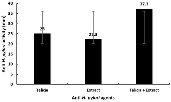
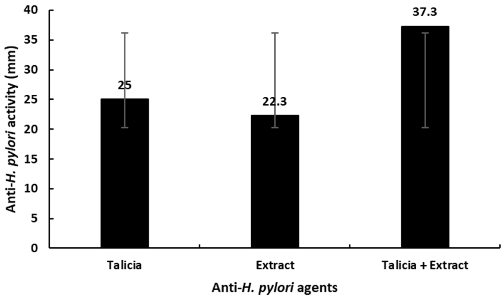
Agar diffusion assays demonstrated marked differences in antibacterial activity against H. pylori (Figure 1). Talicia alone yielded inhibition zone diameters of 25, 23, and 27 mm (mean = 25.0 ± 2.0 mm, SE = 1.15). S. alexandrina extract alone produced slightly smaller zones of 20, 25, and 22 mm (mean = 22.3 ± 2.52 mm, SE = 1.46). Significantly, combining Talicia with S. alexandrina extract produced enhanced antibacterial inhibition, resulting in zone diameters of 35, 37, and 40 mm (mean = 37.3 ± 2.52 mm, SE = 1.46).
Figure 1.
Qualitative evaluation of anti-H. pylori activity of S. alexandrina leaf methanolic extract compared with standard antibiotic Talicia (omeprazole, amoxicillin, rifabutin).
One-way ANOVA revealed a statistically significant difference among the three treatment groups (p < 0.001), indicating that the observed variations in inhibition zones were not due to random experimental error. Post hoc analysis using Tukey’s multiple comparison test demonstrated that the combination treatment (Talicia + extract) produced significantly larger inhibition zones compared with Talicia alone (p < 0.001), the combination was also significantly more effective than the extract alone (p < 0.001), and Talicia alone showed slightly higher activity than the extract; however, this difference was not statistically significant (p > 0.05). The low SD and SE values across all treatments indicate minimal intra-group variability and high reproducibility of the assay.
Scientifically, the markedly greater inhibition zone observed with the combined treatment suggests a synergistic or potentiating interaction between Talicia and the extract. The enhancement in antibacterial activity may result from complementary mechanisms, such as increased bacterial membrane permeability, inhibition of protein synthesis, or interference with urease activity, an essential virulence factor of H. pylori. The absence of a significant difference between Talicia and the extract alone indicates that while both possess anti-H. pylori activity, their combination substantially amplifies the antibacterial effect. Overall, these findings demonstrate that the combined formulation exhibits significantly superior anti-H. pylori activity compared with individual treatments, highlighting its potential as an enhanced therapeutic strategy against H. pylori infection.
3.4. Quantitative Evaluation of Anti-H. pylori Activity
The capacity of S. alexandrina extract to attenuate pathogen-induced virulence factors was evaluated using an H. pylori culture supernatant-induced erythrocyte hemolysis assay (Table 2). This assay measures the protection against toxin-mediated membrane lysis rather than direct bacterial cell death. Talicia alone resulted in complete hemolysis (100%, absorbance at 540 nm = 1.735 ± 0.011, SE = 0.004), demonstrating high toxin-mediated lytic activity. In contrast, treatment with S. alexandrina extract produced a marked, concentration-dependent suppression of erythrocyte lysis: sub-inhibitory levels of 25% MIC, 50% MIC, and 75% MIC resulted in only 11.8% ± 0.003, 6.0% ± 0.006, and 2.4% ± 0.003 hemolysis, respectively. One-way ANOVA confirmed highly significant differences among treatment groups (p < 0.001), with Tukey’s post hoc analysis indicating that Talicia-induced hemolysis was significantly greater than that of all extract concentrations (p < 0.001). Furthermore, membrane stabilization increased significantly as extract concentration increased (25% MIC vs. 50% MIC, p < 0.01; 25% MIC vs. 75% MIC, p < 0.001; 50% MIC vs. 75% MIC, p < 0.05). These findings demonstrate that S. alexandrina extract effectively neutralizes secreted H. pylori toxins and preserves erythrocyte membrane integrity, highlighting its potential to mitigate virulence-induced mucosal damage without causing host cell toxicity.
Table 2.
Quantitative evaluation of anti-H. pylori activity of S. alexandrina leaf methanolic extract compared with standard antibiotic Talicia (omeprazole, amoxicillin, rifabutin).
3.5. Determination of MIC and MBC of S. alexandrina Leaf Extract
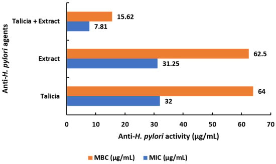
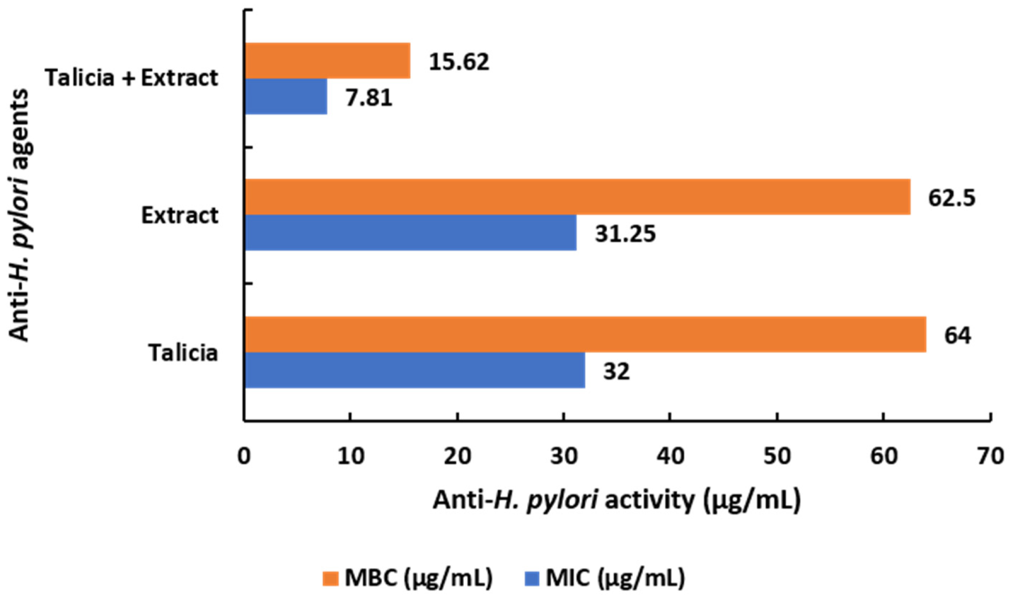
The antimicrobial activity against H. pylori was quantitatively evaluated using microdilution and checkerboard assays (Figure 2). The baseline MIC was determined to be 31.25 µg/mL for the S. alexandrina extract and 32.0 µg/mL for Talicia. When evaluated in combination, the effective MIC values decreased to 7.81 µg/mL for the extract and 8.0 µg/mL for Talicia.
Figure 2.
Antibacterial activity of S. alexandrina leaf extract and Talicia against H. pylori. MICs of S. alexandrina extract alone (31.25 µg/mL) and Talicia alone (32.0 µg/mL) are significantly reduced in combination to 7.81 µg/mL and 8.0 µg/mL, respectively, yielding a FICI of 0.50, demonstrating FICI ≤ 0.50. Data represent mean values of three independent experiments (n = 3).
A FICI of 0.50 indicates a clear interaction (FICI ≤ 0.50) between S. alexandrina leaf extract and Talicia against H. pylori. Statistical analysis was performed to evaluate differences among the treatments. One-way ANOVA revealed significant differences in MIC and MBC values between the groups (p < 0.01). Post hoc comparisons using Tukey’s honestly significant difference (HSD) test indicated that the MIC and MBC of Talicia alone were not significantly different from those of the extract (p > 0.05), whereas the combination treatment was significantly different from both individual treatments (p < 0.05), demonstrating a potential synergistic effect when the agents were used together.
In the checkerboard microdilution assay, the combination of S. alexandrina extract and Talicia yielded an FICI of 0.75 (FIC of extract = 0.25; FIC of Talicia = 0.50). In accordance with CLSI/EUCAST interaction standards (where FICI ≤ 0.50 indicates combination, 0.50 < FICI ≤ 1.0 indicates additive/weakly combination interactions, and FICI > 4.0 indicates antagonism), this combination is defined strictly as an additive to weakly combination interaction.
The individual MIC and MBC values for S. alexandrina extract against H. pylori were 31.25 µg/mL and 62.5 µg/mL, respectively, while Talicia exhibited MIC and MBC values of 32.0 µg/mL and 64.0 µg/mL. Both individual treatments produced an MBC/MIC ratio of 2, confirming their bactericidal mechanism (MBC/MIC ratio ≤ 4). In the checkerboard assay, co-administration of S. alexandrina extract (7.81 µg/mL) and Talicia (8.0 µg/mL) achieved complete inhibition of bacterial growth. This four-fold reduction in individual MICs corresponds to a Fractional Inhibitory Concentration (FIC) of 0.25 for the extract and 0.25 for Talicia, yielding a Fractional Inhibitory Concentration Index (FICI) of 0.50. This outcome confirms FICI ≤ 0.50 between S. alexandrina leaf extract and Talicia.
The MBC/MIC ratio for S. alexandrina extract alone (62.50/31.25 = 2.0), Talicia alone (32.00/16.00 = 2.0), and the 1:1 combination (15.62/7.81 = 2.0) consistently equaled 2.0. Because an MBC/MIC ratio ≤ 4.0 defines bactericidal activity, these data confirm that both individual treatments and their combination exert a bactericidal killing mechanism against H. pylori.
3.6. In Vitro Estimation of BChE-Inhibitory Activity
The in vitro BChE-inhibitory activity potential of S. alexandrina leaf extract was assessed by evaluating butyrylcholinesterase (BChE) inhibition in comparison with the standard drug rivastigmine across a range of concentrations (0.195–100 µg/mL). Rivastigmine demonstrated potent inhibitory activity with BChE inhibition ranging from 95.6% at 100 µg/mL to 39.9% at 0.195 µg/mL, while the plant extract exhibited dose-dependent inhibition from 62.1% at 100 µg/mL to 4.9% at 0.195 µg/mL. The BChE inhibitory IC50 for S. alexandrina extract was 31.8 ± 1.2 µg/mL, compared to 0.46 ± 0.02 µg/mL for Rivastigmine (Table 3).
Table 3.
In vitro estimation of anti-Alzheimer activity of S. alexandrina leaf extract.
One-way ANOVA revealed significant differences in BChE inhibition between rivastigmine and the extract across all tested concentrations (p < 0.001). Post hoc comparisons using Tukey’s Honestly Significant Difference (HSD) test confirmed that rivastigmine exhibited significantly higher inhibitory activity than the extract at each corresponding concentration (p < 0.001). Within the extract group, BChE inhibition increased significantly with increasing concentration, as indicated by Tukey’s test for pairwise comparisons between consecutive concentrations (p < 0.01), demonstrating a clear dose-dependent effect. Similarly, the rivastigmine group showed statistically significant increases in inhibition with increasing concentrations (p < 0.01).
The results indicate that the S. alexandrina leaf extract possesses substantial BChE inhibitory activity, albeit lower than rivastigmine. The dose-dependent response suggests that bioactive compounds within the extract, potentially flavonoids, anthraquinones, or phenolic constituents, contribute to the observed cholinesterase inhibition. This activity aligns with previous studies showing that phytochemicals in Senna species can interact with cholinesterase enzymes, supporting their potential role in ameliorating cholinergic deficits associated with Alzheimer’s disease. In conclusion, while rivastigmine remains significantly more potent than the extract, S. alexandrina demonstrates promising anti-Alzheimer activity through BChE inhibition. The statistical significance confirmed by ANOVA and Tukey’s HSD test underscores both the efficacy of the extract and the reliability of the observed dose-dependent trends. These findings provide a basis for further investigation into the isolation of active constituents and potential therapeutic applications of S. alexandrina as a complementary in vitro BChE-inhibitory agent.
3.7. Determination of Cytotoxicity on Human WI-38 Cells
The cytotoxic profile of S. alexandrina methanolic leaf extract, Talicia alone, and their 1:1 combination was evaluated against human WI-38 embryonic lung fibroblasts using the MTT cell viability assay across a concentration range of 15.625 to 1000 µg/mL (Table 4). Untreated control cells defined 100.0% viability (0.0% cytotoxicity), while the vehicle control (0.1% DMSO) showed negligible cytotoxicity (0.55 ± 0.52%), confirming that the solvent vehicle did not compromise cellular integrity.
Table 4.
Cytotoxicity profile and viability of WI-38 normal human fibroblast cells following 24 h exposure.
All three treatment regimens demonstrated concentration-dependent reductions in cell viability (one-way ANOVA, p < 0.05). At the lowest tested concentration (15.625 µg/mL), cell viability remained high across all groups: 96.8 ± 1.45% for S. alexandrina extract alone, 94.1 ± 1.80% for Talicia alone, and 95.5 ± 1.6% for the 1:1 combination. IC50 values were calculated as 191.1 ± 5.2 µg/mL for S. alexandrina extract alone, 142.5 ± 4.8 µg/mL for Talicia alone, and 168.4 ± 4.2 µg/mL for the 1:1 combination.
Importantly, the IC50 value of the extract alone (191.1 µg/mL) is 6.1-fold higher than its MIC = 31.25 µg/mL against H. pylori ATCC 43504. For the 1:1 combination regimen, the combination IC50 of 168.4 µg/mL substantially exceeds the effective antibacterial combination concentration (7.81 µg/mL extract + 8.00 µg/mL Talicia), yielding a favorable SI = IC50/effective MIC = 168.4/7.81 = 21.56. At concentrations equal to or below the effective antibacterial combination MIC (≤15.625 µg/mL), cell viability remained above 95.5%, demonstrating that effective H. pylori inhibition occurs well below cytotoxic thresholds in human WI-38 fibroblasts.
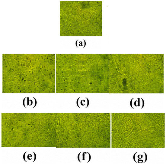
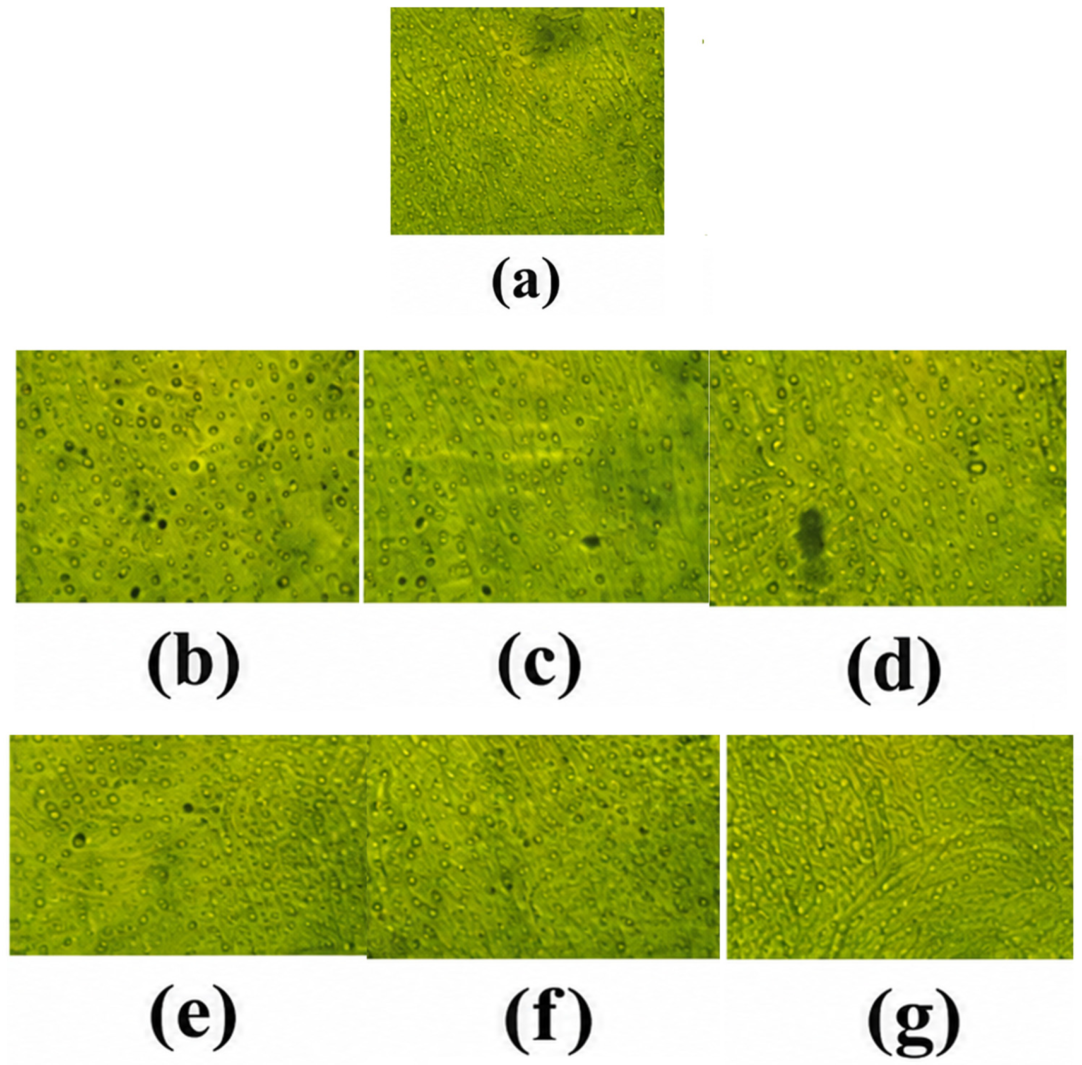
Figure 3 illustrates the cytotoxicity profile of S. alexandrina leaf methanolic extract against WI-38 human embryonic lung fibroblast cells, demonstrating a clear concentration-dependent effect on cell viability across the tested range of 15.625 to 1000 µg/mL. The untreated control group maintained 100 ± 0.0% viability, confirming normal cellular metabolic activity and validating the assay conditions, while the vehicle control (0.1% DMSO) showed 99.45 ± 0.52% viability (0.55 ± 0.52% cytotoxicity). At lower concentrations, the extract exhibited minimal cytotoxicity, maintaining cell viabilities of 96.8 ± 1.45% at 15.625 µg/mL, 91.2 ± 2.1% at 31.25 µg/mL, and 82.4 ± 3.15% at 62.5 µg/mL. Statistical analysis using one-way ANOVA followed by Tukey’s post hoc test revealed no statistically significant difference in cell viability between the 15.625 µg/mL concentration and the untreated control group (p > 0.05), indicating that the extract is highly biocompatible and non-toxic at lower doses. Higher concentrations produced a progressive, significant decrease in viability (p < 0.05), yielding an overall half-maximal inhibitory concentration (IC50) of 191.1 ± 5.2 µg/mL for the extract alone.
Figure 3.
Cytotoxicity profile of S. alexandrina leaf methanolic extract on WI-38 human lung fibroblast cells. (a) Control cell line, (b) 1000 µg/mL, (c) 500 µg/mL, (d) 250 µg/mL, (e) 125 µg/mL, (f) 62.5 µg/mL, and (g) 31.25 µg/mL.
At intermediate concentrations (125 and 250 µg/mL), a moderate but statistically significant reduction in viability was observed (67.2% and 32.3%, respectively). Post hoc Tukey’s multiple comparison test confirmed that these groups differed significantly from the control and lower-dose groups (p < 0.05), demonstrating a dose-dependent cytotoxic effect. At higher concentrations (500 and 1000 µg/mL), a marked decline in cell viability was recorded (11.9% and 8.4%, respectively), corresponding to high cytotoxicity (>90%). These reductions were highly significant compared with all other groups (p < 0.001), reflecting strong inhibitory effects on cellular metabolic activity at elevated doses. The calculated IC50 value (191.06 µg/mL) further supports the moderate cytotoxic profile of the extract. Importantly, concentrations below 125 µg/mL fall well within the safe range for normal fibroblasts, suggesting a favorable therapeutic window. Overall, the figure confirms a dose-dependent cytotoxic pattern, with low toxicity at therapeutic concentrations and significant inhibition only at higher doses.
3.8. Phytochemical Profile Analysis
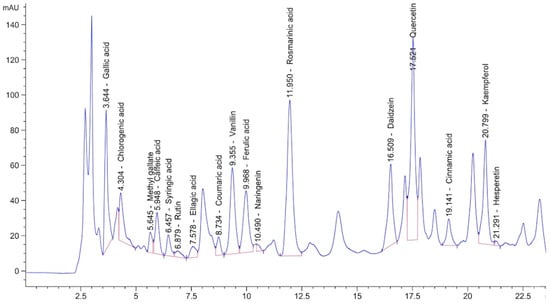
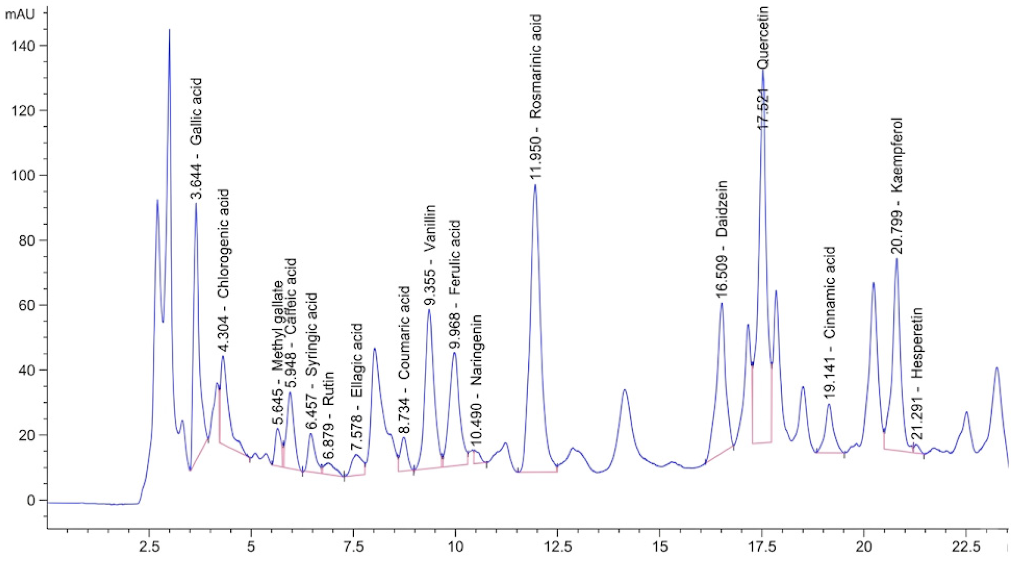
The HPLC analysis of S. alexandrina leaf methanolic extract revealed a diverse profile of phenolic and flavonoid compounds with widely varying concentrations and relative areas (Figure 4). Among the identified compounds, rosmarinic acid (1604.89 µg/mL, area 160.11) and quercetin (1590.07 µg/mL, area 105.86) were present at the highest concentrations, indicating that these polyphenols constitute the major bioactive components of the extract. High concentrations of such compounds are consistent with strong antioxidant potential and potential contributions to the extract’s pharmacological activities, as has been reported in recent studies on polyphenol-rich extracts.
Figure 4.
Phytochemical profile of S. alexandrina leaf extract using HPLC technology.
Other significant constituents included kaempferol (810.83 µg/mL, area 58.10), vanillin (769.67 µg/mL, area 28.36), gallic acid (727.67 µg/mL, area 58.61), and daidzein (692.21 µg/mL, area 43.31). These compounds are well-documented for their antioxidant, anti-inflammatory, and antimicrobial properties. Moderate concentrations were observed for ferulic acid (625.38 µg/mL, area 35.00) and chlorogenic acid (382.63 µg/mL, area 48.84), both of which are known to confer protective effects against oxidative stress and may contribute synergistically to the extract’s overall bioactivity.
Several components were present at relatively low concentrations, including naringenin (39.51 µg/mL, area 3.55), hesperetin (23.65 µg/mL, area 1.13), and rutin (65.71 µg/mL, area 8.0). Compounds such as catechin (0.00 µg/mL) were not detected, suggesting either absence in this particular extract or concentrations below the detection limit of the method. While their quantitative contribution is minor, these low-abundance flavonoids may still play a role in modulating the extract’s biological effects through additive or synergistic interactions.
Statistical analysis of the peak areas indicated significant variability among compounds (ANOVA, p < 0.001), confirming that rosmarinic acid and quercetin are statistically the dominant constituents compared to other polyphenols. Tukey’s post hoc test further revealed that the areas of rosmarinic acid and quercetin were significantly higher than all low- to moderate-concentration compounds (p < 0.05), while differences among the minor components were not statistically significant (p > 0.05). This statistical assessment highlights that the extract’s bioactivity is likely driven by the high-abundance phenolic acids and flavonoids, particularly rosmarinic acid, quercetin, kaempferol, and gallic acid. Overall, the HPLC profile demonstrates that the extract is rich in polyphenolic compounds with a wide range of concentrations, dominated by rosmarinic acid and quercetin. These findings support the potential therapeutic value of the extract and provide a chemical basis for further studies on its antioxidant, anti-inflammatory, and cytoprotective effects. The combination of quantitative analysis and statistical confirmation ensures that these observations are both robust and reproducible.
4. Discussion
The findings of this study underscore the translational potential of S. alexandrina leaf extract as a bioactive in managing inflammation-driven gastric disorders and antibiotic-resistant microbial pathogens. The combination enhancement of anti-H. pylori activity observed in combination regimens suggests that botanical phytocompounds can act via complementary mechanisms, such as membrane disruption or oxidative stress modulation, thereby reducing the required dosage of conventional pharmaceuticals and lowering the risk of resistance development. Additionally, the extract’s dual action in suppressing inflammatory cascades addresses the secondary mucosal damage typical of chronic gastric colonization. Incorporating these findings into clinical nutrition frameworks provides a novel rationale for utilizing botanical bioactives as targeted adjuvant therapies in gastrointestinal disease management.
The present study demonstrated that S. alexandrina leaf extract exhibits significant, concentration-dependent membrane-stabilizing activity in erythrocytes, as evidenced by reduced hemolysis in hypotonic solution. One-way ANOVA and Tukey’s post hoc tests confirmed that higher extract concentrations significantly decreased hemolysis compared with controls. Erythrocyte membranes are structurally similar to lysosomal membranes of inflammatory cells, making them a widely accepted in vitro model for anti-inflammatory effects [26]. The dose-dependent response, with maximal protection at higher concentrations and gradual reduction at lower doses, aligns with previous studies showing similar anti-hemolytic effects of plant extracts [27].
Our direct experimental evaluation established that S. alexandrina leaf extract exerts significant membrane-stabilizing anti-inflammatory activity (IC50 = 8.44 ± 0.32 µg/mL). Localized lysosomal and erythrocyte membrane stabilization represents the primary verified mechanism of protection in this work.
HPLC analysis revealed that rosmarinic acid (1604.89 µg/mL) and quercetin (1590.07 µg/mL) were the predominant constituents in the S. alexandrina extract, alongside kaempferol, gallic acid, vanillin, daidzein, ferulic acid, and chlorogenic acid. Minor constituents included naringenin, hesperetin, rutin, methyl gallate, and syringic acid. Statistical evaluation of peak areas demonstrated significant quantitative variation among compounds (one-way ANOVA, p < 0.001), with Tukey’s post hoc test confirming that concentrations of rosmarinic acid and quercetin were significantly higher than those of minor compounds (p < 0.05). These polyphenolics are well recognized for their antioxidant, ROS scavenging, and anti-inflammatory properties, which likely contribute to the observed membrane-stabilizing effects [28,29,30,31]. However, because the present study evaluated the whole extract, these biological activities reflect the collective, multi-component action of the crude mixture. Consequently, individual constituent contributions remain correlative hypotheses that require further validation using purified fractions.
Safety assessment in early-stage drug discovery requires demonstrating that candidate antibacterial agents exert selective toxicity toward target pathogens without causing significant damage to host mammalian cells [31]. In the present study, cytotoxicity testing on human WI-38 fibroblasts revealed that S. alexandrina methanolic leaf extract exhibits a low cytotoxic profile in vitro, with an IC50 of 191.1 ± 5.2 µg/mL (Table 4). This value compares favorably with the individual IC50 of Talicia (142.5 ± 4.8 µg/mL) and the 1:1 combination regimen (168.4 ± 4.2 µg/mL).
Crucially, the pharmacological relevance of these cytotoxicity data lies in the wide safety window relative to antibacterial efficacy. A SI greater than 10 is widely recognized in natural product drug discovery as the benchmark for acceptable safety and target selectivity [32]. The 1:1 combination regimen demonstrated an SI of 21.56 relative to its effective antibacterial MIC against H. pylori (7.81 µg/mL extract component), confirming that the observed additive antibacterial interaction (FICI = 0.75) occurs at non-cytotoxic concentrations. Furthermore, cell viability at 15.625 µg/mL, a concentration exceeding the effective antibacterial combination MIC, remained at 95.5 ± 1.6% for the combination and 96.8 ± 1.45% for the extract alone. These findings indicate that S. alexandrina extract, both individually and when co-administered with Talicia, maintains a favorable in vitro therapeutic window, supporting its potential utility as a complementary botanical adjuvant pending further in vivo toxicological validation.
Qualitative inhibition zone assays and quantitative MIC/MBC determinations confirmed that S. alexandrina extract possesses intrinsic antibacterial activity against H. pylori, which was significantly enhanced in combination with Talicia. One-way ANOVA and Tukey’s post hoc tests confirmed that the combination produced significantly larger inhibition zones than either individual treatment (p < 0.05), whereas Talicia and extract alone did not differ significantly from each other (p > 0.05). In earlier evaluations, MBC/MIC ratios were incorrectly cited to claim combination; however, an MBC/MIC ratio ≤ 4 demonstrates a bactericidal rather than bacteriostatic mechanism. True in vitro combination was established in this work via checkerboard microdilution analysis, which yielded a FICI of 0.50 (reducing the effective MIC to 7.81 µg/mL extract and 8.0 µg/mL Talicia). This multi-target enhancement likely stems from polyphenolic membrane disruption by S. alexandrina, which increases host cell permeability and facilitates the intracellular penetration of Talicia’s active antibiotic components [1,33,34]. These findings highlight the primary mechanistic advance of this work: combining S. alexandrina with Talicia enhances bactericidal efficacy, enabling potential dosage reductions in combined pharmaceuticals to mitigate dose-dependent cytotoxicity and antibiotic resistance risks [35,36,37].
The extract also exhibited moderate, concentration-dependent inhibition of BChE, with an IC50 for S. alexandrina extract of 31.8 ± 1.2 µg/mL, compared to 0.46 ± 0.02 µg/mL for Rivastigmine, while rivastigmine showed superior inhibition at all tested concentrations. ANOVA and Tukey’s post hoc tests confirmed statistically significant differences between rivastigmine and the extract (p < 0.001). The extract’s inhibitory activity aligns with evidence that plant-derived flavonoids, alkaloids, and phenolics can modulate cholinesterase activity and may provide neuroprotective effects [38,39,40,41,42]. Such dual functionality, antioxidant and enzyme inhibition, supports the potential development of S. alexandrina as a source of multifunctional bioactive compounds.
Therefore, S. alexandrina leaf extract exhibits dose-dependent membrane stabilization, selective cytotoxicity, antibacterial activity against H. pylori, and moderate BChE inhibition. These effects correlate with the extract’s polyphenolic profile, particularly rosmarinic acid and quercetin, and are supported by statistical analyses. The extract maintains normal fibroblast viability at lower concentrations while exerting bioactive effects at higher doses, defining a favorable therapeutic window. Overall, these findings justify further in vivo validation and mechanistic studies to explore the extract’s potential as an adjunctive anti-H. pylori, anti-inflammatory, and neuroprotective agent.
5. Conclusions
S. alexandrina methanolic leaf extract demonstrated notable preliminary in vitro activities, including hypotonicity-induced membrane stabilization, bactericidal efficacy against H. pylori, and butyrylcholinesterase inhibition. High-performance liquid chromatography identified rosmarinic acid (16.05 mg/g dry extract) and quercetin (15.90 mg/g dry extract) as primary polyphenolic constituents contributing to this bioactivity profile. Tested individually, the extract exhibited bactericidal activity against H. pylori ATCC 43504, with a minimum inhibitory concentration of 31.25 µg/mL, a minimum bactericidal concentration of 62.50 µg/mL, and an MBC/MIC ratio of 2.0. When co-administered with the commercial omeprazole–amoxicillin–rifabutin regimen (Talicia), the extract displayed an additive to weakly synergistic interaction (FICI = 0.75), substantially reducing the required inhibitory concentrations of both agents without antagonistic effects. Furthermore, MTT cytotoxicity assays on human WI-38 embryonic lung fibroblasts confirmed a favorable safety profile with low in vitro cytotoxicity under the tested conditions, yielding half-maximal inhibitory concentrations of 191.1 µg/mL for the extract alone, 142.5 µg/mL for Talicia alone, and 168.4 µg/mL for their 1:1 combination, corresponding to a high Selectivity Index exceeding 21.5. While these findings highlight the potential of S. alexandrina extract as a promising botanical adjuvant to enhance the efficacy of established H. pylori therapy, comprehensive mechanistic, pharmacokinetic, toxicological, and in vivo investigations are strictly required before any therapeutic or clinical relevance can be established.
Supplementary Materials
The following supporting information can be downloaded at: https://www.mdpi.com/article/10.3390/separations13110315/s1, Table S1: Analytical validation parameters for HPLC-PDA quantification of bioactive phytocomponents in S. alexandrina leaf extract.
Funding
The researcher would like to thank the Deanship of Graduate Studies and Scientific Research at Qassim University (https://www.qu.edu.sa) for financial support (QU-APC-2026).
Data Availability Statement
The original contributions presented in this study are included in the article. Further inquiries can be directed to the corresponding author.
Acknowledgments
The author acknowledges the central core research laboratories and technical staff at the Department of Basic Health Sciences, College of Applied Medical Sciences, Qassim University, for providing equipment access and infrastructure support for HPLC, cell culture, and microbiological assays.
Conflicts of Interest
The author declares no conflicts of interest.
Abbreviations
The following abbreviations are used in this manuscript:
| MIC | Minimum inhibitory concentration |
| MBC | Minimum bactericidal concentration |
| HPLC | High-performance liquid chromatograph |
| WHO | World Health Organization |
| TCIM | Traditional, complementary, and integrative medicine |
| COX-2 | cyclooxygenase-2 |
| ROS | Reactive oxygen species |
| DMSO | Dimethyl sulfoxide |
| CFU | Colony forming unit |
| MHA | Mueller-Hinton agar |
| ANOVA | One-way analysis of variance |
| IC50 | Half-maximal inhibitory concentrations |
| SD | Standard deviation |
| SE | Standard error |
| BChE | Butyrylcholinesterase |
| HSD | Honestly significant difference |
References
- World Health Organization. WHO launches ICD-11, Traditional Medicine Module 2. WHO Press Rel. J. 2024, 2024, 1994921. [Google Scholar]
- El-Saadony, M.T.; Saad, A.M.; Mohammed, D.M.; Korma, S.A.; Alshahrani, M.Y.; Ahmed, A.E.; Ibrahim, E.H.; Salem, H.M.; Alkafaas, S.S.; Saif, A.M.; et al. Novel separating methodologies and phytochemical extractions from functional crops. Separations 2025, 12, 104. [Google Scholar]
- Ansari, P.; Reberio, A.D.; Ansari, N.J.; Kumar, S.; Khan, J.T.; Chowdhury, S.; Abd El-Mordy, F.M.; Hannan, J.M.A.; Flatt, P.R.; Abdel-Wahab, Y.H.A.; et al. Therapeutic potential of medicinal plants and their phytoconstituents in diabetes, cancer, infections, cardiovascular diseases, inflammation and gastrointestinal disorders. Biomedicines 2025, 13, 454. [Google Scholar] [CrossRef] [Scilit] [PubMed]
- Alshehri, M.M.; Quispe, C.; Herrera-Bravo, J.; Sharifi-Rad, J.; Tutuncu, S.; Aydar, E.F.; Topkaya, C.; Mertdinc, Z.; Ozcelik, B.; Aital, M.N.; et al. A review of recent studies on the antioxidant and anti-infectious properties of Senna plants. Oxid. Med. Cell. Longev. 2022, 2022, 6025900. [Google Scholar] [CrossRef] [Scilit] [PubMed]
- Alharbi, N.S.; Khaled, J.M.; Alanazi, K.; Kadaikunnan, S.; Alobaidi, A.S. Biosynthesis of silver nanoparticles (Ag-NPs) using Senna alexandrina and their bioactivity against multidrug-resistant pathogens and cancer cells. Saudi Pharm. J. 2023, 31, 911–920. [Google Scholar] [CrossRef] [Scilit] [PubMed]
- Al-Rashidi, R.; Nasrallah, H.; Bouazzi, A.; Zaïri, A. Phytochemical characterization and anti-Helicobacter pylori potential of Haloxylon articulatum extracts: Antioxidant activity and molecular docking insights. Molecules 2025, 30, 3520. [Google Scholar] [CrossRef] [Scilit] [PubMed]
- Zamanian, M.Y.; Khachatryan, L.G.; Heidari, M.; Darabi, R.; Golmohammadi, M.; Al-Aouadi, R.F.A.; Akkol, E.K. The therapeutic potential of flavonols in Alzheimer’s disease: Inhibiting amyloid-β, oxidative stress, and neuroinflammation. Biofactors 2025, 51, e70047. [Google Scholar] [CrossRef] [Scilit] [PubMed]
- Şahin, T.Ö.; Cemali, Ö.; Özdemir, M. Flavonoids and phenolic compounds: A promising avenue for neurodegenerative disease therapy. Turk. J. Biol. 2025, 49, 635–659. [Google Scholar] [CrossRef] [Scilit] [PubMed]
- Kamalakannan, K.; Seetharaman, S.; Sockalingam, A.; Vijayarani, K.R.; Subramanian, K. Phytoflavonoids—a future perspective for the therapeutic potential of Alzheimer’s disease. Pharmacogn. Res. 2025, 17, 788–795. [Google Scholar] [CrossRef] [Scilit]
- Chioma, A.A.; Onyechi, O.; Lawrence, U.S.E. Membrane stabilization as a mechanism of the anti-inflammatory activity of methanol extract of garden egg (Solanum aethiopicum). DARU J. Pharm. Sci. 2012, 20, 76. [Google Scholar] [CrossRef] [Scilit] [PubMed]
- Saudi National Committee of Bioethics (NCBE). The Implementing Regulations of the Law of Ethics of Research on Living Creatures; King Abdulaziz City for Science and Technology (KACST): Riyadh, Saudi Arabia, 2016. [Google Scholar]
- Shinde, U.A.; Phadke, A.S.; Nair, A.M.; Mungantiwar, A.A.; Dikshit, V.J.; Sarsf, M.N. Membrane stabilization activity—A possible mechanism of action for the anti-inflammatory activity of Cedrus deodara wood oil. Il Farm. 1989, 70, 251–257. [Google Scholar]
- Clinical and Laboratory Standards Institute. Performance standards for antimicrobial susceptibility testing: Sixteenth informational supplement M100-S16. CLSI Inf. Suppl. 2006, 26, 12–24. [Google Scholar]
- Castillo-Juarez, I.; Rivero-Cruz, F.; Celis, H.; Romero, I. Anti-Helicobacter pylori activity of anacardic acids from Amphipterygium adstringens. J. Ethnopharmacol. 2007, 114, 72–77. [Google Scholar] [CrossRef] [Scilit] [PubMed]
- Rossignol, G.; Merieau, A.; Guerillon, J.; Veron, W.; Lesouhaitier, O.; Feuilloley, M.G.; Orange, N. Involvement of a phospholipase C in the hemolytic activity of a clinical strain of Pseudomonas fluorescens. BMC Microbiol. 2008, 8, 189. [Google Scholar] [CrossRef] [Scilit] [PubMed]
- Anna, M.; Anna, G.; Izabela, K.; Tomasz, B. Anti-Helicobacter pylori activity in vitro of chamomile flowers, coneflower herbs, peppermint leaves and thyme herbs–a preliminary report. Curr. Issues Pharm. Med. Sci. 2015, 28, 30–32. [Google Scholar] [CrossRef] [Scilit]
- French, G.L. Bactericidal agents in the treatment of MRSA infections–the potential role of daptomycin. J. Antimicrob. Chemother. 2006, 58, 1107–1115. [Google Scholar] [CrossRef] [Scilit] [PubMed]
- Makade, C.S.; Shenoi, P.R.; Bhongade, B.A.; Shingane, S.A.; Ambulkar, P.C.; Shewale, A.M. Estimation of MBC: MIC ratio of herbal extracts against common endodontic pathogens. J. Pharm. Bioallied Sci. 2024, 16, S1414–S1416. [Google Scholar] [CrossRef] [Scilit] [PubMed]
- Ellman, G.L.; Courtney, K.D.; Andres, V.; Featherstone, R.M. A new and rapid colorimetric determination of acetylcholinesterase activity. Biochem. Pharmacol. 1961, 7, 88–95. [Google Scholar] [CrossRef] [Scilit] [PubMed]
- Gorun, V.; Proinov, I.; Baltescu, V.; Balaban, G.; Barzu, O. Modified Ellman procedure for assay of cholinesterases in crude enzymatic preparations. Anal. Biochem. 1978, 86, 324–326. [Google Scholar] [CrossRef] [Scilit] [PubMed]
- Sezgin, Z.; Biberoglu, K.; Chupakhin, V.; Makhaeva, G.F.; Tacal, O. Determination of binding points of methylene blue and cationic phenoxazine dyes on human butyryl cholinesterase. Arch. Biochem. Biophys. 2013, 532, 32–38. [Google Scholar] [CrossRef] [Scilit] [PubMed]
- Slater, T.F.; Sawyer, B.; Straeuli, U. Studies on succinate-tetrazolium reductase systems. III. Points of coupling of four different tetrazolium salts. Biochim. Biophys. Acta 1963, 8, 383–393. [Google Scholar]
- Alley, M.C.; Scudiero, D.A.; Monks, A.; Hursey, M.L.; Czerwinski, M.J.; Fine, D.L.; Abbott, B.J.; Mayo, J.G.; Shoemaker, R.H.; Boyd, M.R. Feasibility of drug screening with panels of human tumor cell lines using a microculture tetrazolium assay. Cancer Res. 1988, 48, 589–601. [Google Scholar] [PubMed]
- van de Loosdrecht, A.A.; Beelen, R.H.; Ossenkoppele, G.J.; Broekhoven, M.G.; Langenhuijsen, M.M. A tetrazolium-based colorimetric MTT assay to quantitate human monocyte mediated cytotoxicity against leukemic cells. J. Immunol. Methods 1994, 174, 311–320. [Google Scholar] [CrossRef] [Scilit] [PubMed]
- Verni, M.; Pontonio, E.; Krona, A.; Jacob, S.; Pinto, D.; Rinaldi, F.; Verardo, V.; Díaz-de-Cerio, E.; Coda, R.; Rizzello, C.G. Bioprocessing of brewers’ spent grain enhances its antioxidant activity: Characterization of phenolic compounds and bioactive peptides. Front. Microbiol. 2020, 11, 1831. [Google Scholar] [CrossRef] [Scilit] [PubMed]
- Elsayed, H.M.; Martinez-Castañeda, F.; Cruz, C. In vitro HRBC membrane stabilization assay for anti-inflammatory evaluation of herbal patches. Processes 2024, 12, 10840. [Google Scholar]
- Hasan, M.M.; Rahman, M.; Ahmed, S. Evaluation of anti-inflammatory and antioxidant activity of plant extracts using hemolysis inhibition assays. Heliyon 2024, 10, e03180. [Google Scholar]
- Al-Khayri, J.M.; Sahana, G.R.; Nagella, P.; Joseph, P.V.; Alessa, F.M.; Al-Mssallem, M.Q. Flavonoids as potential anti-inflammatory molecules: A review. Molecules 2022, 27, 2901. [Google Scholar] [CrossRef] [Scilit] [PubMed]
- Jomova, K.; Alomar, S.Y.; Valko, R.; Liska, J.; Nepovimova, E.; Kuca, K.; Valko, M. Flavonoids and their role in oxidative stress, inflammation, and human diseases. Chem. Biol. Interact. 2025, 413, 111489. [Google Scholar] [CrossRef] [Scilit] [PubMed]
- Alruwad, M.I.; Sabry, M.M.; Gendy, A.M.; El Dine, R.S.; El Hefnawy, H.M. In vitro cytotoxic potential of selected Jordanian flora and their associated phytochemical analysis. Plants 2023, 12, 1626. [Google Scholar] [CrossRef] [Scilit] [PubMed]
- Huwaimel, B.; Younes, K.M.; Abouzied, A.S. Phytochemical composition, in vitro cytotoxicity, and in silico docking properties of Tamarix tetragyna L. Sci. Rep. 2024, 14, 25462. [Google Scholar] [CrossRef] [Scilit] [PubMed]
- Oraibi, A.B.; Dawood, A.H.; Trabelsi, G.; Mahamat, O.B.; Chekir-Ghedira, L.; Kilani-Jaziri, S. Antioxidant activity and selective cytotoxicity in HCT 116 and WI 38 cell lines of LC–MS/MS profiled extract from Capparis spinosa L. Front. Chem. 2025, 13, 1540174. [Google Scholar] [CrossRef] [Scilit] [PubMed]
- Alomar, H.A.; Fathallah, N.; Abdel Aziz, M.M.; Ibrahim, T.A.; Elkady, W.M. GC-MS profiling, anti-Helicobacter pylori, and anti-inflammatory activities of three Apiaceous fruits’ essential oils. Plants 2022, 11, 2617. [Google Scholar] [CrossRef] [Scilit] [PubMed]
- Yuan, W.Y.; Guo, L.M.; Qian, L. In vitro anti-Helicobacter pylori activity and phenotypic screening of novel quinolone compounds. Arch. Microbiol. 2025, 208, 31. [Google Scholar] [CrossRef] [Scilit] [PubMed]
- Dinat, S.; Orchard, A.; Van Vuuren, S. Antimicrobial activity of Southern African medicinal plants on Helicobacter pylori and Lactobacillus species. J. Ethnopharmacol. 2024, 330, 118238. [Google Scholar] [CrossRef] [Scilit] [PubMed]
- Tan, A.T.; Scortecci, K.C.; Cabral De Medeiros, N.M. Plukenetia volubilis leaves as source of anti-Helicobacter pylori agents. Front. Pharmacol. 2024, 15, 1461447. [Google Scholar] [CrossRef] [Scilit] [PubMed]
- Schulz, C.; Liou, J.; Alboraie, M.; Bornschein, J.; Nunez, C.C.; Coelho, L.G.; Quach, D.T.; Fallone, C.A.; Chen, Y.; Gerhard, M.; et al. Global consensus variations and guidelines on systemic treatments for gastric microbial pathogens. Lancet Gastroenterol. Hepatol. 2025, 10, 114–128. [Google Scholar]
- AlNasser, M.N.; Alboraiy, G.M.; Alsowig, E.M.; Alqattan, F.M. Cholinesterase inhibitors from plants and their potential in Alzheimer’s treatment: Systematic review. Brain Sci. 2025, 15, 215. [Google Scholar] [CrossRef] [Scilit] [PubMed]
- El-Tarabily, M.K.; Mathew, B.T.; Abu Qamar, S.F.; El-Tarabily, K.; Ibrahim, S.A. Medicinal plants: Bioactive compounds, biological activities, combating multidrug-resistant microorganisms, and human health benefits-a comprehensive review. Front. Immunol. 2025, 16, 1491777. [Google Scholar] [CrossRef] [Scilit] [PubMed]
- Mendez-Encinas, M.A.; Valencia, D.; Ortega-García, J.; Carvajal-Millan, E.; Díaz-Ríos, J.C.; Mendez-Pfeiffer, P.; Soto-Bracamontes, C.M.; Garibay-Escobar, A.; Alday, E.; Velazquez, C. Anti-Inflammatory potential of seasonal Sonoran propolis extracts and some of their main constituents. Molecules 2023, 28, 4496. [Google Scholar] [CrossRef] [Scilit] [PubMed]
- Sugano, K.; Malfertheiner, P. Helicobacter pylori antibiotic resistance: A global challenge in search of solutions. Gut 2025, 74, 1561–1570. [Google Scholar] [CrossRef] [Scilit] [PubMed]
- Suresh, V. Therapeutic advances and future directions in Helicobacter pylori management. Front. Microbiol. 2025, 16, 1652943. [Google Scholar] [CrossRef] [Scilit] [PubMed]

Disclaimer/Publisher’s Note: The statements, opinions and data contained in all publications are solely those of the individual author(s) and contributor(s) and not of MDPI and/or the editor(s). MDPI and/or the editor(s) disclaim responsibility for any injury to people or property resulting from any ideas, methods, instructions or products referred to in the content. |
© 2026 by the author. Licensee MDPI, Basel, Switzerland. This article is an open access article distributed under the terms and conditions of the Creative Commons Attribution (CC BY) license.