


Summary
Friedreich’s ataxia (FRDA), the most common autosomal recessive ataxia, is characterised by progressive ataxia with dysarthria of speech, loss of deep-tendon reflexes, impaired vibratory and proprioceptive sensations and corticospinal weakness with a Babinski’s sign. Patients eventually also develop kyphoscoliosis, cardiomyopathy and diabetes mellitus.The disease is a GAA repeat disorder resulting in severely reduced levels of frataxin, with secondary increased sensitivity to oxidative stress.The anti-oxidative drug, idebenone, is effective against FRDA-associated cardiomyopathy.We provide detailed clinical, electrophysiological and biochemical data from 20 genetically confirmed FRDA patients and have analysed the relationship between phenotype, genotype and malondialdehyde (MDA), which is a marker of superoxide formation. We assessed the effects of idebenone biochemically by measuring blood MDA and clinically by serial measurements of the International Cooperative Ataxia Rating Scale (ICARS). The GAA repeat length influenced the age at onset (p <0.001), the severity of ataxia (p = 0.02), the presence of cardiomyopathy (p = 0.04) and of lowfrequency hearing loss (p = 0.009). Multilinear regression analysis showed (p = 0.006) that ICARS was dependent on the two variables of disease duration (p = 0.01) and size of the GAA expansion (p = 0.02). We found no correlation to bilateral palpebral ptosis, visual impairment, diabetes mellitus or skeletal deformities, all of which appear to be signs of disease progression rather than severity. We discuss more thoroughly two underrecognised clinical findings: palpebral ptosis and GAA length-dependent low-frequency hearing loss.The average ICARS remained unchanged in 10 patients for whom follow-up on treatment was available (mean 2.9 years), whereas most patients treated with idebenone reported an improvement in dysarthria (63%), hand dexterity (58%) and fatigue (47%) after taking the drug for several weeks or months. Oxidative stress analysis showed an unexpected increase in blood MDA levels in patients on idebenone (p = 0.04), and we discuss the putative underlying mechanism for this result, which could then explain the unique efficacy of idebenone in treating the FRDA-associated cardiomyopathy, as opposed to other antioxidative drugs. Indeed, idebenone is not only a powerful stimulator of complexes II and III of the respiratory chain, but also an inhibitor of complex I activity, then promoting superoxide formation. Our preliminary clinical observations are the first to date supporting an effect of idebenone in delaying neurological worsening. Our MDA results point to the dual effect of idebenone on oxidative stress and to the need for controlled studies to assess its potential toxicity at high doses on the one hand, and to revisit the exact mechanisms underlying the physiopathology of Friedreich’s ataxia on the other hand, while recent reports suggest nonoxidative pathophysiology of the disease.
Introduction
Friedreich’s ataxia (FRDA), the most common autosomal recessive ataxia, usually emerges before the age of 25 years and is characterised by progressive ataxia with dysarthria of speech, disappearance of deep-tendon reflexes, loss of vibratory and proprioceptive sensations and by corticospinal weakness with a Babinski’s sign [1].The disease evolves relentlessly and the ability to walk is usually lost after several years. Friedreich’s ataxia has been shown to be a nuclear-encoded mitochondrial disease [2,3,4]. In about 98% of cases, the disease-causing defect consists of an expanded GAA repeat in intron 1 of the frataxin gene on chromosome 9q13 [5]. Levels of frataxin are severely reduced and this results in an increased sensitivity to oxidative stress [6], with mitochondrial iron accumulation [7] and impaired cardiac and skeletal muscle-tissue energy metabolism [6,8], most likely through involvement in the biosynthesis of Fe-S clusters [9,10].
Idebenone, a synthetic analogue of coenzyme Q10, which is a potent free radical scavenger, has been shown to be effective in controlling FRDA heart hypertrophy [11,12]. However, its efficacy on neurological functions is still not established [13,14]. Changes in the levels of biochemical markers of oxidative stress, such as malondialdehyde (MDA, a lipid peroxidation end product), could be useful to monitor the effect of idebenone [15,16]. However, these recent data come from quite small samples (19 and 11 patients respectively) and correlations between genotype, phenotype and biochemical markers are still debated.
We here report our experience with this disorder and provide detailed clinical, electrophysiological and biochemical data from 20 genetically confirmed FRDA patients. Our study was initially designed to assess whether the clinical findings were related to the genetic status and biochemical markers. As several patients were already receiving idebenone before entry to the study and reported a neurological improvement, we performed in a subgroup of the patients a clinical follow-up under treatment with serial ICARS measurements.
Patients and methods
Patients
We examined 20 patients from 15 unrelated families attending our Service in whom the diagnosis of Friedreich’s ataxia was confirmed genetically. All gave their informed consent to participate in the study.
All were examined by the same neurologist (P. A.) so as to guarantee a uniform assessment of the clinical deficits and the ataxia score.For this we used the International Cooperative Ataxia Rating Scale (ICARS) [17]. This semi-quantitative scale allows a measurement of postural and gait ataxia (7 tasks, 0–34 points), limb ataxia (6 tasks on both sides, with one writing task, 0–52 points), dysarthria of speech (2 items, 0–8 points) and oculomotor signs (3 items, 0–6 points), the overall score ranging from 0 (normal) to 100 (worst). Patients unable to perform a task receive the worst score, whatever the cause.
Electrophysiological studies
In 19 patients sensory and motor nerve conduction parameters, including spinal responses (F-waves), were studied in both upper and lower limbs, and 18 patients could be tested for brain-stem auditory- evoked potentials (BAEPs) coupled with pure-tone audiometry and for visual evoked potentials.
Cardiological studies
All patients underwent bedside cardiac auscultation, electrocardiography (ECG) and transthoracic echocardiography (TTE).
Genetic analysis
Only patients with genetically confirmed Friedreich’s ataxia were included, and all were found to have GAA repeat expansion on both alleles.
Biochemical analysis
Except for one patient who denied evaluation, fasting plasma glucose levels were determined in all patients. An oral glucose tolerance test was carried out on 6, and plasma levels of glycosylated haemoglobin (HbA1c) were measured in 16.
Malondialdehyde was used as a biochemical marker of oxidative stress.MDA levels were measured on a freeze/thaw-lysed whole blood-EDTA sample, using a lipid peroxidation assay kit (Calbiochem, San Diego, USA). Each measurement included a sample blank to exclude optical interference due to idebenone. Analysis was carried out on all 20 patients and 6 normal controls.
Idebenone treatment
Sixteen patients were already taking idebenone before the start of the study (11 on 5 mg/kg/day and 5 on 10 mg/kg/day; less than 1 year of treatment in 7 cases, less than 2 years in the others), and, in 2 of these, medication was temporarily interrupted soon after the beginning of the study for 5 and 7 weeks, respectively. Of the 4 cases that were not taking idebenone before the start of the study, one refused treatment, while the remaining 3 were started on 5 mg/kg/day. MDA levels in the same patient off and on idebenone (5 mg/kg/day) could thus be performed on 5 cases and, within the group as a whole, on 19 treated and 6 untreated patients.
Statistics
Pearson coefficients were used to assess correlations between clinical data, biochemical measures and the GAA repeat length on the short allele. Groups with and without a specific sign or symptom were compared using one way analysis of variance (ANOVA). The severity of disease (i.e. ICARS) was correlated by multilinear regression analysis using the two variables of the size of the GAA repeat and disease duration. Modifications in variables with time were measured using a paired t-test. Differences were defined as significant when the p value was <0.05.
Results
The FRDA phenotype: spectrum and characteristics
The clinical findings are summarised in Table 1. Three patients (15%) developed a late-onset form, with symptoms first appearing at the ages of 25, 32 and 54 years, respectively. Two of these, examined after 6 and 14 years of evolution respectively, had increased tendon reflexes, but in the third patient, who had a longer period of evolution of 33 years, they were absent. At the time of the first examination, 16 patients were wheelchair bound and the total ICARS ranged from 21 to 85 (mean 66.4/100). Postural and gait scores ranged from 7 to 33 (mean 29.1/34), kinetic scores from 10 to 42 (mean 30.2/52), speech scores from 1 to 6 (mean 3.3/8) and the oculomotor score from 2 to 5 (mean 3.7/6).
Table 1.
Clinical features, distribution of GAA repeat lengths and laboratory abnormalities in the 20 patients (14 females). Ataxia could not be evaluated in detail in 7 patients because of severe paresis. Deep reflexes were increased in the upper limbs in 3 cases, 2 of which were late-onset cases (*). One patient refused to undergo electrophysiological and audiometric evaluation.
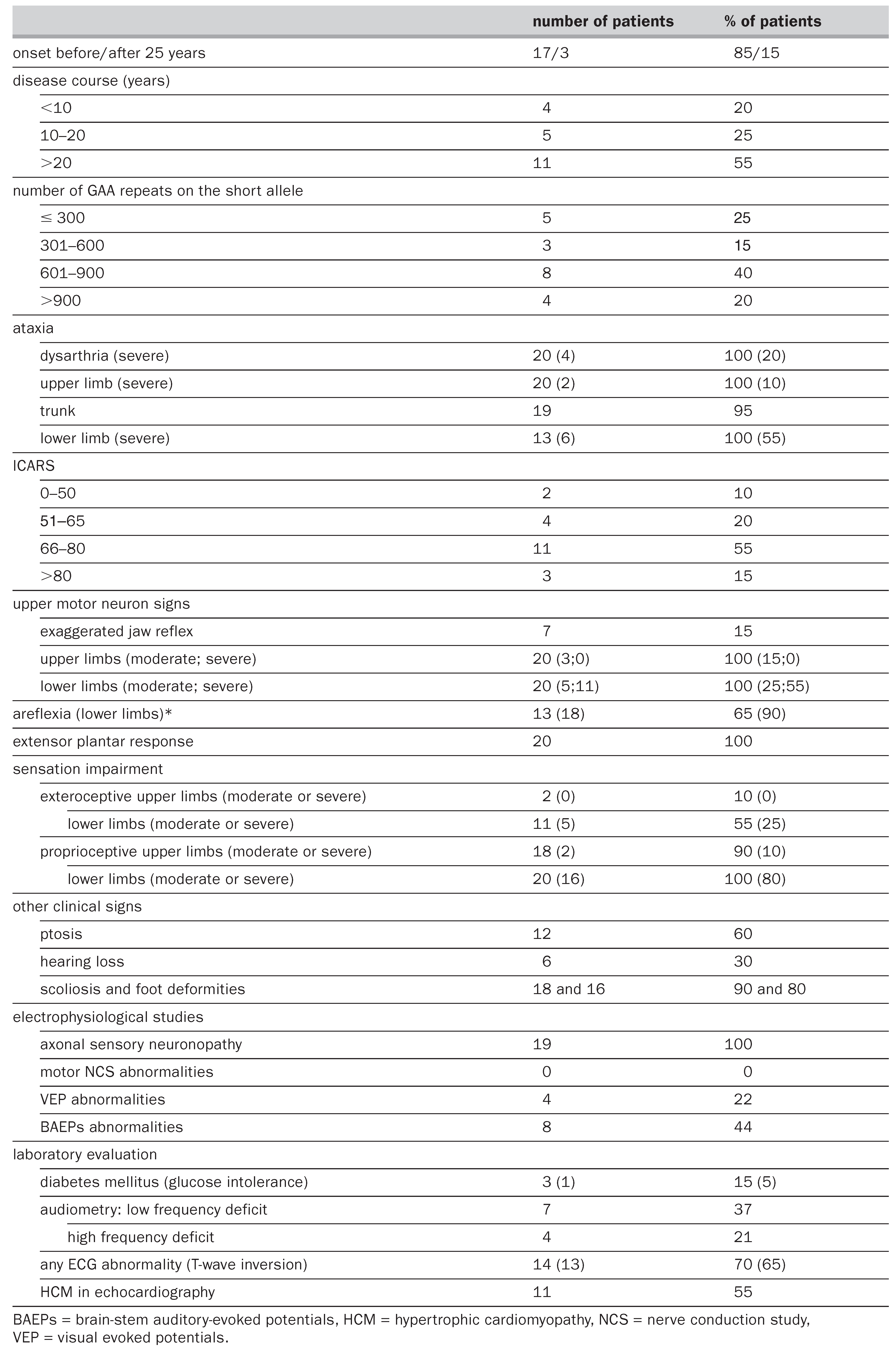
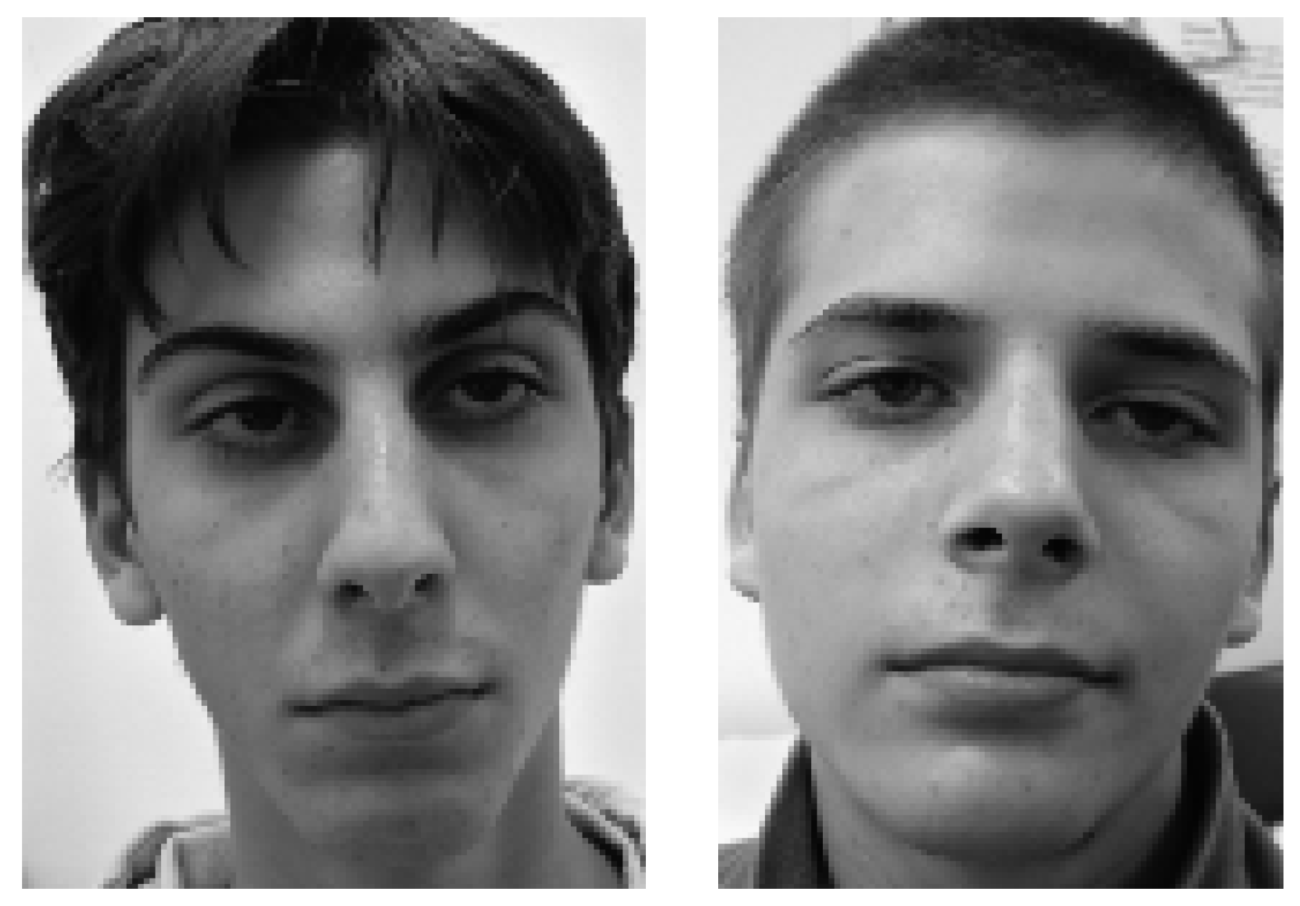
Decreased visual acuity was reported by 4 patients (20%) and optic atrophy was seen in 2 of these. Oculomotor abnormalities were always present, without external ophthalmoplegia, even if bilateral ptosis was observed in 12 patients (60%) (Figure 1). Seven patients complained of hypoacusis, and hearing loss was found on pure-tone audiometry in 11 (58%); four of them had a high-frequency deficit explainable by their age (38 to 68, mean 56), this finding being comparable to the frequency of presbycusis found in the general population. On the other hand, 7 of these 11 patients (37%) had a low-frequency deficit and were younger patients (13 to 36 years, mean 24). Two of these found the deficit very disturbing, but none required a hearing aid.
Figure 1.
Photographs of two brothers demonstrating the bilateral, but asymmetrical ptosis. The picture was taken when the patients were aged 19 and 17 years respectively. Both gave their written consent for the pictures to be published.
Sensory impairment and upper motor neuron signs were universally observed, with increased jaw reflex in 7/20 cases (35%). No patient reported neuropathic pain.
Electrophysiological studies
The sensory nerve conduction parameters were abnormal in all (Table 1). The shape and latencies of BAEPs were abnormal in 8 of the 18 available recordings, responses being absent in one case, desynchronised or prolonged in the 7 others. Visual evoked potentials were abnormal in 4 patients (22%), the P100 latency being either prolonged up to 138 ms (3 cases; normal <120 ms) or not measurable (one case).
Cardiological findings
ECG or TTE evidence of cardiomyopathy was found in 14/20 patients (70%) at the time of the first examination (Table 1). All 11 patients with TTE abnormalities also had an abnormal ECG. The most frequent ECG abnormality was T-wave inversion (13/14 cases). TTE showed non-obstructive ventricular hypertrophy in 10 patients, whereas only one patient presented obstructive cardiomyopathy.
Genetic analysis
We found an inverted and significant correlation between the size of the GAA repeat on the shorter allele and age of onset (p <0.001). Multilinear regression analysis (p = 0.006) showed that the ICARS was dependent on the following two variables: disease duration (p = 0.01) and size of the GAA expansion (p = 0.02) with the equation ICARS = 32.383 + 0.857 × (disease duration) + 0.025 × (GAA repeat length). All 3 late-onset cases had small GAA repeats on the short arm of chromosome 9q13, one of 200 repeats and two of 300. A larger GAA repeat on the short allele was correlated with low-frequency hearing loss (p = 0.009) and the presence of a cardiomyopathy (p = 0.04).
Biochemical analysis
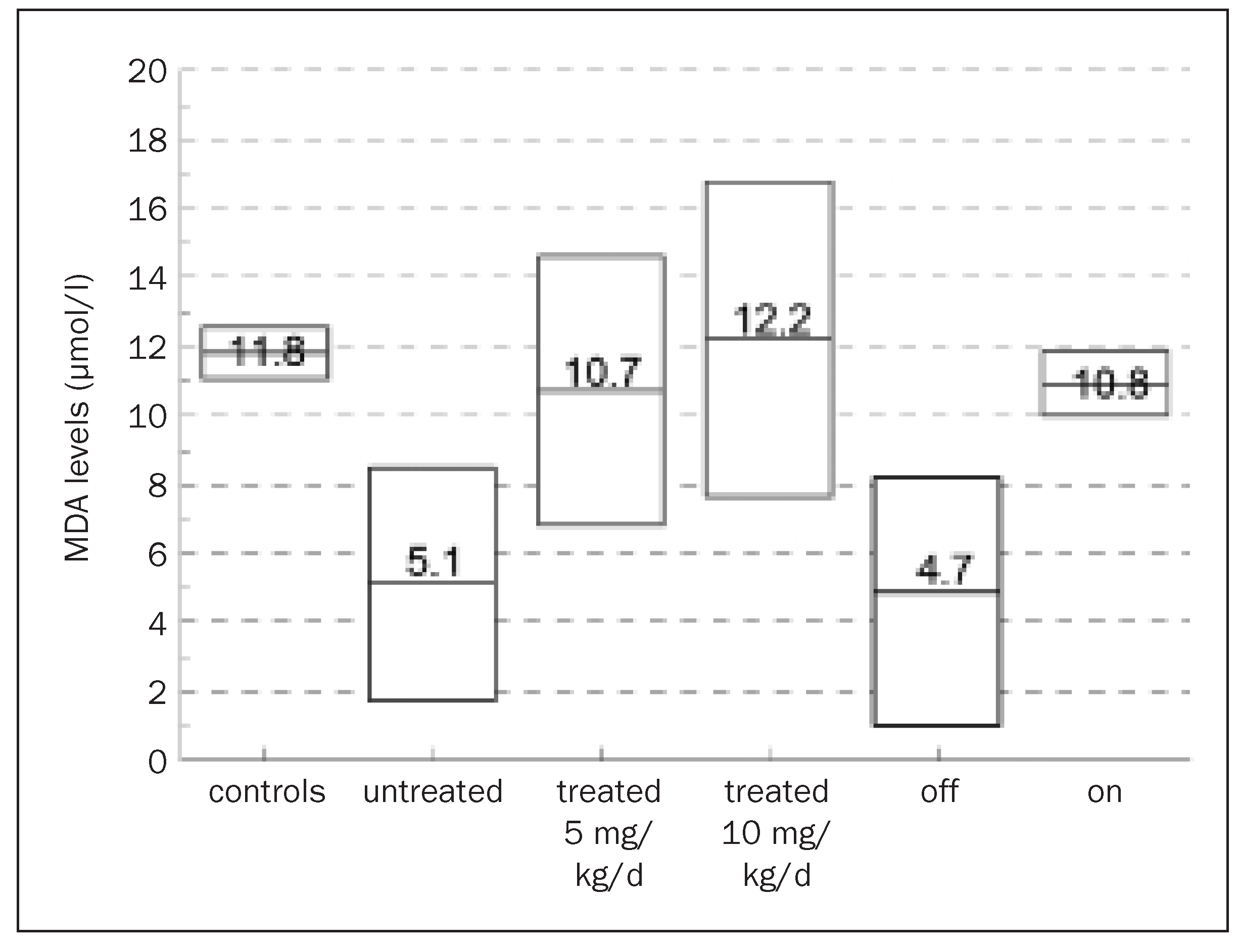
Diabetes mellitus (3 patients) or glucose intolerance (one patient) was present in 21% of our patients. Whole blood MDA levels were 10.8–12.8 μmol/l (11.8 ± 1.0, mean and SD, n = 6) in controls, 0–11.5 μmol/l (5.1 ± 4.5, n = 6) in patients with no treatment and 0–17.7 μmol/l (10.7 ± 5.2, n = 19) in patients on treatment. Within the on-treatment group, no significant difference was found between those already treated at the start of the study and those who started treatment at this time (10.7 ± 5.9 vs 10.7 ± 1.3) or between those on 10 or 5 mg/kg/day (12.2 ± 6.1 vs 10.7 ± 5.2). No correlation could be found between MDA levels, GAA repeat length and disease severity, as estimated by ICARS, the limiting factor being the low number of patients analysed without treatment. However, within the group as a whole, the 19 patients on idebenone had higher MDA levels than the 6 patients off treatment, and measurement of MDA levels performed before and after introduction of idebenone in the same patient (5 subjects) showed a significant increase (Figure 2 on the right, p = 0.028).The two patients in whom treatment was stopped showed subjective and objective worsening of ataxia, which resolved after reintroduction of idebenone, and MDA levels increased from 7.09 to 11.48 and from 4.86 to 10.20 μmol/l, respectively, after reintroduction of treatment.
Figure 2.
MDA levels off and on idebenone treatment. Results are presented with mean values and standard deviation. On the left, untreated and treated (5 and 10 mg/kg/day) patientsare compared to controls. The results of the 5 patients analysed before (off) and again after (on) introduction of idebenone are presented on the right (paired t-test: F ratio = 11.38, p = 0.028).
Clinical effect of idebenone treatment
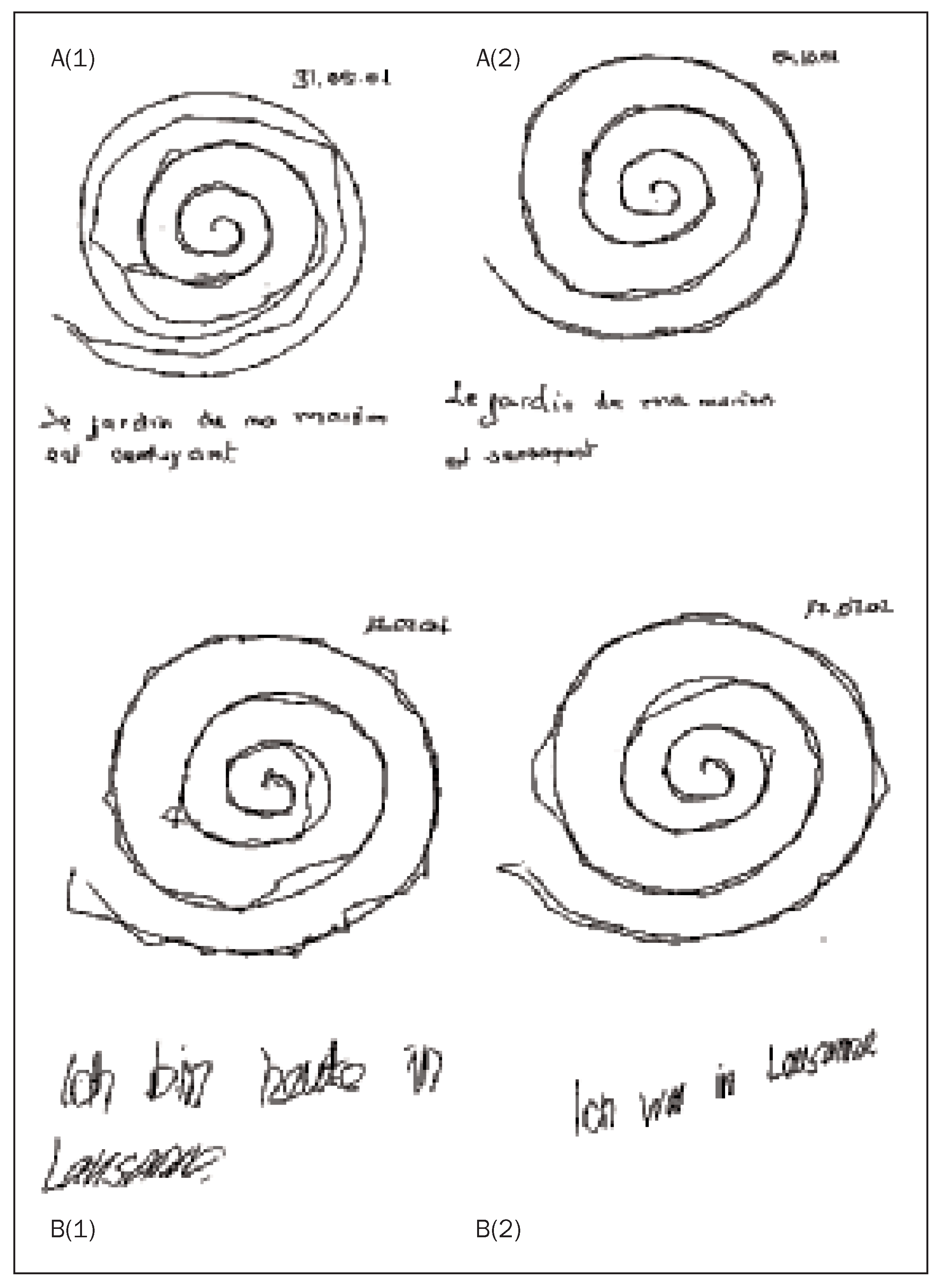
Fifteen of the 19 patients (79%) reported improvement on idebenone treatment in terms of dysarthria (63%), hand dexterity or handwriting(58%; Figure 3), and fatigue (47%).This improvement appeared gradually on treatment (within 6 week sin 3 patients and within 6 months in the other 12).No side effects were reported. Although our study was not designed to monitor patients on treatment, follow-up (1.6–3.5 years, mean 2.9) with ICARS serial measure was available in 10 patients. The mean score did not change during follow-up(66.4 vs 66.8).
Figure 3.
Archimedes’ spiral copy and sample of writing for two patients (A and B) before (1) and on idebenone treatment (2). Idebenone was started immediately after the first test. The date of the test is indicated in the figure. The same pen was used for each test.
Discussion
In the first part of the study, we investigated the relationship between clinical phenotype, GAA expansion and biochemical status in a reasonably large number of FRDA patients. The main points of interests are the late-onset forms, palpebral ptosis and increased jaw reflex, and the correlation between the size of the GAA repeat and disease severity measured by ICARS, and low-frequency hearing loss. In the second part of the study we provided pilot data supporting an efficacy of idebenone in improving ataxia and in modifying the measure of a biochemical marker of oxidative stress.
Whereas most of the abnormalities found in this series are congruent with the typical clinical feature of Friedreich’s ataxia reported before the molecular cause of the disease was discovered [1,18,19], our findings confirm the broad spectrum of FRDA phenotype described afterwards [20,21].The relationship between genotype and phenotype is measured in our series through the ICARS, which could be predicted by the disease duration and the GAA repeat length.We found 15% of late-onset forms, all with small GAA expansions, a finding that confirms the usefulness of an FDRA test for all patients with idiopathic ataxia and sensory neuropathy [21]. The size of the GAA is not sufficient to explain the phenotypic variation encountered in Friedreich’s ataxia; one reason is the somatic mosaicism encountered by a meiotic instability of the expanded GAA repeats, with a secondary unequal repeat length between leukocytes and other tissues [21]; another might be an influence of associated mitochondrial DNA haplogroups [22]. This implies that a large GAA expansion does not necessarily predict a bad course of the disease into a given individual and this has to be taken into account when the prognosis is discussed at the time of diagnosis. Whereas Harding [1] reported less than 10% of patients suffering deafness, one third of our patients were found to have a low-frequency hearing loss on pure-tone audiometry, a feature reported rarely [23] for which we have found a correlation with the size of the GAA repeat. By analogy of what is encountered in mitochondrial encephalomyopathies, deafness has been considered to be secondary to degeneration of the spiral ganglion cells [24,25,26], but the damage is probably more diffuse with associated pontine lesions, as indicated by our BAEPs recordings. Another interesting finding in our series was the bilateral palpebral ptosis, a sign not statistically associated with disease severity. Again palpebral ptosis is a well-known sign in mitochondrial muscle disorders [27] and recent studies have indeed shown that a skeletal muscle mitochondrial dysfunction can be demonstrated in Friedreich’s ataxia, either by in vivo 31P-magnetic resonance spectroscopy [28] or by a decrease in respiratory chain function in muscle biopsies [29]. Ptosis could thus be the clinical expression of the mitochondrial dysfunction in FRDA muscles. By contrast, limb weakness is the hallmark of costicospinal involvement, and in this regard, it is worth noting that a brisk jaw reflex was observed in about one-third of our patients, a bit more frequently than previously reported [30].
Most patients treated with idebenone reported improvement in dysarthria, hand dexterity (Figure 3), and fatigue, and all of this appeared within 6 months after the beginning of the drug treatment. Whereas our findings were found in a pilot and open trial, a stabilisation of the ICARS was observed, but this should be considered with caution because several patients were already in an advanced disease stage at the time they were followed. While two recent studies have failed to demonstrate a beneficial effect of idebenone [13,14], our preliminary results need to be verified by a longitudinal placebo-controlled study.
Oxidative stress analysis in our patients showed decreased blood MDA levels in the untreated FRDA patients, which were increased to values comparable to those of normal controls on idebenone treatment. This seems at first sight in contradiction to the physiopathology of the disease implying impaired oxidative mechanisms and with two previous studies reporting increased values in plasma samples of 19 and 11 patients respectively [15,16]. However, idebenone is not only a powerful stimulator of complexes II and III of the respiratory chain, but has been shown to inhibit complex I activity [31] and then promotes superoxide formation, for which MDA levels act as a marker [32]. This dual effect of idebenone on the respiratory chain explains our results; it is perhaps also the reason why idebenone was found to be effective in FRDA cardiomyopathy [11,12], whereas other anti-oxidant molecules were not, implying that the changes in oxidative stress in Friedreich’s ataxia are more complex than currently believed. A recent study underscores this complexity; in the Seznec et al. study it was indeed demonstrated that an increased superoxide production could not explain by itself the FRDA pathophysiology [33] and that contrary to the general assumption, Friedreich’s ataxia is a neurodegenerative disease not associated with oxidative damage.With the reserve that our results come out of the analysis of a rather small cohort of patients and have to be confirmed by analysis of a larger sample of patients, our study further strengthens the necessity of controlled studies. If confirmed, it should though be suspected that the beneficial effects of idebenone in FRDA patients might be outweighed by toxic effects at higher drug concentrations.
Acknowledgments
We gratefully acknowledge the cooperation of the patients and family members. We are also grateful to C. Gander, R. Poljicak, M. Vaglio and Dr M. Markert for their excellent technical support and to the staff of the Division of Cardiology of our hospital for performing the cardiologic studies.
References
- Harding, AE. Friedreich’s ataxia: a clinical and genetic study of 90 families with an analysis of early diagnostic criteria and intrafamilial clustering of clinical features. Brain 1981, 104, 589–620. [Google Scholar] [CrossRef] [Scilit] [PubMed]
- Gibson, TJ; Koonin, EV; Musco, G; Pastore, A; Bork, P. Friedreich’s ataxia protein: phylogenetic evidence for mitochondrial dysfunction. Trends Neurosci 1996, 19, 465–8. [Google Scholar] [CrossRef] [Scilit] [PubMed]
- Koutnikova, H; Campuzano, V; Foury, F; Dolle, P; Cazzalini, O; Koenig, M. Studies of human, mouse and yeast homologues indicate a mitochondrial function for frataxin. Nat Genet 1997, 16, 345–51. [Google Scholar] [CrossRef] [Scilit]
- Priller, J; Scherzer, CR; Faber, PW; MacDonald, ME; Young, AB. Frataxin gene of Friedreich’s ataxia is targeted to mitochondria. Ann Neurol 1997, 42, 265–9. [Google Scholar] [CrossRef] [Scilit]
- Campuzano, V; Montermini, L; Molto, MD; Pianese, L; Cossee, M; Cavalcanti, F. Friedreich’s ataxia: autosomal recessive disease caused by an intronic GAA triplet repeat expansion. Science 1996, 271, 1423–7. [Google Scholar] [CrossRef] [Scilit]
- Bradley, JL; Blake, JC; Chamberlain, S; Thomas, PK; Cooper, JM; Schapira, AH. Clinical, biochemical and molecular genetic correlations in Friedreich’s ataxia. Hum Mol Genet 2000, 9, 275–82. [Google Scholar] [CrossRef] [Scilit]
- Chantrel-Groussard, K; Geromel, V; Puccio, H; Koenig, M; Munnich, A; Rotig, A. Disabled early recruitment of antioxidant defenses in Friedreich’s ataxia. Hum Mol Genet 2001, 10, 2061–7. [Google Scholar] [CrossRef] [Scilit] [PubMed]
- Lodi, R; Hart, PE; Rajagopalan, B; Taylor, DJ; Crilley, JG; Bradley, JL. Antioxidant treatment improves in vivo cardiac and skeletal muscle bioenergetics in patients with Friedreich’s ataxia. Ann Neurol 2001, 49, 590–6. [Google Scholar] [CrossRef]
- Chen, OS; Hemenway, S; Kaplan, J. Inhibition of Fe-S cluster biosynthesis decreases mitochondrial iron export: evidence that Yfh1p affects Fe-S cluster synthesis. Proc Natl Acad Sci U S A 2002, 99, 12321–6. [Google Scholar] [CrossRef] [Scilit]
- Muhlenhoff, U; Richhardt, N; Ristow, M; Kispal, G; Lill, R. The yeast frataxin homolog Yfh1p plays a specific role in the maturation of cellular Fe/S proteins. Hum Mol Genet 2002, 11, 2025–36. [Google Scholar] [CrossRef] [Scilit]
- Hausse, AO; Aggoun, Y; Bonnet, D; Sidi, D; Munnich, A; Rotig, A. Idebenone and reduced cardiac hypertrophy in Friedreich’s ataxia. Heart 2002, 87, 346–9. [Google Scholar] [CrossRef] [Scilit]
- Rustin, P; Rotig, A; Munnich, A; Sidi, D. Heart hypertrophy and function are improved by idebenone in Friedreich’s ataxia. Free Radic Res 2002, 36, 467–9. [Google Scholar] [CrossRef] [Scilit]
- Buyse, G; Mertens, L; Di Salvo, G; Matthijs, I; Weidemann, F; Eyskens, B. Idebenone treatment in Friedreich’s ataxia: neurological, cardiac, and biochemical monitoring. Neurology 2003, 60, 1679–81. [Google Scholar] [CrossRef] [Scilit] [PubMed]
- Mariotti, C; Solari, A; Torta, D; Marano, L; Fiorentini, C; Di Donato, S. Idebenone treatment in Friedreich patients: one-year-long randomized placebo-controlled trial. Neurology 2003, 60, 1676–9. [Google Scholar] [CrossRef] [Scilit]
- Bradley, JL; Homayoun, S; Hart, PE; Schapira, AH; Cooper, JM. Role of oxidative damage in Friedreich’s ataxia. Neurochem Res 2004, 29, 561–7. [Google Scholar] [CrossRef] [Scilit]
- Emond, M; Lepage, G; Vanasse, M; Pandolfo, M. Increased levels of plasma malondialdehyde in Friedreich’s ataxia. Neurology 2000, 55, 1752–3. [Google Scholar] [CrossRef] [Scilit]
- Trouillas, P; Takayanagi, T; Hallett, M; Currier, RD; Subramony, SH; Wessel, K. International Cooperative Ataxia Rating Scale for pharmacological assessment of the cerebellar syndrome. The Ataxia Neuropharmacology Committee of the World Federation of Neurology. J Neurol Sci 1997, 145, 205–11. [Google Scholar] [CrossRef] [Scilit] [PubMed]
- Harding, AE. The Hereditary Ataxias and Related Disorders; Churchill Livingstone: London, 1984. [Google Scholar]
- Manyam, BV. Friedreich’s ataxia. In Handbook of Clinical Neurology; Vinken, PJ, Bruyn, GW, Klawans, HL, Eds.; Elsevier Science Publisher: Amsterdam, 1991; Vol. 16, 60, pp. 299–333. [Google Scholar]
- Montermini, L; Richter, A; Morgan, K; Justice, CM; Julien, D; Castellotti, B. Phenotypic variability in Friedreich’s ataxia: role of the associated GAA triplet repeat expansion. Ann Neurol 1997, 41, 675–82. [Google Scholar] [CrossRef] [Scilit] [PubMed]
- Schols, L; Amoiridis, G; Przuntek, H; Frank, G; Epplen, JT; Epplen, C. Friedreich’s ataxia. Revision of the phenotype according to molecular genetics. Brain 1997, 120, 2131–40. [Google Scholar] [CrossRef] [Scilit]
- Giacchetti, M; Monticelli, A; De Biase, I; Pianese, L; Turano, M; Filla, A. Mitochondrial DNA haplogroups influence the Friedreich’s ataxia phenotype. J Med Genet 2004, 41, 293–5. [Google Scholar] [CrossRef] [Scilit]
- Ell, J; Prasher, D; Rudge, P. Neuro-otological abnormalities in Friedreich’s ataxia. J Neurol Neurosurg Psychiatry 1984, 47, 26–32. [Google Scholar] [CrossRef] [Scilit]
- Satya-Murti, S; Cacace, A; Hanson, P. Auditory dysfunction in Friedreich’s ataxia: result of spiral ganglion degeneration. Neurology 1980, 30, 1047–53. [Google Scholar] [CrossRef] [Scilit]
- Chinnery, PF; Elliott, C; Green, GR; Rees, A; Coulthard, A; Turnbull, DM. The spectrum of hearing loss due to mitochondrial DNA defects. Brain 2000, 123, 82–92. [Google Scholar] [CrossRef] [Scilit]
- Jabbari, B; Schwartz, DM; MacNeil, DM; Coker, SB. Early abnormalities of brainstem auditory evoked potentials in Friedreich’s ataxia: evidence of primary brainstem dysfunction. Neurology 1983, 33, 1071–4. [Google Scholar] [CrossRef] [Scilit]
- Montagnani, S; De Rosa, P. Morphofunctional features of human extrinsic ocular muscles. Doc Ophthalmol 1989, 72, 119–28. [Google Scholar] [CrossRef] [Scilit]
- Vorgerd, M; Schols, L; Hardt, C; Ristow, M; Epplen, JT; Zange, J. Mitochondrial impairment of human muscle in Friedreich’s ataxia in vivo. Neuromuscul Disord 2000, 10, 430–5. [Google Scholar] [CrossRef] [Scilit] [PubMed]
- Schols, L; Reichmann, H; Amoiridis, G; Seibel, P; Wagener, S; Seufert, S; et al. Mitochondrial disorders in degenerative ataxias. Eur J Neurol 1996, 3, 56–60. [Google Scholar] [CrossRef] [Scilit]
- Auger, RG. Preservation of the masseter reflex in Friedreich’s ataxia. Neurology 1992, 42, 875–8. [Google Scholar] [CrossRef] [Scilit] [PubMed]
- Esposti, MD; Ngo, A; Ghelli, A; Benelli, B; Carelli, V; McLennan, H. The interaction of Q analogs, particularly hydroxydecyl benzoquinone (idebenone), with the respiratory complexes of heart mitochondria. Arch Biochem Biophys 1996, 330, 395–400. [Google Scholar] [CrossRef] [Scilit] [PubMed]
- Genova, ML; Ventura, B; Giuliano, G; Bovina, C; Formiggini, G; Parenti Castelli, G; et al. The site of production of superoxide radical in mitochondrial Complex I is not a bound ubisemiquinone but presumably iron-sulfur cluster N2. FEBS Lett 2001, 505, 364–8. [Google Scholar] [CrossRef] [Scilit]
- Seznec, H; Simon, D; Bouton, C; Reutenauer, L; Hertzog, A; Golik, P. Friedreich’s ataxia: the oxidative stress paradox. Hum Mol Genet 2005, 14, 463–74. [Google Scholar] [CrossRef] [Scilit] [PubMed]
This is an open-access article distributed under the terms of the Creative Commons Attribution Non-Commercial No Derivatives (CC BY-NC-ND) 4.0 License.