


Multiple choice questions
- ■
- P. Michel
Neurologie, CHUV, Lausanne
Select the one correct answer.
Neuroradiology/Neuroanatomy
- ■
- P. Michel
Neurologie, CHUV, Lausanne
Read for you
- ■
- P. Michel
Neurologie, CHUV, Lausanne
Should we use the new definition of TIA? When is a TIA a TIA?
Many clinicians felt that the classical definition of transient ischaemic attacks (TIAs) of “a sudden focal neurological deficit lasting for less than 24 hours, of presumed vascular origin ...” needed a revision. This is particularly true since there is a relatively poor correlation between the duration of the neurological deficit and the radiological and pathological findings, especially when the “transient” deficit lasts for more than 30–60 minutes. Furthermore, patients whose symptoms have not resolved after 1 hour are highly unlikely to be symptom-free after 24 hours and are therefore good candidates for acute treatment such as thrombolysis.
The new proposed definition is: “A TIA is a brief episode of neurological dysfunction caused by focal brain or retinal ischaemia, with clinical symptoms typically lasting less than one hour, and without evidence of acute infarction.”
Advantages of the new definition are:
- 1
- It emphasises the very acute aspect of brain ischaemia and encourages physicians (and patients) to consider giving acute treatment without waiting several hours to see whether symptoms resolve spontaneously. This is especially important because it has been shown that the earlier the treatment is initiated the more the patient benefits.
- 2
- There might be a better clinico-pathological correlation as TIAs lasting less than one hour are less likely to leave radiological or pathological lesions.
Problems with the new definition are:
- 1
- It mentions an exclusion criterion of no “evidence of acute infarction” on imaging, but does (correctly) not require that each patient has an MRI. Consequently, a TIA in one patient might not be a TIA in another, depending on the imaging studies the patients may get.
- 2
- The new time limit is given as “typically […] less than one hour” and remains voluntarily imprecise. Again, a TIA according to one physician’s opinion might not be a TIA according to another physician.
Unfortunately, it remains unclear whether this new definition should be adopted worldwide now; although it is published in a very prestigious medical journal, it is only formulated as a “proposal” by leading American stroke neurologists; should it not have been published in the same place after an international consensus had been reached?
Independent of whether the patient has an “olddefinition” TIA or a “new-definition” TIA (i.e. no matter how short symptoms last), the most important messages are that (1) TIAs should be considered as serious events, (2) acute ischaemic symptoms are a medical emergency, (3) an acute referral to the “best” treatment facility according to duration, type and severity of ischaemic symptoms is made, (4) a thorough search for a cause is undertaken and (5) that effective secondary prevention is assured.
Multiple sclerosis: which interferon regimen is “better”?
A randomised, controlled trial over 6 months found in patients with relapsing-remitting multiple sclerosis treated with IFNβ-1a (Rebif®) 44 µg s.c. three times a week a relapse rate of 25%, compared with 37% in patients treated with IFNβ-1a (Avonex®) 30 µg i.m. (p = 0.0005 for the odds ratio; Panitch et al.). However, the incidence of neutralising antibodies (25 vs 2%), injection-site reactions (83 vs 28%), abnormalities of liver enzymes and altered leukocytecounts was higher in the “more effective” group. The impact on disability was not assessed.
These results raise again the important question which dose, treatment frequency and route of administration are most effective and best tolerated. The trial of Panitch et al. still does not give a clear answer to these questions. It may be useful to the practising neurologist, however, for advising him and his patients on potential benefits and side effects of different regimens.
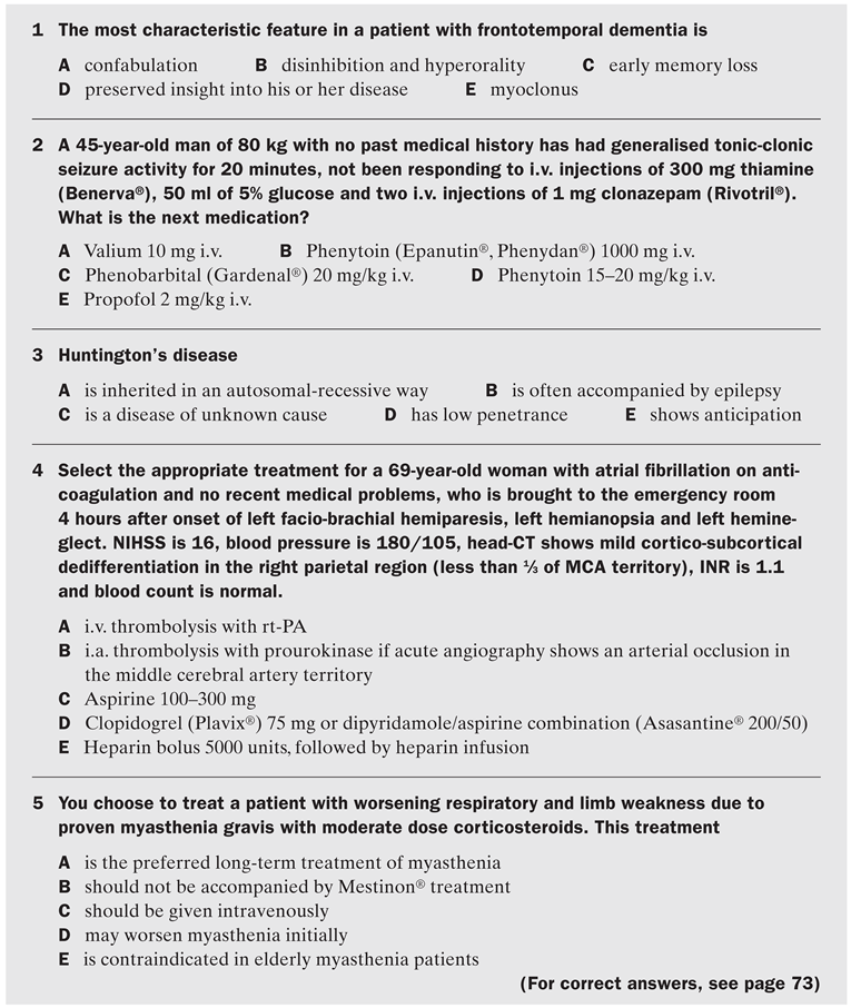
Answers to MCQ
- 1
- B: Disinhibition with socially inadequate behaviour is an early characteristic finding in frontotemporal dementia. Prominent early memory loss is more typical of Alzheimer’s disease, myoclonus might be seen in later stages of most dementias but should raise suspicion of prion diseases and metabolic encephalopathies.
- 2
- D: One of the most frequent mistakes in treating convulsive status epilepticus is the underdosing of phenytoin. Intravenous valproate (Depakine®) loading might be an alternative, but is less well studied. As a next step, phenobarbital may be used if sufficient phenytoinloading failed to stop the seizures. Dormicum (Midazolam®) or propofol infusions may also be considered instead of phenobarbital, but large controlled studies for these drugs in status epilepticus are currently lacking.
- 3
- E: Huntington’s disease is an autosomal-dominant trinucleotide repeat disorder (chromosome 4) characterised by chorea, cognitive and psychiatric manifestations. Anticipation (onset at an earlier age in the next generation) is a feature of most trinucleotide repeat disorders, mainly following transmission by the father.
- 4
- B: Intravenous thrombolysis more than 3 hours after onset of ischaemic stroke is probably not efficient in the majority of patients. The PROACT-II study has, however, shown a benefit for patients with MCA-occlusion treated intraarterially in the 3- to 6-hour window. Oral anticoagulation is not a contraindication for thrombolysis in this patient because her INR is normal.
- 5
- D: Because corticosteroids may worsen myasthenia gravis initially (typically after 5–7 days), many neurologists start with progressive doses and hospitalise these patients. Corticosteroids are also effective in elderly patients, but long-term treatment can usually be avoided (side effects!) with the help of other immunosuppressive treatment (such as azathioprine [Imurek®]).
Answers to Neuroradiology/Neuroanatomy
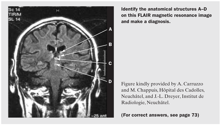
Anatomical structures:
- A
- Fornix
- B
- Dorsomedial thalamic nuclei
- C
- Mamillary bodies
- D
- Periaquaeductal grey
Diagnosis and commentary: Wernicke’s encephalopathy. This 39-year-old alcoholic and epileptic man displayed two of the three features of the classical triad (confusional state, gait ataxia, but no oculomotor abnormalities). This disease is due to vitamin B1 (thiamine) deficiency and may be missed if only one of the classical features is present or if the level of suspicion is too low (such as in patients with hyperemesis gravidarum or prolonged intravenous feeding). Pathologically, necrosis (sometimes haemorrhagic) is found in the paraventricular regions of the thalamus and hypothalamus, mamillary bodies, periaquaeductal region of the midbrain, floor of the fourth ventricle and superior cerebellar vermis. It is only recently that the MRI-features of this disease have been identified: diffusion-MR, FLAIR-MR, proton-density and to a lesser degree T2-weighted images are more useful and may show abnormalities in the structures mentioned above.
Apparent diffusion coefficient (ADC) map revealed the high signal intensity lesions in bilateral medial thalami, suggestive of vasogenic oedema.
References
- Albers, G.W.; Caplan, L.R.; Easton, J.D.; Fayad, P.B.; Mohr, J.P.; Saver, J.L.; et al. Transient ischemic attack – proposal for a new definition. N Engl J Med 2002, 347, 1713–6. [Google Scholar] [CrossRef] [Scilit] [PubMed]
- Marler, J.R.; Tilley, B.C.; Lu, M.; Brott, T.G.; Lyden, P.C.; Grotta, J.C.; et al. Early stroke treatment associated with better outcome: the NINDS rt-PA stroke study. Neurology 2000, 55, 1649–55. [Google Scholar] [CrossRef] [Scilit] [PubMed]
- Panitch, H.; Goodin, D.S.; Francis, G.; Chang, P.; Coyle, P.K.; O’Connor, P.; et al. Randomized, comparative study of inter feron beta-1a treatment regimens in MS: The EVIDENCE Trial. Neurology 2002, 59, 1496–506. [Google Scholar] [CrossRef] [Scilit] [PubMed]
- Kashihara, K.; Irisawa, M. Diffusion-weighted magnetic resonance imaging in a case of acute Wernicke’s encephalopathy. J Neurol Neurosurg Psychiatry 2002, 73, 181. [Google Scholar] [CrossRef] [Scilit] [PubMed]
- Shikata, E.; Mizutani, T.; Kokubun, Y.; Takasu, T. “Iatrogenic” Wernicke’s encephalopathy in Japan. Eur Neurol 2000, 44, 156–61. [Google Scholar] [CrossRef] [Scilit] [PubMed]
This is an open-access article distributed under the terms of the Creative Commons Attribution Non-Commercial No Derivatives (CC BY-NC-ND) 4.0 License.