


Summary
Sleep-related breathing disorders cause significant morbidity through excessive hypersomnolence, cognitive impairment, unrefreshing sleep and other symptoms. Potential consequences include accidents related to sleepiness and cardiovascular diseases. In the most common obstructive sleep apnoea syndrome, apnoeas and hypopnoeas are related to intermittent upper airways collapse. In central sleep apnoea and Cheyne-Stokes respiration associated with congestive heart failure, the waxing and waning of ventilation is caused by an unstable respiratory motor output. Chronic sleep-related hypoventilation may occur in extreme obesity, in neuro-muscular disorders that affect respiratory muscles and in patients with chest-wall deformities and lung diseases. The diagnosis of sleep-related breathing disorders is suggested by typical symptoms and confirmed by a sleep study. Treatment options include various forms of positive pressure ventilation applied by a nasal or face mask, removable oral appliances that increase the upper airway lumen by advancing the mandible, and surgery in selected cases.
Introduction
As early as in the fourth century BC, the first report of a sleep apnoea syndrome is found. Dionysius of Heraclea, a very obese person, adopted an unusual method of keeping himself awake: his servants thrust needles through his sides to awaken him every time he fell asleep to allow him to breathe [1]. In Charles Dickens’s famous novel, “The Posthumous Papers of the Pickwick Club”, published in 1830 [2], the lively account of the fat boy Joe who was extraordinarily sleepy and snoring loudly, provided the paradigm for the term “Pickwickian Syndrome”. By simultaneous recording of neurophysiologic and cardiorespiratory variables during sleep the French neurologist Gastaut in 1965 [3] was able to attribute the excessive sleepiness in patients with Pickwickian syndrome to sleep disruption due to recurrent apnoeas. The first successful treatment of obstructive sleep apnoea by a tracheotomy was described by Kuhlo et al. in Freiburg, Germany, in 1969 [4]. Yet it was not before 1982, when Sullivan in Australia had invented continuous nasal positive airway pressure ventilation (CPAP), that a very effective but less drastic treatment for obstructive sleep apnoea became available [5].
Today, although sleep apnoea and other sleeprelated breathing disorders can be treated successfully, their diagnosis is often delayed or missed [6]. This is because respiration during wakefulness in affected patients is usually normal. Symptoms are played down, misinterpreted or attributed to other causes such as sleep restriction, internal medical, neurologic or psychiatric diseases. Therefore, the purpose of the current paper is to review the clinical presentation, diagnosis and treatment of sleep-related breathing disorders in order to promote awareness of these treatable causes of significant morbidity and even mortality. Several forms of sleep-related breathing disorders are distinguished according to predominant breathing patterns and pathophysiologic aspects [7]. In the following discussion we will focus on three major forms, the obstructive sleep apnoea syndrome,
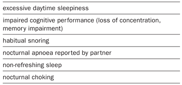
Table 1.
Symptoms of the obstructive sleep apnoea syndrome.
syndromes with central sleep apnoea and periodic breathing during sleep, and alveolar hypoventilation syndromes.
Obstructive sleep apnoea syndrome
The obstructive sleep apnoea/hypopnoea syndrome (OSAS) affects 2–4% of middle-aged men and 1–2% of middle-aged women [8]. It results from repetitive occlusion of the upper airway during sleep, causing hypopnoeas or apnoeas with oxygen desaturations and arousals [9]. The upper airway lumen in patients with OSAS may be reduced even during wakefulness [10]. In particular, in obese individuals, lateral fat deposits and hypertrophic pharyngeal walls encroach upon the lumen of the pharynx and thereby predispose to OSAS [11]. Furthermore, subtle abnormalities of the cranio-facial skeleton are common in patients with OSAS [12]. However, in many patients no particular anatomic abnormalities can be found and the sleep-related upper airway obstruction is purely functional. It relates to a reduced tonic input to the upper airway dilating muscles during sleep which promotes upper airway collapse [13]. Familial aggregation of OSAS suggests a role of genetic factors [14].
Clinical presentation
Symptoms
The principal symptoms of OSAS are excessive daytime sleepiness, unrefreshing sleep, habitual snoring and nocturnal apnoeas observed by the bed partner (table 1) [7]. It is important to find out whether a patient falls asleep against his or her will in dangerous situations, such as while driving. Some patients realise that their ability to concentrate and their work performance is impaired before they become excessively sleepy. Importantly, in children the leading symptom of OSAS may not be hypersomnolence but hyperactivity and other abnormal daytime behaviour, learning difficulties at school, failure to thrive or obesity [15]. In some cases OSAS may not be diagnosed until pulmonary hypertension and cor pulmonale have developed [16].
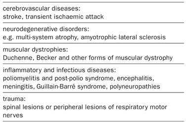
Table 2.
Neurologic disorders associated with sleep-related breathing disturbances.
Findings
Obesity, and a large neck size are common in patients with OSAS [17]. Retrognathia predisposes to OSAS, but visual inspection only detects gross abnormalities [10]. The size of tonsils must be assessed as enlargement is a treatable cause of OSAS, particularly in children [10,15]. The presence of macroglossia and other clinical features may suggest hypothyroidosis or acromegaly as primary causes underlying an OSAS [18]. Impaired nasal respiration which is common in OSAS may aggravate dryness of the throat, a typical complaint of snorers [19]. However, its significance in promoting sleep-related breathing disorders is not clear. As systemic hypertension is associated with OSAS, blood pressure measurement is crucial [20]. In neurologic practice, patients may present with various neurologic disorders that are associated with sleep-related breathing disturbances [21]. Examples are listed in table 2.
Consequences of OSAS
The most relevant and immediate risk from OSAS relates to falling asleep at the wheel [22,23]. This does not only endanger affected patients but others as well. The risk of causing traffic accidents is elevated several-fold in OSAS. The accidents typically occur at night and in the early morning hours, while driving alone on monotonous roads (highways) [24]. As the vehicle usually crashes unbraked into an obstacle, the consequences are particularly grave. The evidence is increasing that OSAS is an independent risk factor for arterial hypertension [20]. Furthermore, OSAS is associated with cardiovascular diseases (stroke, myocardial infarction), coagulopathies and metabolic disturbances [25,26,27,28].
Diagnosis
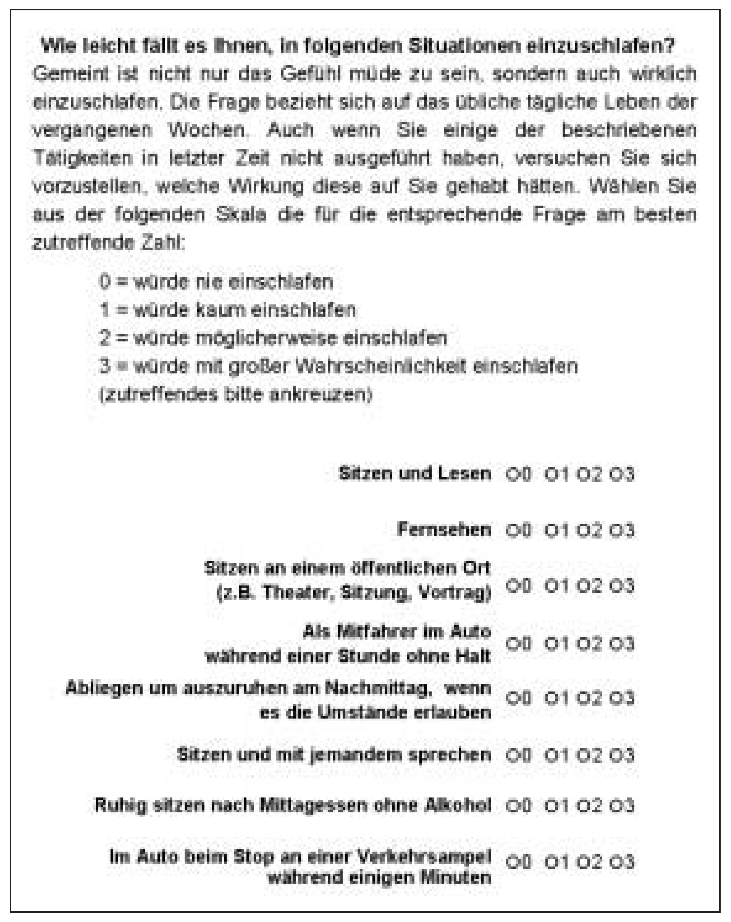
The mainstay of the diagnosis of OSAS consists in a typical history with irresistible daytime sleepiness, habitual snoring and apnoeas during sleep reported by the bed partner. Subjective sleepiness can be quantified by the Epworth sleepiness scale, a widely-used standardized questionnaire (fig. 1) [29,30].The scores derived from this questionnaire correlate with the severity of nocturnal breathing disturbances in patients with OSAS [29,30] and with the risk of traffic accidents in snorers [31,32]. The diagnosis of OSAS is established by a sleep study. Video-polysomnography performed in the sleep laboratory is the standard test that quantifies breathing disturbances in relation to sleep disruption and allows recognition of potential other accompanying sleep disorders [33]. In adults,
Figure 1.
The Epworth sleepiness scale is a widely used questionnaire that evaluates the subjectively perceived tendency of falling asleep in 8 different situations of everyday life. The individual item scores are summed to the total score, which has a range from 0 to 24. Validation of the German version of the scale in 159 subjects without sleep disorders revealed a mean value of 5.7 points and 95% confidence intervals ranging from 0 to 11.6 points [30].
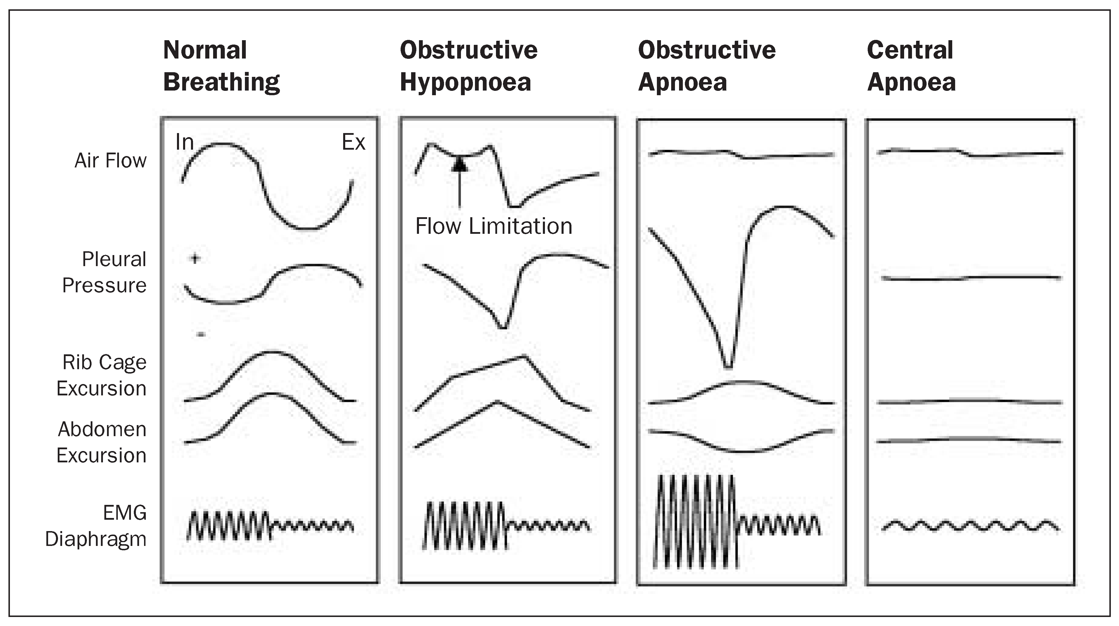
apnoea and hypopnoea are defined as cessation or reduction in airflow for at least 10 seconds, respectively. Depending on whether there is persistent respiratory effort during apnoea/hypopnoea, the event is classified as obstructive (related to upper airway obstruction) or central (reduced or absent respiratory centre drive) (fig. 2 and 3). Whether an oxygen desaturation (e.g. by ≥3%) or an arousal have to be associated with these events and which techniques are employed for monitoring ventilation are still a matter of debate [34,35]. The number of apnoeas and hypopnoeas per hour of sleep is called apnoea/hypopnoea index and serves as a measure of the degree of sleep-disordered breathing in sleep apnoea. The correlation of this index with clinical symptoms is loose, and there is no clear-cut threshold level for abnormal values. In the Wisconsin sleep cohort study, even an apnoea/hypopnoea index of 0 > to 5 per hour was associated with an increased risk of arterial hypertension in comparison to an index of 0 [20].
If the clinical suspicion of OSAS is high, a respiratory polygraphy (without recording of EEG, EOG and EMG) in the hospital ward or at home may confirm the diagnosis [36]. However, these simplified studies do not allow to exclude mild breathing disturbances associated with sleep disruption such as the upper airway resistance syndrome (fig. 4) [37,38]. Nocturnal pulseoximetry may reveal the cyclic oxygen desaturations typical of OSAS but it does not differentiate between obstructive and central events. It is therefore not an appropriate basis for prescribing long-term therapy for OSAS. Daytime vigilance tests such as the multiple sleep latency or maintenance of wakefulness tests [39] and driving simulator tests are performed in OSAS patients to objectively assess sleepiness and the effect of treatment. In the differential diagnosis of OSAS a variety of other disorders that are also associated with excessive daytime sleepiness have to be considered. These include chronic sleep deprivation and poor sleep hygiene, narcolepsy and several internal medical, neurologic and psychiatric disorders [40].
Treatment
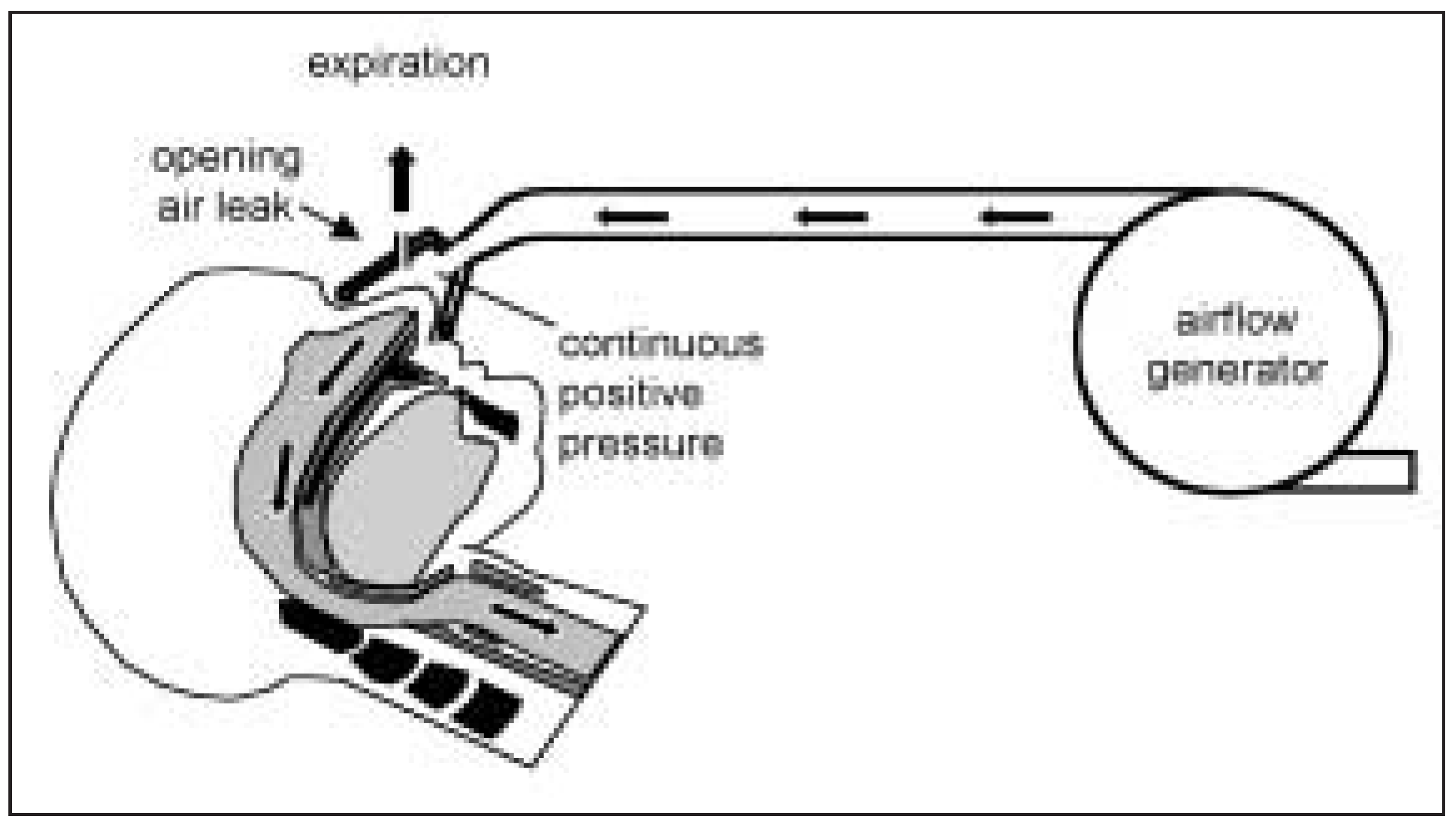
Nocturnal application of continuous positive airway pressure (CPAP) via a nasal mask is the standard therapy for OSAS (fig. 5) [5]. It improves excessive daytime sleepiness and other symptoms, quality of life, vigilance, cognitive performance and nocturnal breathing disturbances and oxygenation [41,42,43]. Furthermore, blood pressure variation associated with obstructive apnoea/hypopnoea
Figure 2.
In adults, apnoea and hypopnoea are defined as a cessation or reduction in airflow for more than 10 seconds. Obstructive and central apnoea/hypopnoea are discriminated by the presence or absence of respiratory efforts as evidenced by oesophageal pressure or diaphragmatic EMG recordings. Asynchrony or paradoxical motion of rib cage and abdomen recorded by inductance plethysmography may provide indirect evidence of increased respiratory effort. During partial upper airway obstruction, inspiratory flow is reduced which results in a flattened contour of inspiratory flow.
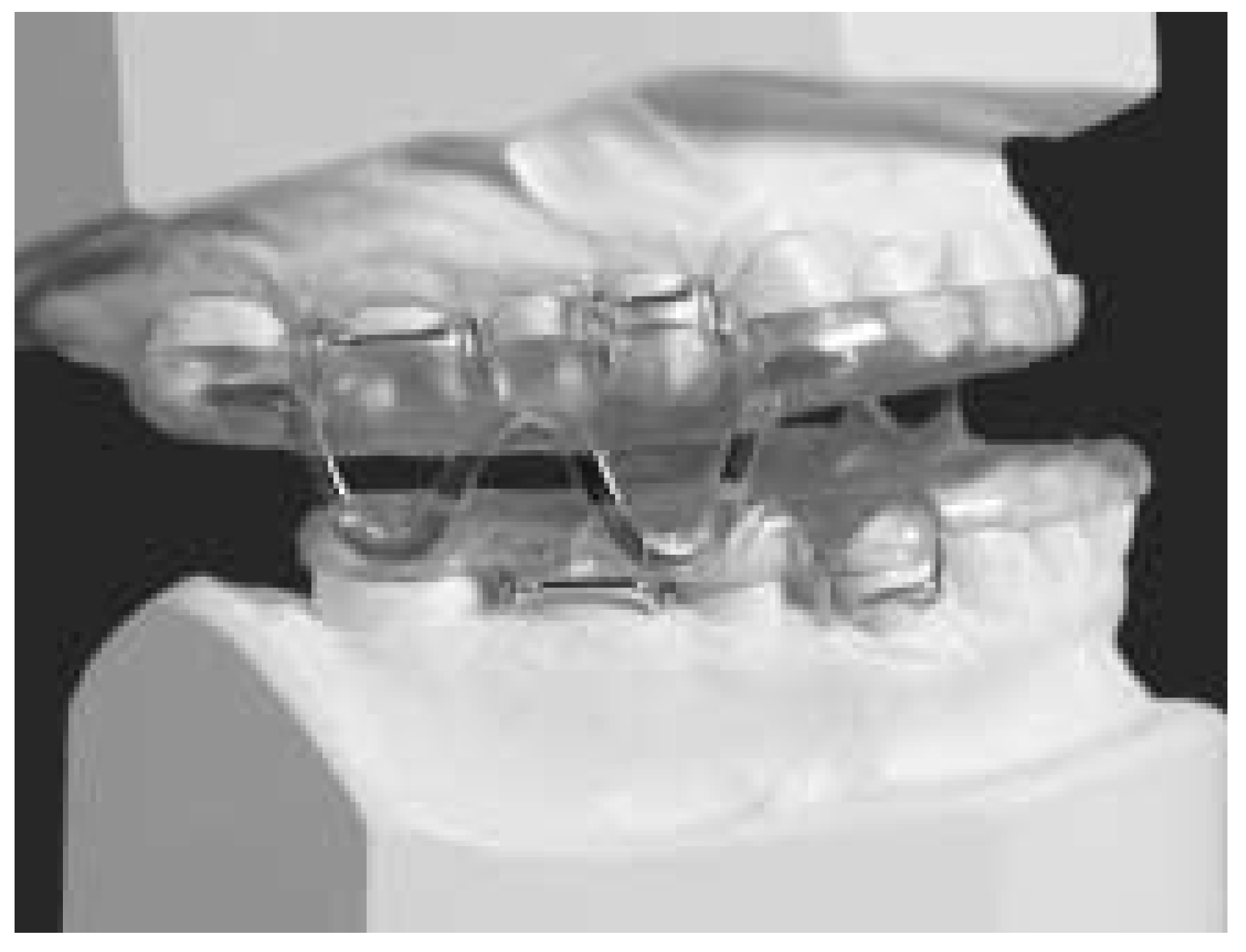
and excessive sympathetic tone are also improved [44]. The indication for CPAP therapy is based on the combination of symptoms and objective sleep study findings. The compliance with CPAP therapy is strongly influenced by the severity of the patients’ symptoms, the benefit they perceive by the treatment and the support given by their physician and other health care workers [45]. The nightly use of CPAP ranges from 5 to 7 hours per night in severe sleep apnoea [42,46] to 2 to 3 hours in mild OSAS [47]. Certain patients refuse CPAP because of the inconvenience, side effects (such as mask discomfort) and for psychologic reasons. For these patients adaptation of a removable oral appliance that is snapped onto the teeth during sleep is a valuable alternative treatment (fig. 6) [48]. The device is designed to advance the mandible (usually by 5 to 10 mm), thereby enlarging the upper airway calibre. Several randomised trials have documented effectiveness of oral appliance therapy for OSAS in alleviation of symptoms, quality of life and sleep-disordered respiration in OSAS [49]. The long-term effects on teeth and temporo-mandibular joints have to be carefully evaluated [50].
With the exception of adeno-tonsillectomy in children and adults with significantly enlarged adenoids and tonsils surgical treatment for OSAS is rarely indicated as first-line therapy for OSAS.
Although commonly performed for snoring and OSAS, uvulopalatopharyngoplasty (UPPP) and novel surgical techniques such as radiofrequency tissue ablation and laser-assisted uvulopalatoplasty have not scientifically been proven to be effective treatments for OSAS [51].
Specific pharmacologic treatment of OSAS is not available. Stimulant drugs (modafinil) are rarely used as an adjunct in selected patients with OSAS in whom excessive sleepiness persists despite optimal adaptation of CPAP therapy [52].
Central sleep apnoea
Central apnoea is the result of insufficient and unstable ventilatory motor output. It occurs occasionally in normal subjects, particularly at sleep onset and in REM sleep, and during stay at high altitude. Patterns of waxing and waning ventilation with repetitive central apnoea/hypopnoea may be associated with cyclical sleep disruption causing nocturnal dyspnoea, unrefreshing sleep and daytime sleepiness [7]. This syndrome, termed central sleep apnoea syndrome, may occur without associated disease or, more frequently, in association with congestive heart failure or neurologic disease, especially stroke. Central sleep apnoea and Cheyne-Stokes respiration in congestive heart
Figure 3.
Upper panel: Polysomnographic recording from a patient with severe congestive heart failure and central sleep apnoea. The waxing and waning of ventilation is associated with repetitive arousals. ECG-derived heart rate shows frequent extrabeats. During apnoea, rib cage and abdominal volumes estimated by calibrated inductance plethysmography show no evidence of volume changes suggesting absence of effort. Pressure at the nares is recorded as a surrogate of airflow. Lower panel: Recordings obtained from a patient with obstructive sleep apnoea. Progressive oesophageal pressure swings recorded with a balloon catheter and paradoxical signals of rib cage and abdominal inductance sensors (arrows) indicatethat the apnoeas are obstructive. They are associated with oxygen desaturations, cyclic alterations in heart rate and arousals (not shown).
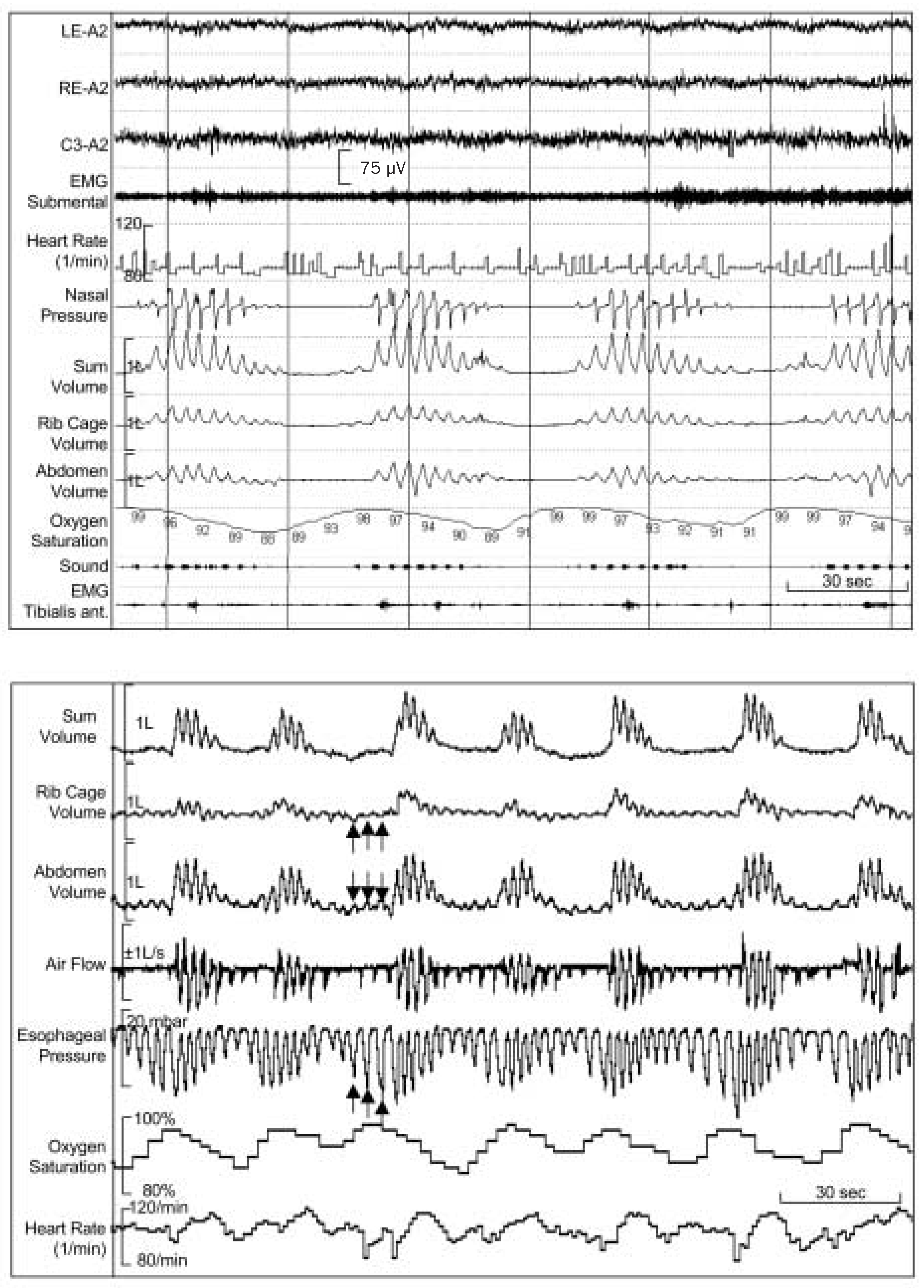
Figure 4.
Polysomnographic recording from a hypersomnic snorer with repetitive partial upper airway obstruction during sleep without apnoea or hypopnoea but with frequent arousals (upper airway resistance syndrome). To overcome increased upper airway resistance a high effort is required. This is reflected in progressive asynchrony of rib cage and abdominal excursions before an arousal occurs (pre). During the arousal (A), rib cage and abdomen move in synchrony but after a few breaths desynchronisation recommences (post). The derivative of the inductance waveform (dV/dt) corresponds to airflow. Before and after the arousal the shape of the inspiratory curve is truncated. This suggests flow limitation (FL). LE-A2, RE-A2, C3A2: electro-oculogram and electroencephalogram channels; EMG: submental electromyogram; Therm.: oral-nasal temperature reflecting airflow; Sum, RC, AB: sum, rib cage and abdominal volume signals recorded by calibrated inductance plethysmography; dV/dtRIP: derivative of inductance plethysmographic sum volume signal reflecting airflow; Sound: snoring sounds recorded by a microphone taped to the skin at the level of the larynx. Adapted from Bloch et al. [38].
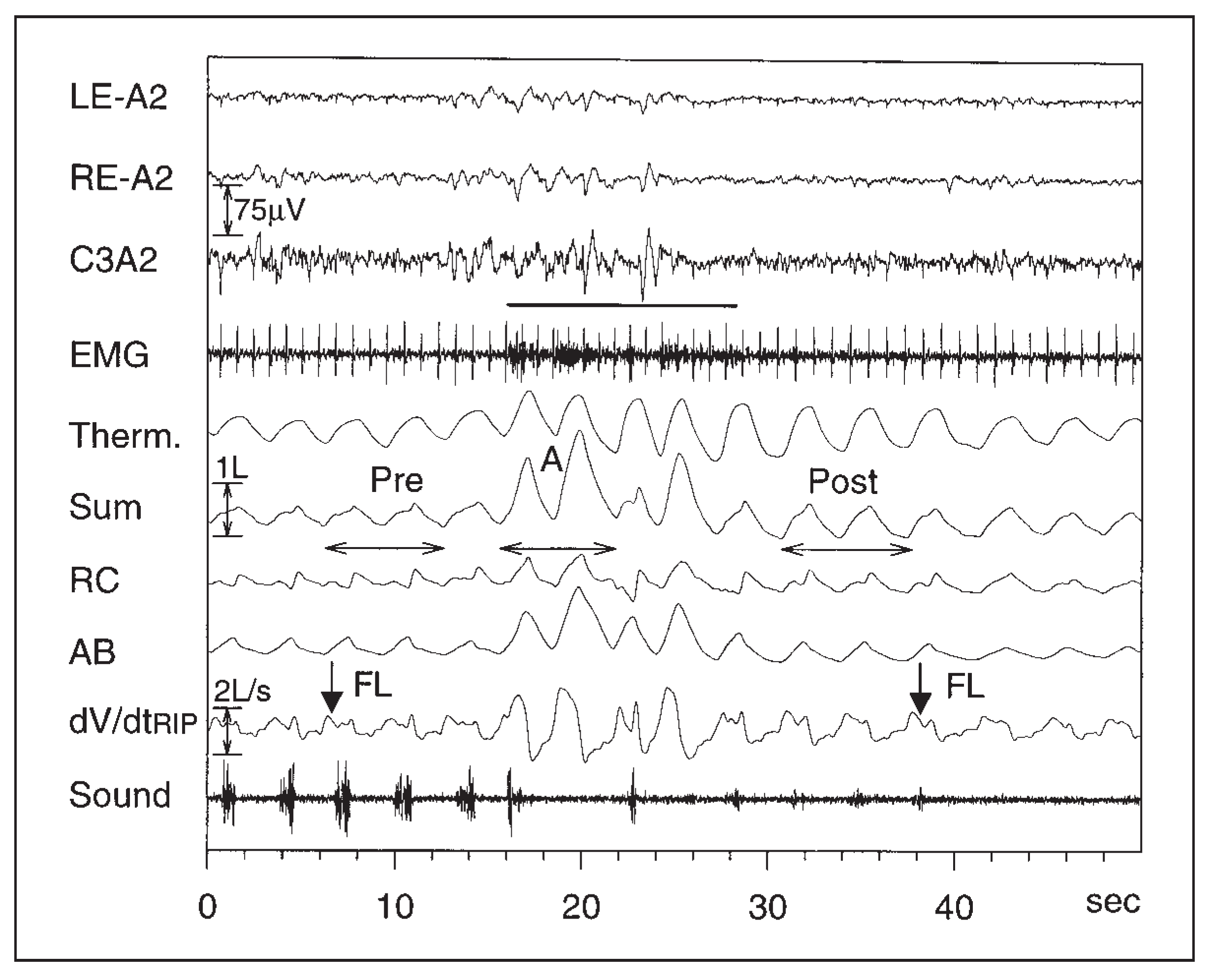
Figure 5.
Continuous positive airway pressure (CPAP) ventilation applied via a nasal mask prevents obstructive apnoea and hypopnoea during sleep by pneumatic splinting of the upper airway. A turbine pump generates a high flow rate, which is continuously adjusted to maintain a constant supra-atmospheric pressure within the tubing, mask and airways. A leak valve built into the tightly fitting nasal mask allows CO2 washout by continuous exsufflation of excessive air.
failure have recently gained renewed attention due to the high prevalence and the associated poor prognosis [53]. In one study [54] 51% of patients with heart failure (left ventricular ejection fraction <45%) had more than 15 apnoeas/hypopnoeas per hour of sleep. Preliminary data from randomised trials suggest that nocturnal CPAP therapy improves symptoms and prognosis in central sleep apnoea due to heart failure [55]. Novel modes of computer-controlled assisted ventilation have provided promising results [56]. Oxygen therapy given by nasal cannulas and theophyllin-medication have been shown to improve central sleep apnoea. However, the effect of these measures on other clinical outcomes has not been evaluated rigorously.
Chronic alveolar hypoventilation syndromes
Hypoventilation refers to insufficient ventilation in relation to the metabolic demands. It may occur secondary to chronic obstructive lung disease, obesity, chest-wall deformities and neurologic disorders (e.g. muscular dystrophy, amyotrophic lateral sclerosis, spinal lesions) or, rarer, as primary central alveolar hypoventilation syndrome. Despite the presence of hypercapnia and hypoxaemia, many patients are free of respiratory complaints but may suffer from excessive daytime sleepiness, disrupted sleep and morning headaches [7].
While long-term oxygen therapy has been shown to improve survival in hypoxaemic patients with chronic obstructive lung disease [57], the effectiveness of this therapy in patients with chronic alveolar hypoventilation due to other causes including obesity (obesity hypoventilation syndrome) is not established by robust scientific evidence. The role of respiratory stimulants and the carboanhydrase inhibitor acetazolamid in the treatment of hypoventilation is controversial. As hypercapnic patients with neuromuscular, chestwall or lung disease generally have an increased respiratory centre drive, administration of acetazolamid may promote respiratory failure due to the increased acid load that stimulates ventilation even more and may lead to respiratory muscle fatigue.Thus, in symptomatic hypoventilation, noninvasive positive airway pressure ventilation via a nasal or face mask is the treatment of choice [58]. It may reverse hypercapnia and improve symptoms, quality of life and survival in certain diseases. For example, in patients with Duchenne’s muscular dystrophy life expectancy has increased from less than 20 to 25–30 years since introduction of
Figure 6.
Removable oral appliances, such as the sleep apnoea monobloc shown here, are snapped onto the teeth during sleep and prevent obstructive apnoea by forcing the mandible in a forward position (protrusion) thereby enlarging the pharyngeal lumen. Adapted from Bloch et al. [48].
long-term ventilation [59]. The indication for assisted ventilation has to be carefully evaluated and is usually based on documentation of both hypercapnia and associated symptoms. Prophylactic assisted ventilation in asymptomatic patients with Duchenne’s muscular dystrophy is not warranted, since it has been associated with an increased death rate [60]. When counselling patients with progressive diseases such as amyotrophic lateral sclerosis, social, ethical and other aspects in addition to the medical ones have to be considered and discussed with the patient and his or her family before long-term home ventilation is initiated.
Conclusions
Sleep-related breathing disorders are common and may cause significant morbidity, mainly through excessive daytime sleepiness and cognitive impairment and even mortality due to accidents and cardiovascular complications. As effective treatment is available, a timely diagnosis is crucial.
References
- Kryger, MH. Sleep apnea. From the needles of Dionysius to continuous positive airway pressure. Arch Intern Med 1983, 143, 2301–3. [Google Scholar] [CrossRef] [Scilit] [PubMed]
- Dickens, C. The Posthumous Papers of the Pickwick Club; Chapman and Hall: London, 1835. [Google Scholar]
- Gastaut, HCA. Diurnal and nocturnal episodal manifestations of Pickwick syndrome. Rev Neurol 1965, 18, 568–79. [Google Scholar]
- Kuhlo, W; Doll, E; Franck, MC. Erfolgreiche Behandlung eines Pickwick-Syndromes durch eine Dauertrachealkanüle. Dtsch Med Wochenschr 1969, 94, 1286–90. [Google Scholar] [CrossRef] [Scilit] [PubMed]
- Sullivan, CE; Issa, FG; Berthon-Jones, M; Eves, L. Reversal of obstructive sleep apnoe by continuous positive airway pressure applied through the nares. Lancet 1981, 1, 862–5. [Google Scholar] [CrossRef] [Scilit]
- Young, T; Evans, L; Finn, L; Palta, M. Estimation of the clinically diagnosed proportion of sleep apnea syndrome in middle-aged men and women. Sleep 1997, 20, 705–6. [Google Scholar] [CrossRef] [Scilit]
- Douglas, NJ. Clinician’s Guide to Sleep Medicine, 1st edition; Arnold: London, 2002. [Google Scholar]
- Young, T; Palta, M; Dempsey, J; Skatrud, J; Weber, S; Badr, S. The occurrence of sleep-disordered breathing among middle-aged adults. N Engl J Med 1993, 328, 1230–5. [Google Scholar] [CrossRef] [Scilit]
- Martin, SE; Engleman, HM; Deary, IJ; Douglas, NJ. The effect of sleep fragmentation on daytime function. Am J Respir Crit Care Med 1996, 153, 1328–32. [Google Scholar] [CrossRef] [Scilit]
- Tsai, WH; Remmers, JE; Brant, R; Flemons, WW; Davies, J; Macarthur, C. A decision rule for diagnostic testing in obstructive sleep apnea. Am J Respir Crit Care Med 2003, 167, 1427–32. [Google Scholar] [CrossRef] [Scilit]
- Schwab, RJ; Pasirstein, M; Pierson, R; Mackley, A; Hachadoorian, R; Arens, R; et al. Identification of upper airway anatomic risk factors for obstructive sleep apnea with volumetric MRI. Am J Respir Crit Care Med 2003, 168, 522–30. [Google Scholar] [CrossRef]
- Kushida, CA; Efron, B; Guilleminault, C. A predictive model for the obstructive sleep apnea syndrome. Ann Intern Med 1997, 127, 581–7. [Google Scholar] [CrossRef] [Scilit]
- Issa, FG; Sullivan, CE. Upper airway closing pressures in obstructive sleep apnea. J Appl Physiol 1984, 57, 520–7. [Google Scholar] [CrossRef] [Scilit] [PubMed]
- Redline, S; Tishler, PV; Tosteson, TD; Williamson, J; Kump, K; Browner, I; et al. The familial aggregation of obstructive sleep apnea. Am J Respir Crit Care Med 1995, 151, 682–7. [Google Scholar] [CrossRef] [Scilit] [PubMed]
- Guilleminault, C; Korobkin, R; Winkle, R. A review of 50 children with obstructive sleep apnea syndrome. Lung 1981, 159, 275–87. [Google Scholar] [CrossRef] [Scilit] [PubMed]
- Bloch, KE; Witztum, A; Wieser, HG; Russi, E. Obstruktives Schlaf-Apnoe-Syndrom bei einem Kind mit Trisomie 21. Monatsschr Kinderheilkd 1990, 138, 817–22. [Google Scholar]
- Viner, S; Szalai, JP; Hoffstein, V. Are history and clinical examination a good screening test for sleep apnea? Ann Intern Med 1991, 115, 356–9. [Google Scholar] [CrossRef] [Scilit]
- Grunstein, RR; Ho, KY; Sullivan, CE. Sleep apnea in acromegaly. Ann Intern Med 1991, 115, 532. [Google Scholar] [CrossRef] [Scilit]
- Young, T; Finn, L; Palta, M. Chronic nasal congestion at night is a risk factor for snoring in a population-based cohort study. Arch Intern Med 2001, 161, 1514–9. [Google Scholar] [CrossRef] [Scilit]
- Peppard, PE; Young, T; Palta, M; Skatrud, J. Prospective study of the association between sleep-disordered breathing and hypertension. N Engl J Med 2000, 342, 1378–84. [Google Scholar] [CrossRef] [Scilit]
- Bassetti, CL; Gugger, M. Sleep-disordered breathing in neurologic diseases. Swiss Med Wkly 2002, 132, 109–15. [Google Scholar] [CrossRef] [Scilit]
- Laube, I; Seeger, R; Russi, EW; Bloch, KE. Accidents related to sleepiness: review of medical causes and prevention with special reference to Switzerland. Schweiz Med Wochenschr 1998, 128, 1487–99. [Google Scholar]
- Horstmann, S; Hess, CW; Bassetti, C; Gugger, M; Mathis, J. Sleepiness-related accidents in sleep apnea patients. Sleep 2000, 23, 383–9. [Google Scholar] [CrossRef] [Scilit] [PubMed]
- Horne, JA; Reyner, LA. Sleep-related vehicle accidents. Br Med J 1995, 310, 565–7. [Google Scholar] [CrossRef] [Scilit] [PubMed]
- Wessendorf, TE; Thilmann, AF; Wang, YM; Schreiber, A; Konietzko, N; Teschler, H. Fibrinogen levels and obstructive sleep apnea in ischemic stroke. Am J Respir Crit Care Med 2000, 162, 2039–42. [Google Scholar] [CrossRef] [Scilit] [PubMed]
- Chin, K; Shimizu, K; Nakamura, T; Narai, N; Masuzaki, H; Ogawa, Y; et al. Changes in intra-abdominal visceral fat and serum leptin levels in patients with obstructive sleep apnea syndrome following nasal continuous positive airway pressure therapy. Circulation 1999, 100, 706–12. [Google Scholar] [CrossRef] [Scilit]
- Punjabi, NM; Sorkin, JD; Katzel, LI; Goldberg, AP; Schwartz, AR; Smith, PL. Sleep-disordered breathing and insulin resistance in middle-aged and overweight men. Am J Respir Crit Care Med 2002, 165, 677–82. [Google Scholar] [CrossRef] [Scilit]
- Bassetti, C; Aldrich, MS. Sleep apnea in acute cerebrovascular diseases: final report on 128 patients. Sleep 1999, 22, 217–23. [Google Scholar] [CrossRef] [Scilit]
- Johns, MW. Daytime sleepiness, snoring and obstructive sleep apnea. The Epworth sleepiness scale. Chest 1993, 103, 30–6. [Google Scholar] [CrossRef] [Scilit]
- Bloch, KE; Schoch, O; Zhang, JN; Russi, EW. German version of the Epworth Sleepiness Scale. Respiration 1999, 66, 440–7. [Google Scholar] [CrossRef] [Scilit]
- Maycock, G. Sleepiness and driving: the experience of heavy goods vehicle drivers in the UK. J Sleep Res 1997, 6, 238–44. [Google Scholar] [CrossRef] [Scilit]
- Maycock, G. Sleepiness and driving: the experience of UK car drivers. J Sleep Res 1996, 5, 229–37. [Google Scholar] [CrossRef] [Scilit]
- Bloch, KE. Polysomnography: a systematic review. Technol Health Care 1997, 5, 285–305. [Google Scholar] [CrossRef] [Scilit]
- American Academy of Sleep Medicine Task Force. Sleeprelated breathing disorders in adults: recommendations for syndrome definition and measurement techniques in clinical research. Sleep 1999, 22, 667–89. [Google Scholar] [CrossRef] [Scilit]
- Thurnheer, R; Xie, X; Bloch, KE. Accuracy of nasal cannula pressure recordings for assessment of ventilation during sleep. Am J Respir Crit Care Med 2001, 164, 1914–9. [Google Scholar] [CrossRef] [Scilit] [PubMed]
- Standards of Practice Committee of the American Sleep Disorders Association. Practice parameters for the use of portable recording in the assessment of obstructive sleep apnea. Sleep 1994, 17, 372–7. [Google Scholar] [CrossRef] [Scilit]
- Guilleminault, C; Stoohs, R; Clerk, A; Cetel, M; Maistros, P. A cause of excessive daytime sleepiness. The upper airway resistance syndrome. Chest 1993, 104, 781–7. [Google Scholar] [CrossRef] [Scilit]
- Bloch, KE; Li, Y; Sackner, MA; Russi, EW. Breathing patterns during sleep disruptive snoring. Eur Respir J 1997, 10, 576–86. [Google Scholar] [CrossRef] [Scilit]
- American Sleep Disorder Association. The clinical use of the multiple sleep latency test. Sleep 1992, 15, 268–76. [Google Scholar] [CrossRef] [Scilit]
- Bassetti, C; Gugger, M. Hypersomnia – etiology, clinic, diagnosis and therapy of excessive sleepiness. Ther Umsch 2000, 57, 421–9. [Google Scholar] [CrossRef] [Scilit]
- Engleman, HM; Martin, SE; Deary, IJ; Douglas, NJ. Effect of continuous positive airway pressure treatment on daytime function in sleep apnoe/hypopnoe syndrome. Lancet 1994, 343, 572–5. [Google Scholar] [CrossRef] [Scilit]
- Jenkinson, C; Davies, RJO; Mullins, R; Stradling, JR. Comparison of therapeutic and subtherapeutic nasal continuous positive airway pressure for obstructive sleep apnoea: a randomised prospective parallel trial. Lancet 1999, 353, 2100–5. [Google Scholar] [CrossRef] [Scilit]
- Senn, O; Brack, T; Matthews, F; Russi, EW; Bloch, KE. Randomized short-term trial of two auto CPAP devices vs. fixed CPAP for treatment of sleep apnea. Am J Respir Crit Care Med In press. 2003. [Google Scholar] [CrossRef] [Scilit] [PubMed]
- Lattimore, JD; Celermajer, DS; Wilcox, I. Obstructive sleep apnea and cardiovascular disease. J Am Coll Cardiol 2003, 41, 1429–37. [Google Scholar] [CrossRef] [Scilit] [PubMed]
- Chervin, RD; Theut, S; Bassetti, C; Aldrich, MS. Compliance with nasal CPAP can be improved by simple interventions. Sleep 1997, 20, 284–9. [Google Scholar] [CrossRef] [Scilit] [PubMed]
- Kaplan, V; Bingisser, R; Li, Y; Hess, T; Russi, EW; Bloch, KE. Compliance mit nasaler Überdruckbehandlung (CPAP) bei obstruktivem Schlafapnoe Syndrom. Schweiz Med Wochenschr 1996, 126, 15–21. [Google Scholar]
- Engleman, HM; Kingshott, RN; Wraith, PK; Mackay, TW; Deary, IJ; Douglas, N. Randomized placebo-controlled crossover trial of continuous positive airway pressure for mild sleep apnea/hypopnea syndrome. Am J Respir Crit Care Med 1999, 159, 461–7. [Google Scholar] [CrossRef] [Scilit]
- Bloch, KE; Iseli, A; Zhang, JN; Xie, X; Stoeckli, PW; Russi, EW. Randomized, controlled trial of two oral appliances for sleep apnea treatment. Am J Respir Crit Care Med 2000, 162, 246–51. [Google Scholar] [CrossRef] [Scilit]
- Bloch, KE; Senn, O; Iseli, A. Oral appliances for treatment of snoring and obstructive sleep apnea. In Surgery for Snoring and Obstructive Sleep Apnea Syndrome, 1st edition; Fabiani, M, Saponara, M, Eds.; Kugler Publications: The Hague, the Netherlands, 2003; pp. 559–75. [Google Scholar]
- Fritsch, K; Iseli, A; Russi, EW; Bloch, KE. Side effects of mandibular advancement devices for sleep apnea treatment. Am J Respir Crit Care Med 2001, 164, 813–8. [Google Scholar] [CrossRef] [Scilit]
- Scherler, MR. Surgical options for treatment of snoring and sleep apnea. Ther Umsch 2000, 57, 454–7. [Google Scholar] [CrossRef] [Scilit]
- Pack, AI; Black, JE; Schwartz, JR; Matheson, JK. Modafinil as adjunct therapy for daytime sleepiness in obstructive sleep apnea. Am J Respir Crit Care Med 2001, 164, 1675–81. [Google Scholar] [CrossRef] [Scilit]
- Bradley, TD; Floras, JS. Sleep apnea and heart failure: Part II: central sleep apnea. Circulation 2003, 107, 1822–6. [Google Scholar] [CrossRef] [Scilit]
- Javaheri, S; Parker, TJ; Liming, JD; Corbett, WS; Nishiyama, H; Wexler, L; et al. Sleep apnea in 81 ambulatory male patients with stable heart failure. Types and their prevalences, consequences, and presentations. Circulation 1998, 97, 2154–9. [Google Scholar] [CrossRef] [Scilit]
- Javaheri, S. Effects of continuous positive airway pressure on sleep apnea and ventricular irritability in patients with heart failure. Circulation 2000, 101, 392–7. [Google Scholar] [CrossRef] [Scilit]
- Wisskirchen, T; Teschler, H. Central sleep apnea syndrome and Cheyne-Stokes respiration. Ther Umsch 2000, 57, 458–62. [Google Scholar]
- Continuous or nocturnal oxygen therapy in hypoxemic chronic obstructive lung disease: a clinical trial. Nocturnal Oxygen Therapy Trial Group. Ann Intern Med 1980, 93, 391–8. [CrossRef] [Scilit]
- Mehta, S; Hill, NS. Noninvasive ventilation. Am J Respir Crit Care Med 2001, 163, 540–77. [Google Scholar] [CrossRef]
- Baydur, A; Gilgoff, I; Prentice, W; Carlson, M; Fischer, DA. Decline in respiratory function and experience with long-term assisted ventilation in advanced Duchenne’s muscular dystrophy. Chest 1990, 97, 884–9. [Google Scholar]
- Raphael, JC; Chevret, S; Chastang, C; Bouvet, F. Randomised trial of preventive nasal ventilation in Duchenne muscular dystrophy. French Multicentre Cooperative Group on Home Mechanical Ventilation Assistance in Duchenne de Boulogne Muscular Dystrophy. Lancet 1994, 343, 1600–4. [Google Scholar] [CrossRef] [Scilit]
This is an open-access article distributed under the terms of the Creative Commons Attribution Non-Commercial No Derivatives (CC BY-NC-ND) 4.0 License.