


Successful therapy of complete middle cerebral artery-(MCA-)territory ischemia by combination of very early hypothermia and decompressive hemicraniotomy: case report
E. Keller, D. Könü, T. Hegner, Y. Yonekawa, Zürich
The 31 years old man presented with SAH after rupture of a left MCA aneurysm. The patient was admitted after repeated generalized seizures with a neurological status rated Grade IV in the Hunt and Hess classification. CT scans were classified as Fisher’s Grade 3. The aneurysm was successfully clipped on day 1. Temporary clipping of the M1 segment was less than 10 minutes. After craniotomy the dura was under high tension and the brain was extensively swollen. Therefore the bone was not replaced at the end of the operation and a small duraplasty was performed. The following day the patient remained sedated and ventilated because of neurogenic pulmonary edema. On day 2 sedation was stopped and the patient woke up initially moving his arms and legs symmetrically. On day 3 within 1 hour he developed rightsided hemiplegia. CT scans showed early infarct signs of the complete MCA territory. The patient was immediately treated with pentobarbital 5 mg/KG i.v. as a loading dose, followed by EEG-controlled barbiturate coma until day 8. Mild hypothermia (33 °C–34 °C) was already induced before leftsided extention of decompressive recraniectomy was performed within 4 hours after symptom onset of ischemia. The following days transcranial doppler (TCD)-blood flow mean velocity in the left MCA increased to 230 cm/sec, highly suspicious for cerebral vasospasms. ICP remained elevated instead of mild hyperventilation, treatment with mannitol, hypertonic NaCI-hydroxyethylstarch solution and THAM-buffer. Therefore and with highly suspicion for cerebral vasospasm barbiturate coma was maintained for further 5 days and mild hypothermia for totally 8 days. CT scans on day 12 showed leftsided gyral contrast enhancement (fig 2) but no demarcating MCA-infarction developed in the course of the illness. After 2 months the patient showed no hemiparesis, walked alone and was talking short sentences. Imminent complete MCAterritory infarction could be prevented by an aggressive treatment regimen with combination of decompressive hemicraniotomy and neuroprotection with very early hypothermia and barbiturate coma.
Quality assessment of stroke unit management after modification of inclusion criteria
S.T. Engelter, P.A. Lyrer, S. Papa, H. Plansky, Basel
Background: Two-year evaluation after implementation of our multidisciplinary, stroke unit concept, showed, that only 36% of those patient who fulfilled the criteria for stroke unit treatment (SUT), had SUT, mainly due to limited resources. There were two consequences: first, a time-budget for SUT was given by the hospital administration. Second, criteria for participation on SUT were modified.
Objective: To evaluate, (1), whether the modified criteria were realized in clinical practice, (2), whether SUT within the given time budget was feasible, (3), whether the change in SUT criteria results in a different stroke profile.
Methods: Management of all patients with acute ischemic stroke was prospectively evaluated between April and October 1999 using standardized, prespecified criteria. For each patient stroke syndrome, (and in case of SUT) reason for SUT, duration of SUT, and medication were assessed and compared to a historical control population in 1997.
Results: In 1999, 71 of 195 (36%) patients had SUT. One patient (0.5%) had no SUT, despite SUT indication, whereas 4 of 71 (5.6%) received SUT, although criteria were not present. In comparison, in 1997, 41 of 162 (27%) patients had SUT (p <0.05), and another 18 (11%) had no SUT despite SUT indications (p <0.5 ). Mean SUT time was 39.8 ± 33.3 h, which was not significantly different to 1997 (mean SUT: 44.5 ± 51.1h; p >0.5).Total SUT time was 2902 h, which was higher than in 1997 (1821 h), but lower than the given time limit of 4416 h (calculated as “24 h SUT each day”). In 1999 SUT for lacunar infarcts (10%) was significantly less frequent than in 1997 (27%) (p <0.01), whereas frequencies of other stroke syndromes did no differ significantly.
Conclusion: Modified SUT criteria were realized with a low rate of misplacement. SUT was available for more patients than previously, but total SUT duration was shorter than the given time limit. Patients with lacunar syndromes seems to be less likely to receive SUT.
Schlaganfall und Wissenserwerb: Wissen über die Krankheit bei Patienten mit akutem Schlaganfall – Pilotstudie
H. Rettke, Ph. Lyrer, Basel
Einleitung: Der Schlaganfall stellt ein häufiges und komplikationsreiches Geschehen dar, das die Gesundheitssituation davon betroffener Patienten völlig und auf eine für sie bisher unbekannte Weise verändert. Zur Komplexität dieser Gesundheitssituation tragen weitere Faktoren bei, z.B. ein subjektives Krankheitserleben, das mit möglichen psychosozialen Verhaltensweisen nicht übereinstimmt oder der Beginn umfangreicher Diagnostik und Therapie bei Spitaleintritt. Um die Gesundheitssituation so selbständig wie möglich meister zu können, müssen die Betroffenen über entsprechendes Wissen verfügen. Dazu müssen sie wiederum lernfähig und lernbereit sein.
Untersuchungsfrage und Hypothesen: Ziel der Studie war, die Frage nach dem Ausmass zu beantworten, zu dem akute Schlaganfallpatienten fähig sind, gesundheitsbezogene und rehabilitationsbezogene Informationen aufzunehmen und wieder abzurufen. Die Hypothesen besagten, dass akute Schlaganfallpatienten dazu in der Lage sind, dass sich deren Leistungen nach Läsionsort und von Patienten ohne Schlaganfall unterscheiden.
Methode: Eine Gelegenheitsstichprobe ergab 12 Teilnehmer mit hemisphärischem Infarkt, 8 Teilnehmer mit lakunärem Infarkt und 9 Teilnehmer ohne Schlaganfall in der Kontrollgruppe. Mittels standardisierter Befragung wurde der Wissensstand aller Teilnehmer erhoben. Dem schloss sich eine strukturierte Wissensvermittlung an, nach der wiederum der Wissensstand erhoben wurde. Nach fünf Tagen wurde die standardisierte Befragung nochmals vorgelegt.
Resultate: Akute Schlaganfallpatienten waren in der Lage, die gebotenen Informationen aufzunehmen und wieder abzurufen. Darin unterschieden sie sich nicht von der Kontrollgruppe. Unterschiedliche Leistungen in Bezug auf den Läsionsort konnten nicht nachgewiesen werden.
Schlussfolgerungen: Die Fähigkeit akuter Schlaganfallpatienten, Wissen erwerben und wieder abrufen zu können, stellt eine Ressource für die Rehabilitation in der Akutphase dieser Patienten dar. Sowohl die Form als auch der Inhalt strukturierter Wissensvermittlung beugen einer zufälligen und inkonsistenten Wissensweitergabe vor. Dabei dürfen die situationsspezifischen Informationsbedürfnisse der Betroffenen nicht unbeachtet bleiben. Ebenso müssen Informationsbedürfnisse von Angehörigen berücksichtigt werden. Weitere Forschung ist notwendig, um das Lernverhalten akuter Schlaganfallpatienten zu erhellen und den Nutzen strukturierter Wissensvermittlung für diese Patientengruppe nachzuweisen.
Better outcome after stroke with higher serum cholesterol levels
G.R. de Freitas, C. Vauthey, G. van Melle, G. Devuyst, J. Bogousslavsky, Lausanne
Objective: To examine whether serum cholesterol levels have any prognostic value in the first month following acute ischemic stroke.
Background: Although the association between serum cholesterol levels and cerebrovascular disorders has been extensively studied, the relationship between cholesterol levels and outcome following ischemic stroke has not been investigated.
Methods: Using data from 3,273 consecutive patients with first-ever ischemic stroke, we compared poor functional outcome (severe disability or death) at one month in patients with high cholesterol (total serum cholesterol greater than 6.5 mmol/l or 250 mg/dl) and normal cholesterol (level equal to, or less than, 6.5 mmol/l or 250 mg/dl). Data were analyzed by univariate and multivariate analysis.
Results: In comparison with patients with normal cholesterol levels, patients with high cholesterol levels had a 2.2-fold lower risk of death (P = 0.002) and a 2.1-fold lower risk of poor functional outcome at one month (P < 0.001). After adjustment for known confounding variables, multivariate analysis showed that higher cholesterol levels remained an independent predictor of better functional outcome (OR: 0.5, CI: 0.35–0.71, p <0.001).
Conclusions: Our findings suggest that higher levels of cholesterol are associated with a better outcome in the early phase after ischemic stroke.
Combined superficial and deep posterior cerebral artery infarcts in the Lausanne Stroke Registry
N. Afsar, V. Castillo, F. Ghika-Schmidt, G. Van Melle, J. Bogousslavsky, Lausanne
Objective and background: To determine the clinical, etiological and topographic correlations of combined superficial and deep posterior cerebral artery (PCA) infarct, with an emphasis on the neuropsychological findings.
Patients and methods: Consecutive first-ever stroke patients with combined superficial and deep PCA territory infarct were collected from the Lausanne Stroke Registry. Patients with concomitant lesions were excluded. All patients were investigated according a standard protocol including CT or MRI, MRA, Doppler ultrasound. A complete neuropsychological battery was applied to a selected group of patient.
Results: Among a total of 3901 patients, there were 383 (10%) with a PCA infarct, with only 57 patients (35 man and 22 woman) having a combined superficial and deep territory lesion (32 left, 25 right sided). The superficial lesions involved the occipital (100%) infero-medial temporal (51%) and posterior parietal (12%) areas, and deep lesions involved the thalamus (91%) and the midbrain (23%). The most common clinical findings were visual field defects (91%), neuropsychological abnormalities (77%), motor (65%) and sensory (68%) deficits. Movement disorders and oculomotor abnormalities were less frequent. Three patients died during early follow-up. The stroke mechanism was most commonly embolic from either cardiac or arterial sources.
Conclusion: This first report emphasizes the fact that combined superficial and deep PCA territory infarcts are rather uncommon in a communitybased registry. Moreover, the frequent association of visual field defects and neuropsychological findings to various degrees of motor or sensory losses seems to reflect the rather extensive lesions encountered in these cases.
Dural arteriovenous fistulas: detection by dynamic MR projection angiography
S. G. Wetzel, D. Bilecen, P. Lyrer, E. W. Radü, K. Scheffler, Basel und Freiburg i. Br.
Purpose: A new, contrast enhanced, two-dimensional MR projection angiography sequence that provides a high temporal resolution and enables a depiction of the entire cervicocranial vasculature was applied in three patients suspected to have a dural arteriovenous fistula (DAVF).
Methods: MR projection angiograms were acquired with a RF-spoiled, steady state gradient echo (GRASS) sequence [8] (flip angle 400, TE 1.8 ms, TR 4.5 ms, matrix size196 x 256, field of fiew 188 x 250 mm) without slice selection in the coronal plane. The sequence was run continuously to acquire 60 images with a temporal resolution of 900 ms per image. Start of the sequence was simultaneous with the injection of the contrast agent bolus (0.1 mmol per kg of body weight GdDTPA-BMA (Omniscan 0.5 mmol/ml, Nycomed Imaging AS, Norway). The bolus was injected at a flow rate of 3 ml/s by use of a power injector (Spectris MR Injector System, Medrad Inc., Pittsburgh, PA, USA), immediately followed by a 20 ml saline solution flush through the right antecubital vein. In all patients an intraarterial DSA and a standard SE-MRI in axial plane was performed.
Results: In all three patients, a DAVF was clearly recognized as confirmed by intraarterial DSA, while MR images did not allow to establish the diagnosis. The information about contrast agent dynamics by MR projection angiography enabled thereby to define the type of DAVF.
Conclusion: These preliminary results indicate, that MR projection angiography is a highly useful and easy-to-perform tool to detect DAVF.
Diabète et AVC sous-corticaux
G. Devuyst, G. R. de Freitas, N. Cals, N. Afsar, J. Bogousslavsky, Lausanne
Objectifs: Bien que le diabète soit un facteur de risque cérébrovasculaire établi, des incertitudes demeurent sur le type d’AVC induit par le diabète. Le but de ce travail est d’examiner les sous-types d’AVC et leur pronostic chez les patients diabétiques. Nous avons également voulu explorer l’association diabète-infarctus sous-cortical chez des patients diabétiques nonhypertendus.
Méthodes: Nous avons étudiés les facteurs de risques, l’étiologie, la topographie, les signes cliniques et le pronostic des 3362 patients admis pour un premier AVC dans notre centre (LSR). Les patients étaient considérés comme diabétiques si ils avaient une histoire connue de diabète avant leur AVC (au moins deux résultats de glycémie >6 mmol/L).
Résultats: 472 patients avec AVC et diabète furent sélectionnés pour cette étude. Les patients diabétiques étaient plus âgés de 5.3 ans en moyenne que les non-diabétiques et avaient deux fois moins d’hémorragies cérébrales (p = 0.002). La microangiopathie cérébrale et l’atteinte athéromateuse des gros vaisseaux étaient plus fréquentes chez les diabétiques alors que les cardiopathies nonischémiques, les dissections artérielles et les causes indéterminées étaient moins fréquentes chez les patients diabétiques (p <0.001). Les infarctus cérébraux sous-corticaux étaient plus fréquents chez les diabétiques par rapport aux non-diabétiques (p <0.001). La sévérité de l’AVC, la mortalité et le pronostic fonctionnel neurologique étaient comparables entre les diabétiques et non-diabétiques. 39 patients diabétiques mais non-hypertendus présentaient un AVC sous-cortical: parmi ces 39 patients, 20 n’avaient que le diabète comme facteur de risque cérébrovasculaire.
Conclusions: Le diabète influence l’étiologie et la topographie de l’AVC mais pas le pronostic fonctionnel. Notre étude suggère que le diabète puisse être associé de façon indépendante aux infarctus cérébraux sous-corticaux.
Marfan’s syndrome with TIAs and multiple extracranial aneurysms: case report
R. Sztajzel, J. Le Floch-Rohr, Genf
Dans le syndrome de Marfan les anévrismes disséquants des artères cervicales extracrâniennes sont le plus souvent causés par une extension de la dissection aortique. Des anévrismes spontanés et isolés des artères cervicales ont aussi été rapportés dans le cadre de ce syndrome, mais beaucoup plus rarement. Dans ces cas-là, les anévrismes sont le plus souvent asymptomatiques ou se manifestent alors comme une masse cervicale pulsatile. Des accidents ischémiques transitoires ou constitués n’ont été que très rarement mentionnés dans cette pathologie. Nous rapportons l’observation d’un patient souffrant d’un syndrome de Marfan, présentant plusieurs AIT et chez lequel nous découvrons des anévrismes fusiformes des deux carotides internes ainsi qu'un anévrisme sacciforme de l’artère vertebrale.
Developmental venous anomalies with arteriovenous shunt: should they be considered benign lesions? Report of a case with thrombotic complication
S. Agazzi, L. Regli, A. Uske, Ph. Maeder, N. de Tribolet, Lausanne
Developmental venous anomalies (DVA), previously known as venous angioma, are the most common type of intracranial vascular malformations documented by autopsy series. They are thought to be congenital variations of normal venous drainage with a benign natural history and a low complication rate. A rare variant of DVA has been described where an angiographically identifiable arteriovenous shunt is present. We report the case of a patient who presented hemorrhage secondary to thrombosis in a complex anomalous hemispheric venous drainage, including two distinct DVAs, one of them with arteriovenous shunt (AVS). In the 45 cases of DVA with AVS reported in the literature, none presented a thrombosis, and thrombosis was only described in 7 cases of DVA. Controversial opinions are found in the literature as to whether DVAs with AVS are benign lesions, or if they should be considered as more aggressive vascular pathologies like arteriovenous malformations. The increasing number of reports of symptomatic DVAs with arteriovenous shunt raises the question whether symptoms, in a patient with a DVA, are related to the presence of the shunt and its concomitant hemodynamic alterations. If this hypothesis is accepted, angiography should be performed in all symptomatic DVAs as the presence of an AVS would represent a risk factor for potentially aggressive clinical behavior.
Ergebnisse und Stellenwert der Karotisendarterektomie
A. Barth, A. Lukes, L. Remonda, K.-O. Lövblad, H. Mattle, R.W. Seiler, Bern
Hintergrund: Die Karotisendarterektomie bei hochgradigen Abgangsstenosen der Arteria carotis interna hat sich in den letzten Jahren zur Hirnschlagprophylaxe gut etabliert. Eine ständige Qualitätskontrolle der chirurgischen Resultate ist notwendig, um die postoperative Komplikationsrate zu vermindern und den Wert der Operation zu stärken.
Methoden: In dieser retrospektiven Studie werden die operativen Ergebnisse von 272 konsekutiven Karotisendarterektomien vorgestellt und mit den heute geltenden Standards verglichen. Die Patienten wurden unter Vollnarkose, milder Hypothermie und medikamentöser Neuroprotektion operiert. Die Hirndurchblutung wurde mittels transkranieller Dopplersonographie intraoperativ überwacht und die Operationsstrategie dementsprechend laufend angepasst.
Resultate: Der postoperative Verlauf war bei 249 Endarterektomien (91.5%) unauffällig. Bei 7 Patienten (2.6%) wurde eine zerebrale ischämische Komplikation beobachtet: eine leichte Hirnischämie bei 4 (1.5%) und eine schwere Hirnischämie bei 3 (1.1%). Drei Patienten verstarben nach der Operation, was eine Mortalitätsrate von 1.1% ergibt. Die kombinierte ischämische Morbiditäts- und Mortalitätsrate betrug 3.7%. Wenn nur die schweren hirnischämischen Defizite mit den Sterbefällen gerechnet wurden, sank diese kombinierte Rate auf 2.2%. Verschiedene nicht zerebrale Komplikationen ereigneten sich bei 13 Patienten (4.8%).
Schlussfolgerung: Die Karotisendarterektomie stellt eine sichere und effiziente Behandlung der Karotisstenosen am Hals dar. Mit Hilfe von nicht invasiver Diagnostik und intraoperativem Monitoring der Hirndurchblutung kann eine sehr tiefe Komplikationsrate erreicht werden.
Wie gefährlich ist die Karotisendarterektomie für das Gehirn?
A. Barth, P. Senn, L. Remonda, K.-O. Lövblad, Bern
Hintergrund: Die kombinierte ischämische Morbiditäts- und Mortalitätsrate der Karotisendarterektomie (CEA) ist aktuell wohlbekannt und sollte 6% für symptomatische und 3% für asymptomatische Stenosen nicht überschreiten. Um das Komplikationsrisiko dieser Operation genauer zu bestimmen, haben wir die Inzidenz von asymptomatischen embolisch-ischämischen Hirnläsionen nach CEA mittels Diffusions-MRI prospektiv untersucht.
Methoden: 48 Patienten mit unverändertem neurologischem Status nach CEA wurden in die Studie einbezogen. Sie gehörten zu einer konsekutiven Serie von 53 CEA-Patienten, aus welcher 5 ausgeschlossen wurden (1 mit einem symptomatischen Hirninfarkt, 2 mit Pacemaker, 2 wegen Klaustrophobie). Ein Schädel-Diffusions-MRI wurde am Tag vor und am Tag nach der Operation systematisch durchgeführt. Die Bilder wurden von zwei Neuroradiologen, die den Neurostatus der Patienten nicht kannten, auf Suche nach neuen hyperintensen Hirnläsionen als Zeichen von postoperativen Ischämien untersucht.
Resultate: Die postoperative Schädel-MR-Untersuchung war unverändert bei 46 von 48 Patienten (95.8%). Nur bei 2 Patienten (4.2%) wurde eine neue kleine hyperintense Läsion beobachtet, die mit einer postoperativen embolisch bedingten Hirnischämie auf Seite der CEA vereinbar war.
Schlussfolgerung: Unsere Resultate zeigen, dass die Inzidenz von asymptomatischen embolischen Hirnläsionen nach CEA tief (ca. 4%) ist. Sie geben einen Hinweis auf die Sicherheit dieser Operation und setzen einen Standard, an welchem sich neue Methoden messen sollten.
Kontrastmittelverstärkte MR-Angiographie in der präoperativen Abklärung von Karotisstenosen
P. Senn, A. Barth, H. Mattle, K.-O. Lövblad, L. Remoda, Bern
Hintergrund: Die Karotisendarterektomie (CEA) ist eine Operation zur Prophylaxe des Hirnschlages. Es ist besonders wichtig, die Risiken der Operation und der präoperativen Abklärungen tief zu halten. Die konventionelle Angiographie (DSA) hat ein kleines, aber signifikantes Risiko von ca. 1%. Die Kontrastmittel-verstärkte MRAngiographie (MRA) ist eine neue, nichtinvasive Methode, die das kleine Risiko der DSA umgeht, deren klinische Anwendung aber noch nicht weit verbreitet ist.
Methoden: 68 Patienten mit einer dopplersonographisch nachgewiesenen hochgradigen Karotisstenose wurden mittels MRA präoperativ abgeklärt. Auf einem 1,5 T MR-Gerät wurden nach manueller intravenöser Gadolinium-Injektion während 9.5 Sekunden mittels einer schnellen Sequenz dreidimensionelle Bilder der Karotiden acquiriert. Die Kriterien zur Evaluation der Karotisläsionen waren: Grad, Länge und Lage der Stenose, Vorhandensein von Plaquesulcerationen und Gefässanatomie im Bereich der Halsarterien.
Resultate: Unter den 68 Patienten traten keine Komplikationen auf, die durch eine ungenügende präoperative Abklärung bedingt waren. Bei 5 Patienten (7.3%) mit Pseudo-Occlusion oder Karotishypoplasie war die MRA ungenügend und musste mit einer DSA ergänzt werden. Bei 4 Patienten (5.8%) war der Stenosegrad kleiner im Vergleich zur Dopplerabklärung, was aber keinen Einfluss auf die Operationsindikation hatte. Die Länge und Lage der Stenose sowie die Morphologie der Bifurkationsgefässe konnten bei 63 von 68 Patienten problemlos dargestellt werden. Hingegen konnte aufgrund der MRA das Vorhandensein von Plaqueulcerationen nicht sicher vorausgesagt werden.
Schlussfolgerung: Die MR-Angiographie bewährt sich im klinischen Alltag, weil sie bei guter Bildqualität billig und sicher ist. Sie kann somit in den meisten Fällen die herkömmliche, konventionelle Angiographie ersetzen und dadurch das Gesamtrisiko der Karotisendarterektomie verringern.
Veränderung der Perfusionsreserve nach dem Anlegen einer extra-intrakraniellen arteriellen Anastomose zur Überbrückung einer atherosklerotisch bedingten Obstruktion zerebraler Gefässe
N. Khan, S. Kneifel, F. Buck, Y. Yonekawa, H.-G. Imhof, Zürich und Basel
Einleitung: Patienten mit symptomatischen Karotisverschlüssen haben ein jährliches Stroke-Risiko von annähernd 6%, sei es infolge thromboembolischen Ereignissen oder infolge gestörter Hämodynamik. Beim Verschlusses der A. carotis interna (ICA) sinkt der zerebrale Perfusionsdruck, das zerebrale Flussvolumen wird durch kompensatorische Dilatation nachgeschalteter Gefässe (verminderter Anstieg der Perfusion unter Diamox und/oder Hyperkapnie → verminderte Perfusionsreserve) und den Einsatz von Kollateralen aufrechterhalten. Bei erschöpftem Dilatationsvermögen steigt kompensatorisch die O2-Ausschöpfung. Von einem Vorgehen, das die Hämodynamik verbessern oder normalisieren kann, darf angenommen werden, dass es das Risiko zukünftiger Schlaganfälle vermindert. Zweck unserer Untersuchung ist es, den Effekt des extra-intrakraniellen Bypass (EIAB) auf die zerebrale Perfusion zu überprüfen.
Patientengut: Das Krankengut umfasst 7 symptomatische Patienten – 5 Männer und 2 Frauen – mit einem Durchschnittsalter von 54 (39–73) Jahren, denen zur Verbesserung der infolge einer ICAOkklusion (6 Patienten) bzw. Stenose der A. cerebri media (MCA) (1 Patient) kompromittierten hämodynamischen Situation in der Neurochirurgischen Klinik USZ ein EIAB angelegt worden war. Zwei der 7 Patienten wurden wegen einer kontralateralen ICA-Stenose zusätzlich die Karotis endarterektomiert. Bei allen 7 Patienten war präund postoperativ eine zerebrale 4-Gefässangiographie, eine Doppler-Untersuchung und eine Positron-Emissions-Tomographie («Wasser-PET») durchgeführt worden.
Resultate: Alle 7 Patienten wiesen anlässlich der Nachuntersuchung durchgängige Anastomosen auf. Die (quantitative) PET zeigte bei 5 der 7 Patienten einen Anstieg der Perfusionsreserven in mehreren Regionen, am häufigsten frontal und parietal. Bei 3 der 7 Patienten war ein Anstieg in Bereichen zu verzeichnen, in welchen präoperativ unter Diamox ein Abfall der Perfusion zu verzeichnen war.
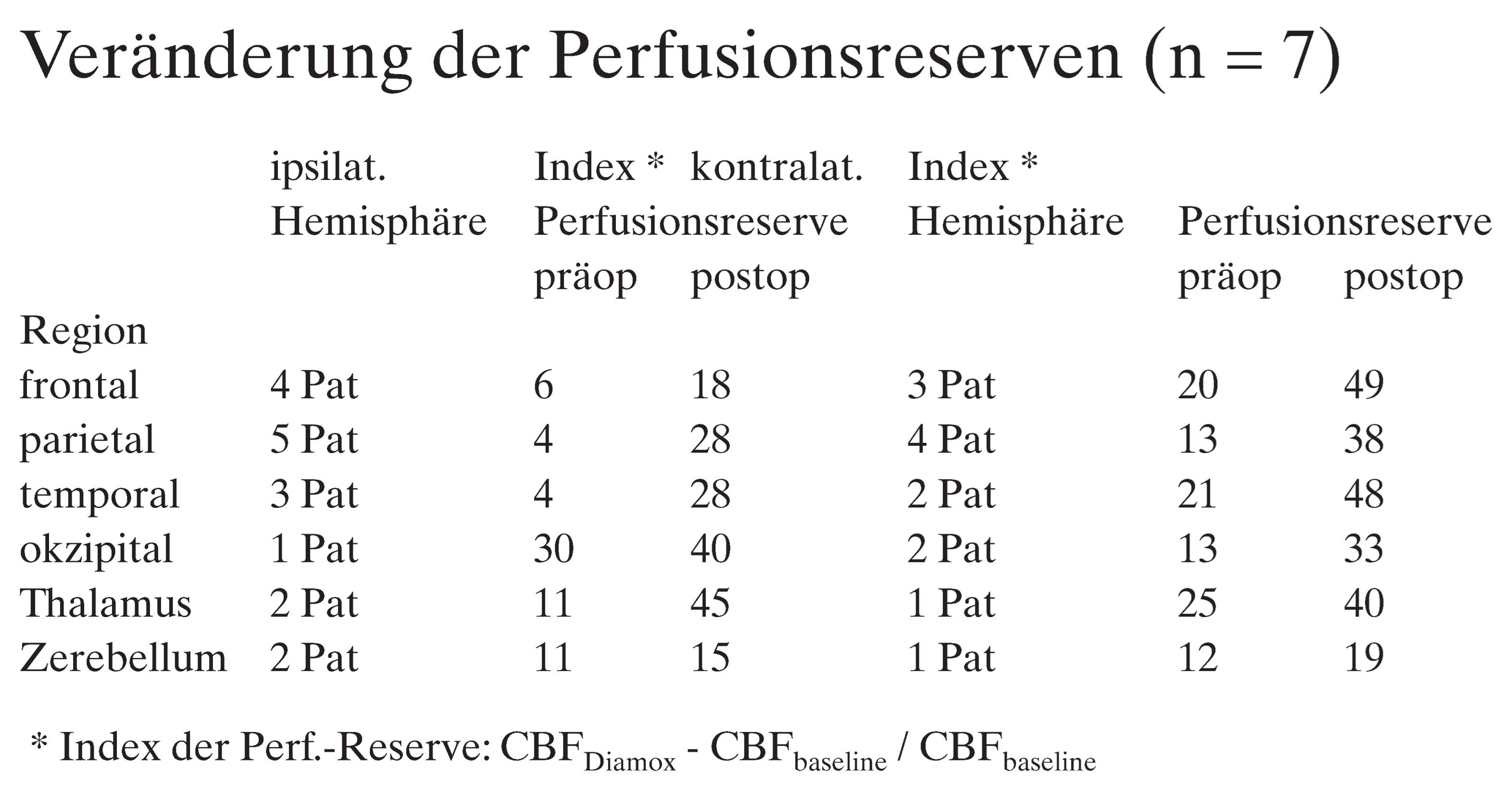
Schlussfolgerung: Drei Monate nach dem Anlegen eines EIAB lässt sich bei Patienten mit ICA- oder MCA-Obstruktionen eine Verbesserung der zerebralen Perfusion feststellen. Auch wenn der prophylaktische Effekt des EIAB bis anhin nicht belegt ist, so lassen sich doch die Überlegungen, auf denen das Vorgehen basiert, erfüllen.
Der extra-intrakranielle Bypass bei therapieresistenten Vasospasmen nach Subarachnoidalblutungen: Erfahrungen mit 3 Patienten
N. Krayenbühl, E. Keller, Y. Yonekawa, H.-G. Imhof, Zürich
Einleitung: Vasospasmen nach Subarachnoidalblutungen (SAB) können trotz intensiver Therapie mit hypertensiver, hypervolämer Hämodilution (3-H), superselektiver intraarterieller Instillation von Papaverin oder Ballondilatation des stenosierten Gefässabschnitts zu zerebralen Ischämien führen. Bewirken die obigen Behandlungsmodalitäten keine Verbesserung, kann versucht werden, durch Anlegen eines extra-intrakraniellen arteriellen Bypass (EIAB) die Perfusion des betroffenen Gefässterritoriums anzuheben.
Patientengut: Wir haben bei 3 Patienten mit nach SAB therapieresistenten Vasospasmen einen EIAB angelegt. Die Patienten (2 Männer und 1 Frau) mit einem Durchschnittsalter von 58 Jahren waren bei Klinikeintritt wach – entsprechend der Klassifikation von Hunt/Hess Grad II, nach der Einteilung der World Federation of Neurological Surgeons (WFNS) Grad 1. Sie wiesen in der Computertomographie eine Blutung, entsprechend Grad 3 nach Fisher auf. Bei den rupturierten Aneurysmen handelte es sich um 2 sakuläre Aneurysmen, wovon eines an der A. communicans anterior (A.com.ant.), das zweite an der A. cerebri media (MCA) lokalisiert war; beim 3. Aneurysma handelte es sich um ein dissezierendes Aneurysma der A. carotis interna (ICA). Alle 3 Aneurysmen waren zwischen Tag 3 und 7 nach der initialen Blutung geklippt worden. Die Vasospasmen traten zwischen dem 5. und 9. Tag nach der initialen Blutung auf und erwiesen sich als therapierefraktär. Um die spasmenbedingte Minderdurchblutung, die bei 2 Patienten durch eine Karotisstenose akzentuiert wurde, zu vermindern, wurde eine Anastomose zwischen der A. temporalis superficialis und einem Ast der A. cerebri media (STA-MCA) angelegt.
Resultat: Im postoperativen Verlauf – die Anastomosen waren durchgängig – wurde 1 Patientin asymptomatisch, die 2 anderen Patienten zeigten eine deutliche Verbesserung ihres neurologischen Zustandes.
Schlussfolgerung: Der EC-IC-Bypass, eingesetzt «als ultima ratio», scheint die spasmenbedingt verminderte zerebrale Durchblutung verbessern zu können.
Changes in cerebral perfusion reserves after revascularisation procedures in Moya Moya disease
N. Khan, Y. Yonekawa, A. Buck, B. Schuknecht, E. Boltshauser, Zürich
Introduction and objectives: Moya Moya disease is a bilateral steno-occlusive disease of the carotid arteries at the level of the siphon with the formation of an anastomotic collateral arterial network. The disease is typically present in the paediatric age group, where 81% of clinical presentation is ischemic in nature.
We advocate multiple, direct vascularisation using microsurgical anastomosis as the treatment of choice. Indirect revascularisation being performed additionally in cases where the latter is not satisfactory alone. Keeping this in mind we examined the changes in perfusion reserves in patients with bilateral Moya Moya disease after such selective multiple revascularisation procedures and compared it to the pre-operative perfusion reserves. Method and patient selection: We examined the pre-operative cerebral blood flow and perfusion reserves in 9 patients (male: female = 4:5) with Moya Moya disease. Mean age at examination was 7 yrs (range 3 – 15) in 8 patients. Only one adult case, age = 38 yrs, was examined. In all patients bilateral disease was present. Eight of the 9 patients have undergone bilateral revascularisation procedures: STA-MCA anastomosis (n = 9), STA-ACA anastomosis (n = 6), Occipital artery – MCA anastomosis (n = 1), EDAS (n = 1), dural synangiosis (n = 4).
We have, to date, compared the pre-operative and post-operative perfusion reserves in 7 of these patients. In these 7 patients a pre- and post-operative cerebral perfusion H20 – PET scan (baseline and Diamox challenge) was performed. Two of the 9 patients studied, have undergone pre-operative HMPAO-SPECT scans.
Results: Post-op perfusion reserve increased focally in 6 of the 7 patients who underwent pre- and post-operative H20 – PET scanning (Cases 1, 2, 3, 5, 6, 7: Frontal: R 31%, L 27%, Parietal: R 30%, L 21%, Temporal: R 40%, L 49%, Occipital: R 40%, L 59%, Cerebellum: R 52%, L 53%, Thalamus: R 47%, L 47%). In one patient (case 6) a global decrease in perfusion reserve was seen. In majority of cases with focal areas of developed infarcts, no increase in the preoperative cerebral perfusion reserve was seen in the infarcted regions. At the same time there were other focal non-infarct areas that also showed unchanged perfusion after diamox challenge, showing a low perfusion state. In 4 of these 7 patients (cases 1, 2, 3, 5), these regions of preoperative unchanged perfusion state showed an increase of upto 44% (range 20% – 64%) of perfusion reserves. Interestingly also in 4 patients (1, 2, 5, 6) an increase in perfusion reserves of upto 39% (range 23% – 64%) occurred in infarcted regions.
Conclusions: A bilateral focal increase in perfusion reserve was seen in 86% of cases after revascularisation surgery for Moya Moya disease. This increase was also present in areas where preoperatively perfusion reserve was unchanged. The benefit of multiple, selective, direct bilateral revascularisation is reiterated.
This is an open-access article distributed under the terms of the Creative Commons Attribution Non-Commercial No Derivatives (CC BY-NC-ND) 4.0 License.