


Summary
We assessed the prevalence and characteristics of neuro-ophthalmological manifestations (NOMs) in patients with stroke due to internal carotid artery (ICA) disease. NOMs were evaluated in 81 patients with ICA dissection and 229 patients with ICA atherosclerotic occlusion from the Lausanne Stroke Registry. NOMs, seen in 57% of patients with ICA dissection consisted of visual field defect (32%), Horner’s syndrome (26%), transient monocular blindness (5%), ocular ischaemic syndrome (4%) and ocular motor paresis (1%). The prevalence of NOMs was lower with ICA atherosclerotic occlusion (42%). Similarly, they consisted of visual field defects (26%), Horner’s syndrome (7%), transient monocular blindness (8%) and ocular ischaemic syndrome (3%). NOMs, consisting predominantly of homonymous hemianopia and transient monocular blindness, were the presenting signs in 16% of ICA dissection and 9% of ICA occlusion. Recognition of initial NOMs in these disorders may provide a window of opportunity for interventive therapies to avert stroke.
Introduction
Internal carotid artery (ICA) dissection is found in 2.5 to 4% of patients with ischaemic stroke [20,24], with particular incidence in young age groups [12,14,32,40]. Clinical features include, among others, headache, tinnitus, neuro-ophthalmological manifestations (NOMs) and focal neurological deficits [5,16,38].
NOMs can follow chronic occlusion of the ICA [23,25,43,46], but frequently they can occur before or simultaneously at the onset of stroke. These include: Horner’s syndrome [1,7,9,20,24,31,39], ocular motor paresis [7,21,27,30], transient monocular blindness [16,20,24,28,39], visual field defects [31], and ocular ischaemic syndrome [6,9,16,21,33,45]. In the latter, ophthalmic vasoocclusion may result in sudden and irreversible loss of vision and has been termed ocular stroke [33]. At times, isolated neuro-ophthalmological symptoms may be the sole manifestation of an ICA disease; thus, raising the question that early diagnosis based on these symptoms may predict the occurrence of stroke.
The aim of this study was to evaluate the prevalence and clinical characteristics of NOMs in patients with stroke due to ICA disease (atherosclerotic occlusion and dissection).
Materials and methods
The present study is based on consecutive cases of ICA disease (dissection or occlusion) with a first time ischaemic stroke examined from the Lausanne Stroke Registry [13]. Patients with isolated transient ischaemic attacks (TIA), or headache due to ICA dissection, were excluded from this study. All patients underwent a thorough and standardised neurological examination. During hospitalisation all the patients underwent a diary system of ophthalmoscopic evaluation by neuroophthalmologists. Finally, the patients were blinded evaluated by the same senior neurologist (JB). Investigation of ocular troubles prior to stroke was retrospectively assessed according to history although it was sometimes difficult to identify precisely which type of visual deficit the patient had experienced. Transient monocular blindness was defined by transient monocular obscuration or loss of vision with or without positive phenomena of at least several seconds of duration. [44]. The diagnosis of ocular ischaemic syndrome was based on a history of monocular visual loss and ophthalmologic evidence of permanent loss of visual acuity or visual field with retinal ischaemia at ophthalmologic evaluation (central retinal artery occlusion or branch retinal artery occlusion [44]). The diagnosis of dissection or occlusion was established by angiography and Doppler ultrasonography findings according to the previous criteria [17,18,19,20,22,26]. MRI with angiographic sequences was performed on selected patients (since the time the technique became available) as an alternative to conventional angiography [34]. We defined the presumed cause of ICA occlusion as atherosclerotic according to the previous criteria [13]. On account of the difficulties in separating traumatic and spontaneous dissection [4,31,39], in this current study, we classified dissection as traumatic when occurring after an obvious head or neck trauma, or, after unusual sudden neck movements. In other cases, dissections were considered spontaneous, even if minor or trivial trauma was evidenced on historical examination of the patients.
Characteristics of patients with ICA dissection or atherosclerotic occlusion were compared using a chi square test.
Results
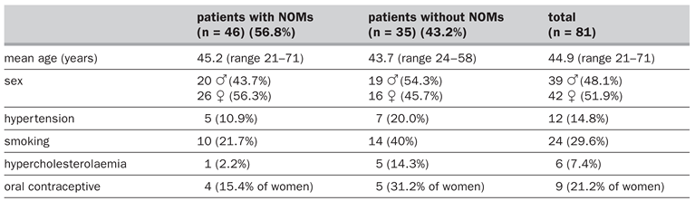
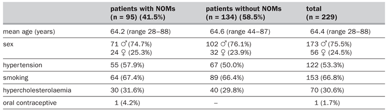
The population characteristics and risk factors of patients with ICA disease are summarised in
Table 1
(dissection) and
Table 2
(occlusion). Patients with dissection were younger (p = 0.002), had fewer vascular risk factors (14.8% of patients with ICA dissection had hypertension vs. 53.3% of patients with ICA occlusion, p = 0.001; 29.6% smoked vs. 66.8%, p = 0.001; 7.4% had hyperlipidaemia vs. 30.6%, p = 0.001; but 21.4% of women with ICA dissection used oral contraceptive vs. 4.2% of women with ICA occlusion, p = 0.001), and were more likely to be female (p = 0.003) than those with atherosclerotic occlusion. However, there were no overt differences in these characteristics between patients with and without NOMs for either aetiology.
Table 1.
Demographic characteristics and risk factors of patients with internal carotid artery dissection.
Table 2.
Demographic characteristics and risk factors of patients with atherosclerotic internal carotid artery occlusion.
Internal carotid artery dissection
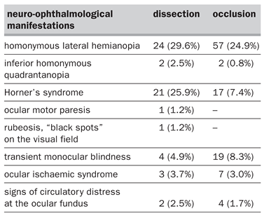
Eighty-one patients (2.2% of the 3628 of Lausanne Stroke Registry) (39 men and 42 women, mean age 44.9 years, range 21–72) had ICA dissection. A bilateral dissection was found in 2 patients and simultaneous carotid and vertebral dissection in one. History of trauma clearly related to the dissection was established in 12 patients, while the possibility of previous minor trauma (trivial trauma) was found in 8 patients. NOMs were present in 46 (56.8%) patients (Table 3). In 12 patients (10 with Horner’s syndrome and homonymous lateral hemianopia, 1 with Horner’s syndrome and ocular motor paresis and 1 with Horner’s syndrome with rubeosis and “black spots” on the visual field) there were 2 NOMs associated.
Table 3.
Frequency of neuro-ophthalmological manifestations in carotid disease.
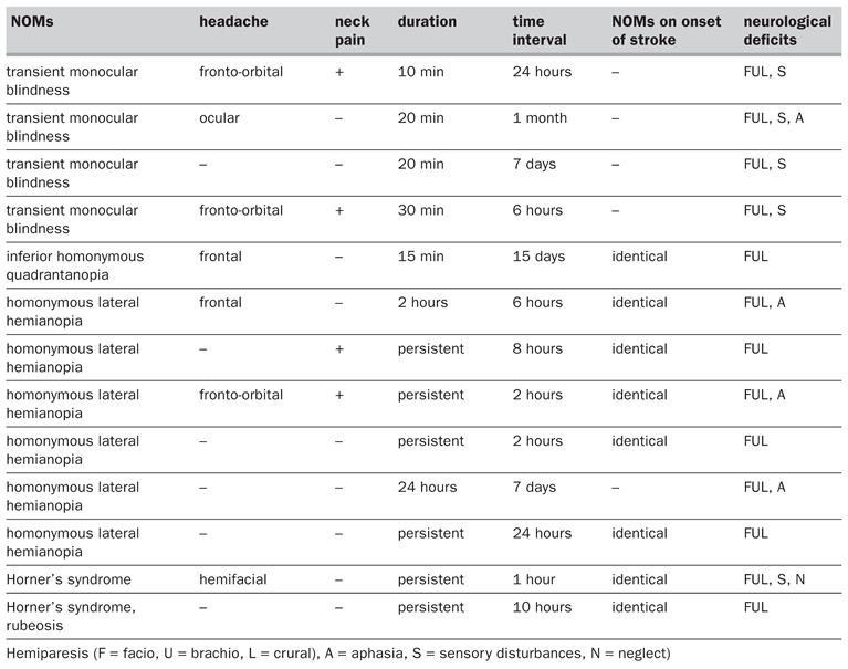
Visual field defects were the most frequent NOMs present in 26 (32.1%) patients, with homonymous lateral hemianopia in 24 (3 cases of traumatic dissection and 21 cases of spontaneous dissection) and inferior homonymous quadrantanopia in 2 (spontaneous dissection). Among these patients, visual field defects were the sole initial symptom in 7 patients (see Table 4). In 3 of these patients, the field defect resolved within 24 hours, but a disabling hemispheric syndrome occurred between 6 hours to 15 days later. In the other 4 patients, the field defect persisted and evolved into a hemispheric syndrome over the next 2 to 24 hours. Visual field defects were also identified simultaneously with hemispheric deficits in an additional 19 patients with stroke due to dissection of ICA.
Table 4.
Characteristics of patients with carotid dissection and NOMs prior stroke.
Horner’s syndrome
(complete or incomplete) was present in 21 (25.9%) patients (all 7 cases of traumatic dissection and 14 cases of spontaneous dissection, including 2 patients with history of minor/trivial trauma). Horner’s syndrome was associated with homonymous hemianopia in 10, with ocular motor paresis in 1 and with rubeosis in another (this patient complained of “black spots” in the visual field). In 2 cases it was the initial symptom. Time lag from the onset of Horner’s syndrome to the onset of stroke was 1 hour and 10 hours respectively.
Transient monocular blindness
was present in 4 (4.9%) cases and associated with spontaneous carotid dissection. Time lag from the onset of transient monocular blindness to the onset of stroke ranged from 6 hours to 1 month.
Ocular motor paresis
was verified only in one case (1.2%), simultaneously involving the III, IV and VI cranial nerves associated with Horner’s syndrome. Ocular motor paresis was present during the stroke.
Ocular ischaemic syndrome
was evident in 3 (3.7%) cases (spontaneous dissection). In another 2 patients (2.5%), there were signs of circulatory distress at the ocular fundus (pallor of the optic disk and venous congestion) without signs of any visual symptoms either previously or at admission, and without a former history of ocular or intracranial disease.
In summary, in 13 (16.0%) patients with ICA dissection, NOMs constituted the initial clinical picture (see Table 4). Among these patients, fronto-ocular pain was also present in 7 patients and neck pain in 4. Time lag from the onset of NOMs to the onset of stroke ranged from 1 hour to 1 month, although in 9 (69.2% of the 13 patients) it ranged from 1 to 24 hours. NOMs were transient in 7 patients and persisted until the onset of stroke in 6.
In total, headache was present in 22 (47.8%) patients with NOMs compared to 17 (48.6%) patients without NOMs and neck pain in 9 (19.6%) patients with NOMs compared to 2 (5.7%) patients without NOMs. The headache characteristics were similar between the group with NOMs and the group without NOMs even if there was a little prevalence for fronto-ocular and ocular pain in patients with NOMs (7 vs. 3).
Atherosclerotic internal carotid artery occlusion
Two hundred and twenty-nine patients (6.3% of the 3628 of Lausanne Stroke Registry; 173 men and 56 women, mean age 64.4 years, range 28–88) had ICA occlusion.
NOMs were present in 95 (41.5%) patients (Table 3). In 11 patients 2 forms of NOMs related incidences coexisted (6 evidenced with Horner’s syndrome and homonymous lateral hemianopia, 3 with transient monocular blindness and homonymous lateral hemianopia and 2 with transient monocular blindness and Horner’s syndrome).
Visual field defects
were the most frequent NOMs present in 59 (25.7%) patients, with homonymous lateral hemianopia in 57 and inferior homonymous quadrantanopia in 2. Homonymous lateral hemianopia alone, was an initial symptom in 2 patients (2 and 5 hours prior to stroke).
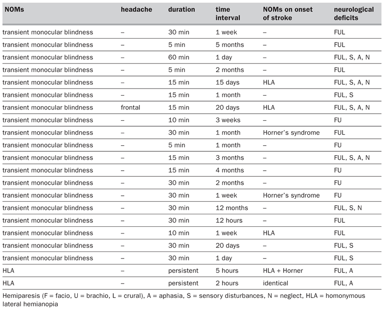
Transient monocular blindness was present in 19 (8.3%) cases. In all the patients the initial sign and the time lag from the onset of transient monocular blindness to the onset of stroke ranged from 12 hours to 12 months. In 5 patients the subsequent stroke presented other NOMs (3 cases with homonymous lateral hemianopia and 2 cases with Horner’s syndrome).
Horner’s syndrome
(complete or incomplete) was present in 17 (7.4%) patients. Horner’s syndrome was associated with homonymous lateral hemianopia in 6 patients and with transient monocular blindness in 2 patients.
Ocular ischaemic syndrome
was evident in 7 (3.0%) cases. In another 4 (1.8%) patients, there were signs of circulatory distress at the ocular fundus without signs of any visual symptoms either previously or at admission, and without a former history of ocular or intracranial disease.
In summary, in 21 (9.2%) patients with ICA occlusion, NOMs constituted the initial clinical picture (see Table 5). Among these patients, frontal headache was also present in only 1 patient. Time lag from the onset of NOMs to the onset of stroke ranged from 2 hours to 12 months. In 5 patients it ranged from 1 to 24 hours. NOMs were transient in 19 patients (transient monocular blindness) and persisted until the onset of stroke in 2 (homonymous lateral hemianopia).
Table 5.
Characteristics of patients with carotid occlusion and NOMs prior stroke.
In total, headache was present in 11 (11.6%) patients with NOMs compared to 18 (13.4%) patients without NOMs. Neck pain not was present.
Discussion
NOMs associated with stroke from ICA disease seem to have characteristic patterns. In general, they represent the particular susceptibility of the optic system to the effects of ischaemia, the close anatomical relations between the ICA and structures involved in different aspects of the regulation of this system, or perhaps, they form part of a group of deficits secondary to brain ischaemia.
Horner’s syndrome, which is usually attributed to the damage of the sympathetic fibres traveling along the carotid artery [2,31], was detected in 58.3% of the patients (7 of 12) having traumatic dissection, and in 7.4% of patients with ICA occlusion, underlining the possible role of direct trauma in this process. Patients with incomplete syndrome probably had involvement of the sympathetic fibres that accompany the ICA, with sparing of the external carotid plexus [3].
Transient monocular blindness prior to stroke from ICA disease may be due to embolisation from the artery or to a haemodynamic mechanism or to a vasospasm [15]. In each case, the results are decreased blood supply to the posterior ciliary artery and consequent ischaemia of the optic nerve [8,16]. The amaurosis fugax in a middle aged or older patient is an indication of atherosclerotic stenosis in the carotid system.
The same mechanisms that search to explain transient monocular blindness may also explain the occurrence of visual field defects before the onset of stroke [42]. Except for 2 patients having ICA dissection, the onset of visual field defects preceded the onset of stroke in a time interval which was less than 24 hours. Perhaps this may be related to the type of stroke itself and may be only the first of multiple focal neurological deficits. On the contrary, in 2 cases with ICA dissection, there was a time interval greater than one week which may indicate that the process is still reversible and this time interval could be sufficient for intervention.
Ocular ischaemic syndrome (ocular stroke) appears to be caused by occlusion of the central retinal artery and consequent infarct of the optic nerve head or by retinal ischaemia. Ocular stroke was associated with simultaneous cerebral infarction (optico-cerebral syndrome) [11]. Once again, both embolic and haemodynamic mechanisms have been postulated [16]. A significant reduction of the visual acuity was the principal complaint of these patients. Although there are reports of retro-bulbar ischaemia with visual impairment associated with dissection (posterior optic neuropathy) [6,37], our study did not find any patient with this clinical pattern. In fact, apart from these patients with optic ischaemic syndrome, we found 6 other patients that presented signs of circulatory distress at the optic fundus suggesting retinal ischaemia, but without signs of any visual impairment. Our results showed that it may be present early after the occlusion and that the systematic search of this type of lesion is important even when there were no visual complaints. The ophthalmic artery system is not a terminal system; isolated ophthalmic artery occlusion may not be clinically significant and presentation can vary greatly [10].
Ocular motor paresis, which was present in only one patient (with ICA dissection), may be due to either the extension of the dissection to the cavernous sinus or occlusion (mechanic, embolic or haemodynamic) of the vessels supplying the vasa nervorum that feed the peripheral branches of the cranial nerves [30,35].
A previous study reported 3 cases of ICA dissection, which presented itself with visual symptoms resembling the migraine aura (scintillating scotoma). Our study did not find any patients with this clinical pattern [36].
Headache and neck pain, which are often attributed to referred pain from the ICA dissection [41], were the only accompanying symptoms of NOMs that occurred prior to stroke. It is possible that NOMs may also participate in the genesis of such headache even if the pain in ICA dissection is considered to be due to distention of the artery, or to ischaemia of the pericarotid plexus, or even to involvement of the trigemino-vascular system [3,29,38].
In our patients NOMs were the sole manifestation of ICA disease with brain ischaemia in 24 (29.6%) patients with ICA dissection and in 84 (36.7%) patients having ICA occlusion but a limitation of this study is that we cannot determine the exact predictive value of NOMs because we have not data regarding patients with isolated NOMs who did not ultimately present a stroke such as patients with transient neurological deficits and monocularobscurationof vision with exercise or dinning of vision during exposition to bright light (in fact, the Registry included only patients with stroke).
Our study suggests a high frequency of NOMs in ICA disease, with a higher prevalence in ICA dissection. Our findings also showed differences between NOMs with ICA dissection and NOMs with atherosclerotic ICA occlusion. In fact, Horner’s syndrome was less frequent in ICA occlusion and it was never present prior to the onset of stroke; moreover, headache and neck pain were frequently the accompanying symptoms of NOMs in ICA dissection and also prior to stroke, while neck pain was not present in ICA occlusion but headache was found in this group of patients only in few cases. In conclusion, isolated neuro-ophthalmological symptoms may be the initial clinical picture and sole manifestation in patients with ICA disease and acute stroke and the time intervals between neuro-ophthalmic signs and stroke is sufficient for intervention; early diagnosis based on these symptoms and early treatment may significantly improve the outcome.
References
- Ameline-Audelan V. Painful Claude Bernard Horner’s syndrome: apropos of a painless carotid artery dissection. J Fr Ophthalmol 1998;21:591–5.
- Bilbao R, Amoros S, Murube J. Horner’s syndrome as an isolated manifestation of an intrapetrous carotid artery dissection. Am J Ophthalmol 1997;123:562–4.
- Biousse V, D’Anglejan-Chatillon J, Massiou H, Bousser MG. Head pain in non-traumatic carotid artery dissection: a series of 65 patients. Cephalalgia 1994;14:33–6.
- Biousse V, D’Anglejan-Chatillon J, Touboul PJ, Amarenco P, Bousser MG. Time course of symptoms in extracranial carotid artery dissections. A series of 80 patients. Stroke 1995;26:235–9.
- Biousse V, Touboul PJ, D’Anglejan-Chatillon J, Levy C, Schaison M, Bousser MG. Ophthalmologic manifestations of internal carotid artery dissection. Am J Ophthalmol 1998;126:565–77.
- Biousse V, Schaison M, Touboul PJ, D’Anglejan-Chatillon J, Bousser MG. Ischaemic optic neuropathy associated with internal carotid artery dissection. Arch Neurol 1998;55:715–9.
- Blunt SB, Galton C. Cervical carotid or vertebral artery dissection. An underdiagnosed cause of stroke in the young. Br Med J 1997;314:243.
- Bogousslavsky J. Amauroses fugaces: diagnostic et prise en charge. Med Et Hyg 1985;43:3025–34.
- Bogousslavsky J, Despland PA, Regli F. Spontaneous carotid dissection with acute stroke. Arch Neurol 1987;44:137–401.
- Bogousslavsky J, Pedrazzi PL, Borruat FX, Regli F. Isolated complete orbital infarction: a common carotid artery occlusion syndrome. Eur Neurol 1991;31:72–6.
- Bogousslavsky J, Regli F, Despland PA, Zografos L. Optic nerve head infarction. Small-artery disease, large-artery disease and cardioembolism. Cerebrovasc Dis 1991;1:341–4.
- Bogousslavsky J, Regli F. Ischaemic stroke in adults younger than 30 years of age. Cause and prognosis. Arch Neurol 1987;44:479–82.
- Bogousslavsky J, Van Melle G, Regli F. The Lausanne Stroke Registry: analysis of 1000 consecutive patients with first stroke. Stroke 1988;19:1083–92.
- Bui LN, Brant-Zawadzky M, Verghese P, Gillan G. Magnetic resonance angiography of cervico cranial dissection. Stroke 1993;24:126–31.
- Burger SK, Saul RF, Selhorst JB, Thurston SE. Transient monocular blindness caused by vasospasm. New Engl J Med 1991;325:870–3.
- Chatillon JA, Ribeiro V, Mas JL, Bousser MG, Laplane D. Dissection de l’artère carotide interne extracranienne. Soixante-deux observations. La Presse Médicale 1990;19:661–7.
- De Bray JM, Dubas F, Joseph PA, Causeret H, Pasquier JP, Emile J. Etude ultrasonique de 22 cas de dissection carotidiennes. Rev Neurol (Paris) 1989;145:702–9.
- Desfontaines Ph, Despland PA. Dissection of the internal carotid artery: aetiology, symptomatology, clinical and neurosonological follow-up, and treatment in 60 consecutive cases. Acta Neurol Belg 1995;95:226–34.
- Eljamel MSM, Humphrey PRD, Shaw MDM. Dissection of the cervical internal carotid artery. The role of Doppler/ Duplex studies and conservative management. J Neurol Neurosurg Psychiatry 1990;53:379–83.
- Fisher CM, Ojemann RG, Roberson GH. Spontaneous dissection of cervico-cerebral arteries. Can J Neurol Sci 1978;5:9–19.
- Galetta SL, Leahey A, Nichols CW, Raps EC. Orbital ischemia, ophthalmoparesis, and carotid dissection. J Clin Neuro-Ophthalm 1991;11:284–7.
- Gardner DJ, Gosink BB, Kallman CE. Internal carotid artery dissections: Duplex Ultrasound Imaging. J Ultrasound Med 1991;10:607–14.
- Hankey GJ, Warlow CP, editors. Transient ischaemic attacks of the brain and eye. London: Sanders; 1994.
- Hart RG, Easton JD. Dissection of cervical and cerebral arteries. Neurol Clin 1983;1:155–82.
- Hayreh SS. Anterior ischaemic optic neuropathy. Arch Ophthalm 1981;38:675.
- Hennerici M, Steinke W, Rautenberg W. High-resistance Doppler flow pattern in extracranial carotid dissection. Arch Neurol 1989;46:670–2.
- Maitland CG, Black JL, Smith WA. Abducens nerve palsy due to spontaneous dissection of the internal carotid artery. Arch Neurol 1983;40:448–9.
- McNeill DH, Dreisbach J, Marsden RJ. Spontaneous dissection of the internal carotid artery: its conservative management with heparin sodium. Arch Neurol 1980;37:54–5.
- Moskowitz MA. Mechanisms in vascular headache. Neurol Clin 1990;8:801–15.
- Mokri B, Silbert PL, Schievink WI, Piepgras DG. Cranial nerve palsy in spontaneous dissection of the extracranial internal carotid artery. Neurology 1996;46:356–9.
- Mokri B, Sundt TM, Houser OW, Piepgras DG. Spontaneous dissection of the cervical internal carotid artery. Ann Neurol 1986;19:126–38.
- Mullges W, Ringelstein EB, Leibold M. Non-invasive diagnosis of internal carotid artery dissections. J Neurol Neurosurg Psychiatry 1992;55:98–104.
- Newman NJ, Kline LB, Leifer D, Lessell S. Ocular stroke and carotid artery dissection. Neurology 1989;39:1462–4.
- Ozdoba C, Sturzenegger M, Schroth G. Internal carotid artery dissection: MR Imaging features and clinical-radiologic correlation. Radiology 1996;199:191–8.
- Panisset M, Eidelman BH. Multiple cranial neuropathy as a feature of carotid artery dissection. Stroke 1990;21:141–7.
- Ramadan NM, Tietjen GE, Levine SR, Welch KMA. Scintillating scotoma associated with internal carotid artery dissection: report of three cases. Neurology 1991;41:1084–7.
- Rivkin MJ, Hedges TR, Logigian EL. Carotid dissection presenting as posterior ischaemic optic neuropathy. Neurology 1990;40:1469.
- Rosebraugh CJ, Griebel DJ, DiPette DJ. A case of carotid artery dissection presenting as cluster headache. Am J Med 1997;102:418–9.
- Schievink WI, Mokri B, O’Fallon WM. Recurrent spontaneous cervical-artery dissection. N Engl J Med 1994;330:393–7.
- Schievink WI, Mokri B, Piepgras G. Spontaneous dissections of cervicocephalic arteries in childhood and adolescence. Neurology 1994;44:1607–12.
- Silbert PL, Mokri B, Schievink WI. Headache and neck pain in spontaneous internal carotid and vertebral artery dissections. Neurology 1995;45:1517–22.
- Steinke W, Schwartz A, Hennerici M. Topography of cerebral infarction associated with carotid artery dissection. J Neurol 1996;243:323–8.
- Sturrock GA, Mueller MR. Chronic ocular ischemia. Br J Ophthalmol 1984;68:716–23.
- Tippin J, Corbett JJ, Kerber RE, Schroeder E, Thompson HS. Amaurosis fugax and ocular infarction in adolescents and young adults. Ann Neurol 1989;26:69–77.
- Wilson WB, Leavengood JM, Ringel SP, Bott AD. Transient ocular motor paresis associated with acute internal carotid artery occlusion. Ann Neurol 1989;25:286–90.
- Wray SH. Visual symptoms (eye). In: Bogousslavsky J, Caplan L, editors. Stroke syndromes. Cambridge: Cambridge University Press; 1995. pp. 68–79.

This is an open-access article distributed under the terms of the Creative Commons Attribution Non-Commercial No Derivatives (CC BY-NC-ND) 4.0 License.