


Introductory remarks
The following article is a summary of the introduction to a two-hour satellite symposium on Cognitive Evoked Potentials (CEPs) to which the following authors contributed: C. E. Elger and T. Grunwald (Bonn): Kognitive Potentiale innerhalb des Temporallappens unter dem Gesichtspunkt der Neurobiologie des menschlichen deklarativen Gedächtnisses; M. Seek, T. Landis and C. Michel (Geneva): Zeitlich-räumliche Lokalisation komplex-visueller Prozesse mit Tiefen- und Oberflächenableitung; K. Gutbrod, A. Nirkko, C. Ozdoba and A. Schnider (Berne): Aktivierung von Spracharealen im funktionellen MRI (fMRI); B. Weber, K. Lehnertz, C. Helmstaedter, H. G. Wieser and C. E. Elger (Zurich, Bonn): Neuronale Komplexität des intrahippokampalen EEGs während kognitiver Aufgaben; D. Brandeis and D. Lehmann (Zurich): Funktionelles Brainmapping mit kognitiv evozierten Potentialen.
Historical flashback
As early as 1875 Richard Caton stated: “... The electric currents of the grey matter appear to have a relation to its function ... When ... the gray matter is in state of functional activity, its electric current usually exhibits negative variation ...” [1]. In 1929 Hans Berger [2] wrote: “... From the beginning, it was my hope to succeed in detecting from the human scalp ... electrical oscillations ... and thus to realise the words of Fleischl von Marxow: It may even be possible ... to detect current produced by various psychic events in one’s own brain” and: “In the experiments with my son Klaus, I gained the impression that during strenuous intellectual effort, and also during intense concentration, the smaller and shorter waves predominate” [2]. In 1964 W. Grey Walter et al. [3] published an article in ‘Nature’ about their finding of what they called “contingent negative variation” and described it as an electric sign of sensorimotor association and expectancy in the human brain. In their own words “The non-specific responses to conditional stimuli usually consist of 3 main components. A brief s+ wave, a brief s– wave and a much more prolonged s– wave, which may last several seconds”, and further: “The features which seem most closely related to the contingency of the situation and the attitude of the subject are the prolonged secondary negative waves and their interaction with the subsequent responses to the imperative stimuli.”
The basic principles of evoked potentials (EPs)
EPs are based on the principle of averaging the EEG with respect to a repeated behavioural event. By this procedure only that part of the EEG related to the behavioural event will remain. (→ event-related potentials = ERPs).
The undisputed advantages of EEG and EPs are their direct relation to neuronal information-processing and their great temporal resolution.
EPs are a series of components, each defined by its
- –
- latency;
- –
- polarity;
- –
- (scalp) topography;
- –
- behavioural correlates.
Successive EP components are related to successive stages in information-processing, from a strictly sensory to the highest integrative levels, termed “endogenous” [4]. From this it follows, that if the intracranial generators of scalp EP components could be identified, then the intensity, onset and duration of activation of specific brain systems involved in a task could be monitored. Unfortunately from the scalp topography of EPs one cannot unambiguously infer the location of the synapses that generate an EP component.
The generators of EPs and their estimation
Estimating cerebral generators from observed scalp EP topography is ambiguous and known as “solving the inverse problem”. An infinite number of different generator configurations in the brain could – at least theoretically – result in the same EP topography on the scalp. Gevins [5] and Koles [6] could show that with the use of Laplacian transforms and spatial deconvolutions the voltage distribution at the exterior cortical surface can be estimated with a 2-cm accuracy. However, only about 30% of the cortex can be monitored in this fashion [7].
The simplest still most common solution of the inverse problem assumes that the scalp EP distribution arises from a single dipole. At a first step, the location, orientation and strength of that dipole is approximated. Then, the propagation of electrical potentials from that dipole to the recording electrodes at the scalp is calculated analytically by modelling the head as concentric spheres (with the shells brain,CSF,skull and scalp) of differing conductances. Subsequently the error between the calculated and measured electrical field pattern is then used to modify the dipole’s parameters, and the resulting field is re-calculated. This process is repeated in an iterative manner until the dipole’s characteristics no longer change significantly between iterations.
The accuracy of such models in localising an assumed single intracranial generating dipole from extracranial EEG is fairly high when compared with electrocorticography (ECoG) [8], or – even better – with intracranial microstimulation [9,10,11]. Unfortunately the assumption that the sources of EEG are current dipoles will probably seldom be correct in practice, and in particular cognitive EPs do not fit this assumption. A more likely model for the source is the current dipole sheet. The image of a dipole sheet on the surface of a model head can be calculated by simply summing the contributions of the individual dipoles in the sheet.
Significant progress in EEG source localisation has been made by the use of more refined and sophisticated monopolar and dipolar source models and more realistic head models based on boundary and finite elements. Some refined methods are [6]:
- –
- equivalent dipole localisation;
- –
- single and multiple time-slice source localisation;
- –
- weighted minimum norm localisation.
In the multiple time-slice source localisation method an analysis of the spatial covariance of the EEG enables the estimation of the number of active sources. This is an advantage over the single time-slice approach, where there is no quantitative way of estimating the multiplicity of the sources. The minimum norm approaches to the localisation of the sources of EEG [12] lead to estimates of the current density everywhere in the three-dimensional volume of the head. The main advantage of the weighted minimum norm localisation is that knowledge of the source multiplicity is not required.
Cognitive evoked potentials (CEPs)
As already mentioned CEPs do not satisfy the single dipole assumption, because CEPs are generated by extended surfaces in multiple brain areas, rather than by a simple dipole.
Using intracranial recorded EEG (stereotactic depth electrode recording, stereo-electroencephalography, SEEG) in the context of presurgical evaluation of candidates for epilepsy surgery, studies of intracerebral potentials to rare target and distractor auditory and visual stimuli (the classical “standard oddball paradigm”) have been undertaken [7] and led to the detection and the description of:
- –
- modality-specific responses to rarity;
- –
- the P3a system for the orientation of attention;
- –
- the P3b event-encoding system;
- –
- intracerebral potentials to novel and repeated words and figures;
- –
- other late CEPs;
- –
- the CNV;
- –
- the RP (Bereitschaftspotiental, “readiness” potential).
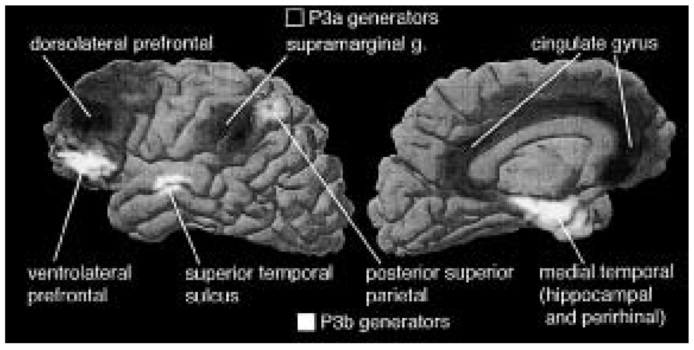
In the context of this introduction and some other papers dealing with temporal lobe epilepsy and memory the P3 systems are of particular importance. The P3a system for the orientation of attention comprises the inferior parietal, cingulate, and dorsolateral prefrontal cortex, whereas the P3b event-encoding system comprises the hippocampus, the superior temporal sulcus, the lateral orbito-frontal cortex, and the intraparietal sulcus ([7], Fig. 1).
Figure 1.
Summary of brain areas where P3s were found to be generated by simple rare stimuli in signal detection tasks. The P3a is evoked by rare stimuli, regardless of whether they are targets or non-targets, overtly attended or unattended, auditory or visual. It is generated in a frontoparietocingulate system that has been associated with the orientation of attention (areas shaded dark in the figure). It is associated with an electrodermal response and represents the cortical component of the orienting response. The P3b (lighter areas) is evoked by attended visual or auditory stimuli that must be definitively processed. Its principle generators are in the hippocampus, superior temporal sulcus, ventrolateral prefrontal cortex, and (probably) intraparietal sulcus. The P3aud (not indicated) is generated in the superior temporal plane to auditory stimuli only, and is insensitive to attention. Lateral (left) and medial (right) views are shown. Diagram based on recordings from about 4000 intracranial sites [21,22,23,24,28]. (From Halgren et al., 1998, with permission of the authors and the publisher, Elsevier, Amsterdam).
The importance of cognitive evoked potentials for prediction of postoperative memory
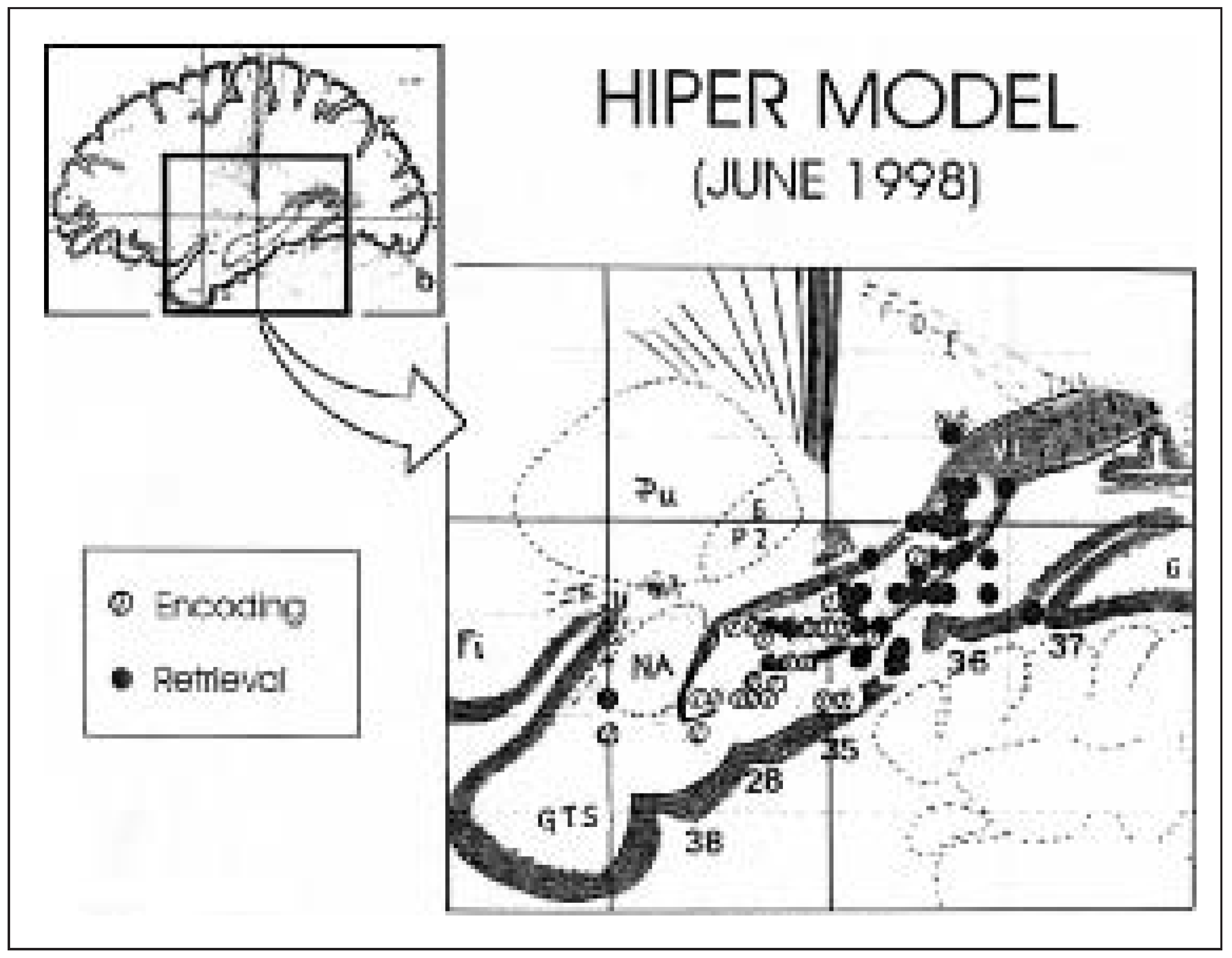
Animal and human lesion studies have revealed the importance of the hippocampal formation and the amygdala for learning and memory. More precisely encoding of new information has been associated with the hippocampal formation, and in particular with the perirhinal and entorhinal cortices (Fig. 2)
Disorders of learning and memory are clearly associated with temporal lobe (TL) dysfunction and have been studied in temporal lobe epilepsy (TLE) before and after TL surgery. Early investigators noted severe mnestic deficits following either bilateral mesial temporal resections or unilateral resections in the presence of occult lesions in the contralateral TL. These observations gave rise to the functional reserve model of hippocampal function in which post-surgical memory deficits were believed to be mediated by the capacity of the contralateral TL structures to support memory. However, material-specific learning and memory deficits, found in most patients with TLE, might be specifically related to the involved TL. When the language-dominant hemisphere is involved, memory deficits are usually more pronounced and in particular verbal memory is insufficient.Verbal memory impairment has been found to correlate with hippocampal pyramidal cell loss. There is, however, no doubt that dysfunction of other structures than the mesiobasal temporal ones, in particular diencephalic structures and probably also thalamic ones, also can produce severe memory deficits.
Pre- and post-operative comparison of per- formances of patients with TLE have contributed quite a lot to the understanding of TL functions. There are, however, some limitations due to the following reasons: most patients with a long history of TL seizures (1) have preoperatively a memory deficit and the degree of this deficit influences post-operative outcome; (2) take highdosed antiepileptic drugs (AEDs) with known side effects regarding higher cognitive functions preoperatively and reduce or discontinue AEDs postoperatively; (3) become seizure-free and thus escape the negative consequences of seizures on cognitive performance and motivation; finally (4) in lesional cases and in patients with resective surgery compensatory mechanisms, i. e. plasticity, play an important role.
Thus, although substantial evidence has been accumulated that the TL and particularly its mesiobasal and basal structures are very important for memory, the exact role of the hippocampal formation is still a matter of research. Recently (O15)-H2O PET studies [13], which show increased blood flow at the site of activation, selective memory temporal lobe-Amytal tests [14], which allow the temporary inactivation of restricted mesial temporal areas, and intrahippocampal cognitive evoked potential studies have revealed some interesting results. The intrahippocampal cognitive evoked potential studies will be shortly summarized:
In TLE patients, intracranial event-related potentials (ERPs) show alterations (increase in latency and decrease of amplitude) which correlate well with the Wechsler Memory Scale and the hippocampal P300 correlates well with neuronal cell density. The recently described N400 recorded from the anterior fusiform gyrus is thought to reflect access of semantic memory, whereas the P300 is thought to reflect rules-based mapping of stimuli onto discrete covert or overt responses, i.e. encoding processes [16,17,18]. Paller et al. [19] found that late potentials around 500–900 msec account for the subject’s engagement in recollection processing, and ERPs differentiate priming and recognition to familiar and unfamiliar faces [20]. Several authors have described limbic correlates of the scalp P300 elicited in visual and auditory odd-ball paradigms using depth electrodes in epileptic patients [21,22,23,24,25,26,27] and to the scalp N400 in word recognition paradigms [27,28]. Therefore it can be assumed that the careful study of ERPs recorded from mesiobasal temporal structures in TLE patients has the potential to clarify the role of these structures in the various kinds of memory and to quantify pre-operative deficits. Indeed, Grunwald et al. [27] recently reported that the amplitude of the intrahippocampal N400 elicited in a word recognition paradigm is an excellent predictor of post-operative memory performance in patients suffering from TLE.
Figure 2.
The HIPER (Hippocampal Encoding/Retrieval) model of Lepage et al. [15]. These authors conducted a meta-analysis of experimentally induced changes in blood flow (“activations”) in positron emission tomography (PET) studies of memory and found an orderly functional anatomic pattern: Activations in the hippocampal region that are associated with episodic memory encoding are located primarily in the rostral portions of the region whereas activations that are associated with episodic memory retrieval are located primarily in the caudal portions. These findings are based on an analysis of a sample of 56 “hippocampal encoding and retrieval” activations that were culled from an overall database consisting of 53 published PET studies of memory. The authors referred to this general pattern of rostrocaudal gradient of encoding and retrieval PET activations as the HIPER (Hippocampal Encoding/Retrieval) model. The model suggests a division of memory-related labor between the rostral and caudal portions of the hippocampal formation. (From [15] with permission of authors and publisher).
References
- Caton R. Br Med J 1875;2:278. Abstract in: Zentralblatt für Physiologie 1890;4(25). According to Bechterew: Die Energie des lebenden Organismus. Wiesbaden; 1902. S. 102.
- Berger H. Über das Elektroenkephalogramm des Menschen. Arch Psychiatr Nervenkrankheiten 1929;87:551–3, 567–70.
- Walter WG, Cooper R, Aldridge VJ, McCallum WC, Winter AL. Contingent negative variation: an electric sign of sensorimotor association and expectancy in the human brain. Nature 1964;203:380–4.
- Wallesch CW, Deuschl G. Elektrophysiologie am Menschen. In: Kischka U, Wallesch CW, Wolf G (editors). Methoden der Hirnforschung. Heidelberg, Berlin: Spektrum Akademischer Verlag; 1997. pp. 167–86.
- Gevins A. The future of electroencephalography in assessing neurocognitive functioning. Electroenceph Clin Neurophysiol 1998;106:165–72.
- Koles JK. Trends in EEG source localization. Electroenceph Clin Neurophysiol 1998;106:127–37.
- Halgren E, Marinkovic K, Chauvel P. Generators of the late cognitive potentials in auditory and visual oddball tasks. Electroenceph Clin Neurophysiol 1998;106:156–64.
- Nakasato N, Levesque MF, Barth DS, et al. Comparison of MEG, EEG and ECoG source localization in neocortical partial epilepsy in humans. Electroenceph Clin Neurophysiol 1994;91:171–8.
- Cohen D, Cuffin BN, Yunokuchi K, et al. MEG versus EEG localization test using implanted sources in the human brain. Ann Neurol 1990;28:811–7.
- Cuffin BN, Cohen D, Yunokuchi K, et al. Tests of EEG localization accuracy using implanted sources in the human brain. Ann Neurol 1991;29:132–8.
- Gharib S, Sutherling WW, Nakasato N, et al. MEG and ECoG localization accuracy test. Electroenceph Clin Neurophysiol 1995;94:109–14.
- Pasqual-Marqui RD, Michel CM, Lehmann D. Low resolution electromagnetic tomography: a new method for localizing electrical activity in the brain. Int J Psychophysiol 1994;18:49–65.
- Henke K, Buck A, Weber B, Wieser HG. Human hippocampus establishes associations in memory. Hippocampus 1997;7:249–56.
- Wieser HG, Müller S, Schiess R, Khan N, Regard M, Landis T, et al. The anterior and posterior selective temporal lobe amobarbital tests: angiographical, clinical, electroencephalographical, PET and SPECT findings, and memory performance. Brain Cogn 1997;33:71–97.
- Lepage M, Habib R, Tulving E. Hippocampal PET activations of memory encoding and retrieval: the HIPER model. Hippocampus 1998;8:313–22.
- Nobre CC, Allison T, McCarthy G. Word recognition in the human inferior temporal lobe. Nature 1994;372:260–3.
- McCarthy G, Nobre CC, Bentin S, Spencer DD. Languagerelated field potentials in the anterior-medial temporal lobe. J Neurosci 1995;15:1080–9.
- Nobre CC, McCarthy G. Language-related field potentials in the anterior-medial temporal lobe: effects of word-type and semantic priming. J Neurosci 1995;15:1090–8.
- Paller KA, Kutas M, McIsaac HK. Monitoring conscious recollection via the electrical activity of the brain. Psychol Sci 1995;6:107–11.
- Begleiter H, Porjesz B, Wang W. Event-related brain potentials differentiate priming and recognition to familiar and unfamiliar faces. Electroencephalogr Clin Neurophysiol 1996;94:41–9.
- Halgren E, Squires NK, Wilson CL, Rohrbaugh J, Babb T, Crandall P. Endogenous potentials generated in the human hippocampus formation and amygdala by infrequent events. Science 1980;210:803–5.
- Halgren E, Baudena P, Clarke JM, Heit G, Liégeois-Chauvel C, Chauvel P, et al. Intracerebral potentials to a rare target and distractor auditory and visual stimuli. I: Superior temporal plane and parietal lobe. Electroencephalogr Clin Neurophysiol 1995;94:191–220.
- Halgren E, Baudena P, Clarke JM, Heit G, Marinkovic K, Devaux B, et al. Intracerebral potentials to a rare target and distractor auditory and visual stimuli. II: Medial, lateral and posterior temporal lobe. Electroencephalogr Clin Neurophysiol 1995;94:229–50.
- Stapelton JM, Halgren E. Endogenous potentials evoked in simple cognitive tasks: depth components and task correlates. Electroencephalogr Clin Neurophysiol 1987;67:44–52.
- Halgren E. Evoked potentials. In: Boulton AA, Baker G, Vanderwolf C (editors). Neuromethods, vol. 15 Neurophysiological techniques – applications to neural systems. New York: Humana, Clifton; 1990. pp. 147–275.
- Loring DW, Meador KJ, King DW, Gallagher BB, Smith JR, Flanigin HF. Relationship of limbic evoked potentials to recent memory performance. Neurology 1988;38:45–8.
- Grunwald T, Elger CE, Lehnertz K, Van Roost D, Heinze HJ. Alterations of intrahippocampal cognitive potentials in temporal lobe epilepsy. Electroencephalogr Clin Neurophysiol 1995;95:53–62.
- Smith ME, Stapelton JM, Halgren E. Human medial temporal lobe potentials evoked in memory and language tasks. Electroencephalogr Clin Neurophysiol 1986;63:145–59.
This is an open-access article distributed under the terms of the Creative Commons Attribution Non-Commercial No Derivatives (CC BY-NC-ND) 4.0 License.