


Simple Summary
Secretory carcinoma is a low-grade malignant tumor that predominantly arises in the salivary glands and breast but occurs only rarely in the skin. We report an exceptionally rare case of primary cutaneous secretory carcinoma of the eyelid. This case highlights the importance of integrating histological, immunohistochemical, and molecular findings, particularly the detection of the ETV6::NTRK3 fusion, to distinguish secretory carcinoma from other eyelid malignancies, including apocrine carcinoma and sebaceous carcinoma.
Abstract
Secretory carcinoma is a low-grade malignant neoplasm characterized histologically by microcystic and tubular structures containing intraluminal secretions and molecularly by the frequent presence of an ETV6::NTRK3 fusion. It most commonly arises in the salivary glands and breast, whereas primary cutaneous secretory carcinoma is exceedingly rare. We report a case of secretory carcinoma arising in the skin of the right lower eyelid in an 82-year-old woman. A gradually enlarging nodule had been present for six months. Excision revealed a dermal tumor composed of mildly atypical polygonal cells forming microcystic and tubular structures with intraluminal secretions. The neoplastic cells expressed S100 protein, mammaglobin, MUC1, MUC4, GATA3, SOX10, and pan-TRK. Fluorescence in situ hybridization demonstrated fusion signals consistent with an ETV6::NTRK3 fusion. Taken together, these findings favored a diagnosis of secretory carcinoma rather than other adnexal malignancies, particularly apocrine and sebaceous carcinomas. This case illustrates the value of integrating morphology, immunohistochemistry, and appropriately interpreted molecular testing in the evaluation of rare eyelid adnexal tumors.
1. Introduction
Secretory carcinoma is a low-grade malignant neoplasm that typically arises in the salivary glands or breast and frequently harbors an ETV6::NTRK3 fusion [1,2,3]. Histologically, it is characterized by microcystic, tubular, and solid growth patterns and tumor cells with abundant eosinophilic or vacuolated cytoplasm.
Although the clinicopathological features of secretory carcinoma are relatively well established at its usual sites, its occurrence outside the salivary glands and breast is rare. At unusual anatomical sites, it may mimic other adnexal or glandular neoplasms, making diagnosis particularly challenging in limited biopsy specimens. Therefore, integrating morphological, immunohistochemical, and molecular findings is important for establishing an accurate diagnosis in such cases.
In routine dermatopathology practice, rare primary cutaneous carcinomas present a diagnostic challenge because site-specific clinical impressions can be misleading. For example, a lesion at the eyelid margin may initially raise concern for sebaceous carcinoma, while a gland-forming tumor with eosinophilic cytoplasm may suggest apocrine carcinoma [4,5]. The identification of intraluminal secretions on histological examination is helpful but not pathognomonic. Immunohistochemistry can narrow the differential diagnosis, although expression patterns overlap across adnexal lineages [5,6]. In this setting, the diagnostic question is not whether a tumor expresses a single secretory-associated marker, but whether its overall phenotype is consistent with secretory carcinoma and whether molecular analysis corroborates that interpretation. The clinical significance of this distinction extends beyond nomenclature. A diagnosis based solely on anatomical location or a single immunohistochemical marker may be misleading, because several adnexal neoplasms share overlapping glandular morphology and immunophenotypic features. A stepwise diagnostic approach therefore requires careful assessment of architecture, cytology, lineage-associated markers, and, when available, fusion gene testing [5,6].
Here, we report an extremely rare case of primary cutaneous secretory carcinoma arising in the lower eyelid. Distinguishing this tumor from other adnexal malignancies, particularly cutaneous apocrine carcinoma and sebaceous carcinoma, was histologically challenging. Detection of the ETV6::NTRK3 fusion played an important role in establishing the definitive diagnosis.
2. Case Presentation
The patient was an 82-year-old woman with a medical history of diabetes mellitus, hypertension, and a lumbar vertebral fracture. Six months before presentation, she noticed a nodule on the medial aspect of her right lower eyelid that had gradually increased in size over the preceding months. She presented to the Department of Plastic Surgery at a general hospital, where a granuloma-like nodular lesion was identified. Squamous cell carcinoma or sebaceous carcinoma was clinically suspected, and an excisional biopsy with a 1-mm surgical margin was performed on the same day.
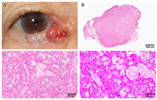
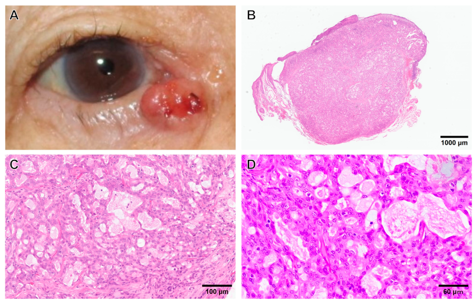
On gross examination, a shiny, reddish-brown nodular lesion with focal surface erosion was present on the medial aspect of the right lower eyelid (Figure 1A). Histologically, the well-circumscribed tumor measured approximately 6 mm in greatest dimension and extended from immediately beneath the epidermis into the deep dermis. The overlying epidermis showed focal erosion (Figure 1B). The neoplasm consisted of mildly atypical polygonal epithelial cells with eosinophilic cytoplasm forming closely packed microcystic and tubular structures containing PAS-positive intraluminal secretions (Figure 1C). Marked nuclear pleomorphism, extensive solid growth, tumor necrosis, and lymphovascular invasion were not identified. Mitotic activity was low, ranging from 0 to 2 mitoses per 10 high-power fields (HPFs), with an average of 1 mitosis per 10 HPFs (Figure 1D).
Figure 1.
Clinical appearance and histopathological findings. (A) A shiny, reddish-brown nodule with focal erosion is present on the medial aspect of the right lower eyelid. (B) A well-circumscribed neoplastic lesion extends from the subepidermal region to the deep dermis. Surface erosion is seen (hematoxylin and eosin [H&E] stain; whole-slide image; scale bar = 1000 μm). (C) Mildly atypical epithelial cells with eosinophilic cytoplasm form closely packed microcystic and tubular structures containing amphophilic secretions (H&E, scale bar = 100 μm). (D) The tumor cells show mild nuclear atypia; mitotic count: 0–2 mitoses per 10 HPFs (mean, 1 mitosis per 10 HPFs) (H&E, scale bar = 60 μm).
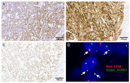
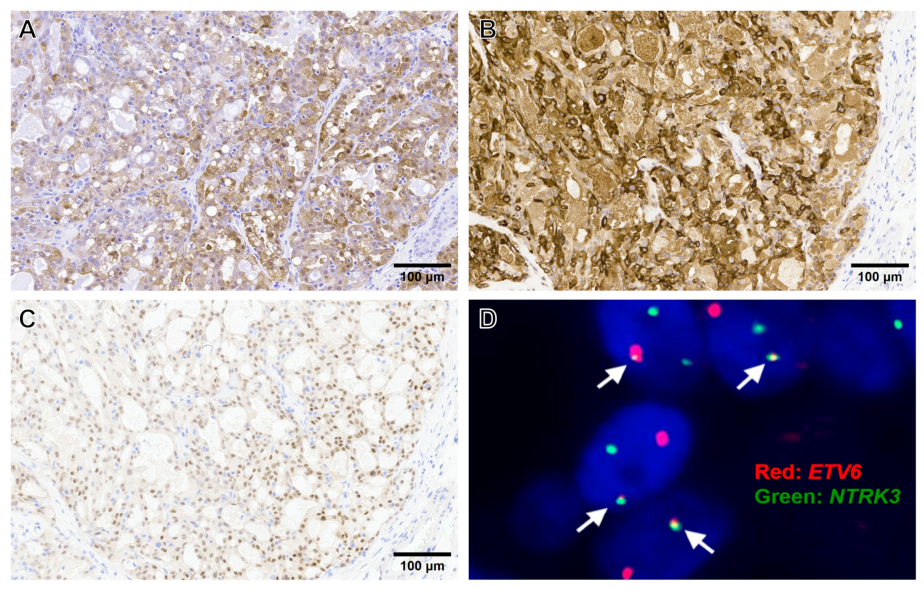
Immunohistochemically, the neoplastic cells were diffusely positive for S100 protein (Figure 2A), mammaglobin (Figure 2B), GATA3, MUC1, MUC4, SOX10, and pan-TRK (Figure 2C). GCDFP15 immunoreactivity was observed in luminal secretions. The neoplastic cells were negative for DOG1, p63, CK14, αSMA, androgen receptor (AR), and HER2. Given the morphology and immunophenotype, fluorescence in situ hybridization (FISH) was performed using a commercially available “ETV6/NTRK3 fusion probe” (Jokoh Co., Ltd., Kawasaki-shi, Japan, product code J17969) according to the testing laboratory’s protocol. The assay demonstrated ETV6::NTRK3 fusion signals in 165 of 200 evaluated tumor cell nuclei (82.5%) (Figure 2D). The integrated findings supported secretory carcinoma arising in the skin of the right lower eyelid.
Figure 2.
Immunohistochemical findings and fluorescence in situ hybridization. Tumor cells show positivity for S100 protein (A), mammaglobin (B), and pan-TRK (C) (scale bar = 100 μm). (D) Dual-color FISH using a commercial ETV6/NTRK3 fusion probe (Jokoh Co., Ltd.; J17969) demonstrates signals consistent with an ETV6::NTRK3 fusion; arrows indicate representative fusion signals.
Considering the patient’s advanced age, no additional treatment was administered, and the patient was instead managed by clinical observation; she died of natural causes one month later.
3. Discussion
Secretory carcinoma is a low-grade malignant neoplasm characterized by distinctive secretory morphology and a molecular profile that is characteristic but not entirely specific. Histologically, these tumors commonly exhibit a combination of microcystic, tubular, papillary, and solid growth patterns, often accompanied by eosinophilic intraluminal secretions. Tumors arising in the salivary glands or breast typically occur in adults, with reported median ages of 46.5 years for salivary gland secretory carcinoma and 53 years for secretory carcinoma of the breast. Salivary gland secretory carcinoma shows no apparent sex predilection, whereas secretory carcinoma of the breast predominantly affects women [1,2,3]. Despite these differences in demographic characteristics, secretory carcinomas arising at these two anatomical sites share considerable morphological and molecular similarities. ETV6 rearrangements are identified in most cases, with ETV6::NTRK3 being the predominant fusion [1]. This fusion results in a chimeric tyrosine kinase that contributes to oncogenic signaling and tumor cell proliferation. More recently, alternative ETV6 fusions, including ETV6::RET, ETV6::MET, and ETV6::MAML3, have also been reported in salivary gland secretory carcinomas [7,8,9]. These findings indicate that secretory carcinoma is associated with a spectrum of molecular alterations rather than a single pathognomonic fusion event.
The term secretory carcinoma refers to a tumor category defined by characteristic epithelial differentiation rather than simply by the presence of luminal material. Its microscopic spectrum includes microcysts, tubules, papillary formations, and sometimes solid nests [3]. Intracytoplasmic vacuoles and eosinophilic luminal material may be prominent, although the relative proportions of these features may vary. Consequently, a single small biopsy may capture only a subset of the typical architectural patterns. This variability is particularly relevant to eyelid lesions, where other adnexal carcinomas can form glands and contain secretions.
Secretory carcinoma can also arise in the eyelid skin, although its occurrence at this anatomical site is exceptionally rare. To date, only eight cases of primary cutaneous secretory carcinoma of the eyelid, including the present case, have been reported in the literature (Table 1) [10,11,12,13,14,15,16]. These patients comprised five females and three males, ranging in age from 35 to 82 years. The lower eyelid was involved in six cases, whereas the upper eyelid was affected in two cases. The maximum tumor diameter varied among the reported cases, ranging from 4 to 20 mm in those with available measurements. Surgical excision was performed in seven cases, while treatment information was not available for the remaining case. No local recurrence or metastasis was documented in the reported cases during the available follow-up periods. However, one patient subsequently developed a metachronous secretory carcinoma of the parotid gland 24 months after the diagnosis of the eyelid lesion [14]. The follow-up periods were relatively short or unavailable in several reports, limiting conclusions regarding long-term prognosis. Although the available evidence suggests that most reported eyelid lesions have followed an indolent clinical course after excision, the small number of cases and limited follow-up data preclude definitive conclusions regarding their biological behavior.
Table 1.
Summary of reported cases of primary cutaneous secretory carcinoma of the eyelid, including the present case.
The histopathological findings of these reported tumors revealed considerable diversity in their architectural patterns (Table 1). Solid growth was the most frequently reported pattern, often accompanied by microcystic, papillary, or tubular structures. Some tumors exhibited predominantly microcystic or papillary patterns, whereas the present case was characterized by microcystic and tubular structures without a prominent solid component. This morphological variability may contribute to diagnostic difficulty, particularly in small biopsy specimens, because the architectural patterns can overlap with those of other cutaneous adnexal neoplasms. Molecular findings also varied among the reported cases, partly reflecting differences in the diagnostic methods employed. The ETV6::NTRK3 fusion was directly demonstrated in three cases, including the present case, using FISH and/or next-generation sequencing. In three additional cases, ETV6 rearrangement was demonstrated by break-apart FISH without direct confirmation of the fusion partner. In the remaining two cases, only pan-TRK immunohistochemistry was reported without molecular confirmation of the fusion or rearrangement.
Published cases of eyelid secretory carcinoma show considerable histopathological variation despite sharing the same diagnostic designation. Some tumors show solid architecture, whereas others are chiefly microcystic or papillary (Table 1). This is important because the present lesion showed predominantly microcystic and tubular structures, which could be mistaken for a different gland-forming adnexal neoplasm if the broader morphological spectrum were not considered. The small number of cases precludes reliable estimates of site-specific incidence, recurrence rates, or metastatic risk. In particular, the absence of reported recurrence in series with relatively short follow-up periods should not be interpreted as evidence that recurrence cannot occur.
Comparison across reports is further limited by differences in the methods for molecular testing. An ETV6 break-apart assay can demonstrate rearrangement of the ETV6 locus but does not identify the partner gene. In contrast, a dual-fusion assay designed for ETV6 and NTRK3 can directly support the specific fusion when the validated signal pattern is present. Pan-TRK immunoreactivity may raise suspicion of an NTRK-associated alteration but is not equivalent to direct molecular confirmation of a specific fusion. Accordingly, the molecular results presented in Table 1 should be interpreted appropriately, taking into account the methods used.
The clinicopathological features of the present case were generally consistent with those of previously reported cases, although our patient was the oldest reported to date. Previous reports have suggested that the glands of Moll or the lacrimal glands may represent possible sites of origin for secretory carcinoma of the eyelid [10,16]. In the present case, the clinical location was compatible with an origin near the lower eyelid margin; however, no histological continuity between the tumor and the pre-existing sweat or lacrimal glands was identified. Overall, the morphological, molecular, and clinical features of eyelid secretory carcinoma appear broadly similar to those of secretory carcinomas arising in the salivary glands or breast. Nevertheless, additional cases with long-term follow-up are needed to clarify its histogenesis and biological behavior.
Detection of the ETV6::NTRK3 fusion is valuable for diagnosing cutaneous secretory carcinoma of the eyelid [5]. The main differential diagnoses in this location include apocrine carcinoma and sebaceous carcinoma, both of which may show overlapping morphological features. Apocrine carcinoma typically exhibits more pronounced cytological atypia and frequently expresses AR, whereas sebaceous carcinoma generally shows moderate-to-severe atypia and morphological evidence of sebaceous differentiation [4,5,6]. In the present case, the tumor cells exhibited only mild nuclear atypia, lacked sebaceous differentiation, and were negative for AR. These findings argued against apocrine carcinoma and sebaceous carcinoma. Furthermore, ETV6::NTRK3 is not a characteristic molecular alteration of conventional apocrine carcinoma or sebaceous carcinoma; therefore, its detection by FISH provided strong support for the diagnosis of secretory carcinoma [5].
The results of the immunohistochemical panel should be interpreted in combinations rather than treating each marker as individually specific [5,6]. S100 protein and mammaglobin are frequently expressed in secretory carcinoma and, together with the microcystic and tubular morphology, favor secretory differentiation. GATA3 can be expressed in a variety of glandular and adnexal tumors, and thus positivity for GATA3 alone is not sufficient to distinguish among these tumors. MUC1 and MUC4 are compatible with a secretory phenotype but are not exclusive to this tumor. SOX10 positivity may provide additional support in the appropriate clinicopathological context, although its sensitivity and specificity vary with tumor site and assay. Similar secretory-associated staining profiles have been described in salivary gland secretory carcinoma; however, direct equivalence of expression frequencies across cutaneous, salivary, and mammary sites cannot be assumed from the limited eyelid literature [5].
Apocrine carcinoma is a particularly important mimic because it may contain eosinophilic cells arranged in glands or papillary structures [5,6]. Apical snouts and AR expression can support apocrine differentiation, but neither feature is completely sensitive or specific on its own. In the present case, AR negativity, the absence of convincing apical decapitation secretion, and the combined S100/mammaglobin/pan-TRK profile favored secretory carcinoma. GCDFP15 positivity restricted to luminal material should not be interpreted as diffuse tumor-cell expression, and it does not establish apocrine carcinoma by itself. The lack of HER2 expression and absence of convincing neoplastic-cell GCDFP15 staining were additional, albeit nonspecific, findings in the differential diagnosis. Negative DOG1 staining likewise did not establish the diagnosis but contributed to the overall immunophenotypic assessment.
Sebaceous carcinoma is an essential consideration in an eyelid tumor because of its well-recognized association with the meibomian glands [4]. Morphological evidence of sebaceous differentiation includes cells with finely vacuolated or foamy cytoplasm, often accompanied by a more basaloid component. Adipophilin and other lipid-associated stains may assist when sebaceous differentiation is subtle. In this specimen, no convincing sebaceous component was identified. The negative staining results for p63, CK14, and αSMA provide additional diagnostic context but cannot independently exclude sebaceous carcinoma. Thus, the distinction rests on the totality of findings, with molecular testing particularly useful when morphology and immunohistochemistry are inconclusive [4,5].
Surgical resection is the primary treatment for localized secretory carcinoma. In the reported cases of cutaneous secretory carcinoma of the eyelid, surgery alone was performed in most patients, and no local recurrence or metastasis has been documented (Table 1). However, systemic therapy may be considered for advanced, unresectable, or metastatic disease. TRK inhibitors, including larotrectinib and entrectinib, have demonstrated clinical activity against tumors harboring NTRK fusions, including secretory carcinoma [17]. Thus, identification of an NTRK fusion may have important implications for both diagnosis and treatment. Although ETV6::NTRK3 is the predominant fusion in secretory carcinoma, alternative ETV6 fusion partners have also been reported [7,8,9]. Comprehensive fusion analysis may therefore be warranted when conventional testing is inconclusive or systemic therapy is being considered. Excision and appropriate clinical surveillance remain the principal considerations for localized disease, while treatment decisions for advanced tumors require individual assessment and confirmation of an actionable fusion.
The ETV6::NTRK3 fusion produces an oncogenic chimeric protein that retains the kinase domain of NTRK3. Fusion-associated oligomerization promotes constitutive tyrosine kinase signaling, which can activate downstream pathways such as RAS–MAPK and PI3K–AKT, thereby supporting proliferation and survival of tumor cells [18,19]. This mechanism provides a biological rationale for the recurrent association between the fusion and secretory carcinoma, as well as for the potential sensitivity of NTRK fusion-positive tumors to selective TRK inhibitors [17,18,20]. These mechanistic considerations are based on the broader literature on NTRK fusions; no direct functional experiments were performed in the present case.
Importantly, ETV6::NTRK3 is not specific to secretory carcinoma. It has been identified in other tumor categories, including congenital/infantile fibrosarcoma and cellular mesoblastic nephroma, among others [19,21]. Therefore, molecular findings must be interpreted in the appropriate anatomical and histological context. Conversely, a negative assay for ETV6::NTRK3 does not exclude secretory carcinoma, because alternative fusion partners have been reported [7,8,9]. A broad RNA-based fusion assay may be considered in diagnostically unresolved cases, subject to specimen quality and assay availability [18].
This report has several limitations. First, as a single-case report, it provides limited information regarding the clinicopathological spectrum of this rare tumor. Second, the histological origin of the tumor remains uncertain because no direct continuity with pre-existing eyelid glands was identified. Finally, the follow-up period was limited to one month because the patient died of unrelated causes, precluding an assessment of long-term clinical outcomes. Nevertheless, this case provides valuable diagnostic information regarding the morphological, immunohistochemical, and molecular features of primary cutaneous secretory carcinoma of the eyelid.
4. Conclusions
In summary, secretory carcinoma can arise in the skin of the eyelid and can resemble other adnexal carcinomas. In particular, differentiation from apocrine carcinoma and sebaceous carcinoma may be challenging because of overlapping morphological and immunohistochemical features. Recognition of its characteristic morphology and immunophenotype should prompt appropriately selected molecular testing. A comprehensive evaluation integrating histological findings, immunohistochemical profiles, and molecular results is therefore essential for establishing an accurate diagnosis. The precise molecular conclusion must reflect whether the assay demonstrates an ETV6 rearrangement or directly identifies an ETV6::NTRK3 fusion. Long-term behavior remains uncertain because reported cases are few and follow-up is limited. The accumulation of additional well-characterized cases with extended follow-up is needed to clarify the clinicopathological characteristics and biological behavior of this rare tumor.
Author Contributions
Conceptualization, K.H., S.B. and Y.E.; investigation, T.T., J.T. and S.B. (clinical examination, medical history, clinical pictures); writing—original draft preparation, K.H. and Y.E.; writing—review and editing, T.T., J.T., S.M., T.I. and S.B. All authors have read and agreed to the published version of the manuscript.
Funding
This research received no external funding.
Institutional Review Board Statement
Ethical review and approval were not required for the Institutional Review Board of Hamamatsu University School of Medicine.
Informed Consent Statement
Informed consent was obtained from all subjects involved in the study. Written informed consent for the publication has been obtained from the patient to publish this paper.
Data Availability Statement
The original contributions presented in this study are included in the article. Further inquiries can be directed to the corresponding author.
Acknowledgments
We would like to thank all staff of the Department of Regenerative and Infectious Pathology and of the Department of Pathology at Hamamatsu University School of Medicine, for their assistance.
Conflicts of Interest
The authors declare no conflicts of interest.
References
- Skalova, A.; Vanecek, T.; Sima, R.; Laco, J.; Weinreb, I.; Perez-Ordonez, B.; Starek, I.; Geierova, M.; Simpson, R.H.W.; Passador-Santos, F.; et al. Mammary analogue secretory carcinoma of the salivary glands, containing the ETV6-NTRK3 fusion gene: A hitherto undescribed salivary gland tumor entity. Am. J. Surg. Pathol. 2010, 34, 599–608. [Google Scholar] [CrossRef] [Scilit] [PubMed]
- Skalova, A.; Bishop, J.A.; Kholova, I.; Urano, M. Secretory carcinoma. In WHO Classification of Tumours Head and Neck Tumors Part A, 5th ed.; The WHO Classification of Tumours Editorial Board, Ed.; IARC Press: Lyon, France, 2019; pp. 210–212. [Google Scholar]
- Krings, G.; Chen, Y.Y.; Sorensen, P.H.B.; Yang, W.T. Secretory carcinoma. In WHO Classification of Tumours Breast Tumors, 5th ed.; The WHO Classification of Tumours Editorial Board, Ed.; IARC Press: Lyon, France, 2019; pp. 146–148. [Google Scholar]
- Yunoki, T.; Miyakoshi, A.; Hayashi, A. Clinicopathologic Features of Eyelid Sebaceous Gland Carcinoma Requiring Immunohistochemical Diagnosis. Ocul. Oncol. Pathol. 2024, 10, 131–138. [Google Scholar] [CrossRef] [Scilit] [PubMed]
- Macagno, N.; Sohier, P.; Kervarrec, T.; Pissaloux, D.; Jullie, M.L.; Cribier, B.; Battistella, M. Recent Advances on Immunohistochemistry and Molecular Biology for the Diagnosis of Adnexal Sweat Gland Tumors. Cancers 2022, 14, 476. [Google Scholar] [CrossRef] [Scilit] [PubMed]
- Płachta, I.; Kleibert, M.; Czarnecka, A.M.; Spałek, M.; Szumera-Ciećkiewicz, A.; Rutkowski, P. Current Diagnosis and Treatment Options for Cutaneous Adnexal Neoplasms with Apocrine and Eccrine Differentiation. Int. J. Mol. Sci. 2021, 22, 5077. [Google Scholar] [CrossRef] [Scilit] [PubMed]
- Skalova, A.; Vanecek, T.; Martinek, P.; Weinreb, I.; Stevens, T.M.; Simpson, R.H.W.; Hyrcza, M.; Rupp, N.J.; Baneckova, M.; Michal, M.; et al. Molecular profiling of mammary analog secretory carcinoma revealed a subset of tumors harboring a novel ETV6-RET translocation: Report of 10 cases. Am. J. Surg. Pathol. 2018, 42, 234–246. [Google Scholar] [CrossRef] [Scilit] [PubMed]
- Guilmette, J.; Dias-Santagata, D.; Nosé, V.; Lennerz, J.K.; Sadow, P.M. Novel gene fusions in secretory carcinoma of the salivary glands: Enlarging the ETV6 family. Hum. Pathol. 2019, 83, 50–58. [Google Scholar] [CrossRef] [Scilit] [PubMed]
- Rooper, L.M.; Karantanos, T.; Ning, Y.; Bishop, J.A.; Gordon, S.W.; Kang, H. Salivary secretory carcinoma with a novel ETV6-MET fusion: Expanding the molecular spectrum of a recently described entity. Am. J. Surg. Pathol. 2018, 42, 1121–1126. [Google Scholar] [CrossRef] [Scilit] [PubMed]
- Bao, Y.; Li, J.; Zhu, Y. Mammary analog secretory carcinoma with ETV6 Rearrangement arising in the conjunctiva and eyelid. Am. J. Dermatopathol. 2018, 40, 531–535. [Google Scholar] [CrossRef] [Scilit] [PubMed]
- Kastnerova, L.; Luzar, B.; Goto, K.; Grishakov, V.; Gatalica, Z.; Kamarachev, J.; Martinek, P.; Hájková, V.; Grossmann, P.; Imai, H.; et al. Secretory carcinoma of the skin: Report of 6 cases, including a case with a novel NFIX-PKN1 translocation. Am. J. Surg. Pathol. 2019, 43, 1092–1098. [Google Scholar] [CrossRef] [Scilit] [PubMed]
- Tsutsui, K.; Takahashi, A.; Mori, T.; Namikawa, K.; Yamazaki, N. Secretory carcinoma of the skin arising on the eyelid, distinguished by immunohistochemical markers and fluorescence in situ hybridization. J. Dermatol. 2020, 47, e99–e100. [Google Scholar] [CrossRef] [Scilit] [PubMed]
- Armijos, P.O.; Uhlenhake, E.; Milman, T. Secretory carcinoma of the eyelid arising in an adnexal gland. Ophthalmology 2022, 129, 1218. [Google Scholar] [CrossRef] [Scilit] [PubMed]
- Llamas-Velasco, M.; Kiss, K.; Melchior, L.; Mentzel, T. Metachronous primary secretory carcinomas of the eyelid and the parotid gland. J. Cutan. Pathol. 2024, 51, 1–4. [Google Scholar] [CrossRef] [Scilit] [PubMed]
- Kebin, P.; Maillard, M.; Lemaitre, S.; Moulin, A. Secretory Carcinoma: A very rare tumor arising in the eyelid skin. Klin. Monbl. Augenheilkd. 2026, 243, 277–279. [Google Scholar] [CrossRef] [Scilit] [PubMed]
- Takano, E.; Nakazato, S.; Otsuka, T.; Horiuchi, M.; Honda, S.; Kimura, C. Primary cutaneous secretory carcinoma of the eyelid margin: A case report. J. Dermatol. 2026, 53, e572–e574. [Google Scholar] [CrossRef] [Scilit] [PubMed]
- Drilon, A.; Laetsch, T.W.; Kummar, S.; DuBois, S.G.; Lassen, U.N.; Demetri, G.D.; Nathenson, M.; Doebele, R.C.; Farago, A.F.; Pappo, A.S.; et al. Efficacy of Larotrectinib in TRK Fusion–Positive Cancers in Adults and Children. N. Engl. J. Med. 2018, 378, 731–739. [Google Scholar] [CrossRef] [Scilit] [PubMed]
- Nguyen, M.A.; Colebatch, A.J.; Beek, D.V.; Tierney, G.; Gupta, R.; Cooper, W.A. NTRK fusions in solid tumours: What every pathologist needs to know. Pathology 2023, 55, 596–609. [Google Scholar] [CrossRef] [Scilit] [PubMed]
- Ruiz-Cordero, R.; Ng, D.L. Neurotrophic receptor tyrosine kinase (NTRK) fusions and their role in cancer. Cancer Cytopathol. 2020, 128, 775–779. [Google Scholar] [CrossRef] [Scilit] [PubMed]
- Pikul, J.; Rzepakowska, A. Molecular landscape of salivary gland malignancies. What is already known? Contemp. Oncol. 2024, 28, 201–216. [Google Scholar] [CrossRef] [Scilit] [PubMed]
- Albert, C.M.; Davis, J.L.; Federman, N.; Casanova, M.; Laetsch, T.W. TRK Fusion Cancers in Children: A Clinical Review and Recommendations for Screening. J. Clin. Oncol. 2019, 37, 513–524. [Google Scholar] [CrossRef] [Scilit] [PubMed]

Disclaimer/Publisher’s Note: The statements, opinions and data contained in all publications are solely those of the individual author(s) and contributor(s) and not of MDPI and/or the editor(s). MDPI and/or the editor(s) disclaim responsibility for any injury to people or property resulting from any ideas, methods, instructions or products referred to in the content. |
© 2026 by the authors. Published by MDPI on behalf of the European Society of Dermatopathology. Licensee MDPI, Basel, Switzerland. This article is an open access article distributed under the terms and conditions of the Creative Commons Attribution (CC BY) license.