


Abstract
Considering the growing global population, attention must be paid to secure human protein supply. Animal protein is highly valuable and should be utilized in a sustainable manner. Microbial contamination of meat can occur due to initial microbial colonization from animal husbandry, or via cross-contamination during slaughter and processing. This study investigates whether the microbicidal effect of plasma-activated water (Tb-PAW) can improve the safety and shelf life of experimentally contaminated buffalo and beef meat. Escherichia coli, Salmonella Typhimurium, and Listeria monocytogenes are treated with Tb-PAW (1 to 15 min), and the reduction in bacterial count, the effect on myoglobin variants, and the color of the meat are analyzed. In fact, the treatment results in a reduction of up to 0.50 log10 CFU/g and 0.82 log10 CFU/g (E. coli), 0.47 log10 CFU/g and 0.69 log10 CFU/g (S. Typhimurium), and 0.5 log10 CFU/g as well as 0.54 log10 CFU/g (L. monocytogenes) in buffalo and beef meat, respectively. The application of Tb-PAW leads to a higher proportion of metmyoglobin. Therefore, meat samples became darker and more gray. Finally, Tb-PAW can be used to reduce bacteria on buffalo and beef meat; however, further optimization is needed regarding its efficacy and the reduction in sensory effects.
1. Introduction
The world’s population is growing; by the mid-2080s, it is projected to reach 10.3 billion people [1]. To ensure that everyone has access to a healthy diet, available resources must be used carefully. Despite the development of new sources—including plant-based ones—a valuable source of protein comes from animal sources [2,3,4]. Beef consumption plays a significant role worldwide [5]; in some countries, such as India, Pakistan and Brazil, the consumption of buffalo meat is also particularly important [6,7]. In other countries, such as Italy and Germany, buffalo are raised for mozzarella production and also for landscape conservation [8,9,10,11]. In some cases, the male animals are fattened, while older animals are sent to slaughter [12,13]. Because of its nutritional benefits compared to beef—such as fewer calories, lower cholesterol levels, higher iron content, and higher levels of vitamin B12—it is becoming attractive to additional consumer groups [14]. Buffalo meat is therefore a niche product in various countries, but from an ecological perspective, it will become increasingly important in the future [6,10,12].
From a food hygiene perspective, various factors must be considered in meat production. In principle, controlled livestock farming and careful ante-mortem and post-mortem inspections ensure very high quality. Nevertheless, it is possible for the meat to be contaminated with pathogens due to initial microbial colonization originating from the farm [15]. Furthermore, (cross-)contamination can occur during the slaughter and processing of the animals [15,16]. In both cases, the shelf life of the products may be reduced, and consumer safety may even be compromised. Escherichia (E.) coli are facultative anaerobic, sometimes flagellated, and motile, Gram-negative rods. They serve as a good indicator of personal hygiene (e.g., in cases of inadequate handwashing), as they occur naturally in the human intestine. However, there are also pathogenic strains of E. coli that, as zoonotic pathogens, can cause gastrointestinal diseases, some of which are severe [17,18]. Salmonella (S.) spp. are Gram-negative, facultatively anaerobic rods that are predominantly motile [18]. The infectious dose ranges from 105 to 106 upon oral ingestion [18]. Since 2020, the number of cases has risen significantly, with S. Typhimurium being one of the relevant serovars, associated in particular with bovine animals and bovine meat [19]. Listeria (L.) monocytogenes is another cause of serious illnesses associated with the consumption of foods of animal origin. These Gram-positive rod-shaped bacteria are facultative anaerobes, motile, and widely distributed in the environment [18]. Nearly all individuals who develop the disease require hospitalization and suffer from septicemia, influenza-like symptoms, and meningitis; some experience a miscarriage or even die. Here, too, the number of cases has risen since 2020 [19]. People particularly among the YOPI group (young, old, pregnant, and immunosuppressed) are at risk [20].
In the EU, in accordance with Regulation (EU) No. 853/2004, only the removal of visible contaminants is permitted to reduce bacterial load [21]. For beef, there are additional uses for lactic acid, which are subjected to Regulation (EU) No. 101/2013 [22]. Other forms of decontamination are not yet permitted, as maintaining clean working conditions during the production of food derived from animal products is a top priority, and there is generally low public acceptance of such methods [23].
Given the increasing emphasis on sustainability in the future, decontamination methods have once again become the focus of increased interest. For example, there have already been studies on the effectiveness of UV-C rays and organic acids such as peracetic acid [24,25]. This study aims to investigate the microbicidal efficacy of plasma-activated water—generated by applying a low-temperature plasma to a buffered liquid—on artificially contaminated beef and buffalo meat. In recent years, the transfer of the favorable properties of plasmas to liquids, so-called PALs (plasma-activated liquids), has generated an increasing interest of numerous research groups worldwide. Plasma activation leads to the formation of various reactive oxygen and nitrogen species (RONS) in the liquid [26], which makes the PALs suitable for a wide range of applications. For example, potential in the field of wound management, e.g., for diabetes-related foot ulcers [27], was demonstrated. Research is also focusing on the use of PALs to reduce biofilms in the context of dental hygiene and skin conditions [28]. With regard to veterinary applications, studies have investigated the microbicidal effect on pathogens relevant to infectious hoof diseases (Mortellaro) and have demonstrated a pathogen-dependent reduction of 3 to 5 log levels [29].
Regarding food production, PALs can also be used in the production of cured products due to its nitrate and nitrite content (depending on the manufacturing conditions) [30,31]. Research is also being conducted into its use as a decontaminant for food contact surfaces [32], and its application to poultry meat itself showed a significant influence of the meat matrix on the microbicidal effect [33].
Overall, the current literature shows that there is a broad range of applications for plasma-activated liquids. However, to the best of our knowledge, no one used buffered liquid to avoid negative effects on sensory properties caused by the low pH value, which is common in PALs. In addition, this approach offers the advantage that the microbicidal effects are attributable solely to the PAL itself and not to the pH effect of an acidic solution. The potential for microbicidal activity is significantly influenced by external factors [33,34,35], which could pose a challenge in a planned industrial application. In addition to its microbicide potential and the opportunity to improve the shelf life of important protein sources like meat or other food products and to boost exports, there are some challenges to overcome. A uniform, reproducible production of sufficiently large quantities of liquid with a stable, storable consistency must be addressed before a market-ready launch. To date, the mode of action of these liquids has not yet been fully elucidated due to the diversity of production conditions and the dynamically changing composition of the substances. Not least for this reason, there is still a lack of approval for their use on food products.
It is precisely against this backdrop that it is important to gain new insights and foster understanding and acceptance of a new procedure. With a view to the sustainable management of the world’s protein resources, the suitability of this innovative decontamination method for beef and buffalo meat must be investigated more closely. In particular, no studies have yet been conducted on buffalo meat to examine the bacterial reduction achieved by buffered PAW and its effects on sensory characteristics.
The objective of this study is to (a) document visually perceptible changes in the surface of buffalo and beef resulting from the application of a TRIS-buffered PAW (Tb-PAW), (b) find a possible explanation for these color changes by determining myoglobin variants after treatment, and (c) quantify the potential of Tb-PAW to reduce microbial contamination in artificially contaminated meat with regard to E. coli, S. Typhimurium, and L. monocytogenes.
The results are expected to make a significant contribution to assessing the potential for future application of Tb-PAWs on raw beef and buffalo meat as an innovative decontamination method. Since successful application depends not only on the treatment’s effectiveness but also on the subsequent marketability (appearance) of the treated products, findings regarding the impact on surface color are a crucial aspect that must be considered.
2. Materials and Methods
All experiments were conducted in laboratories at the University of Veterinary Medicine Hannover, Hannover, Germany. Tb-PAW was manufactured at the University of Applied Sciences and Arts, Göttingen, Germany.
2.1. Bacterial Strains and Culture Preparation
The bacterial strains E. coli (DSM 682), S. enterica serovar Typhimurium (DSM 19587) and L. monocytogenes (DSM 20600) were used in this study. All strains originated from the German Collection of Microorganisms and Cell Cultures (DSMZ, Braunschweig, Germany). The strains were stored in cryopreservation tubes (Mast Diagnostica GmbH, Reinfeld, Germany) at −80 °C. For strain revival, one cryopellet was taken from each tube, streaked onto Columbia agar with sheep blood (Oxoid GmbH, Wesel, Germany) and incubated for 24 h at 37 °C. For cultivation, an overnight culture was subsequently prepared on Columbia agar supplemented with sheep blood and incubated at 37 °C for 24 h. The bacterial cells from this culture were then suspended in 4–7 mL of sterile 0.85% NaCl solution (0.85% NaCl and 0.1% peptone; Carl Roth GmbH & Co. KG, Karlsruhe, Germany) to produce the respective inoculation suspension. The turbidity of the suspensions was adjusted to a McFarland standard of 1.5 using a Densimat device (bioMérieux Deutschland GmbH, Nürtingen, Germany), which corresponds to a concentration of approximately 108 CFU/mL. Microbiological analyses were conducted according to the respective ISO standards [36,37,38].
2.2. In Vitro Experiments with Tb-PAW
Preparation/Production of TRIS and TRIS-buffered PAW with the amounts of the most important chemical constituents (NO3, NO2, H2O2) are described in Scheib et al. [39] and Große-Peclum et al. [29,33]. The experimental procedure was performed according to Große-Peclum et al. [33].
For each bacterial strain, an inoculation suspension was first prepared in accordance with Section 2.1. For each experiment, 9 mL of Tb-PAW or TRIS-buffer was pipetted into sterile reagent tubes and mixed with 1 mL of the respective bacterial suspension. The suspensions were mixed carefully (Vortex, IKA® Genius 3, IKA®-Werke GmbH & Co. KG, Staufen, Germany) and then incubated at room temperature. For E. coli and S. Typhimurium, exposure times of 1, 5, 10 and 15 min were investigated. For L. monocytogenes, additional exposure times of 20, 25 and 30 min were investigated, as preliminary experiments had shown heterogeneous reaction behavior towards Tb-PAW. At each time point, 1 mL of the suspension was taken, diluted tenfold in NaCl–peptone solution (0.85% NaCl and 0.1% peptone, Carl Roth GmbH & Co. KG, Karlsruhe, Germany) according to ISO 6887-1:2017 [40], and 100 µL of the corresponding dilutions were plated in duplicate on selective agar. For E. coli, Brilliance™ E. coli/Coliform Selective Agar (Thermo Scientific™, Fisher Scientific GmbH, Schwerte, Germany) was used, while Brilliance™ Salmonella Agar (Oxoid GmbH, Wesel, Germany) was used for S. Typhimurium and Brilliance™ Listeria Agar (Oxoid GmbH) for L. monocytogenes. Incubation (Memmert GmbH & Co. KG, Schwabach, Germany) was carried out aerobically at 37 °C for 24 h for E. coli and for 48 h for L. monocytogenes and S. Typhimurium. After incubation, the colonies were counted, and the bacterial counts were calculated as CFU/mL. Three independent experiments were performed for E. coli and S. Typhimurium. Due to variable reaction behavior, five independent experiments were conducted for L. monocytogenes.
2.3. Application of Tb-PAW on Meat Samples
2.3.1. Meat Samples and Sample Preparation
Meat samples from water buffalo (Bubalus bubalis) and cattle (Bos taurus) were used for the decontamination experiments. One M. longissimus dorsi was analyzed per experiment and animal species. The meat was vacuum-packed and frozen, sourced from a commercial supplier (Büffel Bill Deutschland GmbH, Konstanz, Germany) and stored at −18 °C until use. At 48 h prior to the experiments, the meat pieces were removed from the freezer and thawed at 4 °C in a refrigerator (Liebherr-Hausgeräte GmbH, Ochsenhausen, Germany). On the day of the experiment, the meat was cut into samples measuring 1 × 3 × 3 cm. The individual samples were then transferred to separately labeled Petri dishes (Sarstedt AG & Co. KG, Nümbrecht, Germany). The dimensions of the samples were chosen so that they could be positioned in the center of the Petri dish without the lid or side walls touching the meat surface.
2.3.2. Design of the Prototype System
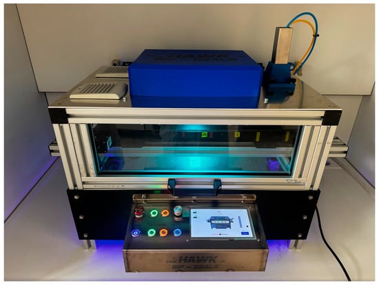
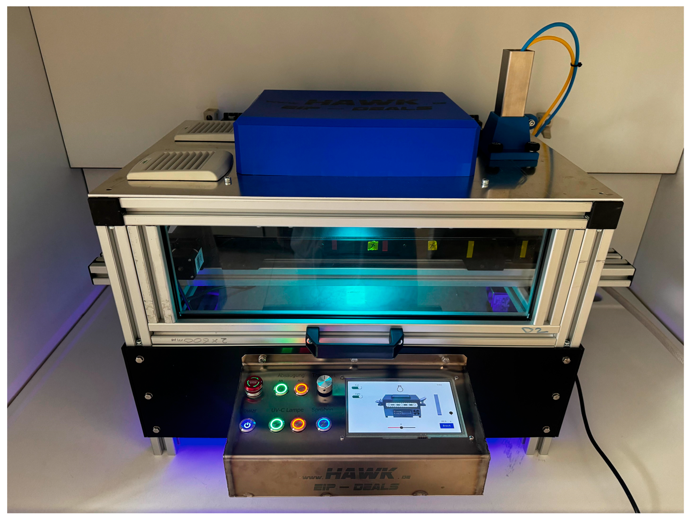
A specially designed prototype system developed and manufactured at HAWK was used to treat the meat samples (Figure 1). The system enabled the semi-automated treatment of meat samples via spray application of Tb-PAW. The prototype was operated using a standard 230 V power supply and was equipped with a central control unit. An integrated conveyor belt was used to transport the samples during the treatment process. The speed and direction of the conveyor belt could be set manually via the control unit. In addition, the system was equipped with a switchable extraction unit for the controlled removal of aerosols and gases generated during the treatment process. The spray application unit consisted of a pneumatic atomizing nozzle (M5 B, Landefeld Druckluft und Hydraulik GmbH, Kassel, Germany), operated with filtered compressed air at an operating pressure of 1–5 bar and an atomization rate of 0.05–0.2 L/min. Compressed air was supplied by an external compressor (TE-AC 270/50 Silent Plus, Einhell, Landau/Isar, Germany) with a 50-liter compressed air reservoir. The nozzle had an orifice diameter of 0.3 mm and was used for the atomization of Tb-PAW, enabling atomization of the Tb-PAW. The spray head was vertically adjustable, allowing the distance between the nozzle and the sample surface to be varied as required. The spraying process was activated via the control unit. The spray duration could be adjusted in millisecond increments and thereby controlled the amount of Tb-PAW applied to the sample.
Figure 1.
Functional model for semi-automated spray treatment with plasma-activated water (PAW). Meat samples are placed in Petri dishes in the protective chamber from the left or right and can be moved along a conveyor belt. The spray head assembly, which is supplied with PAW via a hose system, is visible in the upper right corner. The system is operated via the touchscreen display, and settings can be adjusted for conveyor belt movement, ventilation, and spray application. To enable future combination with other application methods, the protective chamber was designed in an elongated shape, allowing for sequential decontamination steps.
2.3.3. Tb-PAW Treatment of Inoculated Buffalo and Beef Samples
The meat samples were inoculated with 100 µL of the respective bacterial suspension. The suspension was spread evenly over the sample surface (20.25 cm2) using a sterile spatula (Delta™, Carl Roth GmbH & Co. KG, Karlsruhe, Germany). The samples were then incubated for 20 min at room temperature to allow the microorganisms to attach to the sample surface [24,25]. The treatments with Tb-PAW were carried out on inoculated buffalo and beef samples in three independent experiments. The meat samples were treated with Tb-PAW and the corresponding TRIS-buffer control according to the functional model described. The spray volume was set to 0.5 mL to ensure complete wetting of the sample without promoting possible rinsing of the bacteria and was verified gravimetrically using a tared Petri dish. Each sample was sprayed once, with the spray head positioned perpendicular to the sample surface at a constant distance. Exposure times of 1, 5, 10 and 15 min were selected for the treatment, based on preliminary experiments conducted in-house. Each sample treated with Tb-PAW was compared with a corresponding TRIS-treated control sample with an identical exposure time. This comparison served to control a possible rinsing effect to ensure that any observed reduction in bacteria was attributable to the effect of the Tb-PAW. Between applications of TRIS-buffer and Tb-PAW, the tubing was rinsed three times with sterile distilled water. In addition, an inoculated but untreated control sample was examined for each microorganism tested. The microbiological examination of the meat samples was carried out in accordance with ISO 17604:2015 [41] using a two-stage swab method. Due to the moisture film on the sample surface caused by the spray treatment, two dry sterile swabs were used. The sample surface was swabbed twice in a meandering pattern, rotating the swab over the surface and turning the sample 90° between the two passes. Both swab tips were then transferred to a 10 mL tube containing NaCl-peptone solution (0.85% NaCl and 0.1% peptone, Carl Roth GmbH & Co. KG, Karlsruhe, Germany) and homogenized using a vortex mixer. The suspensions were then serially diluted in powers of ten and plated on selective agar. Further processing of the samples, including the culture media used and incubation conditions, was performed as described for the in vitro experiments (Section 2.2.). After incubation, the colonies were counted and calculated as CFU/cm2.
2.3.4. Color Analysis
Immediately after swabbing, the CIE L*a*b* color values of the sample surface were determined at three different measurement points using a colorimeter (Minolta CR-400, Konica-Minolta GmbH, Langenhagen, Germany) and subsequently averaged.
2.3.5. Myoglobin Redox Form Analysis
The influence of the treatments on the myoglobin redox forms of buffalo and beef samples (deoxymyoglobin (DeoMb), oxymyoglobin (OxyMb), and metmyoglobin (MetMb)) was determined. The determination of the myoglobin redox forms followed the method described by [42] with minor modifications. Samples were prepared as described above. Three treatment groups were considered for each animal species: an untreated control, a sample treated with TRIS-buffer and a sample treated with Tb-PAW. After treatment of the meat samples (1 min, plus a uniform delay period for all samples until the start of the analysis), a layer approximately 1 cm thick was removed from the surface. The removed tissue was then cut into small pieces and stored at −18 °C until analysis. To determine the myoglobin redox forms, 2.8–3.0 g of the frozen sample material was weighed into centrifuge tubes (Carl Roth GmbH + Co. KG, Karlsruhe, Germany) and mixed with 7 mL of phosphate-buffered saline (PBS; 1 tablet per 1 L distilled water, Carl Roth GmbH + Co. KG). The samples were then homogenized (1 min at 30,000 rpm) on ice using a rotor-stator homogenization unit (MiniBatch D-9, MICCRA GmbH, Heitersheim, Germany). After each sample, the homogenization unit was rinsed three times with cold water to avoid cross-contamination. The homogenized samples were kept on ice and subsequently centrifuged at 4 °C and 35,000× g for 30 min (SORVALL RC-5C Plus, Kendro Laboratory Products, Newton, MA, USA). The supernatant was then transferred to disposable cuvettes (BRAND GmbH + Co. KG, Wertheim, Germany) and analyzed at room temperature in a UV-VIS spectrophotometer (Evolution 201, Thermo Scientific, Langenselbold, Germany). A total of three independent experiments were analyzed. The relative proportions of DeoMb, OxyMb and MetMb were calculated according to Tang et al. [43].
2.4. Statistical Analysis
The statistical analysis of the data was performed using SAS Enterprise Guide 7.1 (SAS Institute Inc., Cary, NC, USA). First, the data were tested for normality using the Shapiro–Wilk test and for homogeneity of variances using the Levene test. If the assumptions were met, a one-way analysis of variance (ANOVA) was performed, followed by Tukey’s HSD test for multiple comparisons. If the assumptions were not met, the non-parametric Kruskal–Wallis test was used, followed by pairwise comparisons with Bonferroni correction. Differences were considered statistically significant at p ≤ 0.05.
3. Results
3.1. Effects of In Vitro Application of Tb-PAW and TRIS-Buffer
The results of the in vitro experiments can be seen in Table 1. Comparing Tb-PAW and TRIS-buffer, the bacterial count for all three microorganisms decreased progressively as the treatment time of Tb-PAW was extended. However, there are differences among the bacterial species.
Table 1.
Mean value and standard deviation of the bacterial counts achieved for E. coli, S. Typhimurium and L. monocytogenes as a function of exposure time following in vitro treatment with Tb-PAW 1 compared to treatment with TRIS-buffer 2.
3.1.1. Effect on E. coli
Tb-PAW-treatment led to lower microbial counts of E. coli compared to TRIS-buffer-treated samples. The results for the effects on E. coli are similar to the findings of other studies, as Gram-negative bacteria are highly sensitive to PAW [29,34,44]. A prolonged treatment time led to a higher reduction, as treatment of 10 and 15 min resulted in significantly reduced microbial counts. However, in this study, no further reduction or complete eradication of E. coli could be stated.
3.1.2. Effect on S. Typhimurium
Similar to E. coli, in vitro experiments on S. Typhimurium showed lower microbial counts after Tb-PAW treatment. Whereas no reduction was achieved after 1 min treatment, a treatment of 5 min and longer led to a significant reduction. Additionally, a 15 min treatment showed a reduced microbial count compared to a 5 min treatment.
3.1.3. Effect on L. monocytogenes
For L. monocytogenes, at least 10 min of treatment time are necessary to reduce the microbial counts compared to 1 and 5 min of treatment. In addition, after 25 min of treatment, growth of L. monocytogenes was also reduced compared to 10 min of treatment.
3.1.4. General Observation
The in vivo experiments provided valuable insights into the underlying processes. Whereas E. coli was already significantly reduced by 1 min of treatment, a reduction in S. Typhimurium and L. monocytogenes can be seen after 5 min and 10 min, respectively. Since the latter two types of bacteria are Gram-positive, this also shows that they exhibit a delayed response to reactive nitrogen and oxygen species [29,35]. It is hypothesized that the thicker peptidoglycan layer provides protection [18,45].
Consistently, time-dependent inactivation was observed in all microorganisms studied. A longer treatment time leads to a greater reduction in microbial growth. Overall, the findings of this study are consistent with other published data on the antimicrobial activity of PAW. Rothwell et al. [46] were able to achieve a bacterial reduction of up to 6 log orders after just one minute. This research group used PAW from a DBD system and treated L. monocytogenes, as well as E. coli and S. enterica. Zhao et al. [35] used an atmospheric cold plasma jet to treat E. coli. After 30 min, a reduction of approximately 3 log levels was demonstrated; after 3 h of treatment, E. coli was no longer detectable. Using the same plasma system as in this study, Große-Peclum et al. [33] were also able to demonstrate significant reductions in E. coli: an exposure time of 1 min resulted in a reduction of approximately 5 log levels.
In a recent study, Myers et al. [44] investigated the effects of PAW on E. coli using single-cell impedance flow cytometry. First, the E. coli cell shrinks as a result of the treatment, as other authors have already observed [47]. Furthermore, they were able to demonstrate a correlation between the impedance signal and a phase shift in the E. coli populations (nuances between viable and nonviable cells) depending on the plasma water manufacturing time. Longer activation leads to a higher proportion of RONS and, consequently, to a stronger antibacterial effect. These results may provide insight into the mechanisms of action of PAW, which may operate in similar or modified forms in other microorganisms.
3.2. Antimicrobial Effect of Tb-PAW Treatment
The results in Table 2 show the reduction in bacterial counts after the application of Tb-PAW compared to untreated samples. The bacterial counts of the untreated but inoculated samples of buffalo and beef, respectively, were 4.91 ± 0.07 log10 CFU/g and 5.21 ± 0.07 log10 CFU/g for E. coli, 5.25 ± 0.07 log10 CFU/g and 5.26 ± 0.10 log10 CFU/g for S. Typhimurium, 4.55 ± 0.04 log10 CFU/g and 4.62 ± 0.07 log10 CFU/g for L. monocytogenes. For buffalo and beef meat, results of 1 min to 15 min (E. coli and S. Typhimurium) and up to 30 min (L. monocytogenes) are given, showing the potential of Tb-PAW by significant reductions for all three microorganisms. Although the use of Tb-PAW also reduces bacterial counts in meat, this effect is significantly weaker compared to the in vitro studies.
Table 2.
Mean values and standard deviation of the bacterial count reduction achieved for E. coli, S. Typhimurium and L. monocytogenes as a function of exposure time following treatment with Tb-PAW 1 compared to treatment with TRIS-buffer 2, for buffalo meat and beef, respectively.
3.2.1. E. coli
For E. coli, similar effects are found for buffalo and beef meat, respectively. On buffalo meat, treatment times of 1, 5 and 15 min led to a lower bacterial count. However, this effect was not observed for a 10 min treatment time. For beef meat, a reduction for all treatment times was found. It must be noted that prolonged treatment (1 to 15 min) does not lead to a higher reduction in bacteria counts.
3.2.2. S. Typhimurium
For S. Typhimurium, again, similar results are found for buffalo and beef meat. Treatment with Tb-PAW for 1, 5 and 15 min led to a reduced microbial growth compared to TRIS-buffer. As for E. coli in buffalo meat, there was no significant difference for 10 min treatment. In beef meat, 1, 10 and 15 min treatments reduce the growth of S. Typhimurium, but no reduction was found for a 5 min treatment.
3.2.3. L. monocytogenes
As for E. coli and S. Typhimurium, Tb-PAW can be used to significantly reduce the growth of L. monocytogenes on buffalo and beef meat. However, on buffalo meat only a 5 min treatment time led to a significant reduction. This effect could not be strengthened by a prolonged treatment duration. In contrast, 1, 5 as well as 15 min Tb-PAW-treatments reduce the growth of L. monocytogenes on beef in a similar manner. Again, there was no stronger effect due to a longer treatment time.
3.2.4. General Observation
Generally, the antimicrobial effect of Tb-PAW is comparable for buffalo and beef meat. In contrast to in vitro experiments, a prolonged treatment time does not increase the reductive effect of Tb-PAW on meat samples.
When considering the comparability of the results from different publications, it is important to keep in mind that the settings used during the production of plasma water have a decisive influence on the composition and inactivation capacity of the liquid. In addition, there are individual conditions specific to each experimental setup—such as meat-related factors, temperature, the method of applying the plasma water, and others—that can ultimately lead to differences in the study results.
Methodologically, this study employed spraying of the liquid to specifically wet the surface and avoid rinse-off effects. On a positive note, it is worth mentioning that this approach allows for resource-efficient use of the PAW itself. In contrast, Barrales Astorga et al. [48] completely immersed the samples in PAW for up to 300 s. In their publication, they were able to reduce S. Typhimurium and E. coli by 5.9 and 4.0 log levels, respectively.
A lower reduction effect on meat matrices compared to in vitro studies has already been reported on several occasions. Große-Peclum et al. [33] were unable to reduce E. coli on poultry skin—neither immediately after PAW-application nor during a storage period of 14 days. Similarly, Sammanee et al. [49], who investigated E. coli and S. Typhimurium, were able to reduce the CFU/g by only 0.08 log when using a combination of PAW and hydrogen peroxide (60 ppm). Interestingly, unlike the use of pure water, this treatment did not result in a significant reduction in the bacterial load. In contrast, after two weeks of storage, a significant difference in E. coli counts was observed in duck skin—but only in the Tb-PAW samples, not in the control samples or in the TRIS-buffer-treated samples [33]. This suggests the possibility of long-term effects that may be relevant in the future, depending on the shelf life of the individual foods. Nevertheless, the microbicidal effect of (Tb)-PAW on animal-derived matrices (e.g., meat, skin) is evidently reduced.
The more complex structure of animal tissue can be assumed to be the cause for the lower microbicide potential here. Both an irregular surface and cavities can provide microorganisms with better adhesion. On the skin, hair, fats, proteins, and other interfering factors such as dirt also play a significant role (e.g., by shielding effects); similar to what happens on the meat itself, they can interact with the PAW and weaken its microbicidal effect [34,50,51,52]. The fact that no differences were observed between the animal species indicates that the meat has similar characteristics and that Tb-PAW exhibits fundamentally comparable antimicrobial efficacy in buffalo and beef. In contrast to the in vitro experiments, the efficacy cannot be enhanced by extending the treatment time. It can be assumed that the short-lived reactive species react relatively more quickly on a complex meat matrix, e.g., through interactions with interfering factors such as moisture, fat, or proteins [53,54].
Experiments on the influence of an interfering factor were also conducted by Große-Peclum et al. [29], which were presented in the context of the efficacy of Tb-PAW against pathogens associated with a hoof disease (Mortellaro) in cattle. In this study, bovine serum albumin (BSA) (concentration 5%) was used to simulate the presence of an animal protein on the skin surface. It was found that as the concentration of BSA increased, a decrease in the microbicidal activity of Tb-PAW was observed. In conclusion, it should be noted that the interaction of PAW with factors on the surface of animal products results in a reduced effect.
As can be seen in Table 2, all three bacteria strains are reduced on buffalo and beef when treated with Tb-PAW compared to the buffer. It is interesting to note that a longer exposure time does not necessarily result in a greater reduction in bacterial counts; in some cases, the reduction is actually smaller. This is likely due to the interaction of the Tb-PAW with matrix-specific factors [33,34,35]. Depending on which substance the RONS encounter, they react differently. In the case of contact with a bacterium, this can lead to a reduction in bacterial counts [28,29,32,33]. On the other hand, the initial contact may also occur with a different cell, whose reaction does not lead to a reduction in bacterial counts but nevertheless results in the loss or redirection of the RONS’s reactivity. Furthermore, the observation that the microbicidal effect is reduced with longer exposure times compared to a one-minute application should not be overemphasized, as the reported values are based on the results of three independently repeated runs. The low standard deviations indicate that the results were consistent across the three replicates. Since a plate count method was used here, inaccuracies of up to 0.3 log10 are inherent to the method [55].
In general, the reduction ranges from 0.41 to 0.82 for E. coli, from 0.05 to 0.69 for S. Typhimurium, and from 0.33 to 0.54 for L. monocytogenes. It is debatable whether this reduction is actually attributable to a microbicidal effect of PAW. With regard to possible wash-off effects, it can be ruled out that these results are intentionally based on the reduction relative to treatment with TRIS-buffer, since in that case a potential error relative to the untreated control would have occurred due to the methodology as well (cf. 2.3.3.). However, it should be kept in mind that the evaluation of the TRIS-buffer-treated samples was carried out using the same procedure, and relatively speaking, a reduction in bacterial counts due to PAW is still evident for all three bacteria, for all four exposure times, and on both meat matrices.
Overall, therefore, the PAW has been shown to have microbicidal efficacy; however, this efficacy is still low compared to that of other methods used for disinfection. According to the guidelines for disinfectant testing [56], a reduction of 4 log levels is required for a substance to be labeled as such. In this regard, further adjustments to PAW and research into interactions with potential (interfering) factors are needed to enhance its microbicidal efficacy.
3.3. Influence on Meat Color
For untreated samples, there were no differences between buffalo (L* = 37.67; a* = 19.65; b* = 9.56) and beef meat (L* = 39.16; a* = 17.40; b* = 8.67) over the course of treatment (storage of up to 15 min in parallel to the treated samples with TRIS-buffer and Tb-PAW, respectively).
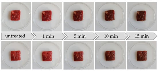
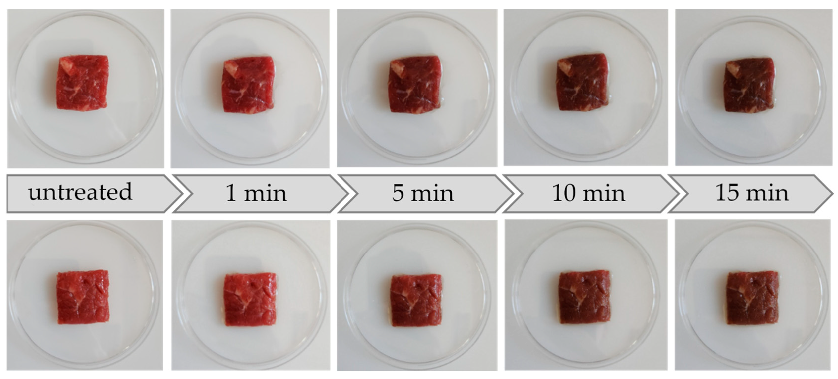
The color differences that are related to the treatment are presented in Table 3, and the relevance for visible color changes is shown in Table 4. Applying the TRIS-buffer did not result in any color change in buffalo and beef meat, respectively. In contrast, the treatment with Tb-PAW resulted in color differences, which increased with treatment duration and led to visible color differences (Figure 2).
Table 3.
Mean values and standard deviations of the difference in CIE-Lab color values between the respective treatment times with Tb-PAW 1 and the untreated samples of buffalo and beef meat, respectively.
Table 4.
Color distance (ΔE) of buffalo and beef meat samples between untreated samples, TRIS-buffer 1-treated samples and Tb-PAW 2-treated samples.
Figure 2.
Color differences in buffalo (top row) and beef meat (bottom row) of untreated samples and after application in relation to treatment time (1, 5, 10, 15 min) with Tb-PAW.
3.3.1. Buffalo Meat
For buffalo samples, Tb-PAW-treatment led to lower a* results compared to the control and TRIS-buffer groups. There are effects of the treatment duration, as 5, 10 and 15 min led to lower a* values after 1 min, but 10 and 15 min also resulted in significantly lower a* values than 5 min treatment.
As significant differences might be detected via instrumental analysis, a different color perception by the consumers is not given in all cases. Therefore, the ΔE values are used [57]. Values of ΔE between 0 < 1 are assumed to be an unnoticeable difference, whereas ΔE between 1 < 2 indicates that color differences are only noticed by experienced observers. For ΔE between 2 < 3.5, the differences are also noticed by unexperienced observers and for ΔE 3.5 < 5 the color difference is clearly noticeable. Finally, values > 5 give the impression of two different colors [58].
Regarding Table 4, treatment with TRIS-buffer does not lead to visible color differences in buffalo meat. In contrast, Tb-PAW led to relevant differences, even after 1 min of treatment, compared to untreated meat samples. A treatment duration of 5 min (as well as for prolonged treatment times) resulted in the perception of two different colors of the buffalo meat samples.
3.3.2. Beef Meat
Similar to this, no effects of TRIS-buffer are found for beef meat. However, Tb-PAW treatment (5, 10, 15 min) led to lower a* values in beef samples compared to control and TRIS-buffer-treated samples. Regarding the Tb-PAW-treated samples, a* values are significantly lower for treatment duration of 5, 10 and 15 min compared to 1 min treatment.
Regarding the ΔE values (Table 4), TRIS-buffer treatment does not result in relevant color changes compared to the beef control sample. For Tb-PAW, similar results as in buffalo meat are observed. Treatment times for 5 min and longer led to the perception of two different colors of the beef meat samples compared to untreated samples.
3.3.3. General Observation
As color of meat is one of the most contributing factors to consumers’ decision to buy a product [57], this aspect is of high relevance. Results presented in Figure 2 demonstrate the changes in color during the treatment time in an impressive manner. As can be seen in Table 4, the effect of Tb-PAW is higher on buffalo meat than on beef meat at all treatment times (ΔE of 10.47 to 8.48, respectively, after 15 min of treatment).
Compared to the untreated sample and the sample treated with buffer, a color change was observed in the meat of both animal species. However, a difference becomes apparent upon closer examination: the reduction in the a* value after 15 min was significantly greater in buffalo meat than in beef. Results might be interpreted as indicating that buffalo meat is more sensitive to oxidative processes.
Similar findings have also been published previously. For example, Große-Peclum et al. [33] were able to demonstrate species-specific differences in poultry meat: Tb-PAW did not result in any visual changes in chicken meat, whereas in duck meat, the a* value decreased by 4.0 units.
Barrales Astorga et al. [48] made similar observations with beef. Their PAW experiments resulted in a reduction of a* values by 0.9 units after 20 min, while L* and b* remained unchanged. This research group considers the deviations to be moderate and comparable to color changes that can also be observed after the application of lactic acid.
However, the present changes show a significantly greater deviation from the untreated meat—both for buffalo and beef. This may be due to differences in sample preparation methods or the nature of the meat itself. However, the individual composition of the plasma waters used very likely plays a role here. Depending on the manufacturing process, storage time, temperature, and other factors, different proportions of reactive species are present. Accordingly, the reaction of the PAW with the respective meat matrix varies significantly from case to case.
Due to the reduction in the red pigment content and the presence of reactive oxygen and nitrogen species, changes in the myoglobin variants in the meat may have occurred.
3.4. Influence on Proportions of Myoglobin
As presented in Table 5, significant differences can be found for MetMb and OxyMb for buffalo and beef meat. Again, there were no differences between untreated control and TRIS-buffer-treated samples for both species. However, the Tb-PAW-treated samples developed a higher proportion of MetMb and a lower proportion of OxyMb compared to untreated or TRIS-buffer-treated meat samples. Higher proportions of MetMb lead to a diminished red coloration and an overall impression of a brown or gray color [59,60].
Table 5.
Mean values and standard deviations of the proportions of myoglobin variants in the untreated control and after treatment with TRIS-buffer 1 or Tb-PAW 2, respectively, for buffalo and beef meat.
As expected, a shift in these myoglobin variants is likely to result in a change in meat color. Oxidized forms of myoglobin typically exhibit brownish-gray hues [61]. Fröhling et al. [62] describe how hydrogen peroxide (H2O2) is capable of oxidizing OxyMb (iron as Fe2+, O2 bound on the 6th ligand) to MetMb (iron as Fe3+, H2O or OH− on the 6th ligand) [63]. Since hydrogen peroxide, among others, is a confirmed component of Tb-PAW [64,65], a similar mechanism is suggested for the present results. Hydrogen peroxide can react with Fe2+, producing both hydroxide ions (OH−) and highly reactive hydroxyl radicals (·OH), a radical formation process called “Fenton reaction” [66]. Other research groups have also reported that contact between hydrogen peroxide and myoglobin led to color changes [67]. Chen et al. claimed the accumulation of active H2O2-species to be the main reason, leading to green discoloration of myoglobin [67]. This assumption is additionally supported by the fact that no shift in myoglobin variants is observed during treatment with TRIS-buffered solution in this study.
Given that appearance and color are decisive factors in the decision to purchase meat products [60], these findings must be taken into account when considering future applications. Studies by Hood and Riordan [68] have already shown that a MetMb content of 10% can lead to a drop in sales of up to 40%. Experienced test subjects describe the meat color as brownish when the MetMb content is 37% [69]. Accordingly, a noticeable brownish discoloration is clearly to be expected in the meat samples treated with Tb-PAW. This is consistent with the interpretation of the ΔE-values (Table 4) [58], according to which both the untreated and the TRIS-treated meat exhibit a noticeably different color than the Tb-PAW-treated meat.
Interestingly, Barrales Astorga et al. [48] were unable to confirm these color changes through treatment with PAW. The levels of OxyMb, DeoxyMb, and MetMb remained comparable to those in untreated samples. As previously suspected when examining the color values, the different preparation and composition of the plasma waters may play a decisive role here.
This is further supported by the finding that Große-Peclum et al. [33], using the same Tb-PAW prepared as in this study, also observed changes in the myoglobin fractions. In contrast to the findings of this study, however, measurable shifts in chicken meat did not occur until after 14 days of storage. Similarly, this was also observed in duck meat during the storage period.
It should be noted, however, that the immediate changes observed by Große-Peclum et al. [33] were significantly smaller: MetMb levels increased by only 2% and 3%, respectively, in chicken and duck on the day of treatment.
The effects on buffalo and beef, however, were much more pronounced immediately after treatment. As shown in Figure 1, the color change in the Tb-PAW-treated samples is immediately visible to the naked eye in both species. The MetMb content increased by approximately 56% in each case, suggesting a strong oxidative reaction in the muscle tissue of buffalo and cattle.
Although the presented results show only a slight (but significant) microbicidal effect of Tb-PAW on the meat of the two animal species, in light of the good in vitro reductions, it appears worthwhile to continue working on the application of Tb-PAW on meat. However, the sensory effect on meat color is a particularly relevant observation that must not be neglected in follow-up studies. Following Chen et al., future research could focus on applying this method to meat with a lower myoglobin content or on using PALs with low oxidation potential to prevent discoloration of the meat [67].
Furthermore, it is also interesting to determine whether the treatment results in further changes in meat quality in terms of sensory characteristics, nutritional values, and shelf life. In this regard, a particularly relevant approach may be to examine the oxidative processes in fats—which can lead to rancid flavors—and to conduct corresponding storage tests. Interestingly, the research group led by Barrales Astorga et al. [48] found no evidence of increased lipid oxidation in beef compared to treatment with lactic acid. They suspect that the presence of NO2 in the plasma solution is the cause, but also note that this result may vary depending on the specific plasma-activated liquid used. This must be specifically analyzed prior to any future industrial use of Tb-PAW.
4. Conclusions
Bacterial counts of E. coli, S. Typhimurium and L. monocytogenes can be reduced by the application of Tb-PAW. Whereas the microbicidal effect in vitro is higher, reductions on meat samples from buffalo and beef are lower regarding matrix-related interactions. There are differences in the efficacy of the reduction in relation to the treatment time as well as the microorganism itself. However, reductions up to 0.8 (E. coli), 0.69 (S. Typhimurium) and 0.54 (L. monocytogenes) can be achieved on meat matrix (beef), but also for buffalo meat (0.50; 0.47 and 0.50 for E. coli, S. Typhimurium and L. monocytogenes, respectively). No species-related differences in microbicidal effect were detected. Also, a clear relation between reduction in microbial growth and treatment duration could not be shown.
For sensory appearance, application of Tb-PAW resulted in clearly visible color changes, both in buffalo and in beef meat samples. As a strong influence of the proportions of myoglobin-redox forms was stated, the darker and more gray impression of meat can be due to a higher proportion of MetMb and lower OxyMb content.
Given the significant role that the visual appearance of food plays in purchasing decisions, further research is needed in this area. At this time, it can be assumed that, due to sensory differences, the chosen form and timing of application cannot be used for the treatment of buffalo and beef. Furthermore, a better understanding of the PAW–matrix interaction is of interest in order to be able to use PAW specifically as a decontamination agent even on complex matrices. The results highlight both the potential and the knowledge gaps of research in the application of plasma-activated liquid in the context of meat and food hygiene.
Author Contributions
Conceptualization, L.S. (Lisa Siekmann) and L.S. (Louisa Singer); methodology, V.G.-P.; software, L.S. (Louisa Singer); validation, L.S. (Lisa Siekmann), V.G.-P. and L.S. (Louisa Singer); formal analysis, L.S. (Lisa Siekmann) and L.S. (Louisa Singer); investigation, L.S. (Louisa Singer); resources, G.A., C.O. and M.P.; data curation, L.S. (Louisa Singer) and L.S. (Lisa Siekmann); writing—original draft preparation, L.S. (Lisa Siekmann); writing—review and editing, V.G.-P., G.A., C.O. and M.P.; visualization, L.S. (Louisa Singer); supervision, L.S. (Lisa Siekmann) and M.P.; project administration, L.S. (Lisa Siekmann) and M.P.; funding acquisition, L.S. (Lisa Siekmann). All authors have read and agreed to the published version of the manuscript.
Funding
This research was funded by Fritz-Ahrberg-Stiftung; the development of the functional model was funded by the European Innovation Partnership (EIP-AGRI) project “DEALS” and by the European Agricultural Fund for Rural Development (EAFRD 276 03 201 000 1523) and the Deutsche Forschungsgemeinschaft (DFG, German Research Foundation)—491094227 “Open Access Publication Funding” and the University of Veterinary Medicine Hannover.
Data Availability Statement
The original contributions presented in this study are included in the article. Further inquiries can be directed to the corresponding author.
Acknowledgments
The authors would like to thank Wolfgang Viöl for the preliminary work and excellent collaboration regarding the application of PAW in veterinary medicine and food hygiene. Furthermore, we are very grateful for the provision of the PAW and would like to thank Markus Harms and Rinat Ortmann. A big thank you also goes to Rabea Bernhardt for her help with the lab work.
Conflicts of Interest
The authors declare no conflicts of interest. The funders had no role in the design of the study; in the collection, analyses, or interpretation of data; in the writing of the manuscript; or in the decision to publish the results.
Abbreviations
The following abbreviations are used in this manuscript:
| * | Significant reduction |
| ΔE | Delta E |
| µmol | Micromole |
| a* | Color value, CIElab, red to green axis |
| °C | Degree Celsius |
| b* | Color value, CIElab, blue to yellow axis |
| CFU | Colony Forming Units |
| cm, cm2 | Centimeter, square centimeters |
| E. | Escherichia |
| et al. | et alii, and others |
| e.g., | For example |
| g | Gram(s) |
| × g | g-force, standard Earth gravity (9.81 m/s2) |
| Fe2+ | Ferrous ion |
| Fe3+ | Ferric ion |
| h | Hour(s) |
| H2O | Dihydrogen monoxide, water |
| H2O2 | Hydrogen peroxide |
| kHz | Kilohertz |
| kV | Kilovolt |
| L | Liter(s) |
| L. | Listeria |
| L* | Color value, CIElab, lightness |
| MetMb | Metmyoglobin |
| min | Minute(s) |
| mL | Milliliter |
| mm | Millimeter |
| mol | Mole |
| N | Size of the entire population, total number of observations |
| ns | Non-significant |
| OH− | Hydroxide ion |
| ·OH | Hydroxyl radical |
| OxyMb | Oxymyoglobin |
| PAL | Plasma-activated liquid |
| PAW | Plasma-activated water |
| pH | Potential of hydrogen, pondus hydrogenii |
| ppm | Parts per million |
| RONS | Reactive oxygen and nitrogen species |
| s | Significant |
| s, s2 | Second(s), square seconds |
| S. | Salmonella |
| Tb-PAW | TRIS-buffered-Plasma-activated water |
| TRIS-buffer | Buffer from TRIS(hydroxymethyl)aminomethane (≥99.8%) and TRIS(hydroxymethyl)aminomethane hydrochloride (≥99.0%) |
| V | Volt |
| W | Watt |
References
- United Nations. World Population Prospects 2024: Summary of Results; UN DESA/POP/2024/TR/NO. 9; United Nations: New York, NY, USA, 2004; Available online: https://population.un.org/wpp/publications?year=2024 (accessed on 15 July 2026).
- Beal, T.; Gardner, C.D.; Herrero, M.; Iannotti, L.L.; Merbold, L.; Nordhagen, S.; Mottet, A. Friend of Foe? The Role of Animal-Source Foods in Healthy and Environmentally Sustainable Diets. J. Nutr. 2023, 153, 409–425. [Google Scholar] [CrossRef] [Scilit]
- Herreman, L.; Nommensen, P.; Pennings, B.; Laus, M.C. Comprehensive Overview of the Quality of Plant- and Animal-sourced Proteins Based on the Digestible Indispensable Amino Acid Score. Food Sci. Nutr. 2020, 8, 5379–5391. [Google Scholar] [CrossRef] [Scilit]
- Godfray, H.C.J.; Aveyard, P.; Garnett, T.; Hall, J.W.; Key, T.J.; Lorimer, J.; Pierrehumbert, R.T.; Scarborough, P.; Springmann, M.; Jebb, S.A. Meat Consumption, Health, and the Environment. Science 2018, 361, eaam5324. [Google Scholar] [CrossRef] [Scilit]
- United States Department of Agriculture (USDA), Foreign Agricultural Service. Livestock and Poultry: World Markets and Trade. PSD Publications; 2026. Available online: https://apps.fas.usda.gov/psdonline/app/index.html#/app/downloads (accessed on 15 July 2026).
- Vaz, R.Z.; de Sá, H.A.O.M.; Sartori, D.B.S.; Costa, P.T.; Fluck, A.C.; Kröning, A.B.; Ferreira, O.G.L.; Costa, O.A.D.; Restle, J. Trade and consumption of buffalo meat in Brazil. Meat Sci. 2024, 208, 109399. [Google Scholar] [CrossRef] [Scilit]
- Cruz, L.C. Trends in Buffalo Production in Asia. Ital. J. Anim. Sci. 2007, 6, 9–24. [Google Scholar] [CrossRef] [Scilit]
- Siekmann, L.; Loreck, K.; Plötz, M. Das Potenzial von Büffelfleisch. Rundsch. Für Fleischhygiene Und Leb. RFL 2022, 4, 112–114. [Google Scholar]
- Cappelo, G.; Di Vuolo, G.; Gerini, O.; Noschese, R.; Bufano, F.; Capacchione, R.; Rosini, S.; Limone, A.; De Carlo, E. Italian Tracing System for Water Buffalo Milk and Processed Milk Products. Animals 2021, 11, 1737. [Google Scholar] [CrossRef] [Scilit]
- Zahn, A.; Herzog, F. Water Buffalo as Habitat Designers. Anliegen Nat. 2015, 37, 46–54. [Google Scholar]
- Calanni Macchio, A.; Santinello, M.; Bifulco, G.; Matera, R.; Biffani, S.; Gomez-Carpio, M.; Campanile, G.; Neglia, G. The Role of Age at First Calving in Shaping Production and Reproductive Outcomes in Italian Buffaloes. J. Dairy Sci. 2005, 108, 7235–7247. [Google Scholar] [CrossRef] [Scilit]
- Zicarelli, F.; Iommelli, P.; Musco, N.; Wanapat, M.; Lotito, D.; Lombardi, P.; Infascelli, F.; Tudisco, R. Growth Performance of Buffalo Calves in Response to Different Diets with and without Saccharomyces cerevisiae Supplementation. Animals 2024, 14, 1245. [Google Scholar] [CrossRef] [Scilit]
- Rodrigues, L.S.; da Silva, J.A.R.; da Silva, W.C.; da Silva, É.B.R.; Belo, T.S.; Sousa, C.E.L.; Guimaraes de Carvalho Rodrigues, T.C.; Maciel e Silva, A.G.; Prates, J.A.M.; Lourenço-Júnior, J.B. A Review of the Nutritional Aspects and Composition of Meat, Liver and Fat of Buffaloes in the Amazon. Animals 2024, 14, 1618. [Google Scholar] [CrossRef] [Scilit]
- Tamburrano, A.; Tavazzi, B.; Callà, C.A.M.; Amorini, A.M.; Lazzarino, G.; Vincenti, S.; Zottola, T.; Campagna, M.C.; Moscato, U.; Laurentini, P. Biochemical and Nutritional Characteristics of Buffalo Meat and Potential Implications on Human Health for a Personalized Nutrition. Ital. J. Food Saf. 2019, 8, 8317. [Google Scholar] [CrossRef] [Scilit]
- Ali, S.; Alsayeqh, F. Review of major meat-borne zoonotic bacterial pathogens. Front. Public Health 2022, 10, 1045599. [Google Scholar] [CrossRef] [Scilit]
- Sofos, J.N.; Geornaras, I. Overview of Current Meat Hygiene and Safety Risks and Summery of Recent Studies on Biofilms, and Control of Escherichia coli O157:H7 in Nonintact, and Listeria monocytogenes in Ready-to-eat, Meat Products. Meat Sci. 2010, 86, 2–14. [Google Scholar] [CrossRef] [Scilit]
- Herawati, C.; Endayani, H.; Indragiri, S.; Kristanti, I.; Supriatin; Wahyuni, N.T.; Dani, A.H.; Hikmawati, I. Sanitary Hygiene and Behaviour of Food Handlers in the Presence of Escherichia coli Bacteria. J. Pure Appl. Microbiol. 2023, 17, 2098–2103. [Google Scholar] [CrossRef] [Scilit]
- Valentin-Weigand, P. Bakterielle Zellwand. In Tiermedizinische Mikrobiologie, Infektions- und Seuchenlehre, 11th ed.; Selbitz, H.-J., Truyen, U., Valentin-Weigand, P., Eds.; Thieme: Stuttgart, Germany, 2023. [Google Scholar] [CrossRef] [Scilit]
- European Food Safety Authority (EFSA); European Centre for Disease Prevention and Control (ECDC). The European Union One Health 2024 Zoonoses Report. EFSA J. 2025, 23, e9759. [Google Scholar] [CrossRef] [Scilit]
- Disson, O.; Charlieer, C.; Pérot, P.; Leclercq, A.; Paz, R.N.; Kathariou, S.; Tsai, Y.-H.; Lecuit, M. Listeriosis. Nat. Rev. Dis. Primers 2025, 11, 71. [Google Scholar] [CrossRef] [Scilit]
- Regulation (EC) No. 853/2004 of the European Parliament and of the Council of 29 April 2004 Laying Down Specific Hygiene Rules for Food of Animal Origin, Last Consolidated Version 07. May 2026. Available online: https://eur-lex.europa.eu/legal-content/EN/TXT/HTML/?uri=CELEX:02004R0853-20260507&qid=1791740956617 (accessed on 15 October 2026).
- Commission Regulation (EU) No. 101/2013 of 4 February 2013 Concerning the Use of Lactic Acid to Reduce Microbiological Surface Contamination on Bovine Carcases. Available online: https://eur-lex.europa.eu/legal-content/EN/TXT/HTML/?uri=CELEX:32013R0101&qid=1791741117332 (accessed on 15 October 2026).
- USDA, Economic Research Service U.S. Department of Agriculture. Charts of Note. EU Sanitary and Phytosanitary Measures Limit Imports of U.S. Poultry. 2014. Available online: https://www.ers.usda.gov/data-products/charts-of-note/77863 (accessed on 13 October 2026).
- Koller, V.; Seinige, D.; Saathoff, J.; Kehrenberg, C.; Krischek, C. Impact of a combination of UV-C Irradiation and peracetic acid spray treatment on Brochothrix thermosphacta and Yersinia enterocolitica contaminated Pork. Foods 2021, 10, 204. [Google Scholar] [CrossRef] [Scilit]
- Reichel, J.; Kehrenberg, C.; Krischek, C. Inactivation of Yersinia enterocolitica and Brochothrix thermosphacta on pork by UV-C irradiation. Meat Sci. 2019, 158, 107909. [Google Scholar] [CrossRef] [Scilit]
- Thirumdas, R.; Kothakota, A.; Annapure, U.; Siliveru, K.; Blundell, R.; Gatt, R.; Valdramidis, V.P. Plasma activated water (PAW): Chemistry, physico-chemical properties, applications in food and agriculture. Trends Food Sci. Technol. 2018, 77, 21–31. [Google Scholar] [CrossRef] [Scilit]
- Abdo, A.I.; Mcmillan, N.; Lao, L.; Pena, G.N.; Fitridge, R.; Richter, K. Plasma-activated water as a wound rinse solution in patiens with diabetes-related food ulcers in two Australian hospitals: Study protocol for a phase I double-blinded, randomized controlled trial. BMJ Open 2026, 10, e118420. [Google Scholar] [CrossRef] [Scilit]
- Stornelli, G.; Musella, G.; Balice, G.; Morari, D.; Gioco, G.; Lajolo, C.; Stoffels, M.; Moran, G.P.; Grande, R.; Caponio, V.C.A.; et al. Antimicrobial Effect of Plasma-Treated Liquids on Skin and Oral Biofilms: A Systematic Review of In Vitro Studies. Appl. Sci. 2026, 16, 7187. [Google Scholar] [CrossRef] [Scilit]
- Große-Peclum, V.; Siekmann, L.; Krischek, C.; Avramidis, G.; ten Bosch, L.; Harms, M.; Ochs, C.; Ortmann, R.; Hoedemaker, M.; Ahlfeld, B.; et al. An In Vitro Model Using TRIS-Buffered Plasma-Activated Water to Reduce Pathogenic Microorganisms Involved in Digital Dermatitis Infection in Cattle. Appl. Sci. 2022, 12, 12325. [Google Scholar] [CrossRef] [Scilit]
- Hadinoto, K.; Niemira, B.A.; Trujillo, F.J. A review on plasma-activated water and its application in the meat industry. Compr. Rev. Food Sci. Food Saf. 2023, 22, 4993–5019. [Google Scholar] [CrossRef] [Scilit]
- Inguglia, E.S.; Oliveira, M.; Burgess, C.M.; Kerry, J.P.; Tiwari, B.K. Plasma-activated water as an alternative nitrite source for the curing of beef jerky: Influence on quality and inactivation of Listeria innocua. Innov. Food Sci. Emerg. Technol. 2020, 59, 102276. [Google Scholar] [CrossRef] [Scilit]
- Steinhausser, L.; Gotzmann, G. Plasma-treated liqids as an environmentally friendly alternative to conventional disinfectants for the hygienization of food processing facilities-first insights in shelf life characteristics and antibacterial efficacy. Food Bioprod. Process. 2026, 156, 152–160. [Google Scholar] [CrossRef] [Scilit]
- Große-Peclum, V.; Siekmann, L.; Krischek, C.; Avramidis, G.; Ochs, C.; Viöl, W.; Plötz, M. Using TRIS-Buffered Plasma-Activated Water to Reduce Pathogenic Microorganisms on Poultry Carcasses with Evaluation of Physicochemical and Sensory Parameters. Foods 2023, 12, 1113. [Google Scholar] [CrossRef] [Scilit]
- Moiseienko, Y.; Shahbaz, H.M.; Saad, S.; Avison, M.B.; Stratakos, A.C. Effect of plasma-activated water against E. coli and S. aureus: Influence of organic matter and impact on skin cell viability. Appl. Microbiol. Biotechnol. 2025, 109, 244. [Google Scholar] [CrossRef] [Scilit]
- Zhao, Y.M.; Patage, A.; Sun, D.W.; Tiwari, B. Plasma-activated water: Physicochemical properties, microbial inactivation mechanisms, factors influencing antimicrobial effectiveness, and applications in the food industry. Compr. Rev. Food Sci. Food Saf. 2020, 19, 3951–3979. [Google Scholar] [CrossRef] [Scilit]
- ISO 16649-2:2001; Microbiology of Food and Animal Feeding Stuffs—Horizontal Method for the Enumeration of Beta-Glucuronidase-Positive Escherichia coli. Part 2: Colony-Count Technique at 44 Degrees C Using 5-Bromo-4-chloro-3-indolyl Beta-D-Glucuronide. International Organization for Standardization: Geneva, Switzerland, 2001.
- ISO 6579-1:2017; Microbiology of the Food Chain—Horizontal Method for the Detection, Enumeration and Serotyping of Salmonella. Part 1: Detection of Salmonella spp. International Organization for Standardization: Geneva, Switzerland, 2017.
- ISO 11290-1:2017; Microbiology of the Food Chain—Horizontal Method for the Detection and Enumeration of Listeria monocytogenes and of Listeria spp. Part 1: Detection Method. International Organization for Standardization: Geneva, Switzerland, 2017.
- Scheib, S.; Leimbach, S.; Avramidis, G.; Bellmann, M.; Nitz, J.; Ochs, C.; Tellen, A.; Wente, N.; Zhang, Y.; Viöl, W.; et al. Intermediate cluster disinfection: Which disinfection solution is most effective on milking liners? A comparison of microorganism reduction on liner inner surfaces using quantitative swab sampling technique. Pathogens 2023, 12, 560. [Google Scholar] [CrossRef] [Scilit]
- ISO 6887-1:2017; Microbiology of the Food Chain—Preparation of Test Samples, Initial Suspension and Decimal Dilutions for Microbiological Examination. Part 1: General Rules for the Preparation of the Initial Suspension and Decimal Dilutions. International Organization for Standardization: Geneva, Switzerland, 2017.
- ISO 17604:2015; Microbiology of the Food Chain—Carcass Sampling for Microbiological Analysis. International Organization for Standardization: Geneva, Switzerland, 2015.
- Bruna, J.M.; OrdóÑez, J.A.; Fernández, M.; Herranz, B.; de la Hoz, L. Microbial and physico-chemical changes during the ripening of dry fermented sausages superficially inoculated with or having added an intracellular cell-free extract of Penicillium aurantiogriseum. Meat Sci. 2001, 59, 87–96. [Google Scholar] [CrossRef] [Scilit]
- Tang, J.; Faustman, C.; Hoagland, T.A. Krzywicki Revisited: Equations for spectrophotometric determination of myoglobin redox forms in aqueous meat extracts. Food Chem. Toxicol. 2004, 69, 717–720. [Google Scholar] [CrossRef] [Scilit]
- Myers, B.; Agus, R.; Bouharrak, B.; Avino, F.; Furno, I. Investigating the effects of plasma-activated water on E. coli with the novel application of single-cell impedance flow cytometry. Sci. Rep. 2025, 15, 24064. [Google Scholar] [CrossRef] [Scilit]
- Ursache, M.; Moraru, R.; Hnatiuc, E.; Nastase, V.; Mares, M. Comparative assessment of the relation between energy consumption and bacterial burden reduction using plasma activated water. In Proceedings of the 2024 International Conference on Optimization of Electrical and Electronic Equipment (OPTIM), Bran, Romania, 22–24 May 2014; pp. 1036–1041. [Google Scholar] [CrossRef] [Scilit]
- Rothwell, J.G.; Alam, D.; Carter, D.A.; Soltani, B.; McConchie, R.; Zhou, R.; Cullen, P.J.; Mai-Prochnow, A. The Antimicrobial Efficacy Of Plasma Activated Water Is Modulated By Reactor Design And Water Composition. J. Appl. Microbiol. 2021, 132, 2490–2500. [Google Scholar] [CrossRef] [Scilit]
- Zhao, Y.M.; Ojha, S.; Burgess, C.M.; Sun, D.W.; Tiwari, B.K. Inactivation efficacy of plasma-activated water: Influence of plasma treatment time, exposure time and bacterial species. Int. J. Food Sci. Technol. 2021, 56, 721–732. [Google Scholar] [CrossRef] [Scilit]
- Barrales Astorga, J.; Hadinoto, K.; Cullen, P.; Prescott, S.; Trujillo, F.J. Effect of plasma activated water on the nutritional composition, storage quality and microbial safety of beef. LWT 2022, 154, 112794. [Google Scholar] [CrossRef] [Scilit]
- Sammanee, P.; Ngamsanga, P.; Jainonthee, C.; Chupia, V.; Sawangrat, C.; Kerdjana, W.; Lampang, K.N.; Meeyam, T.; Pichpol, D. Decontamination of Pathogenic and Spoilage Bacteria on Pork and Chicken Meat by Liquid Plasma Immersion. Foods 2022, 11, 1743. [Google Scholar] [CrossRef] [Scilit]
- Lis, K.A.; Boulaaba, A.; Binder, S.; Li, Y.; Kehrenberg, C.; Zimmermann, J.L.; Klein, G.; Ahlfeld, B. Inactivation of Salmonella Typhimurium and Listeria monocytogenes on ham with nonthermal atmospheric pressure plasma. PLoS ONE 2018, 13, e0197773. [Google Scholar] [CrossRef] [Scilit]
- Noriega, E.; Shama, G.; Laca, A.; Díaz, M.; Kong, M.G. Cold atmospheric gas plasma disinfection of chicken meat and chicken skin contaminated with Listeria innocua. Food Microbiol. 2011, 28, 1293–1300. [Google Scholar] [CrossRef] [Scilit]
- Auty, M.; Duffy, G.; O’beirne, D.; McGovern, A.; Gleeson, E.; Jordan, K. In Situ Localization of Escherichia coli O157:H7 in Food by Confocal Scanning Laser Microscopy. J. Food Prot. 2005, 68, 482–486. [Google Scholar] [CrossRef] [Scilit]
- Li, Y.; Liu, Y.; Yu, X.; Zhang, F.; Ma, R.; Wang, R.; Wang, Z. Bactericidal effect of plasma-activated water generated by a novel super-potable plasma device as a novel antibactericidal method: An in vitro study. BMC Oral Health 2025, 25, 934. [Google Scholar] [CrossRef] [Scilit]
- Piseh, F.Z.; Falah, F.; Sanaei, F.; Vasiee, A.; Zanganeh, H.; Yazdi, F.T.; Ibrahim, S.A. The Effect of Plasma-Activated Water Combined with Rosemary Extract (Rosmarinus officinalis L.) on the Physicochemical Properties of Frankfurter Sausage during Storage. Foods 2022, 12, 4022. [Google Scholar] [CrossRef] [Scilit]
- European Union Reference Laboratory for Listeria monocytogenes. EURL Lm Technical Guidance Document on challenge Tests and Durability Studies for Assessing Shelf-Life of Ready-to-Eat Foods Related to Listeria monocytogens. 2021. Version 4 of 1 July 2021, Amendment 1 of 26 February 2026. Available online: https://zenodo.org/records/18783767 (accessed on 15 October 2026).
- DIN EN 13697:2024-01; Chemical Disinfectants and Antiseptics—Quantitative Non-Porous Surface Test for the Evaluation of Bactericidal and Yeasticidal and/or Fungicidal Activity of Chemical Disinfectants Used in Food, Industrial, Domestic and Institutional Areas Without Mechanical Action—Test Method and Requirements Without Mechanical Action (Phase 2, Step 2). International Organization for Standardization: Geneva, Switzerland, 2023. (In German)
- Lukić, M.; Jovanović, J.; Milijašević, J.B. Key parameters affecting the sensory quality in beef: A review. Meat Tech. 2025, 66, 388–392. [Google Scholar] [CrossRef] [Scilit]
- Mokrzycki, W.S.; Tatol, M. Color difference Delta E—A survey. Mach. Graph. Vis. 2011, 20, 383–411. [Google Scholar]
- Tushar, Z.; Rahman, M.; Hashem, M. Metmyoglobin reducing activity and meat color: A review. Meat Res. 2023, 3. [Google Scholar] [CrossRef] [Scilit]
- Thies, A.J.; Altmann, B.A.; Countryman, A.M.; Smith, C.; Nair, M.N. Consumer willingness to pay (WTP) for beef based on color and price discounts. Meat Sci. 2024, 217, 109597. [Google Scholar] [CrossRef] [Scilit]
- Bueno, L.O.; Massingue, A.A.; Ramos, A.d.L.S.; Ferreira, D.D.; Ramos, E.M. Meat color by numbers: Evaluation of the myoglobin redox forms by different methods and its relationship to CIE color incides. J. Food Compost. Anal. 2024, 133, 106365. [Google Scholar] [CrossRef] [Scilit]
- Fröhling, A.; Durek, J.; Schnabel, U.; Ehlbeck, J.; Bolling, J.; Schlüter, O. Indirect plasma treatment of fresh pork: Decontamination efficiency and effects on quality attributes. Innov. Food Sci. Emerg. Technol. 2012, 16, 381–390. [Google Scholar] [CrossRef] [Scilit]
- Yusa, K.; Shikama, K. Oxidation of oxymyoglobin to metmyoglobin with hydrogen peroxide: Involvement of ferryl intermediate. Biochemistry 1987, 26, 6684–6688. [Google Scholar] [CrossRef] [Scilit]
- Barjasteh, A.; Dehghani, Z.; Lamichhane, P.; Kaushik, N.; Choi, E.H.; Kaushik, N.K. Recent Progress in Applications of Non-Thermal Plasma for Water Purification, Bio-Sterilization, and Decontamination. Appl. Sci. 2021, 11, 3372. [Google Scholar] [CrossRef] [Scilit]
- Nguyen, T.S.; Rond, C.; Vega, A.; Duten, X.; Forget, S. Investigation of Hydrogen Peroxide Formation After Underwater Plasma Discharge. Plasma Chem. Plasma Process. 2020, 40, 955–969. [Google Scholar] [CrossRef] [Scilit]
- Abe, C.; Miazawa, T.; Miyazawa, T. Current Use of Fenton Reaction in Drugs and Food. Molecules 2022, 27, 5451. [Google Scholar] [CrossRef] [Scilit]
- Chen, J.-H.; Chen, Y.-Q.; Sun, D.-W. Effects of plasma activated solution on the colour and structure of metmyoglobin and oxymyoglobin. Food Chem. 2021, 353, 129433. [Google Scholar] [CrossRef] [Scilit]
- Hood, D.E.; Riordan, E.B. Discolouration in pre-packeged beef: Measurement by reflectance spectrophotometry and shopper discrimination. Int. J. Food Sci. Technol. 1973, 8, 33–343. [Google Scholar] [CrossRef] [Scilit]
- Mancini, R.A.; Ramanathan, R.; Hunt, M.C.; Kropf, D.H.; Mafi, G.G. Interrelationships between visual and instrumental measures of ground beef color. Meat Muscle Biol. 2022, 6, 1–8. [Google Scholar] [CrossRef] [Scilit]

Disclaimer/Publisher’s Note: The statements, opinions and data contained in all publications are solely those of the individual author(s) and contributor(s) and not of MDPI and/or the editor(s). MDPI and/or the editor(s) disclaim responsibility for any injury to people or property resulting from any ideas, methods, instructions or products referred to in the content. |
© 2026 by the authors. Licensee MDPI, Basel, Switzerland. This article is an open access article distributed under the terms and conditions of the Creative Commons Attribution (CC BY) license.