


Highlights
What are the main findings?
- •
- D/EE-SWAS is genetically heterogeneous, with monogenic causes found in up to one-third of patients, and these discoveries are increasingly enabling targeted, mechanism-based treatments such as L-serine for GRIN loss-of-function variants and primidone for TRPM3-related disease.
- •
- Corticosteroids currently have the best-supported efficacy, though still with low-to-moderate certainty, for cognitive improvement; benzodiazepines are effective first-/second-line alternatives; and epilepsy surgery achieves SWAS resolution in roughly 80% of carefully selected patients—yet no drug has been formally approved and the evidence base rests mainly on small, retrospective studies.
What are the implications of the main findings?
- •
- Clinicians should consider genetic testing early in the diagnostic workup of D/EE-SWAS, since it may uncover an actionable, etiology-specific treatment option and shift management from a syndrome-driven to a precision-medicine approach.
- •
- Because current treatment evidence is of low to moderate certainty and highly heterogeneous, therapy should be individualized pragmatically while the field awaits adequately powered, controlled trials with standardized EEG and neurocognitive outcome measures.
Abstract
Developmental and/or epileptic encephalopathy with spike-wave activation in sleep (D/EE-SWAS), previously described as continuous spike-wave during slow-wave sleep (CSWS) or electrical status epilepticus in sleep (ESES), is a childhood-onset disorder characterized by cognitive, language, behavioral, and/or motor regression or stagnation associated with marked activation of epileptiform discharges during non-rapid eye movement sleep. Three themes are central to the evolving understanding of D/EE-SWAS. First, it is a network disorder in which thalamocortical dysfunction, impaired sleep-dependent synaptic homeostasis, and potentially neuroinflammatory mechanisms contribute to neurodevelopmental deterioration. Second, increasing recognition of its genetic and structural heterogeneity is reshaping diagnostic evaluation. Monogenic etiologies are identified in up to one-third of cases, with a higher yield in the developmental and epileptic encephalopathy subtype, supporting early genomic testing alongside prolonged sleep EEG, MRI, and serial neuropsychological assessment. Third, treatment remains empiric and constrained by limited comparative evidence. Corticosteroids retain the strongest evidence base for cognitive improvement, although the overall certainty of this evidence remains low to moderate. Benzodiazepines are commonly used alternatives, and epilepsy surgery can provide substantial benefit in appropriately selected patients with focal structural abnormalities. Emerging etiology-directed therapies, including L-serine for selected GRIN loss-of-function variants and primidone for TRPM3-related disease, illustrate a broader transition from syndrome-based to precision management. However, no pharmacological therapy is specifically approved for D/EE-SWAS, long-term neurodevelopmental morbidity remains common, and adequately powered trials using standardized EEG and neurocognitive outcomes are urgently needed.
1. Introduction
Developmental and/or epileptic encephalopathy with spike-wave activation in sleep (D/EE-SWAS), formerly referred to as epileptic encephalopathy with continuous spike-wave during slow sleep (CSWS) or electrical status epilepticus in sleep (ESES), is a childhood-onset epileptic encephalopathy. It is characterized by cognitive, language, behavioral, and/or motor regression or stagnation, accompanied by marked activation of spike-and-wave discharges during non-rapid eye movement (NREM) sleep [1,2]. D/EE-SWAS accounts for approximately 0.2–1.3% of all pediatric epilepsies [2,3,4,5,6,7]. The earliest reported description of a similar condition with cognitive regression has been attributed to Kennedy and Hill’s 1942 report of “dementia infantilis with cortical dysrhythmia” [8]. In 1957, Landau and Kleffner described five children with acquired epileptic aphasia and diffuse interictal epileptiform discharges, with prominent temporal localization [9]. Sleep activation of epileptiform discharges was later described in 1971 by Patry, Lyagoubi, and Tassinari as subclinical electrical status epilepticus induced by sleep in six children [10]. In 1989, the ILAE reclassified it as CSWS, and in 2022 the ILAE Task Force on Nosology and Definitions proposed the current terminology to minimize confusion [1]: D/EE-SWAS for cases with pre-existing neurodevelopmental disorder, and EE-SWAS for cases with previously normal development who subsequently regress [1,11]. The umbrella term D/EE-SWAS encompasses both subtypes. Previously described atypical evolution of self-limited epilepsy with centrotemporal spikes, formerly termed atypical BECTS, and epileptic/acquired opercular forms characterized by drooling, dysarthria, speech arrest, orofacial-lingual weakness or dyspraxia, and impaired swallowing, may also fall within this syndrome when accompanied by SWAS on electroencephalography (EEG).
Beyond recent nomenclature refinements that have brought greater clarity to the clinical and EEG features of the syndrome, a growing number of genetic causes are now associated with D/EE-SWAS, some with potential for precision therapy. Concurrently, the convergence of structural and genetic etiologies is deepening our understanding of the pathophysiological mechanisms underlying D/EE-SWAS, positioning it as a valuable model for studying epilepsy-associated cognitive dysfunction—one of the most common and disabling comorbidities in epilepsy. Converging evidence implicates perisylvian network activation, thalamocortical dysfunction, and remote cortical inhibition, with complex interactions among cortical maturation, neuroplasticity, excitation–inhibition imbalance, and potential neuroinflammation driving the pathogenesis of D/EE-SWAS [12,13,14,15]. At the network level, an imbalance between excitatory glutamatergic dorsal thalamic neurons and inhibitory GABAergic reticular thalamic neurons has been proposed as the fundamental mechanism of SWAS [16,17]. Supporting a mechanistic link between nocturnal epileptiform activity and impaired offline memory consolidation, one study using simultaneous thalamic and cortical recordings demonstrated that sleep-activated cortical spikes propagate to the thalamus, prolong the thalamic spindle refractory period, and suppress spindle production for up to 30 s following thalamic spike discharge [18]. At the cellular level, this disruption manifests as an impairment of synaptic homeostasis (the synaptic homeostasis hypothesis, SHY). Under normal conditions, overnight NREM sleep produces a progressive decrease in slow-wave slope, reflecting net synaptic weakening that is essential for cognitive consolidation and synaptic renormalization. In D/EE-SWAS, spike-wave discharges abolish this overnight slope reduction at the epileptic focus, disrupting synaptic homeostasis and promoting aberrant synapse formation during critical developmental windows [19,20,21].
These exciting developments—spanning refined electroclinical characterization, expanding genetic landscapes, network-level insights, and emerging targeted therapies—underscore the need for a comprehensive and current review. This article aims to address that need, using a narrative synthesis of the literature conducted according to the methods outlined in Table 1.
Table 1.
Literature search and study selection methods.
2. Clinical and EEG Features
2.1. Clinical Presentation
The clinical spectrum of DEE-SWAS and EE-SWAS is characterized by seizure onset between 2 and 12 years of age, with a peak at 4–5 years. Both sexes are affected equally. Birth history is often normal, although premorbid neurological deficits and developmental delay may be present depending on the underlying etiology. In the initial phase, typically between 2 and 5 years of age, most patients have infrequent, drug-responsive focal motor seizures, with or without impaired consciousness, and focal to bilateral tonic–clonic seizures. Seizure onset is usually earlier in DEE-SWAS than in EE-SWAS. After 1–2 years from seizure onset, the EEG typically evolves to show characteristic spike-and-wave activation during sleep, associated with cognitive and/or behavioral regression or plateauing [22,23,24,25].
Regression in cognitive, behavioral, or psychiatric functioning is the cardinal feature of this syndrome, and clinical seizures may not be present or necessary for diagnosis [1]. Regression can fluctuate. All cognitive domains may be affected, including language and communication, temporospatial orientation, attention, executive function, learning/memory, and academic performance. Motor regression, including unilateral weakness (e.g., change in handedness), dyspraxia, ataxia, hyperkinesia, or dystonic features, may also occur [26]. Unusual manifestations include acquired visual agnosia [13]. Behavioral disturbances may include attention deficit, hyperactivity, aggressiveness, impaired social interaction, change in eating and sleeping habits, and, occasionally, autistic features [27]. In LKS (a subtype of D/EE-SWAS), the primary deficit is aphasia with an acquired auditory agnosia [1]. Pre-existing language or behavioral disorders are common in LKS prior to the onset of aphasia.
During this phase with regression, additional seizure types beyond the initial focal seizures may emerge, including typical and atypical absence seizures, atonic seizures, and focal motor seizures with negative myoclonus. Rare cases of absence status epilepticus have been reported at this stage, occasionally requiring intravenous therapy with agents such as midazolam or propofol. The presence of tonic seizures or epileptic spasms should prompt consideration of an alternative diagnosis.
Clinical seizures typically remit around puberty, even in patients with structural lesions. Neurocognitive and behavioral improvement is often observed; however, many patients have residual impairments that may be severe enough to limit independent functioning [23,28].
2.2. EEG Features
The hallmark EEG feature is marked activation of epileptiform activity during drowsiness and sleep, characterized by slow 1.5–2 Hz spike-and-wave complexes during NREM sleep; rarely, higher frequencies up to 3.5 Hz have also been reported [29]. SWAS is usually diffuse, but it may also be focal, often with frontal predominance, multifocal, regional, or lateralized over one hemisphere (Figure 1).
Figure 1.
EEG showing diffuse activation of epileptiform discharges during sleep. 21-channel bipolar EEG recording, standard longitudinal anteroposterior (“double banana”) montage with parasagittal (Fp1-F3-C3-P3-O1, Fp2-F4-C4-P4-O2), temporal (Fp1-F7-T3-T5-O1, Fp2-F8-T4-T6-O2), and midline (Fz-Cz, Cz-Pz, C3-Cz, C4-Cz) chains, plus a single EKG channel. The tracing demonstrates a marked increase in diffuse, high-amplitude, bilaterally synchronous spike-and-slow-wave discharges during NREM sleep.
The diffuse spike-wave pattern may reflect synchronous bilateral spike activation, often with a 20–60 ms lead-in from one hemisphere, and a tangential or oblique dipole localizing to the perirolandic cortex [29]. Spikes may show both intraspike and interspike dipole instability, with changing dipole orientation likely reflecting local propagation around the cortical source and into the depth of the sulcus [29]. The clinical significance of the region of maximal continuous epileptiform activity amplitude—anterior versus posterior—or the broader distribution of epileptiform activity remains uncertain [30]. During REM sleep, these abnormalities become less frequent and may be absent. Normal sleep architecture, including vertex sharp waves, sleep spindles, and K complexes, is absent or difficult to distinguish. However, disturbed sleep architecture on EEG may have adverse cognitive consequences [31].
By contrast to the EEG during sleep, the waking background is variable: it may be normal, show focal or diffuse slowing, or contain focal or multifocal epileptiform abnormalities, but epileptiform discharges are typically much less prominent than during sleep.
The spike-wave index (SWI)—percentage of one-second bins occupied by at least one spike-wave during NREM sleep—is the primary quantitative metric for D/EE-SWAS diagnosis [32]. The original 85% threshold has been progressively supplanted; contemporary studies commonly use 50% as the diagnostic threshold, and current ILAE guidelines no longer mandate a specific percentage for diagnosis (neurobehavioral regression and improvement with steroids may occur in association with lower SWI) [1,33]. There is no consensus regarding the sleep stage (NREM vs. stages 3 and 4) or duration of sleep that should be used for scoring. Because quantifying SWI over an entire night is time-consuming, methods using the first 100 or 300 s of NREM sleep after the first sleep spindle have been reported [34,35,36]. Prior studies have varied in the SWI activation gradient from wakefulness to sleep, EEG duration, the number of sleep epochs analyzed, the choice of epochs analyzed (e.g., first epochs, first and last epochs, or selected NREM epochs), and epoch length [31,37,38,39]. An alternative metric, spike-wave frequency (SWF, spikes per unit time), avoids the ceiling effect intrinsic to SWI and may provide complementary information, though the relative superiority of SWF versus SWI for clinical correlation remains unresolved [24]. Some authors have suggested grading scales to quantify epileptiform discharges or evaluate various epileptiform and non-epileptiform activities [40,41]. Semi-automated or neural network-based automated SWI quantification systems are now available and perform much faster than expert visual scoring [24,42,43,44]. One study showed that higher gamma and ripple energy correlated with higher SWI, suggesting that high-frequency oscillations may track the severity of ESES [45].
D/EE-SWAS may rarely arise as the electroclinical evolution of self-limited epilepsy with centrotemporal spikes (SeLECTS) or self-limited epilepsy with autonomic seizures (SeLAS), though it may also follow other early-onset DEEs [16,46,47]. A SWAS pattern is frequently seen in these self-limited epilepsies, in which minor cognitive changes can occur and make differentiation from D/EE-SWAS difficult. Machine learning algorithms have been applied to resting-state EEG to predict which children will develop D/EE-SWAS [48]. Younger age at first seizure, higher pretreatment seizure frequency, and post-treatment seizure recurrence may be potential risk factors [49]. Two or more consecutive Rolandic spikes or polyspikes, particularly when accompanied by slow waves, may be associated with a higher sleep SWI [50]. In children with new-onset SeLECTS, higher sleep spike-wave burden (SWI ≥ 50%) was associated with worse WISC-IV cognitive performance and band-specific under-representation of MEG source activity in cognitive network regions, particularly alpha-band medial frontal cortex and beta-band posterior cingulate cortex activity [51]. The language effects may be subtle, with impaired complex sentence formulation and word production that may not be captured by standard objective testing but can be detected using narrative assessment tools, particularly through deficits in complex syntactic production [52]. In summary, a SWAS pattern without cognitive or behavioral regression should not be diagnosed as D/EE-SWAS. However, careful serial clinical and neuropsychological assessment is needed to avoid missing evolving D/EE-SWAS.
3. Diagnostic Evaluation
3.1. EEG
Overnight video-EEG polysomnography is the diagnostic gold standard. In practice, prolonged ambulatory EEG and nap recordings are also used [53,54,55]. A wearable two-channel EEG device with semi-automatic spike quantification has demonstrated utility for serial monitoring of interictal activity [42]. Serial EEGs are recommended every 1–6 months to assess treatment response. Emerging monitoring tools include subcutaneous EEG for continuous ambulatory surveillance—still investigational but potentially valuable for dynamic treatment monitoring [12]. One study reported an individualized hybrid expert system using biogeography-based optimization to automate ESES quantification from EEG, achieving low estimation error in 20 children and suggesting that patient-specific machine-learning approaches may support more reproducible SWI assessment [56]. Using difference visibility graph-based analysis of NREM sleep EEG, one study reported higher degree entropy in patients with D/EE-SWAS, particularly over central and left parietal regions, supporting its potential utility as a quantitative biomarker [57]. Efforts to identify biomarkers in high-risk children suggest that spike frequency and delta power may help predict D/EE-SWAS development after perinatal stroke [58].
3.2. Neuroimaging
High-field (3T preferred) brain magnetic resonance imaging (MRI) is essential in all patients to identify structural lesions associated with D/EE-SWAS, including thalamic lesions from perinatal hypoxic–ischemic injury, thalamic hemorrhage, polymicrogyria, focal cortical dysplasia, schizencephaly, porencephaly, and hydrocephalus [59,60,61,62,63]. Reduced relative thalamic volume has been demonstrated even without MRI-visible lesions [64,65,66]. One study reported advanced MRI analyses and found altered thalamic diffusivity, increased temporal cortical thickness, and additional white matter and subcortical volume changes in genetically positive patients, suggesting involvement of corticothalamic, corticostriatal, and cerebellar networks [67]. Functional neuroimaging has further illuminated the network-level consequences. 18F-fluorodeoxyglucose positron emission tomography (FDG-PET) consistently demonstrates hypermetabolism in perisylvian, superior temporal, inferior parietal, and central cortical areas corresponding to epileptic foci, alongside hypometabolism in default mode network (DMN) regions (precuneus, posterior cingulate cortex, prefrontal cortex, parahippocampus) and thalamus attributable to remote inhibition [68,69,70]. A greater number of hypometabolic regions may be associated with a worse behavioral profile [68]. EEG-fMRI studies demonstrate positive BOLD signal changes in perisylvian regions, prefrontal cortex, anterior cingulate, and thalamus, with negative BOLD in DMN regions—a pattern independent of etiology [15,71]. In an fMRI study, lower WISC-IV FSIQ and perceptual reasoning scores were accompanied by altered resting-state ReHo across frontal, sensorimotor, cerebellar, precuneus, and Rolandic-opercular regions, suggesting disrupted local network synchrony as a potential neurofunctional correlate of cognitive impairment [72].
When conventional MRI is normal but focal onset is suspected, advanced techniques including FDG-PET, ictal single-photon emission computed tomography (SPECT), and magnetoencephalography (MEG) are indicated, particularly for pre-surgical evaluation—these may reveal hidden focal cortical dysplasia as the underlying cause [73,74].
3.3. Genetic Testing
Early studies from the 1990s identified predominantly structural causes in 30–60% of cases, while etiology remained unknown in a substantial proportion of patients, approximately 40–70% [75,76]. Genetic causes were essentially unrepresented in these earlier cohorts. By 2009, structural causes were recognized in approximately 40% of cases while genetic causes were still largely absent from series [77]. The landscape shifted dramatically with the advent of next-generation sequencing. The first gene specifically related to SWAS was GRIN2A, identified in 2013 in 9% of patients with the epilepsy-aphasia spectrum [78]. Subsequent studies revealed that many variants previously associated with other DEEs—including SCN2A, KCNA2, KCNQ2, KCNQ3, SLC9A6, CNKSR2, ZEB2, SHANK3, KMT5B, CHD2, CDKL5, and SYNGAP1—are also associated with D/EE-SWAS [12,79]. In a cohort of 52 patients, one study identified GRIN2A variants in 15.5% of cases and additional likely pathogenic variants in GABBR2, SCN1A, TRPC1, ERRFI1, CTXN3, IRX6, and IQCA1 [80]. The identification of DLG4-related synaptopathy as causing D/EE-SWAS in more than 25% of affected individuals is a recent significant finding [81].
Contemporary studies report genetic etiologies in 10–37% of cases [82,83,84,85,86,87]. A recent study reported that genetic causes are far more prevalent in DEE-SWAS (55%) than in EE-SWAS (15%), suggesting that pre-existing developmental impairment warrants comprehensive genetic evaluation as a priority [85]. Whole exome or genome sequencing (WES/WGS) is the preferred first-tier genetic test when MRI is non-informative; chromosomal microarray (CGH-array) should be performed when there is clinical suspicion of 17q21.31 and 15q13.3 deletions or Xp11.22–p11.23 duplication [79]. Some structural causes of D/EE-SWAS may have identifiable genetic etiologies, including mTOR-pathway variants in focal cortical dysplasia type II and polymicrogyria associated with 22q11.2 deletion, 1p36 deletion, or variants in GPR56, TUBB2B, SRPX2, PAX6, EOMES, WDR62, TUBA8, KIAA1279, and COL18A1 [88,89]. Cases with normal or uncertain genetic results should undergo periodic reanalysis and consideration of whole genome sequencing, RNA sequencing, and epigenetic studies [12].
3.4. Neuropsychological Assessment
Standardized serial neuropsychological testing is indispensable but chronically underutilized in clinical practice. Among 28 studies reporting cognitive outcomes in a systematic review, only 12 used validated instruments—including the Wechsler Intelligence Scales, Strengths and Difficulties Questionnaire, and Terman–Merrill Scales [90]. The remainder relied on subjective parental or clinician reports or did not define the assessment method. Language assessment in non-native speakers presents an additional challenge [91]. Serial evaluations using standardized instruments at baseline and at 6-month intervals are strongly recommended, covering domains of language, attention, executive function, motor skills, and adaptive behavior. The frequency of cognitive testing is a barrier in clinical practice due to time and insurance coverage limitations [92]. Testing at very short intervals may also introduce test–retest bias, as improved performance may reflect familiarity with the test rather than true cognitive change.
3.5. Additional Investigations
Cerebrospinal fluid analysis is indicated to exclude infectious and autoimmune encephalitis; onconeuronal and neuronal surface antibody panels should be obtained where indicated [93]. One study reported elevated cerebrospinal fluid levels of IL-1β, TNF-α, IL-1α, and caspase-1 in D/EE-SWAS [94]. Serum inflammatory markers—particularly interleukin-6 and 8, which correlates with D/EE-SWAS disease activity and decreases with immunomodulatory treatment—may serve as a supplementary biomarker [95,96]. One study reported reduced native and total thiol levels, increased ischemia-modified albumin, and higher disulfide/native thiol ratios in children with D/EE-SWAS compared with controls, suggesting a shift toward systemic oxidative stress [97]. Thiol levels negatively correlated with SWI, although these findings require validation before clinical use as biomarkers. Another study reported no change in serum neuron-specific enolase in D/EE-SWAS compared to control subjects, suggesting no neuronal cell loss from interictal activity [98]. Figure 2 presents the recommended diagnostic algorithm.
Figure 2.
Recommended Diagnostic Algorithm for DEE-SWAS. Abbreviations: CGH = comparative genomic hybridization; CSF = cerebrospinal fluid; EEG = electroencephalogram; FCD = focal cortical dysplasia; FDG-PET = fluorodeoxyglucose PET; MEG = magnetoencephalography; N-REM = non-rapid eye movement; SWI = spike-wave index; VUS = variant of uncertain significance; WES/WGS = whole exome/genome sequencing.
4. Differential Diagnosis
D/EE-SWAS must be distinguished from several other conditions. Lennox–Gastaut syndrome shares cognitive regression and drug-resistant epilepsy but is distinguished by tonic seizures, slow spike-and-wave on waking EEG (<2.5 Hz), and paroxysmal fast activity during sleep [99]. Uncomplicated SeLECTS is readily differentiated by the absence of cognitive regression and SWI typically below 50% on sleep EEG. Drug-induced D/EE-SWAS—precipitated by carbamazepine, oxcarbazepine, phenytoin, and phenobarbital—resolves with medication withdrawal and must be excluded before embarking on immunotherapy [100]. Autoimmune encephalitis, particularly NMDAR encephalitis, may produce EEG patterns mimicking D/EE-SWAS and can be excluded with a comprehensive antibody panel [93]. DEE–not otherwise specified may be considered when a SWAS pattern is present without clear developmental regression, particularly in individuals with baseline intellectual disability or autism and a suspected genetic etiology.
5. Treatment
No pharmacological agent is currently approved for D/EE-SWAS, and the treatment evidence base consists almost entirely of retrospective case series and a handful of small prospective trials [27,101,102,103,104,105,106,107,108,109,110,111,112,113,114,115,116,117,118,119,120,121,122,123,124,125,126,127,128,129,130,131,132,133,134,135,136,137,138,139,140,141,142,143] (Table 2).
Table 2.
Key studies in D/EE-SWAS.
Taken together, the studies summarized in Table 2 suggest a rough hierarchy of treatment response across modalities, tempered throughout by small sample sizes, retrospective designs, and heterogeneous outcome definitions. Corticosteroids show the most consistent cognitive benefit and rest on the largest aggregate patient numbers, though relapse after taper is common. Benzodiazepines, particularly diazepam and clobazam, produce meaningful spike-wave index reduction but are generally positioned as first- or second-line adjuncts rather than substitutes for steroids, as illustrated by the RESCUE ESES trial. Other antiseizure medications (levetiracetam, valproate, sulthiame, and others) and the ketogenic diet offer modest, variably reported benefit and are most useful within combination regimens. Neuromodulation data remain the most limited, drawn almost exclusively from case reports and small series, and should be interpreted cautiously. Epilepsy surgery achieves the highest reported rates of SWAS resolution, but these outcomes come from highly selected, non-randomized surgical cohorts and are not directly comparable to outcomes with medical therapy. Overall, no single therapy is curative, relapse is frequent across modalities, and the certainty of the underlying evidence remains low to moderate. Treatment duration and sequence outcome measures used for response assessment, including EEG, seizure, and cognitive endpoints, relapse rates, and short- versus long-term outcomes, are heterogeneously reported, making cross-study comparisons challenging (Figure 3).
Figure 3.
Proposed treatment algorithm for DEE-SWAS: syndrome-driven + etiology-driven approach. Abbreviations: ASM = antiseizure medication; CDKL5 = cyclin-dependent kinase-like 5; ESES = electrical status epilepticus during sleep; FCD = focal cortical dysplasia; GoF = gain of function; IVIG = intravenous immunoglobulin; LKS = Landau–Kleffner syndrome; LoF = loss of function; MEG = magnetoencephalography; NMDA = N-methyl-D-aspartate; SWAS = spike-wave activation in sleep; SWI = spike-wave index; TSC = tuberous sclerosis complex.
5.1. Corticosteroids, ACTH, and IVIG
Given the proposed inflammatory or immune-mediated mechanism, corticosteroids, adrenocorticotropic hormone (ACTH), and intravenous immunoglobulin (IVIG) have been used [40,144]. Corticosteroids represent the treatment of choice based on the strongest available evidence, recommended as first-line therapy either alone or in combination with ASMs, including benzodiazepines [86,100,136,145,146,147]. A pooled analysis of 575 patients demonstrated steroid-associated clinical or EEG improvement in 75–81% of cases, significantly outperforming ASMs alone (49%) and benzodiazepines (68%), with surgery achieving the highest rates at 90% [141]. The landmark RESCUE ESES randomized controlled trial compared steroids versus clobazam as primary outcomes for cognitive function at 6 months; data favored early steroid use (an improvement of >11.25 IQ points was noted in 5 out of 25 (25%) children) [118]. However, this change was not reflected in the cognitive sum score. Changes in SWI, EEG responder rate, defined as a ≥25% reduction in SWI from baseline, and the proportion of patients achieving SWI < 50% did not differ significantly between groups at either 6 or 18 months. In a 10-center retrospective cohort of 72 children with D/EE-SWAS, initial corticosteroid therapy (n = 24) was associated with greater improvement in daily functioning at 6 months than clobazam (84% vs. 51%) and greater SWI reduction, with both treatments generally well tolerated and intravenous methylprednisolone better tolerated than daily oral prednisolone [127]. In a prospective cohort of 39 patients treated with corticosteroids or IVIg, SWI improved from 80% (65–91) to 37% (24–65) over 12 months, alongside improvements in psychiatric symptoms, language function, communication, and disability [148]. Later onset and concomitant benzodiazepine therapy may be associated with higher steroid response rates, whereas earlier onset and frontal epileptiform discharges may increase the risk of recurrence after steroid therapy [149].
Multiple steroid formulations and regimens have been used: oral prednisone/prednisolone (2 mg/kg/day), pulse intravenous methylprednisolone (15–20 mg/kg for 3 days followed by oral prednisolone or repeat pulse dose every 4 weeks), oral dexamethasone (0.15 mg/kg/day orally for 4 weeks, followed by 2–3 months of maintenance and gradual taper in responder), and hydrocortisone (5 mg/kg/day for 1 month, then 4 mg/kg/day in month 2 and 3 mg/kg/day in month 3; if EEG/clinical response, tapered to 2 mg/kg/day for 9 months, then slowly tapered off over a total treatment duration of 21 months) [114,118,126]. Prospective open-label studies of combined methylprednisolone plus prednisolone and oral dexamethasone each reported approximately 47–50% of patients achieving meaningful seizure reduction, SWI improvement, and cognitive gains [115,126]. A minimum of 6 months of treatment is generally recommended, with no consensus on the optimal regimen. Relapse rates are high: 29–59% within the first year [116,150]. Major adverse effects—Cushing syndrome (reported in >70% in some series), hypertension, and metabolic disturbance—require careful monitoring [115,126]. To mitigate cushingoid adverse effects, one study reported successful use of weekly oral prednisone at 2 mg/kg/week for a mean duration of 10 months [117]. Deflazacort, an oxazoline derivative of prednisolone with a potentially more favorable metabolic profile, was used in two patients in one study, both of whom were seizure-free at 6 months [151].
Reports of ACTH use are rare. ACTH produced 50–59% SWI reductions and seizure freedom in 57–70% of patients with seizures in two retrospective studies, with notable improvements in ADHD/ASD-like symptoms [27,125,152]. ACTH was administered at 0.03 to 0.06 mg/kg/day, with treatment duration based on SWI severity: 6 to 10 days for SWI of 15% to 30%, and 12 to 15 days for SWI above 30%. Patients received an average of 3 to 4 repeated treatment courses. Speech recovery occurred in only a minority (12–16%) of patients.
Reports on IVIG use are scarce [153,154,155]. In a small open-label series of five patients, IVIG was associated with significant short-term clinical and EEG improvement, including complete symptom resolution in two patients, but efficacy remains uncertain given the uncontrolled design and small sample size [40].
Other immunomodulatory agents, including anakinra, an interleukin-1β inhibitor, and sirolimus, have been associated with improvements in cognition, speech, and behavior in anecdotal reports [156,157].
5.2. Benzodiazepines
High-dose benzodiazepines—particularly clobazam (0.5–1.2 mg/kg/day) and diazepam (0.5–1 mg/kg/day in short cycles of 1–3 weeks)—are commonly used as first- or second-line therapy and are more effective than other ASMs [45,132,158,159,160]. A first-night oral diazepam dose of 1 mg/kg is still used to assess EEG spike-wave suppression, whereas rectal administration is no longer routinely employed [117,161]. A series of 29 patients receiving high-dose diazepam demonstrated a mean SWI decrease of 36% [107]. Retrospective data from three diazepam studies reported SWI reductions of 47–65% from baseline, with improvements in language (48%), cognition (38%), and behavior (14%) [107,108]. In a prospective cohort of 18 children, high-dose nocturnal diazepam (0.5 mg/kg/d, maximum 15 mg) for 6 months reduced mean sleep SWI from 85.7% to 32.6% and aggregate sleep EEG score from 6.5 to 3.1, although EEG relapse occurred in 10 children and mild side effects in 5 [162]. Clobazam (response rate 33–73%) use has increased relative to diazepam in recent years, likely because its 1,5-benzodiazepine structure may allow easier chronic use with less concern for tolerance [163]. The RESCUE ESES trial directly compared steroids with clobazam with data favoring steroids; benzodiazepines are thus best positioned as second-line agents or adjuncts. Relapse is common after benzodiazepine discontinuation, and adverse effects including irritability and behavioral disinhibition require monitoring [139]. Other benzodiazepines, including clonazepam, lorazepam, midazolam, and nitrazepam, have been used only rarely [132,133,164,165].
5.3. Levetiracetam
Levetiracetam is often considered a preferred ASM for D/EE-SWAS, although careful monitoring is needed because of potential behavioral adverse effects. Two RCTs of levetiracetam (20–25 mg/kg, 12-week crossover design) demonstrated significant SWI reductions versus placebo (40% vs. 17% and 44% vs. 8%) but neither assessed seizure frequency or demonstrated cognitive benefit [105,120]. Higher-dose retrospective studies (30–60 mg/kg) reported SWI normalization in 21–34%, seizure freedom or reduction in 63–70%, and cognitive recovery in a minority [106,124]. Brivaracetam, which also binds synaptic vesicle protein 2A (SV2A), may be a suitable alternative for some patients because of its more favorable behavioral tolerability profile [166].
5.4. Valproic Acid and Ethosuximide
Valproic acid (30–45 mg/kg/day), alone or in combination with ethosuximide or clobazam, has demonstrated 43–65% response rates in seizure control, cognitive improvement, and EEG suppression in small series [102,103,104]. It remains a useful drug in multi-agent regimens. Ethosuximide blocks T-type calcium channels in thalamic neurons and theoretically exerts effects on the thalamocortical epileptic network in D/EE-SWAS; it is most commonly used in combination with valproic acid or benzodiazepines [167]. One study suggested that a higher pretreatment modulation index—reflecting stronger slow-wave coupling with gamma or ripple oscillations across wider brain regions—may identify a thalamocortical network phenotype associated with better response to ethosuximide [167].
5.5. Sulthiame
Sulthiame (a sulfonamide carbonic anhydrase inhibitor, 5–30 mg/kg/day) is particularly effective as an add-on therapy in D/EE-SWAS arising from SeLECTS, with reported responder rates of 60–75% in this subgroup [122]. Retrospective studies reported EEG normalization in 24–28% of patients, seizure freedom or reduction in 58–63%, and cognitive improvement in 38% [111,122,123]. Its use is limited by significant adverse effects: hyperventilation/agitated respiration (6–14%), ataxia, headache, anorexia, and metabolic acidosis [90].
5.6. Other ASMs
Several other ASMs have been evaluated in a limited number of studies. Topiramate (2–5.5 mg/kg/day) improved EEG in 48% and produced cognitive/behavioral improvement in 90% of EEG responders in 21 patients, with high relapse rates [109]. Amantadine (NMDA antagonist, 3–11 mg/kg/day) reduced median SWI by 47% and produced language, cognitive, or behavioral improvement in 69% of 20 patients with refractory ESES over a median 11.5 months [112]. Another study showed EEG normalization and seizure control in 4 of 5 patients (80%) [168]. Perampanel (AMPA antagonist) resolved ESES in 53.7% of 54 children with focal epilepsy and ESES in one retrospective study [169]. Lacosamide add-on achieved responder status in 6/8 children after 6 months [110], and acetazolamide produced CSWS resolution in 3/6 patients with ESES or LKS [170]. Felbamate was associated with clinical and electrographic improvement in a cohort of 22 patients, including 16 without seizures and 6 with seizures: 4 of 16 seizure-free patients had complete EEG resolution with 100% reduction in spike-wave index, while 2 of 6 patients with seizures achieved seizure resolution [171]. In a small open-label exploratory study of six children with DEE-SWAS, fenfluramine was well tolerated over a median 12-month follow-up, with SWI improvement in 4/6 patients (66.7%), including ≥50% SWI reduction in 2/6 and near-resolution in one, while concomitant corticosteroid and antiseizure medication burden was reduced in 4/6 patients (66.7%) [172]. Executive functioning also showed a trend toward improvement in 4/6 patients (66.7%) based on BRIEF-2 scores, while Vineland-3 scores remained stable and caregivers reported perceived improvements in behavior, cognition, and sleep [172].
Sodium channel blockers (carbamazepine, oxcarbazepine, phenobarbital, phenytoin) should be avoided in idiopathic D/EE-SWAS due to risk of precipitating or worsening ESES, though lacosamide appears to be a safe exception [12].
5.7. Ketogenic Diet
A review of six studies (1999–2016) covering 38 children with D/EE-SWAS treated with the ketogenic diet (KD) found EEG improvement in 53%, seizure reduction > 50% in 41%, and cognitive improvement in 45%, with EEG normalization in only 9% [119]. The KD may exert its effects in D/EE-SWAS partly through influence on GABA neurotransmission and anti-inflammatory mechanisms [173]. Combination KD plus oral steroids enabled steroid discontinuation in 8/13 steroid-dependent patients in one monocenter study [174]. In some genetic etiologies, such as Koolen–de Vries syndrome, dietary therapy may be particularly beneficial [175].
5.8. Neuromodulation
Data on neuromodulation in D/EE-SWAS remain very limited. Because the available literature is drawn almost entirely from isolated case reports and very small case series, the observations below should be regarded as preliminary and hypothesis-generating rather than as evidence of established therapeutic benefit, and are presented as illustrative examples rather than practice recommendations pending confirmation in larger, controlled studies.
Vagus nerve stimulation (VNS) showed a 50% seizure response rate in 6 patients in one published study [176]. In another case report, a 12-year-old child became seizure-free, with complete resolution of SWAS and significant cognitive improvement following VNS therapy [177]. Deep brain stimulation (DBS)—routinely used in Lennox–Gastaut syndrome targeting the centromedian nucleus— has not been formally evaluated in D/EE-SWAS; its hypothetical thalamic targeting could be beneficial in super-refractory cases, but clinical data are absent [178,179,180,181,182]. Transcranial direct current stimulation (tDCS) remains investigational [183]. One study reported that simultaneous MEG-guided tDCS–rTMS was safe and associated with improved language function in two of three patients, with MEG connectivity suggesting enhanced bilateral language-network information flow [184]. An ongoing clinical trial (NCT04034030) is evaluating repetitive transcranial oscillatory magnetic stimulation (rTOMS) in D/EE-SWAS patients. In another study of nine patients, 10 days of low-frequency rTMS or continuous theta burst stimulation reduced SWI, increased sleep spindle density and spindle–slow wave coupling, left 75% seizure-free at 6 months, and was associated with improved IQ and mental age [185].
5.9. Epilepsy Surgery
Surgery achieves the highest documented treatment efficacy in D/EE-SWAS. A recent systematic review and meta-analysis reported SWAS resolution rates of 79.7% for hemispherectomies, 78.6% for focal resections, 63.9% for multiple subpial transections (MST; parallel, shallow cortical transections are made to interrupt horizontal intracortical epileptiform propagation while preserving vertical columnar pathways and thereby reducing risk to language or motor function), and only 8.3% for corpus callosotomy [142].
Resective surgeries in structural lesion patients (hemispherectomy/lesionectomy) produced seizure and SWAS resolution in 77–80% and cognitive improvement in 58–71% [186,187]. Importantly, D/EE-SWAS may be surgically remediable even without MRI-visible lesions [73], and a genetic diagnosis no longer represents an absolute surgical contraindication. MST may improve seizures, EEG abnormalities, and behavior, but language outcomes are variable, cognitive outcomes are poorly characterized, comparative benefit over nonsurgical management is unproven, and reported complications include cerebral edema and transient neurological deficits [188,189]. The use of MST has declined at many centers as more precise resective and neuromodulatory techniques have become available. Corpus callosotomy, while rarely achieving SWAS resolution (8%), commonly converts bilateral to hemispheric SWAS and may arrest cognitive regression [190]. Pre-surgical workup should include MEG, FDG-PET, ictal SPECT, and stereo-EEG with depth electrodes as necessary [12].
These favorable surgical outcomes derive almost entirely from retrospective cohorts of highly selected patients and are therefore not directly comparable to outcomes with medical therapy, given the substantial selection bias inherent in surgical case series. Surgical morbidity, including hemiparesis (particularly after hemispherectomy), visual field deficits, and other focal neurological deficits, must be weighed against these outcomes. MRI-negative epilepsy surgery in particular requires highly specialized, multimodal presurgical evaluation and is generally reserved for carefully selected patients with concordant electrophysiological or functional imaging findings.
5.10. Precision Medicine: Etiology-Driven Treatment
The most significant recent development in D/EE-SWAS management is the emergence of precision medicine approaches guided by specific genetic diagnoses [12]. These strategies represent a fundamental shift from the traditional syndrome-driven paradigm and encompass optimizing known ASM indications, repurposing existing medications, and developing new targeted agents.
GRIN2A-related D/EE-SWAS, the most common monogenic cause (up to 20% of epilepsy-aphasia spectrum cases), exemplifies this approach [191]. Pathogenic GRIN2A variants produce either gain-of-function (GoF) or loss-of-function (LoF) of the NMDA receptor GluN2A subunit [191]. For LoF variants, NMDA receptor co-agonist supplementation with L-serine is a promising strategy: a Phase 2A non-randomized open-label trial in 18 patients with GRIN LoF (GRIN1, GRIN2A, GRIN2B) demonstrated improvements in adaptive behavior, motor function, and quality of life, with complete resolution of epileptic activity in 5/18 (28%) and seizure frequency reduction in 1/3 patients with seizures (75–90% responder rate) [143]. For GoF variants, NMDA receptor antagonists—memantine and radiprodil (preclinical GRIN2A DEE mouse models and clinical trial)—are the targeted approach [191,192]. Patients with GRIN2A mutations also show favorable responses to IVIG [193]. Additionally, two children with TRPM3 gain-of-function-associated D/EE-SWAS showed resolution of SWAS, cessation of developmental regression, and psychomotor improvement following treatment with primidone, which inhibits TRPM3 activity [194]. Additional potential precision-therapy options are summarized in Table 3.
Table 3.
Genetic causes of D/EE-SWAS and precision medicine approaches.
However, these targeted approaches are applicable to only a small, genetically defined subset of patients, and reported efficacy is drawn largely from genotype-specific cohorts (e.g., GRIN-related or TRPM3-related disease) rather than from the D/EE-SWAS phenotype as a whole; genetic testing results should therefore be interpreted with this distinction in mind.
6. Outcomes
6.1. Seizure Outcomes
Seizure outcomes in D/EE-SWAS are generally more favorable than neurodevelopmental outcomes [140]. The natural history tends toward seizure remission by adolescence in most patients, mirroring the self-limiting trajectory of related childhood focal epilepsies. Seizure freedom rates at last follow-up range from 38% to 90% across observational studies, with higher rates in longer follow-up studies and in non-lesional etiology [128,129,130]. Treatment-related seizure control ranges from 47 to 100% across drug evaluation studies but varies substantially by agent and population [90,122].
6.2. Neurodevelopmental Outcomes
Neurodevelopmental outcomes are frequently unsatisfactory; complete cognitive recovery after EEG remission occurs in only a minority of patients, and up to half are left with deficits that limit independent functioning [23,28,38,195]. Disruptive behaviors and poor adaptive functioning may contribute to reduced health-related quality of life; structured psychological interventions, including parent training, may help address these challenges [196]. For LKS, complete language recovery occurs in approximately 28% at 12-year follow-up [130].
The most consistently identified adverse prognostic factors are longer ESES duration and younger age at onset—both associated with more severe and enduring cognitive and language deficits across multiple studies [39,124,128,130]. Additional unfavorable factors include structural lesions, slow EEG background, high SWI, drug resistance, and polypharmacy [39,197,198]. Favorable prognostic factors include older age at onset, shorter D/EE-SWAS duration, normal MRI, normal development before encephalopathy, SeLECTS phenotype at onset, late-onset aphasia, and early EEG normalization with treatment [28,141,199] (Table 4).
Table 4.
Prognostic factors in DEE-SWAS.
Sleep slow-wave slope analysis has emerged as a biomarker of cognitive trajectory: impaired overnight slope decrease during ESES at the epileptic focus correlates with worse cognitive outcomes, while post-remission normalization of slope predicts cognitive recovery [20,200]. HFOs may provide steroid-responsive prognostic information in D/EE-SWAS, appearing more treatment-sensitive than spikes, with greater steroid-related reduction in genetic or unknown etiologies than in structural etiologies, while persistent HFOs after steroid therapy have been associated with seizure relapse and unfavorable cognitive outcomes [115,135]. Several studies associate EEG improvement with subjective cognitive or IQ and language gains, while others—particularly smaller studies with shorter follow-up—find no such correlation [90,117,130,137,138].
7. Future Research
The field of D/EE-SWAS is at an inflection point: decades of predominantly syndrome-driven, low-quality evidence are giving way to a precision medicine era anchored in genetic diagnosis and targeted therapeutics. Several priority areas define the research agenda.
First, adequately powered randomized controlled trials with prespecified EEG, seizure, and standardized neurocognitive endpoints remain urgently needed, including sensitive outcome measures beyond IQ, which may not change meaningfully in the short term. Several recently completed or ongoing trials are promising, including RESCUE ESES comparing corticosteroids with clobazam (ISRCTN42686094), cannabidiol versus placebo (NCT04721691), fenfluramine in DEEs including D/EE-SWAS (NCT05232630), the phase 2 NBI-827104 T-type calcium channel blocker study and open-label extension (NCT04625101/NCT05301894), and rTOMS (NCT04034030). However, additional rigorously designed studies are still needed.
Second, the phase 2A L-serine trial in GRIN loss-of-function disorders, which included one patient with D/EE-SWAS, demonstrates the feasibility of genotype-guided therapy and provides a template for future precision-medicine trials [143]. Drug repurposing—applying existing agents with new mechanistic rationale—offers a faster pathway than de novo drug development.
Third, consensus on standardized diagnostic criteria (agreed SWI thresholds, calculation methods) and a validated international neuropsychological assessment battery are prerequisites for meaningful cross-study comparability [1].
Fourth, novel biomarkers—including HFOs, sleep slow-wave slope analysis, interleukin-6, and MRI-based thalamic volumetrics—may supplement SWI as dynamic treatment monitoring tools and prognostic indicators. Using predictive biomarkers to identify at-risk children before the onset of D/EE-SWAS and to guide preventive therapy may represent the next frontier [58].
Fifth, ambulatory and subcutaneous EEG monitoring hold promise for continuous, real-time surveillance of SWAS burden and treatment response [201]. Finally, adequately designed prospective studies with longitudinal neurodevelopmental follow-up into adulthood are needed to fully characterize the long-term burden of D/EE-SWAS.
8. Conclusions
D/EE-SWAS is a clinically heterogeneous and neurodevelopmentally consequential spectrum of childhood epileptic encephalopathies, for which traditional treatments have shown limited effectiveness in improving long-term cognitive outcomes. Its diagnosis requires sleep EEG with SWI quantification, high-resolution MRI, comprehensive genetic testing, and serial standardized neuropsychological assessment. Early diagnosis and treatment are critical: longer ESES duration during critical developmental windows drives irreversible cognitive sequelae.
Corticosteroids currently have the strongest available evidence for cognitive benefit, although the overall certainty of evidence remains low to moderate, and may therefore be considered first-line therapy. Benzodiazepines may be used as first- or second-line treatment. Other antiseizure medications that may be useful include levetiracetam, valproate, ethosuximide, and sulthiame. Surgery achieves the highest documented response rates in appropriately selected lesional or non-lesional focal cases and should be considered early in the management of drug-resistant D/EE-SWAS. Given the pervasive impact of D/EE-SWAS on neurodevelopmental trajectories, optimal management also extends beyond pharmacologic and surgical therapy to multidisciplinary care involving neurologists, neuropsychologists, rehabilitation specialists (speech-language, occupational, and physical therapy), educators, and genetic counselors. Most importantly, the field is at a time of genuine paradigm shift: the growing identification of genetic causes opens the door to precision medicine approaches. The optimal future management strategy will combine syndrome-driven and etiology-driven approaches, individualized to each patient’s clinical presentation, genetic diagnosis, and treatment response—guided by a systematic diagnostic algorithm and monitored with objective neurocognitive and EEG biomarkers.
Funding
This research received no external funding.
Institutional Review Board Statement
Not applicable.
Informed Consent Statement
Not applicable.
Data Availability Statement
No new data were created or analyzed in this study. Data sharing is not applicable to this article.
Conflicts of Interest
The author declares no conflicts of interest.
References
- Specchio, N.; Wirrell, E.C.; Scheffer, I.E.; Nabbout, R.; Riney, K.; Samia, P.; Guerreiro, M.; Gwer, S.; Zuberi, S.M.; Wilmshurst, J.M.; et al. International League Against Epilepsy classification and definition of epilepsy syndromes with onset in childhood: Position paper by the ILAE Task Force on Nosology and Definitions. Epilepsia 2022, 63, 1398–1442. [Google Scholar] [CrossRef] [Scilit] [PubMed]
- Singhal, N.S.; Sullivan, J.E. Continuous Spike-Wave during Slow Wave Sleep and Related Conditions. ISRN Neurol. 2014, 2014, 619079. [Google Scholar] [CrossRef] [Scilit] [PubMed]
- Chipaux, M.; Szurhaj, W.; Vercueil, L.; Milh, M.; Villeneuve, N.; Cances, C.; Auvin, S.; Chassagnon, S.; Napuri, S.; Allaire, C.; et al. Epilepsy diagnostic and treatment needs identified with a collaborative database involving tertiary centers in France. Epilepsia 2016, 57, 757–769. [Google Scholar] [CrossRef] [Scilit] [PubMed]
- Kramer, U.; Nevo, Y.; Neufeld, M.Y.; Fatal, A.; Leitner, Y.; Harel, S. Epidemiology of epilepsy in childhood: A cohort of 440 consecutive patients. Pediatr. Neurol. 1998, 18, 46–50. [Google Scholar] [CrossRef] [Scilit] [PubMed]
- Van Hirtum-Das, M.; Licht, E.A.; Koh, S.; Wu, J.Y.; Shields, W.D.; Sankar, R. Children with ESES: Variability in the syndrome. Epilepsy Res. 2006, 70, S248–S258. [Google Scholar] [CrossRef] [Scilit] [PubMed]
- Eksioglu, Y.Z.; Tas, E.; Takeoka, M.; Sarco, D.; Rotenberg, A.; Kothare, S.V.; Duffy, F.H.; Jensen, F.E.; Bourgeois, B.F.; Loddenkemper, T. Clinical presentation and acute treatment of electrical status epilepticus in sleep and sleep potentiated spikes. In Proceedings of the Neurology; Williams & Wilkins: Philadelphia, PA, USA, 2009; p. A434. [Google Scholar]
- Morikawa, T. Clinical relevance of continuous spike-wave during slow wave sleep. Adv. Epileptol. 1989, 17, 359–363. [Google Scholar]
- Kennedy, A.; Hill, D. Dementia infantilis with cortical dysrhythmia. Arch. Dis. Child. 1942, 17, 122. [Google Scholar] [CrossRef] [Scilit] [PubMed]
- Landau, W.M.; Kleffner, F.R. Syndrome of acquired aphasia with convulsive disorder in children. Neurology 1957, 7, 523–530. [Google Scholar] [CrossRef] [Scilit] [PubMed]
- Patry, G.; Lyagoubi, S.; Tassinari, C.A. Subclinical “electrical status epilepticus” induced by sleep in children. A clinical and electroencephalographic study of six cases. Arch. Neurol. 1971, 24, 242–252. [Google Scholar] [CrossRef] [Scilit] [PubMed]
- Fernández, I.S.; Chapman, K.E.; Peters, J.M.; Kothare, S.V.; Nordli, D.R., Jr.; Jensen, F.E.; Berg, A.T.; Loddenkemper, T. The tower of Babel: Survey on concepts and terminology in electrical status epilepticus in sleep and continuous spikes and waves during sleep in North America. Epilepsia 2013, 54, 741–750. [Google Scholar] [CrossRef] [Scilit] [PubMed]
- Perez-Navarro, V.M.; Perry, M.S.; Fernandez-Perrone, A.L.; Romero-Del-Rincon, C.; Soto-Insuga, V.; Sanchez-Suarez, A.; Gonzalez-Alguacil, E.; Barcia-Aguilar, C.; Garcia-Peñas, J.J.; Aledo-Serrano, A. Current and Future Treatment Strategies in Developmental and/or Epileptic Encephalopathy With Spike-Wave Activation in Sleep (DEE-SWAS): A Time for Precision Medicine? Pediatr. Neurol. 2025, 170, 87–97. [Google Scholar] [CrossRef] [Scilit] [PubMed]
- Gibbs, S.A.; Nobili, L.; Halász, P. Interictal epileptiform discharges in sleep and the role of the thalamus in Encephalopathy related to Status Epilepticus during slow Sleep. Epileptic Disord. 2019, 21, 54–61. [Google Scholar] [CrossRef] [Scilit] [PubMed]
- Kamila, G.; Jauhari, P.; Gulati, S.; Jain, S.; Chakrabarty, B.; Kumar, A.; Sankar, J.; Pandey, R.M. ‘Remote inhibition’ of motor cortex in Epileptic encephalopathy with spike-wave activation in sleep (EE-SWAS): A TMS based cortical excitability study. Seizure 2024, 121, 133–140. [Google Scholar] [CrossRef] [Scilit] [PubMed]
- Siniatchkin, M.; Groening, K.; Moehring, J.; Moeller, F.; Boor, R.; Brodbeck, V.; Michel, C.M.; Rodionov, R.; Lemieux, L.; Stephani, U. Neuronal networks in children with continuous spikes and waves during slow sleep. Brain 2010, 133, 2798–2813. [Google Scholar] [CrossRef] [Scilit] [PubMed]
- Issa, N.P. Neurobiology of continuous spike-wave in slow-wave sleep and Landau-Kleffner syndromes. Pediatr. Neurol. 2014, 51, 287–296. [Google Scholar] [CrossRef] [Scilit] [PubMed]
- Li, Q.; Westover, M.B.; Zhang, R.; Chu, C.J. Computational evidence for a competitive thalamocortical model of spikes and spindle activity in rolandic epilepsy. Front. Comput. Neurosci. 2021, 15, 680549. [Google Scholar] [CrossRef] [Scilit] [PubMed]
- Wodeyar, A.; Chinappen, D.; Mylonas, D.; Baxter, B.; Manoach, D.S.; Eden, U.T.; Kramer, M.A.; Chu, C.J. Thalamic epileptic spikes disrupt sleep spindles in patients with epileptic encephalopathy. Brain 2024, 147, 2803–2816. [Google Scholar] [CrossRef] [Scilit] [PubMed]
- Tassinari, C.A.; Rubboli, G. Cognition and paroxysmal EEG activities: From a single spike to electrical status epilepticus during sleep. Epilepsia 2006, 47, 40–43. [Google Scholar] [CrossRef] [Scilit] [PubMed]
- Bölsterli Heinzle, B.K.; Fattinger, S.; Kurth, S.; Lebourgeois, M.K.; Ringli, M.; Bast, T.; Critelli, H.; Schmitt, B.; Huber, R. Spike wave location and density disturb sleep slow waves in patients with CSWS (continuous spike waves during sleep). Epilepsia 2014, 55, 584–591. [Google Scholar] [CrossRef] [Scilit] [PubMed]
- Bölsterli, B.K.; Schmitt, B.; Bast, T.; Critelli, H.; Heinzle, J.; Jenni, O.G.; Huber, R. Impaired slow wave sleep downscaling in encephalopathy with status epilepticus during sleep (ESES). Clin. Neurophysiol. 2011, 122, 1779–1787. [Google Scholar] [CrossRef] [Scilit] [PubMed]
- Loddenkemper, T.; Fernández, I.S.; Peters, J.M. Continuous spike and waves during sleep and electrical status epilepticus in sleep. J. Clin. Neurophysiol. 2011, 28, 154–164. [Google Scholar] [CrossRef] [Scilit] [PubMed]
- Seegmüller, C.; Deonna, T.; Dubois, C.M.; Valenti-Hirsch, M.P.; Hirsch, E.; Metz-Lutz, M.N.; de Saint Martin, A.; Roulet-Perez, E. Long-term outcome after cognitive and behavioral regression in nonlesional epilepsy with continuous spike-waves during slow-wave sleep. Epilepsia 2012, 53, 1067–1076. [Google Scholar] [CrossRef] [Scilit] [PubMed]
- Posar, A.; Visconti, P. Continuous Spike-Waves during Slow Sleep Today: An Update. Children 2024, 11, 169. [Google Scholar] [CrossRef] [Scilit] [PubMed]
- Arzimanoglou, A.; Cross, H.J. Cognitive impairment and behavioral disorders in Encephalopathy related to Status Epilepticus during slow Sleep: Diagnostic assessment and outcome. Epileptic Disord. 2019, 21, 71–75. [Google Scholar] [CrossRef] [Scilit] [PubMed]
- Faust, S.L.; Mattson, K.S.; Felker, M.; Jordan, B.; Miller, D.J. Focal Epileptic Encephalopathy with Spike-Wave Activation in Sleep: A Case Report of Hemispheric Involvement with Change in Handedness. Neurodiagnostic J. 2025, 65, 89–100. [Google Scholar] [CrossRef] [Scilit] [PubMed]
- Altunel, A.; Altunel, E.; Sever, A. Response to adrenocorticotropic in attention deficit hyperactivity disorder-like symptoms in electrical status epilepticus in sleep syndrome is related to electroencephalographic improvement: A retrospective study. Epilepsy Behav. 2017, 74, 161–166. [Google Scholar] [CrossRef] [Scilit] [PubMed]
- Pera, M.C.; Brazzo, D.; Altieri, N.; Balottin, U.; Veggiotti, P. Long-term evolution of neuropsychological competences in encephalopathy with status epilepticus during sleep: A variable prognosis. Epilepsia 2013, 54, 77–85. [Google Scholar] [CrossRef] [Scilit] [PubMed]
- Balaram, N.; Jose, J.; Gafoor, A.V.; Balachandran, S. Classification of electrical status epilepticus in sleep based on EEG patterns and spatiotemporal mapping of spikes. Epileptic Disord. 2022, 24, 1060–1072. [Google Scholar] [CrossRef] [Scilit] [PubMed]
- Gencpinar, P.; Dundar, N.O.; Tekgul, H. Electrical status epilepticus in sleep (ESES)/continuous spikes and waves during slow sleep (CSWS) syndrome in children: An electroclinical evaluation according to the EEG patterns. Epilepsy Behav. 2016, 61, 107–111. [Google Scholar] [CrossRef] [Scilit] [PubMed]
- Scheltens-de Boer, M. Guidelines for EEG in encephalopathy related to ESES/CSWS in children. Epilepsia 2009, 50, 13–17. [Google Scholar] [CrossRef] [Scilit] [PubMed]
- Kulkarni, N.; Albert, D.V.F.; Klamer, B.; Drees, M.; Twanow, J.D. The Spike-Wave Index of the First 100 Seconds of Sleep Can Be a Reliable Scoring Method for Electrographic Status Epilepticus in Sleep. J. Clin. Neurophysiol. 2023, 40, 547–552. [Google Scholar] [CrossRef] [Scilit] [PubMed]
- Jauhari, P.; Madaan, P.; Sirolia, V.; Sharma, S.; Chakrabarty, B.; Gulati, S. Neurobehavioral deterioration associated with sleep-augmented epileptiform abnormalities: A steroid responsive state in children. Epilepsy Behav. 2022, 129, 108505. [Google Scholar] [CrossRef] [Scilit] [PubMed]
- Weber, A.B.; Albert, D.V.; Yin, H.; Held, T.P.; Patel, A.D. Diagnosis of Electrical Status Epilepticus During Slow-Wave Sleep With 100 Seconds of Sleep. J. Clin. Neurophysiol. 2017, 34, 65–68. [Google Scholar] [CrossRef] [Scilit] [PubMed]
- Agarwal, R.; Kumar, A.; Tiwari, V.N.; Chugani, H. Thalamic abnormalities in children with continuous spike-wave during slow-wave sleep: An F-18-fluorodeoxyglucose positron emission tomography perspective. Epilepsia 2016, 57, 263–271. [Google Scholar] [CrossRef] [Scilit] [PubMed]
- Fernández, I.S.; Peters, J.M.; Hadjiloizou, S.; Prabhu, S.P.; Zarowski, M.; Stannard, K.M.; Takeoka, M.; Rotenberg, A.; Kothare, S.V.; Loddenkemper, T. Clinical staging and electroencephalographic evolution of continuous spikes and waves during sleep. Epilepsia 2012, 53, 1185–1195. [Google Scholar] [CrossRef] [Scilit] [PubMed]
- Sánchez Fernández, I.; Loddenkemper, T.; Peters, J.M.; Kothare, S.V. Electrical status epilepticus in sleep: Clinical presentation and pathophysiology. Pediatr. Neurol. 2012, 47, 390–410. [Google Scholar] [CrossRef] [Scilit] [PubMed]
- Sánchez Fernández, I.; Chapman, K.E.; Peters, J.M.; Harini, C.; Rotenberg, A.; Loddenkemper, T. Continuous Spikes and Waves during Sleep: Electroclinical Presentation and Suggestions for Management. Epilepsy Res. Treat. 2013, 2013, 583531. [Google Scholar] [CrossRef] [Scilit] [PubMed]
- Maltoni, L.; Posar, A.; Parmeggiani, A. Long-term follow-up of cognitive functions in patients with continuous spike-waves during sleep (CSWS). Epilepsy Behav. 2016, 60, 211–217. [Google Scholar] [CrossRef] [Scilit] [PubMed]
- Mikati, M.A.; Saab, R.; Fayad, M.N.; Choueiri, R.N. Efficacy of intravenous immunoglobulin in Landau-Kleffner syndrome. Pediatr. Neurol. 2002, 26, 298–300. [Google Scholar] [CrossRef] [Scilit] [PubMed]
- Aeby, A.; Poznanski, N.; Verheulpen, D.; Wetzburger, C.; Van Bogaert, P. Levetiracetam efficacy in epileptic syndromes with continuous spikes and waves during slow sleep: Experience in 12 cases. Epilepsia 2005, 46, 1937–1942. [Google Scholar] [CrossRef] [Scilit] [PubMed]
- Carvalho, D.; Mendes, T.; Dias, A.I.; Leal, A. Interictal spike quantification in continuous spike-wave of sleep (CSWS): Clinical usefulness of a wearable EEG device. Epilepsy Behav. 2020, 104, 106902. [Google Scholar] [CrossRef] [Scilit] [PubMed]
- Bonardi, C.M.; Mignot, C.; Serratosa, J.M.; Giraldez, B.G.; Moretti, R.; Rudolf, G.; Reale, C.; Gellert, P.M.; Johannesen, K.M.; Lesca, G.; et al. Expanding the clinical and EEG spectrum of CNKSR2-related encephalopathy with status epilepticus during slow sleep (ESES). Clin. Neurophysiol. 2020, 131, 1030–1039. [Google Scholar] [CrossRef] [Scilit] [PubMed]
- Joshi, C.N.; Chapman, K.E.; Bear, J.J.; Wilson, S.B.; Walleigh, D.J.; Scheuer, M.L. Semiautomated Spike Detection Software Persyst 13 Is Noninferior to Human Readers When Calculating the Spike-Wave Index in Electrical Status Epilepticus in Sleep. J. Clin. Neurophysiol. 2018, 35, 370–374. [Google Scholar] [CrossRef] [Scilit] [PubMed]
- Zhang, H.; Yan, L.; Peng, X.; Jiang, L.; Zhang, J.; Chen, J.; Hu, Y. The prospective study of 54 children with electrical status epilepticus during sleep: How to simplify the electroencephalogram diagnosis and guide the treatment. Epileptic Disord. 2023, 25, 690–701. [Google Scholar] [CrossRef] [Scilit] [PubMed]
- İriş, M.; Yaşgüçlükal, M.A.; Yalçınkaya, C.; Demirbilek, V. Evolution into spike-and-wave activation in sleep in patients with self-limited focal epilepsies. Seizure 2026, 135, 34–38. [Google Scholar] [CrossRef] [Scilit] [PubMed]
- Nickels, K.; Wirrell, E. Electrical status epilepticus in sleep. Semin. Pediatr. Neurol. 2008, 15, 50–60. [Google Scholar] [CrossRef] [Scilit] [PubMed]
- Ye, X.; Hu, P.; Yang, B.; Yang, Y.; Gao, D.; Zeng, G.Q.; Wang, K. Using scalp EEG to predict seizure recurrence and electrical status epilepticus in children with idiopathic focal epilepsy. Seizure 2024, 118, 8–16. [Google Scholar] [CrossRef] [Scilit] [PubMed]
- Wang, X.; Zhang, Y.; Sun, R.; Kong, N. Risk factors of electrical status epilepticus during sleep in children with benign childhood epilepsy with centro-temporal spikes. Pak. J. Med. Sci. 2024, 40, 612–616. [Google Scholar] [CrossRef] [Scilit] [PubMed]
- Lu, G.; Cheng, Y.; Wang, Y.; Hu, J.; Zhang, F.; Li, W.; Xia, M.; Lu, X.; Yang, W. The Prevalence and Risk Factors of Electrical Status Epilepticus During Slow-Wave Sleep in Self-Limited Epilepsy With Centrotemporal Spikes. Clin. EEG Neurosci. 2024, 55, 265–271. [Google Scholar] [CrossRef] [Scilit] [PubMed]
- Li, Y.; Li, Y.; Sun, J.; Niu, K.; Wang, P.; Xu, Y.; Wang, Y.; Chen, Q.; Zhang, K.; Wang, X. Relationship between brain activity, cognitive function, and sleep spiking activation in new-onset self-limited epilepsy with centrotemporal spikes. Front. Neurol. 2022, 13, 956838. [Google Scholar] [CrossRef] [Scilit] [PubMed]
- Savaş, M.; Tunçer, A.M.; Çelik, İ.; Yaşgüçlükal, M.A.; Demirbilek, A.V.; Çokar, A. Does Electrical Status Epilepticus in Sleep Adversely Affect Language in Self-Limiting Focal Epilepsies of Childhood? Noro Psikiyatr. Ars. 2023, 60, 62–67. [Google Scholar] [CrossRef] [Scilit] [PubMed]
- Borlot, F.; Kozlik, S.; Alfaro, L.; Payne, E.T.; Ho, A.W.; Appendino, J.P.; Scantlebury, M.H.; Jacobs, J. Proposed Pathway for the Utilization of Pediatric Ambulatory EEG. J. Clin. Neurophysiol. 2023, 40, 443–449. [Google Scholar] [CrossRef] [Scilit] [PubMed]
- Kulkarni, N.; Klamer, B.; Drees, M.; Twanow, J.D. The First 100 Seconds of Sleep of rEEGs Can Be a Reliable Scoring Method for D/EE-SWAS. J. Clin. Neurophysiol. 2025, 42, 172–175. [Google Scholar] [CrossRef] [Scilit] [PubMed]
- Shah, S.; Ghosh, S.; Nagarajan, L. Is sleep captured during a standard daytime EEG sufficient to diagnose Electrical Status Epilepticus in Sleep. Epilepsy Behav. Rep. 2023, 23, 100611. [Google Scholar] [CrossRef] [Scilit] [PubMed]
- Zhou, W.; Zhao, X.; Wang, X.; Zhou, Y.; Wang, Y.; Meng, L.; Fan, J.; Shen, N.; Zhou, S.; Chen, W.; et al. A Hybrid Expert System for Individualized Quantification of Electrical Status Epilepticus During Sleep Using Biogeography-Based Optimization. IEEE Trans. Neural Syst. Rehabil. Eng. 2022, 30, 1920–1930. [Google Scholar] [CrossRef] [Scilit] [PubMed]
- He, Z.; Peng, X.; Tang, S.; Li, Y.; Liu, Y.; Yang, R.; Man, J.; Chen, Z.; Zhou, Y. Complex temporal network analysis based on the difference visibility graph for epilepsy with and without electrical status epilepticus during sleep (ESES) patients. Health Inf. Sci. Syst. 2026, 14, 42. [Google Scholar] [CrossRef] [Scilit] [PubMed]
- Azeem, A.; Kirton, A.; Appendino, J.P.; Kozlik, S.; Mineyko, A. Automated quantification of spike-wave activity may be used to predict the development of electrical status epilepticus in sleep (ESES) in children with perinatal stroke. Clin. Neurophysiol. 2021, 132, 146–153. [Google Scholar] [CrossRef] [Scilit] [PubMed]
- Kersbergen, K.J.; de Vries, L.S.; Leijten, F.S.; Braun, K.P.; Nievelstein, R.A.; Groenendaal, F.; Benders, M.J.; Jansen, F.E. Neonatal thalamic hemorrhage is strongly associated with electrical status epilepticus in slow wave sleep. Epilepsia 2013, 54, 733–740. [Google Scholar] [CrossRef] [Scilit] [PubMed]
- Guerrini, R.; Genton, P.; Bureau, M.; Parmeggiani, A.; Salas-Puig, X.; Santucci, M.; Bonanni, P.; Ambrosetto, G.; Dravet, C. Multilobar polymicrogyria, intractable drop attack seizures, and sleep-related electrical status epilepticus. Neurology 1998, 51, 504–512. [Google Scholar] [CrossRef] [Scilit] [PubMed]
- Sánchez Fernández, I.; Takeoka, M.; Tas, E.; Peters, J.M.; Prabhu, S.P.; Stannard, K.M.; Gregas, M.; Eksioglu, Y.; Rotenberg, A.; Riviello, J.J., Jr.; et al. Early thalamic lesions in patients with sleep-potentiated epileptiform activity. Neurology 2012, 78, 1721–1727. [Google Scholar] [CrossRef] [Scilit] [PubMed]
- Caraballo, R.H.; Cersósimo, R.O.; Fortini, P.S.; Ornella, L.; Buompadre, M.C.; Vilte, C.; Princich, J.P.; Fejerman, N. Congenital hemiparesis, unilateral polymicrogyria and epilepsy with or without status epilepticus during sleep: A study of 66 patients with long-term follow-up. Epileptic Disord. 2013, 15, 417–427. [Google Scholar] [CrossRef] [Scilit] [PubMed]
- Caraballo, R.H.; Bongiorni, L.; Cersósimo, R.; Semprino, M.; Espeche, A.; Fejerman, N. Epileptic encephalopathy with continuous spikes and waves during sleep in children with shunted hydrocephalus: A study of nine cases. Epilepsia 2008, 49, 1520–1527. [Google Scholar] [CrossRef] [Scilit] [PubMed]
- Auteur, J.; Güngör, O.; Güngör, G.; Kaban, Y.E.; Orhan, O.; Aygün, D.; Yüzbaşı, B.K.; Sağtaş, E.; Egemen, E. Evaluation of Thalamic Volume in Patients Diagnosed with BECTS and ESES Using the MRI-Cloud Method. Niger. J. Clin. Pract. 2024, 27, 1473–1478. [Google Scholar] [CrossRef] [Scilit] [PubMed]
- Sánchez Fernández, I.; Peters, J.M.; Akhondi-Asl, A.; Klehm, J.; Warfield, S.K.; Loddenkemper, T. Reduced thalamic volume in patients with Electrical Status Epilepticus in Sleep. Epilepsy Res. 2017, 130, 74–80. [Google Scholar] [CrossRef] [Scilit] [PubMed]
- Kamila, G.; Jauhari, P.; Kumar, A.; Singh, S.; Chakrabarty, B.; Gulati, S.; Pandey, R.M. Thalamic volumetric analysis in Developmental and/or Epileptic Encephalopathy with Spike Wave Activation in Sleep (D/EE-SWAS): A cross-sectional study. Seizure 2025, 127, 94–100. [Google Scholar] [CrossRef] [Scilit] [PubMed]
- Düzkalir, H.G.; Genç, B.; Sağer, S.G.; Türkyilmaz, A.; Günbey, H.P. Microstructural evaluation of the brain with advanced magnetic resonance imaging techniques in cases of electrical status epilepticus during sleep (ESES). Turk. J. Med. Sci. 2023, 53, 1840–1851. [Google Scholar] [CrossRef] [Scilit] [PubMed]
- Srivastava, M.K.; Shaik, A.J.; Yareeda, S.; Nallapareddy, K.; Lingappa, L.; Moturi, P.; Gaddamonugu, P.; Kandadai, R.M.; Borgohain, R. Neurocognitive Profile and (18) F-Fluorodeoxyglucose Positron Emission Tomography Brain Imaging Correlation in Children with Electrical Status Epilepticus during Sleep. World J. Nucl. Med. 2023, 22, 174–182. [Google Scholar] [CrossRef] [Scilit] [PubMed]
- De Tiège, X.; Trotta, N.; Op de Beeck, M.; Bourguignon, M.; Marty, B.; Wens, V.; Nonclercq, A.; Goldman, S.; Van Bogaert, P. Neurophysiological activity underlying altered brain metabolism in epileptic encephalopathies with CSWS. Epilepsy Res. 2013, 105, 316–325. [Google Scholar] [CrossRef] [Scilit] [PubMed]
- Ligot, N.; Archambaud, F.; Trotta, N.; Goldman, S.; Van Bogaert, P.; Chiron, C.; De Tiège, X. Default mode network hypometabolism in epileptic encephalopathies with CSWS. Epilepsy Res. 2014, 108, 861–871. [Google Scholar] [CrossRef] [Scilit] [PubMed]
- Siniatchkin, M.; Van Bogaert, P. Pathophysiology of encephalopathy related to continuous spike and waves during sleep: The contribution of neuroimaging. Epileptic Disord. 2019, 21, 48–53. [Google Scholar] [CrossRef] [Scilit] [PubMed]
- Mo, T.; Wang, X.; Luo, J.; Lin, J.; Meng, X.; Hu, Z.; Chen, L.; Zhang, L.; Chen, Y.; Cao, D.; et al. Cognitive impairments and neurofunctional alterations in children with electrical status epilepticus during sleep: Insights from WISC-IV and resting-state fMRI. BMC Med. 2026. [Google Scholar] [CrossRef] [Scilit] [PubMed]
- Alawadhi, A.; Appendino, J.P.; Hader, W.; Rosenblatt, B.; Moreau, J.T.; Dubeau, F.; Dudley, R.W.R.; Myers, K.A. Surgically Remediable Secondary Network Epileptic Encephalopathies With Continuous Spike Wave in Sleep: Lesions May Not Be Visible on Brain Magnetic Resonance Imaging (MRI). J. Child. Neurol. 2022, 37, 992–1002. [Google Scholar] [CrossRef] [Scilit] [PubMed]
- Magara, S.; Komatsubara, T.; Hojo, M.; Kobayashi, Y.; Yoshino, M.; Saitoh, A.; Tohyama, J. The association of epileptic focus estimated by magnetoencephalography with cognitive function in non-lesional epilepsy with continuous spikes and waves during slow wave sleep (ECSWS) children. Brain Dev. 2019, 41, 163–172. [Google Scholar] [CrossRef] [Scilit] [PubMed]
- Jayakar, P.B.; Seshia, S.S. Electrical status epilepticus during slow-wave sleep: A review. J. Clin. Neurophysiol. 1991, 8, 299–311. [Google Scholar] [CrossRef] [Scilit] [PubMed]
- Veggiotti, P.; Beccaria, F.; Guerrini, R.; Capovilla, G.; Lanzi, G. Continuous spike-and-wave activity during slow-wave sleep: Syndrome or EEG pattern? Epilepsia 1999, 40, 1593–1601. [Google Scholar] [CrossRef] [Scilit] [PubMed]
- Kramer, U.; Sagi, L.; Goldberg-Stern, H.; Zelnik, N.; Nissenkorn, A.; Ben-Zeev, B. Clinical spectrum and medical treatment of children with electrical status epilepticus in sleep (ESES). Epilepsia 2009, 50, 1517–1524. [Google Scholar] [CrossRef] [Scilit] [PubMed]
- Carvill, G.L.; Regan, B.M.; Yendle, S.C.; O’Roak, B.J.; Lozovaya, N.; Bruneau, N.; Burnashev, N.; Khan, A.; Cook, J.; Geraghty, E.; et al. GRIN2A mutations cause epilepsy-aphasia spectrum disorders. Nat. Genet. 2013, 45, 1073–1076. [Google Scholar] [CrossRef] [Scilit] [PubMed]
- Freibauer, A.E.; RamachandranNair, R.; Jain, P.; Jones, K.C.; Whitney, R. The genetic landscape of developmental and epileptic encephalopathy with spike-and-wave activation in sleep. Seizure Eur. J. Epilepsy 2023, 110, 119–125. [Google Scholar] [CrossRef] [Scilit]
- Ngoh, A.; Clark, M.; Greenaway, R.; Chen, X.; Reid, K.M.; Barwick, K.; Meyer, E.; Moulding, D.; Trump, N.; Cross, J.H.; et al. Clinical and Molecular Genetic Characterization of Landau Kleffner Syndrome: An Observational Cohort and Experimental Study. Ann. Neurol. 2025, 98, 951–966. [Google Scholar] [CrossRef] [Scilit] [PubMed]
- Kassabian, B.; Levy, A.M.; Gardella, E.; Aledo-Serrano, A.; Ananth, A.L.; Brea-Fernández, A.J.; Caumes, R.; Chatron, N.; Dainelli, A.; De Wachter, M. Developmental epileptic encephalopathy in DLG4-related synaptopathy. Epilepsia 2024, 65, 1029–1045. [Google Scholar] [CrossRef] [Scilit] [PubMed]
- Kessi, M.; Peng, J.; Yang, L.; Xiong, J.; Duan, H.; Pang, N.; Yin, F. Genetic etiologies of the electrical status epilepticus during slow wave sleep: Systematic review. BMC Genet. 2018, 19, 40. [Google Scholar] [CrossRef] [Scilit] [PubMed]
- Arican, P.; Gencpinar, P.; Olgac Dundar, N.; Tekgul, H. Electrical Status Epilepticus During Slow-wave Sleep (ESES): Current Perspectives. J. Pediatr. Neurosci. 2021, 16, 91–96. [Google Scholar] [CrossRef] [Scilit] [PubMed]
- Sonnek, B.; Döring, J.H.; Mütze, U.; Schubert-Bast, S.; Bast, T.; Balke, D.; Reuner, G.; Schuler, E.; Klabunde-Cherwon, A.; Hoffmann, G.F.; et al. Clinical spectrum and treatment outcome of 95 children with continuous spikes and waves during sleep (CSWS). Eur. J. Paediatr. Neurol. 2021, 30, 121–127. [Google Scholar] [CrossRef] [Scilit] [PubMed]
- Viswanathan, S.; Oliver, K.L.; Regan, B.M.; Schneider, A.L.; Myers, C.T.; Mehaffey, M.G.; LaCroix, A.J.; Antony, J.; Webster, R.; Cardamone, M. Solving the etiology of developmental and epileptic encephalopathy with spike–wave activation in sleep (D/EE-SWAS). Ann. Neurol. 2024, 96, 932–943. [Google Scholar] [CrossRef] [Scilit] [PubMed]
- Alsini, H.; Alghamdi, A.; Alshafi, S.; Hundallah, K.; Almehmadi, S.; Alsowat, D.; Al-Yamani, S.; Almuzaini, H.; Alwadie, A.; Al-Otaibi, A.; et al. Developmental and/or epileptic encephalopathy with spike-and-wave activation in sleep in Saudi Arabia: Electroclinical, etiologic, genetic, and outcome multicenter study. Seizure 2023, 107, 146–154. [Google Scholar] [CrossRef] [Scilit] [PubMed]
- Barcia, G.; Chemaly, N.; Gobin-Limballe, S.; Losito, E.; Aubart, M.; Sarda, E.; Assouline, Z.; Plante-Bordeneuve, P.; Hully, M.; Barrois, R.; et al. Genetic etiologies with a large NGS panel in a monocentric cohort of 1000 patients with pediatric onset epilepsies. Epilepsia Open 2025, 10, 1065–1073. [Google Scholar] [CrossRef] [Scilit] [PubMed]
- Marsan, E.; Baulac, S. Review: Mechanistic target of rapamycin (mTOR) pathway, focal cortical dysplasia and epilepsy. Neuropathol. Appl. Neurobiol. 2018, 44, 6–17. [Google Scholar] [CrossRef] [Scilit] [PubMed]
- James, J.; Iype, M.; Surendran, M.O.; Anitha, A.; Thomas, S.V. The genetic landscape of polymicrogyria. Ann. Indian Acad. Neurol. 2022, 25, 616–626. [Google Scholar] [CrossRef] [Scilit] [PubMed]
- Chapman, K.E.; Haubenberger, D.; Jen, E.; Tishchenko, A.; Nguyen, T.; McMicken, C. Unmet needs in epileptic encephalopathy with spike-and-wave activation in sleep: A systematic review. Epilepsy Res. 2024, 199, 107278. [Google Scholar] [CrossRef] [Scilit] [PubMed]
- Tomal, D.; Hodecker, S.; Mechler, K.; Gerstner, S.; Syrbe, S.; Haege, A. Diagnostic Challenges and Consideration of Landau-Kleffner Syndrome as a Differential Diagnosis for Language Disorders: A Case Report. Clin. Case Rep. 2025, 13, e71595. [Google Scholar] [CrossRef] [Scilit] [PubMed]
- Triplett, R.L.; Asato, M.R. Brief cognitive and behavioral screening in children with new-onset epilepsy: A pilot feasibility trial. Pediatr. Neurol. 2015, 52, 49–55. [Google Scholar] [CrossRef] [Scilit] [PubMed]
- Madhu, G.; Singh, R.; Peer, S.; Wander, A. Acquired epileptic aphasia in NMDA encephalitis: A unique electroclinical presentation with pathophysiological considerations. BMJ Case Rep. 2025, 18, e266881. [Google Scholar] [CrossRef] [Scilit] [PubMed]
- Ye, X.G.; Dong, S.W.; Wu, B.Z.; Liu, Z.G. Increased expression of proinflammatory cytokines in the cerebrospinal fluid of patients with a history of electrical status epilepticus in sleep. Neurol. Sci. 2025, 46, 3997–4000. [Google Scholar] [CrossRef] [Scilit] [PubMed]
- van den Munckhof, B.; de Vries, E.E.; Braun, K.P.; Boss, H.M.; Willemsen, M.A.; van Royen-Kerkhof, A.; de Jager, W.; Jansen, F.E. Serum inflammatory mediators correlate with disease activity in electrical status epilepticus in sleep (ESES) syndrome. Epilepsia 2016, 57, e45–e50. [Google Scholar] [CrossRef] [Scilit] [PubMed]
- Jauhari, P.; Kaur, P.; Gulati, S.; Meena, A.K.; Pandey, T.; Upadhyay, A. Diagnostic and prognostic significance of serum interleukins in epileptic encephalopathy with spike wave activation in sleep (EE-SWAS) syndrome. Eur. J. Paediatr. Neurol. 2024, 53, 33–38. [Google Scholar] [CrossRef] [Scilit] [PubMed]
- Ceylan, N.; Çelik, H.; Akif Özdemir, F.M.; Öztoprak, Ü.; Neşelioğlu, S.; Erel, Ö. Evaluation of Thiol-Disulfide Homeostasis with Electrical Status Epilepticus in Slow Sleep (ESES). Neurol. India 2023, 71, 49–54. [Google Scholar] [CrossRef] [Scilit] [PubMed]
- Kılıç, H.; Yılmaz, K.; Uyur Yalçın, E.; Kucur, M.; Saltık, S. Are Interictal Discharges Associated with Neuronal Cell Loss in Electrical Status Epilepticus in Sleep? Turk. Arch. Pediatr. 2022, 57, 611–615. [Google Scholar] [CrossRef] [Scilit] [PubMed]
- Caraballo, R.H.; Soraru, A.; Cersósimo, R.O. Electroclinical overlap of two types of epileptic encephalopathy occurring in the same children in a certain age period? Epilepsy Res. 2012, 101, 185–190. [Google Scholar] [CrossRef] [Scilit] [PubMed]
- Dulac, O. Epileptic encephalopathy. Epilepsia 2001, 42, 23–26. [Google Scholar] [CrossRef] [Scilit] [PubMed]
- Moresco, L.; Bruschettini, M.; Calevo, M.G.; Siri, L. Pharmacological treatment for continuous spike-wave during slow wave sleep syndrome and Landau-Kleffner Syndrome. Cochrane Database Syst. Rev. 2020, 11, Cd013132. [Google Scholar] [CrossRef] [Scilit] [PubMed]
- Ohtsuka, Y.; Amano, R.; Mizukawa, M.; Oka, E.; Ohtahara, S. Treatment of intractable childhood epilepsy with high-dose valproate. Epilepsia 1992, 33, 158–164. [Google Scholar] [CrossRef] [Scilit] [PubMed]
- Ribacoba, R.; Salas-Puig, J.; Solar, D.M.; Otero, B.; Herranz, J.L. The efficacy of the valproic acid-ethosuximide combination in the continuous slow point wave syndrome during sleep. Neurologia 1997, 12, 335–338. [Google Scholar] [PubMed]
- Inutsuka, M.; Kobayashi, K.; Oka, M.; Hattori, J.; Ohtsuka, Y. Treatment of epilepsy with electrical status epilepticus during slow sleep and its related disorders. Brain Dev. 2006, 28, 281–286. [Google Scholar] [CrossRef] [Scilit] [PubMed]
- Larsson, P.G.; Bakke, K.A.; Bjørnæs, H.; Heminghyt, E.; Rytter, E.; Brager-Larsen, L.; Eriksson, A.S. The effect of levetiracetam on focal nocturnal epileptiform activity during sleep—A placebo-controlled double-blind cross-over study. Epilepsy Behav. 2012, 24, 44–48. [Google Scholar] [CrossRef] [Scilit] [PubMed]
- Chen, X.Q.; Zhang, W.N.; Yang, Z.X.; Zhao, M.; Cai, F.C.; Huang, S.P.; Gao, L.; Pang, B.D.; Chen, X.; Zou, L.P. Efficacy of levetiracetam in electrical status epilepticus during sleep of children: A multicenter experience. Pediatr. Neurol. 2014, 50, 243–249. [Google Scholar] [CrossRef] [Scilit] [PubMed]
- Sánchez Fernández, I.; Hadjiloizou, S.; Eksioglu, Y.; Peters, J.M.; Takeoka, M.; Tas, E.; Abdelmoumen, I.; Rotenberg, A.; Kothare, S.V.; Riviello, J.J., Jr.; et al. Short-term response of sleep-potentiated spiking to high-dose diazepam in electric status epilepticus during sleep. Pediatr. Neurol. 2012, 46, 312–318. [Google Scholar] [CrossRef] [Scilit] [PubMed]
- Francois, D.; Roberts, J.; Hess, S.; Probst, L.; Eksioglu, Y. Medical management with diazepam for electrical status epilepticus during slow wave sleep in children. Pediatr. Neurol. 2014, 50, 238–242. [Google Scholar] [CrossRef] [Scilit] [PubMed]
- Vrielynck, P.; Marique, P.; Ghariani, S.; Lienard, F.; de Borchgrave, V.; van Rijckevorsel, K.; Bonnier, C. Topiramate in childhood epileptic encephalopathy with continuous spike-waves during sleep: A retrospective study of 21 cases. Eur. J. Paediatr. Neurol. 2017, 21, 305–311. [Google Scholar] [CrossRef] [Scilit] [PubMed]
- Grosso, S.; Parisi, P.; Giordano, L.; di Bartolo, R.; Balestri, P. Lacosamide efficacy in epileptic syndromes with continuous spike and waves during slow sleep (CSWS). Epilepsy Res. 2014, 108, 1604–1608. [Google Scholar] [CrossRef] [Scilit] [PubMed]
- Topçu, Y.; Kılıç, B.; Tekin, H.G.; Aydın, K.; Turanlı, G. Effects of sulthiame on seizure frequency and EEG in children with electrical status epilepticus during slow sleep. Epilepsy Behav. 2021, 116, 107793. [Google Scholar] [CrossRef] [Scilit] [PubMed]
- Wilson, R.B.; Eliyan, Y.; Sankar, R.; Hussain, S.A. Amantadine: A new treatment for refractory electrical status epilepticus in sleep. Epilepsy Behav. 2018, 84, 74–78. [Google Scholar] [CrossRef] [Scilit] [PubMed]
- Marescaux, C.; Hirsch, E.; Finck, S.; Maquet, P.; Schlumberger, E.; Sellal, F.; Metz-Lutz, M.N.; Alembik, Y.; Salmon, E.; Franck, G. Landau-Kleffner syndrome: A pharmacologic study of five cases. Epilepsia 1990, 31, 768–777. [Google Scholar] [CrossRef] [Scilit] [PubMed]
- Buzatu, M.; Bulteau, C.; Altuzarra, C.; Dulac, O.; Van Bogaert, P. Corticosteroids as treatment of epileptic syndromes with continuous spike-waves during slow-wave sleep. Epilepsia 2009, 50, 68–72. [Google Scholar] [CrossRef] [Scilit] [PubMed]
- Cao, D.; Chen, Y.; Liao, J.; Nariai, H.; Li, L.; Zhu, Y.; Zhao, X.; Hu, Y.; Wen, F.; Zhai, Q. Scalp EEG high frequency oscillations as a biomarker of treatment response in epileptic encephalopathy with continuous spike-and-wave during sleep (CSWS). Seizure 2019, 71, 151–157. [Google Scholar] [CrossRef] [Scilit] [PubMed]
- Chen, J.; Yang, Z.; Liu, X.; Ji, T.; Fu, N.; Wu, Y.; Xiong, H.; Wang, S.; Chang, X.; Zhang, Y.; et al. Efficacy of methylprednisolone therapy for electrical status epilepticus during sleep in children. Zhonghua Er Ke Za Zhi 2014, 52, 678–682. [Google Scholar] [PubMed]
- Hempel, A.; Frost, M.; Agarwal, N. Language and behavioral outcomes of treatment with pulse-dose prednisone for electrical status epilepticus in sleep (ESES). Epilepsy Behav. 2019, 94, 93–99. [Google Scholar] [CrossRef] [Scilit] [PubMed]
- van Arnhem, M.M.L.; van den Munckhof, B.; Arzimanoglou, A.; Perucca, E.; Metsähonkala, L.; Rubboli, G.; Søndergaard Khinchi, M.; de Saint-Martin, A.; Klotz, K.A.; Jacobs, J.; et al. Corticosteroids versus clobazam for treatment of children with epileptic encephalopathy with spike-wave activation in sleep (RESCUE ESES): A multicentre randomised controlled trial. Lancet Neurol. 2024, 23, 147–156. [Google Scholar] [CrossRef] [Scilit] [PubMed]
- Kelley, S.A.; Kossoff, E.H. How effective is the ketogenic diet for electrical status epilepticus of sleep? Epilepsy Res. 2016, 127, 339–343. [Google Scholar] [CrossRef] [Scilit] [PubMed]
- Bjørnæs, H.; Bakke, K.A.; Larsson, P.G.; Heminghyt, E.; Rytter, E.; Brager-Larsen, L.M.; Eriksson, A.-S. Subclinical epileptiform activity in children with electrical status epilepticus during sleep: Effects on cognition and behavior before and after treatment with levetiracetam. Epilepsy Behav. 2013, 27, 40–48. [Google Scholar] [CrossRef] [Scilit] [PubMed]
- Sánchez Fernández, I.; Peters, J.M.; An, S.; Bergin, A.M.; Takeoka, M.; Rotenberg, A.; Kothare, S.V.; Riviello, J.J., Jr.; Loddenkemper, T. Long-term response to high-dose diazepam treatment in continuous spikes and waves during sleep. Pediatr. Neurol. 2013, 49, 163–170.e4. [Google Scholar] [CrossRef] [Scilit]
- Fejerman, N.; Caraballo, R.; Cersósimo, R.; Ferraro, S.M.; Galicchio, S.; Amartino, H. Sulthiame add-on therapy in children with focal epilepsies associated with encephalopathy related to electrical status epilepticus during slow sleep (ESES). Epilepsia 2012, 53, 1156–1161. [Google Scholar] [CrossRef] [Scilit] [PubMed]
- Kanmaz, S.; Simsek, E.; Serin, H.M.; Yilmaz, S.; Aktan, G.; Tekgul, H.; Gokben, S. Sulthiame add-on treatment in children with epileptic encephalopathy with status epilepticus: An efficacy analysis in etiologic subgroups. Neurol. Sci. 2021, 42, 183–191. [Google Scholar] [CrossRef] [Scilit] [PubMed]
- Chen, J.; Cai, F.; Jiang, L.; Hu, Y.; Feng, C. Levetiracetam efficacy in children with epilepsy with electrical status epilepticus in sleep. Epilepsy Behav. 2015, 44, 73–77. [Google Scholar] [CrossRef] [Scilit] [PubMed]
- Altunel, A.; Sever, A.; Altunel, E. ACTH has beneficial effects on stuttering in ADHD and ASD patients with ESES: A retrospective study. Brain Dev. 2017, 39, 130–137. [Google Scholar] [CrossRef] [Scilit] [PubMed]
- Chen, J.; Cai, F.; Jiang, L.; Hu, Y.; Feng, C. A prospective study of dexamethasone therapy in refractory epileptic encephalopathy with continuous spike-and-wave during sleep. Epilepsy Behav. 2016, 55, 1–5. [Google Scholar] [CrossRef] [Scilit] [PubMed]
- van Arnhem, M.M.L.; Vijn, L.J.; Rubboli, G.; Khinchi, M.S.; Dimova, P.; Perucca, E.; De Giorgis, V.; Metsähonkala, L.; Ramantani, G.; Jansen, A.; et al. Clobazam versus corticosteroid for developmental and epileptic encephalopathy with spike-wave activation in sleep ((D)EE-SWAS): Results of a multicenter observational study. Epilepsia 2026, 67, 634–645. [Google Scholar] [CrossRef] [Scilit] [PubMed]
- Arhan, E.; Serdaroglu, A.; Aydin, K.; Hırfanoglu, T.; Soysal, A.S. Epileptic encephalopathy with electrical status epilepticus: An electroclinical study of 59 patients. Seizure 2015, 26, 86–93. [Google Scholar] [CrossRef] [Scilit] [PubMed]
- Caraballo, R.H.; Veggiotti, P.; Kaltenmeier, M.C.; Piazza, E.; Gamboni, B.; Lopez Avaria, M.F.; Noli, D.; Adi, J.; Cersosimo, R. Encephalopathy with status epilepticus during sleep or continuous spikes and waves during slow sleep syndrome: A multicenter, long-term follow-up study of 117 patients. Epilepsy Res. 2013, 105, 164–173. [Google Scholar] [CrossRef] [Scilit] [PubMed]
- Caraballo, R.H.; Cejas, N.; Chamorro, N.; Kaltenmeier, M.C.; Fortini, S.; Soprano, A.M. Landau-Kleffner syndrome: A study of 29 patients. Seizure 2014, 23, 98–104. [Google Scholar] [CrossRef] [Scilit] [PubMed]
- Caraballo, R.H.; Fortini, S.; Flesler, S.; Pasteris, M.C.; Caramuta, L.; Portuondo, E. Encephalopathy with status epilepticus during sleep: Unusual EEG patterns. Seizure 2015, 25, 117–125. [Google Scholar] [CrossRef] [Scilit] [PubMed]
- Değerliyurt, A.; Yalnizoğlu, D.; Bakar, E.E.; Topçu, M.; Turanli, G. Electrical status epilepticus during sleep: A study of 22 patients. Brain Dev. 2015, 37, 250–264. [Google Scholar] [CrossRef] [Scilit] [PubMed]
- Fatema, K.; Rahman, M.M.; Begum, S. Characteristics and Management of Children with Continuous Spikes and Waves during Slow Sleep. Mymensingh Med. J. 2015, 24, 806–812. [Google Scholar] [PubMed]
- Fortini, S.; Corredera, L.; Pastrana, A.L.; Reyes, G.; Fasulo, L.; Caraballo, R.H. Encephalopathy with hemi-status epilepticus during sleep or hemi-continuous spikes and waves during slow sleep syndrome: A study of 21 patients. Seizure 2013, 22, 565–571. [Google Scholar] [CrossRef] [Scilit] [PubMed]
- Gong, P.; Xue, J.; Qian, P.; Yang, H.; Liu, X.; Cai, L.; Bian, K.; Yang, Z. Scalp-recorded high-frequency oscillations in childhood epileptic encephalopathy with continuous spike-and-wave during sleep with different etiologies. Brain Dev. 2018, 40, 299–310. [Google Scholar] [CrossRef] [Scilit] [PubMed]
- Raha, S.; Shah, U.; Udani, V. Neurocognitive and neurobehavioral disabilities in Epilepsy with Electrical Status Epilepticus in slow sleep (ESES) and related syndromes. Epilepsy Behav. 2012, 25, 381–385. [Google Scholar] [CrossRef] [Scilit] [PubMed]
- Saraf, U.U.; Asranna, A.; Menon, R.N.; Mohan, P.M.; VP, V.; Radhakrishnan, A.; Cherian, A.; Thomas, S.V. Predictors of one-year language and seizure outcomes in children with epileptic encephalopathy with continuous spike-and-wave during sleep (CSWS). Seizure 2020, 81, 315–324. [Google Scholar] [CrossRef] [Scilit] [PubMed]
- van den Munckhof, B.; Alderweireld, C.; Davelaar, S.; van Teeseling, H.C.; Nikolakopoulos, S.; Braun, K.P.J.; Jansen, F.E. Treatment of electrical status epilepticus in sleep: Clinical and EEG characteristics and response to 147 treatments in 47 patients. Eur. J. Paediatr. Neurol. 2018, 22, 64–71. [Google Scholar] [CrossRef] [Scilit] [PubMed]
- Wiwattanadittakul, N.; Depositario-Cabacar, D.; Zelleke, T.G. Electrical status epilepticus in sleep (ESES)-Treatment pattern and EEG outcome in children with very high spike-wave index. Epilepsy Behav. 2020, 105, 106965. [Google Scholar] [CrossRef] [Scilit] [PubMed]
- Yilmaz, S.; Serdaroglu, G.; Akcay, A.; Gokben, S. Clinical characteristics and outcome of children with electrical status epilepticus during slow wave sleep. J. Pediatr. Neurosci. 2014, 9, 105–109. [Google Scholar] [CrossRef] [Scilit] [PubMed]
- van den Munckhof, B.; van Dee, V.; Sagi, L.; Caraballo, R.H.; Veggiotti, P.; Liukkonen, E.; Loddenkemper, T.; Sánchez Fernández, I.; Buzatu, M.; Bulteau, C.; et al. Treatment of electrical status epilepticus in sleep: A pooled analysis of 575 cases. Epilepsia 2015, 56, 1738–1746. [Google Scholar] [CrossRef] [Scilit] [PubMed]
- Ma, Y.; Kaminski, M.; Kim, A. Surgical treatment of epileptic encephalopathy with spike-and-wave activation in sleep: A systematic review and meta-analysis. Seizure Eur. J. Epilepsy 2024, 119, 78–83. [Google Scholar] [CrossRef] [Scilit]
- Juliá-Palacios, N.; Olivella, M.; Sigatullina Bondarenko, M.; Ibáñez-Micó, S.; Muñoz-Cabello, B.; Alonso-Luengo, O.; Soto-Insuga, V.; García-Navas, D.; Cuesta-Herraiz, L.; Andreo-Lillo, P.; et al. L-serine treatment in patients with GRIN-related encephalopathy: A phase 2A, non-randomized study. Brain 2024, 147, 1653–1666. [Google Scholar] [CrossRef] [Scilit] [PubMed]
- McKinney, W.; McGreal, D.A. An aphasic syndrome in children. Can. Med. Assoc. J. 1974, 110, 637. [Google Scholar] [PubMed]
- Russo, A.; Mazzone, S.; Landolina, L.; Colucci, R.; Baccari, F.; Fetta, A.; Boni, A.; Cordelli, D.M. Efficacy and Safety of Pulse Intravenous Methylprednisolone in Pediatric Epileptic Encephalopathies: Timing and Networks Consideration. J. Clin. Med. 2024, 13, 2497. [Google Scholar] [CrossRef] [Scilit] [PubMed]
- Veggiotti, P.; Pera, M.C.; Olivotto, S.; De Giorgis, V. How to Manage Electrical Status Epilepticus in Sleep. J. Clin. Neurophysiol. 2016, 33, 3–9. [Google Scholar] [CrossRef] [Scilit] [PubMed]
- Canbay, D.; Jansen, F.E.; Schönberger, J.; San Antonio-Arce, V.; Jacobs, J.; Klotz, K.A. European experience of steroid therapy in children with developmental and epileptic encephalopathy with spike wave activation in sleep ((D)EE-SWAS). Orphanet J. Rare Dis. 2025, 20, 204. [Google Scholar] [CrossRef] [Scilit] [PubMed]
- Emekli, A.S.; Dörtkol, Ş.O.; Savaş, M.; Öz, F.; İşcen, P.; Topaloğlu, P.; Tarhan, G.; Soylu, S.; Yılmaz, V.; Küçükali, C.; et al. Effects of immune modulatory treatment on language and psychiatric profile in patients with electrical status epilepticus in sleep (ESES). Epilepsy Behav. 2025, 163, 110225. [Google Scholar] [CrossRef] [Scilit] [PubMed]
- Xiaoyue, H.; Hongwei, T.; Jianbiao, W.; Jingbo, M.; Ying, H. Exploratory analysis of high-dose corticosteroid therapy on epileptic encephalopathy with spike-and-wave activation in sleep. Front. Pediatr. 2024, 12, 1388008. [Google Scholar] [CrossRef] [Scilit] [PubMed]
- Meng, L.P.; Dai, Y.Y. A clinical analysis of electrical status epilepticus during sleep in children and a follow-up study of methylprednisolone pulse therapy. Zhongguo Dang Dai Er Ke Za Zhi 2019, 21, 348–353. [Google Scholar] [CrossRef] [PubMed]
- Grosso, S.; Farnetani, M.; Mostardini, R.; Cordelli, D.; Berardi, R.; Balestri, P. A comparative study of hydrocortisone versus deflazacort in drug-resistant epilepsy of childhood. Epilepsy Res. 2008, 81, 80–85. [Google Scholar] [CrossRef] [Scilit] [PubMed]
- Altunel, A.; Muduroglu-Kirmizibekmez, A.; Onder, A.; Altunel, O.; Sever, A.; Kara, I. Efficacy of ACTH therapy in children with Landau-Kleffner Syndrome and Autism Spectrum Disorder: A retrospective analysis. Epilepsy Behav. 2025, 165, 110308. [Google Scholar] [CrossRef] [Scilit] [PubMed]
- Geva-Dayan, K.; Shorer, Z.; Menascu, S.; Linder, I.; Goldberg-Stern, H.; Heyman, E.; Lerman-Sagie, T.; Ben Zeev, B.; Kramer, U. Immunoglobulin treatment for severe childhood epilepsy. Pediatr. Neurol. 2012, 46, 375–381. [Google Scholar] [CrossRef] [Scilit] [PubMed]
- Lagae, L.G.; Silberstein, J.; Gillis, P.L.; Casaer, P.J. Successful use of intravenous immunoglobulins in Landau-Kleffner syndrome. Pediatr. Neurol. 1998, 18, 165–168. [Google Scholar] [CrossRef] [Scilit] [PubMed]
- Mikati, M.A.; Saab, R. Successful use of intravenous immunoglobulin as initial monotherapy in Landau-Kleffner syndrome. Epilepsia 2000, 41, 880–886. [Google Scholar] [CrossRef] [Scilit] [PubMed]
- Jyonouchi, H.; Geng, L. Resolution of EEG findings and clinical improvement in a patient with encephalopathy and ESES with a combination of immunomodulating agents other than corticosteroids: A case report. Epilepsy Behav. Rep. 2020, 14, 100379. [Google Scholar] [CrossRef] [Scilit] [PubMed]
- Ng, A.C.; Scantlebury, M.H. Successful treatment of epileptic encephalopathy with spike wave activation in sleep with anakinra. Epilepsy Behav. Rep. 2024, 27, 100678. [Google Scholar] [CrossRef] [Scilit] [PubMed]
- Kılıç, B.; Acar, M.; Topçu, Y.; Turanlı, G. Epileptic encephalopathy with electrical status epilepticus during slow sleep: Evaluation of treatment response from a tertiary center. Turk. J. Pediatr. 2022, 64, 302–311. [Google Scholar] [CrossRef] [Scilit] [PubMed]
- Baumer, F.M.; McNamara, N.A.; Fine, A.L.; Pestana-Knight, E.; Shellhaas, R.A.; He, Z.; Arndt, D.H.; Gaillard, W.D.; Kelley, S.A.; Nagan, M.; et al. Treatment Practices and Outcomes in Continuous Spike and Wave during Slow Wave Sleep: A Multicenter Collaboration. J. Pediatr. 2021, 232, 220–228.e3. [Google Scholar] [CrossRef] [Scilit] [PubMed]
- DeMarco, P. Electrical status epilepticus during slow sleep: One case with sensory aphasia. Clin. Electroencephalogr. 1988, 19, 111–113. [Google Scholar] [CrossRef] [Scilit] [PubMed]
- Singh, S.; Ochi, A.; Otsubo, H.; Cortez, M.; Chau, V.; Wang, L.; Sharma, R.; Flynn, K.; Donner, E.; Jain, P. Short-term EEG Outcomes in Children With Developmental and/or Epileptic Encephalopathy With Spike and Wave Activation in Sleep (DEE-SWAS) Treated With High-Dose Diazepam. J. Child. Neurol. 2025, 40, 448–454. [Google Scholar] [CrossRef] [Scilit] [PubMed]
- Kiani, H.; Whitney, R.; Go, C.; Puka, K.; Connolly, M.B.; Jones, K.C.; Smith, M.L.; RamachandranNair, R. EEG response to high dose nocturnal diazepam in children with developmental/epileptic encephalopathy with spike-and-wave activation in sleep (DEE/EE-SWAS). Epilepsy Behav. 2025, 171, 110498. [Google Scholar] [CrossRef] [Scilit] [PubMed]
- Humayun, M.J.; Samanta, D.; Carson, R.P. Clobazam. In StatPearls; StatPearls Publishing LLC.: Treasure Island, FL, USA, 2026. [Google Scholar]
- Liu, X.; Jiang, T.; Xu, L.; Zhang, W.; Gao, F. Efficacy of propofol and midazolam combination in managing refractory epileptic encephalopathy with spike-wave activation in sleep. Epilepsy Behav. Rep. 2025, 29, 100732. [Google Scholar] [CrossRef] [Scilit] [PubMed]
- Nobutoki, T.; Sugai, K.; Fukumizu, M.; Hanaoka, S.; Sasaki, M. Continuous midazolam infusion for refractory nonconvulsive status epilepticus in children. NO Hattatsu 2005, 37, 369–373. [Google Scholar]
- Willems, L.M.; Bertsche, A.; Bösebeck, F.; Hornemann, F.; Immisch, I.; Klein, K.M.; Knake, S.; Kunz, R.; Kurlemann, G.; Langenbruch, L.; et al. Efficacy, Retention, and Tolerability of Brivaracetam in Patients With Epileptic Encephalopathies: A Multicenter Cohort Study From Germany. Front. Neurol. 2018, 9, 569. [Google Scholar] [CrossRef] [Scilit] [PubMed]
- Shibata, T.; Tsuchiya, H.; Akiyama, M.; Akiyama, T.; Kobayashi, K. Modulation index predicts the effect of ethosuximide on developmental and epileptic encephalopathy with spike-and-wave activation in sleep. Epilepsy Res. 2024, 202, 107359. [Google Scholar] [CrossRef] [Scilit] [PubMed]
- Soto-Insuga, V.; González-Alguacil, E.; Cantarín Extremera, V.; García Fernández, M.; Gutiérrez Delicado, E.; Mansilla Lozano, D.; Duat Rodríguez, A.; Esteso Orduña, B.; García Peñas, J.J. Old drugs, new indications: Effectiveness of amantadine for epileptic encephalopathy in paediatric patients with spike-wave activation in sleep or refractory absence seizures. Neurologia 2026, 41, 501936. [Google Scholar] [CrossRef] [Scilit] [PubMed]
- Yu, T.; Teng, Z.T.; Liu, X.Y.; Wang, H. Effectiveness of perampanel in the treatment of pediatric patients with focal epilepsy and ESES: A single-center retrospective study. Front. Pharmacol. 2022, 13, 1026836. [Google Scholar] [CrossRef] [Scilit] [PubMed]
- Fine, A.L.; Wirrell, E.C.; Wong-Kisiel, L.C.; Nickels, K.C. Acetazolamide for electrical status epilepticus in slow-wave sleep. Epilepsia 2015, 56, e134–e138. [Google Scholar] [CrossRef] [Scilit] [PubMed]
- Rabinowicz, S.; Schreiber, T.; Heimer, G.; Bar-Yosef, O.; Nissenkorn, A.; E, Z.D.; Arkush, L.; Hamed, N.; Ben-Zeev, B.; Tzadok, M. Felbamate for pediatric epilepsy-should we keep on using it as the last resort? Front. Neurol. 2022, 13, 979725. [Google Scholar] [CrossRef] [Scilit] [PubMed]
- Parra-Díaz, P.; Gil-Nagel, A.; Román, I.S.; Pascual-Zapatero, I.; Valls-Carbó, A.; Aledo-Serrano, Á.; Beltrán-Corbellini, Á. Fenfluramine for developmental and epileptic encephalopathy with spike-wave activation in sleep (DEE-SWAS): An exploratory study. Epilepsy Res. 2025, 218, 107687. [Google Scholar] [CrossRef] [Scilit] [PubMed]
- Rho, J.M. How does the ketogenic diet induce anti-seizure effects? Neurosci. Lett. 2017, 637, 4–10. [Google Scholar] [CrossRef] [Scilit] [PubMed]
- Ville, D.; Chiron, C.; Laschet, J.; Dulac, O. The ketogenic diet can be used successfully in combination with corticosteroids for epileptic encephalopathies. Epilepsy Behav. 2015, 48, 61–65. [Google Scholar] [CrossRef] [Scilit] [PubMed]
- Khan, A.Q.; Coorg, R.K.; Gill, D.; Marini, C.; Myers, K.A. Koolen-de Vries syndrome associated with continuous spike-wave in sleep. Epileptic Disord. 2022, 24, 928–933. [Google Scholar] [CrossRef] [Scilit] [PubMed]
- Park, Y.D. The effects of vagus nerve stimulation therapy on patients with intractable seizures and either Landau-Kleffner syndrome or autism. Epilepsy Behav. 2003, 4, 286–290. [Google Scholar] [CrossRef] [Scilit] [PubMed]
- Carosella, C.M.; Greiner, H.M.; Byars, A.W.; Arthur, T.M.; Leach, J.L.; Turner, M.; Holland, K.D.; Mangano, F.T.; Arya, R. Vagus Nerve Stimulation for Electrographic Status Epilepticus in Slow-Wave Sleep. Pediatr. Neurol. 2016, 60, 66–70. [Google Scholar] [CrossRef] [Scilit] [PubMed]
- Joshi, C.N.; Karakas, C.; Eschbach, K.; Samanta, D.; Auguste, K.; Desai, V.; Singh, R.; McGoldrick, P.; Wolf, S.; Abel, T.J.; et al. Pediatric neuromodulation for drug-resistant epilepsy: Survey of current practices, techniques, and outcomes across US epilepsy centers. Epilepsia Open 2024, 9, 785–792. [Google Scholar] [CrossRef] [Scilit] [PubMed]
- Samanta, D.; Aungaroon, G.; Albert, G.W.; Karakas, C.; Joshi, C.N.; Singh, R.K.; Oluigbo, C.; Perry, M.S.; Naik, S.; Reeders, P.C.; et al. Advancing thalamic neuromodulation in epilepsy: Bridging adult data to pediatric care. Epilepsy Res. 2024, 205, 107407. [Google Scholar] [CrossRef] [Scilit] [PubMed]
- Samanta, D.; Aungaroon, G.; Fine, A.L.; Karakas, C.; Chiu, M.Y.; Jain, P.; Seinfeld, S.; Knowles, J.K.; Mohamed, I.S.; Stafstrom, C.E.; et al. Neuromodulation Strategies in Lennox-Gastaut Syndrome: Practical Clinical Guidance from the Pediatric Epilepsy Research Consortium. Epilepsy Res. 2025, 210, 107499. [Google Scholar] [CrossRef] [Scilit] [PubMed]
- Samanta, D.; Haneef, Z.; Albert, G.W.; Naik, S.; Reeders, P.C.; Jain, P.; Abel, T.J.; Al-Ramadhani, R.; Ibrahim, G.M.; Warren, A.E.L. Neuromodulation strategies in developmental and epileptic encephalopathies. Epilepsy Behav. 2024, 160, 110067. [Google Scholar] [CrossRef] [Scilit] [PubMed]
- Samanta, D.; Jain, P.; Cunningham, J.; Arya, R. Comparative efficacy of neuromodulation therapies in Lennox-Gastaut syndrome: A systematic review and meta-analysis of vagus nerve stimulation, deep brain stimulation, and responsive neurostimulation. Epilepsia 2025, 66, 4324–4342. [Google Scholar] [CrossRef] [Scilit] [PubMed]
- Faria, P.; Fregni, F.; Sebastião, F.; Dias, A.I.; Leal, A. Feasibility of focal transcranial DC polarization with simultaneous EEG recording: Preliminary assessment in healthy subjects and human epilepsy. Epilepsy Behav. 2012, 25, 417–425. [Google Scholar] [CrossRef] [Scilit] [PubMed]
- Chen, R.; Liu, X.; Qi, X.; Shen, W.; Leng, X.; Zhang, X.; Zhang, L.; Zhang, Y.; Zhao, X.; Wang, X.; et al. Simultaneous tDCS-rTMS stimulation to regulate the language network and improve language ability in Landau-Kleffner syndrome. Epilepsia Open 2025, 10, 1997–2008. [Google Scholar] [CrossRef] [Scilit] [PubMed]
- Han, Y.; Gao, J.; Zhou, Y.; Deng, J.; Yang, H.; Wang, X.; Wang, J.; Yang, Y.; Ren, J.; Chen, L.; et al. rTMS Improves Cognition in Patients With Self-Limited Epilepsy With Centrotemporal Spikes With Electrical Status Epilepticus in Sleep via Increase of the Sleep Spindle. CNS Neurosci. Ther. 2025, 31, e70501. [Google Scholar] [CrossRef] [Scilit] [PubMed]
- Fohlen, M.; Dorfmüller, G.; Ferrand-Sorbets, S.; Dorison, N.; Chipaux, M.; Taussig, D. Parasagittal hemispherotomy in hemispheric polymicrogyria with electrical status epilepticus during slow sleep: Indications, results and follow-up. Seizure 2019, 71, 190–200. [Google Scholar] [CrossRef] [Scilit] [PubMed]
- Cossu, M.; Pelliccia, V.; Gozzo, F.; Casaceli, G.; Francione, S.; Nobili, L.; Mai, R.; Castana, L.; Sartori, I.; Cardinale, F. Surgical treatment of polymicrogyria-related epilepsy. Epilepsia 2016, 57, 2001–2010. [Google Scholar] [CrossRef] [Scilit] [PubMed]
- Duda, P.; Duda, N.; Kostelecka, K.; Woliński, F.; Góra, J.; Granat, M.; Bryliński, Ł.; Teresińska, B.; Karpiński, R.; Czyżewski, W.; et al. Multiple Subpial Transection for the Treatment of Landau-Kleffner Syndrome-Review of the Literature. J. Clin. Med. 2024, 13, 7580. [Google Scholar] [CrossRef] [Scilit] [PubMed]
- Downes, M.; Greenaway, R.; Clark, M.; Helen Cross, J.; Jolleff, N.; Harkness, W.; Kaliakatsos, M.; Boyd, S.; White, S.; Neville, B.G. Outcome following multiple subpial transection in Landau-Kleffner syndrome and related regression. Epilepsia 2015, 56, 1760–1766. [Google Scholar] [CrossRef] [Scilit] [PubMed]
- Yokosako, S.; Muraoka, N.; Watanabe, S.; Kosugi, K.; Takayama, Y.; Iijima, K.; Kimura, Y.; Kaneko, Y.; Sumitomo, N.; Saito, T.; et al. Corpus callosotomy in pediatric patients with non-lesional epileptic encephalopathy with electrical status epilepticus during sleep. Epilepsy Behav. Rep. 2021, 16, 100463. [Google Scholar] [CrossRef] [Scilit] [PubMed]
- Samanta, D. GRIN2A-related epilepsy and speech disorders: A comprehensive overview with a focus on the role of precision therapeutics. Epilepsy Res. 2023, 189, 107065. [Google Scholar] [CrossRef] [Scilit] [PubMed]
- Bertocchi, I.; Cifarelli, L.; Oberto, A.; Eva, C.E.; Sprengel, R.; Mirza, N.R.; Muglia, P. Radiprodil, a selective GluN2B negative allosteric modulator, rescues audiogenic seizures in mice carrying the GluN2A (N615S) mutation. Br. J. Pharmacol. 2024, 181, 1886–1894. [Google Scholar] [CrossRef] [Scilit] [PubMed]
- Fainberg, N.; Harper, A.; Tchapyjnikov, D.; Mikati, M.A. Response to immunotherapy in a patient with Landau-Kleffner syndrome and GRIN2A mutation. Epileptic Disord. 2016, 18, 97–100. [Google Scholar] [CrossRef] [Scilit] [PubMed]
- Becker, L.L.; Horn, D.; Boschann, F.; Van Hoeymissen, E.; Voets, T.; Vriens, J.; Prager, C.; Kaindl, A.M. Primidone improves symptoms in TRPM3-linked developmental and epileptic encephalopathy with spike-and-wave activation in sleep. Epilepsia 2023, 64, e61–e68. [Google Scholar] [CrossRef] [Scilit] [PubMed]
- Jansen, F.E.; Nikanorova, M.; Peltola, M. Current treatment options for Encephalopathy related to Status Epilepticus during slow Sleep. Epileptic Disord. 2019, 21, 76–81. [Google Scholar] [CrossRef] [Scilit] [PubMed]
- Jellinek, E.R.; Duda, T.A.; Fein, R.H. The RUBI Parent Training for Disruptive Behavior in a Child with Electrical Status Epilepticus in Sleep (ESES): A Case Report. J. Clin. Psychol. Med. Settings 2023, 30, 770–779. [Google Scholar] [CrossRef] [Scilit] [PubMed]
- Zhang, Y.; Li, C.; Zhou, Y.; Yu, L.; Zhang, L.; Wang, Y.; Zhou, S. Clinical analysis of developmental and/or epileptic encephalopathy with spike-and-wave activation in sleep: A single tertiary care center experience in China. Seizure 2024, 119, 52–57. [Google Scholar] [CrossRef] [Scilit] [PubMed]
- Kessi, M.; Yan, F.; Pan, L.; Chen, B.; Olatoutou, E.; Li, D.; He, F.; Rugambwa, T.; Yang, L.; Peng, J.; et al. Treatment for the Benign Childhood Epilepsy With Centrotemporal Spikes: A Monocentric Study. Front. Neurol. 2021, 12, 670958. [Google Scholar] [CrossRef] [Scilit] [PubMed]
- Hanci, F.; Daniş, A. Electrophysiological patterns and therapeutic outcomes in DEE-SWAS: A cohort study from a tertiary pediatric neurology center. Epileptic Disord. 2026, 28, 799–808. [Google Scholar] [CrossRef] [Scilit] [PubMed]
- Bölsterli, B.K.; Gardella, E.; Pavlidis, E.; Wehrle, F.M.; Tassinari, C.A.; Huber, R.; Rubboli, G. Remission of encephalopathy with status epilepticus (ESES) during sleep renormalizes regulation of slow wave sleep. Epilepsia 2017, 58, 1892–1901. [Google Scholar] [CrossRef] [Scilit] [PubMed]
- Weisdorf, S.; Zibrandtsen, I.C.; Kjaer, T.W. Subcutaneous EEG Monitoring Reveals AED Response and Breakthrough Seizures. Case Rep. Neurol. Med. 2020, 2020, 8756917. [Google Scholar] [CrossRef] [Scilit] [PubMed]
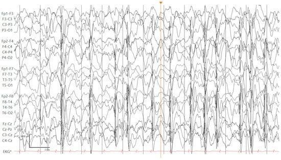
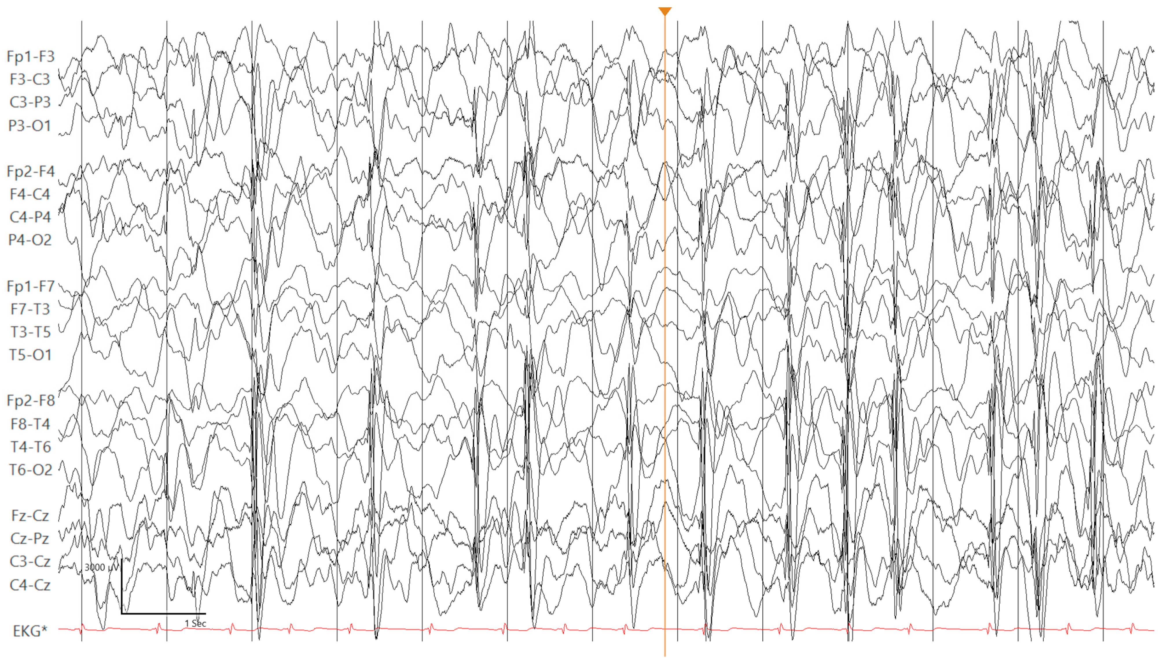
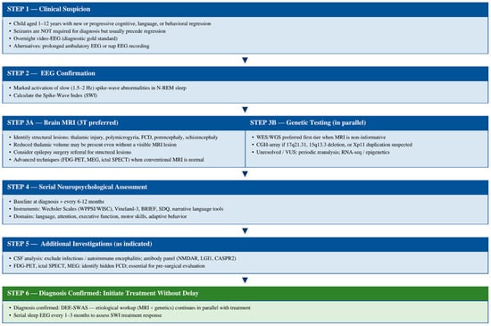
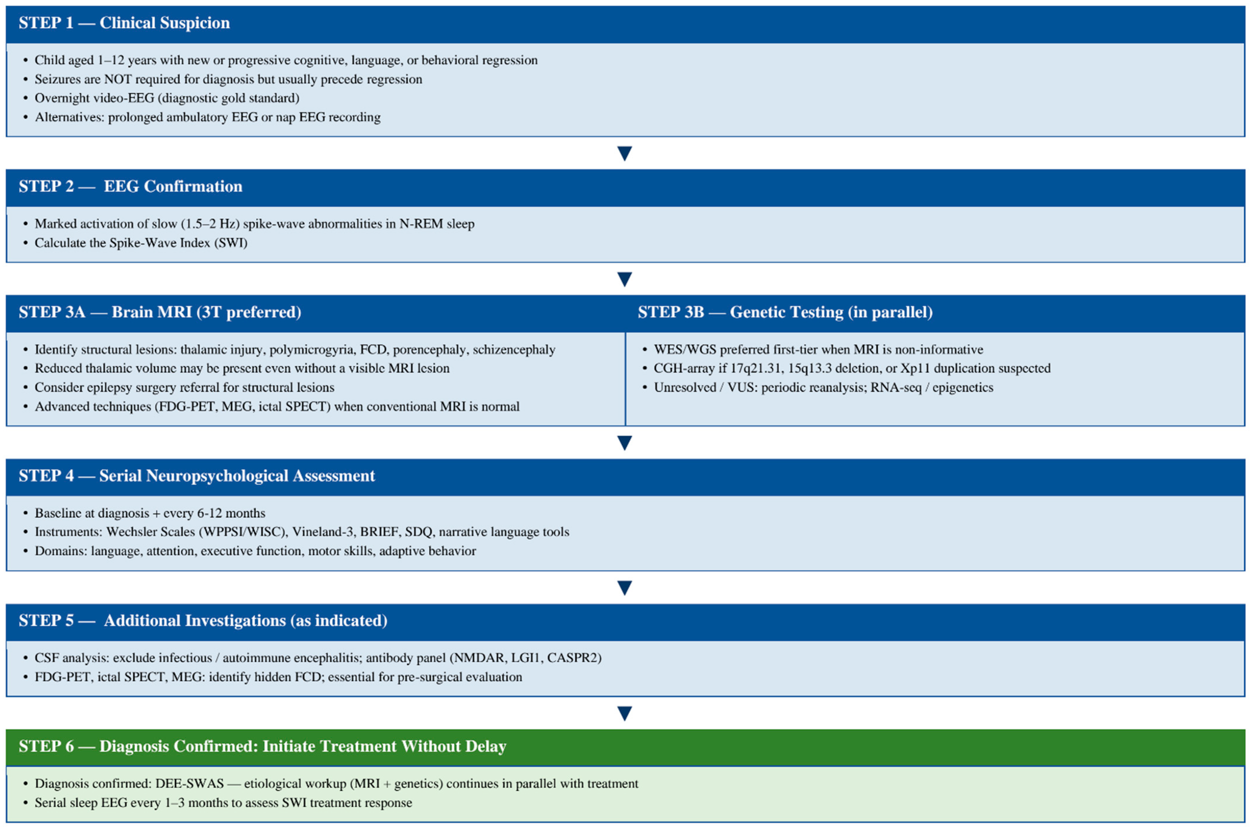
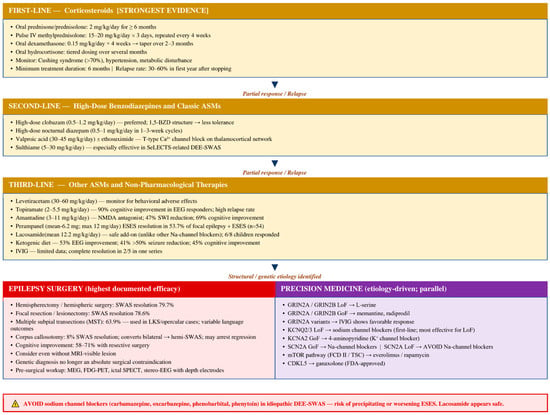
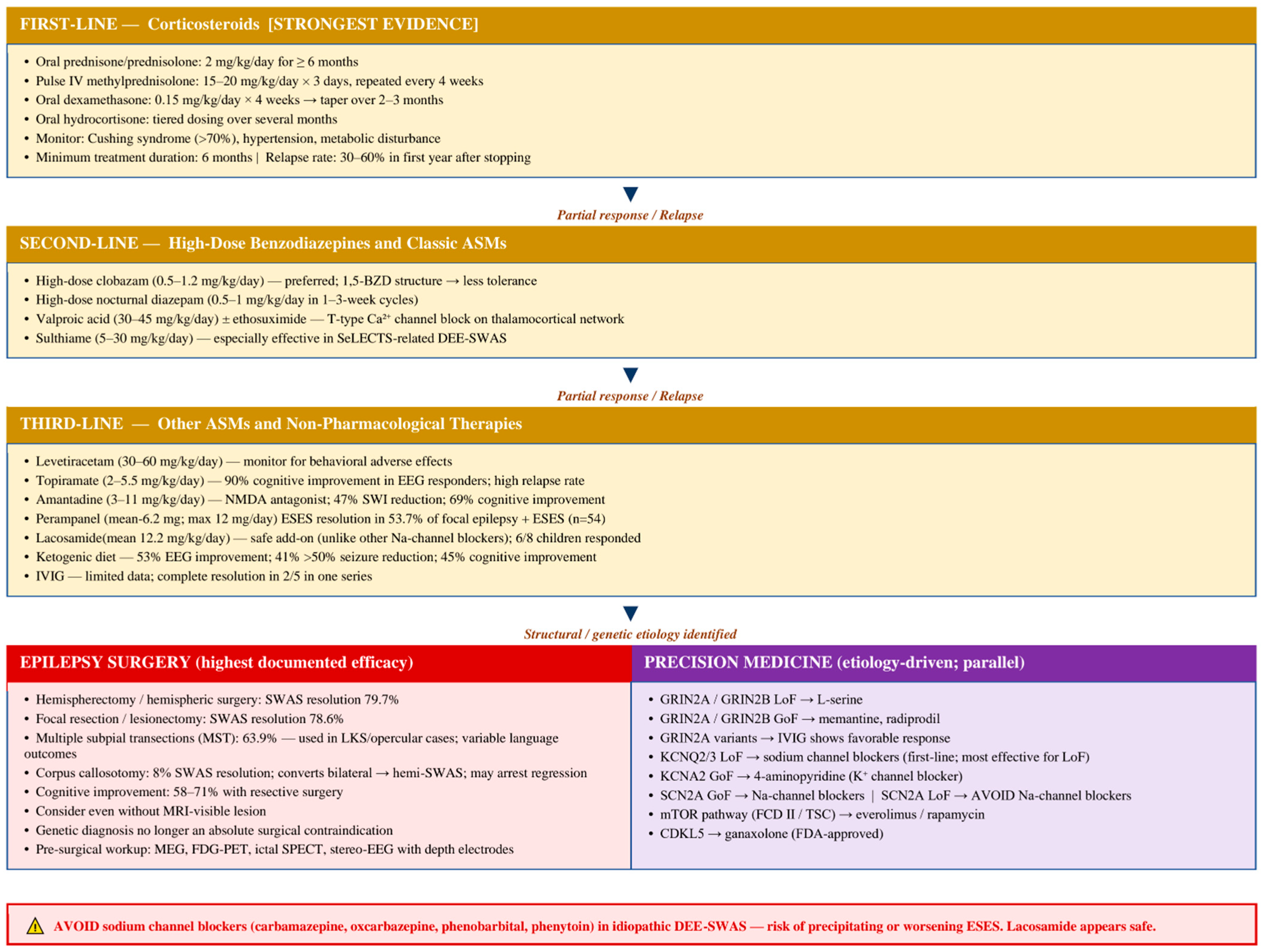

Disclaimer/Publisher’s Note: The statements, opinions and data contained in all publications are solely those of the individual author(s) and contributor(s) and not of MDPI and/or the editor(s). MDPI and/or the editor(s) disclaim responsibility for any injury to people or property resulting from any ideas, methods, instructions or products referred to in the content. |
© 2026 by the author. Licensee MDPI, Basel, Switzerland. This article is an open access article distributed under the terms and conditions of the Creative Commons Attribution (CC BY) license.