


Highlights
What are the main findings?
- Children with unilateral cleft lip and palate and obstructive sleep apnea showed significantly reduced palatal dimensions on three-dimensional CBCT analysis.
- Among the measured parameters, palatal volume was identified as the only variable independently associated with obstructive sleep apnea severity in this study.
What are the implications of the main findings?
- Reduced palatal dimensions may represent an anatomical phenotype associated with sleep-disordered breathing in children with unilateral cleft lip and palate.
- Early three-dimensional palatal assessment may support multidisciplinary cleft care and serve as a preliminary screening parameter to identify children at elevated risk for obstructive sleep apnea.
Abstract
Background/Objectives: Obstructive sleep apnea (OSA) is highly prevalent in children with unilateral cleft lip and palate (UCLP) due to maxillary retrusion and upper airway compromise. While palatal morphology may influence pediatric OSA, three-dimensional (3D) evaluations in this population remain limited. This study evaluated palatal dimensions and maxillary widths in UCLP children with and without OSA using cone-beam computed tomography (CBCT) and examined their associations with OSA severity (apnea–hypopnea index, AHI). Methods: Forty CBCT scans of Thai children with non-syndromic UCLP (mean age 8.98 ± 1.99 years) were analyzed. Participants were categorized into OSA (n = 20; AHI ≥ 1) and non-OSA (n = 20; AHI < 1) groups. Reconstructed palatal structures were measured for surface area, volume, height, and transverse maxillary widths. Group differences were assessed using independent t-tests, and associations with AHI were examined via Pearson’s correlation and linear regression (p < 0.05). Results: Children with OSA exhibited significantly reduced palatal surface area, volume, height, and buccal alveolar crest width compared with those without OSA (p < 0.05). Other transverse widths showed no significant intergroup differences. Linear regression identified palatal volume as the only variable independently associated with AHI (β = −0.631, p < 0.001). Conclusions: Children with UCLP and OSA exhibit significantly constricted palatal morphology. Among the measured parameters, reduced three-dimensional palatal volume was the only variable independently associated with increased OSA severity in this sample. Thus, CBCT-based palatal volume assessment may serve as a preliminary screening parameter to help identify OSA risk within multidisciplinary cleft care, though further validation is needed.
1. Introduction
Obstructive sleep apnea (OSA) is a common pediatric sleep-related breathing disorder characterized by recurrent episodes of upper airway obstruction during sleep. Its prevalence in otherwise healthy children is estimated at 1% to 5%, and it has been linked to adverse neurocognitive, cardiovascular, behavioral, and growth-related outcomes [1]. Overnight polysomnography (PSG) is the gold standard for diagnosis, and the apnea–hypopnea index (AHI) derived from PSG is the most widely accepted metric for defining the presence and severity of OSA in children [2]. Adenotonsillar hypertrophy is the primary cause of pediatric OSA; however, craniofacial anomalies, particularly cleft lip and palate (CLP), are well-recognized risk factors because of their characteristic maxillary deficiency and compromised upper airway anatomy [3].
CLP arises from the disruption of the embryologic fusion of the medial nasal, lateral nasal, and maxillary processes [4], with a reported incidence of approximately 1.7 per 1000 live births in Thailand [5]. Children with CLP, especially those with unilateral cleft lip and palate (UCLP), have a significantly higher prevalence of OSA than those without CLP. Population-based data indicate that the prevalence of PSG-confirmed OSA in children with cleft lip and/or palate is approximately 6.3%, whereas no cases were identified in non-cleft controls, highlighting the markedly elevated risk in this population [6]. Structural anomalies such as midfacial retrusion, maxillary hypoplasia, and alterations in the nasal cavity, nasopharynx, oropharynx, and palate contribute to airway narrowing and increased OSA susceptibility [7]. These craniofacial deficiencies, particularly a retrusive maxilla, reduce cross-sectional airway volume and further increase the likelihood of OSA in this population [8]. Collectively, these findings demonstrate the importance of investigating specific craniofacial correlates, particularly palatal dimensions and maxillary constriction, that may underlie OSA susceptibility in children with CLP.
Recent evidence has identified palatal morphology as a potential anatomical correlate of pediatric OSA. Reduced palatal dimensions have also been found to correlate with smaller oropharyngeal airway volumes, suggesting a functional association between maxillary constriction and upper airway patency [9,10]. Furthermore, three-dimensional assessments of cleft populations have demonstrated that children with UCLP exhibit significantly smaller palatal volumes and surface areas compared with those without UCLP, findings that coincide with reduced posterior airway spaces [11]. These observations indicate that alterations in palatal morphology not only reflect craniofacial deficiency but may represent structural features associated with OSA susceptibility and severity. Importantly, because these structural constraints are potentially modifiable through early targeted orthodontic interventions, such as maxillary expansion, a detailed understanding of three-dimensional palatal configurations may aid in comprehensive treatment planning to potentially improve respiratory outcomes [1].
Although conventional assessments of palatal and maxillary morphology using two-dimensional radiographs or dental casts [12] are adequate for basic linear analysis, they offer limited insight into volumetric configuration and asymmetry [13]. Although laser scanning and digital models enhance surface visualization, they remain inadequate for defining skeletal boundaries in cleft-related deformities [14]. By contrast, cone-beam computed tomography (CBCT) enables high-resolution, three-dimensional evaluation with isotropic voxel precision and minimal geometric distortion, allowing accurate quantification of palatal surface area and volume [14,15]. This modality provides a reliable platform for examining craniofacial correlates of OSA in children with UCLP, a population in which complex skeletal asymmetries require detailed volumetric assessment.
Despite these recognized anatomical risks, comprehensive evaluations of the relationship between three-dimensional palatal volume, surface area, and OSA severity in children with UCLP remain limited. Therefore, this study quantitatively assessed palatal morphology and transverse maxillary widths in children with UCLP with and without OSA using CBCT. Associations between palatal dimensions and AHI were also analyzed to clarify the relationship between palatal configuration and OSA severity and to explore their potential relevance for clinical risk assessment.
2. Materials and Methods
2.1. Study Population and Ethical Approval
This cross-sectional study included participants who attended the Ear, Nose, and Throat (ENT) snoring clinic at the Faculty of Medicine, Chiang Mai University Hospital, as well as the Orthodontic Division, Department of Orthodontics and Pediatric Dentistry, Chiang Mai University, between 2019 and 2024. Ethical approval was granted by the Human Experimentation Committee of the Faculty of Dentistry, Chiang Mai University, Thailand (Approval No. 56/2022; approval date: 17 October 2022). Written informed consent was obtained from each participant and their caregivers prior to enrollment, permitting the use of CBCT images, polysomnographic findings, and associated medical information for research purposes.
2.2. Sample Size and Participants
Sample size was determined using G*Power software (version 3.1.9.4; University of Kiel, Germany) [16]. The calculation was based on an effect size of 0.96, derived from a prior study examining three-dimensional palatal morphology in patients with OSA [9], with a statistical power of 0.80 and a significance level of α = 0.05 for a two-tailed test, yielding 20 participants per group.
A total of 40 Thai children (5–12 years old) diagnosed with non-syndromic UCLP were recruited. Prior to study inclusion, all candidates underwent a comprehensive clinical assessment by an otolaryngologist. Demographic data—including age, sex, weight, and height—were recorded, and body mass index (BMI) was computed to evaluate nutritional status. During the examination, tonsillar size was graded according to the Brodsky classification [17], and adenoid size percentage was recorded for each participant. To ensure airway stability prior to data collection, participants identified with adenotonsillar hypertrophy were prescribed a standardized six-week course of medical therapy under otolaryngological supervision before proceeding with polysomnography (PSG) and CBCT imaging.
To eliminate potential anatomical and physiological confounders, strict exclusion criteria were applied. Children were excluded if they presented with extreme BMI values (below the 5th or above the 95th percentile), persistent Grade III–IV tonsillar hypertrophy following medical management, or a diagnosis of central or mixed sleep apnea. Furthermore, a history of orthodontic interventions, continuous positive airway pressure (CPAP) therapy, secondary alveolar bone grafting, or major craniofacial surgery disqualified candidates from the study, as did the presence of incidental pathological lesions on CBCT scans.
2.3. PSG and OSA Diagnosis
All eligible patients underwent overnight PSG using a Type III portable device, SOMNOlab-2 (Weinmann, Hamburg, Germany) in the Department of Otolaryngology, Faculty of Medicine, Chiang Mai University. PSG recordings included oronasal airflow, snoring, thoracoabdominal movements, pulse rate, and arterial oxygen saturation. The AHI was calculated as the number of apnea and hypopnea events per hour of sleep. Pediatric OSA was diagnosed when the AHI was ≥1 events/h, and cases were further classified as mild (AHI ≥ 1 to <5), moderate (AHI ≥ 5 to <10), or severe (AHI ≥ 10) [1]. Following these criteria, the sample was divided into two groups: an OSA group (AHI ≥ 1, n = 20) and a non-OSA group (AHI < 1, n = 20).
2.4. CBCT Assessment and Measurements
Image Acquisition and Safety Protocol. To minimize temporal variation, PSG and CBCT examinations were performed within one month of each other. Importantly, CBCT imaging was performed strictly as part of routine pre-surgical and orthodontic diagnostic planning prior to alveolar bone grafting; no additional radiation exposure was incurred solely for research purposes.
All CBCT scans were acquired using a MobiiScan unit (NECTEC, Pathumthani, Thailand) operating with a voxel resolution of 0.4 mm, a total scanning duration of 26 s, and a field of view of 16 cm × 16.8 cm [18]. During image acquisition, participants were positioned supine with the maxillary and mandibular teeth in maximum intercuspation. Strict instructions were given to avoid swallowing or head movement to prevent motion artifacts.
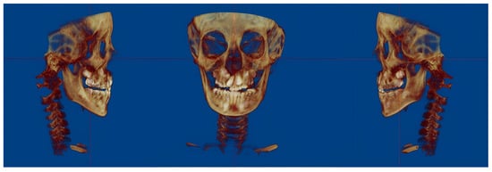
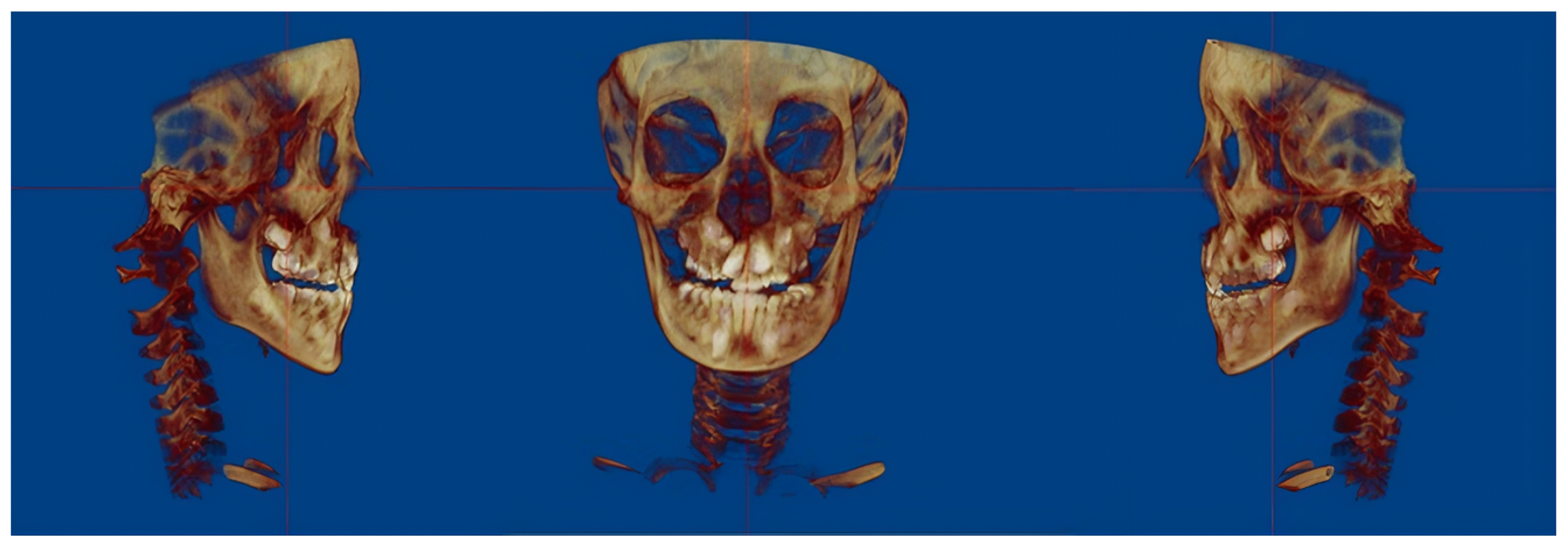
Image Orientation and Linear Measurements. CBCT datasets were imported into Dolphin Imaging software (version 11.9; Dolphin Imaging & Management Solutions, Chatsworth, CA, USA) and standardized using the Frankfort horizontal (FH) plane for skull orientation (Figure 1).
Figure 1.
Standardization of CBCT datasets using the Frankfort horizontal plane for skull orientation in Dolphin Imaging software.
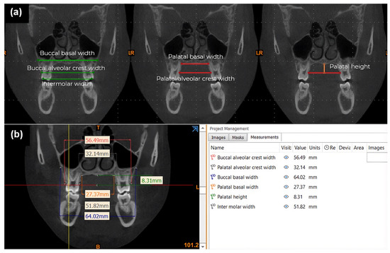
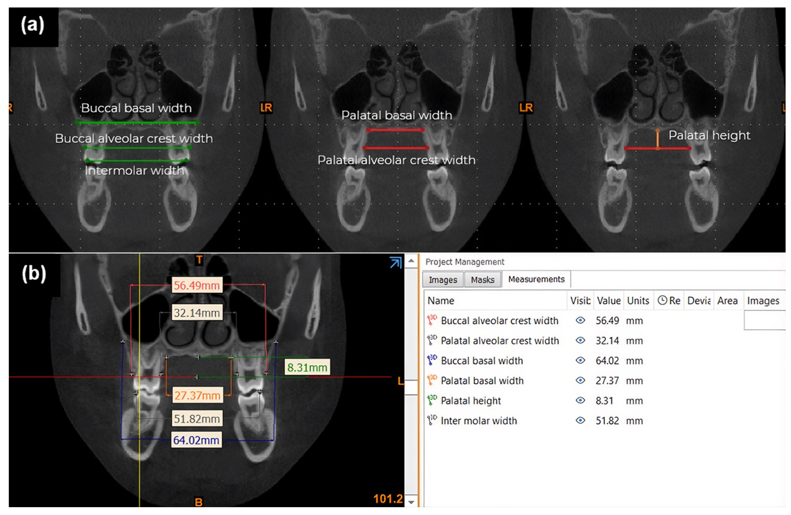
Linear measurements of palatal dimensions and maxillary width were performed using Mimics Medical software version 21.0 (Materialise, Leuven, Belgium). Transverse maxillary dimensions were obtained according to the method described by Podesser et al. [19], whereas vertical palatal height was measured following the protocols described by Yassaei et al. [15]. The most anterior coronal slice displaying the complete palatal roots of the maxillary first molars was selected for analysis (Table 1; Figure 2). To minimize measurement bias from dental anomalies common in UCLP (crowding, ectopic eruption, and missing teeth), transverse dimensions were defined using skeletal landmarks independent of dental position. The assessed parameters included intermolar width, buccal and palatal alveolar crest widths, and buccal and palatal basal widths. Detailed definitions and anatomical landmarks for all linear measurements are comprehensively outlined in Table 1.
Table 1.
Definitions of palatal and maxillary measurements used in this study.
Figure 2.
Linear measurements of palatal dimensions and transverse maxillary widths on the most anterior coronal CBCT slice at the level of the maxillary first molars in Mimics Medical software. (a) Illustration of measurement landmarks. (b) Representative coronal CBCT slice showing linear measurements used for analysis.
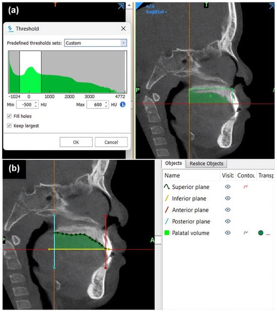
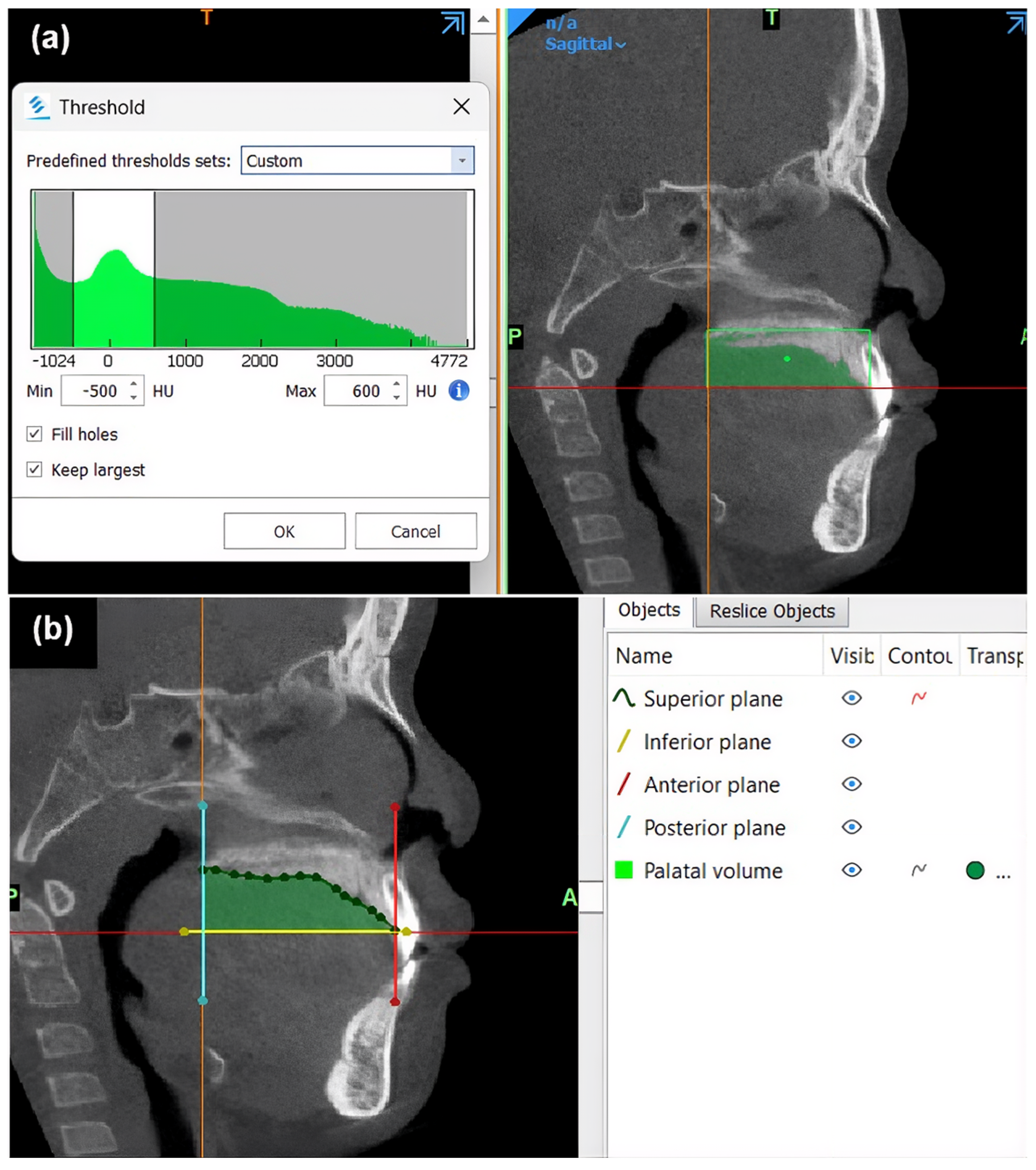
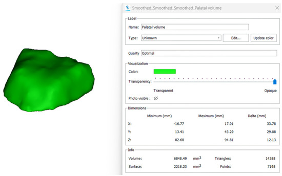
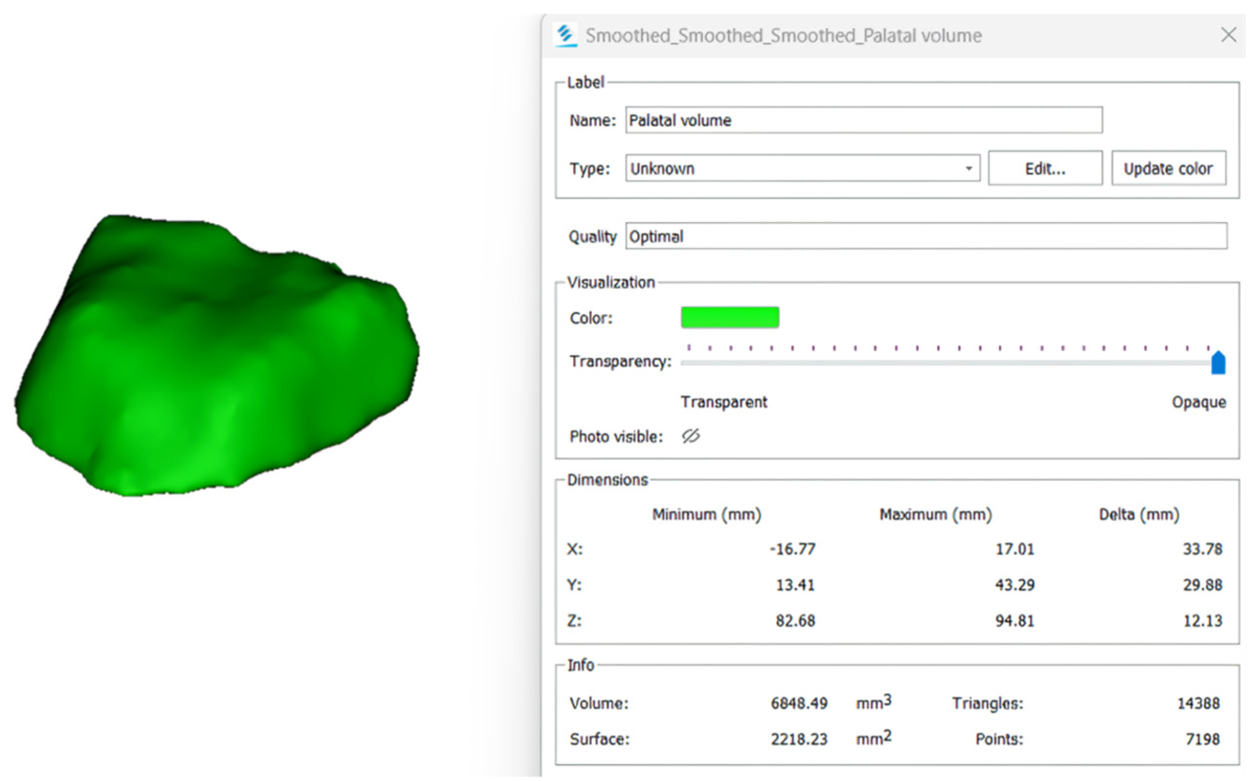
Three-Dimensional Volumetric Assessment. Volumetric assessment of the palate was based on tissue density using a Hounsfield unit (HU) threshold of −500 to 600 in Mimics Medical software on the same Digital Imaging and Communications in Medicine (DICOM) datasets (Figure 3a). The superior, inferior, anterior, posterior, and lateral boundaries of the palatal morphology were then defined according to a standardized multiplanar protocol adapted from Yu et al. [14] and Huang et al. [20]. The superior plane corresponded to the nasal floor, ensuring inclusion of the hard palate without overlap from nasal or sinus structures. The inferior plane was defined by three reference points: (1) the palatal side of the anterior alveolar crest along the mid-sagittal plane and (2–3) the midpoints of the palatal alveolar crests of the right and left first molars. The anterior and posterior boundaries were delineated by coronal planes passing through the anterior alveolar crest and the posterior nasal spine, respectively, whereas the lateral borders followed the internal contours of the palatal bone (Figure 3b). Manual delineation and slice-by-slice segmentation were performed to ensure accurate boundary definition, allowing the software to automatically calculate palatal surface area (mm2) and palatal volume (mm3) (Figure 4).
Figure 3.
Palatal volumetric assessment in Mimics Medical software. (a) Threshold-based segmentation of the palatal region (indicated by the green area). (b) Delineation of boundary planes used for palatal volume measurement. The colored lines represent the superior (green), inferior (yellow), anterior (red), and posterior (cyan) boundary planes.
Figure 4.
Three-dimensional reconstruction of palatal morphology following manual segmentation, used for palatal surface area and volume calculation in Mimics Medical software.
2.5. Reliability Analysis
To assess intra-examiner reliability, the primary investigator repeated all linear and volumetric measurements on 10 randomly selected CBCT datasets after a four-week interval. The mean of the two readings was subsequently used for the main analysis. To evaluate inter-examiner reliability, the same 10 CBCT datasets were independently analyzed by a second calibrated orthodontist. Agreement within and between examiners was quantified using the intraclass correlation coefficient (ICC).
2.6. Statistical Analysis
All statistical analyses were conducted using IBM SPSS Statistics version 25.0 (IBM Corp., Armonk, NY, USA). Descriptive statistics (mean ± standard deviation) were computed for continuous variables. Data normality was confirmed using the Shapiro–Wilk test. Chi-square tests were employed to evaluate differences in categorical variables between the groups, while independent samples t-tests were used to compare intergroup differences for continuous variables (palatal dimensions and maxillary widths). Pearson’s correlation analysis was performed to examine the associations between the studied variables and the AHI.
To identify variables independently associated with OSA severity, a multivariable linear regression model was constructed. Only variables demonstrating statistically significant univariate correlations with AHI were included in the regression model to prevent model overfitting and ensure that variables lacking significant associations were appropriately excluded. Standardized β coefficients and R2 values were reported. For all statistical tests, a p-value of <0.05 was considered statistically significant.
3. Results
3.1. Demographic and Clinical Characteristics
Table 2 summarizes the demographic data of the 40 Thai patients with UCLP (16 females, 24 males; mean age, 8.98 ± 1.99 years). Participants were classified into an OSA group (n = 20; mean age = 9.25 ± 1.71 years) and a non-OSA group (n = 20; mean age, 8.70 ± 2.25 years). The two groups demonstrated similar mean BMI values (OSA: 17.94 ± 4.27 kg/m2; non-OSA: 17.37 ± 2.70 kg/m2). No significant intergroup differences were observed in adenoid size percentage or tonsil grade distribution (p = 0.733 and p = 0.388, respectively). The mean AHI was 3.79 ± 3.30 events/h in the OSA group and 0.42 ± 0.26 events/h in the control group. Among children in the OSA group, OSA severity was classified as mild in 13 participants, moderate in 5 participants, and severe in 2 participants.
Table 2.
Descriptive statistics and group comparisons of subject characteristics, polysomnographic findings, and palatal and maxillary measurements.
3.2. Measurement Reliability
According to the 95% confidence intervals, the intraclass correlation coefficients (ICCs) for intra- and inter-examiner reliability demonstrated excellent agreement across all parameters, confirming high measurement reproducibility:
- Palatal volume: 0.998 and 0.997, respectively;
- Palatal surface area: 0.995 and 0.976, respectively;
- Palatal height: 0.986 and 0.934, respectively;
- Intermolar width: 0.980 and 0.940, respectively;
- Buccal basal width: 0.984 and 0.953, respectively;
- Palatal basal width: 0.993 and 0.986, respectively;
- Buccal alveolar crest width: 0.994 and 0.978, respectively;
- Palatal alveolar crest width: 0.995 and 0.987, respectively.
3.3. Intergroup Comparison of Palatal and Maxillary Dimensions
All data were normally distributed (Shapiro–Wilk normality test; p > 0.05). The OSA group exhibited significantly smaller palatal volume, palatal surface area, and palatal height than the non-OSA group (p < 0.05). By contrast, none of the maxillary width parameters differed significantly between groups, except for the buccal alveolar crest width, which was significantly narrower in the OSA group (p < 0.05) (Table 2).
3.4. Correlations with OSA Severity
Pearson’s correlation analysis was performed to assess the relationships between the studied variables and the AHI (Table 3). No significant correlations were observed between AHI and the following parameters (all p > 0.05):
Table 3.
Pearson’s correlations between AHI and study variables.
- Age (r = 0.064);
- Intermolar width (r = −0.260);
- Buccal basal width (r = −0.205);
- Palatal basal width (r = −0.302).
However, statistically significant negative correlations were identified for the remaining dimensions:
- Weak negative correlations: Palatal height (r = −0.380; p < 0.05) and palatal alveolar crest width (r = −0.392; p < 0.05);
- Moderate negative correlations: Palatal volume (r = −0.631; p < 0.001), palatal surface area (r = −0.619; p < 0.001), and buccal alveolar crest width (r = −0.524; p < 0.05).
3.5. Multivariable Linear Regression Analysis
Multivariable linear regression analysis identified palatal volume as the only variable independently associated with AHI (p < 0.001). Conversely, palatal surface area, palatal height, and both buccal and palatal alveolar crest widths showed no significant independent associations with OSA severity (Table 4).
Table 4.
Multivariable linear regression analysis of AHI as the dependent variable (five covariates).
4. Discussion
The present study showed that children with UCLP who had OSA exhibited significantly reduced palatal dimensions, including palatal volume, surface area, and height, compared with those without OSA. Among the various transverse and vertical parameters evaluated, multivariable linear regression analysis identified palatal volume as the sole variable independently associated with OSA severity (AHI). This finding suggests that three-dimensional palatal constriction may be associated with pediatric sleep-disordered breathing in this cohort.
To minimize potential confounders such as adenotonsillar hypertrophy, obesity, and sex, individuals with Grade III–IV tonsillar hypertrophy or a BMI above the 95th percentile were excluded. In addition, no significant intergroup differences were observed in tonsil grade distribution or adenoid size percentage, indicating comparable baseline adenotonsillar status within this cohort. Enlarged tonsils may constrict the oropharyngeal airway [21]; furthermore, in general pediatric populations, severe adenotonsillar hypertrophy and obesity are recognized as the primary clinical determinants of OSA, and their presence may obscure the respiratory contribution of craniofacial structures [2]. This methodological approach was intended to reduce confounding and improve the specificity of the observed associations between palatal dimensions and AHI, thereby reducing the likelihood that these associations were driven primarily by adenotonsillar enlargement, soft-tissue obstruction, or obesity-related factors [22]. Consequently, although BMI, tonsil grade, and adenoid size were documented to characterize the baseline status of the cohort, they were not included in the primary correlation and regression models, which focused on skeletal palatal and maxillary parameters. Consistent with this rationale, previous studies have shown that when extreme obesity is controlled, the associations between BMI and AHI become weak [23]. Furthermore, the literature confirms a lack of sex-related differences in OSA among patients with cleft palate [24], as the cleft anomaly itself exerts a fundamentally greater influence on maxillary morphology than sex-related growth patterns [25]. Findings from Silvestre et al. and Ho et al. have similarly indicated that sex-related variations in OSA risk are minimal in CLP populations [24,26]. Together, these methodological considerations allowed a focused evaluation of the association between palatal morphology and OSA severity within this cohort.
Historically, characterization of the palate and maxilla has primarily relied on two-dimensional linear and angular measurements from dental cast analysis [12]. Although this method is reliable, it is time-consuming and provides limited information on three-dimensional morphology [13]. To address these limitations, several studies have introduced three-dimensional imaging systems employing laser scanning to generate dental models, facilitating improved visualization and documentation of the upper arch in patients with clefts [10]. However, such surface-based techniques may not fully represent the intricate palatal anatomy [14]. In the present study, CBCT was used to obtain precise measurements of hard tissue structures, including the palate and maxilla. This technique supports both linear and volumetric assessments [15] and enables high-resolution three-dimensional reconstruction that accurately captures the complexity of palatal morphology [14,15]. CBCT-based evaluation provides reproducible and clinically applicable data for orthodontic diagnosis and interdisciplinary management of patients with OSA.
Previous studies have demonstrated craniofacial structural alterations in patients with OSA, particularly reduced maxillary dimensions and retrusion [27]. The association between maxillary morphology and upper airway dimensions indicates that they may play a role in OSA pathogenesis [7]. Kecik et al. reported significantly smaller palatal dimensions in individuals with OSA [9]. Children with UCLP are especially predisposed to OSA because of midfacial deficiency and maxillary retrusion, which contribute to reduced airway volume [8]. Comparative analyses have consistently shown smaller palatal dimensions in patients with UCLP than in non-cleft individuals [13,28], suggesting increased OSA susceptibility. In our analysis, the employment of skeletal landmarks to define transverse dimensions provided a more accurate representation of true maxillary width [19]. Most transverse parameters, including intermolar, palatal basal, and buccal basal widths, showed no significant differences between groups, except for the buccal alveolar crest width, which was significantly smaller in the OSA cohort. These findings are consistent with studies by Ciavarella et al. [10] and Johal and Conaghan [7], who reported no significant posterior transverse discrepancies between individuals with and without OSA, but contrast with the study by Kecik [9], who observed narrower dental arches in patients with OSA. Intercanine width was not included in the present analysis because many subjects (ages 5–12) were in early mixed dentition, where ongoing canine eruption may compromise measurement reliability. Nevertheless, AHI demonstrated significant negative correlations with palatal height, buccal alveolar crest width, and palatal alveolar crest width, whereas intermolar width showed no significant correlation. Maxillary constriction is a known risk factor for OSA [27]. Although Seto et al. suggested that palatal height alone may not reflect maxillary constriction [27], our findings, consistent with Ciavarella et al. [10], indicate that reduced palatal height correlates with greater OSA severity, possibly due to posterior and inferior tongue displacement leading to airway narrowing [29]. Beyond linear parameters, the three-dimensional analyses in the present study showed significant reductions in palatal surface area and volume among patients with OSA, consistent with the findings of Kecik [9] and Ciavarella [10], who reported strong negative correlations between palatal dimensions and OSA indices. In patients with UCLP, maxillary constriction and reduced palatal dimensions may contribute to compromised airway anatomy through altered tongue–palate spatial relationships and retroglossal airway narrowing, which may aggravate obstructive events during sleep [14,30].
The observed associations between palatal dimensions and OSA severity have practical implications for the multidisciplinary management of children with UCLP. Three-dimensional CBCT assessment of palatal volume may serve as a preliminary screening parameter to help identify children at elevated risk for OSA, who could then be considered for polysomnographic evaluation. However, it should be emphasized that while three-dimensional palatal volume may indicate a structural predisposition to upper airway compromise, it serves merely as an adjunctive screening tool and cannot substitute for definitive polysomnography. These findings align with reports by Watanabe et al. [31], who demonstrated that craniofacial skeletal deficiencies are associated with airway narrowing. Incorporating palatal dimension assessment into routine orthodontic planning may support early identification of OSA risk. Recognizing this structural susceptibility highlights the potential clinical value of early intervention. Targeted orthodontic treatments, such as maxillo-mandibular expansion, may help increase this three-dimensional anatomical volume. By widening the skeletal and nasal cavity base, these interventions have been shown to improve upper airway volume and potentially reduce airway resistance, offering beneficial effects for growing subjects with OSA [30]. Left untreated, OSA in patients with UCLP can lead to neurocognitive deficits, behavioral issues, impaired growth, and cardiovascular complications, making early identification and management essential [1,2].
This study has several strengths. Strict exclusion criteria (orthodontics, CPAP, alveolar bone grafting, central/mixed apnea, severe adenotonsillar hypertrophy, extreme BMI) minimized confounding and strengthened internal validity. Intra- and inter-examiner reliability were excellent (ICCs ≥ 0.93 for all parameters), confirming high measurement reproducibility. CBCT scans were acquired using a standardized protocol, and palatal volumetry used a validated HU threshold (−500 to 600) with manual segmentation blinded to group status. Children aged 5–12 years were intentionally selected because this period corresponds to active maxillary growth and routine pre–alveolar bone graft imaging, providing ample data. Palatal volume was the strongest independent correlate of AHI among the measured parameters, indicating an association between smaller palatal volume and increased OSA severity within this study cohort.
Despite these strengths, several limitations should be acknowledged. First, the cross-sectional design precludes causal inference; however, strict exclusion criteria minimized confounding, and palatal volume remained the strongest independent correlate of AHI among the measured parameters. Longitudinal studies are needed to establish causality. Second, the age range (5–12 years) corresponds to active maxillary growth, and transverse dimensions continue to change during this period [32]; groups were matched by age to minimize residual variation. Third, while manual slice-by-slice segmentation remains the reference standard for precise three-dimensional volumetric reconstruction, the process is highly labor-intensive, time-consuming, and inherently subject to inter- and intra-expert variability [33,34]. Fourth, the lack of detailed records on primary palatoplasty technique and timing limited our ability to assess whether surgical factors influenced maxillary growth, palatal morphology, and subsequent airway outcomes, including OSA [35,36]. Fifth, while strict exclusion criteria strengthened internal validity, results may not generalize to UCLP children with severe adenotonsillar hypertrophy or extreme BMI. Sixth, the utilization of a Type III portable sleep monitor (SOMNOlab-2), which inherently lacks electroencephalography (EEG) channels, presents a minor technical limitation. Because the device computes the AHI based on total recording time rather than true total sleep time, the severity of OSA could theoretically be underestimated if a child experienced significant periods of wakefulness during the night. However, this limitation was substantially mitigated in our study protocol, as all sleep examinations were performed during formal hospital admission under the direct supervision of clinical staff, ensuring that the total recording period closely reflected the actual sleep time. Seventh, the current investigation focused primarily on transverse skeletal widths, vertical height, and three-dimensional volumetric configuration, without incorporating independent linear measurements in the sagittal plane. Although sagittal boundaries were strictly controlled during the three-dimensional volume segmentation, the absence of standalone sagittal parameters limits a fully comprehensive multiplanar assessment.
Future studies with larger sample sizes should examine the effects of surgical technique, timing of palatal repair, velopharyngeal insufficiency (VPI) management, and tongue volume on OSA in UCLP. Furthermore, incorporating standalone sagittal parameters alongside longitudinal designs integrating 3D airway and soft tissue parameters would provide further insight into the anatomical mechanisms associated with OSA in this population. Automated or Artificial Intelligence (AI)-based segmentation methods could also reduce the labor-intensive nature of current 3D reconstruction workflows.
5. Conclusions
In conclusion, the findings of this study suggest that children with UCLP who have OSA exhibit a significantly constricted palatal morphology—specifically reduced palatal volume, surface area, height, and buccal alveolar crest width—compared to those without OSA. Furthermore, among the measured parameters, reduced three-dimensional palatal volume was the only variable independently associated with increased OSA severity in this specific sample (β = −0.631, p < 0.001). While two-dimensional measurements showed limited independent correlations, 3D volumetric analysis provided a more comprehensive understanding of the skeletal structural parameters evaluated. Incorporating CBCT-based palatal volume assessment into routine pre-surgical and orthodontic evaluations may serve as a preliminary screening parameter to help identify children with UCLP who are at elevated risk for OSA. Identifying this structural predisposition offers potential clinical utility by providing a rationale for early orthodontic interventions to expand palatal volume. However, further validation is needed before widespread clinical implementation. Ultimately, because these associations reflect only the hard-tissue dimensions evaluated in this sample, timely polysomnographic evaluation and comprehensive multidisciplinary management remain essential.
Author Contributions
Conceptualization, C.P. and M.C.; methodology, C.P. and M.C.; software, C.P.; validation, C.P., N.S. and M.C.; formal analysis, C.P.; investigation, C.P. and N.S.; resources, N.S. and M.C.; data curation, C.P.; writing—original draft preparation, C.P.; writing—review and editing, C.P., N.S. and M.C.; visualization, C.P.; supervision, M.C.; project administration, M.C. All authors have read and agreed to the published version of the manuscript.
Funding
This research received no external funding.
Institutional Review Board Statement
The study was conducted in accordance with the ethical standards of the Human Experimental Committee of the Faculty of Dentistry, Chiang Mai University, Thailand (Approval No. 56/2022; approval date: 17 October 2022) and with the 1964 Helsinki declaration and its later amendments or comparable ethical standards.
Informed Consent Statement
Informed consent was obtained from legal guardians and all individual participants included in the study.
Data Availability Statement
The data presented in this study are available on request from the corresponding author. The data are not publicly available due to privacy and ethical reasons.
Acknowledgments
The authors gratefully acknowledge the Princess Sirindhorn IT Foundation Craniofacial Center and the Cleft Center, Faculty of Dentistry, Chiang Mai University, Thailand, for their support in providing access to patient records used in this study. The authors also thank NECTEC for providing the MobiiScan system, which was used for CBCT data acquisition. In addition, the authors acknowledge Ornicha Laowong, D.D.S., for independently performing measurements of palatal surface area, volume, height, and maxillary transverse dimensions, which were used to assess inter-rater reliability.
Conflicts of Interest
The authors declare no conflicts of interest.
Abbreviations
The following abbreviations are used in this manuscript:
| 2D | Two-Dimensional |
| 3D | Three-Dimensional |
| AHI | Apnea–Hypopnea Index |
| AI | Artificial Intelligence |
| BMI | Body Mass Index |
| CBCT | Cone-Beam Computed Tomography |
| CLP | Cleft Lip and Palate |
| CPAP | Continuous Positive Airway Pressure |
| DICOM | Digital Imaging and Communications in Medicine |
| ENT | Ear, Nose, and Throat |
| FH | Frankfort Horizontal |
| HU | Hounsfield Unit |
| ICC | Intraclass Correlation Coefficient |
| OSA | Obstructive Sleep Apnea |
| PSG | Polysomnography |
| UCLP | Unilateral Cleft Lip and Palate |
| VPI | Velopharyngeal Insufficiency |
References
- Behrents, R.G.; Shelgikar, A.V.; Conley, R.S.; Flores-Mir, C.; Hans, M.; Levine, M.; McNamara, J.A.; Palomo, J.M.; Pliska, B.; Stockstill, J.W.; et al. Obstructive sleep apnea and orthodontics: An American Association of Orthodontists White Paper. Am. J. Orthod. Dentofac. Orthop. 2019, 156, 13–28.e11. [Google Scholar] [CrossRef] [Scilit] [PubMed]
- Marcus, C.L.; Brooks, L.J.; Draper, K.A.; Gozal, D.; Halbower, A.C.; Jones, J.; Schechter, M.S.; Ward, S.D.; Sheldon, S.H.; Shiffman, R.N.; et al. Diagnosis and management of childhood obstructive sleep apnea syndrome. Pediatrics 2012, 130, e714–e755. [Google Scholar] [CrossRef] [Scilit] [PubMed]
- Wiwattanadittakul, P.; Sonsuwan, N.; Prapayasatok, S.; Chaiworawitkul, M. Comparative evaluation of the upper pharyngeal airway among children with/without UCLP and with/without OSA. Sleep Breath. 2024, 29, 37. [Google Scholar] [CrossRef] [Scilit] [PubMed]
- Cielo, C.M.; Marcus, C.L. Obstructive sleep apnoea in children with craniofacial syndromes. Paediatr. Respir. Rev. 2015, 16, 189–196. [Google Scholar] [CrossRef] [Scilit] [PubMed]
- Chaiworawitkul, M. Incidence, etiology, and prevention of cleft lip and/or palate. CM Dent. J. 2012, 33, 45–55. [Google Scholar]
- Carlson, A.; Sobol, D.; Pien, I.; Allori, A.; Marcus, J.; Watkins, S.; Aylsworth, A.; Meyer, R.; Pimenta, L.; Strauss, R.; et al. Obstructive sleep apnea in children with cleft lip and/or palate: Results of an epidemiologic study. Dent. Oral Craniofacial Res. 2017, 3, 1000212. [Google Scholar] [CrossRef] [Scilit]
- Johal, A.; Conaghan, C. Maxillary morphology in obstructive sleep apnea: A cephalometric and model study. Angle Orthod. 2004, 74, 648–656. [Google Scholar] [CrossRef] [Scilit] [PubMed]
- Robison, J.G.; Otteson, T.D. Increased prevalence of obstructive sleep apnea in patients with cleft palate. Arch. Otolaryngol. Head Neck Surg. 2011, 137, 269–274. [Google Scholar] [CrossRef] [Scilit] [PubMed]
- Kecik, D. Three-dimensional analyses of palatal morphology and its relation to upper airway area in obstructive sleep apnea. Angle Orthod. 2017, 87, 300–306. [Google Scholar] [CrossRef] [Scilit] [PubMed]
- Ciavarella, D.; Campobasso, A.; Conte, E.; Burlon, G.; Guida, L.; Montaruli, G.; Cassano, M.; Laurenziello, M.; Illuzzi, G.; Tepedino, M. Correlation between dental arch form and OSA severity in adult patients: An observational study. Prog. Orthod. 2023, 24, 19. [Google Scholar] [CrossRef] [Scilit] [PubMed]
- Tabellion, M.; Lisson, A. Three-dimensional analysis of palatal morphology and PAS in patients with cleft lip and palate prior to orthodontic treatment. Head Face Med. 2024, 20, 40. [Google Scholar] [CrossRef] [Scilit] [PubMed]
- Garrahy, A.; Millett, D.T.; Ayoub, A.F. Early assessment of dental arch development in repaired unilateral cleft lip and unilateral cleft lip and palate versus controls. Cleft Palate Craniofacial J. 2005, 42, 385–391. [Google Scholar] [CrossRef] [Scilit] [PubMed]
- Generali, C.; Primozic, J.; Richmond, S.; Bizzarro, M.; Flores-Mir, C.; Ovsenik, M.; Perillo, L. Three-dimensional evaluation of the maxillary arch and palate in unilateral cleft lip and palate subjects using digital dental casts. Eur. J. Orthod. 2017, 39, 641–645. [Google Scholar] [CrossRef] [Scilit] [PubMed]
- Yu, C.; Ahn, H.W.; Kim, S.H. Three-dimensional morphological evaluation of the hard palate in Korean adults with mild-to-moderate obstructive sleep apnea. Korean J. Orthod. 2018, 48, 133–142. [Google Scholar] [CrossRef] [Scilit] [PubMed]
- Yassaei, S.; Safi, Y.; Valian, F.; Mohammadi, A. Evaluation of maxillary arch width and palatal volume and depth in patients with maxillary impacted canine by CBCT. Heliyon 2022, 8, e10854. [Google Scholar] [CrossRef] [Scilit] [PubMed]
- Faul, F.; Erdfelder, E.; Lang, A.-G.; Buchner, A. G*Power 3: A flexible statistical power analysis program for the social, behavioral, and biomedical sciences. Behav. Res. Methods 2007, 39, 175–191. [Google Scholar] [CrossRef] [Scilit] [PubMed]
- Brodsky, L. Modern assessment of tonsils and adenoids. Pediatr. Clin. N. Am. 1989, 36, 1551–1569. [Google Scholar] [CrossRef] [Scilit] [PubMed]
- National Electronics and Computer Technology Center (NECTEC). MobiiScan. Available online: https://www.nectec.or.th/innovation/innovation-hardware-electronics/mobiiscan.html (accessed on 7 June 2026).
- Podesser, B.; Williams, S.; Bantleon, H.P.; Imhof, H. Quantitation of transverse maxillary dimensions using computed tomography: A methodological and reproducibility study. Eur. J. Orthod. 2004, 26, 209–215. [Google Scholar] [CrossRef] [Scilit] [PubMed]
- Huang, X.; Hu, X.; Zhao, Y.; Wang, Y.; Gu, Y. Preliminary comparison of three-dimensional reconstructed palatal morphology in subjects with different sagittal and vertical patterns. BMC Oral Health 2020, 20, 55. [Google Scholar] [CrossRef] [Scilit] [PubMed]
- Abdel-Aziz, M. The effectiveness of tonsillectomy and partial adenoidectomy on obstructive sleep apnea in cleft palate patients. Laryngoscope 2012, 122, 2563–2567. [Google Scholar] [CrossRef] [Scilit] [PubMed]
- Pourhoseingholi, M.A.; Baghestani, A.R.; Vahedi, M. How to control confounding effects by statistical analysis. Gastroenterol. Hepatol. Bed Bench 2012, 5, 79–83. [Google Scholar] [PubMed]
- Ciavarella, D.; Tepedino, M.; Chimenti, C.; Troiano, G.; Mazzotta, M.; Foschino Barbaro, M.P.; Lo Muzio, L.; Cassano, M. Correlation between body mass index and obstructive sleep apnea severity indexes—A retrospective study. Am. J. Otolaryngol. 2018, 39, 388–391. [Google Scholar] [CrossRef] [Scilit] [PubMed]
- Silvestre, J.; Tahiri, Y.; Paliga, J.T.; Taylor, J. Incidence of positive screening for obstructive sleep apnea in patients with isolated cleft lip and/or palate. Plast. Surg. 2014, 22, 259–263. [Google Scholar] [CrossRef] [Scilit]
- da Silva Filho, O.G.; Valladares Neto, J.; Capelloza Filho, L.; de Souza Freitas, J.A. Influence of lip repair on craniofacial morphology of patients with complete bilateral cleft lip and palate. Cleft Palate Craniofacial J. 2003, 40, 144–153. [Google Scholar] [CrossRef] [Scilit] [PubMed]
- Ho, A.C.H.; Savoldi, F.; Wong, R.W.K.; Fung, S.C.; Li, S.K.Y.; Yang, Y.; Gu, M. Prevalence and Risk Factors for Obstructive Sleep Apnea Syndrome Among Children and Adolescents with Cleft lip and Palate: A Survey Study in Hong Kong. Cleft Palate Craniofacial J. 2023, 60, 421–429. [Google Scholar] [CrossRef] [Scilit] [PubMed]
- Seto, B.; Gotsopoulos, H.; Sims, M.; Cistulli, P. Maxillary morphology in obstructive sleep apnoea syndrome. Eur. J. Orthod. 2001, 23, 703–714. [Google Scholar] [CrossRef] [Scilit] [PubMed]
- Monga, N.; Kharbanda, O.P.; Balachandran, R.; Neelapu, B.C. Palatal volume estimation in operated unilateral and bilateral cleft lip and palate subjects using digital study models. Orthod. Craniofacial Res. 2020, 23, 284–290. [Google Scholar] [CrossRef] [Scilit] [PubMed]
- Ciavarella, D.; Lo Russo, L.; Mastrovincenzo, M.; Padalino, S.; Montaruli, G.; Giannatempo, G.; Cassano, M.; Laino, L.; Lo Muzio, L. Cephalometric evaluation of tongue position and airway remodelling in children treated with swallowing occlusal contact intercept appliance (S.O.C.I.A.). Int. J. Pediatr. Otorhinolaryngol. 2014, 78, 1857–1860. [Google Scholar] [CrossRef] [Scilit] [PubMed]
- McNamara, J.A.; Lione, R.; Franchi, L.; Angelieri, F.; Cevidanes, L.H.S.; Darendeliler, M.A.; Cozza, P. The role of rapid maxillary expansion in the promotion of oral and general health. Prog. Orthod. 2015, 16, 33. [Google Scholar] [CrossRef] [Scilit] [PubMed]
- Watanabe, T.; Isono, S.; Tanaka, A.; Tanzawa, H.; Nishino, T. Contribution of body habitus and craniofacial characteristics to segmental closing pressures of the passive pharynx in patients with sleep-disordered breathing. Am. J. Respir. Crit. Care Med. 2002, 165, 260–265. [Google Scholar] [CrossRef] [Scilit] [PubMed]
- Bishara, S.E.; Jakobsen, J.R.; Treder, J.; Nowak, A. Arch width changes from 6 weeks to 45 years of age. Am. J. Orthod. Dentofac. Orthop. 1997, 111, 401–409. [Google Scholar] [CrossRef] [Scilit] [PubMed]
- Guijarro-Martínez, R.; Swennen, G.R. Cone-beam computerized tomography imaging and analysis of the upper airway: A systematic review of the literature. Int. J. Oral Maxillofac. Surg. 2011, 40, 1227–1237. [Google Scholar] [CrossRef] [Scilit] [PubMed]
- Park, J.H.; Hamimi, M.; Choi, J.J.E.; Figueredo, C.M.S.; Cameron, A.B. Comparisons of artificial intelligence automated segmentation techniques to manual segmentation techniques of the maxilla and maxillary sinus for CT or cone-beam CT scans-a systematic review. Dentomaxillofac. Radiol. 2025, 54, 529–539. [Google Scholar] [CrossRef] [Scilit] [PubMed]
- Liao, Y.F.; Mars, M. Hard palate repair timing and facial growth in cleft lip and palate: A systematic review. Cleft Palate Craniofacial J. 2006, 43, 563–570. [Google Scholar] [CrossRef] [Scilit] [PubMed]
- Bergeron, M.; Cohen, A.P.; Maby, A.; Babiker, H.E.; Pan, B.S.; Ishman, S.L. The Effect of Cleft Palate Repair on Polysomnography Results. J. Clin. Sleep Med. 2019, 15, 1581–1586. [Google Scholar] [CrossRef] [Scilit] [PubMed]

Disclaimer/Publisher’s Note: The statements, opinions and data contained in all publications are solely those of the individual author(s) and contributor(s) and not of MDPI and/or the editor(s). MDPI and/or the editor(s) disclaim responsibility for any injury to people or property resulting from any ideas, methods, instructions or products referred to in the content. |
© 2026 by the authors. Licensee MDPI, Basel, Switzerland. This article is an open access article distributed under the terms and conditions of the Creative Commons Attribution (CC BY) license.