


Abstract
Necrotising enterocolitis (NEC) remains an important disease for neonatologists, with diagnostic and management challenges and impacts on mortality and neurodisability. NEC can present in a non-specific way, and differentiating from late-onset sepsis (LOS), focal perforation (FIP) and feed intolerance can be difficult. Biomarkers have been extensively explored as a way to help more definitively identify NEC or rule it out. Many biomarkers that have been studied are blood biomarkers, and several other extensive reviews of biomarkers in NEC exist. In this narrative review, we focus on non-invasive samples, namely stool, urine and saliva, and on tests that are already available as point-of-care tests (POCTs) or are likely to be available as POCTs soon given current technologies. Faecal calprotectin and urinary intestinal fatty acid-binding protein (IFABP) have the most data to currently support their use in larger multi-centre studies and appear most likely to achieve translation into clinical practice. Saliva appears the most under-researched potential source of a non-invasive POCT for a biomarker for NEC. For faecal calprotectin and urinary IFABP, data that are most lacking relate to specificity, particularly the performance of these tests to differentiate NEC from FIP or LOS (occurring in the absence of NEC). We suggest a study design to facilitate moving towards the clinical use of non-invasive biomarkers in NEC.
1. Introduction
1.1. NEC
Necrotising enterocolitis (NEC) is a life-threatening bowel condition most commonly affecting preterm or very low-birth-weight (VLBW, <1500 g) infants. The precise aetiology is poorly understood but is underpinned by interactions between gut microbes and the host that precipitate exacerbated gut and systemic inflammation leading to necrosis [1]. Associated tight-junction breakdown increases the risk of bacterial translocation, meaning that around 10–20% of infants with NEC also have associated bacteraemia. Mortality rates are around 30–50% of infants with NEC, and there is significant neurodisability in survivors [2]. Current United Kingdom (UK) incidence reported by the National Neonatal Audit Programme (NNAP) showed that 5.1% of VLBW babies born in the UK in 2024 developed NEC [3]. Management includes antibiotics, usually stopping feeds, commencing parenteral nutrition (PN) and consideration for surgery [4]. An international study identified at least nine differing NEC definitions used in key publications, including Bells and the modified Bells criteria [5,6,7]. These mainly rely on clinical and radiological features (which are subjective); ‘routine’ blood results (platelet count, leucocytosis, pH, or c-reactive protein (CRP)) alone or in combination also contribute to some definitions or help classify severity [8], but problems with defining NEC are well-recognised [1]. There are no gold-standard diagnostic criteria for NEC. Intestinal symptoms that mimic NEC (abdominal distension, bilious aspirates and blood in stool) also occur with isolated late-onset sepsis (iLOS, bacteraemia without NEC), focal intestinal perforation (FIP) or feed intolerance. Treatments for FIP and iLOS differ from treatments for NEC, and improved discrimination of these conditions would benefit patients. Better discrimination would allow for better decision making around transfers to surgical centres [9], durations of ‘nil-by-mouth’ (NBM) and antibiotic duration. This would improve outcomes, facilitate better resource utilisation and reduce antimicrobial resistance. A good biomarker would increase NEC discrimination. This review seeks to address the specific complexities of biomarker use in NEC and review the existing literature in relation to non-invasive biomarkers to identify existing gaps in evidence and practice. We focus on point-of-care testing and suggest ways forward for the neonatal community. For practical applicability, we present potential biomarkers by non-invasive sample type.
1.2. Biomarkers
A biomarker can be defined as a measurable parameter that provides meaningful information regarding a specific clinical condition—here, NEC. Generic properties of good biomarkers include characteristics that relate to aetiology, with understanding of the timing of response. This allows for the interpretation of results and testing within appropriate time frames [10]. Biomarkers with early increase followed by decline with patient recovery allow for therapeutic monitoring and treatment adaption. Ideal biomarkers are sensitive (do not miss cases) and specific (do not confuse other pathologies for this specific disease) [11]. For preterm infants, an ideal NEC biomarker would also use a non-invasive sample (stool, urine or saliva rather than blood) and be undertaken on small-volume samples. Tests available as point-of-care tests facilitate rapid clinical decision making that improves outcomes [12]. For an NEC biomarker attempting to differentiate NEC from FIP or iLOS, cost is also important given the number of septic screens and episodes of abdominal concern experienced by this population [13]. It should also perform well across gestations, including the most immature (i.e., from 22 weeks), where aetiology and physiological responses may differ. It should perform well at varying postnatal ages and be independent of other intensive care processes and physiological adaptions that occur after birth. This includes performing well for those on parenteral or enteral nutrition [14]. Table 1 summarises properties of the ideal NEC biomarker.
Table 1.
Properties of the ideal NEC biomarker.
Studies of potential biomarkers are common in medicine in general and within neonatology but are not always designed and reported optimally. The Standards for Reporting of Diagnostic Accuracy (STARD) Checklist for reporting diagnostic studies should be used for biomarker studies [15]. Studies should assess and demonstrate biomarker modification in diseased patients compared to controls, assess and report diagnostic properties of the biomarker, compare the diagnostic properties of the biomarker to existing tests, and demonstrate that the diagnostic properties of the biomarker improve physicians’ decision making. In addition, they should assess the overall usefulness of the biomarker, taking into consideration ease of use, invasiveness, timing of results, costs and consequences of therapeutic options. A successful biomarker study will show that use of the biomarker improves outcomes. For NEC, this may mean shortened time to surgery, reduced days NBM, or reducing the proportion of babies transferred to surgical centres who do not undergo surgery. Studies reporting all these elements for biomarkers are rare, and the appropriate statistical analysis is not always undertaken or presented. Thresholds for acceptable levels of sensitivity and specificity are not widely agreed or appropriately powered [16]. A recent systematic review (SR) of blood biomarkers in NEC used sensitivity and specificity values as high (>85%), moderate (70–85%) and low (<70%), with overall biomarker accuracy deemed high if both sensitivity and specificity exceeded 85%. This SR used values to determine the discriminative power of a biomarker based on Area Under Curve (AUC) values: poor (<0.5), moderate (>0.7), good (>0.8) and excellent (>0.9) [17]. Choice of cut-off values for receiver operator curves (ROCs) often does not acknowledge the grey zone [18], which lies between the areas of ‘exclusion with near-certainty’ and ‘diagnosis with near-certainty’ and is the area where additional testing may be clinically beneficial [19].
1.3. Specific Challenges in Biomarker Studies for NEC
The lack of a gold-standard test and the inability to always distinguish NEC from FIP or iLOS add additional difficulties to the search for an NEC biomarker [20,21]. Some studies include stage 1 NEC, whilst others exclude this; some classify NEC as ‘surgical’ or ‘medical’, and these variations make comparison difficult. NEC incidence also means that large sample sizes are needed but rarely undertaken in biomarker studies. Identified biomarkers need validation in larger populations before clinical use, and many potential biomarkers identified in the literature have not been tested further to facilitate their use in routine clinical practice [14]. For NEC, poor aetiological understanding, no gold-standard diagnosis and misclassifications within populations used in studies add to challenges in identifying and interpreting biomarker studies. Despite extensive publication of biomarker studies for NEC a standardised approach by clinicians to assessing, validating and using biomarkers in clinical practice has not yet been achieved. Although physiological and radiological biomarkers have been explored for NEC, such as heart rate variability [22], abdominal ultrasound [23] and near-infrared spectroscopy (NIRS) [24], this paper will focus on potential molecular biomarkers. Given the recent blood-focused biomarker SR by Molloy et al., we have limited our review to biomarkers using non-invasive samples—stool, urine and saliva, available or potentially adaptable as point-of-care tests (POCTs) [17].
2. Potential NEC Biomarkers
2.1. Faecal Biomarkers
Stool is generally easily obtained from the nappy with minimal disturbance to the infant. It may be limited in the first few days (when NEC is unusual) in infants that have never tolerated enteral feeds and in infants with paralytic ileus secondary to iLOS. Despite these limitations, many infants do stool regularly, and a successful faecal biomarker would be of clinical use.
2.1.1. Faecal Proteins
Some faecal proteins, elevated by inflammation, are indicative of intestinal distress and the loss of barrier integrity that accompanies NEC. At present, several faecal proteins can be rapidly measured in stool and are (or are potentially) translatable into clinical practice by POCTs, discussed below.
2.1.2. S100 Proteins
S100 proteins are a group of small calcium-binding proteins with both intra- and extracellular functions involved in inflammation [25,26].
2.1.3. Calprotectin
Calprotectin is a heterodimer of S100A8 and S100A9 [25,26] released from activated granulocytes, particularly neutrophils, during inflammation [27] and in gut inflammation, and can be detected in stool. Faecal calprotectin (fCAL) is well-established in the diagnosis and monitoring of inflammatory bowel disease (IBD) [28]. Laboratory-based calprotectin testing is performed by enzyme-linked immunosorbent assay (ELISA) [29], which utilises antibodies to detect fCAL in stool. Validated POCTs are available utilising lateral-flow technology [30]. Normal levels of fCAL in preterm infants have been explored, are variable, and influenced by postnatal age, milk type [31,32,33,34] and corrected gestations [32,35]. A systematic review of articles analysing fCAL and NEC in preterm infants was published in 2016 [36], concluding that fCAL showed promise, but there were insufficient data to recommend clinical use. Ten years later, we identified 16 studies comparing fCAL in preterm infants with NEC against appropriate controls [37,38,39,40,41,42,43,44,45,46,47,48,49,50,51,52]. Although fCAL levels varied and many individual studies were small (six had <20 NEC cases), most (fourteen) studies identified statistically significant differences in NEC infants compared to controls (Table 2). Elevation both at clinical presentation and preceding this is described [53], and particularly high fCAL at diagnosis (>664.2 µg/g) was predictive of post-NEC stricture (sensitivity 86% and specificity 92%), supporting the concept that levels could be used to guide surgical decision making [54]. Whether fCAL can adequately discriminate NEC from iLOS or FIP is currently inadequately explored. One ‘NEC study’ above also included an iLOS population, showing that an fCAL level of 238 µg/g differentiated between NEC and iLOS [45]. Latif separately reports fCAL in a preterm population with iLOS, showing the utility of fCAL to discriminate between iLOS and healthy preterm infants (p < 0.001) [55]. The fCAL values for the iLOS group were median 94.56 (range 0.81–432.08)—units used are not clearly reported in this study, making comparison to NEC studies difficult, but the g/g values appear higher than in most reported NEC studies. Altamimi et al., in abstract form only, showed that fCAL levels were elevated in iLOS compared to healthy controls (average 320 µg/g vs. 190 µg/g), without further analysis [56]. One report identifies two cases of FIP where fCAL was lower than in three NEC cases (<2000 vs. >2000 µ/g) [57]. fCAL thus appears to have potential to act as a sensitive NEC biomarker and is available as a POCT, but differences in assay methodology, reporting units, gestational age, timing of sample collection, feeding status, and NEC severity or case definitions may contribute to the wide range of reported fCAL thresholds and limit the establishment of a universally applicable diagnostic threshold. There are currently inadequate data to confirm its ability to distinguish FIP and iLOS from NEC.
Table 2.
Case–control studies of fCAL in preterm NEC vs. healthy controls.
2.1.4. S100A12
Faecal S100A12 (fS100A12, calgranulin C) is a calcium-binding antimicrobial protein used as a biomarker of intestinal inflammation in other populations [26] measured by ELISA. In eight cases of NEC at presentation and sixteen matched controls, a fS100A12 cut-off of 54.9 ng/mg had sensitivity 38% and specificity 94% [58]. In 145 VLBW infants (18 NEC), fS100A12 was significantly higher in severe NEC at onset and at 10 days before symptoms [59]. A cut-off value of 65 µg/g had sensitivity 0.76 and specificity 0.56, suggesting that fS100A12 may have a role in both predicting NEC and differentiating disease severity. However, significant inter- and intra-individual variability was reported. To the best of our knowledge, fS100A12 levels in iLOS or FIP have not been reported in the literature.
2.1.5. Neutrophil Gelatinase-Associated Lipocalin (NGAL)
NGAL (lipocalin-2) is a protein produced by gut epithelial cells in response to inflammation, measured in stool by ELISA [60]. In eight stage 3 NEC and fourteen matched controls, faecal NGAL (fNGAL) was discriminatory up to 10 days before presentation, and when combined with fCAL, predicted 50% of cases of NEC [61]. We have not found literature exploring fNGAL in medical NEC, iLOS, FIP or feeding intolerance.
2.1.6. Intestinal Fatty Acid-Binding Protein (I-FABP)
I-FABP is a protein exclusively synthesised in enterocytes, released on their death, and measurable by ELISA in serum, urine and stool [62]. In a recent case–control study of eight NEC infants (Stage ≥IIB), Avula et al. found that faecal I-FABP (fI-FABP) was significantly increased in NEC compared to controls at diagnosis (p < 0.05). At a cut-off of 0.36 ng/mg, sensitivity was 75% and specificity 94% with an AUC of 0.81 [58]. We have not identified further literature on fI-IFABP nor literature exploring stool I-FABP in iLOS, FIP or feeding intolerance.
2.1.7. Keratin 8 (K8)
Keratin 8 is a cytoskeletal protein found in enterocytes, detected in stool by ELISA. In five infants with NEC, faecal K8 (fK8) was significantly increased from 48 h before diagnosis [63]; fK8 has not been further reported in NEC, nor in iLOS, feeding intolerance or FIP.
2.1.8. Intestinal Alkaline Phosphatase (IAP)
IAP is an enzyme acting on the lipopolysaccharide (LPS) component of gram-negative bacterial cell walls. Faecal IAP (fIAP) activity increases upon initiation of the inflammatory cascade. In 136 preterm infants, 25 with severe NEC and 19 suspected, Heath et al. Showed that fIAP abundance and activity differentiated severe and suspected NEC from healthy infants (99% vs. 123% vs. 4.8%; AUC 0.97 for severe and suspected NEC). Twenty-six iLOS cases did not differ in fIAP measures from healthy infants, making IAP a potential early discriminator between NEC and iLOS, although causative organisms were not reported [64]. Unfortunately, others have not replicated this; Thibault et al. found no significant difference between fIAP measures between NEC and controls [61] but were underpowered (eight NEC) based on Heath’s data [64].
2.1.9. Human Beta-Defensin-2 (HBD-2)
HBD-2 is an antimicrobial peptide quantifiable in stool via ELISA. Jenke et al. found that faecal HBD-2 (fHBD-2) increased in extremely low-birth-weight (ELBW) infants (<1000 g) with moderate (n = 6) but not severe NEC (n = 6) in the 72 h before diagnosis compared to controls [65]. It may therefore have a role in predicting severity, but not for diagnosis.
2.1.10. Stool Proteomics
Proteomic technology is expanding the ability to identify faecal proteins as biomarkers for diseases, but current studies are limited and not suitable for POCTs. However, technological advances may change this, and pilot work suggests potential utility [66]. Metaproteomic analysis of stool is feasible, but the management of the data produced by this technology is challenging given the large number of proteins present [67,68].
2.1.11. Surrogate Markers of Gut Microbiome
NEC is preceded by dysbiosis [69], but faecal microbiome changes are difficult to measure in a clinical setting, have high intra-individual variability, and depend on hospital sites [70]. Hooven et al. utilised machine learning with microbiome and clinical data generated from 45 NEC infants with 116 VLBW controls to develop a predictive model for NEC, predictive 24 h before disease onset (AUC 0.9) [71]. Dysbiosis-related measures may therefore be useful biomarkers of NEC, but a POCT of the microbiome is currently challenging. Stool metabolites represent metabolic interactions of the gut microbiome and host, and similar to microbiomic studies, stool metabolomics have been explored in NEC. Changes have been detected in amino acids and the tricarboxylic acid (TCA) cycle [72]. The amino acid profile of 31 NEC infants differed up to 3 days before NEC onset compared to controls [73]. Paired microbial and metabolomic profiling of 7 infants with NEC compared to 28 matched controls likewise identified metabolites of interest associated with NEC [74]. Infants with iLOS metabolite profiles have not been directly compared to NEC infants but have been assessed [75]. Faecal Volatile Organic Compounds (fVOCs) are volatile metabolites in stool and can be analysed using a handheld electronic nose (eNose) device to produce qualitative data or by gas chromatography–mass spectrometry (GC–MS) to produce quantitative results [76]. In small numbers of NEC cases, specific VOCs were absent in NEC up to 4 days prior to onset of symptoms [77]. De Meij et al. compared fVOCs in NEC, controls and iLOS from 5 days before to the day of diagnosis, showing NEC discrimination 2–3 days before disease onset, but not the day before or day of diagnosis [78]. In a multi-centre study, fVOCs were found to identify NEC but not in a way that could translate to clinical use [79]. More recently, Hassani et al. used GC–MS in 44 preterm NEC and matched controls. VOC analysis clearly distinguished cases and controls (AUC 0.82), with fourteen unique VOC features contributing to this [80]. In other settings, metabolites released by gut microbiota have been noted to induce epigenetic modifications in the host [81] and DNA methylation in NEC-associated genes (TLR4, VEGFA and DEFA5), which differed in NEC compared to controls [82]. As technology advances, ‘omic’ technologies, machine learning and artificial intelligence may support multi-omic POCTs of these proxy measures of microbe–host interactions that are not currently possible.
2.1.12. Summary for Faecal Biomarkers
Currently, fCAL holds the most potential for clinical use as a point-of-care biomarker for NEC, differentiating NEC from controls, but with limited data to confirm whether iLOS and other ‘NEC-mimicking’ pathologies would be discriminated. More limited evidence suggests that fCAL may be useful for prognostication and has potential to guide management, including surgical decision making. The other stool biomarkers explored are reported in a small number of studies, each with limited numbers of NEC cases, and their ability to differentiate between iLOS and NEC is inadequately explored.
2.2. Urine Biomarkers
Urine can easily be obtained non-invasively using cotton wool in the nappy, although care is needed to avoid stool contamination [83]. Urine biomarkers may require normalisation with creatinine.
2.2.1. Gut-Specific Proteins in Urine
Gut-specific proteins increase in urine with the loss of gut mucosal integrity that accompanies NEC [84].
Intestinal Fatty Acid-Binding Proteins (I-FABPs)
Fatty acid-binding proteins (FABPs) are a family of intracellular proteins with a variety of functions relating to transport, storage and metabolism of phospholipids. They have tissue-specific functions and are named after the organs in which they are expressed [85]. I-FABP is the most extensively explored urinary biomarker for NEC—enterocyte death results in the liberation of cytoplasmic I-FABP [62,86]. It is measurable in urine by ELISA or radioimmunoassay. Urinary I-FABP (uI-FABP) correlates with serum levels [87,88]. Studies assessing uI-FABP in NEC in comparison to preterm non-NEC controls are summarised in Table 3.
Table 3.
Studies of urinary I-FABP.
uI-FABP values were discriminatory in most studies [86,87,89,91,92,94,96,97,98,99]. Several studies showed a positive correlation between uI-FABP and NEC severity, in terms of staging [90,93,95] or other measures including resection length [101] or death as an outcome [86] (Table 3). However, not all studies support the utility of uI-FABP for either diagnosis [90,95] or staging/prognosticating [100]. Mannonia et al. measured uI-FABP within 90 h of birth, finding a significant difference between those who developed NEC and those who did not, suggesting changes may occur early [91]. Similarly, Gregory et al. found a significant difference in uI-FABP levels between NEC and controls 3 and 7 days before onset of symptoms [94]. Two studies explored whether uI-FABP could be useful in predicting response to treatment. Reisinger et al. found that higher urinary I-FABP levels were associated with unsuccessful reintroduction of enteral feeds following the NEC episode, and Kuik et al. found that higher uI-FABP levels were associated with the development of post-NEC strictures [93,102].
Unlike some other biomarkers, uI-FABP has been explored in iLOS and appears to discriminate between iLOS and NEC. Coufal et al. conducted an initial study identifying a significant difference in uI-FABP levels between NEC and LOS, substantiated in 2020 (AUC 0.831) [97,98]. A more recent study confirmed the utility of uI-FABP in a secondary analysis of the preterm neuroprotection trial, where infants with NEC had higher uI-FABP levels (median 11 vs. 2.6 ng/mL, p = 0.006) [103]. Urinary I-FABP is therefore a promising non-invasive biomarker for NEC, differentiating both health and iLOS from NEC.
2.2.2. Liver Fatty Acid-Binding Protein (L-FABP)
Coufal et al. also measured urinary L-FABP (uL-FABP) in their study (NEC (n = 20) or iLOS (n = nine), and eight healthy controls). Infants with NEC had significantly higher uL-FABP levels than healthy infants or those with iLOS [98]. We are not aware of other studies of uL-FABP.
2.2.3. Trefoil Factor-3 (TFF-3)
TFF-3 is a peptide expressed in the intestinal tract with a role in maintaining mucosal barrier integrity. It is elevated in patients with IBD and can be measured by ELISA. Coufal et al. also measured urinary TFF-3 (uTFF-3) in their preterm case–control study discussed above. Infants with NEC had a significantly higher uTFF-3 level compared to control infants, but uTFF-3 did not distinguish between NEC and iLOS [98].
2.2.4. Claudins
Claudins are tight-junction proteins found in intestinal epithelial cells helping maintain mucosal integrity. A study of six neonates, three with NEC, measured urinary claudin-2 (uclaudin-2) by Western blot longitudinally. In two NEC patients, uclaudin-2 was elevated at presentation, and uclaudin-2 differentiated NEC and control neonates (p = 0.05), and active NEC and NEC-free periods in NEC patients (p < 0.0001) [104]. Likewise, Thuijls et al. showed that uclaudin-3 differentiated confirmed from suspected NEC. A proposed cut-off of 800.8 INT gave a sensitivity of 71% and specificity of 81%, with an AUC of 0.76. Claudin-3 levels did not distinguish NEC stages [86].
2.2.5. Caveolin
Caveolins are tight-junction proteins that act synergistically with claudins to maintain the gut mucosal barrier [105]. They can be measured in urine using an ELISA. A single-centre study of 12 preterm NEC infants and 12 healthy preterm controls measured urine caveolin-1 (ucaveolin-1) levels on day 3 and day 5 of life. Day 3 ucaveolin-1 ≥17.81 ng/dL predicted NEC with a sensitivity of 87.5% and specificity of 75.0% [106]. We have not found studies exploring ucaveolin-1 levels at the onset of NEC, in iLOS, feeding intolerance or FIP.
2.2.6. Epidermal Growth Factor (EGF)
EGF is a peptide secreted into the intestinal lumen throughout the gastrointestinal tract and can be measured in urine (and saliva and serum) by ELISA. It has a role in preserving intestinal barrier function [107]. A study from 1991 showed that there was a significant increase in urinary EGF (uEGF) at diagnosis of NEC (n = 28) compared to healthy controls (n = 47) [108]. However, this was not replicated in a subsequent study; although salivary and serum EGF levels were different in NEC and controls, uEGF was not [109]. Urinary EGF has not been explored in LOS, FIP or feeding intolerance.
2.2.7. Non-Specific Inflammatory Mediators in Urine
Serum Amyloid A (SAA)
Serum amyloid A is an acute-phase protein with a small molecular size, produced by the liver and kidneys in response to pro-inflammatory cytokines and can be measured in both serum and urine [100]. Reisinger et al. performed a retrospective study of 29 infants with NEC. Urine SAA (uSAA) was significantly higher in those with surgical NEC compared to medical NEC and stage II NEC compared to stage III NEC [100]. Coufal et al. found that uSAA levels significantly differed between NEC and controls, but also with iLOS, which uSAA did not differentiate from. When combined with I-FABP, the AUC for differentiating between NEC and controls was 0.941 [98]. Given its lack of specificity to gut mucosal insults, uSAA may have a role in severity prediction in NEC but may not be able to differentiate NEC and iLOS.
2.2.8. Urinary Metabolomics
Urinary metabolites can be measured similarly to those in stool, but urine is a potentially easier sample to collect and work with, and the renal excretion of metabolites makes urinary metabolomics a potentially promising area for future biomarkers for NEC [83]. As noted earlier, metabolomics is generally not currently suitable for POCT but may identify suitable candidate molecules for the development of a POCT [110]. A urinary alanine:histidine ratio of >4 had a sensitivity of 82% and specificity of 75% for predicting NEC when taken between day four and nine of life [111]. Picaud et al. performed a pilot longitudinal study of 18 preterm infants using nuclear magnetic resonance (NMR) spectroscopy for metabolomic analysis of 96 urine samples. More complex differences were seen in urinary lactate, betaine, myo-inositol, urea, creatinine and N,N-dimethylglycine in infants who developed NEC compared to healthy infants [112]. Urinary tyrosine, arginine and riboflavin differentiated NEC from controls (AUC 0.963) in a case–control study using a combination of non-targeted NMR spectroscopy and targeted liquid chromatography MS of 108 metabolites [113]. However, the different metabolites identified in different studies suggests caution in clinical use of urinary metabolites as an NEC biomarker and may again raise the importance of the site and its influence on microbiome–host interactions. A recent critical appraisal of metabolomics in iLOS did not identify any studies seeking to discriminate iLOS from NEC [114].
2.2.9. Urinary Proteomics
The urinary proteome contains >1500 different peptides. MS can identify a large number of different peptides and identify patterns of peptides in different disease processes. These peptides can then be validated using ELISA or other methods [115]. Sylvester et al. identified a 7-biomarker urinary panel with an AUC of 0.98 for differentiating NEC from iLOS and medical from surgical NEC (AUC of 0.984) in a multi-centre cohort [116]. A further prospective study identified three urinary fibrinogen peptide biomarkers, which produced an AUC of 0.856 for predicting surgical NEC [117]. More recently, Mackay et al. used aptamer-based proteomic technology in 20 infants with NEC matched to eight age-matched and 12 self-matched controls. Ninety-nine proteins significantly differed between NEC and controls. They developed two panels, one of intestinal inflammatory proteins (AUC of 0.9) and one that included proteins with the largest differences between NEC and controls (AUC of 0.98) [118].
2.2.10. Summary for Urine Biomarkers
Currently, uI-FABP is promising in terms of an NEC biomarker as it can predict the onset of NEC and shows promise in terms of prognostication and guiding management decisions, and it appears to differentiate between NEC and iLOS. There is currently an available POCT for uLFABP but not IFABP, but technologically, a uIFABP POCT is achievable. As omic technologies advance, the development of POCTs may allow for a POCT of urinary metabolites to be used either as a direct biomarker of NEC, or to identify metabolites that could be selected for individual POCT assays.
2.3. Saliva
Saliva is routinely suctioned from preterm neonates for their comfort/care and also easily collected using specifically designed collection devices [119,120,121]. Salivary biomarkers are generally measured using ELISA. The COVID-19 pandemic promoted the production and use of POCTs on saliva through lateral-flow devices, with an acceleration of research and technology in this area [122,123].
2.3.1. Salivary Markers of Healthy Gut Development
Salivary biomarkers may be of benefit in NEC; however, some are also found in breastmilk, and given the increase in the routine use of buccal colostrum, this may be problematic. Below, we explore current data on salivary biomarkers.
2.3.2. Epidermal Growth Factor (EGF)
Epidermal growth factor (EGF) is found in multiple bodily fluids, the EGF receptor is found on enterocytes, and EGF has been implicated in NEC development [124]. Salivary EGF (sEGF) can be measured by ELISA. sEGF was significantly lower in 15 NEC infants at diagnosis compared to 12 controls (p < 0.001) [125], and in surgical NEC infants post-operatively compared to controls (p < 0.001) [109]. sEGF has also been shown to be lower in the first week of life in infants later developing NEC [126,127]. Studies measuring salivary EGF in iLOS or FIP are lacking.
2.3.3. Salivary Inflammatory Markers
Salivary inflammatory markers have been explored in the context of neonatal sepsis but not NEC, and their potential is discussed below.
2.3.4. Salivary C-Reactive Protein (CRP)
C-reactive protein (CRP) is an acute-phase reactant with clinical value in the serum in NEC prognostication and guiding surgical decision making [128]. In serum, it can be measured using ELISA, and POCTs are available. Salivary CRP (sCRP) correlates to serum and can discriminate a serum CRP of ≥10 mg/L in infants with mixed disease aetiology (including NEC and FIP) [129]. The POCT showed that sCRP had the same sensitivity (72%) and a higher specificity (89% vs. 70%) compared to serum CRP for detecting iLOS [130]. Given the current understanding, it is unlikely that sCRP would be specific for NEC, but it may help in differentiating feed intolerance from NEC or iLOS, allowing for early refeeding.
2.3.5. Salivary Procalcitonin
Procalcitonin (PCT) is a pre-hormone produced by the thyroid gland with a role in calcium homeostasis; extra-thyroid synthesis occurs in response to iLOS [131]. In NEC without associated LOS, procalcitonin levels were elevated compared to healthy infants in one study [132], but in a second study, serum procalcitonin levels were comparable in NEC to healthy infants, and lower in NEC than in iLOS [133], and serum procalcitonin acted as a marker of NEC severity in a third study [134]. In infants below one year of age with iLOS, a significant correlation between salivary PCT (sPCT) and serum procalcitonin levels has been shown (r = 0.250, p = 0.001), and sPCT differentiated bacterial from viral infections [135]. There are no direct studies of sPCT in NEC, but sPCT appears useful to separate NEC from healthy infants. Further studies are required to identify whether sPCT separates NEC from iLOS or FIP.
2.3.6. Salivary ‘-Omics’
There are limited published studies exploring salivary omic technology in NEC. The potential of newborn salivary proteomics in diagnosis has been recognised [136], and in ten infants with NEC, lower amounts of interleukin-1 receptor antagonist were detected in saliva in the two to three weeks before onset compared to controls [137]. Characteristic cytokine profiles have been identified in other bodily fluids [138] associated with NEC, and salivary proteomics may have potential to detect cytokines non-invasively [139], but this has not currently been studied.
Transcriptomics measures RNA transcripts produced by the genome and is a marker of gene expression [140]. Maron et al. have studied the salivary transcriptome of healthy preterm infants and established a small (five preterm infants) control cohort that could be utilised to identify gene expression changes in conditions such as NEC [141,142,143]. This work of Maron has formed the foundation of neonatal salivary omic research; the next steps require comparing preterm proteomes and transcriptomes from healthy infants to those with NEC, iLOS and FIP.
2.3.7. Summary for Saliva
Salivary biomarkers for NEC are relatively underexplored compared to other biological fluids and have considerable potential in terms of the development of non-invasive POCTs but are currently not in a position to directly translate to clinical use.
3. Discussion
NEC is a heavily researched and much discussed neonatal topic, and the difficulties in diagnosis are well-established. Accurate diagnosis matters for each baby and for any research where NEC or its potential mimickers are outcomes. A biomarker that facilitated early diagnosis or allowed for the early exclusion of a diagnosis of NEC would be clinically important. NEC biomarkers are heavily researched, most commonly in the blood, and we have focused here on biomarkers in non-invasive samples, as well as those already available as POCTs or potentially modifiable to a POCT technology. Despite the extensive literature, many studies are of small numbers of NEC cases, and variation in methodology, reporting units, sample timing and case selection make interpretation limited. However, of these, the most well-studied and promising faecal biomarker to date is fCAL, already available as a POCT, where extensive data suggest that fCAL is sensitive for NEC, but specificity, and especially the ability to differentiate NEC from iLOS or FIP, requires further work. The most promising urine biomarker to date is uI-FABP, which appears moderately sensitive and specific, although the number of infants studied to date remains small, and a current POCT is not available, although a POCT for uL-FABP is available, making a POCT possible for uI-FABP. Salivary NEC biomarkers are the least well-explored and only offer research potential at this stage.
4. Conclusions and Future Work
Perhaps the most surprising aspect of biomarkers for NEC is that despite promising non-invasive biomarkers for NEC diagnosis and prognostication, none have yet translated into clinical practice. This may relate to ongoing uncertainty about which infants have been included in studies to date, the lack of a gold-standard diagnosis, the single-site nature of many studies, and the small size of individual studies. We propose the ‘ideal’ NEC biomarker study for these non-invasive biomarkers in Figure 1.
Figure 1.
Suggested design for POCT biomarker study. POCT = point-of-care test, NEC = necrotising enterocolitis, iLOS = isolated late-onset sepsis, FIP = focal intestinal perforation, LOS = late-onset sepsis, fCAL = faecal calprotectin, CRP = C reactive protein, uIFABP = urinary intestinal fatty acid-binding protein, uNGAL = urinary neutrophil gelatinase-associated lipocalin.
This involves the consentual salvaging of samples already taken for clinical reasons to obtain ‘control’ samples, as described in our SERVIS (supporting enhanced research in vulnerable infants) [144] and suggests separately consenting infants undergo additional sampling when being screened for possible infection, NEC or feed intolerance. This would ideally be undertaken in multiple centres, use actual POCTs and leave the clinical team unaware of the results. Performance, measured by sensitivities and specificities of individual assays, and combinations of assays could then be evaluated across centres explored, and potential clinical roll out may result. Once in use clinically, these POCTs could be additionally evaluated to ensure meaningful clinical impact on families and babies, as well as on services, including cost effectiveness. However, given the technological advances in POCT testing brought about by the COVID pandemic and the huge cost to health services of NEC, it is highly likely that a POCT could be made available that would be cost-effective. These could be measured by reduced time to diagnosis, faster transfer of babies who need surgery, reduced transfer of babies who never need surgery, impact on time spent NBM and on antibiotics, and potential other measures. Although several non-invasive biomarkers show promise, further large-scale multi-centre validation studies are required before routine clinical implementation can be achieved. For NEC biomarkers, we are not quite ‘there’ but could be soon.
Author Contributions
J.B., A.J. and M.C.N. contributed to the writing and review of this article. All authors have read and agreed to the published version of the manuscript.
Funding
This review received no external funding.
Institutional Review Board Statement
Not applicable.
Informed Consent Statement
Not applicable.
Data Availability Statement
Not applicable.
Conflicts of Interest
The authors declare no conflicts of interest.
Abbreviations
The following abbreviations are used in this manuscript:
| AUC | Area under the curve |
| CAL | Calprotectin |
| CRP | C-reactive protein |
| EGF | Epidermal growth factor |
| f | Faecal |
| ELISA | Enzyme-linked immunosorbent assay |
| FIP | Focal intestinal perforation |
| GM–CS | Gas chromatography–mass spectrometry |
| HBD-2 | Human beta-defensin-2 |
| IAP | Intestinal alkaline phosphatase |
| IBD | Inflammatory bowel disease |
| I-FABP | Intestinal fatty acid-binding protein |
| iLOS | Isolated late-onset sepsis |
| K8 | Keratin 8 |
| LOS | Late-onset sepsis |
| IQR | Interquartile range |
| NBM | Nil by mouth |
| NMR | Nuclear magnetic resonance |
| NEC | Necrotising enterocolitis |
| NIRS | Near-infrared spectroscopy |
| NNAP | National neonatal audit programme |
| NGAL | Neutrophil gelatinase-associated lipocalin |
| PLR | Positive likelihood ratio |
| NLR | Negative likelihood ratio |
| PN | Parenteral nutrition |
| PCT | Procalcitonin |
| POCT | Point-of-care test |
| ROC | Receiver operator curve |
| s | Salivary |
| SAA | Serum amyloid A |
| SERVIS | Supporting enhanced research in vulnerable infants |
| STARD | Standards for Reporting of Diagnostic Accuracy |
| SR | Systematic review |
| TCA | Tricarboxylic acid |
| TFF3 | Trefoil factor-3 |
| u | Urinary |
| UK | United Kingdom |
| VLBW | Very low birthweight |
| VOCS | Volatile organic compounds |
References
- Ishiyama, A.; Jang, H.-S.; Dintaman, J.M.; Duess, J.W.; Tragesser, C.; Sodhi, C.P. Necrotizing Enterocolitis: A Comprehensive Review on Toll-like Receptor 4-Mediated Pathophysiology, Clinical, and Therapeutic Insights. Biomedicines 2025, 13, 2288. [Google Scholar] [CrossRef] [Scilit] [PubMed]
- Schulzke, S.M.; Deshpande, G.C.; Patole, S.K. Neurodevelopmental Outcomes of Very Low-Birth-Weight Infants With Necrotizing Enterocolitis: A Systematic Review of Observational Studies. Arch. Pediatr. Adolesc. Med. 2007, 161, 583–590. [Google Scholar] [CrossRef] [Scilit] [PubMed]
- NNAP National Neonatal Audit Programme. Summary Report on 2024 Data [Internet]. 2025. Available online: www.rcpch.ac.uk/ (accessed on 26 March 2026).
- Ruttenstock, E.; Pierro, A.; Zani, A. Necrotizing Enterocolitis. In Essential Paediatric Surgery: A Practical Guide; CRC Press: Boca Raton, FL, USA, 2023; pp. 13–86. [Google Scholar] [CrossRef] [Scilit] [PubMed]
- Battersby, C.; Santhalingam, T.; Costeloe, K.; Modi, N. Incidence of neonatal necrotising enterocolitis in high-income countries: A systematic review. Arch. Dis. Child.-Fetal Neonatal Ed. 2018, 103, F182–F189. [Google Scholar] [CrossRef] [Scilit] [PubMed]
- Bell, M.J.; Ternberg, J.L.; Feigin, R.D.; Keating, J.P.; Marshall, R.; Barton, L.; Brotherton, T. Neonatal necrotizing enterocolitis: Therapeutic decisions based upon clinical staging. Ann. Surg. 1978, 187, 1–7. [Google Scholar] [CrossRef] [Scilit] [PubMed]
- Walsh, M.C.; Kliegman, R.M. Necrotizing enterocolitis: Treatment based on staging criteria. Pediatr. Clin. North Am. 1986, 33, 179–201. [Google Scholar] [CrossRef] [Scilit] [PubMed]
- Lueschow, S.R.; Boly, T.J.; Jasper, E.; Patel, R.M.; McElroy, S.J. A critical evaluation of current definitions of necrotizing enterocolitis. Pediatr. Res. 2021, 91, 590–597. [Google Scholar] [CrossRef] [Scilit] [PubMed]
- Granger, C.L.; Mukherjee, K.; Embleton, N.D.; Tinnion, R.J.; Berrington, J.E. Impact of Transfer for Surgical Management of Preterm Necrotising Enterocolitis or Focal Intestinal Perforation. J. Pediatr. Surg. 2023, 58, 1976–1981. [Google Scholar] [CrossRef] [Scilit] [PubMed]
- Biomarkers Definitions Working Group; Atkinson, A.J., Jr.; Colburn, W.A.; DeGruttola, V.G.; DeMets, D.L.; Downing, G.J.; Hoth, D.F.; Oates, J.A.; Peck, C.C.; Spilker, B.A.; et al. Biomarkers and surrogate endpoints: Preferred definitions and conceptual framework. Clin. Pharmacol. Ther. 2001, 69, 89–95. [Google Scholar] [CrossRef] [Scilit] [PubMed]
- Holland, R.L. What makes a good biomarker? Adv. Precis. Med. 2016, 1, 66. [Google Scholar] [CrossRef] [Scilit]
- Bethell, G.S.; Knight, M.; Hall, N.J. Surgical necrotizing enterocolitis: Association between surgical indication, timing, and outcomes. J. Pediatr. Surg. 2021, 56, 1785–1790. [Google Scholar] [CrossRef] [Scilit] [PubMed]
- Coggins, S.A.; Zevallos Barboza, A.; Puopolo, K.M.; Flannery, D.D. Late Antibiotic Use Among Preterm Infants Admitted to the Neonatal Intensive Care Unit. Pediatrics 2025, 156, e2025071372. [Google Scholar] [CrossRef] [Scilit] [PubMed]
- Gilfillan, M.; Bhandari, V. Biomarkers for the diagnosis of neonatal sepsis and necrotizing enterocolitis: Clinical practice guidelines. Early Hum. Dev. 2017, 105, 25–33. [Google Scholar] [CrossRef] [Scilit] [PubMed]
- Bossuyt, P.M.; Reitsma, J.B.; Bruns, D.E.; Gatsonis, C.A.; Glasziou, P.P.; Irwig, L.M.; Moher, D.; Rennie, D.; de Vet, H.C.; Lijmer, J.G. The STARD statement for reporting studies of diagnostic accuracy: Explanation and elaboration. Ann. Intern. Med. 2003, 138, W1–W12. [Google Scholar] [CrossRef] [Scilit] [PubMed]
- Bujang, M.A.; Adnan, T.H. Requirements for Minimum Sample Size for Sensitivity and Specificity Analysis. J. Clin. Diagn. Res. 2016, 10, YE01–YE06. [Google Scholar] [CrossRef] [Scilit] [PubMed]
- Faizan, M.A.; Khalid, I.; Yeo, A.; Fadel, M.M.; Mcmahon, A.; Gavigan, P.; O’nEill, S.; Isweisi, E.; Semova, G.; Roche, E.F.; et al. Necrotising enterocolitis biomarkers: A systematic review. Front. Pediatr. 2026, 13, 1652566. [Google Scholar] [CrossRef] [Scilit] [PubMed]
- Obuchowski, N.A.; Lieber, M.L.; Wians, F.H. ROC curves in clinical chemistry: Uses, misuses, and possible solutions. Clin. Chem. 2004, 50, 1118–1125. [Google Scholar] [CrossRef] [Scilit] [PubMed]
- Ray, P.; Le Manach, Y.; Riou, B.; Houle, T.T. Statistical Evaluation of a Biomarker. Anesthesiology 2010, 112, 1023–1040. [Google Scholar] [CrossRef] [Scilit] [PubMed]
- Berrington, J.; Embleton, N.D. Discriminating necrotising enterocolitis and focal intestinal perforation. Arch. Dis. Child.-Fetal Neonatal Ed. 2022, 107, 336–339. [Google Scholar] [CrossRef] [Scilit] [PubMed]
- Berrington, J.E.; Embleton, N.D. Time of Onset of Necrotizing Enterocolitis and Focal Perforation in Preterm Infants: Impact on Clinical, Surgical, and Histological Features. Front. Pediatr. 2021, 9, 724280. [Google Scholar] [CrossRef] [Scilit] [PubMed]
- Moorman, J.R.; Carlo, W.A.; Kattwinkel, J.; Schelonka, R.L.; Porcelli, P.J.; Navarrete, C.T.; Bancalari, E.; Aschner, J.L.; Walker, M.W.; Perez, J.A.; et al. Mortality reduction by heart rate characteristic monitoring in very low birth weight neonates: A randomized trial. J. Pediatr. 2011, 159, 900–906.e1. [Google Scholar] [CrossRef] [Scilit] [PubMed]
- van Druten, J.; Khashu, M.; Chan, S.S.; Sharif, S.; Abdalla, H. Abdominal ultrasound should become part of standard care for early diagnosis and management of necrotising enterocolitis: A narrative review. Arch. Dis. Child.-Fetal Neonatal Ed. 2019, 104, F551–F559. [Google Scholar] [CrossRef] [Scilit] [PubMed]
- Mishra, V.; Mathur, A.A.; Mohamed, S.; Maheshwari, A. Role of Near-infrared Spectroscopy in the Diagnosis and Assessment of Necrotizing Enterocolitis. Newborn 2022, 1, 177–181. [Google Scholar] [CrossRef] [Scilit] [PubMed]
- Donato, R.; Cannon, B.R.; Sorci, G.; Riuzzi, F.; Hsu, K.; Weber, D.J.; Geczy, C.L. Functions of S100 Proteins. Curr. Mol. Med. 2013, 13, 24–57. [Google Scholar] [CrossRef] [Scilit] [PubMed]
- Gonzalez, L.L.; Garrie, K.; Turner, M.D. Role of S100 proteins in health and disease. Biochim. Biophys. Acta (BBA) -Mol. Cell Res. 2020, 1867, 118677. [Google Scholar] [CrossRef] [Scilit] [PubMed]
- Fagerhol, M.K.; Dale, I.; Anderson, T. Release and Quantitation of a Leucocyte Derived Protein (L1). Scand. J. Haematol. 1980, 24, 393–398. [Google Scholar] [CrossRef] [Scilit]
- Jukic, A.; Bakiri, L.; Wagner, E.F.; Tilg, H.; Adolph, T.E. Calprotectin: From biomarker to biological function. Gut 2021, 70, 1978–1988. [Google Scholar] [CrossRef] [Scilit] [PubMed]
- Horlick, C. BiteSized Immunology: Experimental Techniques [Internet]. Enzyme-Linked Immunosorbent Assay (ELISA). Available online: https://www.immunology.org/public-information/bitesized-immunology/experimental-techniques/enzyme-linked-immunosorbent-assay (accessed on 25 November 2024).
- Baskerville, R.; Thompson, M.; Price, C.P.; Heneghan, C.; Plüddemann, A. Point-of-Care Calprotectin Tests: Horizon Scan Report 0034 [Internet]. 2014. Available online: https://www.community.healthcare.mic.nihr.ac.uk/reports-and-resources/horizon-scanning-reports/point-of-care-calprotectin-tests (accessed on 29 April 2026).
- Rougé, C.; Butel, M.-J.; Piloquet, H.; Ferraris, L.; Legrand, A.; Vodovar, M.; Voyer, M.; De La Cochetière, M.-F.; Darmaun, D.; Rozé, J.-C. Fecal Calprotectin Excretion in Preterm Infants during the Neonatal Period. PLoS ONE 2010, 5, e11083. [Google Scholar] [CrossRef] [Scilit] [PubMed]
- MacQueen, B.C.; Christensen, R.D.; Yost, C.C.; Gordon, P.V.; Baer, V.L.; Schlaberg, R.; Lowe, J. Reference intervals for stool calprotectin in preterm neonates and their utility for the diagnosis of necrotizing enterocolitis. J. Perinatol. 2018, 38, 1379–1385. [Google Scholar] [CrossRef] [Scilit] [PubMed]
- Präuner, F.; Weiss, G.A.; Fusch, C.; Seeliger, S.; Knab, K.; Rochow, N. The effect of cow’s milk in the diet of preterm infants on fecal calprotectin. Pediatr. Res. 2025, 99, 1273–1275. [Google Scholar] [CrossRef] [Scilit] [PubMed]
- Campeotto, F.; Butel, M.J.; Kalach, N.; Derrieux, S.; Aubert-Jacquin, C.; Barbot, L.; Francoual, C.; Dupont, C.; Kapel, N. High faecal calprotectin concentrations in newborn infants. Arch. Dis. Child.-Fetal Neonatal Ed. 2004, 89, F353–F355. [Google Scholar] [CrossRef] [Scilit] [PubMed]
- Cekovic, J.R.; Prodanovic, N.S.; Mijailovic, S.S.; Knezevic, S.M.; Vuletic, B.P.; Stojkovic, A.K.; Savic, D.M.; Prodanovic, T.V.; Stanojevic, M.M.; Simovic, A.M. The perinatal factors that influence the excretion of fecal calprotectin in premature-born children. Open Med. 2022, 17, 1275–1281. [Google Scholar] [CrossRef] [Scilit] [PubMed]
- Pergialiotis, V.; Konstantopoulos, P.; Karampetsou, N.; Koutaki, D.; Gkioka, E.; Perrea, D.N.; Papantoniou, N. Calprotectin levels in necrotizing enterocolitis: A systematic review of the literature. Inflamm. Res. 2016, 65, 847–852. [Google Scholar] [CrossRef] [Scilit] [PubMed]
- Abdelkader, M.; Mesbah, B.E.-D.; Khashana, A. Fecal calprotectin level in neonates with necrotizing enterocolitis. Iran. J. Neonatol. 2019, 10, 7–13. [Google Scholar] [CrossRef] [Scilit]
- Albanna, E.; Ahmed, H.; Awad, H. Stool calprotectin in necrotizing enterocolitis. J. Clin. Neonatol. 2014, 3, 16–19. [Google Scholar] [CrossRef] [Scilit] [PubMed]
- Aydemir, G.; Cekmez, F.; Tanju, I.A.; Canpolat, F.E.; Genc, F.; Yildirim, S.; Tunc, T.; Sarici, S.U. Increased fecal calprotectin in preterm infants with necrotizing enterocolitis. Clin. Lab. 2012, 58, 841. [Google Scholar] [CrossRef] [Scilit]
- Aydemir, O.; Aydemir, C.; Sarikabadayi, Y.U.; Canpolat, F.E.; Erdeve, O.; Biyikli, Z.; Dilmen, U. Fecal calprotectin levels are increased in infants with necrotizing enterocolitis. J. Matern. Neonatal Med. 2012, 25, 2237–2241. [Google Scholar] [CrossRef] [Scilit] [PubMed]
- Barekatain, B.; Saneian, H.; Ebrahimi, A.; Mahaki, B. Evaluation and comparison of stool calprotectin level in necrotizing enterocolitis infected and noninfected neonates of <1500 g. J. Clin. Neonatol. 2019, 8, 90–95. [Google Scholar] [CrossRef] [Scilit] [PubMed]
- Bin-Nun, A.; Booms, C.; Sabag, N.; Mevorach, R.; Algur, N.; Hammerman, C. Rapid fecal calprotectin (FC) Analysis: Point of care testing for diagnosing early necrotizing enterocolitis. Am. J. Perinatol. 2015, 32, 337–342. [Google Scholar] [CrossRef] [Scilit] [PubMed]
- Carroll, D.; Corfield, A.; Spicer, R.; Cairns, P. Faecal calprotectin concentrations and diagnosis of necrotising enterocolitis. Lancet 2003, 361, 310–311. [Google Scholar] [CrossRef] [Scilit] [PubMed]
- Eskander, A.E.; Tomerak, R.H.; Safwat, M.; Ghobrial, C.; Almohammady, M.N.; Abdelfattah, W. Fecal Calprotectin: A Screening Marker for the Early Detection of Necrotizing Enterocolitis among Children in Egypt. Iran. J. Neonatol. IJN 2020, 11, 1–9. [Google Scholar] [CrossRef] [Scilit]
- Hong, L.; Huang, Y.; Jiang, S.; Han, J.; Li, S.; Zhang, L.; Zhou, Q.; Cao, X.; Yu, W.; Yang, Y.; et al. Postnatal Dynamics and Clinical Associations of Fecal Calprotectin in Very Preterm Infants: Implications for Necrotizing Enterocolitis and Feeding Intolerance. Clin. Transl. Gastroenterol. 2023, 14, e00604. [Google Scholar] [CrossRef] [Scilit] [PubMed]
- Liu, S.; Liu, Y.; Lai, S.; Xie, Y.; Xiu, W.; Yang, C. Values of serum intestinal fatty acid-binding protein, fecal calprotectin, and fecal human β-defensin 2 for predicting necrotizing enterocolitis. BMC Pediatr. 2024, 24, 1–8. [Google Scholar] [CrossRef] [Scilit] [PubMed]
- MacQueen, B.C.; Christensen, R.D.; Yost, C.C.; Lambert, D.K.; Baer, V.L.; Sheffield, M.J.; Gordon, P.V.; Cody, M.J.; Gerday, E.; Schlaberg, R.; et al. Elevated fecal calprotectin levels during necrotizing enterocolitis are associated with activated neutrophils extruding neutrophil extracellular traps. J. Perinatol. 2016, 36, 862–869. [Google Scholar] [CrossRef] [Scilit] [PubMed]
- Shalaby, A.M.; Mohammed, A.A.; Abdelaleem, R.A.; Qenawy, O.K.; Abdelmoeaz, A.M.; Mohamad, I.L.; Tamer, D.M. Role of calprotectin in diagnosis of necrotizing enterocolitis and Doppler US in detecting its severity and outcome in neonates. J. Neonatal-Perinat. Med. 2025, 18, 519–525. [Google Scholar] [CrossRef] [Scilit] [PubMed]
- van Zoonen, A.G.; Hulzebos, C.V.; Kobold, A.C.M.; Kooi, E.M.; Bos, A.F.; Hulscher, J.B. Serial fecal calprotectin in the prediction of necrotizing enterocolitis in preterm neonates. J. Pediatr. Surg. 2019, 54, 455–459. [Google Scholar] [CrossRef] [Scilit] [PubMed]
- Yoon, J.M.; Park, J.Y.; Ko, K.O.; Lim, J.W.; Cheon, E.J.; Kim, H.J. Fecal calprotectin concentration in neonatal necrotizing enterocolitis. Korean J. Pediatr. 2014, 57, 351–356. [Google Scholar] [CrossRef] [Scilit] [PubMed]
- Zhang, M.; Zhang, X.; Zhang, J. Diagnostic Value of Fecal Calprotectin in Preterm Infants with Necrotizing Enterocolitis. Clin. Lab. 2016, 62, 863–869. [Google Scholar] [CrossRef] [Scilit] [PubMed]
- Chen, Z.; Luo, K.; Jiang, L. Value of single-center fecal calprotectin in the early diagnosis and assessment of necrotizing enterocolitis in premature infants. Front. Pediatr. 2025, 13, 1598448. [Google Scholar] [CrossRef] [Scilit] [PubMed]
- Tang, W.H.; Guan, M.C.; Jiang, X.J.; Teng, C.Z.; Wang, H.T. The clinical significance of fecal calprotectin on premature infant with ne-crotizing enterocolitis. Chin. J. Health Lab. Technol. 2013, 4, 889–891. [Google Scholar]
- Chen, G.; Lv, X.; Tang, W. Fecal calprotectin as a non-invasive marker for the prediction of post-necrotizing enterocolitis stricture. Pediatr. Surg. Int. 2023, 39, 250. [Google Scholar] [CrossRef] [Scilit] [PubMed]
- Latif, L.I.; Febriani, A.D.B.; Alasiry, E.; Baso, A.J.A.; Machmoed, H.A.; Jusli. Elevated fecal calprotectin as a biomarker of sepsis in preterm neonates: A cross-sectional study. Acta Biomed. 2025, 96, 16725. [Google Scholar] [CrossRef]
- Altamimi, E.M.; Murthy, P.; Urbaniak, C.; Grey, V.; Denou, E.; Issenman, R. Prospective Non-Randomized Diagnostic Pilot Study Using Fecal Calprotectin for Diagnosing Necrotizing Enterocolitis (NEC) in Very Low Birth Weight Infants. Gastroenterology 2011, 140, S-685. [Google Scholar] [CrossRef] [Scilit]
- Josefsson, S.; Bunn, S.K.; Domellöf, M. Fecal Calprotectin in Very Low Birth Weight Infants. J. Pediatr. Gastroenterol. Nutr. 2007, 44, 407–413. [Google Scholar] [CrossRef] [Scilit] [PubMed]
- Avula, S.; Nantais-Smith, L.; Monga, R.; Lockwood, L.; Kadrofske, M. Stool Biomarkers to Diagnose Necrotizing Enterocolitis in Preterm Infants: A Pilot Case-Control Study. Pediatrics 2017, 140, 78. [Google Scholar] [CrossRef] [Scilit]
- Däbritz, J.; Jenke, A.; Wirth, S.; Foell, D. Fecal phagocyte-specific S100A12 for diagnosing necrotizing enterocolitis. J. Pediatr. 2012, 161, 1059–1064. [Google Scholar] [CrossRef] [Scilit] [PubMed]
- Nielsen, B.S.; Borregaard, N.; Bundgaard, J.R.; Timshel, S.; Sehested, M.; Kjeldsen, L. Induction of NGAL synthesis in epithelial cells of human colorectal neoplasia and inflammatory bowel diseases. Gut 1996, 38, 414–420. [Google Scholar] [CrossRef] [Scilit] [PubMed]
- Thibault, M.-P.; Tremblay, É.; Horth, C.; Fournier-Morin, A.; Grynspan, D.; Babakissa, C.; Levy, E.; Ferretti, E.; Bertelle, V.; Beaulieu, J.-F. Lipocalin-2 and calprotectin as stool biomarkers for predicting necrotizing enterocolitis in premature neonates. Pediatr. Res. 2022, 91, 129–136. [Google Scholar] [CrossRef] [Scilit] [PubMed]
- Sbragia, L.; Gualberto, I.J.N.; Xia, J.; Gadde, R.; Saulsbery, A.; Hameedi, S.; Dalto, A.L.F.M.; Olutoye, O.O. Intestinal Fatty Acid–Binding Protein as a Marker of Necrotizing Enterocolitis Incidence and Severity: A Scoping Review. J. Surg. Res. 2024, 303, 613–627. [Google Scholar] [CrossRef] [Scilit] [PubMed]
- Wang, K.; Tao, G.; Sun, Z.; Wei, J.; Liu, J.; Taylor, J.; Gibson, M.; Mostaghimi, M.; Good, M.; Sylvester, K.G. Fecal Keratin 8 Is a Noninvasive and Specific Marker for Intestinal Injury in Necrotizing Enterocolitis. J. Immunol. Res. 2023, 2023, 1–8. [Google Scholar] [CrossRef] [Scilit] [PubMed]
- Heath, M.; Buckley, R.; Gerber, Z.; Davis, P.; Linneman, L.; Gong, Q.; Barkemeyer, B.; Fang, Z.; Good, M.; Penn, D.; et al. Association of Intestinal Alkaline Phosphatase With Necrotizing Enterocolitis Among Premature Infants. JAMA Netw. Open 2019, 2, e1914996. [Google Scholar] [CrossRef] [Scilit] [PubMed]
- Jenke, A.C.; Zilbauer, M.; Postberg, J.; Wirth, S. Human β-defensin 2 expression in ELBW infants with severe necrotizing enterocolitis. Pediatr. Res. 2012, 72, 513–520. [Google Scholar] [CrossRef] [Scilit] [PubMed]
- Gagné, D.; Shajari, E.; Thibault, M.-P.; Noël, J.-F.; Boisvert, F.-M.; Babakissa, C.; Levy, E.; Gagnon, H.; Brunet, M.A.; Grynspan, D.; et al. Proteomics Profiling of Stool Samples from Preterm Neonates with SWATH/DIA Mass Spectrometry for Predicting Necrotizing Enterocolitis. Int. J. Mol. Sci. 2022, 23, 11601. [Google Scholar] [CrossRef] [Scilit] [PubMed]
- Blackburn, J.M.; Martens, L. The challenge of metaproteomic analysis in human samples. Expert Rev. Proteom. 2016, 13, 135–138. [Google Scholar] [CrossRef] [Scilit] [PubMed]
- Beck, L.C.; Granger, C.L.; Masi, A.C.; Stewart, C.J. Use of omic technologies in early life gastrointestinal health and disease: From bench to bedside. Expert Rev. Proteom. 2021, 18, 247–259. [Google Scholar] [CrossRef] [Scilit] [PubMed]
- Masi, A.C.; Stewart, C.J. The role of the preterm intestinal microbiome in sepsis and necrotising enterocolitis. Early Hum. Dev. 2019, 138, 104854. [Google Scholar] [CrossRef] [Scilit] [PubMed]
- Embleton, N.; Berrington, J.; Cummings, S.; Dorling, J.; Ewer, A.; Frau, A.; Juszczak, E.; Kirby, J.; Lamb, C.; Lanyon, C.; et al. Lactoferrin impact on gut microbiota in preterm infants with late-onset sepsis or necrotising enterocolitis: The MAGPIE mechanisms of action study. Effic. Mech. Eval. 2021, 8, 1–88. [Google Scholar] [CrossRef] [Scilit] [PubMed]
- Hooven, T.; Lin, Y.C.; Salleb-Aouissi, A. Multiple instance learning for predicting necrotizing enterocolitis in premature infants using microbiome data. In ACM CHIL 2020—Proceedings of the 2020 ACM Conference on Health, Inference, and Learning; Association for Computing Machinery: New York, NY, USA, 2020; pp. 99–109. [Google Scholar] [CrossRef] [Scilit] [PubMed]
- Bosco, A.; Piu, C.; Picciau, M.E.; Pintus, R.; Fanos, V.; Dessì, A. Metabolomics in NEC: An Updated Review. Metabolites 2023, 14, 14. [Google Scholar] [CrossRef] [Scilit] [PubMed]
- Deianova, N.; el Manouni el Hassani, S.; Struijs, E.A.; Jansen, E.E.W.; Bakkali, A.; van de Wiel, M.A.; de Boode, W.P.; Hulzebos, C.V.; van Kaam, A.H.; Kramer, B.W.; et al. Fecal amine metabolite analysis before onset of severe necrotizing enterocolitis in preterm infants: A prospective case–control study. Sci. Rep. 2022, 12, 12310. [Google Scholar] [CrossRef] [Scilit] [PubMed]
- Stewart, C.J.; Embleton, N.D.; Marrs, E.C.L.; Smith, D.P.; Nelson, A.; Abdulkadir, B.; Skeath, T.; Petrosino, J.F.; Perry, J.D.; Berrington, J.E.; et al. Temporal bacterial and metabolic development of the preterm gut reveals specific signatures in health and disease. Microbiome 2016, 4, 67. [Google Scholar] [CrossRef] [Scilit] [PubMed]
- Stewart, C.J.; Embleton, N.D.; Marrs, E.C.L.; Smith, D.P.; Fofanova, T.; Nelson, A.; Skeath, T.; Perry, J.D.; Petrosino, J.F.; Berrington, J.E.; et al. Longitudinal development of the gut microbiome and metabolome in preterm neonates with late onset sepsis and healthy controls. Microbiome 2017, 5, 75. [Google Scholar] [CrossRef] [Scilit] [PubMed]
- Hosfield, B.D.; Pecoraro, A.R.; Baxter, N.T.; Hawkins, T.B.; Markel, T.A. The Assessment of Fecal Volatile Organic Compounds in Healthy Infants: Electronic Nose Device Predicts Patient Demographics and Microbial Enterotype. J. Surg. Res. 2020, 254, 340. [Google Scholar] [CrossRef] [Scilit] [PubMed]
- Garner, C.E.; Ewer, A.K.; Elasouad, K.; Power, F.; Greenwood, R.; Ratcliffe, N.M.; Costello, B.d.L.; Probert, C.S. Analysis of faecal volatile organic compounds in preterm infants who develop necrotising enterocolitis: A pilot study. J. Pediatr. Gastroenterol. Nutr. 2009, 49, 559–565. [Google Scholar] [CrossRef] [Scilit] [PubMed]
- de Meij, T.G.; van der Schee, M.P.; Berkhout, D.J.; van de Velde, M.E.; Jansen, A.E.; Kramer, B.W.; van Weissenbruch, M.M.; van Kaam, A.H.; Andriessen, P.; van Goudoever, J.B.; et al. Early Detection of Necrotizing Enterocolitis by Fecal Volatile Organic Compounds Analysis. J. Pediatr. 2015, 167, 562–567.e1. [Google Scholar] [CrossRef] [Scilit] [PubMed]
- Probert, C.; Greenwood, R.; Mayor, A.; Hughes, D.; Aggio, R.; Jackson, R.E.; Simcox, L.; Barrow, H.; García-Finana, M.; Ewer, A.K. Faecal volatile organic compounds in preterm babies at risk of necrotising enterocolitis: The DOVE study. Arch. Dis. Child.-Fetal Neonatal Ed. 2020, 105, 474–479. [Google Scholar] [CrossRef] [Scilit] [PubMed]
- el Manouni el Hassani, S.; Frerichs, N.M.; Berkhout, D.J.C.; Nijsen, T.; Knobel, H.H.; Weda, H.; Xu, M.; Long, X.; Wijnoltz, L.; van Weissenbruch, M.M.; et al. Longitudinal fecal microbiota and volatile metabolomics preceding necrotizing enterocolitis in preterm infants: A case–control study. Sci. Rep. 2025, 15, 10419. [Google Scholar] [CrossRef] [Scilit] [PubMed]
- Mathebela, P.; Damane, B.P.; Mulaudzi, T.V.; Mkhize-Khwitshana, Z.L.; Gaudji, G.R.; Dlamini, Z. Influence of the Microbiome Metagenomics and Epigenomics on Gastric Cancer. Int. J. Mol. Sci. 2022, 23, 13750. [Google Scholar] [CrossRef] [Scilit] [PubMed]
- Klerk, D.H.; Plösch, T.; Verkaik-Schakel, R.N.; Hulscher, J.B.F.; Kooi, E.M.W.; Bos, A.F. DNA Methylation of TLR4, VEGFA, and DEFA5 Is Associated With Necrotizing Enterocolitis in Preterm Infants. Front. Pediatr. 2021, 9, 630817. [Google Scholar] [CrossRef] [Scilit] [PubMed]
- Sylvester, K.G.; Moss, R.L. Urine biomarkers for necrotizing enterocolitis. Pediatr. Surg. Int. 2015, 31, 421–429. [Google Scholar] [CrossRef] [Scilit] [PubMed]
- Ng, P.C. Biomarkers of necrotising enterocolitis. Semin. Fetal Neonatal Med. 2014, 19, 33–38. [Google Scholar] [CrossRef] [Scilit] [PubMed]
- Xu, H.; Diolintzi, A.; Storch, J. Fatty acid-binding proteins: Functional understanding and diagnostic implications. Curr. Opin. Clin. Nutr. Metab. Care 2019, 22, 407–412. [Google Scholar] [CrossRef] [Scilit] [PubMed]
- Thuijls, G.; Derikx, J.P.M.; van Wijck, K.; Zimmermann, L.J.I.; Degraeuwe, P.L.; Mulder, T.L.; Van der Zee, D.C.; Brouwers, H.A.A.; Verhoeven, B.H.; van Heurn, L.W.E.; et al. Non-invasive markers for early diagnosis and determination of the severity of necrotizing enterocolitis. Ann. Surg. 2010, 251, 1174–1180. [Google Scholar] [CrossRef] [Scilit] [PubMed]
- Shaaban, A.I.E.; Alfqy, O.A.E.; Shaaban, H.M.K.; A-Maqsoud, Y.H.; Assar, E.H. Potential Role of Serum Intestinal Fatty Acid-Binding Protein as a Marker for Early Prediction and Diagnosis of Necrotizing Enterocolitis in Preterm Neonates. J. Indian Assoc. Pediatr. Surg. 2021, 26, 393–400. [Google Scholar] [CrossRef] [Scilit] [PubMed]
- Schurink, M.; Scholten, I.G.; Kooi, E.M.; Hulzebos, C.V.; Kox, R.G.; Groen, H.; Heineman, E.; Bos, A.F.; Hulscher, J.B. Intestinal Fatty Acid-Binding Protein in Neonates with Imminent Necrotizing Enterocolitis. Neonatology 2014, 106, 49–54. [Google Scholar] [CrossRef] [Scilit] [PubMed]
- Derikx, J.P.M.; Evennett, N.J.; Degraeuwe, P.L.J.; Mulder, T.L.; Van Bijnen, A.A.; Van Heurn, L.W.E.; Buurman, W.A.; Heineman, E. Urine based detection of intestinal mucosal cell damage in neonates with suspected necrotising enterocolitis. Gut 2007, 56, 1473–1475. [Google Scholar] [CrossRef] [Scilit] [PubMed]
- Evennett, N.J.; Hall, N.J.; Pierro, A.; Eaton, S. Urinary intestinal fatty acid–binding protein concentration predicts extent of disease in necrotizing enterocolitis. J. Pediatr. Surg. 2010, 45, 735–740. [Google Scholar] [CrossRef] [Scilit] [PubMed]
- Mannoia, K.; Boskovic, D.S.; Slater, L.; Plank, M.S.; Angeles, D.M.; Gollin, G. Necrotizing enterocolitis is associated with neonatal intestinal injury. J. Pediatr. Surg. 2011, 46, 81–85. [Google Scholar] [CrossRef] [Scilit] [PubMed]
- Reisinger, K.W.; Van der Zee, D.C.; Brouwers, H.A.; Kramer, B.W.; van Heurn, L.E.; Buurman, W.A.; Derikx, J.P. Noninvasive measurement of fecal calprotectin and serum amyloid A combined with intestinal fatty acid–binding protein in necrotizing enterocolitis. J. Pediatr. Surg. 2012, 47, 1640–1645. [Google Scholar] [CrossRef] [Scilit] [PubMed]
- Reisinger, K.W.; Derikx, J.P.; Thuijls, G.; van der Zee, D.C.; Brouwers, H.A.; van Bijnen, A.A.; Wolfs, T.G.; van Heurn, L.E.; Buurman, W.A.; Kramer, B.W. Noninvasive measurement of intestinal epithelial damage at time of refeeding can predict clinical outcome after necrotizing enterocolitis. Pediatr. Res. 2013, 73, 209–213. [Google Scholar] [CrossRef] [Scilit] [PubMed]
- Gregory, K.E.; Winston, A.B.; Yamamoto, H.S.; Dawood, H.Y.; Fashemi, T.; Fichorova, R.N.; Van Marter, L.J. Urinary intestinal fatty acid binding protein predicts necrotizing enterocolitis. J. Pediatr. 2014, 164, 1486–1488. [Google Scholar] [CrossRef] [Scilit] [PubMed]
- Schurink, M.; Kooi, E.M.W.; Hulzebos, C.V.; Kox, R.G.; Groen, H.; Heineman, E.; Bos, A.F.; Hulscher, J.B.F. Intestinal Fatty Acid-Binding Protein as a Diagnostic Marker for Complicated and Uncomplicated Necrotizing Enterocolitis: A Prospective Cohort Study. PLoS ONE 2015, 10, e0121336. [Google Scholar] [CrossRef] [Scilit] [PubMed]
- Gollin, G.; Stadie, D.; Mayhew, J.; Slater, L.; Asmerom, Y.; Boskovic, D.; Holden, M.; Angeles, D. Early Detection of Impending Necrotizing Enterocolitis with Urinary Intestinal Fatty Acid-Binding Protein. Neonatology 2014, 106, 195–200. [Google Scholar] [CrossRef] [Scilit] [PubMed]
- Coufal, S.; Kokesova, A.; Tlaskalova-Hogenova, H.; Snajdauf, J.; Rygl, M.; Kverka, M. Urinary Intestinal Fatty Acid-Binding Protein Can Distinguish Necrotizing Enterocolitis from Sepsis in Early Stage of the Disease. J. Immunol. Res. 2016, 2016, 5727312. [Google Scholar] [CrossRef] [Scilit] [PubMed]
- Coufal, S.; Kokesova, A.; Tlaskalova-Hogenova, H.; Frybova, B.; Snajdauf, J.; Rygl, M.; Kverka, M. Urinary I-FABP, L-FABP, TFF-3, and SAA Can Diagnose and Predict the Disease Course in Necrotizing Enterocolitis at the Early Stage of Disease. J. Immunol. Res. 2020, 2020, 3074313. [Google Scholar] [CrossRef] [Scilit] [PubMed]
- Ahmed, A.E.-A.; Hassan, M.H.; Abo-Halawa, N.; Abdel-Razik, G.M.; Moubarak, F.A.; Sakhr, H.M. Lactate and intestinal fatty acid binding protein as essential biomarkers in neonates with necrotizing enterocolitis: Ultrasonographic and surgical considerations. Pediatr. Neonatol. 2020, 61, 481–489. [Google Scholar] [CrossRef] [Scilit] [PubMed]
- Reisinger, K.W.; Kramer, B.W.; Van der Zee, D.C.; Brouwers, H.A.A.; Buurman, W.A.; van Heurn, E.; Derikx, J.P.M. Non-Invasive Serum Amyloid A (SAA) Measurement and Plasma Platelets for Accurate Prediction of Surgical Intervention in Severe Necrotizing Enterocolitis (NEC). PLoS ONE 2014, 9, e90834. [Google Scholar] [CrossRef] [Scilit] [PubMed]
- Heida, F.; Hulscher, J.; Schurink, M.; Timmer, A.; Kooi, E.; Bos, A.; Bruggink, J.; Kasper, D.; Pones, M.; Benkoe, T. Intestinal fatty acid-binding protein levels in Necrotizing Enterocolitis correlate with extent of necrotic bowel: Results from a multicenter study. J. Pediatr. Surg. 2015, 50, 1115–1118. [Google Scholar] [CrossRef] [Scilit] [PubMed]
- Kuik, S.J.; Kalteren, W.S.; Mebius, M.J.; Bos, A.F.; Hulscher, J.B.F.; Kooi, E.M.W. Predicting intestinal recovery after necrotizing enterocolitis in preterm infants. Pediatr. Res. 2019, 87, 903–909. [Google Scholar] [CrossRef] [Scilit] [PubMed]
- Fundora, J.B.; Shores, D.R.; Everett, A.D.; Yanek, L.R.; Northington, F.J.; Gilmore, M.M. Longitudinal analysis of urinary I-FABP in extremely preterm infants that develop necrotizing enterocolitis. Pediatr. Res. 2025, 98, 2249–2255. [Google Scholar] [CrossRef] [Scilit] [PubMed]
- Blackwood, B.P.; Wood, D.R.; Yuan, C.Y.; Nicolas, J.D.; Griffiths, A.; Mestan, K.; Hunter, C.J. Urinary Claudin-2 Measurements as a Predictor of Necrotizing Enterocolitis: A Pilot Study. J. Neonatal Surg. 2015, 4, 43. [Google Scholar] [CrossRef] [Scilit]
- Van Itallie, C.M.; Anderson, J.M. Caveolin binds independently to claudin-2 and occludin. Ann. N. Y. Acad. Sci. 2012, 1257, 103–107. [Google Scholar] [CrossRef] [Scilit] [PubMed]
- Corebima, B.I.; Rohsiswatmo, R.; Santosaningsih, D.; Barlianto, W.; Handono, K. Novel scoring system for early diagnosis of necrotizing enterocolitis: Integrating clinical and laboratory data with urinary caveolin-1 levels. Arch. Med. Sci. 2023, 20, 444–456. [Google Scholar] [CrossRef] [Scilit] [PubMed]
- Coursodon, C.F.; Dvorak, B. Epidermal growth factor and necrotizing enterocolitis. Curr. Opin. Pediatr. 2012, 24, 160–164. [Google Scholar] [CrossRef] [Scilit] [PubMed]
- Scott, S.M.; Rogers, C.; Angelus, P.; Backstrom, C. Effect of necrotizing enterocolitis on urinary epidermal growth factor levels. Am. J. Dis. Child. 1991, 145, 799–803. [Google Scholar] [CrossRef] [Scilit]
- Shin, C.E.; Falcone, R.A.; Stuart, L.; Erwin, C.R.; Warner, B.W. Diminished epidermal growth factor levels in infants with necrotizing enterocolitis. J. Pediatr. Surg. 2000, 35, 173–177. [Google Scholar] [CrossRef] [Scilit] [PubMed]
- Gowda, G.N.; Zhu, W.; Raftery, D. NMR-based metabolomics: Where are we now and where are we going? Prog. Nucl. Magn. Reson. Spectrosc. 2025, 150–151, 101564. [Google Scholar] [CrossRef] [Scilit] [PubMed]
- Morrow, A.L.; Lagomarcino, A.J.; Schibler, K.R.; Taft, D.H.; Yu, Z.; Wang, B.; Altaye, M.; Wagner, M.; Gevers, D.; Ward, D.V.; et al. Early microbial and metabolomic signatures predict later onset of necrotizing enterocolitis in preterm infants. Microbiome 2013, 1, 13. [Google Scholar] [CrossRef] [Scilit] [PubMed]
- Picaud, J.-C.; De Magistris, A.; Mussap, M.; Corbu, S.; Dessì, A.; Noto, A.; Fanos, V.; Marincola, F.C. Urine NMR Metabolomics Profile of Preterm Infants With Necrotizing Enterocolitis Over the First Two Months of Life: A Pilot Longitudinal Case-Control Study. Front. Mol. Biosci. 2021, 8, 680159. [Google Scholar] [CrossRef] [Scilit]
- Thomaidou, A.; Chatziioannou, A.C.; Deda, O.; Benaki, D.; Gika, H.; Mikros, E.; Agakidis, C.; Raikos, N.; Theodoridis, G.; Sarafidis, K. A pilot case-control study of urine metabolomics in preterm neonates with necrotizing enterocolitis. J. Chromatogr. B 2019, 1117, 10–21. [Google Scholar] [CrossRef] [Scilit] [PubMed]
- Baizhanova, A.; Makhammajanov, Z.; Galiyeva, D.; Mironova, O.; Gaipov, A.; Sazonov, V. Metabolomics in neonatal sepsis: A critical appraisal of current evidence. Comput. Struct. Biotechnol. J. 2025, 27, 4459–4468. [Google Scholar] [CrossRef] [Scilit] [PubMed]
- Coon, J.J.; Zürbig, P.; Dakna, M.; Dominiczak, A.F.; Decramer, S.; Fliser, D.; Frommberger, M.; Golovko, I.; Good, D.M.; Herget-Rosenthal, S.; et al. CE-MS analysis of the human urinary proteome for biomarker discovery and disease diagnostics. Proteom.-Clin. Appl. 2008, 2, 964–973. [Google Scholar] [CrossRef] [Scilit] [PubMed]
- Sylvester, K.G.; Ling, X.B.; Liu, G.Y.-G.; Kastenberg, Z.J.; Ji, J.; Hu, Z.; Wu, S.; Peng, S.; Abdullah, F.; Brandt, M.L.; et al. Urine protein biomarkers for the diagnosis and prognosis of necrotizing enterocolitis in infants. J. Pediatr. 2014, 164, 607–612.e7. [Google Scholar] [CrossRef] [Scilit] [PubMed]
- Sylvester, K.G.; Ling, X.B.; Liu, G.Y.; Kastenberg, Z.J.; Ji, J.; Hu, Z.; Peng, S.; Lau, K.; Abdullah, F.; Brandt, M.L.; et al. A novel urine peptide biomarker-based algorithm for the prognosis of necrotising enterocolitis in human infants. Gut 2014, 63, 1284–1292. [Google Scholar] [CrossRef] [Scilit] [PubMed]
- Mackay, S.; Frazer, L.C.; Bailey, G.K.; Miller, C.M.; Gong, Q.; DeWitt, O.N.; Good, M. Improving Diagnostic Precision: Urine Proteomics Identifies Promising Biomarkers for Necrotizing Enterocolitis. medRxiv 2024. medRxiv:2024.03.21.24304374. [Google Scholar] [CrossRef] [Scilit]
- Hassaneen, M.; Maron, J.L. Salivary diagnostics in pediatrics: Applicability, translatability, and limitations. Front. Public Health 2017, 5, 83. [Google Scholar] [CrossRef] [Scilit] [PubMed]
- Yen, E.; Kaneko-Tarui, T.; Maron, J.L. Technical Considerations and Protocol Optimization for Neonatal Salivary Biomarker Discovery and Analysis. Front. Pediatr. 2021, 8, 618553. [Google Scholar] [CrossRef] [Scilit] [PubMed]
- Maron, J.; Johnson, K. Comparative performance analyses of commercially available products for salivary collection and nucleic acid processing in the newborn. Biotech. Histochem. 2015, 90, 581–586. [Google Scholar] [CrossRef] [Scilit] [PubMed]
- Pittman, T.W.; Decsi, D.B.; Punyadeera, C.; Henry, C.S. Saliva-based microfluidic point-of-care diagnostic. Theranostics 2023, 13, 1091–1108. [Google Scholar] [CrossRef] [Scilit] [PubMed]
- Tartaglia, F.C.; Khijmatgar, S.; Del Fabbro, M.; Maspero, C.; Caprioglio, A.; Amati, F.; Sozzi, D. Procalcitonin and C-reactive protein as alternative salivary biomarkers in infection and inflammatory diseases detection and patient care: A scoping review. Adv. Biomark. Sci. Technol. 2025, 7, 111–123. [Google Scholar] [CrossRef] [Scilit]
- Warner, B.W.; Warner, B.B. Role of epidermal growth factor in the pathogenesis of neonatal necrotizing enterocolitis. Semin. Pediatr. Surg. 2005, 14, 175–180. [Google Scholar] [CrossRef] [Scilit] [PubMed]
- Warner, B.W.; Helmrath, M.A.; Shin, C.E.; Fox, J.W.; Erwin, C.R. Epidermal growth factor in saliva and serum of infants with necrotising enterocolitis. Lancet 1998, 351, 266–267. [Google Scholar] [CrossRef] [Scilit] [PubMed]
- Warner, B.B.; Ryan, A.L.; Seeger, K.; Leonard, A.C.; Erwin, C.R.; Warner, B.W. Ontogeny of Salivary Epidermal Growth Factor and Necrotizing Enterocolitis. J. Pediatr. 2007, 150, 358–363. [Google Scholar] [CrossRef] [Scilit] [PubMed]
- Caplan, M. Is EGF the Holy Grail for NEC? J. Pediatr. 2007, 150, 329–330. [Google Scholar] [CrossRef] [Scilit] [PubMed]
- Cheng, H.; Yu, J.; Dai, L. A meta-analysis on the predictive role of CRP in NEC diagnosis and prognosis. Ital. J. Pediatr. 2025, 51, 244. [Google Scholar] [CrossRef] [Scilit] [PubMed]
- Iyengar, A.; Paulus, J.K.; Gerlanc, D.J.; Maron, J.L. Detection and Potential Utility of C-Reactive Protein in Saliva of Neonates. Front. Pediatr. 2014, 2, 131. [Google Scholar] [CrossRef] [Scilit] [PubMed]
- Ramavath, C.; Katam, S.K.; Vardhelli, V.; Deshabhotla, S.; Oleti, T.P. Examining the Utility of Rapid Salivary C-Reactive Protein as a Predictor for Neonatal Sepsis: An Analytical Cross-Sectional Pilot Study. Diagnostics 2023, 13, 867. [Google Scholar] [CrossRef] [Scilit] [PubMed]
- Cleland, D.A.; Eranki, A.P. Procalcitonin; StatPearls: Treasure Island, FL, USA, 2023. [Google Scholar] [PubMed]
- Elfarargy, M.S.; El Farargy, M.S.; Atef, M.M.; El-Deeb, O.S.; Elsharaby, R.M.; Elhady, H.A.E. Early Biomarkers in Neonatal Necrotizing Enterocolitis: A Pilot Study. J. Popul. Ther. Clin. Pharmacol. 2019, 26, e1–e8. [Google Scholar] [CrossRef] [Scilit] [PubMed]
- Turner, D.; Hammerman, C.; Rudensky, B.; Schlesinger, Y.; Wine, E.; Muise, A.; Schimmel, M.S. Low Levels of Procalcitonin During Episodes of Necrotizing Enterocolitis. Dig. Dis. Sci. 2007, 52, 2972–2976. [Google Scholar] [CrossRef] [Scilit] [PubMed]
- Cai, N.; Liao, W.; Chen, Z.; Tao, M.; Chen, S. The Mean Platelet Volume Combined with Procalcitonin as an Early Accessible Marker Helps to Predict the Severity of Necrotizing Enterocolitis in Preterm Infants. Int. J. Gen. Med. 2022, 15, 3789–3795. [Google Scholar] [CrossRef] [Scilit] [PubMed]
- Çınarlı, S.; Yurtseven, A.; Turan, C.; Azarsız, E.; Köse, T.; Saz, E.U. A comparative evaluation of salivary and serum procalcitonin to identify infants with serious bacterial infections. Eur. J. Pediatr. 2026, 185, 92. [Google Scholar] [CrossRef] [Scilit] [PubMed] [PubMed Central]
- Castagnola, M.; Inzitari, R.; Fanali, C.; Iavarone, F.; Vitali, A.; Desiderio, C.; Vento, G.; Tirone, C.; Romagnoli, C.; Cabras, T.; et al. The surprising composition of the salivary proteome of preterm human newborn. Mol. Cell. Proteom. 2011, 10, M110.003467. [Google Scholar] [CrossRef] [Scilit] [PubMed] [PubMed Central]
- Torrazza, R.M.; Li, N.; Young, C.; Kobeissy, F.; Chow, M.; Chen, S.; Mai, V.; Sharma, R.; Hudak, M.; Shuster, J.; et al. Pilot Study Using Proteomics to Identify Predictive Biomarkers of Necrotizing Enterocolitis from Buccal Swabs in Very Low Birth Weight Infants. Neonatology 2013, 104, 234–242. [Google Scholar] [CrossRef] [Scilit] [PubMed]
- Maheshwari, A.; Schelonka, R.L.; Dimmitt, R.A.; Carlo, W.A.; Munoz-Hernandez, B.; Das, A.; McDonald, S.A.; Thorsen, P.; Skogstrand, K.; Hougaard, D.M.; et al. Cytokines associated with necrotizing enterocolitis in extremely-low-birth-weight infants. Pediatr. Res. 2014, 76, 100–108. [Google Scholar] [CrossRef] [Scilit] [PubMed]
- Iyengar, A.; Maron, J.L. Detecting Infection in Neonates: Promises and Challenges of a Salivary Approach. Clin. Ther. 2015, 37, 523–528. [Google Scholar] [CrossRef] [Scilit] [PubMed]
- Srivastava, U.; Kanchan, S.; Kesheri, M.; Gupta, M.K.; Singh, S. Types of omics data: Genomics, metagenomics, epigenomics, tran-scriptomics, proteomics, metabolomics, and phenomics. Integrative Omics: Concept, Methodology, and Application. In Integrative Omics; Academic Press: Cambridge, MA, USA, 2024; pp. 13–34. [Google Scholar] [CrossRef] [Scilit]
- Maron, J.L. Exploring the neonatal salivary transcriptome: Technical optimization and clinical applications. Clin. Biochem. 2011, 44, 467–468. [Google Scholar] [CrossRef] [Scilit] [PubMed]
- Maron, J.L.; Johnson, K.L.; Rocke, D.M.; Cohen, M.G.; Liley, A.J.; Bianchi, D.W. Neonatal Salivary Analysis Reveals Global Developmental Gene Expression Changes in the Premature Infant. Clin. Chem. 2010, 56, 409–416. [Google Scholar] [CrossRef] [Scilit] [PubMed]
- Romano-Keeler, J.; Wynn, J.L.; Maron, J.L. Great expectorations: The potential of salivary ‘omic’ approaches in neonatal intensive care. J. Perinatol. 2014, 34, 169–173. [Google Scholar] [CrossRef] [Scilit] [PubMed]
- Embleton, N.D.; Turnbull, E.; Turner, S.; Berrington, J.E. Successful blood salvaging from preterm infants: Maximizing opportunities, minimizing interventions. Acta Paediatr. 2013, 102, e527–e529. [Google Scholar] [CrossRef] [Scilit] [PubMed]
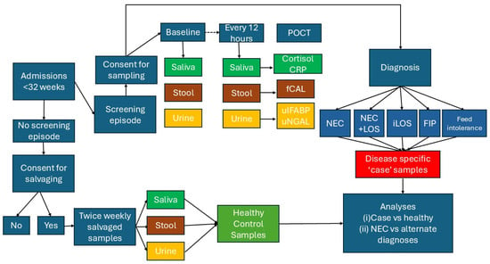
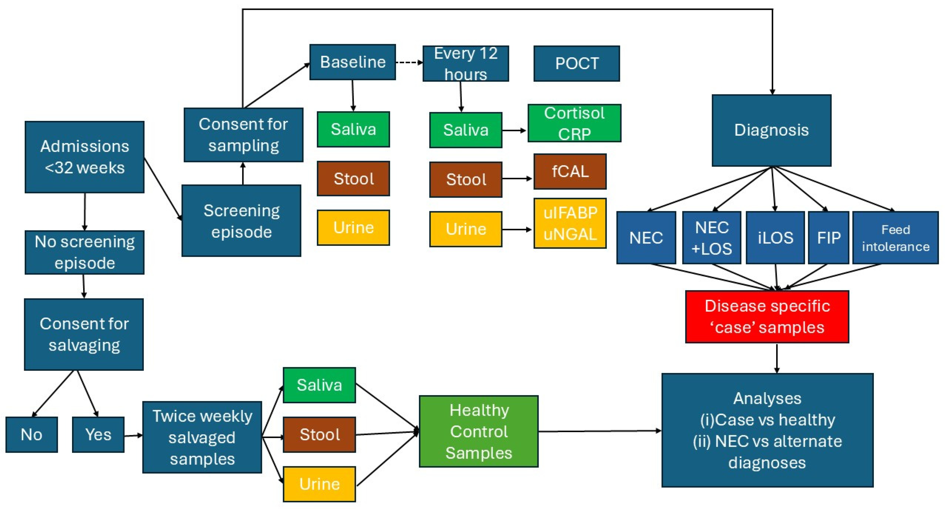

Disclaimer/Publisher’s Note: The statements, opinions and data contained in all publications are solely those of the individual author(s) and contributor(s) and not of MDPI and/or the editor(s). MDPI and/or the editor(s) disclaim responsibility for any injury to people or property resulting from any ideas, methods, instructions or products referred to in the content. |
© 2026 by the authors. Licensee MDPI, Basel, Switzerland. This article is an open access article distributed under the terms and conditions of the Creative Commons Attribution (CC BY) license.