


Highlights
What are the main findings?
- Relapsed clubfoot can be corrected gradually and simultaneously in all three planes with the Ilizarov circular external fixator, using closed soft-tissue distraction in the growing foot and V- or Y-shaped osteotomies in the skeletally mature foot.
- The frame assembly, the distraction protocol and the correction sequence for each component of the deformity are described step by step and illustrated with two representative cases.
What are the implications of the main findings?
- Severe, relapsed or multiply operated clubfeet can be brought to a plantigrade position without bone resection, arthrodesis or shortening of the foot.
- The choice between closed correction and correction through osteotomy is based on skeletal maturity, joint congruity and the presence of structural bony deformity rather than on chronological age alone.
Abstract
Background/Objectives: Relapse of clubfoot deformity may occur in severe cases after either conservative or surgical treatment, and repeated conventional surgery in a multiply operated foot frequently gives poor results. This article describes the treatment of neglected or relapsed idiopathic congenital clubfoot in children older than 6 years with the Ilizarov circular external fixator, using either closed soft-tissue distraction or gradual correction through osteotomy. Methods: The indications, the mechanical and biological principles of gradual correction, the frame assembly and the correction protocol for each component of the deformity are described step by step. Closed correction is reserved for the growing foot (up to approximately 10–12 years of age), whereas V- or Y-shaped osteotomies of the calcaneus, talus or midfoot are used in the skeletally mature foot, with olive “rein-wires” inserted through the surgical approach to ensure that distraction occurs at the osteotomy rather than at the adjacent joints. Results: Two illustrative cases are presented: a 9-year-old boy with a relapsed equino-cavo-varus deformity treated by closed distraction, and a 16-year-old boy with a relapsed clubfoot, previously treated with repeated open soft-tissue releases, corrected through osteotomy. A plantigrade foot was obtained in both cases. Conclusions: The Ilizarov method allows simultaneous three-plane correction of complex deformities, induces new histogenesis of soft tissues and bone, and avoids bone resection, arthrodesis and foot shortening. Strict adherence to the correction protocol and to the post-removal regimen is essential to limit recurrence.
1. Introduction
The first description of congenital clubfoot is attributed to Hippocrates, who first suggested that the deformity is caused by the position of the fetus in the uterus and by external mechanical forces and, secondarily, by adaptive shortening of muscles and ligaments [1]. Congenital clubfoot is a complex deformity of the entire hindfoot–foot complex, involving the skeleton, the joint capsules and the tendons of the foot. Treatment is always started early, and during the first months of life, it is non-operative [2]. The most widely used method today is the Ponseti technique, which consists of weekly manipulations followed by sequential casting, started in the first weeks of life [3,4]. The literature reports success with casting alone in 25–50% of cases [5,6,7,8,9,10]. Even after successful initial correction with the Ponseti method, relapse remains common: a systematic review reported relapse rates ranging from 1.9% to 45%, increasing with the length of follow-up [11], and brace non-compliance and poor evertor muscle activity have been identified as the main risk factors [12]. When conservative treatment fails, many authors recommend surgery between 3 and 6 months of age [13]—as soon as the child’s weight is adequate to reduce the anesthetic risk—in the form of a soft-tissue release (posterior and medial release). Most surgeons prefer an extensive one-stage release to achieve complete surgical correction [4,14,15,16,17,18]. However, at a mean follow-up of 30 years after extensive soft-tissue release, most patients had significant limitation of foot function, and repeated releases resulted in stiff, painful and arthritic feet with impaired quality of life [19]; open surgery thus tends to produce stiffer, scarred and more painful feet in adulthood [20].
Failure and relapse of the deformity after surgical treatment of congenital clubfoot are widely reported, with recurrence rates ranging from 15% to 25% depending on the series [21]. Most authors conclude that residual foot deformities are caused by incomplete initial surgical correction [22]. Each child must therefore be carefully assessed to determine which treatment will best correct his or her individual functional impairment. Moreover, no reliable and prognostic classification of clubfoot exists, and clinical assessment is based solely on the reducibility of the deformity, as expressed by the Diméglio score [23,24]. This 20-point scale is based on four parameters (equinus in the sagittal plane, varus in the frontal plane, derotation of the calcaneo-forefoot block around the talus and adduction of the forefoot on the hindfoot in the horizontal plane) and grades feet from benign (grade I) to very severe (grade IV) [23]. The main residual deformities of the foot described by Sullivan [13] are listed in Table 1.
Table 1.
Residual deformities in congenital clubfoot (modified from Sullivan [13]).
Relapses are initially treated with a series of procedures, choosing the most appropriate surgical option for each type of residual deformity. Equinus is corrected by percutaneous or open lengthening of the Achilles tendon, often combined with posterior capsulotomy. Cavus is often corrected by wedge resection of the midfoot or by resection and arthrodesis of the calcaneocuboid joint [25,26,27]. Calcaneal varus or valgus can be corrected with a Dwyer osteotomy in patients aged 4–6 years [28]. When the residual deformity is located in the forefoot, adduction, supination or both are corrected by multiple metatarsal osteotomies or by a combined opening-wedge osteotomy of the medial cuneiform and closing-wedge osteotomy of the cuboid [21,29]. In the most difficult cases, more disabling procedures such as triple arthrodesis or talectomy are required [30]. Failure may nevertheless occur even after a second operation, making any further salvage attempt with conventional surgery futile.
The Ilizarov technique has made it possible to address the most severe problems effectively and early. The first operations on the foot, performed in the 1980s, were based on the principle formulated by Ilizarov: a well-vascularized bone subjected to the continuous action of an external force calibrated in direction, sense and intensity can increase its volume and change its morphology [31]. This principle implies that a malformation or deformity can be corrected without invasive procedures on bone and soft tissues. Experimental and clinical work confirmed this hypothesis, while limiting its indication to the growing skeleton [32]. For relapsed clubfoot in adolescents and young adults, Ilizarov devised original variants of midfoot and hindfoot osteotomy to be used for correction with his apparatus [33]. The distraction method subsequently spread rapidly in the Western world [34,35,36] and achieved even greater success in developing countries, where limited access to health care results in a higher incidence of severe foot deformities [37]. Completely untreated (neglected) clubfoot in older children, which is still frequent in regions with limited access to early Ponseti treatment, represents one of the most common indications for correction with circular external fixation [20,37]. Many orthopedic textbooks now include chapters on the method, and a monograph entirely dedicated to distraction treatment of the foot has been published [38]. More recently, both the classic Ilizarov frame and hexapod fixators have been increasingly used for recurrent, residual and neglected clubfoot, correcting the deformity either by progressive soft-tissue stretching or by distraction through osteotomies [20,39,40,41,42], and a 2025 meta-analysis of 21 studies reported a pooled success rate of 81.4% and a pooled recurrence rate of 17.7% after external fixation [43].
The aim of this article is didactic: to describe, step by step, the surgical technique for the correction of relapsed and neglected clubfoot with the Ilizarov circular external fixator—both closed correction without osteotomy and correction through osteotomy—to present two technical elements introduced by the senior author, the rein-wires and the Y-osteotomy, and to illustrate the two treatment options with representative clinical cases. The article is not intended to report the outcomes of a clinical series.
2. Surgical Technique
2.1. Indications
The method is indicated in children with severe soft-tissue and bony deformities. It is not a first-line procedure; it is preferably applied to the most severe deformities or after failure of conventional surgery. The Ilizarov method can be used in two ways: closed (“bloodless”) treatment without osteotomy, and open treatment with osteotomy. The choice between the two cannot be reduced to a single age threshold. It is based on a combination of factors: the residual growth potential of the foot (skeletal rather than chronological age), the congruity of the hindfoot and midfoot joints, the presence of structural bony deformity, the rigidity of the foot and the condition of the soft tissues after previous surgery [20]. As a general rule, closed distraction is preferred in the growing foot with congruent joints and no structural bony deformity, whereas osteotomy is required when the skeleton is mature or the deformity is structured at the bone level. The age ranges given in the following sections should be interpreted as indicative.
2.2. Frame Assembly
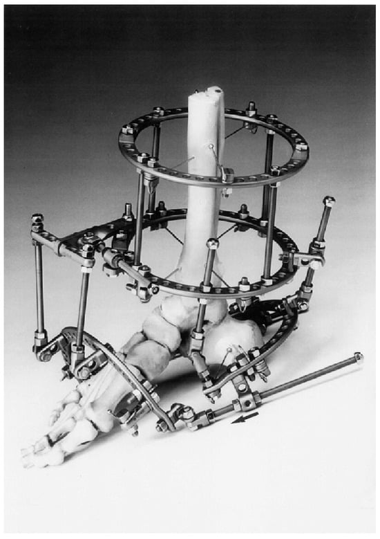
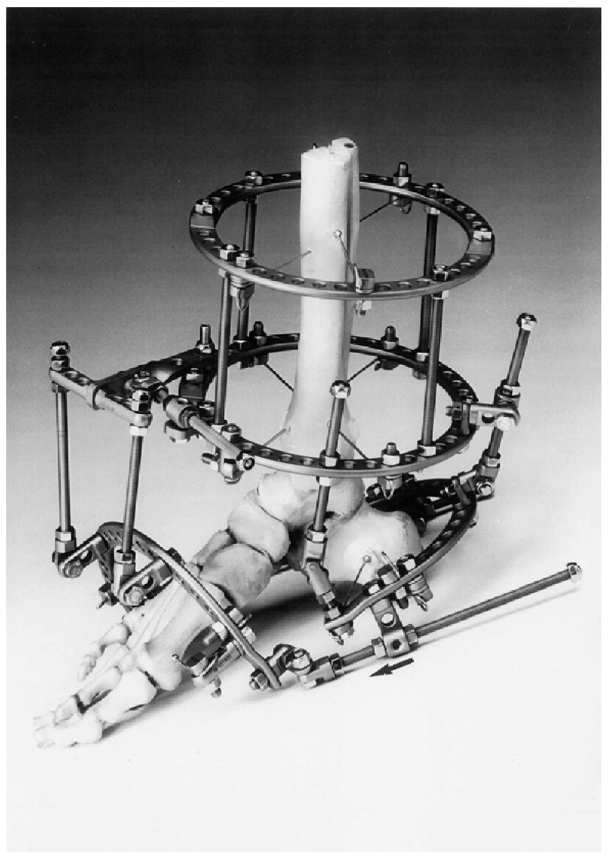
The construction of the fixator follows the same principles in closed treatment and in treatment with osteotomy (Figure 1). In treatment with osteotomy, however, the frame is assembled only after the osteotomy has been performed and the rein-wires have been inserted (Section 2.4).
Figure 1.
Frame assembly for the closed correction of clubfoot, shown on a bone model. The components are applied in the following order (Section 2.2): (1) the tibial base, consisting of two rings connected by threaded rods and fixed to the tibia with crossed wires; (2) the calcaneal (posterior) half-ring, fixed to the calcaneal tuberosity with olive wires and inclined so as to reproduce the equinus and varus of the hindfoot; (3) the anterior (forefoot) half-ring, fixed to the metatarsals with olive wires; and (4) the connecting elements—the threaded rods and hinges between the tibial base and the two half-rings and the plantar rods between the posterior and anterior half-rings—through which the corrective forces are applied.
The frame consists of the following:
- A tibial base (leg support), consisting of two rings with four wires, or of one ring with two wires and half-pins on a Rancho cube.
- In treatment with osteotomy, two crossed wires in the body of the talus, fixed to the tibial base.
- A calcaneal half-ring stabilized with at least three wires with opposing olives (most of them on the medial side), placed as far as possible in different planes; a supplementary self-tapping 4.0 mm or 5.0 mm half-pin may be added. The half-ring, centered on the calcaneal tuberosity, is positioned so as to reproduce the equinus of the foot and the adducted varus of the calcaneus, so that its inclination is corrective for equinovarus. To correct equinus, the half-ring is inclined from proximal to distal and from posterior to anterior; since the calcaneus is also in varus, the plane of the ring must also be inclined from proximal to distal and from medial to lateral. Adduction is a medial rotation of the calcaneal tuberosity in the horizontal plane; therefore, to reproduce the deformity and be corrective, the medial end of the half-ring must be placed more anteriorly than the lateral end.
- An anterior (forefoot) half-ring at the metatarsal level, perpendicular to the longitudinal axis of the forefoot (which corrects cavus and any adduction), is rotated so that its lateral end is more plantar than the medial end in order to correct supination; because of this rotation, the ends of the half-ring lie on the plane tangent to the sole of the forefoot. The half-ring is stabilized with at least three olive wires inserted according to the anatomy of the forefoot, with opposing olives (most of them on the medial side). The opposing olives prevent the half-ring from sliding along the axis of the wires. Unlike other segments, however, such as the tibia, where opposing olive wires are tensioned against a single bone, at the forefoot, the wires cross several metatarsals: tensioning each wire from the side opposite to its olive would squeeze the metatarsals between the olives. For this reason, all the wires are tensioned from the same side.
2.3. Closed Treatment Without Osteotomy
Closed correction relies on the remodeling capacity of the growing skeleton and is therefore effective only before skeletal maturity, generally up to 10–12 years of age (see Section 2.1). Its main advantages are the following: no osteotomy is required, so that bone and cartilage are not injured and further growth of the foot is not compromised; progressive stretching of the soft tissues restores their proportion with the size of the skeleton and neutralizes the deforming pull of contracted structures and unbalanced muscles; the soft tissues can be over-lengthened so that the deformity is overcorrected, which lowers the risk of relapse; and associated shortening or axial deviation of the leg can be addressed during the same treatment.
2.3.1. Mechanical and Biological Factors
The outcome of closed correction depends on the interplay between mechanical factors—frame stability, the magnitude and direction of the corrective forces and the rhythm with which they are applied—and the biological response of the tissues, namely soft-tissue lengthening, the trophism of bone, vessels and nerves, and the changes in shape and size of the skeleton.
Stability. The frame must remain rigidly anchored to bone for the whole duration of treatment and must not loosen under the corrective forces or during weight bearing. Its stability is determined by the number, diameter and tension of the wires, by their crossing angle, by their insertion sites and by the position of the olives. In our practice, 1.5 mm or 1.8 mm wires are used, with the widest crossing angle permitted by the anatomy; wires are tensioned as much as the resistance of the wire and the deformability of the ring allow (approximately 90 kg on half-rings and up to 130 kg on full rings); olives are placed on the side opposite to the direction of the corrective forces; and the insertion sites are chosen according to the local anatomy and the type of deformity.
Magnitude of the corrective force. The corrective force is expressed as the daily displacement, in millimeters, of the nuts along the threaded rods, and may consist of distraction, compression, rotation, translation or a combination of these movements. With correctly positioned hinges and a correctly built frame, correction is started at 1 mm per day and subsequently accelerated or slowed down according to the progress achieved and the response of the tissues.
An excessive rate of correction is signaled by swelling, pain, toe clawing and skin that is overly tight, pale or cold, whereas steady clinical and radiographic progress with absent or minimal trophic changes indicates that the rate is appropriate. The rate is therefore dictated by the biological response. In limb lengthening, the speed of distraction is adjusted to the speed at which the regenerate forms between the bone ends, which thus acts as a biological indicator. In the closed correction of foot deformities, the same role is played by the soft tissues: they yield when tension is appropriate, develop trophic disturbances when it is excessive and fail to correct when it is insufficient.
Rhythm of correction. The daily amount of correction is split into at least four increments, one every six hours, so that the anatomical structures are not overloaded in a short time, which would favor the early appearance of trophic disturbances.
2.3.2. Correction of the Individual Components of the Deformity
- Equinus: The central rod of the posterior (calcaneal) half-ring is lengthened, while the rods connecting the T-shaped module to the anterior half-ring are shortened.
- Varus: The medial rod of the posterior half-ring is lengthened, and the lateral rod is left free to shorten.
- Cavus: The medial plantar rod connecting the anterior and posterior modules is lengthened (the lateral rod shortens passively), and the forefoot is pulled cranially through the threaded rods between the T-shaped module and the anterior half-ring.
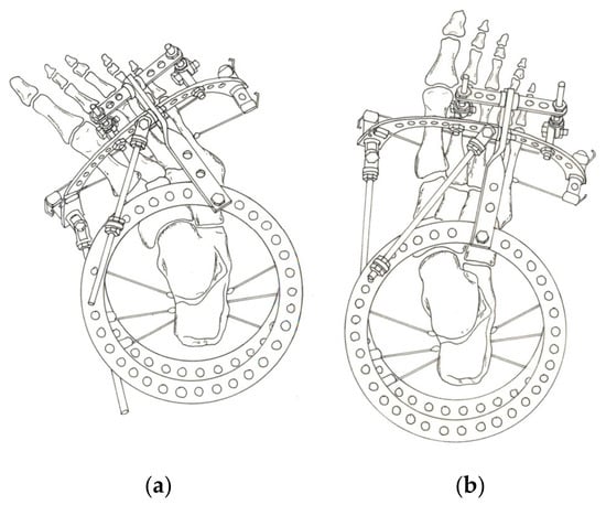
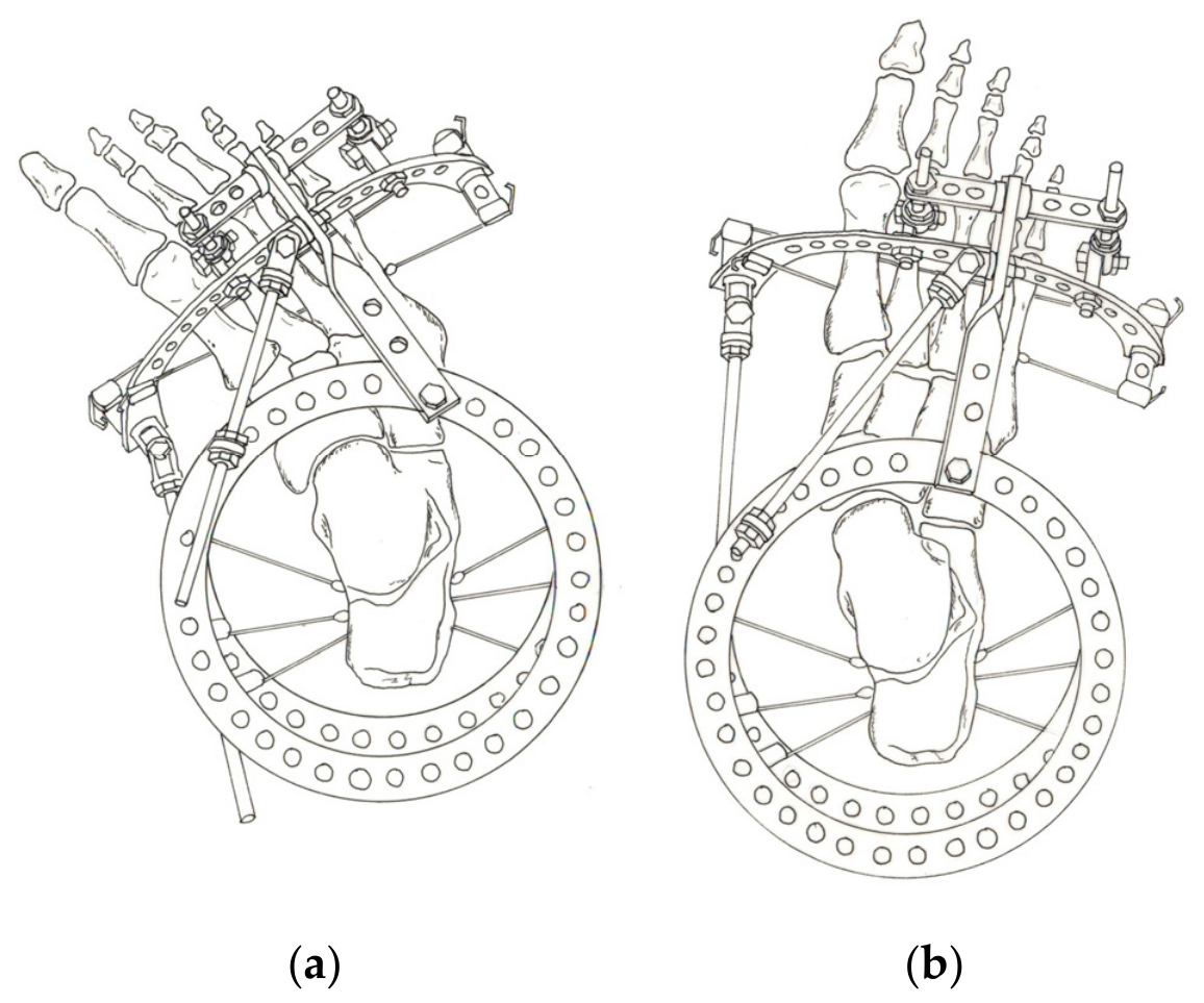
- Adduction: The T-shaped module is rotated externally, and further push is applied through the medial plantar rod (Figure 2).
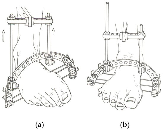
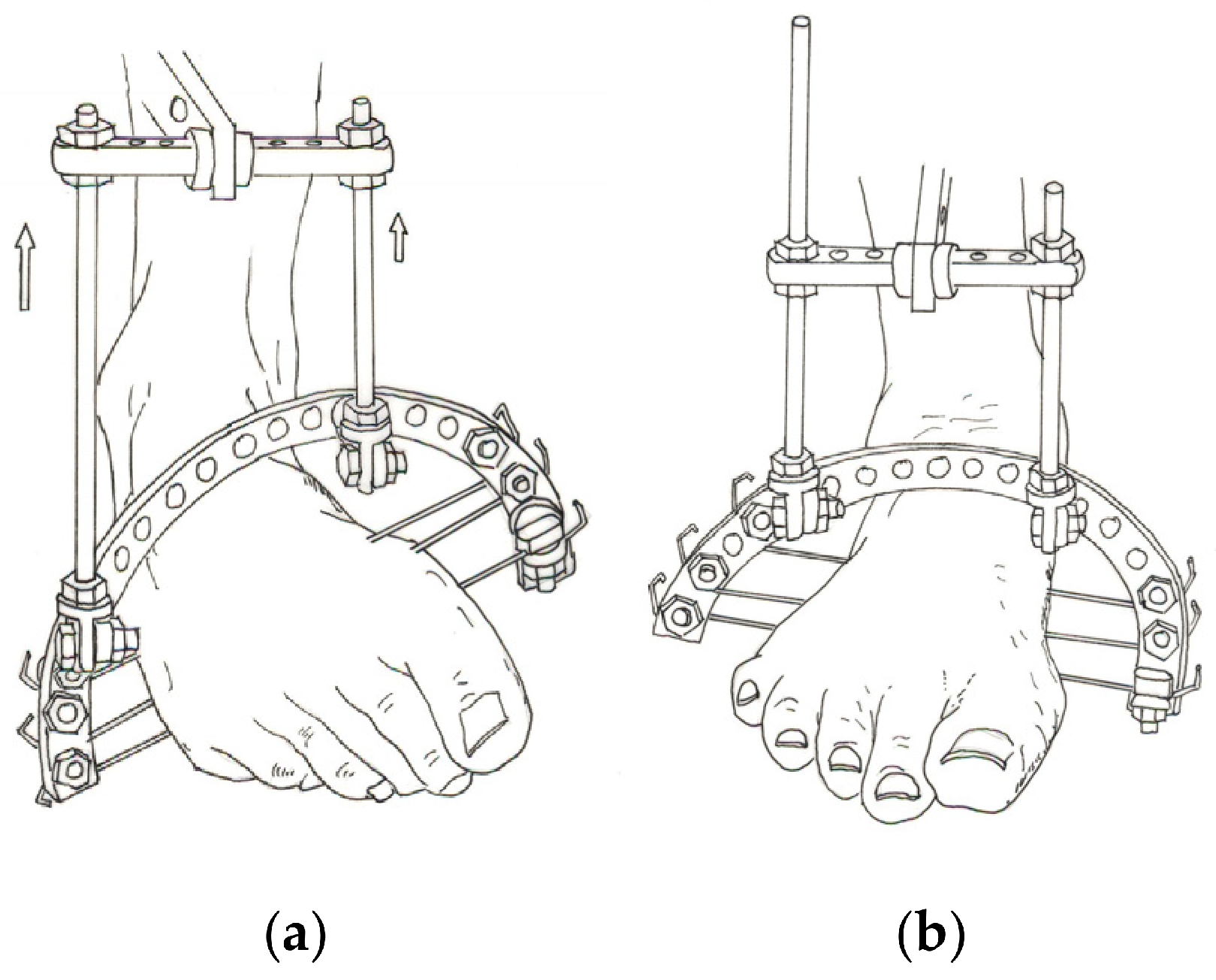
- Supination: It is corrected together with equinus and cavus by shortening the threaded rods between the T-shaped module and the anterior half-ring by different amounts (Figure 3).
- Internal rotation of the calcaneus: (medial rotation of the calcaneal tuberosity in the horizontal plane) is usually corrected acutely at surgery. Alternatively, an additional sub-assembly placed between the tibial base and the posterior half-ring can rotate the calcaneal tuberosity externally; the two plantar rods connecting the posterior and anterior modules must then be left free to adapt, the medial one lengthening and the lateral one shortening during correction.
Figure 2.
Correction of forefoot adduction with the T-shaped module (view from above; the circular ring is the tibial base). (a) Before correction: the forefoot is adducted, and the T-shaped module, connected to the anterior half-ring, is aligned with the adducted forefoot. (b) After correction: external rotation of the T-shaped module, combined with lengthening of the medial plantar rod between the posterior and anterior half-rings, has brought the forefoot into overcorrection (abduction).
Figure 3.
Correction of forefoot supination (anterior view). (a) The anterior half-ring, fixed to the metatarsals with olive wires, is connected to a horizontal bar of the tibial base by two vertical threaded rods; shortening these rods by different amounts (arrows) rotates the forefoot from supination toward pronation. (b) End of correction: the forefoot is overcorrected into pronation.
All components are corrected simultaneously rather than one after the other: initially, all rods are lengthened or shortened by the same amount (for example, up to 2 mm per day), and the rates are then adjusted to the local response. Correction is assessed mainly clinically. Equinus and forefoot adduction usually correct first; their rods are then slowed down or stopped while correction of the remaining components continues. Achilles tenotomy and plantar fasciotomy are always performed beforehand.
The aim is to overcorrect each component by approximately 20° for varus, 30–40° for adduction, 25–30° for equinus, 20° for supination and 10° for cavus. The frame is then kept in place for approximately 45 days. Throughout treatment, the patient walks with crutches and partial weight bearing on a compensating insole, which is modified as correction proceeds. At the end of treatment, radiographs usually show joint diastasis and some degree of osteoporosis. After frame removal, a walking cast holding the foot in overcorrection is worn for 30 days; the cast removes the deforming forces and allows each skeletal element to grow along a correct morphological line, filling the space created by distraction. Bone morphology normalizes within 2–3 months of apparent clinical correction, consolidating the result. The timing and frequency of trophic disturbances depend on several factors.
2.3.3. Complications and Recurrence
Mechanical complications. Because the frame is complex, errors in the position of the hinges or in the direction of the corrective forces can lead to anterior subluxation of the talus or to subluxation at the Chopart or Lisfranc joints, which must be detected by serial radiographs. Correction of one or more components may stop because a hinge has jammed, because the forces are wrongly directed, or because the connecting elements on the side opposite to the corrective movement are prevented from adapting.
Biological complications. The wires may cut through the medial skin and cause linear necrosis; this is prevented by displacing the skin in the anticipated direction of movement (between the posterior half-ring and the tibial base, and between the anterior and posterior half-rings) when the wires are inserted. Trophic disturbances are managed by slowing down or temporarily stopping correction.
Recurrence. Provided that the post-removal protocol is respected, recurrence is uncommon in congenital and post-traumatic clubfoot, whereas it is more frequent in arthrogryposis and in multiply operated feet, in which scar tissue tends to retract.
Congenital clubfoot may coexist with tibial hypoplasia and with a variable degree of external tibial torsion, which is clinically concealed by forefoot adduction and inversion of the foot and may be suspected from a posteriorly displaced lateral malleolus. This external torsion results from external rotation of the talus secondary to medial subluxation of the forefoot or of the Chopart joint. Realignment of the foot removes its cause, so that mild torsion may resolve during growth or be compensated by internal rotation of the foot. Only in extreme untreated deformities, in which the foot is curled medially and the patient walks on its dorsal surface, does the deformity drag the talus and the tibial plafond into internal rotation and produce true internal tibial torsion, which then needs to be corrected. Up to 7–8 years of age, this can be achieved without osteotomy by extending the frame to the leg; tibial osteotomy or closed derotation with the Ilizarov method is only exceptionally needed. Tibial hypoplasia can be addressed by modest tibial lengthening during correction of the foot.
2.4. Open Treatment with Osteotomy
Correction through osteotomy is reserved for adolescents, generally older than 12 years (see Section 2.1), with severe and structured deformities in whom conventional elective procedures—tendon transfers, aponeurotic and muscle releases, limited osteotomies or resections—even when associated with complementary procedures such as tenoplasty or tenolysis, can no longer provide an acceptable foot shape and function.
In addition to the conventional osteotomies of the calcaneus, midfoot and metatarsals, Ilizarov described specific talocalcaneal osteotomies for use with his apparatus. Whenever the anatomy permits, the osteotomy is performed subcutaneously with minimal soft-tissue stripping, to preserve the blood supply. Skin incisions are oriented parallel to the direction of distraction to avoid wound dehiscence. Mobility of the fragments when the osteotome is rotated between the cut surfaces confirms that the osteotomy is complete; fluoroscopic or radiographic control is useful. The osteotomies are generally performed before the wires for frame assembly are inserted.
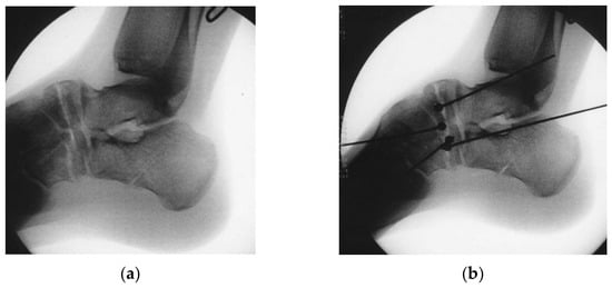
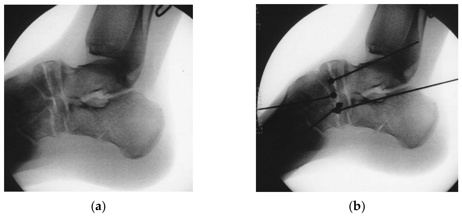
Rein-wires. To control the osteotomy fragments directly, the senior author introduced the use of “rein-wires”, so called because they act on the fragments like the reins of a horse. These are olive wires inserted through the same surgical approach used to perform the osteotomy, so that the surgeon can be sure that each wire does not cross the osteotomy but passes through only one of the two fragments (proximal or distal). The wire is drilled from the osteotomy surface through the chosen fragment and out through the skin on the opposite side; after checking that the wire does not cross the osteotomy, the surgeon retracts it, cuts it just in front of the olive and reinserts it, so that its cut end remains within the osteotomy and the olive rests against the osteotomy surface of the fragment. Only the other end of the wire emerges from the skin, where it is fixed to the frame. In this way, the distraction and corrective forces generated by the frame are transmitted to the fragments on either side of the osteotomy and act at the level of the osteotomy rather than at the adjacent joints (subtalar, talonavicular, calcaneocuboid and ankle joints). Without this direct control, part of the apparent correction may be obtained by joint distraction or subluxation rather than by displacement of the osteotomy fragments; such correction is unstable and exposes the foot to recurrence of the deformity after removal of the fixator. By concentrating the distraction at the osteotomy, rein-wires ensure that the correction is achieved—and subsequently consolidated by the regenerate—at bone level.
V- or Y-shaped osteotomies are used to correct multiplanar deformities in clubfoot. The V-osteotomy is indicated when simultaneous lengthening of the foot is required, whereas the Y-osteotomy is purely corrective.
2.4.1. V-Osteotomy
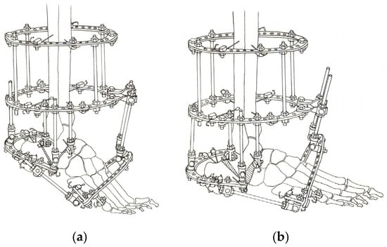
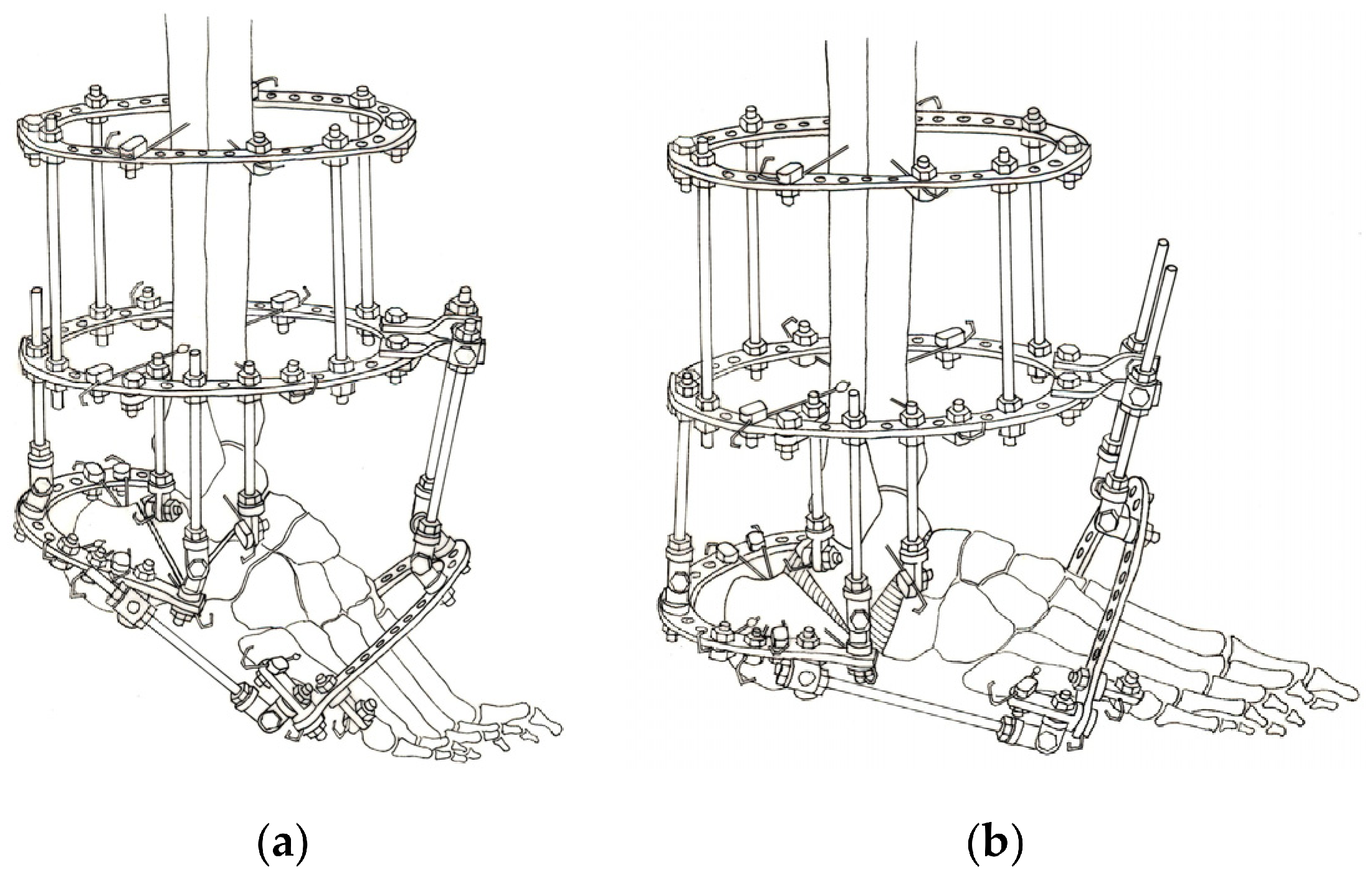
The V-osteotomy is performed subcutaneously or through an open approach, under fluoroscopic or radiographic control; the frame is assembled as described in Section 2.2 (Figure 4).
Figure 4.
V-osteotomy. (a) Frame for correction through the V-osteotomy, assembled after the osteotomy has been performed: tibial base (two rings), crossed wires in the talar body fixed to the tibial base, calcaneal half-ring and anterior (forefoot) half-ring, connected by threaded rods and hinges; the shaded areas indicate the two limbs of the V-osteotomy. (b) The same frame at the end of correction: distraction across the osteotomy has corrected equinus and cavus, and the regenerate fills the osteotomy gap.
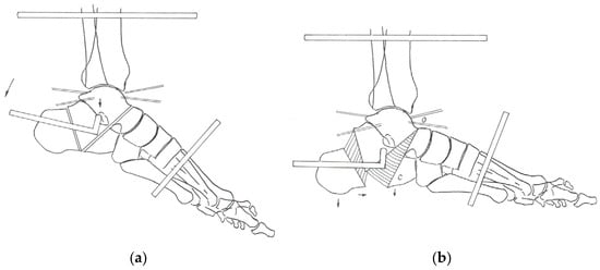
Correction protocol (Figure 5a). Achilles tenotomy and plantar fasciotomy, as well as measures to prevent toe clawing, are mandatory. Distraction begins on the fifth postoperative day: all rods between the tibial base and the posterior half-ring and between the anterior and posterior half-rings are lengthened by 1 mm per day, while the vertical rods between the tibial base and the anterior half-ring are shortened by 1 mm per day. In this way, the osteotomy gap is opened, creating room for the corrective displacement of the fragments and preventing premature consolidation.
Figure 5.
Schematic lateral views of correction through the V-osteotomy. The horizontal bar represents the tibial base, to which the crossed talar wires are fixed; the angled rod represents the calcaneal half-ring and the oblique rod the anterior half-ring. (a) Before correction: the push applied through the calcaneal half-ring (arrow) rotates the calcaneal tuberosity caudally, correcting equinus, and translates it anteriorly. (b) Virtual hinge at the cranial end of the talocalcaneal osteotomy (O). As the forefoot rotates cranially about an axis passing through O, the osteotomized calcaneal fragment (C) forms an abnormal prominence that disrupts the plantar profile of the foot; caudal displacement of the calcaneal tuberosity (arrows) realigns the plantar profile.
A radiograph obtained after 7–10 days confirms the diastasis and excludes subluxation at the midtarsal joint. Correction then continues at different rates for the individual components, depending on their magnitude, on the local response and on any trophic disturbances. Forefoot adduction and supination are best corrected once the regenerate has started to form in the talocalcaneal osteotomy, because its plasticity accommodates pronation and medial distraction.
During correction of cavo-equinus, the forefoot rotates cranially around the cranial end of the talocalcaneal osteotomy, which acts as a virtual hinge. The osteotomized anterior process of the calcaneus, the cuboid or the base of the fifth metatarsal therefore tends to protrude toward the sole; this prominence is eliminated by lowering, rotating caudally and translating the calcaneal tuberosity anteriorly until the plantar profile of the foot is realigned (Figure 5b). Calcaneal varus remaining after acute correction is corrected by lengthening the medial rod of the posterior half-ring, using the frontal-plane mobility of the biplanar hinges.
The frame is removed after approximately 45 days of fixation, once the regenerate has consolidated, and weight bearing with crutches is then progressively increased.
Complications. Mechanical: Arrest of correction due to inaccurate positioning of the hinges or insufficient diastasis at the osteotomies; joint subluxation. Biological: Premature consolidation; trophic disturbances related to correction of cavus.
2.4.2. Y-Osteotomy (Introduced by the Senior Author)
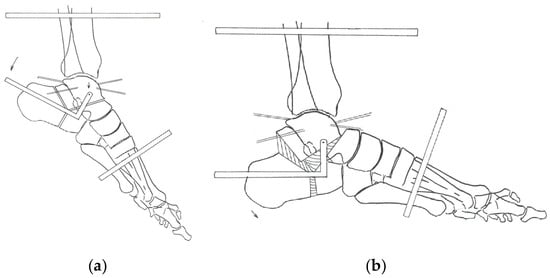
Through a 2–3 cm curved lateral incision below the malleolus, the calcaneal tuberosity is first partially cut (posterior oblique limb of the Y), the calcaneus is then divided vertically (vertical limb), and the talocalcaneal osteotomy is finally completed, producing a three-pointed star with limbs about 120° apart (Figure 6a).
Figure 6.
Schematic lateral views of the Y-osteotomy (horizontal bar: tibial base; angled rod: calcaneal half-ring; oblique rod: anterior half-ring). (a) Before correction: the three limbs of the osteotomy (posterior oblique cut of the calcaneal tuberosity, vertical cut of the calcaneus and talocalcaneal cut) form a three-pointed star; the hinge of the calcaneal half-ring (circle) is placed at the level of the tip of the medial malleolus, approximately 0.5 cm anterior to the anatomical axis of the tibia. (b) After correction: the calcaneal tuberosity has been displaced caudally and anteriorly, and a regenerate (shaded) has formed in the osteotomy. Note that the length of the foot does not increase significantly.
The frame is then assembled as described in Section 2.2; in contrast to the V-osteotomy, no measures against calcaneocuboid diastasis are needed. The biplanar hinges, which allow motion in the sagittal and frontal planes for correction of equinovarus and connect the medial and lateral threaded rods of the calcaneal half-ring, are positioned at the level of the tip of the medial malleolus, about 0.5 cm anterior to the anatomical axis of the tibia.
Equinus is corrected by moving the calcaneal tuberosity downward and the forefoot upward with respect to the talar body, which is held by two crossed wires fixed to the tibial base. Pushed by the central posterior rod, the calcaneal half-ring rotates around the hinge axis and displaces the calcaneal tuberosity caudally and anteriorly more than in the V-osteotomy (Figure 6b). When further lowering of the calcaneus is needed, the three rods on the half-ring are lengthened equally, and varus is corrected simultaneously by additional lengthening of the medial rod.
At the same time, the forefoot is lengthened and pulled anteriorly and cranially to correct equinus and cavus; forefoot supination and equinus are corrected by shortening the two vertical threaded rods between the tibial base and the anterior half-ring by different amounts. Achilles tenotomy and plantar fasciotomy are always required.
Because the fragments move within the narrow space of the osteotomy, this space must first be enlarged by acting on the components that open it most, namely cavus and equinus. As cavus does not need to be corrected rapidly, only the central posterior rod (equinus) is lengthened by 2 mm per day, while all the other rods are lengthened by 1 mm per day. The diastasis is checked radiographically after 7–10 days. Correction proceeds, according to the trophic tolerance of the tissues, until cavus and equinus are corrected; varus is then corrected by lengthening the medial rod of the calcaneal half-ring, supination by rotating the anterior half-ring into pronation, and adduction by lengthening the medial connecting rod between the two half-rings while the lateral rod is allowed to shorten.
The Y-osteotomy does not lengthen the foot skeleton, which has three favorable consequences:
- A smaller amount of regenerate and therefore faster consolidation;
- Fewer trophic complications;
- No need to prevent calcaneocuboid diastasis.
Although less traumatic, the Y-osteotomy still allows differentiated correction of the hindfoot and forefoot, as with the V-osteotomy.
3. Illustrative Cases
3.1. Case 1: Closed Correction Without Osteotomy
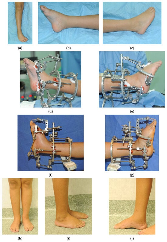
A 9-year-old boy presented with a right congenital clubfoot with a relapsed equino-cavo-varus deformity (Figure 7a–c). Given his age and the residual growth potential of the foot skeleton, closed correction by soft-tissue distraction was chosen. The frame was assembled as described in Section 2.2 (Figure 7d,e). All components of the deformity were corrected gradually and simultaneously over 1.5 months, until overcorrection of the equinus was obtained (Figure 7f,g). The frame was then maintained for a further 2 months, for a total treatment time of 3.5 months. After frame removal, a walking cast in overcorrection was applied for 30 days. No complications occurred in this patient; this observation, based on a single selected case, is not representative of the safety profile of the technique (see Section 4). One year after frame removal, the foot was plantigrade (Figure 7h–j). The patient has been followed for more than 5 years after frame removal, and at the latest follow-up, the correction was maintained, with a plantigrade foot.
Figure 7.
Case 1. Nine-year-old boy with right congenital clubfoot (equino-cavo-varus) treated with closed correction. (a–c) Clinical appearance before treatment; (d,e) the fixator after surgery; (f,g) the fixator at the end of treatment, showing overcorrection of the equinus; (h–j) clinical appearance 1 year after frame removal. Total treatment time was 3.5 months (1.5 months of correction and 2 months of fixation), followed by a walking cast in overcorrection for 30 days. No complications occurred in this patient.
3.2. Case 2: Correction with Osteotomy
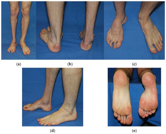
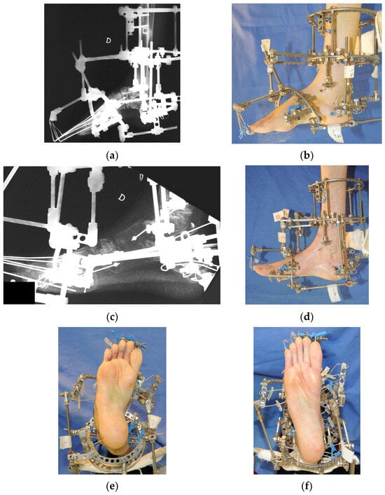
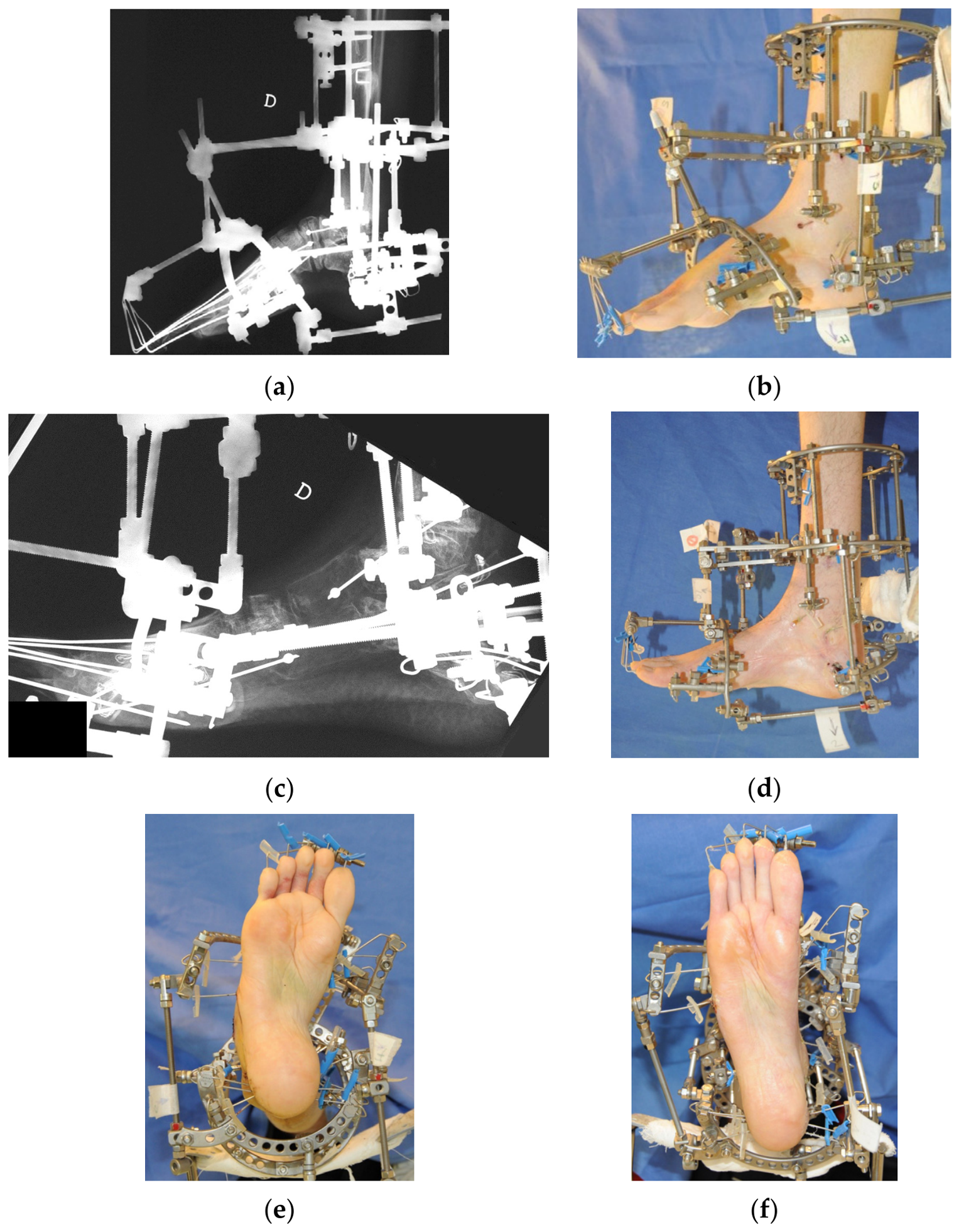
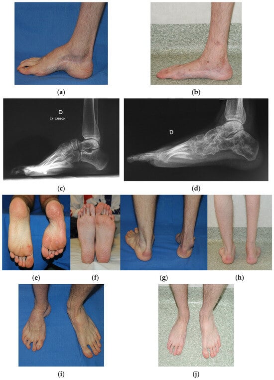
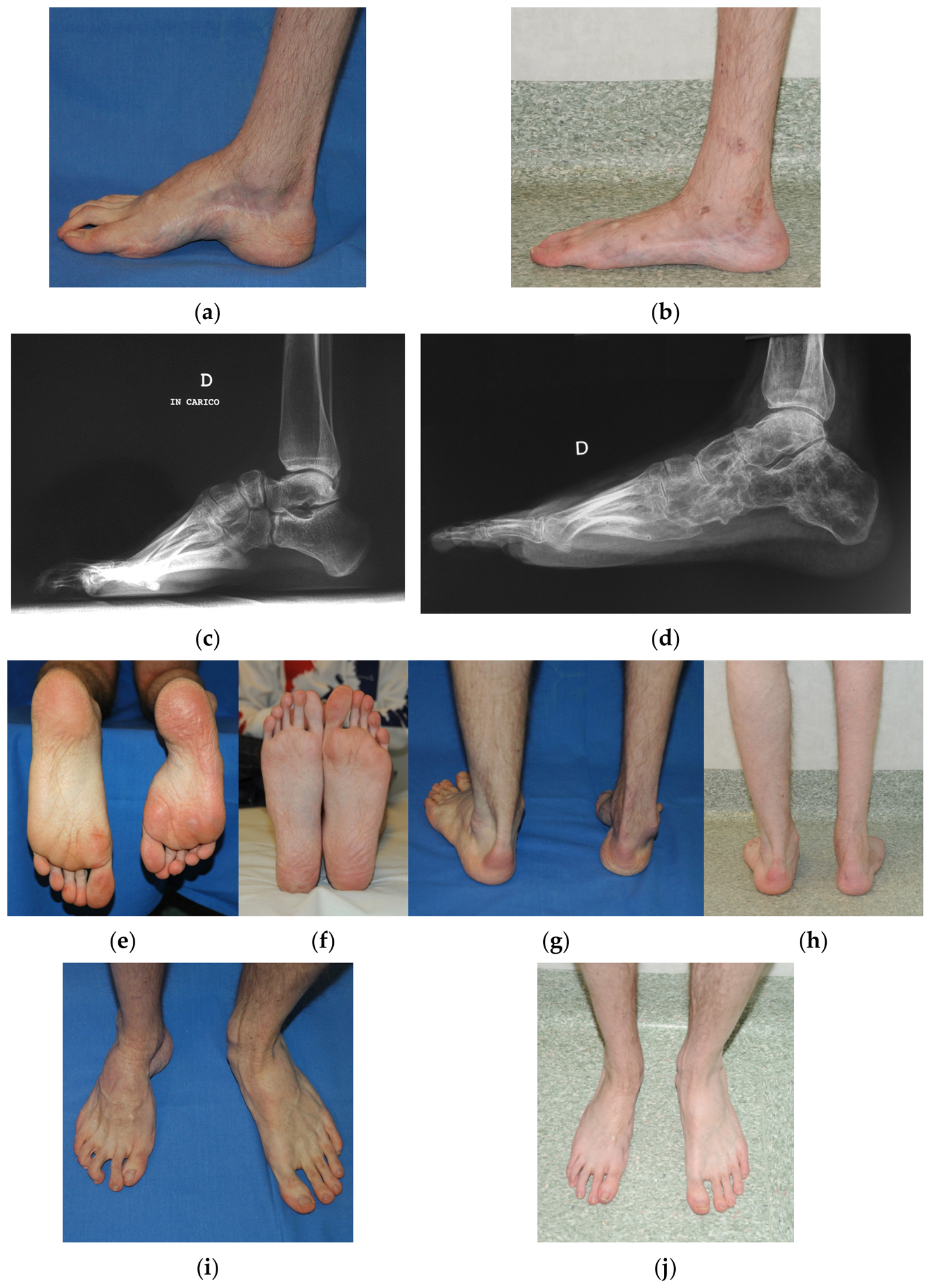
A 16-year-old boy presented with a relapsed right idiopathic congenital clubfoot. He had undergone multiple previous treatments, including repeated open soft-tissue releases, as shown by the surgical scars on the medial side of the foot and ankle. On examination, the right foot showed a rigid relapsed deformity with cavus, forefoot adduction and hindfoot varus (Figure 8). Because the foot skeleton was close to maturity and the soft tissues were stiff and scarred, correction through osteotomy was indicated. The V-osteotomy was performed through a limited surgical approach, and rein-wires were inserted through the same approach under fluoroscopic control (Figure 9). The frame was then assembled as described in Section 2.2 (Figure 10a,b), and the deformity was corrected gradually through the osteotomy (Figure 10). The distraction phase lasted for 8 weeks and was followed by 8 weeks of fixation. At 4 months, the frame was removed and a walking cast in overcorrection was applied for 30 days. No complications occurred in this patient; this observation, based on a single selected case, is not representative of the safety profile of the technique (see Section 4). One year after frame removal, the foot was plantigrade, with correction of cavus, heel varus and forefoot adduction (Figure 11). The patient has been followed for more than 5 years after frame removal, and at the latest follow-up, the correction was maintained.
Figure 8.
Case 2. Sixteen-year-old boy with relapsed right idiopathic congenital clubfoot after multiple previous treatments, including open soft-tissue releases. Preoperative clinical appearance: (a) anterior, (b) posterior, (c) dorsal, (d) medial and (e) plantar views, showing cavus, forefoot adduction, hindfoot varus and the scars of previous surgery.
Figure 9.
Case 2. Rein-wires. (a) Intraoperative lateral fluoroscopic view of the hindfoot and midfoot. (b) Rein-wires in place. The rein-wires, introduced by the senior author, are olive wires that act on the osteotomy fragments like the reins of a horse. Each wire is inserted through the surgical approach used to perform the osteotomy, so that it does not cross the osteotomy but passes through only one fragment (proximal or distal). The wire is then retracted, cut just in front of the olive and reinserted, so that the olive remains within the osteotomy, resting against the cut surface of the fragment; only the other end emerges from the skin and is fixed to the frame. In this way, distraction and correction take place at the osteotomy rather than at the adjacent joints, avoiding an unstable joint-level correction that would expose the foot to recurrence of the deformity after removal of the fixator.
Figure 10.
Case 2. Treatment with the external fixator. (a) Lateral radiograph and (b) clinical appearance after frame application; (c) lateral radiograph and (d) clinical appearance during correction; (e,f) plantar views in the frame at the beginning (e) and at the end (f) of correction, showing correction of forefoot adduction.
Figure 11.
Case 2. Comparison before and after treatment of the right foot. Postoperative photographs and radiographs were obtained 1 year after frame removal; follow-up was continued for more than 5 years, with maintenance of the correction. X-rays and pictures show correction of cavus, heel varus, and forefoot adduction, with a final plantigrade foot. (a) Preoperative and (b) postoperative medial views; (c) preoperative and (d) postoperative lateral weight-bearing radiographs; (e) preoperative and (f) postoperative plantar views; (g) preoperative and (h) postoperative posterior views; (i) preoperative and (j) postoperative dorsal views.
4. Discussion
Tendon lengthening and transfers, capsulotomies, osteotomies, resections, arthroereisis and arthrodesis are the techniques used to correct relapsed clubfoot [27,44,45,46]. Residual deformities in adolescents are difficult to correct and have historically been associated with mediocre results. Soft-tissue procedures alone carry a very high failure rate, and a stable result is difficult to obtain without hindfoot and midfoot arthrodesis. Additional osteotomies allow correction of the deformity, but only in one plane [47]. The Ilizarov method differs from conventional surgery in that it introduces the dimension of time into surgical treatment [48].
The distraction method can be regarded as an extension of progressive correction of the foot with serial casts; the major difference is that the joints are compressed with casting and distracted with the Ilizarov method [49]. Whereas in conventional surgery, the correction is completed in the operating room, which predetermines the result from the outset [50], with the Ilizarov method, the operation is the prologue to subsequent therapeutic steps that reach the goal over time. Accordingly, the Ilizarov method offers the following possibilities: during the correction phase, errors can be corrected and the correction can be guided according to the treatment plan by modifying the frame; treatment spread over time allows resections (e.g., wedge resection for cavus foot) to be replaced by osteotomies which, subjected to distraction, form a regenerate, so that correction is obtained by addition and shortening of the foot is avoided; the plastic regenerate can be molded by changing the direction and action of the external forces applied by the frame; and continuous, prolonged stretching of the soft tissues lengthens them and eliminates their size discrepancy with the skeleton. During treatment, clinical examination shows the progression of the deformity toward correction, while radiographs show diastasis between the joints of the individual deformed bones (e.g., between the navicular and the talar head in varus, at the midtarsal joint in cavus, and at the Lisfranc joint in forefoot adduction). This finding indicates that the corrective force, by lengthening the soft tissues, has created space.
Several authors consider the closed technique suitable for children up to 8 years of age [21,49,51]; more recently, Baar and McClure suggested that correction by soft-tissue distraction alone is appropriate in children aged 8 years or younger with congruent joints and no bony deformity, and recommended reserving circular fixation for severe relapsed or neglected feet (Diméglio grades III and IV) [20]. In our opinion, closed correction of clubfoot can be performed up to approximately 12 years of age, as long as the growth potential of the skeleton is not exhausted and bone can remodel into the new position achieved under distraction and repositioning forces. Once the foot skeleton has reached its definitive structure, closed treatment is no longer applicable, and soft-tissue and bone procedures are required [52]. Although the technique is demanding and should be performed by surgeons familiar with pediatric foot correction and with a thorough knowledge of Ilizarov external fixation [47], the results reported in the literature are predominantly excellent or good [53]. El Barbary et al. reported satisfactory results in both relapsed and neglected severe clubfeet [53], and Lejman et al. obtained mostly good results in children treated without additional procedures [54]. Ankle range of motion remains similar to the preoperative range [55], and lengthening of the leg can be performed simultaneously with correction of the foot deformity [56]. More recent series have confirmed these findings in children, both with short-term application of an Ilizarov frame [57] and with Ilizarov correction combined with limited adjunctive procedures [58], and hexapod frames combined with midfoot osteotomy have been used successfully in older children [42]. Not all reports are favorable, however: Freedman et al. observed residual or recurrent deformity, often requiring revision surgery, in a substantial proportion of resistant clubfeet [59], and recurrence is more frequent in multiply operated and syndromic feet [43,60]. Treatment-related events are common, pin-tract infection being the most frequent, and require careful postoperative surveillance [43]. In older and more rigid feet, combining gradual distraction with osteotomies or tendon procedures appears to improve the durability of correction [43,47].
In our experience, the protocol followed after frame removal plays a key role in preventing recurrence. The walking cast in overcorrection, applied immediately after frame removal for approximately 30 days, protects the correction while the lengthened soft tissues and the regenerate mature and while the bones remodel in the corrected position, and it allows the patient to bear weight. After cast removal, footwear with a lateral wedge may be used to keep the foot in slight eversion and pronation, counteracting the tendency of the hindfoot and forefoot to drift back into varus and supination during the remodeling period. Without these measures, the elastic recoil of the soft tissues—particularly in multiply operated feet with retracting scars—may lead to early loss of correction.
The Y-osteotomy, introduced by the senior author, is described in detail in this article but is not illustrated by a clinical case: its clinical application and results are the subject of a separate paper, and the osteotomy case presented here (Case 2) was treated with a V-osteotomy.
This article has several limitations. First, it is a technical description supported by only two illustrative cases, selected to show the two treatment options; they do not represent a consecutive series, and the number of patients treated with the technique is not reported. Therefore, no conclusions can be drawn on efficacy, safety or recurrence rates, and no meaningful comparison can be made with the pooled success and recurrence rates reported in the literature [43]; in particular, the absence of complications in the two cases should not be interpreted as representative of the safety profile of the technique. Second, there is no control group, and the technique is not compared with other treatment options, such as repeated Ponseti casting, conventional soft-tissue and bony procedures or hexapod frames. Third, although both patients have been followed for more than 5 years, the follow-up was not standardized and did not include systematic objective assessment; given the known tendency of clubfoot to relapse over time [11], late recurrence cannot be completely excluded. Fourth, outcomes were not assessed with objective measures, such as the Diméglio score, radiographic angles, range of motion or patient-reported outcome measures. Fifth, the cases were treated with the classic Ilizarov apparatus, whereas hexapod frames are increasingly used today. Finally, the technique is demanding, has a substantial learning curve and was developed and refined in a single specialized center, so that the experience described may not be generalizable to centers with less experience in circular external fixation. Prospective studies including consecutive patients, standardized outcome measures and long-term follow-up are needed.
5. Conclusions
The Ilizarov method allows very severe foot deformities to be treated with a minimally invasive technique, without bone resection or arthrodesis and without shortening of the foot. Additionally, patients remain ambulatory throughout all phases of treatment, walking with crutches and partial weight bearing during both the correction and the fixation periods and after frame removal. All components of the deformity can be corrected simultaneously, without waiting for the end of growth, to obtain a stable, plantigrade and painless foot; associated deformities and shortening of the lower limb can be treated at the same time. Closed distraction is the treatment of choice in the growing foot, whereas V- or Y-osteotomies extend the indications of the method to the skeletally mature foot.
Author Contributions
Conceptualization, A.K.; methodology, A.K.; writing—original draft preparation, F.V.; writing—review and editing, E.M. and A.K.; visualization, F.V.; supervision, A.K. All authors have read and agreed to the published version of the manuscript.
Funding
This research received no external funding.
Institutional Review Board Statement
Not applicable. This article describes a surgical technique and two illustrative cases treated as part of routine clinical care; no experimental intervention or research protocol was involved, and no data were collected for research purposes. Written informed consent for publication was obtained from the parents/legal guardians of both patients.
Informed Consent Statement
Written informed consent has been obtained from the parents/legal guardians of both patients to publish this paper.
Data Availability Statement
The original contributions presented in this study are included in the article. Further inquiries can be directed to the corresponding author.
Acknowledgments
During the preparation of this manuscript, the authors used Claude Opus 5.5 (Anthropic, San Francisco, CA, USA) for the purposes of English editing. The authors have reviewed and edited the output and take full responsibility for the content of this publication.
Conflicts of Interest
The authors declare no conflicts of interest.
References
- Hippocrates. Oeuvres Complètes d’Hippocrate; J.-B. Baillière: Paris, France, 1844. [Google Scholar]
- Kite, J.H. Nonoperative treatment of congenital clubfoot. Clin. Orthop. Relat. Res. 1972, 84, 29–38. [Google Scholar] [CrossRef] [Scilit]
- Ponseti, I.V. Treatment of congenital club foot. J. Bone Jt. Surg. Am. 1992, 74, 448–454. [Google Scholar] [CrossRef] [Scilit]
- Chu, A.; Labar, A.S.; Sala, D.A.; van Bosse, H.J.; Lehman, W.B. Clubfoot classification: Correlation with Ponseti cast treatment. J. Pediatr. Orthop. 2010, 30, 695–699. [Google Scholar]
- Farsetti, P.; Weinstein, S.L.; Ponseti, I.V. The long-term functional and radiographic outcomes of untreated and non-operatively treated metatarsus adductus. J. Bone Jt. Surg. Am. 1994, 76, 257–265. [Google Scholar] [CrossRef] [Scilit]
- Ikeda, K. Conservative treatment of idiopathic clubfoot. J. Pediatr. Orthop. 1992, 12, 217–223. [Google Scholar] [CrossRef] [Scilit]
- Kite, J.H. Principles involved in the treatment of congenital clubfoot. Clin. Orthop. Relat. Res. 1972, 84, 4–8. [Google Scholar] [CrossRef] [Scilit]
- McKay, D.W. New concept of and approach to clubfoot treatment: Section I—Principles and morbid anatomy. J. Pediatr. Orthop. 1982, 2, 347–356. [Google Scholar]
- McKay, D.W. New concept of and approach to clubfoot treatment: Section II—Correction of the clubfoot. J. Pediatr. Orthop. 1983, 3, 10–21. [Google Scholar] [CrossRef] [Scilit]
- McKay, D.W. New concept of and approach to clubfoot treatment: Section III—Evaluation and results. J. Pediatr. Orthop. 1983, 3, 141–148. [Google Scholar]
- Gelfer, Y.; Wientroub, S.; Hughes, K.; Fontalis, A.; Eastwood, D.M. Congenital talipes equinovarus: A systematic review of relapse as a primary outcome of the Ponseti method. Bone Jt. J. 2019, 101-B, 639–645. [Google Scholar] [CrossRef] [Scilit]
- van Schelven, H.; Moerman, S.; van der Steen, M.; Besselaar, A.T.; Greve, C. Prognostic factors for recurrent idiopathic clubfoot deformity: A systematic literature review and meta-analysis. Acta Orthop. 2022, 93, 11–28. [Google Scholar] [CrossRef] [Scilit]
- Sullivan, R.J. Congenital talipes equinovarus. In Foot and Ankle Disorders; Myerson, M.S., Ed.; W.B. Saunders Company: Philadelphia, PA, USA, 2000; pp. 693–710. [Google Scholar]
- van Gelder, J.H.; van Ruiten, A.G.; Visser, J.D.; Maathuis, P.G. Long-term results of the posteromedial release in the treatment of idiopathic clubfoot. J. Pediatr. Orthop. 2010, 30, 700–704. [Google Scholar] [CrossRef] [Scilit]
- Carroll, N.C. Pathoanatomy and surgical treatment of the resistant clubfoot. Instr. Course Lect. 1988, 37, 93–106. [Google Scholar]
- Howard, C.B.; Benson, M.K. Clubfoot: Its pathological anatomy. J. Pediatr. Orthop. 1993, 13, 654–659. [Google Scholar]
- Simons, G.W. Complete subtalar release in club feet. Part I—A preliminary report. J. Bone Jt. Surg. Am. 1985, 67, 1044–1055. [Google Scholar] [CrossRef] [Scilit]
- Simons, G.W. Complete subtalar release in club feet. Part II—Comparison with less extensive procedures. J. Bone Jt. Surg. Am. 1985, 67, 1056–1065. [Google Scholar] [CrossRef] [Scilit]
- Dobbs, M.B.; Nunley, R.; Schoenecker, P.L. Long-term follow-up of patients with clubfeet treated with extensive soft-tissue release. J. Bone Jt. Surg. Am. 2006, 88, 986–996. [Google Scholar] [CrossRef] [Scilit]
- Baar, A.K.; McClure, P. External fixation for the management of clubfoot sequelae in children and adolescents. Foot Ankle Clin. 2026, 31, 313–329. [Google Scholar] [CrossRef] [Scilit]
- Raab, P.; Krauspe, R. Recurrent club foot. Orthopade 1999, 28, 110–116. (In German) [Google Scholar]
- Vizkelety, T.; Szepesi, K. Reoperation in treatment of clubfoot. J. Pediatr. Orthop. 1989, 9, 144–147. [Google Scholar] [CrossRef] [Scilit]
- Diméglio, A.; Bensahel, H.; Souchet, P.; Mazeau, P.; Bonnet, F. Classification of clubfoot. J. Pediatr. Orthop. B 1995, 4, 129–136. [Google Scholar] [CrossRef] [Scilit]
- van Mulken, J.M.; Bulstra, S.K.; Hoefnagels, N.H. Evaluation of the treatment of clubfeet with the Diméglio score. J. Pediatr. Orthop. 2001, 21, 642–647. [Google Scholar] [CrossRef] [Scilit]
- Abrams, R.C. Relapsed club foot. The early results of an evaluation of Dillwyn Evans’ operation. J. Bone Jt. Surg. Am. 1969, 51, 270–282. [Google Scholar]
- Evans, D. Relapsed club foot. J. Bone Jt. Surg. Br. 1961, 43, 722–733. [Google Scholar] [CrossRef] [Scilit]
- Dierauer, S.; Schäfer, D.; Hefti, F. Osteotomies of the mid- and back-foot in recurrent club foot. Orthopade 1999, 28, 117–124. (In German) [Google Scholar]
- Dwyer, F.C. The treatment of relapsed club foot by the insertion of a wedge into the calcaneum. J. Bone Jt. Surg. Br. 1963, 45, 67–75. [Google Scholar] [CrossRef] [Scilit]
- McHale, K.A.; Lenhart, M.K. Treatment of residual clubfoot deformity—The “bean-shaped” foot—By opening wedge medial cuneiform osteotomy and closing wedge cuboid osteotomy: Clinical review and cadaver correlations. J. Pediatr. Orthop. 1991, 11, 374–381. [Google Scholar] [CrossRef]
- Dias, L.S.; Stern, L.S. Talectomy in the treatment of resistant talipes equinovarus deformity in myelomeningocele and arthrogryposis. J. Pediatr. Orthop. 1987, 7, 39–41. [Google Scholar] [CrossRef] [Scilit]
- Ilizarov, G.A. Clinical application of the tension-stress effect for limb lengthening. Clin. Orthop. Relat. Res. 1990, 250, 8–26. [Google Scholar] [CrossRef] [Scilit]
- Ilizarov, G.A.; Shevtsov, V.I.; Kuz’min, N.V. Method of treating talipes equinocavus. Ortop. Travmatol. Protez. 1983, 5, 46–48. (In Russian) [Google Scholar]
- Ilizarov, G.A. Transosseous Osteosynthesis: Theoretical and Clinical Aspects of the Regeneration and Growth of Tissue; Springer: Berlin/Heidelberg, Germany, 1992. [Google Scholar]
- Paley, D. The correction of complex foot deformities using Ilizarov’s distraction osteotomies. Clin. Orthop. Relat. Res. 1993, 293, 97–111. [Google Scholar] [CrossRef] [Scilit]
- Grant, A.D.; Atar, D.; Lehman, W.B. The Ilizarov technique in correction of complex foot deformities. Clin. Orthop. Relat. Res. 1992, 280, 94–103. [Google Scholar] [CrossRef] [Scilit]
- Grill, F.; Franke, J. The Ilizarov distractor for the correction of relapsed or neglected clubfoot. J. Bone Jt. Surg. Br. 1987, 69, 593–597. [Google Scholar] [CrossRef] [Scilit]
- de la Huerta, F. Correction of the neglected clubfoot by the Ilizarov method. Clin. Orthop. Relat. Res. 1994, 301, 89–93. [Google Scholar] [CrossRef] [Scilit]
- Kirienko, A.; Villa, A.; Calhoun, J.H. Ilizarov Technique for Complex Foot and Ankle Deformities; Marcel Dekker: New York, NY, USA, 2004. [Google Scholar]
- Ferreira, R.C.; Costa, M.T. Recurrent clubfoot—Approach and treatment with external fixation. Foot Ankle Clin. 2009, 14, 435–445. [Google Scholar] [CrossRef] [Scilit]
- Ganger, R.; Radler, C.; Handlbauer, A.; Grill, F. External fixation in clubfoot treatment—A review of the literature. J. Pediatr. Orthop. B 2012, 21, 52–58. [Google Scholar] [CrossRef] [Scilit]
- Meyer, Z.; Zide, J.R.; Cherkashin, A.; Samchukov, M.; Bohl, D.D.; Riccio, A.I. Narrative review of ring fixator management of recurrent club foot deformity. Ann. Transl. Med. 2021, 9, 1104. [Google Scholar] [CrossRef] [Scilit]
- Eidelman, M.; Keren, Y.; Katzman, A. Correction of residual clubfoot deformities in older children using the Taylor spatial butt frame and midfoot Gigli saw osteotomy. J. Pediatr. Orthop. 2012, 32, 527–533. [Google Scholar] [CrossRef] [Scilit]
- Vaccalluzzo, M.S.; Testa, G.; Sodano, A.; Sapienza, M.; Canavese, F.; Aloj, D.C.; Caldaci, A.; Pavone, V. The use of external fixation for the correction of recurrent clubfoot: A systematic review and meta-analysis. Arch. Orthop. Trauma Surg. 2025, 145, 159. [Google Scholar] [CrossRef] [Scilit]
- DeRosa, G.P.; Stepro, D. Results of posteromedial release for the resistant clubfoot. J. Pediatr. Orthop. 1986, 6, 590–595. [Google Scholar] [CrossRef] [Scilit]
- Hjelmstedt, A.; Sahlstedt, B. Talo-calcaneal osteotomy and soft-tissue procedures in the treatment of clubfeet. Acta Orthop. Scand. 1980, 51, 349–357. [Google Scholar] [CrossRef] [Scilit]
- Thompson, G.H.; Richardson, A.B.; Westin, G.W. Surgical management of resistant congenital talipes equinovarus deformities. J. Bone Jt. Surg. Am. 1982, 64, 652–665. [Google Scholar] [CrossRef] [Scilit]
- Burns, J.K.; Sullivan, R. Correction of severe residual clubfoot deformity in adolescents with the Ilizarov technique. Foot Ankle Clin. 2004, 9, 571–582. [Google Scholar] [CrossRef] [Scilit]
- Ilizarov, G.A. Basic principles of transosseous compression and distraction osteosynthesis. Ortop. Travmatol. Protez. 1971, 32, 7–15. (In Russian) [Google Scholar]
- Paley, D. Application of external fixation to foot and ankle reconstruction. In Foot and Ankle Disorders; Myerson, M.S., Ed.; W.B. Saunders Company: Philadelphia, PA, USA, 2000; pp. 1135–1188. [Google Scholar]
- Kose, N.; Gunal, I.; Gokturk, E.; Seber, S. Treatment of severe residual clubfoot deformity by trans-midtarsal osteotomy. J. Pediatr. Orthop. B 1999, 8, 251–256. [Google Scholar] [CrossRef] [Scilit]
- Herzenberg, J. Treatment of complex pediatric foot contractures with Ilizarov-TSF. In Proceedings of the Limb Lengthening and Reconstruction Society Specialty Day, AAOS Annual Meeting, Washington, DC, USA, 26 February 2005. [Google Scholar]
- Kirienko, A. Complex foot deformity correction with osteotomy. In Proceedings of the Limb Lengthening and Reconstruction Society Specialty Day, AAOS Annual Meeting, Washington, DC, USA, 26 February 2005. [Google Scholar]
- El Barbary, H.; Abdel Ghani, H.; Hegazy, M. Correction of relapsed or neglected clubfoot using a simple Ilizarov frame. Int. Orthop. 2004, 28, 183–186. [Google Scholar] [CrossRef] [Scilit]
- Lejman, T.; Sułko, J.; Kowalczyk, B. Treatment for recurrent clubfoot with the Ilizarov technique. Chir. Narzadow Ruchu Ortop. Pol. 1998, 63, 39–45. (In Polish) [Google Scholar]
- Laville, J.M.; Collin, J.F. Treatment of recurrent or neglected clubfoot by Ilizarov’s appliance. Rev. Chir. Orthop. Reparatrice Appar. Mot. 1992, 78, 485–490. (In French) [Google Scholar]
- Song, H.R.; Myrboh, V.; Oh, C.W.; Lee, S.T.; Lee, S.H. Tibial lengthening and concomitant foot deformity correction in 14 patients with permanent deformity after poliomyelitis. Acta Orthop. 2005, 76, 261–269. [Google Scholar] [CrossRef] [Scilit]
- Refai, M.A.; Song, S.H.; Song, H.R. Does short-term application of an Ilizarov frame with transfixion pins correct relapsed clubfoot in children? Clin. Orthop. Relat. Res. 2012, 470, 1992–1999. [Google Scholar] [CrossRef] [Scilit]
- Wang, X.J.; Chang, F.; Su, Y.X.; Wei, X.C.; Wei, L. Ilizarov technique combined with limited adjunctive surgical procedures for correction of relapsed talipes equinovarus in children. J. Int. Med. Res. 2018, 46, 802–810. [Google Scholar] [CrossRef] [Scilit]
- Freedman, J.A.; Watts, H.; Otsuka, N.Y. The Ilizarov method for the treatment of resistant clubfoot: Is it an effective solution? J. Pediatr. Orthop. 2006, 26, 432–437. [Google Scholar] [CrossRef] [Scilit]
- Bradish, C.F.; Noor, S. The Ilizarov method in the management of relapsed club feet. J. Bone Jt. Surg. Br. 2000, 82, 387–391. [Google Scholar] [CrossRef]

Disclaimer/Publisher’s Note: The statements, opinions and data contained in all publications are solely those of the individual author(s) and contributor(s) and not of MDPI and/or the editor(s). MDPI and/or the editor(s) disclaim responsibility for any injury to people or property resulting from any ideas, methods, instructions or products referred to in the content. |
© 2026 by the authors. Licensee MDPI, Basel, Switzerland. This article is an open access article distributed under the terms and conditions of the Creative Commons Attribution (CC BY) license.