


Highlights
What are the main findings?
- Through non-selective adenosine A1 and A2A antagonism, caffeine may produce development- and concentration-dependent cardiovascular responses; this pattern is inferred across studies differing in species, stage, exposure, and outcome, not demonstrated within a single experimental framework.
- At standard doses, acute hemodynamic changes—chiefly increased blood pressure and systemic vascular resistance—have been reported in small studies, while effects on cardiac output and stroke volume remain inconsistent and clinical benefits are unproven. Loading doses carry dose-dependent risks of tachycardia and transient hypoperfusion, and long-term cardiovascular outcomes remain unestablished.
What are the implications of the main findings?
- Cardiovascular effects of caffeine vary with dose, development stage, and clinical context.
- Further mechanistic and clinical studies should place cardiovascular outcomes at the core of caffeine’s use in preterm infants.
Abstract
Caffeine is the most widely used methylxanthine in the neonatal intensive care unit, and its application has expanded from the treatment of apnea in premature infants to the prevention of bronchopulmonary dysplasia (BPD) and beyond. The neonatal heart is one of the targets of caffeine. However, the cardiac effects of caffeine, spanning molecular pharmacology, developmental physiology, and clinical hemodynamics, have not been comprehensively reviewed. This article synthesizes the evidence on the effects of caffeine on the preterm heart and is organized around four dimensions: animal models and mechanisms, clinical studies, current controversies, and knowledge gaps. At the basic animal level, the cardiac effects are mainly mediated through nonselective antagonism of adenosine A1/A2A receptors, suggesting a biphasic pattern that depends on developmental stage and concentration; this pattern is inferred across studies differing in species, stage, exposure and outcomes, not demonstrated within a single experimental framework. In clinical terms, the standard dose of caffeine has been associated with acute hemodynamic changes, most consistently an increase in blood pressure and systemic vascular resistance, whereas effects on cardiac output and stroke volume are inconsistent; a reduced need for PDA treatment has been reported with standard dosing. However, in infants with patent ductus arteriosus, there may be a transient window period during which there is an increase in arterial duct shunting and a reduction in systemic perfusion. Tachycardia is the most common cardiac adverse event and is concentration-dependent. Although long-term data are reassuring, medium- and long-term cardiovascular-specific outcomes remain almost entirely unexplored. We identified eight priority knowledge gaps and proposed research designs to fill them. Considering the multiple factors of developmental stage, dose, and clinical condition, we should comprehensively consider the advantages and disadvantages of caffeine on the preterm heart and take cardiovascular outcomes as the core focus in the clinical use of it.
1. Introduction
Since caffeine entered neonatal clinical practice in 1977, through more than 10 years of clinical trials, it has become the most representative first-line methylxanthine in the neonatal intensive care unit (NICU), mainly used for the treatment of apnea in low-birth-weight infants [1,2]. Among these trials, the largest one is the Caffeine for Apnea of Prematurity (CAP) trial, a randomized controlled trial (RCT, n = 2006, birth weight (BW) 500–1250 g), which established the standard treatment regimen, safety, and efficacy of caffeine [3]. Besides confirming its efficacy for apnea of prematurity (AOP), the CAP trial reported lower rates of patent ductus arteriosus (PDA) requiring pharmacological or surgical intervention in a post hoc analysis, as well as a lower rate of severe retinopathy of prematurity (ROP). It was a difference in clinical outcomes, not evidence of a direct cardiovascular protective benefit: the trial was not designed to separate a hemodynamic mechanism from the other actions of caffeine, and the PDA endpoint reflects treatment decisions as well as ductal biology. Recently, in England and Wales, the early caffeine initiation (<3 days) in extremely preterm infants has risen from 55% to 83% [4], and the indications have expanded to bronchopulmonary dysplasia, neuroprotection, and other off-label early preventive uses [5].
The cardiac effects of caffeine are mediated through at least four distinct pharmacological mechanisms, and the concentration ranges associated with them are approximate values derived from in vitro work [6]. The main mechanism is adenosine receptor antagonism (with a concentration range of 10–100 µM). Caffeine is a non-selective antagonist that can act on adenosine 1, 2A, 2B and 3 receptors (A1R/A2AR/A2BR/A3R). In the heart, A1R mediates a negative chronotropic, dromotropic, and inotropic effect, whereas A2AR promotes vasodilation and exerts a positive inotropic effect in certain specific conditions. Competitive blockade of these receptors relieves the tonic inhibitory effects of endogenous adenosine and enhances β1-adrenergic stimulation, resulting in positive chronotropic and inotropic effects as well as accelerated atrioventricular conduction. At higher concentrations in isolated preparations (100–1000 µM), caffeine inhibits phosphodiesterase (PDE), exerting positive inotropic effects and promoting catecholamine release. On a single concentration scale (molecular weight of caffeine 194.19; 1 mg/L ≈ 5.15 µmol/L), 10–100 µmol/L corresponds to approximately 2–20 mg/L and 100–1000 µmol/L to approximately 20–194 mg/L [7]. The therapeutic plasma concentration range reported for preterm infants (5–20 mg/L ≈ 26–103 µmol/L [8]) falls almost entirely within the first of these ranges. The ranges are approximate values derived from isolated preparations, and 20 mg/L is used only as a reference point on a common scale obtained by unit conversion. It is not a validated threshold for phosphodiesterase inhibition, and no plasma concentration at which this mechanism begins has been established in preterm infants. The concentration-dependent responses described in Section 3 are reported as observations; a dose–response relationship has not been demonstrated. Toxicological reference points on the same scale are transient jitteriness or fine tremor at concentrations above 50 mg/L (observed at 58.4, 61.7 and 84.2 mg/L in three of seven infants receiving a high maintenance dose, and resolving in two of them within 24–48 h despite continued treatment) [9]; serious toxicity has been reported at serum concentrations greater than 50 mg/L, and post-overdose serum concentrations have ranged from approximately 24 to 350 mg/L; no death attributable to caffeine overdose has been reported in preterm infants [10]. In the animal studies summarized in Section 3, exposure is reported as an in vivo dose (mg/kg) or as a bath concentration (mol/L) rather than as a plasma concentration; those values are therefore not placed on this mg/L scale, and the 20 mg/L reference point is not applied to them. Concomitantly, caffeine sensitizes the cardiac ryanodine receptor type 2 (RyR2), promoting Ca2+ release from the sarcoplasmic reticulum of cardiomyocytes and thereby enhancing excitation–contraction coupling [11]. In addition, both Ca2+ channel modulation and sympathetic activation elevate catecholamine and cortisol levels in humans and animals alike, driving systemic cardiovascular stimulation [12].
Under the Food and Drug Administration (FDA)-approved label for caffeine citrate, the recommended intravenous loading dose is 20 mg/kg caffeine citrate (equivalent to 10 mg/kg caffeine base), followed by a maintenance dose of 5 mg/kg caffeine citrate (2.5 mg/kg base) every 24 h [10]. In preterm infants, caffeine elimination is slow and highly variable: the reported serum half-life ranges from approximately 40 to 230 h, more than 17-fold longer than in adults, and shortens rapidly with advancing postmenstrual age, reaching about 2–4 h by 6–8 months of age [13,14]. The volume of distribution is 0.8–0.9 L/kg, and because hepatic metabolism is immature, a substantial proportion of the dose is excreted unchanged by the kidneys [13,15]. This profile maintains a relatively stable plasma concentration but also creates a risk of accumulation. CYP1A2 activity is low but not absent: paraxanthine was detectable in all 186 plasma samples from 31 preterm neonates, the caffeine metabolic ratio increased with postmenstrual age (β = 0.05, p = 0.01) with additional modulation by illness severity, and published physiologically based pharmacokinetic models systematically overpredicted caffeine exposure in this population [16]. Consistent with slow and variable clearance, one third of infants receiving the standard regimen had serum caffeine concentrations above 50 mg/L and several exceeded 80 mg/L; higher concentrations were associated with lower serum sodium and more frequent hyponatremia (72.7% vs. 35.7%; OR 4.8, p = 0.066) and with a twofold higher rate of intraventricular hemorrhage (8% vs. 4%; adjusted OR 3.45, p = 0.156), although neither association reached statistical significance and the study was observational [17].
The preterm heart differs fundamentally from the adult heart, and is characterized by considerable immaturity. Structural immaturity: The contraction of myocardial cells is highly dependent on intracellular Ca2+ influx and heart rate (HR), and cardiac output is largely dependent on heart rate. Autonomic immaturity: Sympathetic–parasympathetic balance is immature and the expression of adenosine receptors (A1R/A2AR) changes with development. Third, most preterm infants have fetal circulation after birth [18,19]. Patent ductus arteriosus (PDA) and patent foramen ovale are common (the incidence of PDA in less than 28 weeks or extremely low-birth-weight (ELBW) preterm infants is over 70%), and the transitional hemodynamics are labile, with hypotension was reported in up to 78% of <28-week infants [20,21].
In this narrative review, we searched the literature on caffeine in preterm infants, extracted the content pertaining to the heart, and analyzed the effects and the underlying mechanisms of caffeine on the heart during pregnancy and postpartum in animal or cell experiments; summarized the effects on cardiac-related function in human studies; and considered the existing controversies and limitations in this field and proposed directions for future research.
2. Materials and Methods
This review is narrative in design. We conducted a structured literature search to identify studies examining the effects of caffeine on the heart in preterm infants. PubMed and Embase were searched from database inception to 31 July 2026, restricted to English-language publications. MeSH terms were used in PubMed and Emtree terms in Embase. The strategy combined four concept blocks: the drug (caffeine, caffeine citrate, methylxanthines); the population (preterm, premature, neonate, newborn, low birth weight); the cardiac outcome (heart, cardiac, cardiovascular, haemodynamics, echocardiography, arrhythmia, QT interval, contractility, ductus arteriosus); and prenatal exposure (in utero, maternal, fetal, pregnancy, embryo). All drug–outcome combinations were restricted to the population block, whereas the prenatal-exposure block was combined with the cardiac outcome block without the population restriction, to capture the preclinical studies discussed in Section 3.1. After duplicate removal, titles and abstracts were screened independently by two authors; full texts were reviewed whenever relevance was uncertain, and disagreements were resolved by discussion, with the senior author arbitrating. Additional records were identified by screening the reference lists of included articles and of relevant reviews. In total, 1698 records were identified, 148 full texts were assessed and 71 studies are cited. Studies were selected according to their relevance to cardiac function, cardiovascular and hemodynamic effects, or the molecular mechanisms underlying caffeine exposure in preterm infants, and articles addressing these domains were prioritized. As this is a narrative rather than a systematic review, no protocol was registered, no PRISMA flow diagram was generated, and no formal risk-of-bias assessment or appraisal of the cited meta-analyses was performed. The included studies were appraised narratively, and where the authors of a cited review reported a GRADE certainty rating, we have reproduced that rating rather than our own. Consequently, the selection of evidence may be subject to selection bias, and the restriction to English-language publications may have led to the omission of relevant studies; we acknowledge both as limitations of the review.
Dose and concentration convention. Caffeine is used clinically as caffeine citrate, and the dose expressed as caffeine base is one-half of the dose expressed as caffeine citrate (20 mg/kg caffeine citrate = 10 mg/kg caffeine base). Throughout this review, doses are given as caffeine citrate with the caffeine base equivalent in parentheses, except where the primary report specified the salt form or stated the dose as caffeine base; in most animal studies the dose is reported simply as “caffeine”, which conventionally denotes caffeine base, and where the salt form was not stated, this is indicated. Concentrations are given in mg/L with the µmol/L equivalent in parentheses, calculated using a molecular weight of 194.19 for caffeine (1 mg/L ≈ 5.15 µmol/L); 5–20 mg/L therefore corresponds to approximately 26–103 µmol/L. In the animal studies summarized in Section 3, exposure is reported as an in vivo dose (mg/kg) or as a bath concentration (mol/L) rather than as a plasma concentration; those values are therefore not placed on this mg/L scale, and the 20 mg/L reference point is not applied to them. The reference point is obtained by unit conversion from in vitro concentration ranges and is not a threshold for any mechanism in preterm infants.
3. Caffeine’s Cardiovascular Effects in Basic Research
3.1. Embryonic and Fetal Heart
During cardiac development, caffeine exerts significant stage-dependent effects that differ from those observed after birth. Buscariollo and colleagues exposed whole murine embryos (E9.5) and isolated embryonic hearts (E12.5) to caffeine in vitro and found a concentration-dependent increase in heart rate at E12.5 (significant from 100 µM; EC50 118.4 µM; Emax +37.7% above baseline), with no response at E9.5, indicating a developmental threshold [22]. Caffeine at 200 µM abolished the hypoxia-mediated reduction in heart rate at E9.5 and blunted it at E12.5, and the chronotropic effect was absent in hearts lacking A1 adenosine receptors, indicating that it is mediated by blockade of A1 rather than A2A receptors [22]. Consistent with a protective role of endogenous adenosine signaling under hypoxia, a single in vivo caffeine exposure of pregnant mice at E8.5 (20 mg/kg, a dose producing circulating concentrations in the dam equivalent to those after roughly two cups of coffee [23]) reduced fetal viability and ventricular myocardial area and reduced HIF-1α protein expression under hypoxic conditions [24]; adult offspring showed decreased cardiac function on echocardiography and increased body fat [24]. Because cardiac A1 adenosine receptors protect the embryo against intrauterine hypoxia, and because their blockade disordered the embryonic heart rate response to hypoxia [22], endogenous A1 receptor signaling appears essential for normal cardiac development and hypoxic tolerance [25]. In rats, teratogenic effects of caffeine on the fetal heart have been observed at doses in excess of 50 mg/kg (reported as caffeine base; the salt form was not specified in the primary reports), with ventricular septal defect being the most frequently reported cardiovascular malformation [23]; reported thresholds vary with species, route and dosing schedule. The above animal data support the notion that caffeine exposure during the embryonic stage would have a negative impact on cardiac development [25], although there is no conclusive evidence on human teratogenicity currently [26,27].
In non-mammalian models, caffeine exposure of zebrafish embryos produces cardiac arrhythmia and structural cardiac malformation. After microinjection into 1–2-cell embryos (0.25–2.5 ng per embryo; 0.75 ng for caffeine), methylxanthines caused a transient increase in heart rate at 48 h post-fertilization that returned to baseline by 96 h, while structural cardiac alteration occurred in 20–40% of embryos at the highest dose and was fatal within 120 h [28]. In contrast, acute bath exposure of 2–3-day-old embryos to millimolar caffeine (1–25 mM) produced a dose-dependent fall in heart rate progressing to complete cessation, together with apparent cardiac arrhythmia; these effects were not reversed by adenosine or ruthenium red or by altering extracellular calcium concentration, raising the possibility that they are mediated by inhibition of erg (ERG) potassium channels [29]. Because the direction of the chronotropic response differs between these two exposure protocols, the corresponding entry in Table 1 describes the effect as arrhythmia rather than tachyarrhythmia.
Table 1.
Mechanisms of caffeine in Animal Models on early heart.
3.2. Neonatal Animal Heart
Mota-Rojas and colleagues summarized the experimental data from neonatal animals, including rats, mice, rabbits, dogs, piglets and lambs, and found that caffeine enhanced myocardial contractility and elevated heart rate and blood pressure [6,12,32]. Cardiorespiratory and neuroprotective effects in newborns have been reported across a dose range of approximately 5–20 mg/kg, expressed here as caffeine base, following Section 2, with species-specific protocols (for example, 10 mg/kg intravenously in rats, and a 20 mg/kg loading dose followed by 10 mg/kg daily in rabbits) [12].
The proposed mechanisms are relief of the tonic negative influence of adenosine receptors on heart rate and myocardial contractility, enhanced β1-adrenergic activity, phosphodiesterase inhibition and RyR2-mediated Ca2+ release [6,12]; tachycardia and a risk of arrhythmia may accompany these effects. In neonatal rat pups and lambs, caffeine increased myocardial and diaphragmatic contractility via RyR2-mediated Ca2+ release, thereby improving their vitality and gas exchange [12]. In pregnant sows, maternal caffeine administration before delivery (210 mg/day subcutaneously on days 113 and 114 of gestation) improved piglet vigour, gas exchange and body weight [33]; this is a prenatal maternal exposure model, and its dose is not comparable with the neonatal doses summarized above. In a neonatal rat model of hyperoxia-induced lung injury, caffeine (20 mg/kg every other day) attenuated pulmonary vascular remodeling and pulmonary arterial hypertension and reduced right ventricular afterload [34]; the cardiac effect here is secondary to the pulmonary vascular effect rather than a direct myocardial action.
Two lines of evidence indicate that these positive effects apply to acute administration and should not be extended to chronic developmental exposure. First, when rat dams received caffeine in the diet at 10 mg/kg/day during lactation only, and the offspring continued on the caffeine diet until 88 days of age, isolated working hearts from the offspring showed significant reductions in cardiac output, stroke volume, mean aortic pressure and estimated myocardial work [30]. When exposure additionally covered gestation, the direction reversed, with higher coronary flow, peak systolic pressure and myocardial work in the offspring [35], so the cardiac outcome depends on the exposure window as well as on the dose. Second, maturation determines the direction of the inotropic response: in isolated right ventricular strips, caffeine at 2–10 mM had a positive inotropic effect in rats aged 3–15 days but a negative inotropic effect in rats older than 22 days [31], a shift attributed to the transition from extracellular to intracellular Ca2+ as the main source of activator calcium during the third postnatal week. Taken together, the positive chronotropic and inotropic effects described above are findings of acute administration in the neonatal period and do not imply that prolonged developmental caffeine exposure is cardioprotective.
Yang and his colleagues used telemetry in mice lacking A1R, A2AR or both adenosine receptors to dissect the receptor pharmacology of caffeine [36]. Baseline heart rate differed by genotype, being higher in male A1R knockout mice and lower in A2AR knockout mice than in wild-type littermates, and these differences were abolished by the non-selective β-blocker timolol (1 mg/kg), indicating that they reflect tonic adenosine receptor signaling rather than caffeine exposure. Caffeine increased locomotor activity, but only in mice expressing A2AR, and caffeine ingestion increased oxygen consumption in wild-type mice and to a lesser extent in the knockout mice, indicating an A2AR-dependent component of caffeine’s stimulant and metabolic effects. By contrast, injection of 30 mg/kg caffeine lowered body temperature most markedly in the knockout animals, and hence in a manner unrelated to A1R or A2AR blockade. The authors described this as a biphasic effect on temperature and suggested phosphodiesterase inhibition as a possible explanation; the study measured heart rate, body temperature, locomotor activity and oxygen consumption, and arrhythmia was not an outcome. This receptor-independent effect was observed for body temperature only and does not establish a receptor-independent arrhythmic mechanism; any link to the biphasic pattern described in clinical studies remains a hypothesis. In support of a cardioprotective role of A1R signaling, Lankford and colleagues showed that increasing cardiac A1R expression enhanced protection against ischemia–reperfusion injury, whereas A1R knockout abolished this protection [37]; that study used genetic modification rather than caffeine administration, and comparable data on the preterm human heart are not available.
Taken together, the available animal data suggest a concentration- and development-dependent pattern of cardiac responses to caffeine: predominantly adverse effects during embryonic and fetal development, positive chronotropic and inotropic effects after acute administration in the neonatal period, and arrhythmic risk at higher concentrations. This pattern is inferred by combining studies that differ in species (mouse, rat, rabbit, dog, pig, sheep and zebrafish), developmental stage (embryonic, fetal, neonatal and adult), exposure route and duration, and outcome measures; a continuous biphasic dose–response relationship has not been demonstrated within a single experimental framework. Two qualifications follow from this heterogeneity. First, the direction of the response is not uniform even within a single species: heart rate rose transiently after embryonic microinjection in zebrafish but fell progressively to asystole during millimolar bath exposure [28,29], and myocardial work fell in rats exposed throughout lactation but rose when exposure also covered gestation [30,35]. Second, acute administration and chronic developmental exposure are not interchangeable, and the positive inotropic effects reported after acute neonatal dosing should not be extrapolated to prolonged developmental exposure. Prenatal maternal exposure and postnatal therapeutic administration also differ substantially in their clinical implications and are therefore discussed separately. In vivo neonatal hearts appear more tolerant than isolated preparations, and intervention thresholds vary among species and developmental stages; caution is warranted when extrapolating these doses to human preterm infants.
The mechanisms proposed for caffeine in animal models, together with the species, developmental stage and the dose or concentration actually used in each study, are summarized in Table 1.
4. Caffeine’s Cardiovascular Effects in Clinical Study
4.1. Hemodynamics: Cardiac Output, Blood Pressure and Vascular Resistance
In the clinical study of 20 preterm infants conducted in 1990, a loading dose of 20 mg/kg caffeine citrate (10 mg/kg caffeine base), followed by 5 mg/kg caffeine citrate (2.5 mg/kg caffeine base) every 24 h, increased left ventricular output and stroke volume from day 1 to day 7 and raised mean arterial pressure (MAP) in the first 3 days [38]. In a prospective observational study of 31 preterm infants without a patent ductus arteriosus, Soloveychik et al. found that intravenous caffeine citrate increased cardiac index in 31 of 31 trials (mean +14.6%), stroke volume in 24 of 31 (+7.8%), heart rate in 28 of 31 (+7.7 bpm) and blood pressure in 25 of 31 (+4.1 mm Hg), all p < 0.001, while systemic vascular resistance was unchanged. Because the study was observational, the dose was chosen by the attending neonatologist rather than fixed by protocol (caffeine citrate 5, 10 or 20 mg/kg; mean 10.2 ± 6.6 mg/kg), and on multivariable analysis the hemodynamic effect was independent of dose, birth weight, gestational age and postnatal age [39]. In the 2015 pilot trial of Katheria et al., 21 premature infants under 29 weeks of gestation were randomized to intravenous caffeine citrate 20 mg/kg (10 mg/kg base) or placebo, and early administration (within 2 h after birth) was associated with higher blood pressure (p = 0.03), higher superior vena cava flow (p = 0.04) and higher right ventricular output (p = 0.03), whereas heart rate, left ventricular output and stroke volume did not differ significantly [40]. Parladori et al., in a 2024 study of 77 preterm infants born before 32 weeks of gestation, reported that maintenance-dose caffeine (5 mg/kg caffeine citrate) during the transitional period increased systemic vascular resistance without altering heart rate, cardiac output, stroke volume, or myocardial contractility [41]. Shivakumar et al. in 2019 found that, among small-for-gestational-age infants, caffeine increased cardiac output with only a modest increase in ejection fraction [42]. Two small studies using a 20 mg/kg loading dose reported a rise in blood pressure [38,40], but whether this translates into a role as an adjunct in hypotension has not been tested. Collectively, these findings indicate that standard-dose caffeine is associated with acute hemodynamic changes, most consistently an increase in blood pressure and systemic vascular resistance, whereas effects on cardiac output and stroke volume are inconsistent across small studies. Such physiological changes should be distinguished from established clinical benefit, which remains unproven.
4.2. Heart Rate, Rhythm, and Electrophysiology
Currently, tachycardia is the most common cardiac adverse effect of caffeine administration [43,44]. In an overview of seven meta-analyses, lower caffeine doses were consistently associated with less tachycardia [45]. In a randomized controlled trial from Malaysia (Hospital Universiti Sains Malaysia; n = 78 infants ≤ 32 weeks’ gestation), tachycardia (heart rate > 170 bpm) occurred in 20% of infants assigned to a higher-dose regimen (caffeine citrate 40 mg/kg loading dose, then 20 mg/kg/day) and in 13% assigned to caffeine citrate 20 mg/kg loading dose, then 10 mg/kg/day, a difference that was not statistically significant [46]. In a retrospective therapeutic drug monitoring study of 115 preterm infants, a higher-dose regimen (caffeine citrate 40/5 mg/kg every 12 h, equivalent to 20/2.5 mg/kg caffeine base) was associated with more tachycardia than the standard regimen (20/5 mg/kg every 24 h, equivalent to 10/2.5 mg/kg base; 44.7% vs. 10.2%, p < 0.001); heart rate correlated significantly but weakly with serum caffeine concentration (r2 = 0.031, p = 0.04) [44]. Huvanandana et al. reported a fall in the heart rate variability scaling exponent α1 from 0.92 to 0.86 (p < 0.01), consistent with increased vagal tone, and a rise in beat-to-beat pulse pressure variability from 2.1 to 2.5 mm Hg (p < 0.01); heart rate itself was not reported as increased [47]. In 21 preterm newborns studied prospectively, conventional-dose caffeine did not alter heart rate, blood pressure, autonomic tone or heart rate variability [48]. Accidental excessive dose presents with tachycardia, irritability, hyperglycaemia and metabolic acidosis. In a 31-week infant given 160 mg/kg (the salt form was not specified in the original report), the first measured serum caffeine concentration was 217.5 mg/L at 36.5 h, with hypertonia, sweating, cardiac failure, pulmonary oedema and metabolic disturbances that resolved by day 7 at a concentration of 60–70 mg/L [49]. In a 28-week infant given 300 mg/kg caffeine base, agitation, tachycardia, tachypnoea, diuresis, electrolyte abnormalities, hyperglycaemia and metabolic acidosis resolved over 96 h; concentrations were not measured [50]. Currently, data on arrhythmias and QTc prolongation remain lacking, and only isolated case reports indicate the occurrence of supraventricular tachycardia at high doses [51].
4.3. Patent Ductus Arteriosus
The CAP trial reported lower rates of pharmacological or surgical treatment for PDA in infants assigned to caffeine; this was a post hoc comparison of an endpoint that was not prespecified [3]. Early intervention (within the first 2 days after birth) is associated with a lower requirement for PDA treatment [52]. Experimental work in mouse ductus arteriosus and human ductus smooth muscle cells suggests a mechanism but reports no clinical treatment outcome: caffeine did not constrict the ductus directly, but pretreatment with caffeine prevented adenosine-induced ductus relaxation and blocked adenosine-stimulated cAMP release in human ductus smooth muscle cells [53]. A meta-analysis of five cohorts reported a 29% lower incidence of PDA and a 59% lower incidence of PDA requiring surgery with early caffeine [54]. The acute physiological finding is that in 25 preterm infants with PDA, Hwang et al. observed that a 20 mg/kg caffeine citrate loading dose (10 mg/kg base) was followed within 1 h by increased ductal shunting and a decrease in superior vena cava flow. Left ventricular output fell progressively over the following hours and remained reduced, while the ductal diameter decreased significantly only by the fourth hour [55]. This was a hemodynamic study: it did not assess intraventricular hemorrhage or any other clinical outcome. It has been proposed that such transient systemic hypoperfusion could increase the risk of intraventricular hemorrhage, but this link has not been demonstrated. The supporting evidence is indirect: separate Doppler studies show that a high loading dose (25 mg/kg caffeine base, ≈50 mg/kg caffeine citrate) reduces blood-flow velocity in the cerebral and mesenteric arteries, an effect that was not abolished by dividing the loading dose, but none of these studies assessed hemorrhagic outcomes [56,57]. The randomized evidence points in a different direction: compared with standard-dose regimens, high-dose caffeine resulted in little to no difference in intraventricular hemorrhage of any grade (RR 0.90, 95% CI 0.63 to 1.27; 2 studies, 361 participants) or in severe intraventricular hemorrhage (RR 1.26, 95% CI 0.67 to 2.36; 5 studies, 686 participants), whereas an increase in cerebellar hemorrhage was reported in a single trial (RR 3.33, 95% CI 1.00 to 11.15; 74 participants) [58].
4.4. Long-Term Cardiovascular Outcomes
At present, the long-term follow-up studies on caffeine have mainly focused on childhood motor function, learning/behavioral outcomes, and pulmonary function [59,60], and none have reported alterations in blood pressure, cardiac structure or cardiovascular function. Furthermore, there are no cohort study data available to explore the relationship between caffeine administration during the neonatal period and long-term cardiac outcomes, such as hypertension, myocardial metabolism, and myocardial remodeling. In contrast, animal studies have shown that prolonged caffeine exposure could lead to impaired cardiac function in adulthood [61,62,63]. This needs to be further investigated in human studies.
See Table 2 for a summary of clinical studies using caffeine treatment and their cardiovascular findings.
Table 2.
Clinical studies evaluating cardiovascular outcomes after caffeine treatment in preterm infants.
5. Current Controversies
5.1. High-Dose Caffeine
Dosing and timing are separate questions and are considered separately here. High-dose regimens (caffeine citrate loading dose > 20 mg/kg, equivalent to >10 mg/kg caffeine base; maintenance dose > 10 mg/kg/day citrate, equivalent to >5 mg/kg/day base) have been evaluated in randomized trials. In a meta-analysis of 13 trials (1515 infants) comparing maintenance doses of 10–20 versus 5–10 mg/kg/day caffeine citrate, the higher dose range was associated with less bronchopulmonary dysplasia (RR 0.79, 95% CI 0.68 to 0.91) and less extubation failure (RR 0.50, 95% CI 0.35 to 0.71), and with more tachycardia (RR 2.02, 95% CI 1.30 to 3.12) [65]. In a second meta-analysis of six trials (620 infants), the reduction in bronchopulmonary dysplasia was confined to regimens continued for more than 14 days (RR 0.72, 95% CI 0.54 to 0.97; the composite of bronchopulmonary dysplasia or death, RR 0.76, 95% CI 0.59 to 0.98), and the certainty of this evidence was low to very low [66]. In the Cochrane review of dosing regimens, higher doses probably reduce bronchopulmonary dysplasia (RR 0.75, 95% CI 0.60 to 0.94; moderate certainty) [58].
Their safety profile is less well defined. In that same review of seven trials (894 infants), high-dose compared with standard-dose strategies resulted in little to no difference in intraventricular hemorrhage of any grade (RR 0.90, 95% CI 0.63 to 1.27; 2 studies, 361 participants) or in severe intraventricular hemorrhage (RR 1.26, 95% CI 0.67 to 2.36; 5 studies, 686 participants) [58]. The one bleeding signal reported was cerebellar hemorrhage, which was increased with high-dose caffeine in a single trial (RR 3.33, 95% CI 1.00 to 11.15; RD 0.19, 95% CI 0.02 to 0.36; NNTH 5; 74 participants). Seventy-four infants of ≤30 weeks’ gestation were randomized to high-dose caffeine citrate given in divided doses (40 mg/kg initially, 20 mg/kg at 12 h and 10 mg/kg at both 24 and 36 h; 80 mg/kg total over 36 h) or to the standard regimen (20 mg/kg initially and 10 mg/kg at 24 h; 30 mg/kg total over 36 h). The high-dose group had a higher incidence of cerebellar hemorrhage (36% vs. 10%, p = 0.03), together with more hypertonia and more deviant neurologic signs at term-equivalent age, whereas diffusion measures and 2-year developmental outcomes did not differ [67,68]. Because the total dose was spread over 36 h, the peak concentration would be lower than after a single infusion of 80 mg/kg, and this bleeding signal should not be read as the effect of a single bolus. Evidence for other harms is of low or very low certainty: side effects leading to dose reduction or withholding of caffeine (tachycardia, agitation or feed intolerance) showed little to no difference (RR 1.66, 95% CI 0.86 to 3.23; 5 studies, 593 participants), and the evidence for seizures is very uncertain (RR 1.42, 95% CI 0.79 to 2.53; 1 study, 74 participants) [58]. These two harm endpoints are not interchangeable (incident tachycardia versus treatment modification for any side effect), which may explain why the meta-analysis of trials found a significant increase in tachycardia [65] whereas the review did not find a significant difference in treatment modification [58]. This signal has not been reproduced: in a retrospective cohort of 218 infants of <28 weeks’ gestation given a caffeine citrate loading dose within the first 36 h, cerebellar hemorrhage detected on cranial ultrasound did not differ between a median high dose of 80 mg/kg (n = 158; 2.5%) and a standard dose of 20 mg/kg (n = 60; 1.7%), and rates of intraventricular hemorrhage did not differ either; detection was by ultrasound rather than MRI, and the overall incidence was substantially lower than in MRI-based studies, so a difference cannot be excluded [69].
Taken together, the randomized evidence suggests, rather than establishes, that higher caffeine doses reduce bronchopulmonary dysplasia at the cost of more tachycardia, and it is insufficient either to support or to exclude harm from routine high-dose therapy; long-term neurodevelopmental data remain limited [68]. The safety signals that have been observed concern cerebellar injury; they have not been shown to extend to intraventricular hemorrhage or to cardiac end points, which remain inadequately assessed.
5.2. Early Initiation
Early initiation (variously defined as <72 h, <48 h or <2 h after birth) is associated with a lower incidence of PDA and BPD in observational studies [54,64,70]. In the largest of these, a propensity score-matched analysis of very-low-birth-weight infants (29,070 infants matched 1:1 into 14,535 pairs, drawn from a cohort of 62,056 infants), early caffeine (within 3 days of birth) was associated with less bronchopulmonary dysplasia (23.1% vs. 30.7%; OR 0.68), less PDA treatment (OR 0.60) and approximately 6 fewer days of ventilation, but with higher mortality (4.5% vs. 3.7%; OR 1.23, 95% CI 1.05 to 1.43) [70]. That excess is difficult to interpret causally: infants treated later had, by definition, to survive long enough to receive treatment, so the comparison is subject to survival bias, and observational studies of timing cannot separate the effect of caffeine from the effect of being well enough to receive it [70].
The randomized evidence is less consistent. In a placebo-controlled trial, 83 ventilated infants of 23–30 weeks’ gestation were randomized to early caffeine citrate (loading dose 20 mg/kg, equivalent to 10 mg/kg caffeine base; maintenance 5 mg/kg/day, equivalent to 2.5 mg/kg/day base) or placebo; the age at first successful extubation did not differ (median 24 vs. 20 days, p = 0.7), and the trial was stopped by the data safety monitoring board at 75% of planned enrolment after a trend toward higher mortality in the early-caffeine group. On unblinded analysis, mortality did not differ significantly (9/41 [22%] vs. 5/42 [12%]; p = 0.22), and the groups were imbalanced at baseline (male sex, 73% vs. 41%) [71]. About one third of the deaths in the caffeine group (three of the nine deaths) occurred after the first successful extubation, at a point when infants in both groups were already receiving open-label caffeine. The contribution of the randomized intervention to these later deaths cannot be separated from that of the subsequent treatment, and the fact that both groups received the same treatment after extubation does not exclude an effect of the earlier randomized intervention on later outcomes [71]. Given the small sample (83 infants), the baseline imbalance and the mortality difference that did not reach statistical significance, this trial can neither establish nor exclude an association between early caffeine administration and mortality. This trial did not include hemodynamic measurements. Any proposed link between early caffeine administration, acute hemodynamic changes and mortality therefore remains speculative and requires prospective evaluation. In particular, the safe dose and timing of an early loading dose in the presence of PDA remain to be defined.
6. Future Directions
Electrophysiologic safety: There is no systematic data on QTc interval, or severe arrhythmias on caffeine in preterm infants.
Dose-finding trials with cardiovascular endpoints: Meta-analyses of higher versus standard caffeine doses show lower rates of BPD but more tachycardia [65,66], and the pilot trial of high-dose caffeine citrate given in divided doses over 36 h (40 mg/kg initially, then 20 mg/kg at 12 h and 10 mg/kg at both 24 and 36 h; 80 mg/kg caffeine citrate in total, equivalent to 40 mg/kg caffeine base) signaled more cerebellar hemorrhage [67]. No dose-finding trial has been powered for cardiovascular outcomes. A randomized comparison of loading and maintenance doses with predefined hemodynamic, electrophysiologic and echocardiographic endpoints is required, particularly in infants with a hemodynamically significant PDA.
Myocardial injury biomarkers: No study has measured B-type natriuretic peptide, N-terminal pro-B-type natriuretic peptide or troponin in relation to caffeine in preterm infants. Echocardiographic and Doppler assessments of cardiac function are described in Section 4; what is missing is a standardized measure of myocardial function applied over time, such as speckle-tracking strain or the myocardial performance index.
Long-term cardiovascular follow-up: No CAP cohort has reported blood pressure or cardiac function at school age and beyond. Echocardiographic measures such as speckle-tracking strain, the myocardial performance index (MPI) and arterial stiffness have not been assessed longitudinally, so the long-term trajectory of myocardial function remains unknown.
Hypotensive or shocked preterm infants: Studies on the application of caffeine and related indicators are needed in infants with circulatory instability.
Drug interactions: In premature infants with PDA, further clinical research is required on the concurrent use of caffeine and cyclooxygenase (COX) inhibitors (ibuprofen/indomethacin), regarding the effects on survival rates and disease-related outcomes.
Pharmacogenetics: Whether caffeine-related gene polymorphisms, such as ADORA1, ADORA2A, and CYP1A2, are associated with tachycardia or therapeutic efficacy needs to be tested in preterm infants and in animal models. The caffeine metabolic ratio measured in 186 plasma samples from 31 preterm neonates shows that CYP1A2 activity is detectable in vivo and varies with postmenstrual age and illness severity [16]; this measures enzyme activity, not genotype. These findings support the feasibility of dose titration informed by developmental age and clinical illness severity rather than a fixed weight-based regimen. Whether CYP1A2 polymorphisms predict caffeine metabolism in preterm infants has not been tested, and genotype-guided dosing remains a subject for future investigation rather than an established clinical approach.
Prenatal versus postnatal differences: Caffeine has markedly divergent effects during the fetal period and postnatal intervention; further mechanisms need to be explained.
Based on the knowledge gaps above, in basic research, it is advisable to establish models of preterm cardiomyocytes or cardiac organoids to validate the proposed biphasic concentration–effect relationship and to bridge the gap between animal and clinical data. Multicenter prospective cohort studies could be conducted in the clinical practice. Additionally, the existing CAP cohorts can be analyzed or follow-up studies can be performed based on registry data.
7. Conclusions
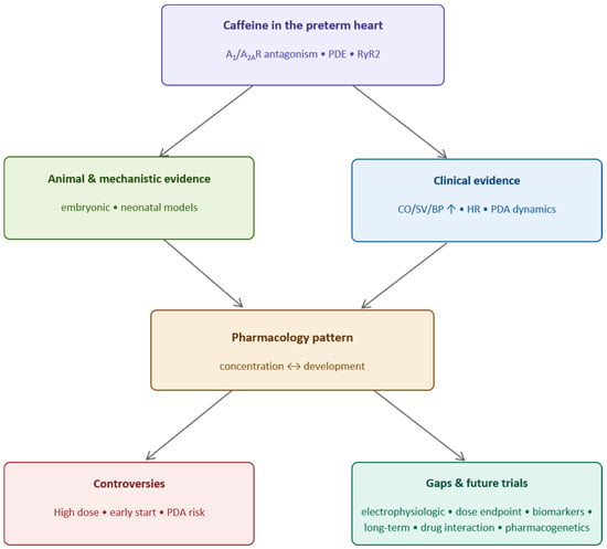
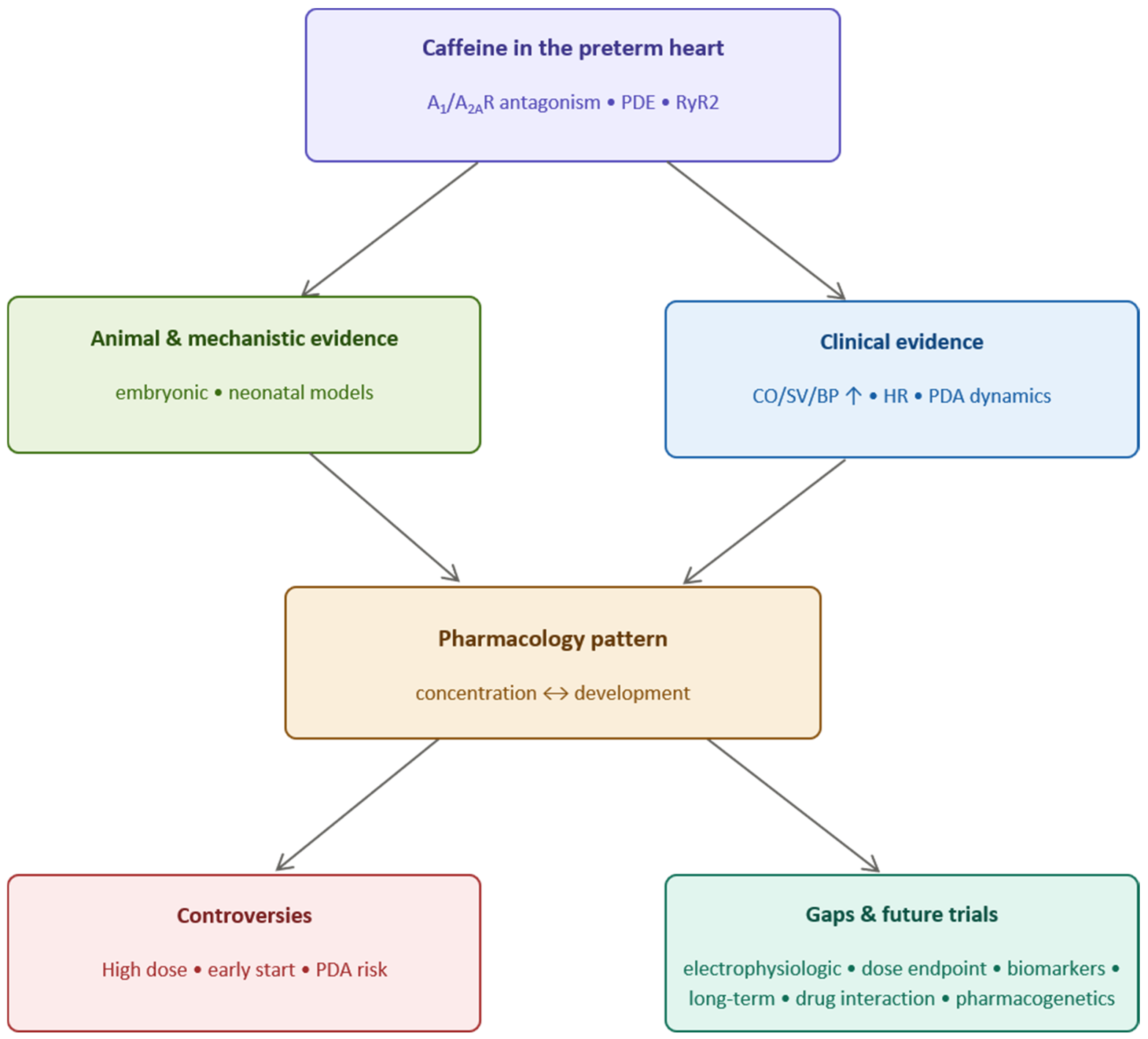
Caffeine has become a fundamental medication in the NICU. However, its cardiac effect is determined by various factors, including dose, developmental stage, and clinical conditions. Animal studies suggest, but do not yet establish, a biphasic pattern of development- and concentration-dependent cardiac responses: (i) during the fetal stage, A1 receptor-dependent developmental heterogeneity; (ii) after birth, a positive chronotropic response after acute administration at therapeutic concentrations, attributed to relief of the tonic inhibitory action of adenosine on nodal conduction and to enhanced β1-adrenergic activity, with the receptor subtype not resolved in neonatal models; (iii) at high concentrations, a risk of arrhythmias for which receptor-independent mechanisms have been proposed but not demonstrated. These inferences are drawn across species and developmental stages rather than from a single experimental framework. For preterm infants, current standard clinical doses have been associated with acute hemodynamic changes—chiefly increased blood pressure and systemic vascular resistance—in small physiological and observational studies, while effects on cardiac output and stroke volume remain inconsistent; whether these physiological changes translate into clinical benefit remains unproven. The primary safety concerns are concentration-dependent tachycardia and transient perfusion abnormalities at the loading dose, particularly in premature infants with PDA. There is still a lack of definitive evidence regarding long-term cardiovascular outcomes. Further animal models and clinical trials, as well as follow-up studies, are needed to fill these gaps. In the application of caffeine in preterm infants, we should place the heart, not merely the lungs, at the core of investigation (Figure 1).
Figure 1.
Description of the search: Caffeine and the preterm heart.
Author Contributions
Conceptualization, Q.W. and T.Y.; methodology, Q.W. and T.Y.; investigation, Q.W. and T.Y.; writing—original draft preparation, Q.W.; writing—review and editing, T.Y. and L.D.; visualization, Q.W.; supervision, L.D. All authors have read and agreed to the published version of the manuscript.
Funding
This research received no external funding.
Institutional Review Board Statement
Not applicable. This article is a narrative review and did not involve human participants or animals.
Informed Consent Statement
Not applicable.
Data Availability Statement
No new data were created or analyzed in this study. Data sharing is not applicable to this article.
Acknowledgments
During the preparation of this article, the authors used GLM (GLM5.3) for the purposes of proofreading and grammar correction. The authors reviewed and edited all outputs and take full responsibility for the content of the publication.
Conflicts of Interest
The authors declare no conflicts of interest.
Abbreviations
The following abbreviations are used in this manuscript:
| NICU | Neonatal intensive care unit |
| BPD | Bronchopulmonary dysplasia |
| CAP | Caffeine for Apnea of Prematurity |
| AOP | Apnea of prematurity |
| PDA | Patent ductus arteriosus |
| ROP | Retinopathy of prematurity |
| g | Grams |
| BW | Birth weight |
| A1R | Adenosine 1 receptor |
| A2AR | Adenosine 2A receptor |
| µM | Micromolar |
| PDE | Phosphodiesterase |
| RyR2 | Ryanodine Receptor type 2 |
| FDA | Food and Drug Administration |
| ELBW | Extremely low birth weight |
| TDM | Therapeutic drug monitoring |
| HR | Heart rate |
| QTc | Corrected QT interval |
| IVH | Intraventricular hemorrhage |
| BNP | B-type Natriuretic Peptide |
| MPI | Myocardial performance index |
| COX | Cyclooxygenase |
| IV | Intravenous |
| TOHRx | Tissue Oxygenation Heart Rate index |
| SVC | Superior Vena Cava |
References
- Aranda, J.V.; Gorman, W.; Bergsteinsson, H.; Gunn, T. Efficacy of caffeine in treatment of apnea in the low-birth-weight infant. J. Pediatr. 1977, 90, 467–472. [Google Scholar] [CrossRef] [Scilit] [PubMed]
- Borys, F.; Wróblewska-Seniuk, K.; Fiander, M.; Soll, R.; Ringsten, M.; Bruschettini, M.; Sibrecht, G. Indications and timings for caffeine initiation in preterm infants. Cochrane Database Syst. Rev. 2025, 11, Cd016274. [Google Scholar] [CrossRef] [Scilit] [PubMed]
- Schmidt, B.; Roberts, R.S.; Davis, P.; Doyle, L.W.; Barrington, K.J.; Ohlsson, A.; Solimano, A.; Tin, W. Caffeine therapy for apnea of prematurity. N. Engl. J. Med. 2006, 354, 2112–2121. [Google Scholar] [CrossRef] [Scilit] [PubMed]
- Szatkowski, L.; Fateh, S.; Abramson, J.; Kwok, T.C.; Sharkey, D.; Budge, H.; Ojha, S. Observational cohort study of use of caffeine in preterm infants and association between early caffeine use and neonatal outcomes. Arch. Dis. Childhood. Fetal Neonatal Ed. 2023, 108, 505–510. [Google Scholar] [CrossRef] [Scilit] [PubMed]
- Neamțu, A.V.; Zlatian, O.M.; Manda, C.V.; Cioboată, R.; Bărbulescu-Mateescu, C.M.; Coteanu, C.; Chiuțu, L.C.; Stanca, L.; Mateescu, O.G.; Neamțu, S.D. Methylxanthines: The Major Impact of Caffeine in Clinical Practice in Patients Diagnosed with Apnea of Prematurity. J. Clin. Med. 2025, 14, 8417. [Google Scholar] [CrossRef] [Scilit] [PubMed]
- Villanueva-García, D.; Mota-Rojas, D.; Miranda-Cortés, A.; Ibarra-Ríos, D.; Casas-Alvarado, A.; Mora-Medina, P.; Martínez-Burnes, J.; Olmos-Hernández, A.; Hernández-Avalos, I. Caffeine: Cardiorespiratory effects and tissue protection in animal models. Exp. Anim. 2021, 70, 431–439. [Google Scholar] [CrossRef] [Scilit] [PubMed]
- Soldin, O.P.; Soldin, S.J. Review: Therapeutic drug monitoring in pediatrics. Ther. Drug Monit. 2002, 24, 1–8. [Google Scholar] [CrossRef] [Scilit] [PubMed]
- Natarajan, G.; Botica, M.L.; Thomas, R.; Aranda, J.V. Therapeutic drug monitoring for caffeine in preterm neonates: An unnecessary exercise? Pediatrics 2007, 119, 936–940. [Google Scholar] [CrossRef] [Scilit] [PubMed]
- Aranda, J.V.; Cook, C.E.; Gorman, W.; Collinge, J.M.; Loughnan, P.M.; Outerbridge, E.W.; Aldridge, A.; Neims, A.H. Pharmacokinetic profile of caffeine in the premature newborn infant with apnea. J. Pediatr. 1979, 94, 663–668. [Google Scholar] [CrossRef] [Scilit] [PubMed]
- Caffeine Citrate Injection and Oral Solution [Prescribing Information]. Sagent Pharmaceuticals; DailyMed, U.S. National Library of Medicine. Available online: https://dailymed.nlm.nih.gov/dailymed/drugInfo.cfm?setid=5f38c395-0093-4afd-89ec-f96e5dc0934a (accessed on 25 October 2026).
- Leng, J.; Yu, J.; Li, Z.; Zhang, Y.; Cui, J. Caffeine and human performance: From molecular mechanisms to exercise and recovery. Front. Physiol. 2026, 17, 1871149. [Google Scholar] [CrossRef] [Scilit] [PubMed]
- Mota-Rojas, D.; Villanueva-García, D.; Hernández-Ávalos, I.; Casas-Alvarado, A.; Domínguez-Oliva, A.; Lezama-García, K.; Miranda-Cortés, A.; Martínez-Burnes, J. Cardiorespiratory and Neuroprotective Effects of Caffeine in Neonate Animal Models. Animals 2023, 13, 1769. [Google Scholar] [CrossRef] [Scilit] [PubMed]
- Moschino, L.; Zivanovic, S.; Hartley, C.; Trevisanuto, D.; Baraldi, E.; Roehr, C.C. Caffeine in preterm infants: Where are we in 2020? ERJ Open Res. 2020, 6, 00330-2019. [Google Scholar] [CrossRef] [Scilit] [PubMed]
- O’Shea, M.; Butler, L.; Holohan, S.; Healy, K.; O’Farrell, R.; Shamit, A.; Cusack, R.; Elhadi, M.; Lynch, S.; Gilcrest, M.; et al. Caffeine and preterm infants: Multiorgan effects and therapeutic creep: Scope to optimise dose and timing. Pediatr. Res. 2026, 99, 462–469. [Google Scholar] [CrossRef] [Scilit] [PubMed]
- He, Y.; Shen, X.; Zhu, J.; Zhang, L.; Wang, X.; Zhou, T.; Zhang, J.; Li, W.; Fan, X. A comprehensive review of caffeine population pharmacokinetics in preterm infants: Factors affecting clearance. Eur. J. Pharm. Biopharm. 2025, 208, 114659. [Google Scholar] [CrossRef] [Scilit] [PubMed]
- Wang, M.C.; Sealey, V.A.; Rahman, M.; Ngugama, M.; Trolat, A.; Wybenga-Groot, L.; Moran, M.F.; Dinh, J.; Lewis, T.R. Developmental age and clinical illness drive in vivo variability of CYP1A2 ontogeny and caffeine metabolism in preterm neonates. Front. Pharmacol. 2026, 17, 1823969. [Google Scholar] [CrossRef] [Scilit] [PubMed]
- Neamțu, A.V.; Zlatian, O.M.; Bălășoiu, A.T.; Manda, C.V.; Bărbulescu-Mateescu, C.M.; Pătrașcu, A.M.; Cioboată, R.; Stanca, L.; Mateescu, O.G.; Pătrașcu, Ș.; et al. Clinical and biological impact of caffeine therapy in premature infants with apnea of prematurity: A prospective observational study. Front. Med. 2026, 13, 1808401. [Google Scholar] [CrossRef] [Scilit] [PubMed]
- Hari Gopal, S.; Parmekar, S.; Dempsey, E.; Pammi, M. Caring for the smallest hearts: Cardiovascular phenotypes and assessment in tiny babies. Pediatr. Res. 2026; in press. [CrossRef] [Scilit] [PubMed]
- Burnstock, G.; Dale, N. Purinergic signalling during development and ageing. Purinergic Signal. 2015, 11, 277–305. [Google Scholar] [CrossRef] [Scilit] [PubMed]
- Ramaswamy, V.V.; Kumar, G.; Pullattayil, S.A.; Aradhya, A.S.; Suryawanshi, P.; Sahni, M.; Khurana, S.; More, K. Active versus restrictive approach to isolated hypotension in preterm neonates: A Systematic Review, Meta-analysis and GRADE based Clinical Practice Guideline. PLoS ONE 2025, 20, e0309520. [Google Scholar] [CrossRef] [Scilit] [PubMed]
- Blixenkrone-Møller, E.; Dannesbo, S.; Dehn, A.M.; Pihl, C.A.; Sillesen, A.S.; Vøgg, R.O.B.; Raja, A.A.; Colan, S.; Mertens, L.; Vejlstrup, N.; et al. Interatrial Communications: Prevalence and Subtypes in 12,385 Newborns-a Copenhagen Baby Heart Study. Pediatr. Cardiol. 2025, 46, 1570–1580. [Google Scholar] [CrossRef] [Scilit] [PubMed]
- Buscariollo, D.L.; Breuer, G.A.; Wendler, C.C.; Rivkees, S.A. Caffeine acts via A1 adenosine receptors to disrupt embryonic cardiac function. PLoS ONE 2011, 6, e28296. [Google Scholar] [CrossRef] [Scilit] [PubMed]
- Rivkees, S.A.; Wendler, C.C. Regulation of cardiovascular development by adenosine and adenosine-mediated embryo protection. Arterioscler. Thromb. Vasc. Biol. 2012, 32, 851–855. [Google Scholar] [CrossRef] [Scilit] [PubMed]
- Wendler, C.C.; Busovsky-McNeal, M.; Ghatpande, S.; Kalinowski, A.; Russell, K.S.; Rivkees, S.A. Embryonic caffeine exposure induces adverse effects in adulthood. FASEB J. 2009, 23, 1272–1278. [Google Scholar] [CrossRef] [Scilit] [PubMed]
- Rivkees, S.A.; Wendler, C.C. Adverse and protective influences of adenosine on the newborn and embryo: Implications for preterm white matter injury and embryo protection. Pediatr. Res. 2011, 69, 271–278. [Google Scholar] [CrossRef] [Scilit] [PubMed]
- James, J.E. Maternal Caffeine Consumption and Pregnancy Outcomes. Adv. Exp. Med. Biol. 2026, 1500, 303–336. [Google Scholar] [CrossRef] [Scilit] [PubMed]
- Lakin, H.; Sheehan, P.; Soti, V. Maternal Caffeine Consumption and Its Impact on the Fetus: A Review. Cureus 2023, 15, e48266. [Google Scholar] [CrossRef] [Scilit] [PubMed]
- Basnet, R.M.; Zizioli, D.; Guarienti, M.; Finazzi, D.; Memo, M. Methylxanthines induce structural and functional alterations of the cardiac system in zebrafish embryos. BMC Pharmacol. Toxicol. 2017, 18, 72. [Google Scholar] [CrossRef] [Scilit] [PubMed]
- Rana, N.; Moond, M.; Marthi, A.; Bapatla, S.; Sarvepalli, T.; Chatti, K.; Challa, A.K. Caffeine-induced effects on heart rate in zebrafish embryos and possible mechanisms of action: An effective system for experiments in chemical biology. Zebrafish 2010, 7, 69–81. [Google Scholar] [CrossRef] [Scilit] [PubMed]
- Temples, T.E.; Geoffray, D.J.; Nakamoto, T.; Hartman, A.D.; Miller, H.I. Effects of chronic caffeine ingestion on growth and myocardial function. Proc. Soc. Exp. Biol. Med. 1985, 179, 388–395. [Google Scholar] [CrossRef] [Scilit] [PubMed]
- Vornanen, M. Effects of caffeine on the mechanical properties of developing rat heart ventricles. Comp. Biochem. Physiol. Part C Comp. Pharmacol. 1984, 78, 329–334. [Google Scholar] [CrossRef] [Scilit] [PubMed]
- Bienboire-Frosini, C.; Muns, R.; Marcet-Rius, M.; Gazzano, A.; Villanueva-García, D.; Martínez-Burnes, J.; Domínguez-Oliva, A.; Lezama-García, K.; Casas-Alvarado, A.; Mota-Rojas, D. Vitality in Newborn Farm Animals: Adverse Factors, Physiological Responses, Pharmacological Therapies, and Physical Methods to Increase Neonate Vigor. Animals 2023, 13, 1542. [Google Scholar] [CrossRef] [Scilit] [PubMed]
- Sánchez-Salcedo, J.A.; Orozco-Gregorio, H.; González-Lozano, M.; Roldán-Santiago, P.; González-Hernández, M.; Ballesteros-Rodea, G.; Bonilla-Jaime, H. Caffeine administered to pregnant sows improves piglet vitality, gas exchange and body weight gain. Anim. Reprod. Sci. 2019, 208, 106120. [Google Scholar] [CrossRef] [Scilit] [PubMed]
- Mielgo, V.; Catozzi, C.; Ricci, F.; Villetti, G.; Gomez-Solaetxe, M.A.; Rey-Santano, C. Caffeine Protects Newborn Rat Lungs Against the Effects of Hyperoxia and Intra-Amniotic Enterotoxin. Pediatr. Pulmonol. 2025, 60, e71395. [Google Scholar] [CrossRef] [Scilit] [PubMed]
- Temples, T.E.; Geoffray, D.J.; Nakamoto, T.; Hartman, A.D.; Miller, H.I. Effect of chronic caffeine intake on myocardial function during early growth. Pediatr. Res. 1987, 21, 391–395. [Google Scholar] [CrossRef] [Scilit] [PubMed]
- Yang, J.N.; Chen, J.F.; Fredholm, B.B. Physiological roles of A1 and A2A adenosine receptors in regulating heart rate, body temperature, and locomotion as revealed using knockout mice and caffeine. Am. J. Physiol. Heart Circ. Physiol. 2009, 296, H1141–H1149. [Google Scholar] [CrossRef] [Scilit] [PubMed]
- Lankford, A.R.; Yang, J.N.; Rose’Meyer, R.; French, B.A.; Matherne, G.P.; Fredholm, B.B.; Yang, Z. Effect of modulating cardiac A1 adenosine receptor expression on protection with ischemic preconditioning. Am. J. Physiol. Heart Circ. Physiol. 2006, 290, H1469–H1473. [Google Scholar] [CrossRef] [Scilit] [PubMed]
- Walther, F.J.; Erickson, R.; Sims, M.E. Cardiovascular effects of caffeine therapy in preterm infants. Am. J. Dis. Child. 1990, 144, 1164–1166. [Google Scholar] [CrossRef] [Scilit] [PubMed]
- Soloveychik, V.; Bin-Nun, A.; Ionchev, A.; Sriram, S.; Meadow, W. Acute hemodynamic effects of caffeine administration in premature infants. J. Perinatol. Off. J. Calif. Perinat. Assoc. 2009, 29, 205–208. [Google Scholar] [CrossRef] [Scilit] [PubMed]
- Katheria, A.C.; Sauberan, J.B.; Akotia, D.; Rich, W.; Durham, J.; Finer, N.N. A Pilot Randomized Controlled Trial of Early versus Routine Caffeine in Extremely Premature Infants. Am. J. Perinatol. 2015, 32, 879–886. [Google Scholar] [CrossRef] [Scilit] [PubMed]
- Parladori, R.; Austin, T.; Smielewski, P.; Czosnyka, M.; Paoletti, V.; Vitali, F.; Corvaglia, L.; Martini, S. Cardiovascular and cerebrovascular effects of caffeine maintenance in preterm infants during the transitional period. Pediatr. Res. 2024, 96, 1267–1274. [Google Scholar] [CrossRef] [Scilit] [PubMed]
- Shivakumar, M.; Nayak, K.; Lewis, L.E.S.; Kamath, A.; Purkayastha, J. Acute hemodynamic effects of methylxanthine therapy in preterm neonates: Effect of variations in subgroups. J. Trop. Pediatr. 2019, 65, 264–272. [Google Scholar] [CrossRef] [Scilit] [PubMed]
- Amponsah, S.K.; Nartey, C.M.; Ofori, E.K. The Use of Caffeine Citrate in the Management of Neonatal Apnea in Low- and Middle-Income Countries: A Rapid Systematic Review. Health Sci. Rep. 2025, 8, e70486. [Google Scholar] [CrossRef] [Scilit] [PubMed]
- Yu, T.; Balch, A.H.; Ward, R.M.; Korgenski, E.K.; Sherwin, C.M. Incorporating pharmacodynamic considerations into caffeine therapeutic drug monitoring in preterm neonates. BMC Pharmacol. Toxicol. 2016, 17, 22. [Google Scholar] [CrossRef] [Scilit] [PubMed]
- Alhersh, E.; Abushanab, D.; Al-Shaibi, S.; Al-Badriyeh, D. Caffeine for the Treatment of Apnea in the Neonatal Intensive Care Unit: A Systematic Overview of Meta-Analyses. Paediatr. Drugs 2020, 22, 399–408. [Google Scholar] [CrossRef] [Scilit] [PubMed]
- Mohd Kori, A.M.; Van Rostenberghe, H.; Ibrahim, N.R.; Yaacob, N.M.; Nasir, A. A Randomized Controlled Trial Comparing Two Doses of Caffeine for Apnoea in Prematurity. Int. J. Environ. Res. Public Health 2021, 18, 4509. [Google Scholar] [CrossRef] [Scilit] [PubMed]
- Huvanandana, J.; Thamrin, C.; McEwan, A.L.; Hinder, M.; Tracy, M.B. Cardiovascular impact of intravenous caffeine in preterm infants. Acta Paediatr. 2019, 108, 423–429. [Google Scholar] [CrossRef] [Scilit] [PubMed]
- Ulanovsky, I.; Haleluya, N.S.; Blazer, S.; Weissman, A. The effects of caffeine on heart rate variability in newborns with apnea of prematurity. J. Perinatol. Off. J. Calif. Perinat. Assoc. 2014, 34, 620–623. [Google Scholar] [CrossRef] [Scilit] [PubMed]
- Anderson, B.J.; Gunn, T.R.; Holford, N.H.; Johnson, R. Caffeine overdose in a premature infant: Clinical course and pharmacokinetics. Anaesth. Intensive Care 1999, 27, 307–311. [Google Scholar] [CrossRef] [Scilit] [PubMed]
- Ergenekon, E.; Dalgiç, N.; Aksoy, E.; Koç, E.; Atalay, Y. Caffeine intoxication in a premature neonate. Paediatr. Anaesth. 2001, 11, 737–739. [Google Scholar] [CrossRef] [Scilit] [PubMed]
- Long, J.Y.; Guo, H.L.; He, X.; Hu, Y.H.; Xia, Y.; Cheng, R.; Ding, X.S.; Chen, F.; Xu, J. Caffeine for the Pharmacological Treatment of Apnea of Prematurity in the NICU: Dose Selection Conundrum, Therapeutic Drug Monitoring and Genetic Factors. Front. Pharmacol. 2021, 12, 681842. [Google Scholar] [CrossRef] [Scilit] [PubMed]
- Lodha, A.; Entz, R.; Synnes, A.; Creighton, D.; Yusuf, K.; Lapointe, A.; Yang, J.; Shah, P.S. Early Caffeine Administration and Neurodevelopmental Outcomes in Preterm Infants. Pediatrics 2019, 143, e20181348. [Google Scholar] [CrossRef] [Scilit] [PubMed]
- Crockett, S.L.; Su, R.L.; Sekulich, D.C.; Goins, K.A.; Montero, A.; Boatwright, N.; Berger, C.D.; Yarboro, M.T.; Paria, B.C.; Herington, J.L.; et al. Caffeine-associated reduction in patent ductus arteriosus is mediated in part by adenosine receptor antagonism. Am. J. Physiol. Heart Circ. Physiol. 2025, 329, H1548–H1559. [Google Scholar] [CrossRef] [Scilit] [PubMed]
- Kua, K.P.; Lee, S.W. Systematic review and meta-analysis of clinical outcomes of early caffeine therapy in preterm neonates. Br. J. Clin. Pharmacol. 2017, 83, 180–191. [Google Scholar] [CrossRef] [Scilit] [PubMed]
- Hwang, J.; Kim, Y.S.; Shin, J.H.; Choi, B.M. Hemodynamic Effects on Systemic Blood Flow and Ductal Shunting Flow after Loading Dose of Intravenous Caffeine in Preterm Infants according to the Patency of Ductus Arteriosus. J. Korean Med. Sci. 2018, 33, e25. [Google Scholar] [CrossRef] [Scilit] [PubMed]
- Hoecker, C.; Nelle, M.; Poeschl, J.; Beedgen, B.; Linderkamp, O. Caffeine impairs cerebral and intestinal blood flow velocity in preterm infants. Pediatrics 2002, 109, 784–787. [Google Scholar] [CrossRef] [Scilit] [PubMed]
- Hoecker, C.; Nelle, M.; Beedgen, B.; Rengelshausen, J.; Linderkamp, O. Effects of a divided high loading dose of caffeine on circulatory variables in preterm infants. Arch. Dis. Childhood. Fetal Neonatal Ed. 2006, 91, F61–F64. [Google Scholar] [CrossRef] [Scilit] [PubMed]
- Bruschettini, M.; Brattström, P.; Russo, C.; Onland, W.; Davis, P.G.; Soll, R. Caffeine dosing regimens in preterm infants with or at risk for apnea of prematurity. Cochrane Database Syst. Rev. 2023, 4, Cd013873. [Google Scholar] [CrossRef] [Scilit] [PubMed]
- Rub, D.M.; Eichenwald, E.C. What is the Role of Caffeine in the Management of Preterm Infants? Curr. Treat. Options Pediatr. 2025, 11, 16. [Google Scholar] [CrossRef] [Scilit] [PubMed]
- Kelly, C.E.; Ooi, W.L.; Yang, J.Y.; Chen, J.; Adamson, C.; Lee, K.J.; Cheong, J.L.Y.; Anderson, P.J.; Doyle, L.W.; Thompson, D.K. Caffeine for apnea of prematurity and brain development at 11 years of age. Ann. Clin. Transl. Neurol. 2018, 5, 1112–1127. [Google Scholar] [CrossRef] [Scilit] [PubMed]
- Rorabaugh, B.R. Does Prenatal Exposure to CNS Stimulants Increase the Risk of Cardiovascular Disease in Adult Offspring? Front. Cardiovasc. Med. 2021, 8, 652634. [Google Scholar] [CrossRef] [Scilit] [PubMed]
- Souza, L.L.; Moura, E.G.; Lisboa, P.C. Can mothers consume caffeine? The issue of early life exposure and metabolic changes in offspring. Toxicol. Lett. 2024, 393, 96–106. [Google Scholar] [CrossRef] [Scilit] [PubMed]
- Fang, X.; Poulsen, R.R.; Rivkees, S.A.; Wendler, C.C. In Utero Caffeine Exposure Induces Transgenerational Effects on the Adult Heart. Sci. Rep. 2016, 6, 34106. [Google Scholar] [CrossRef] [Scilit] [PubMed]
- Borszewska-Kornacka, M.K.; Hożejowski, R.; Rutkowska, M.; Lauterbach, R. Shifting the boundaries for early caffeine initiation in neonatal practice: Results of a prospective, multicenter study on very preterm infants with respiratory distress syndrome. PLoS ONE 2017, 12, e0189152. [Google Scholar] [CrossRef] [Scilit] [PubMed]
- Chen, J.; Jin, L.; Chen, X. Efficacy and Safety of Different Maintenance Doses of Caffeine Citrate for Treatment of Apnea in Premature Infants: A Systematic Review and Meta-Analysis. BioMed Res. Int. 2018, 2018, 9061234. [Google Scholar] [CrossRef] [Scilit] [PubMed]
- Vliegenthart, R.; Miedema, M.; Hutten, G.J.; van Kaam, A.H.; Onland, W. High versus standard dose caffeine for apnoea: A systematic review. Arch. Dis. Child. Fetal Neonatal Ed. 2018, 103, F523–F529. [Google Scholar] [CrossRef] [Scilit] [PubMed]
- McPherson, C.; Neil, J.J.; Tjoeng, T.H.; Pineda, R.; Inder, T.E. A pilot randomized trial of high-dose caffeine therapy in preterm infants. Pediatr. Res. 2015, 78, 198–204. [Google Scholar] [CrossRef] [Scilit] [PubMed]
- McPherson, C.; Lean, R.E.; Cyr, P.E.P.; Inder, T.E.; Rogers, C.E.; Smyser, C.D. Five-year outcomes of premature infants randomized to high or standard loading dose caffeine. J. Perinatol. Off. J. Calif. Perinat. Assoc. 2022, 42, 631–635. [Google Scholar] [CrossRef] [Scilit] [PubMed]
- Firman, B.; Molnar, A.; Gray, P.H. Early high-dose caffeine citrate for extremely preterm infants: Neonatal and neurodevelopmental outcomes. J. Paediatr. Child Health 2019, 55, 1451–1457. [Google Scholar] [CrossRef] [Scilit] [PubMed]
- Dobson, N.R.; Patel, R.M.; Smith, P.B.; Kuehn, D.R.; Clark, J.; Vyas-Read, S.; Herring, A.; Laughon, M.M.; Carlton, D.; Hunt, C.E. Trends in caffeine use and association between clinical outcomes and timing of therapy in very low birth weight infants. J. Pediatr. 2014, 164, 992–998.e3. [Google Scholar] [CrossRef] [Scilit] [PubMed]
- Amaro, C.M.; Bello, J.A.; Jain, D.; Ramnath, A.; D’Ugard, C.; Vanbuskirk, S.; Bancalari, E.; Claure, N. Early Caffeine and Weaning from Mechanical Ventilation in Preterm Infants: A Randomized, Placebo-Controlled Trial. J. Pediatr. 2018, 196, 52–57. [Google Scholar] [CrossRef] [Scilit] [PubMed]

Disclaimer/Publisher’s Note: The statements, opinions and data contained in all publications are solely those of the individual author(s) and contributor(s) and not of MDPI and/or the editor(s). MDPI and/or the editor(s) disclaim responsibility for any injury to people or property resulting from any ideas, methods, instructions or products referred to in the content. |
© 2026 by the authors. Licensee MDPI, Basel, Switzerland. This article is an open access article distributed under the terms and conditions of the Creative Commons Attribution (CC BY) license.