


Highlights
What are the main findings?
- Sequential mapping identified three spatially separated right-sided accessory pathway sites in a child with Wolff–Parkinson–White syndrome.
- Dynamic ECG changes and stepwise QRS shifts after ablation were consistent with changing accessory-pathway dominance.
What are the implications of the main findings?
- A change in QRS morphology after an ablation lesion may indicate residual accessory pathway conduction.
- Sequential mapping, immediate ECG reassessment, and remapping may help identify additional ablation targets in pediatric WPW syndrome.
Abstract
Background: Multiple accessory pathways are uncommon in pediatric Wolff–Parkinson–White syndrome and may cause dynamic changes in ventricular pre-excitation, complicating electrocardiographic localization. Case presentation: A 7-year-old girl with recurrent palpitations and syncope exhibited variable QRS morphology and delta-wave patterns. Electrophysiological findings were consistent with orthodromic atrioventricular reentrant tachycardia. Sequential CARTO mapping identified three spatially separated right-sided accessory pathway sites. Ablation of AP1 (7 o’clock, tricuspid annulus) and AP2 (11 o’clock, tricuspid annulus) produced successive QRS morphological changes without eliminating pre-excitation. AP3 was localized just superior to the coronary sinus ostium at approximately the 4 o’clock position, where ablation eliminated pre-excitation. No tachycardia was inducible immediately afterward. However, pre-excitation reappeared on ECG at 1-month follow-up and persisted at 3 months, with two patient-reported tachycardia episodes but no recurrent syncope. Conclusions: Dynamic pre-excitation patterns may suggest multiple accessory pathways. Sequential mapping and electrocardiographic reassessment can help identify residual pathway conduction, although acute procedural success does not ensure sustained elimination of pre-excitation.
1. Introduction
Wolff–Parkinson–White (WPW) syndrome results from atrioventricular conduction through accessory pathways and may cause orthodromic or antidromic atrioventricular reentrant tachycardia (AVRT), ventricular dyssynchrony during sinus rhythm, and rapid ventricular responses during atrial fibrillation or atrial tachycardia [1]. These manifestations can lead to palpitations, syncope, and, rarely, life-threatening events. Catheter ablation is an established treatment for symptomatic pediatric WPW syndrome, with three-dimensional electroanatomical mapping facilitating pathway localization [2].
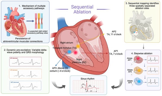
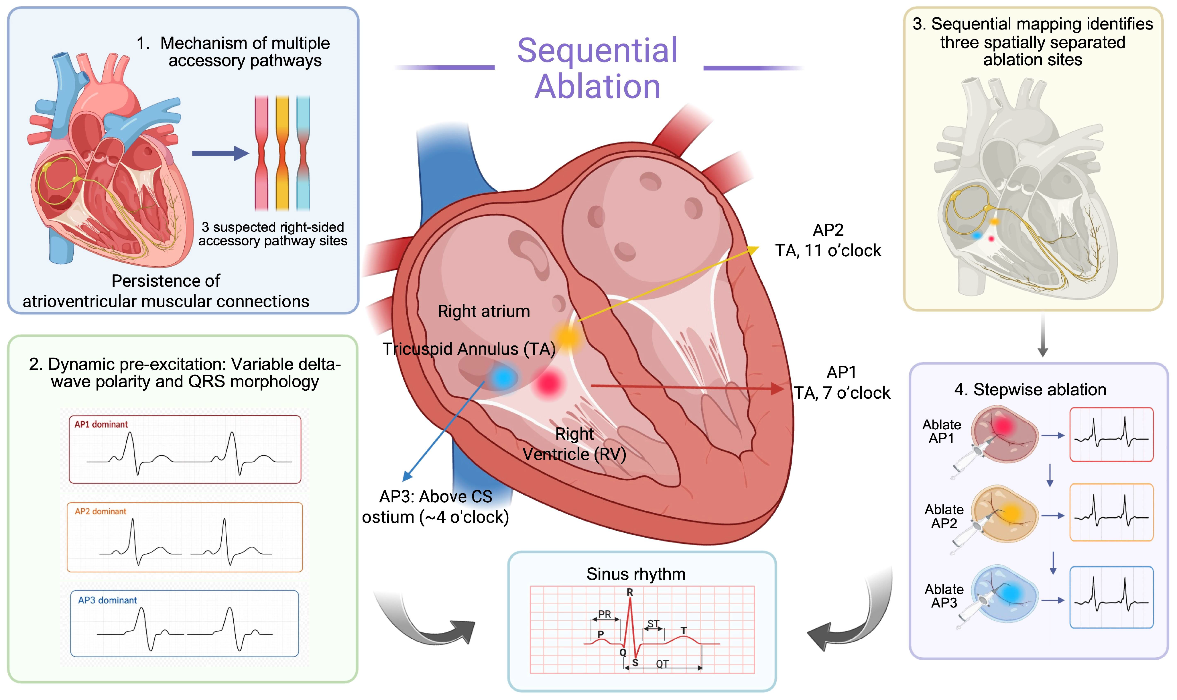
Multiple accessory pathways are uncommon but clinically important in pediatric WPW syndrome. Their coexistence may produce dynamic changes in delta-wave and QRS morphology as the relative contribution of each pathway changes, complicating electrocardiographic localization [3,4]. We report a 7-year-old girl with dynamic pre-excitation and sequential changes in QRS morphology during ablation. This case highlights the value of repeated ECG assessment and electroanatomical remapping in detecting residual conduction through additional pathways (Figure 1).
Figure 1.
Graphical overview of the electrophysiological concept and procedural strategy. Multiple atrioventricular muscular connections can produce competing patterns of ventricular pre-excitation, with different right-sided accessory pathways contributing variably to QRS morphology and delta-wave configuration. Sequential mapping localized AP1 to the 7 o’clock position of the tricuspid annulus, AP2 to the 11 o’clock position of the tricuspid annulus, and AP3 just superior to the coronary sinus ostium at approximately the 4 o’clock position. Sequential ablation with immediate reassessment after each lesion allowed residual accessory pathway conduction to be recognized and treated until pre-excitation was acutely eliminated.
2. Case Presentation
A 7-year-old girl was admitted with a 6-year history of recurrent paroxysmal palpitations and a recent episode of syncope. The episodes had an abrupt onset and termination, lasted from several minutes to several hours, and were accompanied by dizziness, chest discomfort, nausea, vomiting, and limb weakness. Five days before admission, an episode of tachycardia was accompanied by pallor and transient loss of consciousness lasting approximately 30 s. There was no seizure-like activity, cyanosis, fever, cough, or dyspnea. Her growth and development had been normal, and she had no known structural heart disease, previous cardiac surgery, or a family history of sudden cardiac death.
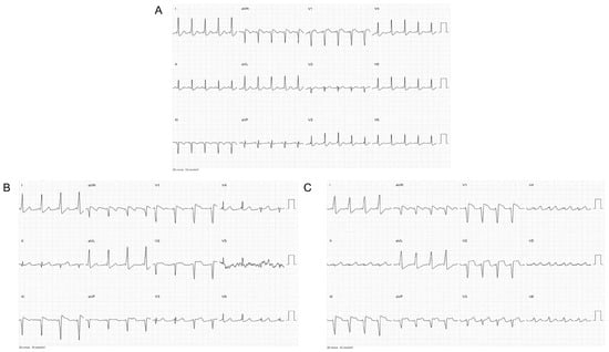
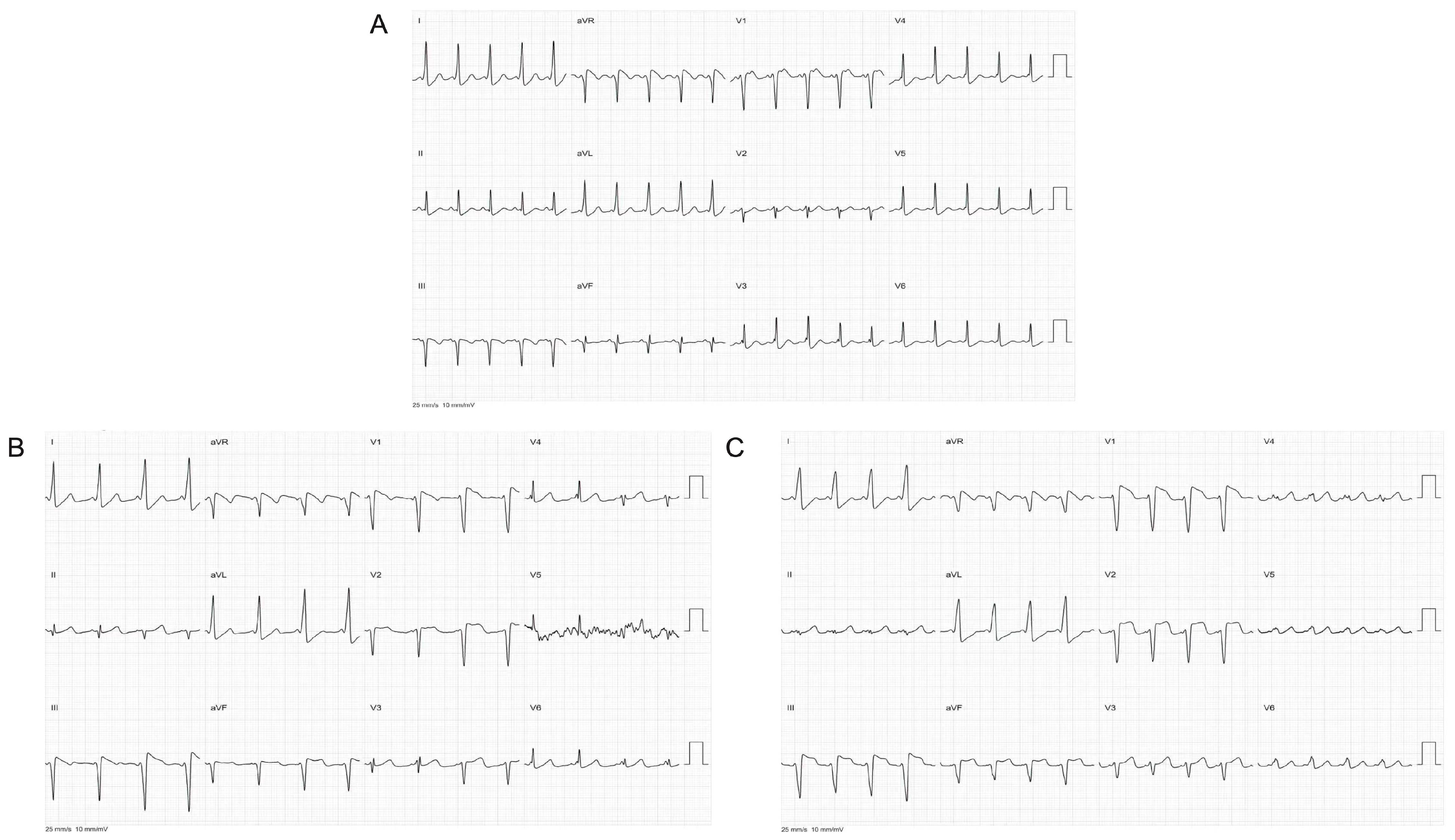
Serial 12-lead electrocardiograms (ECGs) showed sinus rhythm with manifest ventricular pre-excitation, characterized by a short PR interval, widened QRS complexes, and delta waves. The pre-excitation morphology varied across recordings, with marked changes in lead V1 and the inferior leads (Figure 2). Although these changes raised suspicion for multiple right-sided accessory pathways, surface ECGs alone could not establish the number or anatomical locations of the pathways. Transthoracic echocardiography showed normal cardiac structure and function, and routine laboratory findings were unremarkable.
Figure 2.
Representative serial 12-lead electrocardiograms showing dynamic ventricular pre-excitation. (A) Initial ECG demonstrating ventricular pre-excitation. (B) Subsequent ECG showing changes in QRS morphology and delta-wave configuration. (C) Further ECG changes, particularly in lead V1 and the inferior leads, raising suspicion of multiple right-sided accessory pathways and prompting electrophysiological evaluation.
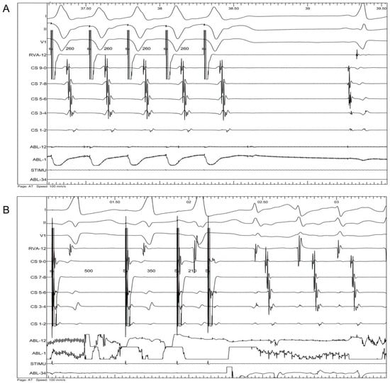
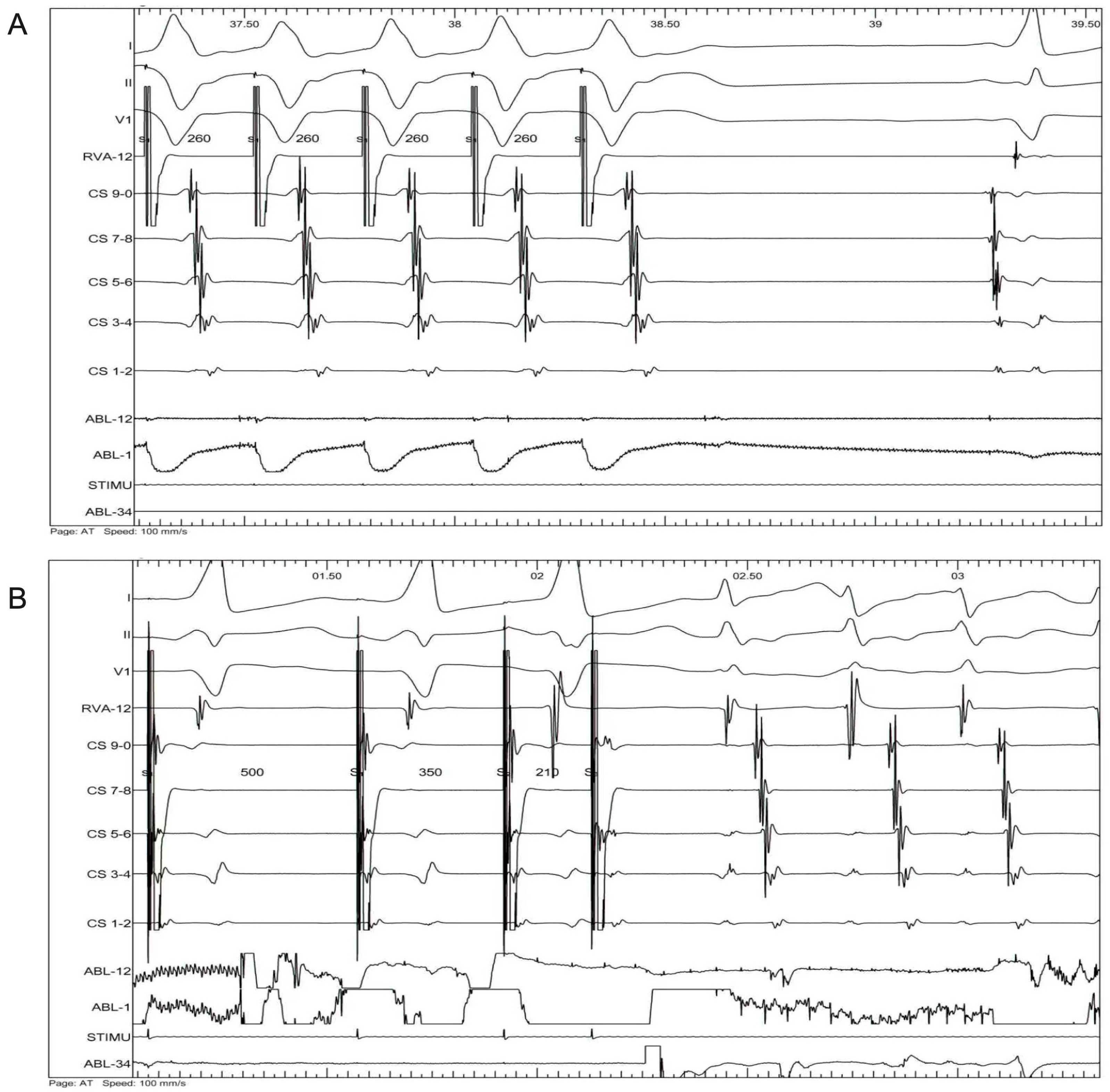
An electrophysiological study was performed under general anesthesia, with a decapolar catheter in the coronary sinus and a quadripolar catheter in the right ventricular apex. Incremental ventricular pacing (500–260 ms) maintained 1:1 ventriculoatrial (VA) conduction. Programmed atrial stimulation using an S1/S2/S3 sequence at 500/350/210 ms induced brief tachycardia with a cycle length of approximately 285 ms (Figure 3). The earliest retrograde atrial activation was near the proximal coronary sinus during both ventricular pacing and tachycardia. These findings were consistent with orthodromic atrioventricular reentrant tachycardia (AVRT), although the retrograde accessory pathway could not be identified because pathway-specific retrograde mapping was not performed. The tachycardia terminated spontaneously, precluding assessment of hemodynamic tolerance during sustained tachycardia. Pathway-specific antegrade effective refractory periods could not be determined; retrograde effective refractory periods were not measured. The shortest pre-excited RR interval during atrial fibrillation (SPERRI) was unavailable because atrial fibrillation was not induced. Baseline AH and HV intervals were unavailable from the procedural records, and the VA interval during induced tachycardia was not measured.
Figure 3.
Electrophysiological findings before ablation. (A) Incremental ventricular pacing down to a cycle length of 260 ms demonstrated 1:1 ventriculoatrial conduction, with the earliest retrograde atrial activation near the proximal coronary sinus. (B) Programmed atrial stimulation at 500/350/210 ms induced tachycardia consistent with orthodromic atrioventricular reentrant tachycardia (AVRT), with a tachycardia cycle length (TCL) of 280–290 ms. The earliest retrograde atrial activation was again recorded near the proximal coronary sinus.
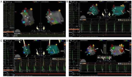
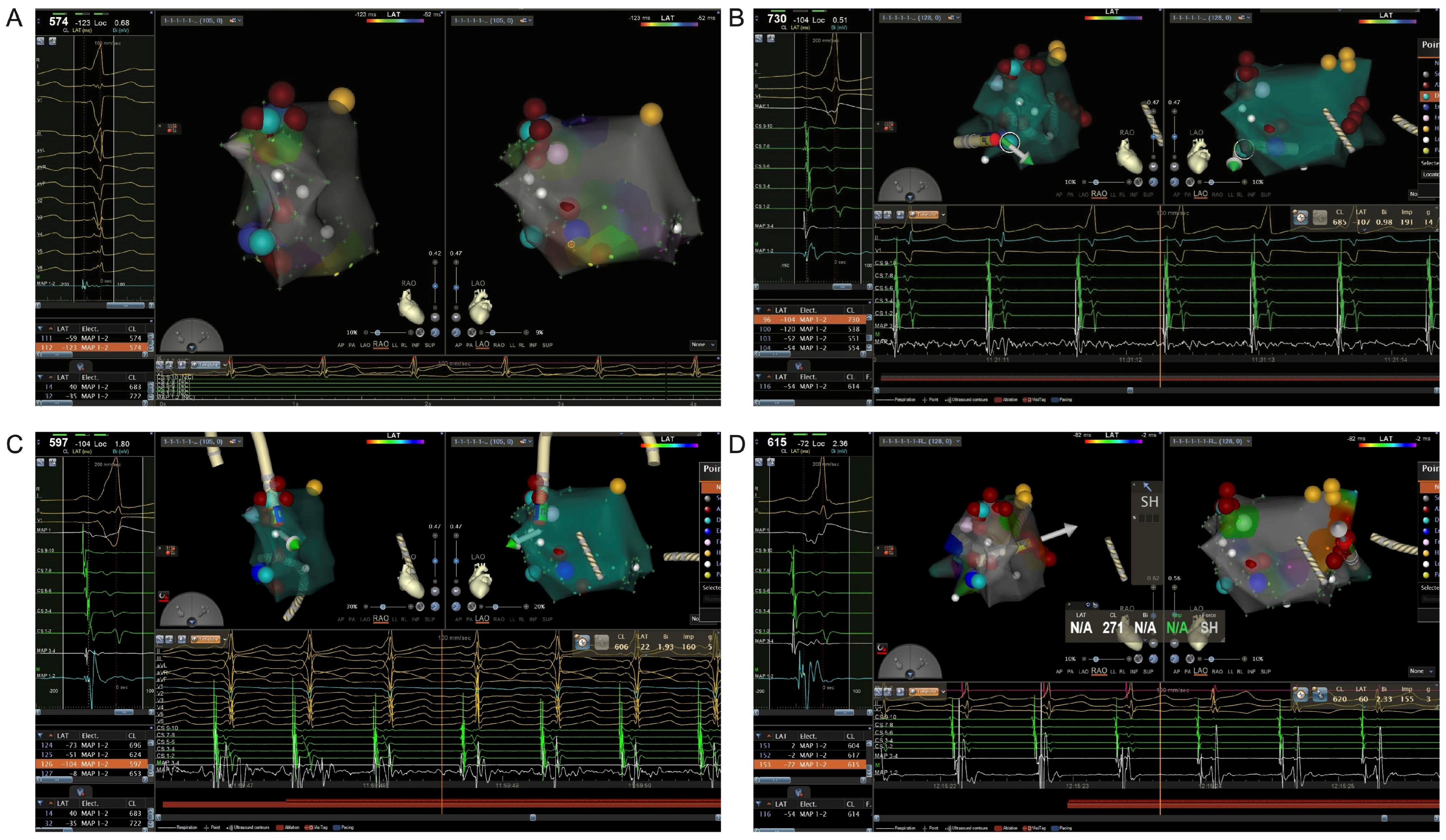
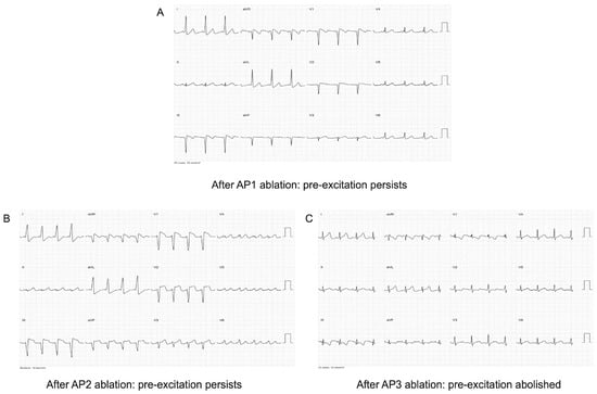
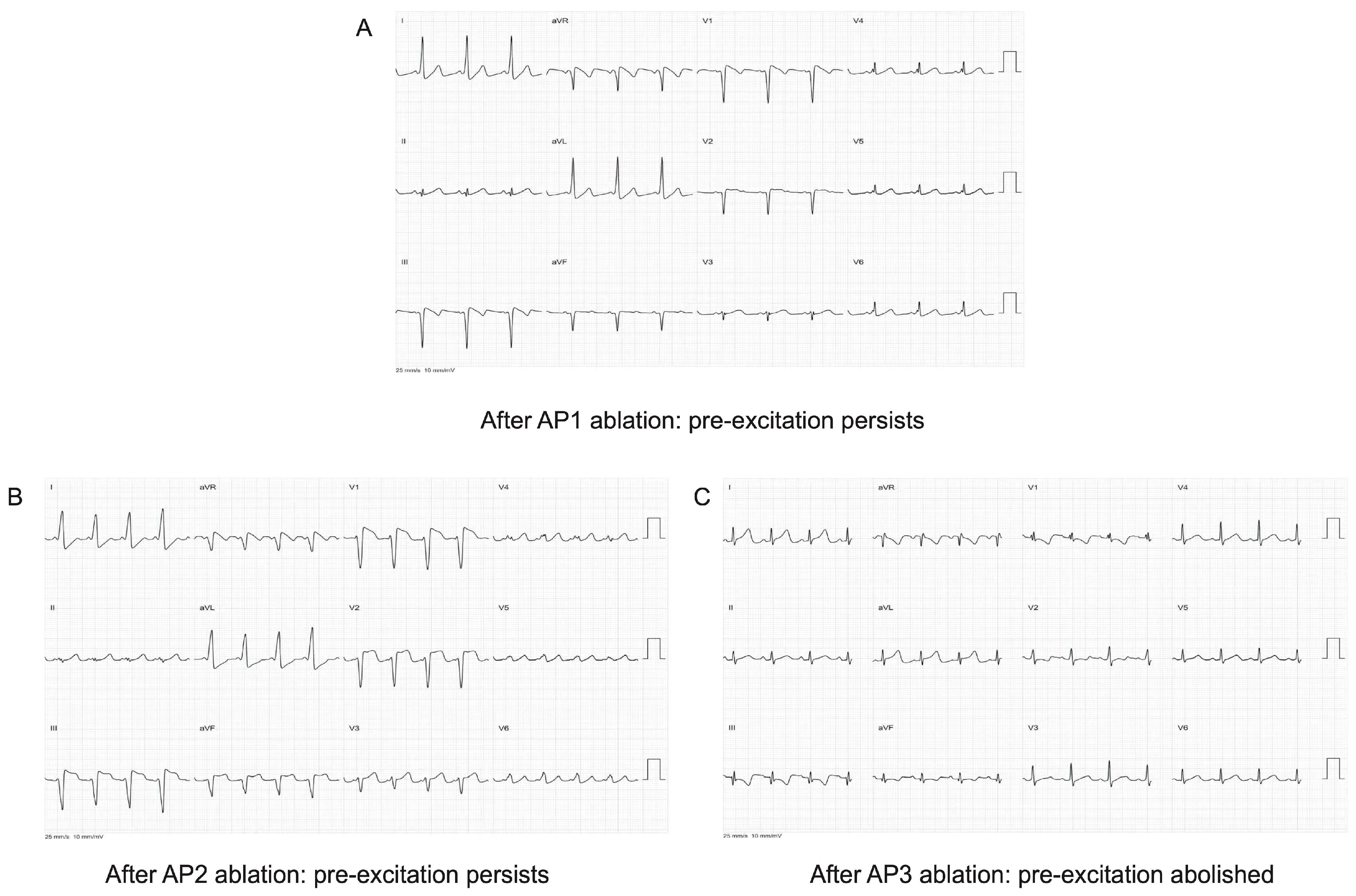
An 8-F, 3.5-mm-tip, contact force-sensing irrigated radiofrequency ablation catheter (THERMOCOOL® SMARTTOUCH®, ST; Biosense Webster, Johnson & Johnson, Irvine, CA, USA) was used with the CARTO three-dimensional electroanatomical mapping system. The catheter featured six electrodes (one distal ablation electrode and five ring electrodes), with an electrode spacing configuration of 1–6–2 mm. It had an effective length of 115 cm, bidirectional deflection, and contact force monitoring capability. The three sites were designated AP1–AP3 in the order identified and treated. Mapping was performed during sinus rhythm, targeting the earliest local ventricular activation. AP1 was located at the 7 o’clock position of the tricuspid annulus, where the local ventricular electrogram preceded the R-wave peak in lead II by 37 ms. Radiofrequency energy was delivered at 35 W, with a temperature limit of 40 °C and an irrigation flow rate of 17 mL/min. After 5 s, the main QRS deflection in lead II changed from negative to positive, but pre-excitation persisted. Total radiofrequency application time, including consolidation lesions, was approximately 120 s, after which the tricuspid annulus was remapped.
Remapping identified AP2 at the 11 o’clock position of the tricuspid annulus, where the local ventricular electrogram preceded the R-wave peak in lead II by 39 ms. Ablation using the same settings as AP1 changed the QRS morphology in lead V1 from rS to QS, but pre-excitation persisted. Total radiofrequency application time, including consolidation lesions, was approximately 120 s. After AP2 ablation, right ventricular pacing at a cycle length of 260 ms demonstrated ventriculoatrial dissociation. AP3 was located just superior to the coronary sinus ostium at approximately the 4 o’clock position, where the local ventricular electrogram preceded the R-wave peak in lead II by 35 ms. Radiofrequency energy was delivered at 30 W, with a temperature limit of 50 °C. The irrigation flow rate was 2 mL/min at baseline and was increased to 30 mL/min during radiofrequency ablation. Pre-excitation disappeared 3 s after radiofrequency application began. Total radiofrequency application time, including consolidation lesions, was approximately 120 s. Individual application durations, lesion counts, and actual contact-force values were unavailable. At all three sites, local atrial and ventricular electrograms were fused before ablation and separated afterward, with no discrete accessory pathway potentials recorded. Sequential CARTO activation maps and corresponding ECG changes are shown in Figure 4 and Figure 5, respectively.
Figure 4.
Sequential CARTO electroanatomical mapping and ablation at three spatially separated right-sided accessory pathway sites. (A) Initial activation mapping during sinus rhythm identified AP1 at the 7 o’clock position of the tricuspid annulus. (B) Following AP1 ablation, repeat activation mapping identified AP2 at the 11 o’clock position of the tricuspid annulus. (C) Further mapping after AP2 ablation identified AP3 just superior to the coronary sinus (CS) ostium. (D) Corresponding CARTO mapping and catheter positioning during the AP3 ablation stage. Right anterior oblique (RAO) and left anterior oblique (LAO) views are shown. The color scale represents local activation time (LAT). Yellow markers indicate the His bundle location, blue markers indicate the target ablation sites, and red markers represent additional consolidation lesions surrounding the target sites.
Figure 5.
Stepwise surface electrocardiographic changes following sequential ablation at three accessory pathway sites. (A) After ablation of AP1 at the 7 o’clock position of the tricuspid annulus, the QRS polarity in lead II changed from predominantly negative to positive. (B) After ablation of AP2 at the 11 o’clock position of the tricuspid annulus, the QRS morphology in lead V1 changed from rS to QS, with persistent ventricular pre-excitation. (C) After ablation of AP3 just superior to the coronary sinus (CS) ostium, manifest ventricular pre-excitation disappeared.
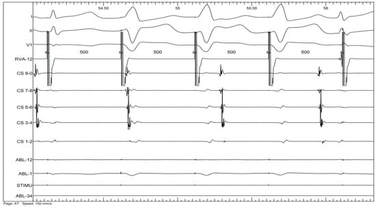
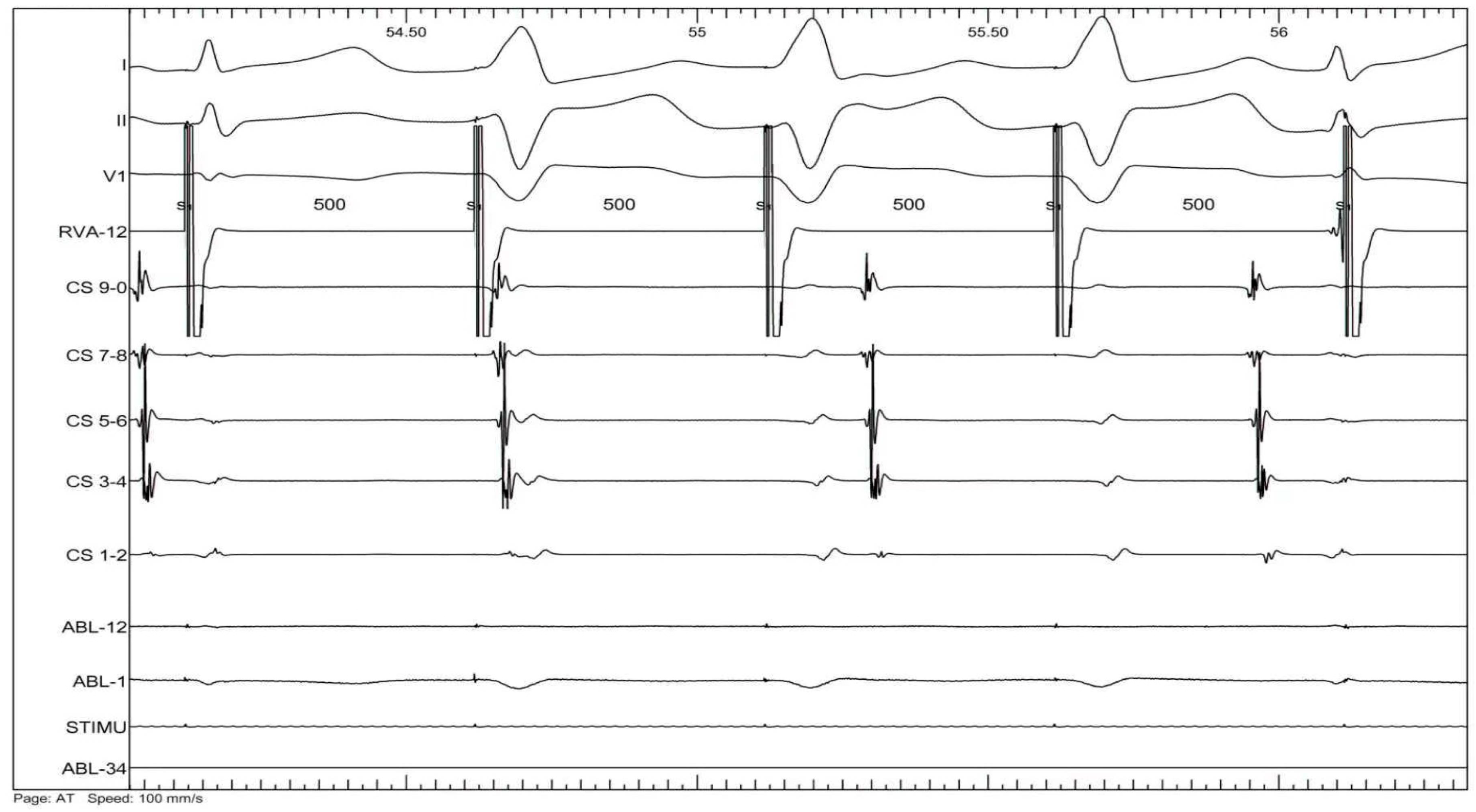
After 25 min of observation, no pre-excitation recurred, including after repeated intravenous adenosine triphosphate (ATP) administration. Post-ablation testing showed ventriculoatrial dissociation during ventricular pacing at 500 ms and atrioventricular block during atrial stimulation at 500/230 ms, with no inducible tachycardia (Figure 6). The procedure lasted 115 min, with 2–3 min of fluoroscopy and a radiation dose of 20 mGy; no acute complications occurred.
Figure 6.
Post-ablation electrophysiological confirmation. Ventricular pacing at a cycle length of 500 ms demonstrated ventriculoatrial dissociation under the tested pacing conditions.
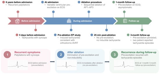
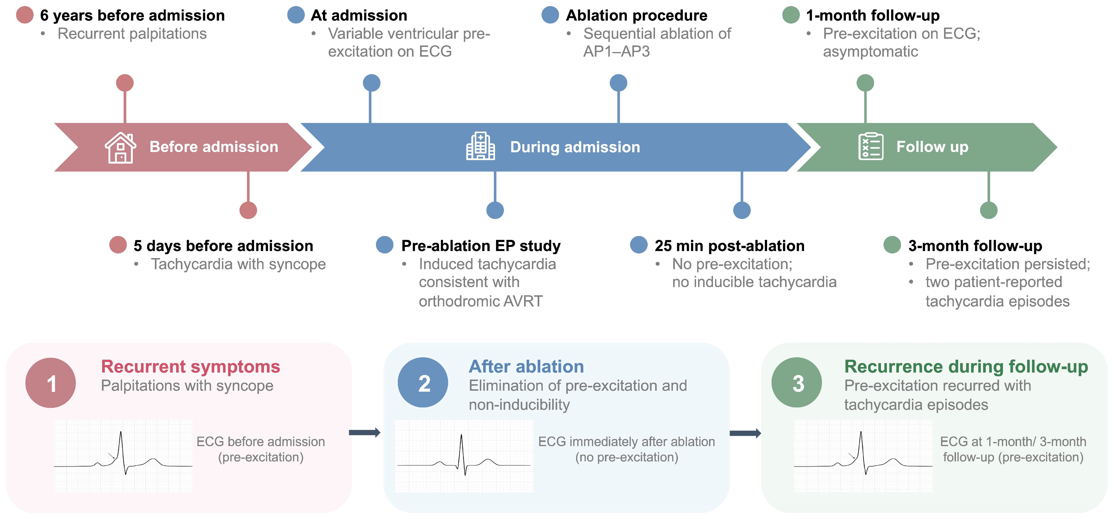
At 1 month, ECG showed recurrent pre-excitation, although the patient reported no palpitations or syncope. Pre-excitation persisted at 3 months, when the patient reported two episodes of tachycardia following a febrile illness without recurrent syncope. Neither episode was documented electrocardiographically (Figure S1). The clinical course, sequential ablation procedures, and follow-up findings are summarized in Figure 7.
Figure 7.
Timeline of ablation and follow-up. Sequential ablation at three accessory pathway sites and electrocardiographic findings at 1- and 3-month follow-up. The arrows indicate the chronological progression from initial symptoms through ablation to follow-up recurrence.
3. Discussion
This case illustrates a diagnostic challenge in pediatric Wolff–Parkinson–White (WPW) syndrome with multiple accessory pathways: surface ECG morphology may reflect the relative electrical contribution of individual pathways rather than the full anatomical substrate. Although multiple accessory pathways are uncommon in children undergoing WPW ablation, their recognition is clinically important [5]. Delta-wave and QRS morphology can provide useful clues to accessory pathway localization, particularly for right-sided and septal pathways [6]. In this patient, variations in lead V1 morphology and inferior-lead QRS polarity across serial ECGs, together with stepwise QRS changes after ablation, suggested changing contributions from accessory pathway conduction at spatially distinct right-sided sites. These findings highlight the importance of interpreting dynamic ECG changes in conjunction with electrophysiological mapping rather than relying on a single ECG pattern for localization.
The sequential ablation process further illustrates the diagnostic value of reassessment after each lesion. Ablation of AP1 at the 7 o’clock position of the tricuspid annulus altered the QRS morphology without eliminating pre-excitation. Ablation of AP2 at the 11 o’clock position produced a further morphological shift, indicating persistent antegrade accessory pathway conduction. Only after AP3, located just superior to the coronary sinus ostium, was ablated did pre-excitation disappear. This sequence demonstrates how interruption of one pathway may change the dominant pre-excitation pattern and reveal conduction through another pathway. Previous studies have described different approaches to accessory pathway localization and ablation [7,8,9]. Accordingly, a new QRS morphology after ablation should prompt evaluation for residual accessory pathway conduction rather than being interpreted solely as procedural failure.
Three-dimensional electroanatomical mapping was instrumental in localizing the three spatially separated targets and documenting successive shifts in the earliest ventricular activation site after AP1 and AP2 ablation. Separation of local atrial and ventricular electrograms after ablation at all three sites provided additional evidence supporting the presence of distinct accessory pathways. Nevertheless, a single broad pathway with multiple insertion sites cannot be definitively excluded. Electroanatomical mapping facilitates anatomical reconstruction and can reduce fluoroscopic exposure during pediatric ablation [10,11]. Right-sided free-wall and septal or paraseptal pathways remain technically challenging because catheter stability, annular anatomy, and proximity to the normal conduction system may affect procedural outcomes; previous pediatric studies have also highlighted recurrence after right free-wall pathway ablation and the complexity of septal pathway ablation [12,13]. In the present case, despite acute elimination of pre-excitation, it recurred at 1 month and persisted at 3 months. The pathway responsible for recurrence could not be identified, and the two reported tachycardia episodes were not documented electrocardiographically. Thus, the subsequent clinical course cannot establish whether recurrence arose from a previously treated pathway or residual conduction through another pathway.
Syncope accompanied by recurrent palpitations in a child with manifest pre-excitation warrants comprehensive electrophysiological assessment. Although pediatric WPW cohorts have demonstrated a small but clinically meaningful risk of life-threatening events, conventional clinical and electrophysiological markers do not identify every child at risk [14,15]. In this patient, recurrent symptoms, syncope, suspected orthodromic atrioventricular reentrant tachycardia (AVRT), and suspected multiple accessory pathways supported invasive evaluation and ablation. However, the absence of pathway-specific antegrade effective refractory periods and SPERRI limited further risk stratification. This case underscores that acute elimination of pre-excitation does not guarantee sustained procedural success. Immediate ECG reassessment after each ablation step, with remapping when pre-excitation persists, and continued clinical and electrocardiographic follow-up are important for detecting residual conduction and recurrence.
Given the recurrence of pre-excitation and the reported tachycardia episodes, repeat electrophysiological evaluation followed by a second catheter ablation is recommended. As the patient currently has minimal symptoms, regular outpatient follow-up is being maintained, with further intervention planned electively.
4. Conclusions
Multiple spatially separated right-sided accessory pathway sites may produce dynamic ventricular pre-excitation and complicate localization and ablation in pediatric Wolff–Parkinson–White syndrome. Changes in QRS morphology and delta-wave configuration across serial ECGs, particularly after individual ablation lesions, should raise suspicion for residual accessory pathway conduction. Three-dimensional electroanatomical mapping, sequential ablation, and reassessment of ECG morphology can help identify additional pathway conduction. Continued follow-up is essential because pre-excitation may recur despite initial procedural success [16,17].
Supplementary Materials
The following supporting information can be downloaded at https://www.mdpi.com/article/10.3390/children13111552/s1. Figure S1: Follow-up ECGs at 1 and 3 months.
Author Contributions
Conceptualization, H.X.; Data Curation, F.X.; Writing—Review and Editing, F.X. and W.W.; Supervision, H.Z. and Y.Z. All authors have read and agreed to the published version of the manuscript.
Funding
This research received no external funding.
Institutional Review Board Statement
The study was conducted in accordance with the Declaration of Helsinki and approved by the Institutional Review Board of Children’s Hospital, Zhejiang University School of Medicine (protocol No. 2026-IRB-0085-P-01; approval date: 9 March 2026).
Informed Consent Statement
Written informed consent for publication of the case and accompanying images was obtained from the patient’s legal guardian before submission.
Data Availability Statement
The data presented in this study are available from the corresponding author upon reasonable request. The data are not publicly available due to privacy and ethical restrictions.
Conflicts of Interest
The authors declare no conflicts of interest.
References
- Vătășescu, R.G.; Paja, C.S.; Șuș, I.; Cainap, S.; Moisa, Ș.M.; Cinteză, E.E. Wolf-Parkinson-White Syndrome: Diagnosis, Risk Assessment, and Therapy-An Update. Diagnostics 2024, 14, 296. [Google Scholar] [CrossRef] [Scilit] [PubMed]
- Brešković, T.; Lisica, L.; Jurišić, Z.; Petrović, D.; Sikirić, I.; Metličić, V.; Anić, A. Ablation of accessory pathways in different anatomic locations using focal pulsed field ablation. Heart Rhythm 2024, 21, 1211–1217. [Google Scholar] [CrossRef] [Scilit] [PubMed]
- Przybylski, R.; Saravu Vijayashankar, S.; O’Leary, E.T.; Hylind, R.J.; Noon, J.; Dionne, A.; DeWitt, E.S.; Bezzerides, V.J.; Abrams, D.J. Hypertrophic Cardiomyopathy and Ventricular Preexcitation in the Young: Cause and Accessory Pathway Characteristics. Circ. Arrhythmia Electrophysiol. 2023, 16, e012191. [Google Scholar] [CrossRef] [Scilit] [PubMed]
- Janson, C.M.; Shah, M.J.; Kennedy, K.F.; Iyer, V.R.; Behere, S.; Sweeten, T.L.; O’Byrne, M.L. Association of Weight with Ablation Outcomes in Pediatric Wolff-Parkinson-White: Analysis of the NCDR IMPACT Registry. JACC Clin. Electrophysiol. 2023, 9, 73–84. [Google Scholar] [CrossRef] [Scilit] [PubMed]
- Bisht, D.S.; Kishor, K. Dual Uncommon Atrioventricular Connections in a Patient with Wolff-Parkinson-White Syndrome. J. Cardiovasc. Electrophysiol. 2025, 36, 900–904. [Google Scholar] [CrossRef] [Scilit] [PubMed]
- Lai, Y.; Gao, M.; Li, Q.; Li, M.; Zhang, J.; Li, X.; Guo, Q.; Huang, L.; Jiang, C.; Li, S.; et al. Characteristics of Right-Sided Accessory Pathways Associated with Right Cardiac Veins. Circ. Arrhythmia Electrophysiol. 2024, 17, e012513. [Google Scholar] [CrossRef] [Scilit] [PubMed]
- Iacopino, S.; Fabiano, G.; Sorrenti, P.; Petretta, A.; Colella, J.; Vilio, A.D.; Statuto, G.; Diomede, N.; Artale, P.; Filannino, P.; et al. Delta-wave automatic mapping of the manifest accessory pathway. Front. Cardiovasc. Med. 2024, 11, 1449038. [Google Scholar] [CrossRef] [Scilit] [PubMed]
- Grubb, C.S.; Melki, L.; Wang, D.Y.; Peacock, J.; Dizon, J.; Iyer, V.; Sorbera, C.; Biviano, A.; Rubin, D.A.; Morrow, J.P.; et al. Noninvasive localization of cardiac arrhythmias using electromechanical wave imaging. Sci. Transl. Med. 2020, 12, eaax6111. [Google Scholar] [CrossRef] [Scilit] [PubMed]
- Moubarak, G.; Zhao, A.; Semichon, M.; Grimard, C.; Sharifzadehgan, A. Catheter ablation of a mechanically traumatized atrioventricular accessory pathway by targeting its ventricular insertion determined by pace mapping: A case report. Eur. Heart J. Case Rep. 2025, 9, ytaf253. [Google Scholar] [CrossRef] [Scilit] [PubMed]
- Krmek, N.; Környei, L.; Kralik, I.; Delić-Brkljačić, D.; Milošević, M.; Rode, M.; Kocsis, F.; Radeljić, V. Should Patient Age and Body Mass Be Taken in Consideration When Zero Fluoro Ablation Is Scheduled? Acta Clin. Croat. 2024, 63, 142–148. [Google Scholar] [CrossRef] [Scilit] [PubMed]
- Fitzpatrick, N.; Mittal, A.; Galvin, J.; Jauvert, G.; Keaney, J.; Keelan, E.; O’Brien, J.; Széplaki, G. The impact of steerable sheath visualization during catheter ablation for atrial fibrillation. Europace 2023, 25, 1345–1351. [Google Scholar] [CrossRef] [Scilit] [PubMed]
- Turan, O.; Akdeniz, C.; Tuzcu, V. Catheter Ablation of Septal Accessory Pathways in Children: A 12-Year Experience at a Tertiary Care Center. J. Cardiovasc. Dev. Dis. 2025, 12, 111. [Google Scholar] [CrossRef] [Scilit] [PubMed]
- Gillette, K.; Winkler, B.; Kurath-Koller, S.; Scherr, D.; Vigmond, E.J.; Bär, M.; Plank, G. A computational study on the influence of antegrade accessory pathway location on the 12-lead electrocardiogram in Wolff-Parkinson-White syndrome. Europace 2025, 27, euae223. [Google Scholar] [CrossRef] [Scilit] [PubMed]
- Barat, M.; Barba, D.T.; Ho, G. Wolff-Parkinson-White syndrome: Diagnostic and management strategies. Clevel. Clin. J. Med. 2025, 92, 119–127. [Google Scholar] [CrossRef] [Scilit] [PubMed]
- Jemtrén, A.; Saygi, S.; Åkerström, F.; Asaad, F.; Bourke, T.; Braunschweig, F.; Carnlöf, C.; Drca, N.; Insulander, P.; Kennebäck, G.; et al. Risk assessment in patients with symptomatic and asymptomatic pre-excitation. Europace 2024, 26, euae036. [Google Scholar] [CrossRef] [Scilit] [PubMed]
- Boyd, C.A.; Bruno, M.A.; Lodeiro, C.A.; Nguyen, D.T.; Banerjee, A.; Menillo, A.M.; Pham, T.D.N.; Miyake, C.Y.; Lam, W.W.; Cannon, B.C.; et al. Detection of Accessory Pathway Recurrence at Standard and Abbreviated Waiting Periods and Implications for Recurrence Following Pediatric Radiofrequency Ablation. J. Cardiovasc. Electrophysiol. 2026. online ahead of print. [Google Scholar] [CrossRef] [Scilit] [PubMed]
- Torun, E.G.; Özdemiroğlu, N.; Yıldız, A.; Ömeroğulları, Z.; Ekşi, M.A.; Koca, S. Multiple Accessory Pathways in Children: Electrophysiological Features and Catheter Ablation Outcomes. Pacing Clin. Electrophysiol. 2026. online ahead of print. [Google Scholar] [CrossRef] [Scilit] [PubMed]
Disclaimer/Publisher’s Note: The statements, opinions and data contained in all publications are solely those of the individual author(s) and contributor(s) and not of MDPI and/or the editor(s). MDPI and/or the editor(s) disclaim responsibility for any injury to people or property resulting from any ideas, methods, instructions or products referred to in the content. |
© 2026 by the authors. Licensee MDPI, Basel, Switzerland. This article is an open access article distributed under the terms and conditions of the Creative Commons Attribution (CC BY) license.