


Highlights
What are the main findings?
- Contact sensitization was identified in 29.8% of children with contact dermatitis who underwent patch testing.
- Nickel (8.3%), fragrance mix and gold sodium thiosulfate (5.8% each) were the most frequent allergens, and bullous reactions occurred only with p-phenylenediamine and black rubber mix.
What are the implications of the main findings?
- Children with concomitant atopic dermatitis had a lower positivity rate, and markers of atopy were not significantly associated with patch test positivity in this cohort.
- Regional data, including allergens outside the European Baseline Series, may inform pediatric patch test panel selection.
Abstract
Background/Objectives: Regional data on contact sensitization in children remain limited and are needed to optimize pediatric patch test series. We aimed to determine the frequency of contact sensitization, identify the most common allergens, and evaluate factors associated with positive patch test results in children with clinically diagnosed contact dermatitis. Methods: We retrospectively reviewed 121 children with clinically diagnosed contact dermatitis who were patch tested with the T.R.U.E. Test to investigate an allergic cause; some also had concomitant atopic dermatitis. Demographic and clinical characteristics, atopic comorbidities, laboratory findings, and patch test results, including reaction intensity, were analyzed. Associations between patch test positivity and clinical variables were assessed using chi-square, Fisher’s exact, and Mann–Whitney U tests. Results: At least one positive reaction was observed in 36 children (29.8%). Nickel sulfate was the most common allergen (8.3%), followed by fragrance mix and gold sodium thiosulfate (5.8% each) and p-phenylenediamine (5.0%). Of 62 positive reactions, 6 were extreme (+++), including three bullous reactions (p-phenylenediamine, n = 2; black rubber mix, n = 1). The positivity rate was lower in children with concomitant atopic dermatitis than in those without (15.8% vs. 36.1%; p = 0.031). No significant associations were found with age, sex, other atopic diseases, family history of atopy, aeroallergen sensitization, total IgE, absolute eosinophil count, or symptom duration. Conclusions: Nearly one-third of children with contact dermatitis showed contact sensitization on patch testing. Markers of atopy were not significantly associated with patch test positivity in this cohort. Allergens outside the European Baseline Series, such as gold, should be considered when designing regional pediatric panels.
Keywords:
contact dermatitis; patch test; contact sensitization; children; contact allergens; nickel 1. Introduction
Contact dermatitis is an inflammatory skin disease caused by exogenous substances reaching the skin surface. In its allergic form, it is a delayed-type hypersensitivity reaction in which haptens penetrating the epidermis bind to host proteins and prime hapten-specific T cells; this sensitization phase is silent, and eczematous lesions appear on re-exposure, characteristically after 24 to 72 h [1]. A meta-analysis of 28 population-based studies covering more than 20,000 patch-tested individuals estimated the pooled prevalence of contact allergy at 20.1% in the general population and 16.5% in those under 18 years of age [2]. Among children actually referred for testing, the yield is considerably higher: a recent meta-analysis of 17 studies and 11 593 children found positive reactions in 36%, led by nickel (11.9%), cobalt (6.6%) and cocamidopropyl betaine (5.5%) [3]. The relevant exposures differ from those of adult practice—costume jewelry, temporary henna tattoos, slime and craft materials, footwear, wet wipes and children’s cosmetics recur throughout the pediatric literature, and their relative weight shifts with local habits and market regulation [4,5].
Whether atopic dermatitis predisposes to or protects against contact sensitization remains unresolved, with published series reporting the association as positive, negative and absent [3,6,7]. This inconsistency may partly reflect differences in allergen potency and the immune pathways involved: classical delayed-type hypersensitivity to potent contact allergens is predominantly associated with Th1-skewed responses, whereas emerging evidence suggests that allergens classified as weak or non-sensitizers in the murine local lymph node assay may induce Th2-skewed sensitization through pathways involving group 2 innate lymphoid cells (ILC2s), particularly within the chronically inflamed, barrier-impaired, and Th2-skewed cutaneous environment characteristic of atopic dermatitis [8,9,10].
A survey by the EBS Working Party of the European Society of Contact Dermatitis found wide variation between clinics in the age at which testing is offered, the panel used, chamber size, occlusion time and reading schedule [11]; the same group subsequently pooled data from 13 centers in 12 countries and proposed a dedicated pediatric series [12]. Reported positivity rates reflect this heterogeneity as much as any real difference in exposure. The European Surveillance System on Contact Allergies (ESSCA) analysis of 6961 European children found 29.3% positive, with nickel sulfate leading at 9.9% [13], whereas single-center series range from 30.6% to 64% within Türkiye alone [6,14,15] and reach 43.6% in Israel [16]. The leading allergens themselves vary by region: textile dye mix and limonene hydroperoxide topped a series of 716 Chinese children [17], while nickel and methylchloroisothiazolinone/methylisothiazolinone (MCI/MI) consistently lead the Turkish data [6,14]. Reliable regional estimates are therefore a prerequisite for any rational pediatric panel.
Against this background, we retrospectively reviewed patch testing in 121 children and adolescents with contact dermatitis evaluated in a pediatric allergy clinic at a single center, with the aim of contributing regional data to the literature in which Turkish pediatric series remain few and discordant. Testing was performed with the Thin-layer Rapid Use Epicutaneous (T.R.U.E.) Test rather than the European Baseline Series used in the Turkish and ESSCA cohorts cited above, which allowed several allergens absent from the baseline series to be assessed. Moreover, most pediatric patch test series, including the three Turkish series cited above, have been reported from dermatology departments; data from pediatric allergy settings, where atopic comorbidity is common, and markers of type 2 sensitization such as total IgE, eosinophil count and skin prick test results are routinely available, remain scarce. We describe the demographic and clinical profile of the cohort, determine the frequency, pattern and intensity of contact sensitization, identify the clinical and laboratory variables associated with a positive result, and compare our findings with national and European data.
2. Materials and Methods
2.1. Study Design and Population
This was a retrospective, descriptive study conducted at the Department of Pediatric Allergy and Clinical Immunology, İzmir Bakırçay University Çiğli Training and Research Hospital. We reviewed the records of all patients aged 1 to 18 years who were followed with a clinical diagnosis of contact dermatitis between 1 January 2023 and 1 June 2026 and who underwent epicutaneous patch testing to investigate a possible allergic cause. The diagnosis of contact dermatitis and the decision to perform patch testing were based on the clinical history and on the morphology and distribution of the lesions, as assessed by the treating pediatric allergist; no standardized pre-screening questionnaire or scoring instrument was used. A subset of children also had concomitant atopic dermatitis; in these children, patch testing was performed because contact dermatitis was diagnosed in addition to atopic dermatitis, and no child was patch tested for atopic dermatitis alone. Patients whose clinical diagnosis could not be confirmed and those with incomplete patch test records were excluded, and 121 children were included.
Age, sex, presenting complaint, duration of symptoms, distribution of lesions, clinical diagnosis, concomitant atopic disease, family history of atopy, aeroallergen sensitization on skin prick testing, total serum IgE, absolute eosinophil count and full patch test results were extracted from the electronic medical records. Total serum IgE and the absolute eosinophil count in peripheral blood were measured at presentation, around the time of patch testing, as part of the routine evaluation; results were available for 95 and 108 children, respectively. The diagnosis of concomitant atopic dermatitis was made clinically by the treating pediatric allergist, on the basis of history and physical examination findings and applying the Hanifin and Rajka criteria [18]. Symptom duration was grouped as 1–6, 7–12 and more than 12 months, and age as 1–6, 7–12 and 13–17 years. No patient identifiers were retained in the analysis dataset.
2.2. Patch Testing
Testing was performed with the T.R.U.E. Test (SmartPractice, Phoenix, AZ, USA), a ready-to-use thin-layer epicutaneous system comprising three adhesive panels containing 35 allergens and allergen mixes together with a negative control. Current ESCD guidance considers patch testing safe in children and recommends testing for the same indications as in adults when allergic contact dermatitis is suspected or needs to be excluded [19]. Although the T.R.U.E. Test is licensed for use from 6 years of age, it was used off-label in younger children when clinical suspicion of allergic contact dermatitis was high. Parents were informed about the procedure and potential risks, and written informed consent was obtained. All children, including those younger than 6 years, were tested with the complete standard T.R.U.E. Test panel; no allergens were omitted.
Patients receiving systemic corticosteroids or other immunosuppressive treatment were not tested; these agents were withheld for at least one month beforehand, and topical corticosteroids were stopped on the test area at least seven days in advance. Panels were applied to clean, dry, hair-free and lesion-free skin of the upper back and left in place under occlusion for 48 h, consistent with the standard occlusion period recommended by ESCD diagnostic patch testing guidance [19]. Children and their parents were instructed to keep the area dry and to avoid vigorous physical activity and sun exposure during this period.
2.3. Reading and Interpretation
No readings were performed during the occlusion period. The first reading was performed at 48 h, immediately after panel removal, followed by a second reading between 72 and 96 h. An additional delayed reading on day 7 was performed when a late reaction was suspected. Reactions were graded according to International Contact Dermatitis Research Group criteria as negative (−), doubtful (?+, faint erythema only), weak positive (+, erythema with infiltration and possible papules), strong positive (++, erythema, infiltration, papules and vesicles) and extreme positive (+++, intense erythema with confluent vesicles or bullae) [20]. A reaction of + or greater at any reading was counted as positive, and doubtful or irritant reactions were recorded as negative. All readings were performed by the same pediatric allergist. Throughout this report, a positive patch test reaction is referred to as contact sensitization. The relevance of positive reactions was assessed clinically as part of routine care, on the basis of the exposure history (for example, jewelry and metal objects for nickel, or hair dye and temporary henna tattoos for p-phenylenediamine), and avoidance advice was given accordingly; however, relevance was not documented in a standardized format (current, past or unknown) for every reaction and could therefore not be analyzed retrospectively.
2.4. Statistical Analysis
Continuous variables are presented as mean ± standard deviation or median (interquartile range [IQR]) according to their distribution, and categorical variables as counts and percentages. Categorical variables were compared using the chi-square or Fisher’s exact test, and continuous variables using the Mann–Whitney U test. A 95% confidence interval (CI) for the overall positivity rate was calculated using the Wilson method, and odds ratios (ORs) with 95% CIs are reported for significant associations. Analyses were performed with IBM SPSS Statistics (Version 27.0.1.0, IBM Corp., Armonk, NY, USA), and a two-sided p-value < 0.05 was considered statistically significant. Given the sample size, no multivariable analysis was performed; all associations are therefore unadjusted and should be considered exploratory.
2.5. Ethics
The study protocol was approved by the Non-Interventional Clinical Research Ethics Committee of İzmir Bakırçay University (approval number 2026–3024), and the study was conducted in accordance with the principles of the Declaration of Helsinki.
3. Results
3.1. Demographic and Clinical Characteristics
The cohort comprised 121 children with a mean age of 10.8 ± 3.7 years (range 4–17); 72 (59.5%) were girls, and 49 (40.5%) were boys. The median symptom duration before testing was 21 months (IQR 6–48, range 1–144); 31 children (25.6%) had been symptomatic for one to six months, 26 (21.5%) for seven to twelve months, and 63 (52.1%) for more than a year; symptom duration was not recorded in one child.
Thirty-eight children (31.4%) had concomitant atopic dermatitis. A concomitant atopic disease, most frequently allergic rhinitis or asthma, was present in 70 children (57.9%), and 30 (24.8%) had a family history of atopy. Skin prick testing identified aeroallergen sensitization in 60 children (49.6%). Lesions were generalized in 29.8% of cases and limited to the hands in 19.0%. Median total IgE was 78 IU/mL (IQR 31–271) in the 95 children in whom it was measured, and the median absolute eosinophil count was 190/µL (IQR 130–325) in 108 children. Table 1 summarizes these characteristics.
Table 1.
Demographic and clinical characteristics of the study population (n = 121).
3.2. Patch Test Positivity and Allergen Profile
Thirty-six children reacted to at least one allergen, an overall positivity rate of 29.8% (95% CI 22.3–38.4). Eighty-five children (70.2%) had no reaction, 21 (17.4%) reacted to a single allergen, 10 (8.3%) to two and 5 (4.1%) to three or more.
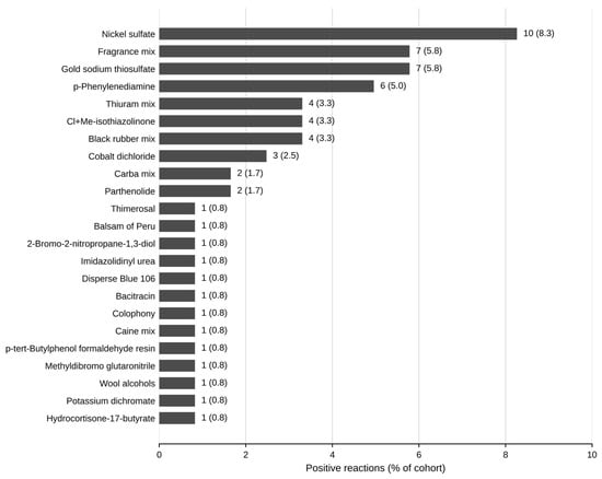
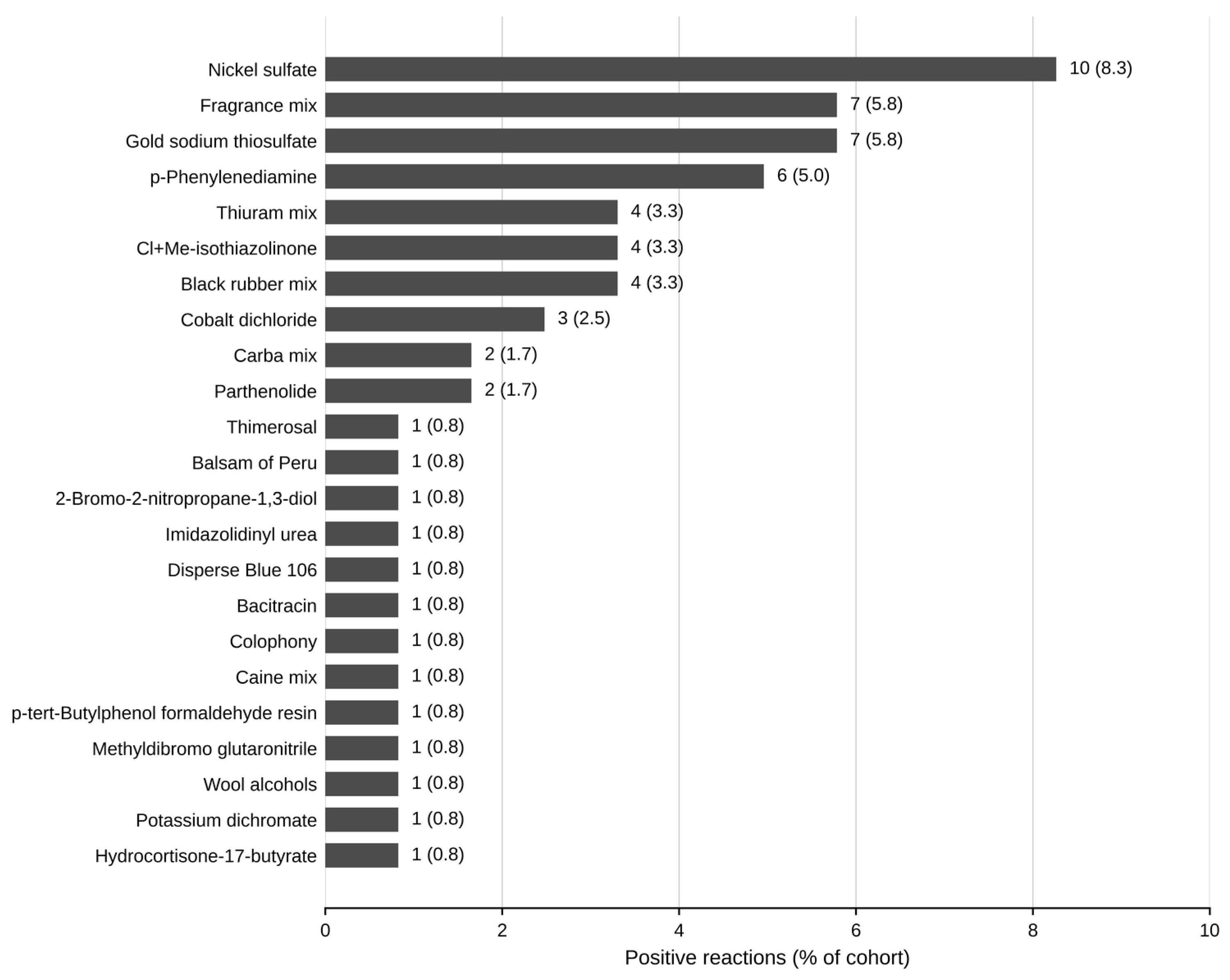
Nickel sulfate was the most frequent allergen, positive in 10 children (8.3%), followed by fragrance mix and gold sodium thiosulfate with seven reactions each (5.8%) and p-phenylenediamine with six (5.0%). Thiuram mix, MCI/MI and black rubber mix each produced four reactions (3.3%), cobalt dichloride three (2.5%), and carba mix and parthenolide two each (1.7%). Thirteen further allergens were positive in one child apiece: thimerosal, balsam of Peru, 2-bromo-2-nitropropane-1,3-diol, imidazolidinyl urea, Disperse Blue 106, bacitracin, colophony, caine mix, p-tert-butylphenol formaldehyde resin, methyldibromo glutaronitrile, wool alcohols, potassium dichromate and hydrocortisone-17-butyrate (Figure 1).
Figure 1.
Frequency of positive patch test reactions to individual allergens in 121 children. Values beside each bar are the number of children and the percentage of the cohort. Thirty-six children accounted for 62 reactions, as some reacted to more than one allergen.
Of the 62 positive reactions, 42 (67.7%) were weak (+), 14 (22.6%) strong (++) and 6 (9.7%) extreme (+++) (Table 2). Extreme reactions were confined to p-phenylenediamine (n = 3), gold sodium thiosulfate (n = 2) and black rubber mix (n = 1); three of these were bullous (p-phenylenediamine, n = 2; black rubber mix, n = 1).
Table 2.
Intensity of positive patch test reactions by allergen (ICDRG grading).
3.3. Factors Associated with Patch Test Positivity
Concomitant atopic dermatitis was the only variable significantly associated with test positivity. The positivity rate was lower in children with concomitant atopic dermatitis than in those without (15.8% [6/38] vs. 36.1% [30/83]; Fisher’s exact test, p = 0.031; OR for positivity in children without vs. with atopic dermatitis, 3.02; 95% CI, 1.13–8.05; unadjusted analysis). No association was observed for sex (21/72 girls vs. 15/49 boys, p = 0.999), family history of atopy (p = 0.098), aeroallergen sensitization (p = 0.536), or symptom duration category (p = 0.891). Positivity increased across the age bands, from 17.6% (3/17) at 1–6 years to 29.7% (19/64) at 7–12 years and 35.0% (14/40) at 13–17 years, although this trend did not reach statistical significance (p = 0.423). Likewise, the positive and negative groups did not differ significantly in age (p = 0.124), symptom duration (p = 0.878), total IgE (p = 0.692), or absolute eosinophil count (p = 0.446) (Table 3).
Table 3.
Association between patch test positivity and demographic and clinical variables.
3.4. Allergen Distribution by Age Group
The allergen profile shifted with age. Among the 17 children aged 1–6 years, three were positive, and the reactions recorded were to fragrance mix (n = 2), p-phenylenediamine (n = 1) and caine mix (n = 1); none reacted to nickel. In the 7–12 year band, 19 of 64 children were positive, with gold sodium thiosulfate the leading allergen (n = 5) ahead of nickel sulfate (n = 4), and thiuram mix, p-phenylenediamine, parthenolide, black rubber mix and Cl + Me-isothiazolinone each contributing two reactions. Among the 13–17-year-olds, 14 of 40 were positive and nickel sulfate predominated (n = 6), followed by fragrance mix (n = 4) and p-phenylenediamine (n = 3).
Expressed as a proportion of all tested children in each age band, nickel sensitization was absent below seven years (0/17) and was found in 6.3% (4/64) of children aged 7–12 years and 15.0% (6/40) of those aged 13–17 years. This gradient did not reach significance on chi-square testing (p = 0.118). Gold sodium thiosulfate showed the opposite tendency, clustering in the school-age band.
4. Discussion
In this single-center series of 121 children with contact dermatitis evaluated in a pediatric allergy clinic, 29.8% had at least one positive patch test reaction, supporting the diagnostic role of patch testing in this setting. Beyond adding to the limited Turkish pediatric data, the study contributes in three respects. First, testing with the T.R.U.E. Test allowed assessment of allergens absent from the European Baseline Series; among these, gold sodium thiosulfate, which was not assessed in the national and European pediatric series used for comparison, ranked joint second and included two extreme reactions. Second, because atopic comorbidity was common (57.9%) and atopy markers were routinely measured, we could examine whether these markers were associated with contact sensitization; they were not significantly associated with patch test positivity in this cohort. Third, we report reaction intensity by allergen, showing that extreme reactions were confined to p-phenylenediamine, gold sodium thiosulfate and black rubber mix, and that all three bullous reactions involved p-phenylenediamine or black rubber mix.
The overall positivity rate of 29.8% was comparable to the 29.3% reported by the European Surveillance System on Contact Allergies (ESSCA) in 6961 children and adolescents [13] and the 30.6% observed in a previous Turkish university-based study [6]. However, it was lower than the pooled prevalence of 36% reported in a recent meta-analysis [3] and substantially lower than the 46.1% and 64% reported by two other centers in Türkiye [14,15]. Although our patch test series included 35 allergens, slightly exceeding the European Baseline Series, this broader allergen panel did not result in a higher positivity rate. These differences may instead reflect variations in referral patterns, disease severity, patient selection, and indications for patch testing, with tertiary referral centers evaluating more highly selected children likely to report higher rates of contact sensitization.
Nickel sulfate was the most common allergen, detected in 8.3% (10/121) of tested children. This prevalence was slightly lower than the 9.9% reported by ESSCA [13] and the 11.9% observed in a recent pediatric meta-analysis [3], and lower than that reported in previous Turkish studies [6,15]. A progressive decline in nickel sensitization has been documented across Europe following implementation of the EU Nickel Directive, and comparable national regulation may have contributed to the prevalence observed in our cohort [21]. More noteworthy than the overall prevalence was the age-specific distribution of nickel sensitization. No nickel sensitization was observed among children younger than seven years, whereas it was found in 6.3% of children aged 7–12 years and 15.0% of adolescents aged 13–17 years. In contrast, ESSCA reported the highest prevalence in younger children, with rates declining with increasing age [13]. However, our youngest age group included only 17 children, three of whom had any positive patch test result, limiting the interpretation of age-specific findings. Moreover, the observed trend did not reach statistical significance (p = 0.118). Differences in patterns of nickel exposure, including the age at ear piercing and other sources of metal contact, may also contribute to these contrasting findings [22]. Because exposure history was not documented in a standardized format, this explanation remains speculative and should be investigated in larger prospective studies.
A similar age-related pattern was observed for p-phenylenediamine (PPD), with positive reactions increasing from one case in the youngest age group to three cases among adolescents. Although the number of sensitized patients was small, this trend is consistent with findings from the ESSCA, which reported an approximately threefold increase in PPD sensitization between children aged 6–12 years and adolescents aged 13–16 years [13]. The higher prevalence of PPD sensitization during adolescence has generally been attributed to increasing exposure to hair dyes and temporary black henna tattoos, which become more common with advancing age [23,24,25]. These exposures have also been implicated in the intense PPD patch test reactions reported in pediatric populations [24,25]. In keeping with this, p-phenylenediamine produced the most intense reactions in our cohort, with three of six positive children showing extreme (+++) reactions, two of them bullous. Given the potential for severe allergic contact dermatitis and the need for long-term avoidance of relevant exposures, recognition of PPD sensitization remains clinically important throughout childhood and adolescence.
Gold sodium thiosulfate was positive in seven children (5.8%), ranking joint second with fragrance mix, with five of the seven reactions occurring in the 7–12 year age group. This allergen is not included in the European Baseline Series and was therefore not tested in any of the cohorts used here for comparison, including the ESSCA network and the three Turkish series [6,13,14,15]. Positive patch test reactions to gold are, however, frequently detected in the absence of an identifiable exposure, and their clinical relevance remains contested [26]. Notably, two of the seven gold reactions were graded +++, indicating that at least some of these reactions were strong rather than borderline, although the strength of a reaction does not establish its clinical relevance. As relevance was not documented in a standardized format in our cohort, the clinical significance of these reactions cannot be determined.
Among the variables evaluated, only concomitant atopic dermatitis was significantly associated with patch test results: children with atopic dermatitis had a lower positivity rate than those without (15.8% vs. 36.1%; p = 0.031; OR 3.02, 95% CI 1.13–8.05). This association was unadjusted and exploratory and should not be interpreted as a protective effect of atopic dermatitis against contact sensitization. Because all children were tested for the same indication, this comparison does not show that clinical suspicion predicts patch test positivity; it shows only that, among children with contact dermatitis, those with concomitant atopic dermatitis were less often sensitized to the allergens tested. This finding is consistent with data from the ESSCA, which reported higher sensitization rates among children without atopic dermatitis [13]. In contrast, Kazan et al. [15], Andre et al. [16], and Kalay Yıldızhan and Boyvat [6] found no significant association, with the latter reporting almost identical positivity rates between children with and without atopic dermatitis. Interestingly, a history of atopic disease—including atopic dermatitis, asthma, and allergic rhinitis—was not associated with patch test positivity in our cohort (p = 1.000). However, cocamidopropyl betaine and propylene glycol, allergens reported to show higher patch test positivity in children with atopic dermatitis [3], were not included in the T.R.U.E. Test panel used in this study. Their absence may therefore have contributed to an underestimation of contact sensitization in the atopic dermatitis subgroup and should be considered when interpreting this null association. This interpretation is also compatible with emerging mechanistic evidence suggesting that chronically inflamed, Th2-skewed skin may favor sensitization to certain weaker allergens, including propylene glycol [10]. Other explanations should also be considered. Eczematous lesions of atopic dermatitis may resemble contact dermatitis, so the threshold for diagnosing contact dermatitis, and therefore the pre-test probability of sensitization, may differ in this subgroup. In addition, the atopic dermatitis subgroup was small (n = 38), and the confidence interval was wide, so the estimate is imprecise. In children with established atopic dermatitis, patch testing may be performed to identify or exclude concomitant allergic contact dermatitis, particularly when contact allergy is suspected as a contributor to persistent or worsening dermatitis. Current ESCD guidance recommends patch testing in patients with atopic dermatitis when allergic contact dermatitis is suspected, including when apparent atopic dermatitis fails to improve despite adequate treatment [19]. However, the relationship between atopic dermatitis and contact sensitization is likely more complex than a simple predisposing or protective effect. Epidermal barrier dysfunction may facilitate allergen penetration, whereas the consequences of the Th2-skewed inflammatory environment, including ILC2-associated pathways, may vary according to allergen potency and the sensitization pathway involved [8,9,10].
Total serum IgE, absolute eosinophil count, and aeroallergen sensitization determined by skin prick testing were not significantly associated with patch test positivity in this cohort. This finding is consistent with the T-cell-mediated pathogenesis of allergic contact dermatitis, which is distinct from IgE-mediated allergic diseases [1]. Although these parameters are routinely assessed in pediatric allergy practice, our findings do not support their use for selecting children for patch testing. These findings support current recommendations that the decision to patch test be based on the clinical history and the morphology and distribution of the dermatitis rather than on markers of atopy [19].
Ten allergens accounted for 49 of the 62 positive reactions observed in our cohort, whereas thirteen allergens each produced only a single positive reaction. Considered in isolation, these findings support the rationale for a compact pediatric baseline panel, consistent with recently proposed recommendations [3,12]. Furthermore, the marked variability in patch test series currently used across Europe, as documented by the European Baseline Series Working Party, further highlights the need for greater standardization [11]. However, the largest Turkish pediatric study reported that nearly one-third of children with allergic contact dermatitis would have been missed if testing had been restricted to the recommended pediatric baseline allergens [14]. Our own findings illustrate this balance. Gold sodium thiosulfate, the joint second most common allergen in our cohort, would not have been detected using an EBS-derived panel, while the thirteen allergens associated with only a single positive reaction collectively accounted for more than one-fifth of all positive patch test results. Taken together, these findings support the use of a standardized core allergen panel supplemented by region-specific allergens, underscoring the importance of local epidemiological data when selecting allergens for routine patch testing.
The main limitations of this study include its retrospective, single-center design and modest cohort of 121 selectively referred children, which limit the generalizability of the findings and the precision of subgroup and age-specific estimates, particularly in the youngest age group and for individual allergens. Although the relevance of positive reactions was evaluated clinically, it was not recorded in a standardized format, so relevance could not be reported per allergen. Because of the sample size, no multivariable analysis was performed; the associations reported here are therefore unadjusted and may be confounded. In addition, some clinical and laboratory variables were incomplete, which reduced the number of children available for individual analyses. Furthermore, testing was performed using the T.R.U.E. Test, a fixed 35-allergen panel not specifically designed for pediatric exposure patterns. Its fixed composition limits individualized allergen selection and excludes potentially relevant allergens such as cocamidopropyl betaine and propylene glycol, which have been reported to show higher positivity in children with atopic dermatitis; their absence may therefore have underestimated sensitization in this subgroup. The complete panel was also used without modification in children younger than 6 years, for whom its use is off-label, which should be considered when interpreting findings in this age group. Recently proposed shortened pediatric panels may offer a more tailored approach for younger children [27]. Finally, because the first assessment was performed at patch removal at 48 h, earlier reaction kinetics were not evaluated. Moreover, all children were tested on the upper back, and no histological assessment was performed, so our data cannot address the early neutrophil infiltration recently described in nickel allergic reactions [28]. Emerging evidence suggests that Th2-skewed reactions in atopic or chronically inflamed skin may differ in morphology and kinetics from classical contact-allergic reactions [10]. Thus, the reading schedule and grading criteria may have influenced the detection of certain reaction patterns, particularly in children with atopic dermatitis. These findings should be interpreted cautiously and confirmed in larger, multicenter studies.
5. Conclusions
Contact sensitization was identified in almost one-third of children with contact dermatitis who underwent patch testing at our center, comparable to contemporary European data. Nickel was the most frequent allergen, and gold sodium thiosulfate, which is absent from the European Baseline Series, ranked joint second with fragrance mix. In an univariable, exploratory analysis, children with concomitant atopic dermatitis had a lower positivity rate, which should not be interpreted as a protective effect; total IgE, eosinophil count and aeroallergen sensitization were not significantly associated with patch test positivity in this cohort, and patch testing should therefore continue to be based on clinical evaluation. Regional data of this kind may help identify allergens for inclusion in a pediatric core panel and its adaptation to local exposure patterns.
Author Contributions
Conceptualization, Ö.S.; data curation, Ö.S. and Ö.K.B.; formal analysis, Ö.S.; writing—original draft preparation, Ö.S.; writing—review and editing, S.B.; supervision, S.B. All authors have read and agreed to the published version of the manuscript.
Funding
This research received no external funding.
Institutional Review Board Statement
The study protocol was approved by the Non-Interventional Clinical Research Ethics Committee of İzmir Bakırçay University (approval number 2026–3024), approval date 10 May 2026, and the study was conducted in accordance with the principles of the Declaration of Helsinki.
Informed Consent Statement
Written informed consent for the patch testing procedure was routinely obtained from the parents/legal guardians before testing as part of our clinical practice.
Data Availability Statement
The data presented in this study are available on request from the corresponding author.
Conflicts of Interest
The authors declare no conflict of interest.
Abbreviations
The following abbreviations are used in this manuscript:
| CI | Confidence interval |
| EBS | European Baseline Series |
| ESCD | European Society of Contact Dermatitis |
| ESSCA | European Surveillance System on Contact Allergies |
| EU | European Union |
| ICDRG | International Contact Dermatitis Research Group |
| IgE | Immunoglobulin E |
| IQR | Interquartile range |
| MCI/MI | Methylchloroisothiazolinone/methylisothiazolinone |
| OR | Odds ratio |
| PPD | p-phenylenediamine |
| T.R.U.E. | Thin-layer Rapid Use Epicutaneous |
References
- Scheinman, P.L.; Vocanson, M.; Thyssen, J.P.; Johansen, J.D.; Nixon, R.L.; Dear, K.; Botto, N.C.; Morot, J.; Goldminz, A.M. Contact dermatitis. Nat. Rev. Dis. Primers 2021, 7, 38. [Google Scholar] [PubMed]
- Alinaghi, F.; Bennike, N.H.; Egeberg, A.; Thyssen, J.P.; Johansen, J.D. Prevalence of contact allergy in the general population: A systematic review and meta-analysis. Contact Dermat. 2019, 80, 77–85. [Google Scholar] [CrossRef] [Scilit] [PubMed]
- Isufi, D.; Jensen, M.B.; Kursawe Larsen, C.; Alinaghi, F.; Schwensen, J.F.B.; Johansen, J.D. Allergens responsible for contact allergy in children from 2010 to 2024: A systematic review and meta-analysis. Contact Dermat. 2025, 92, 327–343. [Google Scholar] [CrossRef] [Scilit] [PubMed]
- Brown, C.; Yu, J. Pediatric allergic contact dermatitis. Immunol. Allergy Clin. N. Am. 2021, 41, 393–408. [Google Scholar] [CrossRef] [Scilit] [PubMed]
- Pigatto, P.; Martelli, A.; Marsili, C.; Fiocchi, A. Contact dermatitis in children. Ital. J. Pediatr. 2010, 36, 2. [Google Scholar] [CrossRef] [Scilit] [PubMed]
- Kalay Yıldızhan, İ.; Boyvat, A. Contact sensitivity of Turkish children and adolescents to European Baseline Series allergens between 2013 and 2023. Dermatol. Pract. Concept. 2024, 14, e2024151. [Google Scholar] [CrossRef] [Scilit] [PubMed]
- Hamann, C.R.; Hamann, D.; Egeberg, A.; Johansen, J.D.; Silverberg, J.; Thyssen, J.P. Association between atopic dermatitis and contact sensitization: A systematic review and meta-analysis. J. Am. Acad. Dermatol. 2017, 77, 70–78. [Google Scholar] [CrossRef] [Scilit] [PubMed]
- Yamaguchi, H.L.; Yamaguchi, Y.; Peeva, E. Role of Innate Immunity in Allergic Contact Dermatitis: An Update. Int. J. Mol. Sci. 2023, 24, 12975. [Google Scholar] [CrossRef] [Scilit] [PubMed]
- Nedorost, S.; Zhang, G.; Fekedulegn, D.; Fluharty, K.; Wang, W.; Frye, B.; Baron, E.; Zug, K.; Yucesoy, B. Weak Sensitizers May Be Associated with CD80 Polymorphisms: Implications for Systemic Contact Dermatitis. JID Innov. 2025, 5, 100382. [Google Scholar] [CrossRef] [Scilit] [PubMed]
- Jacob, S.E.; Scheman, A.; Nedorost, S. Reframing Eczema: Th2-Skewed Contact Sensitization, Atopy Patch Testing, and Systemic Contact Dermatitis. Curr. Allergy Asthma Rep. 2026, 26, 22. [Google Scholar] [CrossRef] [Scilit] [PubMed]
- Aerts, O.; Rustemeyer, T.; Belloni-Fortina, A.; Badulici, S.; Dickel, H.; Abujeta, J.L.G.; Giménez-Arnau, A.M.; Gonçalo, M.; Holden, C.R.; Isaksson, M.; et al. Variability in paediatric patch test practices across Europe: A survey from the European Baseline Series (EBS) Working Party on behalf of the European Society of Contact Dermatitis (ESCD). Contact Dermat. 2025, 93, 285–297. [Google Scholar] [CrossRef] [Scilit] [PubMed]
- Simonsen, A.B.; Rustemeyer, T.; Hadzavdic, S.L.; Spiewak, R.; Fortina, A.B.; Gonçalo, M.; Arnau, A.M.G.; Planella-Fontanillas, N.; Schuttelaar, M.L.A.; Dickel, H.; et al. Contact allergy in children in Europe: A proposal for a paediatric European Baseline Series. Contact Dermat. 2026, 94, 28–39. [Google Scholar] [CrossRef] [Scilit] [PubMed]
- Mazzetto, R.; Uter, W.; Dugonik, A.; Giménez-Arnau, A.M.; Simon, D.; Spiewak, R.; Patruno, C.; Filon, F.L.; Valiukevičienė, S.; Napolitano, M.; et al. Contact sensitization in children and adolescents: Trends from a multicentre European study (2011–2022). Pediatr. Allergy Immunol. 2025, 36, e70245. [Google Scholar] [CrossRef] [Scilit] [PubMed]
- Yılmaz, Z.; Özkaya, E. Patch-test results in terms of the recently recommended allergens in children and adolescents: A retrospective cohort study over 22 years from Turkey. Contact Dermat. 2021, 85, 198–210. [Google Scholar] [CrossRef] [Scilit] [PubMed]
- Kazan, D.; Odyakmaz Demirsoy, E.; Eloğlu, H.; Kiran, R. Evaluation of patch test results in paediatric patients in a tertiary dermatology clinic. Indian J. Dermatol. 2023, 68, 354. [Google Scholar] [CrossRef] [Scilit] [PubMed]
- Andre, N.; Usher, A.; Ofri, M.; Kilimnik, V.; Horev, A. Examining the benefits of extended patch test series in children: A comprehensive analysis. Int. J. Dermatol. 2024, 63, 1200–1204. [Google Scholar] [CrossRef] [Scilit] [PubMed]
- Niu, J.R.; Zhou, H.; Xia, X.; Tian, R. Analysis of patch test results in 716 pediatric patients with type IV hypersensitivity dermatosis. Arch. Dermatol. Res. 2025, 317, 515. [Google Scholar] [CrossRef] [Scilit] [PubMed]
- Hanifin, J.M.; Rajka, G. Diagnostic features of atopic dermatitis. Acta Derm. Venereol. 1980, 60, 44–47. [Google Scholar] [CrossRef] [Scilit]
- Uter, W.; Aerts, O.; Agner, T.; Brans, R.; Bruze, M.; Cannavó, A.; Crépy, M.; Giménez-Arnau, A.M.; Gonçalo, M.; Goossens, A.; et al. European Society of Contact Dermatitis Guideline for Diagnostic Patch Testing-Recommendations on Best Practice (Update 2026). Contact Dermat. 2026, 95, 237–275. [Google Scholar] [CrossRef] [Scilit] [PubMed]
- Bruze, M.; Svedman, C. Clarification and Modification of the International Contact Dermatitis Research Group Classification of Patch Test Reactions on Behalf of the International Contact Dermatitis Research Group. Dermatitis 2025, 36, 440–446. [Google Scholar] [CrossRef] [Scilit] [PubMed]
- Ahlström, M.G.; Thyssen, J.P.; Menné, T.; Johansen, J.D. Prevalence of nickel allergy in Europe following the EU Nickel Directive—A review. Contact Dermat. 2017, 77, 193–200. [Google Scholar] [CrossRef] [Scilit] [PubMed]
- Ahlström, M.G.; Thyssen, J.P.; Wennervaldt, M.; Menné, T.; Johansen, J.D. Nickel allergy and allergic contact dermatitis: A clinical review of immunology, epidemiology, exposure, and treatment. Contact Dermat. 2019, 81, 227–241. [Google Scholar] [CrossRef] [Scilit] [PubMed]
- Andrés Alberola, I.; Jiménez Hernández, E.; Soltero Carracedo, J.F.; Panedas, A.M.; Iranzo, N.M.; Morate, M.B.; López, S.A.; Calleja, J.R. Early presentation of allergic contact dermatitis due to paraphenylenediamine. Allergol. Immunopathol. 2024, 52, 93–96. [Google Scholar] [CrossRef] [Scilit] [PubMed]
- Schubert, S.; Lessmann, H.; Schnuch, A.; Uter, W.; Geier, J.; IVDK. Factors associated with p-phenylenediamine sensitization: Data from the Information Network of Departments of Dermatology, 2008–2013. Contact Dermat. 2018, 78, 199–207. [Google Scholar] [CrossRef] [Scilit] [PubMed]
- Spornraft-Ragaller, P.; Schnuch, A.; Uter, W. Extreme patch test reactivity to p-phenylenediamine but not to other allergens in children. Contact Dermat. 2011, 65, 220–226. [Google Scholar] [CrossRef] [Scilit] [PubMed]
- Yang, B.; Sundquist, B.K.; Pasha, M.A. Determining the clinical relevance of positive patch testing to gold in evaluation of contact dermatitis. J. Allergy Clin. Immunol. Pract. 2019, 7, 292–293. [Google Scholar] [CrossRef] [Scilit] [PubMed]
- Şendal, M.; Çalışkan, N.; Ozceker, D. Pediatric Allergic Contact Dermatitis: Sensitization Patterns for a Practical Panel. Int. Arch. Allergy Immunol. 2026. [Google Scholar] [CrossRef] [Scilit] [PubMed]
- Funch, A.B.; Ahlström, M.G.; Johansen, J.D.; Geisler, C.; Bonefeld, C.M. Neutrophil infiltration in allergic contact dermatitis to nickel. Br. J. Dermatol. 2024, 190, 569–570. [Google Scholar] [CrossRef] [Scilit] [PubMed]

Disclaimer/Publisher’s Note: The statements, opinions and data contained in all publications are solely those of the individual author(s) and contributor(s) and not of MDPI and/or the editor(s). MDPI and/or the editor(s) disclaim responsibility for any injury to people or property resulting from any ideas, methods, instructions or products referred to in the content. |
© 2026 by the authors. Licensee MDPI, Basel, Switzerland. This article is an open access article distributed under the terms and conditions of the Creative Commons Attribution (CC BY) license.