


Highlights
What are the main findings?
- EDH-predominant injuries showed more frequent 90-day good recovery than SDH/contusion-associated injuries, although the effect estimate was imprecise.
- The SDH/contusion-associated group had greater neurological, radiological, and treatment burden, and adjustment for admission GCS attenuated the observed association.
What are the implications of the main findings?
- CT injury pattern provides useful clinical context but should not be used alone to predict outcome after operative pediatric TBI.
- Functional status at discharge may not reflect subsequent recovery; standardized fixed-time follow-up provides a more informative short-term assessment.
Abstract
Background/Objectives: Children who require surgery after traumatic brain injury (TBI) represent a heterogeneous population. We compared 90-day functional status between epidural hematoma (EDH)-predominant injuries and injuries containing acute subdural hematoma (SDH) and/or cerebral contusion, and examined whether quantitative CT measures added useful context. Methods: We retrospectively studied 53 patients aged 1–18 years who underwent cranial surgery for acute TBI between June 2020 and June 2026. The primary comparison included 41 patients aged ≤ 16 years; 40 (97.6%) had assigned 90-day Glasgow Outcome Scale—Extended Pediatric Revision (GOS-E Peds) status. Preoperative CT scans were reviewed using a predefined protocol. Results: SDH/contusion-associated patients had lower admission GCS (median 10 vs. 15) and higher Rotterdam CT scores (3 vs. 1). Good recovery (GOS-E Peds 1–2) at 90 days occurred in 9/13 (69.2%) SDH/contusion-associated patients and 25/27 (92.6%) EDH-predominant patients. The corresponding exact odds ratio for absence of good recovery was 5.28 (95% CI 0.63–68.02; p = 0.075), with an absolute risk difference of +23.4 percentage points (95% CI −0.8 to 50.8). Adjustment for admission GCS attenuated the association (OR 2.57, 95% CI 0.37–19.09). Continuous midline shift was similar between the CT-pattern groups and was not associated with 90-day status. Conclusions: CT-defined injury patterns identified children with different initial injury burdens, but the 90-day association was imprecise and partly tracked admission neurological severity. CT anatomy should therefore be interpreted together with the clinical examination rather than used as a stand-alone prognostic classifier.
1. Introduction
Traumatic brain injury (TBI) remains an important cause of death and acquired disability in children and adolescents. Epidemiology, injury mechanisms, severity, and access to definitive treatment vary across regions [1,2]. Pediatric TBI also differs from adult TBI in cranial anatomy, cerebral physiology, mechanisms of injury, and developmental consequences [3]. Current severe-TBI guidelines therefore emphasize combined neurological, physiological, and radiological assessment rather than reliance on a single severity marker [4].
Only a minority of children with head injury require cranial surgery, and this operative population is not uniform. A neurologically preserved child undergoing evacuation of a focal EDH differs substantially from a child requiring decompression for acute SDH, contusional swelling, diffuse edema, or a complex cranial injury. Previous operative series have described clinical predictors, age-related outcomes, or lesion-specific results [5,6,7], but direct comparison of predefined CT injury patterns at a fixed pediatric functional endpoint is limited.
CT-based classifications, including the Rotterdam score, are associated with mortality and functional outcome in pediatric TBI [8,9,10,11]. MRI studies have also linked diffuse axonal injury, lesion location, diffusion abnormalities, and lesion burden with outcome [12,13,14,15,16]. In contrast, pediatric EDH is often a focal and surgically remediable lesion, and favorable outcomes are common in appropriately selected operative cohorts [17,18,19,20]. These observations suggest that the need for surgery does not by itself define the severity of primary brain injury.
Outcome assessment is also important. GOS-E Peds provides a developmentally adapted measure of global function [21], while recovery after pediatric TBI may continue beyond hospital discharge [22,23,24,25,26]. Global function can also be influenced by extracranial trauma [27]. Discharge status is therefore clinically useful, but it is not a standardized postinjury time point and may not represent short-term recovery.
We therefore examined a consecutive operative pediatric TBI cohort using two clinically recognizable CT patterns: EDH-predominant injury without acute SDH or cerebral contusion, and injury containing acute SDH and/or cerebral contusion. The primary objective was to compare functional status at 90 days in the age range for which GOS-E Peds was originally validated. Secondary objectives were to describe discharge and 180-day status, quantify midline shift and focal lesion volumes, and examine treatment timing. The study was intended to describe heterogeneity within an operative cohort, not to derive a prognostic score or estimate the causal effect of surgery.
2. Materials and Methods
2.1. Study Design, Setting, and Cohort Identification
This retrospective observational cohort study was conducted at Başakşehir Çam and Sakura City Hospital, a tertiary neurosurgical referral center in Istanbul, Türkiye. The institutional operative database identified 68 patients aged 0–18 years who underwent cranial surgery for suspected traumatic cranial injury between June 2020 and June 2026. Fifteen patients operated for non-acute traumatic cranial conditions were excluded. The remaining 53 patients constituted the acute operative TBI cohort. No eligible patient was younger than 1 year.
Qualifying procedures included craniotomy for evacuation of an acute traumatic hematoma, decompressive craniectomy with or without hematoma evacuation, and operative elevation or reconstruction of a depressed skull fracture. Placement of an intracranial pressure monitor or external ventricular drain alone was not considered a qualifying operation. An analyzable preoperative head CT showing an acute traumatic cranial or intracranial abnormality was required.
None of the included patients had documented preinjury neurological or functional impairment expected to alter age-appropriate independence or GOS-E Peds classification. Because the original GOS-E Peds validation included children through 16 years of age [21], patients aged ≤ 16 years formed the primary functional-outcome population. Patients aged 17–18 years remained in the full institutional cohort and were included in sensitivity analysis.
The study was conducted in accordance with the Declaration of Helsinki and was approved by the Başakşehir Çam and Sakura City Hospital Ethics Committee (approval No. 415, 27 August 2026). The final research database was locked on 30 August 2026. Reporting followed the Strengthening the Reporting of Observational Studies in Epidemiology (STROBE) recommendations [28].
2.2. Clinical, Neurological, and Treatment Variables
Clinical information was obtained from electronic medical records, emergency-department records, transfer documents, anesthesia and operative records, intensive-care and ward records, discharge summaries, radiology records, and outpatient follow-up. Variables included age, sex, injury mechanism, clinically significant extracranial trauma, transfer status, intubation before assessment in our emergency department, admission GCS and its components, pupillary reactivity, procedure type, decompressive craniectomy, ICU and ventilation duration, hospital length of stay, and mortality.
Polytrauma was defined as acute TBI with at least one clinically significant extracranial injury requiring specialist assessment or treatment and with the potential to affect mobility or age-appropriate independence. Minor superficial injuries without functional consequence were not classified as polytrauma.
Admission GCS was the first reliable neurological examination documented by a neurosurgeon before surgery. Patients intubated before assessment in our emergency department were identified separately because sedation, neuromuscular blockade, and airway intervention could affect GCS interpretation. Pupillary reactivity was recorded from the first reliable examination. Routine invasive intracranial-pressure monitoring was not used as a standardized institutional protocol, so cohort-level ICP data were unavailable.
Treatment timing was derived from trauma, emergency-department, and operating-room timestamps. Injury-to-operation time was categorized as <3 h, 3–6 h, 6–12 h, or >12 h, and emergency-department-to-incision time was analyzed continuously. Operative decisions were individualized. The on-call attending neurosurgeon integrated neurological status and change in GCS, pupillary findings, lesion morphology and mass effect, midline shift, basal-cistern compression, serial CT progression when available, and surgically relevant skull-fracture features. Borderline cases were discussed at departmental rounds and managed by consensus. No single numerical threshold determined surgery across the entire study period.
2.3. Admission CT Review and Injury-Pattern Classification
The index examination was the noncontrast head CT used for operative decision-making. For transferred patients, an outside-hospital CT was accepted when image quality was adequate and the examination informed the operative decision; otherwise, the first analyzable preoperative CT obtained at our institution was used.
Two neurosurgeons reviewed all CT examinations using a predefined protocol while blinded to discharge, 90-day, and 180-day functional outcomes. Findings were assigned by consensus. Independent pre-consensus classifications were not retained, so formal imaging interobserver reliability could not be calculated. Recorded features included EDH, acute SDH, cerebral contusion, traumatic subarachnoid hemorrhage (tSAH), intraventricular hemorrhage (IVH), skull-fracture morphology, basal-cistern status, Rotterdam CT score, continuous midline shift, and lesion volume.
EDH was defined as an acute traumatic extra-axial collection between the inner table and dura, typically with a lentiform configuration. Acute SDH was defined as an unequivocal traumatic collection between the dura and arachnoid, typically crescentic along the convexity, falx, or tentorium. Cerebral contusion was defined on CT as a focal traumatic cortical or subcortical parenchymal lesion with hemorrhagic foci or a focal traumatic intraparenchymal hematoma, typically accompanied by mixed attenuation and/or surrounding edema and occurring in a distribution compatible with direct, coup, or contrecoup injury. For study classification, traumatic intraparenchymal hemorrhagic lesions meeting these criteria were included within the contusion-associated category. Shearing injury/diffuse axonal injury was not a defining component of the CT-pattern classification. Isolated lesions considered suggestive of traumatic axonal injury did not qualify a patient as contusion-associated unless the preoperative CT independently demonstrated a focal lesion meeting the above definition of cerebral contusion. MRI-only evidence of traumatic axonal injury was therefore not used for group assignment. Injury-pattern assignment was based exclusively on the preoperative CT used for surgical decision-making; MRI findings were not used to assign or modify the CT-pattern group. Equivocal trace hyperdensity was not sufficient to define SDH. tSAH and IVH were defined by acute traumatic blood in the subarachnoid spaces and ventricular system, respectively.
Basal cisterns were categorized as normal, compressed, or absent using the Rotterdam framework. The Rotterdam CT score was calculated from cisternal status, midline shift > 5 mm, epidural mass lesion, and tSAH and/or IVH. Continuous midline shift was measured in millimeters as the maximal displacement of the septum pellucidum from the expected midline on axial CT. EDH, acute SDH, and contusion volumes were recorded in milliliters during the quantitative retrospective CT review. Because these lesion types differ anatomically and biologically, lesion-specific volumes were described separately. Exploratory analyses also considered total extra-axial hematoma volume (EDH + SDH) and aggregate focal lesion volume (EDH + SDH + contusion).
Patients were assigned to mutually exclusive CT patterns by a hierarchical rule defined independently of outcome: (1) EDH-predominant, defined by EDH without acute SDH or cerebral contusion; (2) SDH/contusion-associated, defined by acute SDH and/or cerebral contusion irrespective of concomitant EDH; (3) fracture-dominant, defined by a surgically treated skull fracture without EDH, acute SDH, or cerebral contusion; and (4) other operative injury. The principal comparison involved the first two patterns. A patient with EDH plus unequivocal SDH or contusion was classified as SDH/contusion-associated, avoiding subjective retrospective assignment of a dominant lesion.
2.4. Operative-Target Adjudication and Surgical Treatment
CT injury pattern and operative indication were not treated as synonymous. Operative reports and perioperative documentation for SDH/contusion-associated injuries were reviewed to identify the main surgical target. Targets were categorized as EDH evacuation, SDH evacuation with or without decompression, contusion-related decompression, depressed-fracture elevation/reconstruction, or combined treatment. SDH or contusional pathology was considered directly addressed when it was the primary target or a substantive component of a combined operation. Unclear targets were retained as indeterminate rather than inferred from lesion size.
Craniotomy was defined as creation of a bone flap that was replaced at the end of the operation. Decompressive craniectomy was defined as removal of a bone flap that was intentionally not replaced to provide additional intracranial volume. In this study, decompressive craniectomy was treated as a marker of injury and treatment burden rather than as an independent baseline exposure.
2.5. GOS-E Peds Ascertainment and Follow-Up
Functional status was evaluated with GOS-E Peds, ranging from 1 (upper good recovery) to 8 (death) [21]. When an explicit GOS-E Peds score was documented within the relevant assessment window, that score was used. When no score was recorded but contemporaneous documentation contained sufficient functional information, two neurosurgeons independently reconstructed the category. Disagreements were resolved by consensus. If the record did not support a confident category, the assessment was left missing rather than inferred from another time point.
For reconstructed 90-day assessments, exact ordinal agreement and agreement for the prespecified GOS-E Peds 1–2 versus ≥3 classification were recorded. Chance-corrected coefficients were not estimated because the reconstruction subset was small. Outcome provenance and reconstruction agreement are summarized in Supplementary Table S1.
Discharge status represented function at the end of the index hospitalization. Follow-up assessments were centered at 90 and 180 days after injury, with encounters within ±21 days accepted. If more than one assessment occurred within a window, the encounter closest to the target date was used. Patients whose follow-up window had not matured by database lock were classified as not yet assessable rather than lost to follow-up.
All deaths occurred during the index hospitalization. Death was retained as GOS-E Peds 8 at subsequent nominal time points. Survivors without adequate documentation after a mature follow-up window remained missing; no outcome was imputed.
GOS-E Peds 1–2 was defined as good recovery because these categories correspond to upper and lower good recovery in the instrument. Scores ≥ 3 were described as absence of good recovery rather than “unfavorable outcome,” because this range includes moderate disability. GOS-E Peds was treated as a global functional measure; disability was not retrospectively reassigned according to presumed cerebral or extracranial cause.
2.6. Outcome Hierarchy and Sensitivity Analyses
The primary endpoint was absence of good recovery (GOS-E Peds ≥ 3) at 90 days among patients aged ≤ 16 years in the two principal CT-pattern groups. Discharge GOS-E Peds was a secondary transitional endpoint. The 180-day assessment was exploratory because follow-up maturity was lower. Secondary descriptive outcomes included the ordinal GOS-E Peds distribution and patient-level transitions.
Longitudinal analyses were descriptive. The marked discharge ceiling effect in the EDH-predominant group precluded interpretation of ordinal change as a linear recovery rate, and available-case denominators differed across time points.
Prespecified sensitivity analyses repeated the 90-day comparison in the complete 0–18-year cohort and after excluding patients intubated before assessment at our emergency department, patients with polytrauma, and patients with depressed/displaced fractures; an alternative GOS-E Peds 5–8 endpoint was also examined. A single exploratory Firth logistic model included CT pattern and admission GCS. Exploratory analyses examined continuous midline shift, lesion volumes, and emergency-department-to-incision time in relation to 90-day status.
2.7. Statistical Analysis
Continuous variables are presented as median and interquartile range (IQR), and categorical variables as n/N (%). Continuous or ordinal variables were compared with the Wilcoxon rank-sum test and categorical variables with Fisher’s exact test. Spearman correlation was used for continuous imaging or timing variables versus ordinal GOS-E Peds.
For binary functional outcomes, the SDH/contusion-associated pattern was treated as the exposed group. Conditional exact odds ratios (ORs) and 95% confidence intervals (CIs) were calculated, with absolute risk differences estimated using Newcombe-type CIs. Penalized profile-likelihood CIs and likelihood-ratio p-values were used for the exploratory Firth model. Lesion-specific volume analyses were primarily descriptive because of sparse lesion-positive outcome subgroups.
No formal sample-size calculation was performed because all eligible operations during the study interval were included. Missing outcomes were not imputed. The 90-day CT-pattern comparison was the single prespecified primary inferential analysis; no multiplicity adjustment was applied to that test. Other analyses were secondary or exploratory and were interpreted using effect estimates, confidence intervals, and clinical context rather than isolated p-value thresholds. A Holm adjustment was additionally examined as a sensitivity check across the exploratory quantitative CT tests and did not change their interpretation. Analyses were performed in R version 4.5.2 (R Foundation for Statistical Computing, Vienna, Austria).
3. Results
3.1. Cohort Identification and CT Injury Patterns
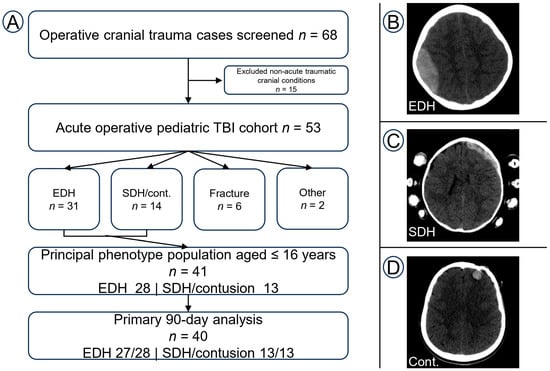
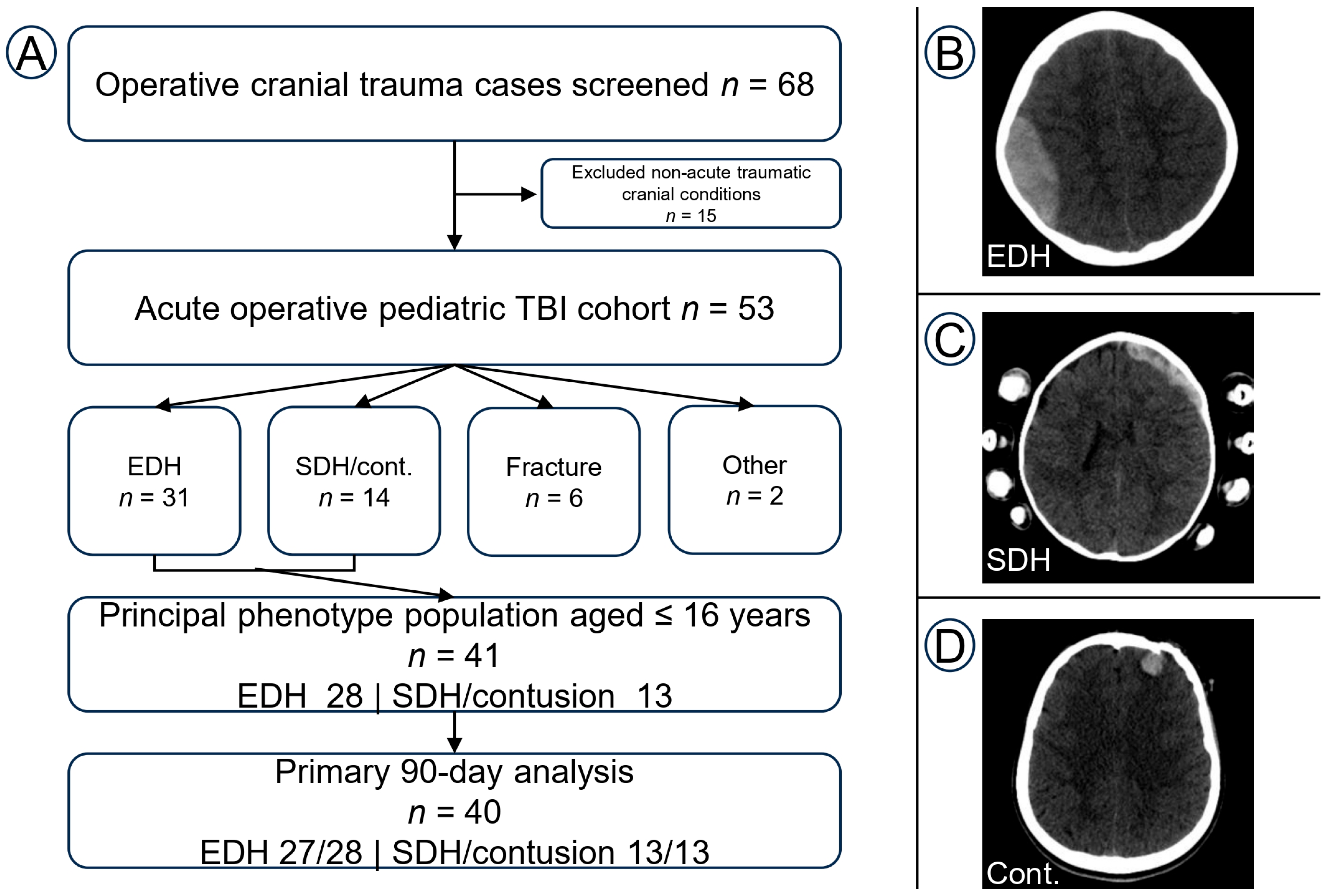
Sixty-eight operative cases were screened. Fifteen non-acute traumatic cranial conditions were excluded, leaving 53 patients in the acute operative TBI cohort (Figure 1). Median age was 9 years (IQR 5–16), and 36/53 patients (67.9%) were male. Traffic-related trauma accounted for 32/53 injuries (60.4%) and falls for 19/53 (35.8%). Nineteen patients (35.8%) had clinically significant polytrauma, 17 (32.1%) were transferred from another institution, and eight (15.1%) had been intubated before assessment in our emergency department.
Figure 1.
Cohort identification, CT injury-pattern classification, and representative CT imaging. (A) Screening and study flow. (B) Representative EDH-predominant injury. (C) Representative acute SDH-associated injury. (D) Representative hemorrhagic contusion/intraparenchymal traumatic hemorrhage classified within the contusion-associated pattern according to the prespecified CT definition. In the patient shown in panel (D), surgery was prompted by neurological deterioration in GCS together with radiographic progression; the CT-pattern label does not imply that every contusion in the cohort was itself the operative target. CT patterns were assigned independently of functional outcome. CT, computed tomography; EDH, epidural hematoma; GOS-E Peds, Glasgow Outcome Scale—Extended Pediatric Revision; SDH, subdural hematoma; TBI, traumatic brain injury.
EDH was present in 35/53 patients (66.0%), acute SDH in 4/53 (7.5%), cerebral contusion in 10/53 (18.9%), tSAH in 17/53 (32.1%), IVH in 1/53 (1.9%), and skull fracture in 46/53 (86.8%). Forty-three patients underwent craniotomy and 10 underwent decompressive craniectomy. Three patients died during the index hospitalization (5.7%).
Across the complete 0–18-year cohort, 31 injuries were EDH-predominant, 14 were SDH/contusion-associated, six were fracture-dominant, and two were other operative injuries. Forty-seven patients were aged ≤ 16 years. Within this age group, 28 had EDH-predominant injury and 13 had SDH/contusion-associated injury, forming the 41-patient principal comparison cohort.
3.2. Initial Clinical, Radiological, and Treatment Burden
The two principal CT-pattern groups differed in initial neurological and radiological severity (Table 1; additional detail in Supplementary Table S2). Median admission GCS was 15 (IQR 13.75–15) in EDH-predominant patients and 10 (IQR 5–14) in SDH/contusion-associated patients (p = 0.004). Median Rotterdam CT scores were 1 (IQR 1–2) and 3 (IQR 2–4), respectively (p < 0.001).
Table 1.
Clinical, radiological, and treatment characteristics of the principal operative injury groups in patients aged ≤ 16 years.
tSAH was present in 2/28 EDH-predominant patients (7.1%) and 9/13 SDH/contusion-associated patients (69.2%; p < 0.001). Depressed or displaced skull fractures were also more frequent in the SDH/contusion-associated group (61.5% vs. 14.3%; p = 0.004), as was decompressive craniectomy (46.2% vs. 3.6%; p = 0.002).
Median emergency-department-to-incision time was similar between groups (127 min [IQR 82–220] vs. 140 min [IQR 73–300]; p = 0.845). Ventilation duration, ICU stay, and hospital length of stay were greater in the SDH/contusion-associated group. These findings describe different acute clinical states and treatment burdens rather than an independent effect of CT pattern.
3.3. Quantitative CT Measures and Treatment Timing
Continuous midline shift did not differ between the principal CT-pattern groups: the median was 0 mm (IQR 0–4) in both EDH-predominant and SDH/contusion-associated patients (p = 0.638). At 90 days, median midline shift was also 0 mm in patients with good recovery (IQR 0–4) and in those without good recovery (IQR 0–9.3; p = 0.933). The correlation between continuous midline shift and ordinal 90-day GOS-E Peds was weak (Spearman ρ = 0.073; p = 0.656). Midline shift > 5 mm was present in 8/40 patients and was not clearly associated with absence of good recovery (OR 2.28, 95% CI 0.17–20.84; p = 0.580).
Among lesion-positive patients in the primary 90-day cohort, median recorded volumes were 12.0 mL (IQR 4.87–24.66) for EDH, 37.7 mL (IQR 11.03–70.85) for acute SDH, and 0.7 mL (IQR 0.30–1.70) for contusion. Lesion-specific outcome groups were too small for stable inferential testing. Exploratory extra-axial hematoma volume and aggregate focal lesion volume showed no clear association with 90-day status (both Wilcoxon p ≈ 0.21; Spearman ρ = −0.166 and −0.153, respectively). Holm adjustment across these exploratory quantitative CT tests did not change the interpretation.
Treatment timing also did not show a simple delay–outcome relationship. Median emergency-department-to-incision time was 142 min (IQR 80.75–266) in patients with 90-day good recovery and 91.5 min (IQR 33.5–114.25) in those without good recovery (p = 0.096). The correlation with ordinal 90-day GOS-E Peds was ρ = −0.263 (p = 0.101). The numerically shorter interval among children with worse outcomes is consistent with more urgent treatment of more severe injuries and does not permit causal inference about timing.
3.4. Operative Target Within the SDH/Contusion-Associated Pattern
The 13 SDH/contusion-associated patients included nine with cerebral contusion and four with acute SDH; four also had concomitant EDH. Because the associated SDH or contusion was not necessarily the lesion that prompted surgery, operative targets were adjudicated separately from CT pattern (Supplementary Table S3).
A principal operative target was assigned in all 13 patients. Three operations primarily addressed acute SDH, one primarily addressed contusional swelling, and three were combined procedures in which SDH or contusional swelling was a substantive component. Two operations were primarily EDH evacuations despite the associated SDH/contusion pattern, and four primarily treated depressed fractures or required cranial reconstruction. Thus, seven of 13 operations directly addressed SDH or contusional pathology. The subsets were too small for stable comparative modeling.
3.5. 90-Day Outcome Ascertainment and Primary Functional Outcome
90-day status was assigned in 40/41 patients (97.6%) in the principal ≤ 16-year cohort: 27/28 EDH-predominant patients and all 13 SDH/contusion-associated patients. Among the 38 survivors with assigned 90-day status, 30 (78.9%) had a directly documented GOS-E Peds score and eight (21.1%) required reconstruction from contemporaneous clinical documentation. Exact ordinal agreement for reconstructed assessments was 7/8 (87.5%), and agreement for the prespecified GOS-E Peds 1–2 versus ≥3 classification was 8/8 (100%) (Supplementary Table S1).
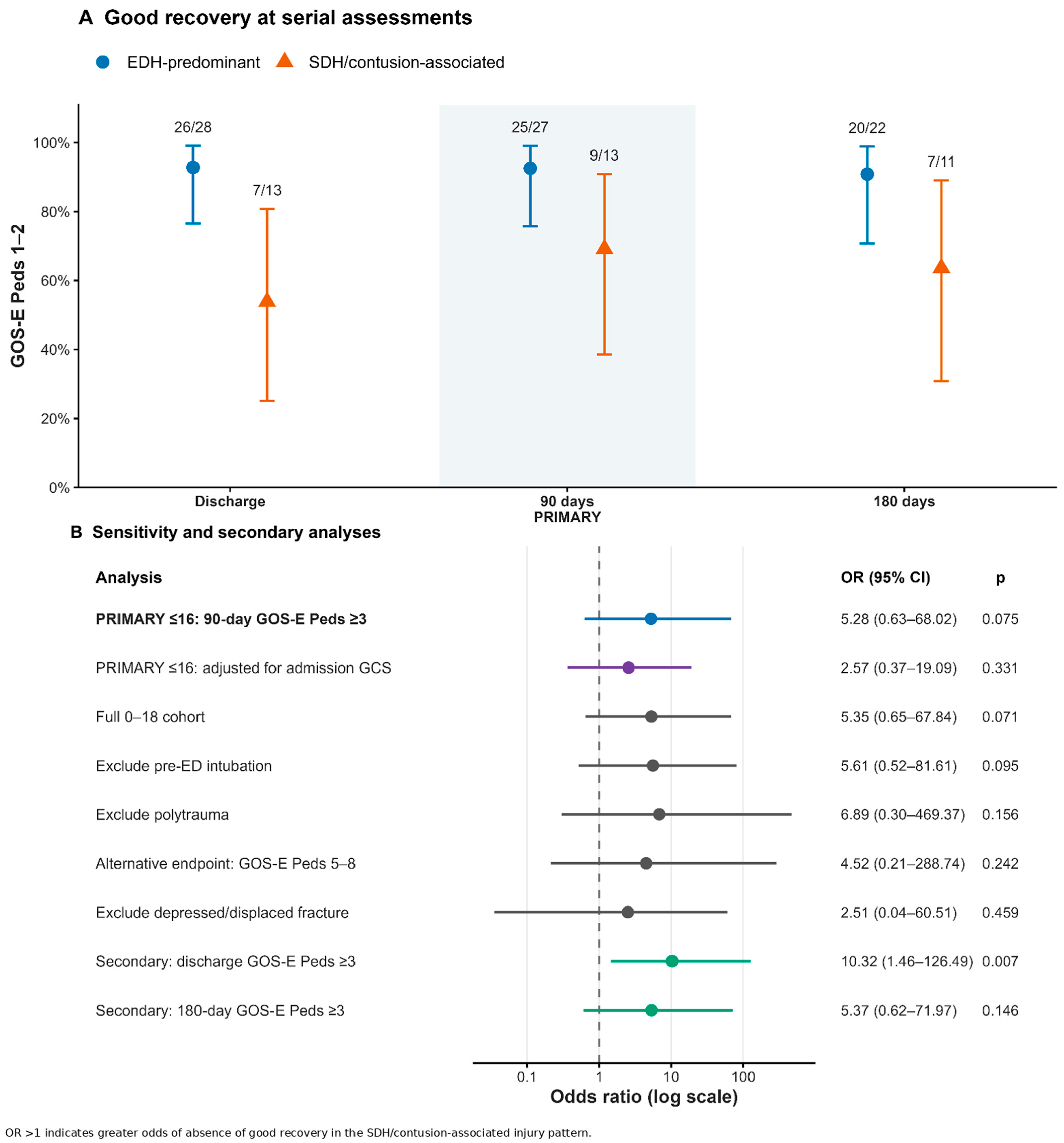
Good recovery at 90 days was present in 25/27 EDH-predominant patients (92.6%) and 9/13 SDH/contusion-associated patients (69.2%). For the prespecified endpoint of GOS-E Peds ≥3, the exact OR was 5.28 (95% CI 0.63–68.02; p = 0.075), and the absolute risk difference was +23.4 percentage points (95% CI −0.8 to 50.8). The point estimate suggested clinically relevant separation, but the confidence interval was wide and included the null.
The ordinal distributions were also different descriptively. In the EDH-predominant group, 24 patients had GOS-E Peds 1, one had category 2, one category 3, and one category 8. In the SDH/contusion-associated group, seven had category 1, two category 2, two category 3, one category 6, and one category 8. Only six patients in the primary analysis were outside the good-recovery range, limiting multivariable precision.
3.6. Discharge State, Exploratory 180-Day Outcome, and Individual Transitions
At discharge, 26/28 EDH-predominant patients (92.9%) had GOS-E Peds 1–2 compared with 7/13 SDH/contusion-associated patients (53.8%). For GOS-E Peds ≥ 3, the exact OR was 10.32 (95% CI 1.46–126.49; p = 0.007), with an absolute risk difference of +39.0 percentage points (95% CI 11.3–64.2). Because discharge occurred after different lengths of hospitalization, this comparison represents an early transitional state rather than a fixed-time outcome.
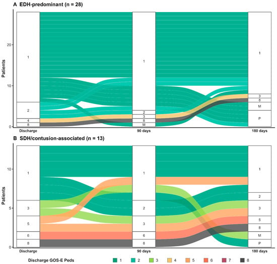
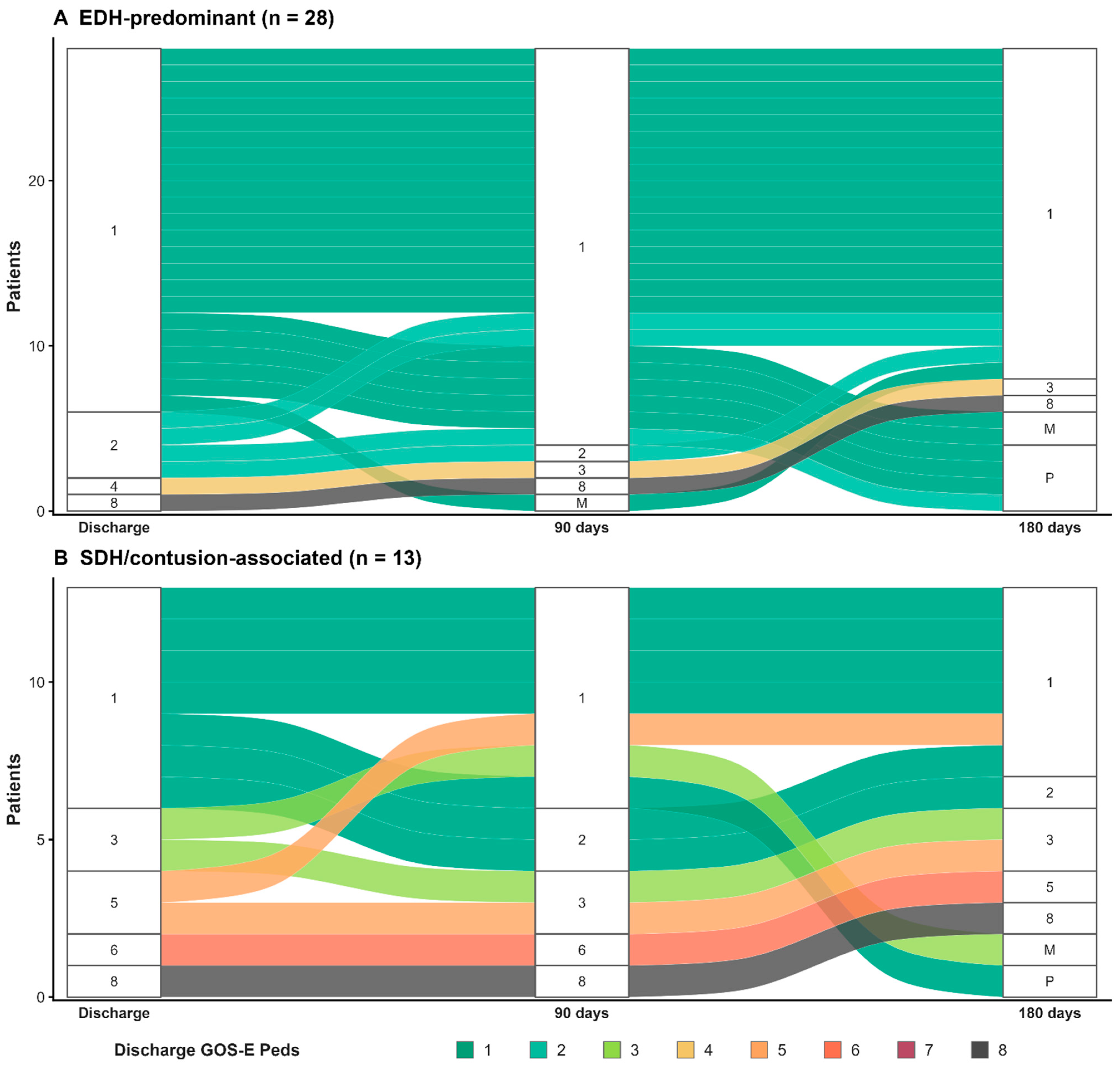
Between discharge and 90 days, four EDH-predominant patients improved and 23 remained unchanged among those with paired observations. In the SDH/contusion-associated group, three improved, eight remained unchanged, and two moved to a numerically higher GOS-E Peds category. Two patients who were outside the good-recovery range at discharge entered GOS-E Peds 1–2 by 90 days. The EDH-predominant group showed a marked ceiling effect from discharge onward. Individual patient-level transitions are shown in Figure 2.
Figure 2.
Individual GOS-E Peds transitions in the principal ≤ 16-year operative injury groups. Alluvial diagrams show patient-level transitions across discharge, 90 days, and 180 days for (A) EDH-predominant and (B) SDH/contusion-associated injuries. Lower GOS-E Peds scores represent better global function and category 8 represents death; known deaths remain in category 8 at subsequent nominal time points. M denotes missing outcome documentation after a mature follow-up window. p denotes pending/not yet assessable because the follow-up window had not matured or remained open at database lock. Ribbon color denotes the discharge GOS-E Peds category. The figure is descriptive because of the discharge ceiling effect and changing available-case denominators over time.
At database lock, 36/41 patients in the principal cohort had completed the 180-day ascertainment window. Functional status was assigned in 33/36 (91.7%), including two known in-hospital deaths retained as GOS-E Peds 8. Five additional patients had not yet completed the accepted assessment window and were considered administratively not yet assessable.
At 180 days, good recovery was present in 20/22 EDH-predominant patients (90.9%) and 7/11 SDH/contusion-associated patients (63.6%). For GOS-E Peds ≥ 3, the exact OR was 5.37 (95% CI 0.62–71.97; p = 0.146). Functional outcomes across the three assessment points are summarized in Table 2. Among patients with both 90- and 180-day assessments, one EDH-predominant patient and two SDH/contusion-associated patients improved by at least one ordinal category; no patient worsened. Detailed distributions and transitions are provided in Supplementary Table S4.
Table 2.
Functional status in the principal ≤ 16-year operative injury groups.
3.7. Sensitivity and Severity-Adjusted Analyses
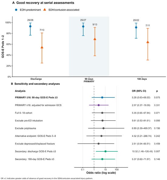
Prespecified sensitivity analyses remained directionally consistent with the primary comparison, but all estimates were imprecise (Supplementary Table S5 and Figure 3B). This was true when the analysis was extended to patients aged 17–18 years, after exclusion of patients intubated before assessment in our emergency department, after exclusion of polytrauma, when GOS-E Peds 5–8 was used as a more stringent endpoint, and after exclusion of depressed/displaced fractures.
Figure 3.
Functional status at serial assessments and sensitivity analyses. (A) Proportion of patients achieving good recovery (GOS-E Peds 1–2) at discharge, 90 days, and 180 days in the principal ≤ 16-year CT-pattern groups, with exact binomial 95% confidence intervals and observed n/N labels. The 90-day assessment is the prespecified primary fixed-time endpoint. Points are not connected because available-case denominators differ. (B) Forest plot of the primary 90-day association, the exploratory estimate adjusted for admission GCS, prespecified sensitivity analyses, and secondary discharge and 180-day estimates. Odds ratios > 1 indicate greater odds of GOS-E Peds ≥ 3 in the SDH/contusion-associated group. Secondary and sensitivity estimates are exploratory and are shown to indicate precision and direction, not as separate confirmatory tests.
In the exploratory Firth model, adjustment for admission GCS attenuated the CT-pattern estimate from an OR of 5.28 to 2.57 (95% profile-likelihood CI 0.37–19.09; p = 0.331). Each one-point higher admission GCS was associated with an OR of 0.84 for absence of good recovery (95% profile-likelihood CI 0.68–1.04; p = 0.107). These estimates were imprecise. The attenuation indicates that part of the crude CT-pattern association tracked neurological severity at presentation.
3.8. Follow-Up Completeness and Administrative Censoring
At 90 days, 40/41 patients in the principal cohort had assigned status (97.6%); one eligible survivor lacked adequate documentation. At 180 days, 33/36 patients whose accepted follow-up window had matured had assigned status (91.7%). Administratively immature observations were not classified as missing and were excluded from the corresponding available-case denominator (Supplementary Table S6).
4. Discussion
In this single-center operative cohort, EDH-predominant and SDH/contusion-associated injuries represented different clinical states. 90-day good recovery was observed more often after EDH-predominant injury, but the estimate was imprecise. The groups also differed substantially in admission GCS, Rotterdam CT score, tSAH, fracture morphology, decompressive-craniectomy use, ventilation, and hospital course. Adjustment for admission GCS reduced the CT-pattern association. The main interpretation is therefore that CT pattern provides context for injury burden; it is not a stand-alone prognostic classifier.
This interpretation is consistent with prior operative and imaging literature. Previous pediatric surgical series have focused on clinical predictors, age-stratified outcomes, or lesion-specific cohorts [5,6,7], while CT and MRI studies have shown that neurological status and global imaging burden are important for outcome [8,9,10,11,12,13,14,15,16]. Pediatric EDH can nevertheless be a focal, surgically remediable lesion with favorable recovery [17,18,19,20]. Our findings extend these observations by comparing predefined CT patterns within one consecutive operative cohort at a fixed 90-day pediatric functional assessment.
Quantitative CT analyses did not identify a single CT measurement that explained outcome. Continuous midline shift was similar between injury-pattern groups and showed little association with 90-day GOS-E Peds. Lesion volumes were highly heterogeneous, and lesion-specific outcome samples were too small for stable modeling. Exploratory aggregate volume measures also showed no clear association with 90-day status. These negative exploratory results should not be interpreted as evidence that mass effect or lesion volume is unimportant; the cohort was small and selected for surgery. They instead support integrated interpretation of imaging and neurological examination.
Treatment timing showed a similar limitation. Children without 90-day good recovery underwent surgery sooner numerically, not later. This likely reflects triage of more severe injuries to more urgent surgery and illustrates confounding by indication. Because the study contains only operated patients and no comparable nonoperative cohort, it cannot estimate the causal effect of time to surgery, the benefit of surgery, or a threshold at which surgery should be performed.
The longitudinal findings also distinguish discharge status from fixed-time outcome. The between-group difference was larger at discharge than at 90 days, and two SDH/contusion-associated patients entered the good-recovery range during this interval. Discharge therefore reflects an early transitional state rather than a definitive short-term outcome. The 180-day findings were directionally similar but exploratory because fewer patients had mature follow-up. Broader cognitive, behavioral, educational, and participation outcomes may continue to evolve beyond the GOS-E Peds categories [22,23,24,25,26].
CT pattern should also not be confused with operative indication. Only seven of 13 SDH/contusion-associated operations directly treated SDH or contusional pathology; other operations primarily addressed EDH or skull-fracture pathology. Operative decisions in routine practice were made by the on-call attending neurosurgeon after integrating the neurological examination, clinical course, and CT findings; borderline cases were discussed by the department and approached by consensus. Figure 1D illustrates this distinction: surgery followed GCS deterioration and radiographic progression rather than the mere presence of a small contusion.
Decompressive craniectomy was more frequent in the SDH/contusion-associated group and is best viewed here as a marker of physiological and treatment burden. The pediatric evidence for decompressive craniectomy remains heterogeneous [29,30,31], and our small retrospective cohort cannot estimate its independent treatment effect.
Outcome ascertainment was a strength. 90-day status was available for 97.6% of the principal cohort, most surviving patients had a directly documented GOS-E Peds score, and reconstructed assessments showed complete agreement for the prespecified binary endpoint. Missing scores were not imputed or carried across time points. GOS-E Peds nevertheless remains a global measure and can be affected by extracranial injury; this was addressed through a prespecified polytrauma sensitivity analysis rather than retrospective attribution of disability [27].
Additional strengths include a consecutive operative cohort, outcome-blinded CT review, an explicit injury-pattern rule, operative-target adjudication for mixed lesions, quantitative CT measures, a fixed 90-day primary endpoint, and transparent handling of follow-up maturity. The statistical analysis was deliberately limited because only six patients in the primary cohort were outside the good-recovery range.
Several limitations are important. This was a retrospective single-center study with 53 acute operative TBI patients, 40 in the primary 90-day analysis, and only six primary endpoint events. Confidence intervals were therefore wide. The CT-pattern groups were constructed for this study and are not externally validated; acute SDH and cerebral contusion are biologically distinct and were grouped only to identify injuries containing subdural or parenchymal components. CT review was consensus-based, so imaging interobserver reliability cannot be reported. Because injury-pattern assignment was based on CT, the classification was not designed to capture the full burden of traumatic axonal/shearing injury, which is more sensitively characterized by MRI. Lesion-volume measurements were exploratory and are vulnerable to measurement error, particularly for irregular lesions. Surgical indications reflected attending judgment and departmental consensus rather than a single protocol, and the absence of a nonoperative comparison group prevents inference about surgical benefit or optimal timing. Admission GCS could be affected by pre-referral airway management. ICP data were not routinely available. Retrospective GOS-E Peds reconstruction is not equivalent to prospective structured administration, and the 180-day analysis had a smaller available-case denominator. Finally, GOS-E Peds does not fully capture cognition, behavior, emotional health, school reintegration, or participation.
5. Conclusions
Among children undergoing cranial surgery for acute TBI, EDH-predominant and SDH/contusion-associated CT patterns identified groups with different initial neurological, radiological, and treatment burdens. 90-day good recovery was more frequent in the EDH-predominant group, but the association was imprecise and attenuated after adjustment for admission GCS. Continuous midline shift and exploratory lesion-volume measures did not show clear independent associations with 90-day status in this small cohort.
The larger disparity at discharge narrowed by 90 days, emphasizing that discharge function is transitional. CT anatomy should therefore be interpreted together with neurological status, serial clinical change, and other measures of injury burden rather than used alone for outcome prediction. Larger multicenter studies with prospective standardized functional and neurocognitive assessment are needed.
Supplementary Materials
The following supporting information can be downloaded at https://www.mdpi.com/article/10.3390/children13111512/s1: Table S1. 90-day GOS-E Peds ascertainment and agreement for reconstructed assessments; Table S2. Detailed injury mechanism, admission GCS components, pupillary findings, treatment timing, and operative characteristics by CT injury pattern in the principal ≤16-year cohort; Table S3. Detailed CT composition and adjudicated operative-target distribution within the SDH/contusion-associated injury pattern in the principal ≤16-year cohort (n = 13); Table S4. Exact GOS-E Peds distributions and transition matrices in the principal ≤16-year operative injury groups; Table S5. Sensitivity analyses for absence of good recovery at 90 days; Table S6. Follow-up eligibility, administrative censoring, and outcome availability at 90 and 180 days.
Author Contributions
Conceptualization, F.D., A.T.B. and F.S.; methodology, F.D., F.S. and A.T.B.; investigation, F.D., F.S., E.K.K., S.D.T.B., S.D. and A.T.B.; data curation, F.D., F.S., E.K.K., S.D.T.B. and S.D.; formal analysis, F.D. and A.T.B.; visualization, F.D.; supervision, A.T.B.; writing—original draft preparation, F.D.; writing—review and editing, all authors. All authors have read and agreed to the published version of the manuscript.
Funding
This research received no external funding.
Institutional Review Board Statement
The study was conducted in accordance with the Declaration of Helsinki and approved by the Başakşehir Çam and Sakura City Hospital Ethics Committee (approval No. 415, 27 August 2026).
Informed Consent Statement
Patient consent was waived because of the retrospective design and use of existing clinical data, in accordance with the institutional ethics committee approval.
Data Availability Statement
The de-identified data supporting the findings of this study are available from the corresponding author upon reasonable request and subject to institutional and ethics requirements.
Acknowledgments
Use of Generative Artificial Intelligence: During manuscript preparation and revision, the authors used ChatGPT (GPT-5.6 Sol; OpenAI, San Francisco, CA, USA) for English-language editing, clarity, organization, and presentation. The tool was not used to generate or alter study data. All analyses, interpretations, references, and final wording were critically reviewed and approved by the authors, who take full responsibility for the manuscript.
Conflicts of Interest
The authors declare no conflicts of interest.
References
- Baticulon, R.E.; Sy, J.J.; Haizel-Cobbina, J.; Du, L.; Figueredo, L.; Hounjet, C.; Mitchell, R.; Shlobin, N.A.; Dewan, M.C. Global epidemiology of pediatric traumatic brain injury: Systematic review and meta-analysis. Neurosurgery 2026, 99, 496–509. [Google Scholar] [CrossRef] [Scilit]
- Dewan, M.C.; Mummareddy, N.; Wellons, J.C., III; Bonfield, C.M. Epidemiology of global pediatric traumatic brain injury: Qualitative review. World Neurosurg. 2016, 91, 497–509.e1. [Google Scholar] [CrossRef] [Scilit]
- Araki, T.; Yokota, H.; Morita, A. Pediatric traumatic brain injury: Characteristic features, diagnosis, and management. Neurol. Med. Chir. 2017, 57, 82–93. [Google Scholar] [CrossRef] [Scilit]
- Kochanek, P.M.; Tasker, R.C.; Carney, N.; Totten, A.M.; Adelson, P.D.; Selden, N.R.; Davis-O’reilly, C.; Hart, E.L.; Bell, M.J.; Bratton, S.L.; et al. Guidelines for the management of pediatric severe traumatic brain injury, Third Edition: Update of the Brain Trauma Foundation guidelines. Pediatr. Crit. Care Med. 2019, 20, S1–S82. [Google Scholar] [CrossRef] [Scilit]
- Lee, S.W.Y.; Ming, Y.; Jain, S.; Chee, S.Y.; Teo, K.; Chou, N.; Lwin, S.; Yeo, T.T.; Nga, V.D.W. Factors predicting outcomes in surgically treated pediatric traumatic brain injury. Asian J. Neurosurg. 2019, 14, 737–743. [Google Scholar] [CrossRef] [Scilit]
- Roach, C.S.; Shawwa, J.J.; Nee, C.; Chetan, S.; Dong, G.; Rios, A.; Brun, J.D.; Brun, J.D.; Lu, V.M. Age-stratified neurosurgical outcomes for traumatic brain injury in a pediatric neurosurgical cohort in La Paz, Bolivia. Childs Nerv. Syst. 2026, 42, 32. [Google Scholar] [CrossRef] [Scilit]
- Echara, M.; Das, A.K.; Agrawal, M.; Gupta, A.; Sharma, A.; Singh, S.K. Prognostic factors and outcome of surgically treated supratentorial versus infratentorial epidural hematoma in pediatrics: A comparative study of 350 patients at a tertiary center of a developing country. World Neurosurg. 2023, 171, e447–e455. [Google Scholar] [CrossRef] [Scilit]
- García-Arellano, M.; Patiño-Córdova, P.E.; Macías-Hernández, S.I.; Reyes-Melendres, D.A.; Marquez-Romero, J. Comparative performance of CT scales for outcome prediction at discharge in pediatric traumatic brain injury: A retrospective cohort study. J. Neurosurg. Pediatr. 2026, 38, 87–94. [Google Scholar] [CrossRef] [Scilit]
- Liesemer, K.; Riva-Cambrin, J.M.; Bennett, K.S.; Bratton, S.L.; Tran, H.; Metzger, R.R.; Bennett, T.D. Use of Rotterdam CT scores for mortality risk stratification in children with traumatic brain injury. Pediatr. Crit. Care Med. 2014, 15, 554–562. [Google Scholar] [CrossRef] [Scilit]
- Talari, H.R.; Hamidian, Y.; Moussavi, N.; Fakharian, E.; Abedzadeh-Kalahroudi, M.; Akbari, H.; Taher, E.B. The prognostic value of Rotterdam computed tomography score in predicting early outcomes among children with traumatic brain injury. World Neurosurg. 2019, 125, e139–e145. [Google Scholar] [CrossRef] [Scilit]
- Katar, S.; Aydin Ozturk, P.; Ozel, M.; Arac, S.; Evran, S.; Cevik, S.; Baran, O. The use of Rotterdam CT score for prediction of outcomes in pediatric traumatic brain injury patients admitted to emergency service. Pediatr. Neurosurg. 2020, 55, 237–243. [Google Scholar] [CrossRef] [Scilit]
- Ferrazzano, P.A.; Rebsamen, S.; Field, A.S.; Broman, A.T.; Mayampurath, A.; Rosario, B.; Buttram, S.; Willyerd, F.A.; Rathouz, P.J.; Bell, M.J.; et al. MRI and clinical variables for prediction of outcomes after pediatric severe traumatic brain injury. JAMA Netw. Open 2024, 7, e2425765. [Google Scholar] [CrossRef] [Scilit]
- Janas, A.M.; Qin, F.; Hamilton, S.; Jiang, B.; Baier, N.; Wintermark, M.; Threlkeld, Z.; Lee, S. Diffuse axonal injury grade on early MRI is associated with worse outcome in children with moderate-severe traumatic brain injury. Neurocrit. Care 2022, 36, 492–503. [Google Scholar] [CrossRef] [Scilit]
- Janas, A.M.; Miller, K.R.; Stence, N.V.; Wyrwa, J.M.; Ruzas, C.M.; Messer, R.; Mourani, P.M.; Fink, E.L.; Maddux, A.B. Utility of early magnetic resonance imaging to enhance outcome prediction in critically ill children with severe traumatic brain injury. Neurocrit. Care 2024, 41, 80–90. [Google Scholar] [CrossRef] [Scilit]
- Smitherman, E.; Hernandez, A.; Stavinoha, P.L.; Huang, R.; Kernie, S.G.; Diaz-Arrastia, R.; Miles, D.K. Predicting outcome after pediatric traumatic brain injury by early magnetic resonance imaging lesion location and volume. J. Neurotrauma 2016, 33, 35–48. [Google Scholar] [CrossRef] [Scilit]
- Baker, C.M.; Cox, A.P.; Hunsaker, J.C.; Scoville, J.; Bollo, R.J. Postoperative magnetic resonance imaging may predict poor outcome in children with severe traumatic brain injuries who undergo cranial surgery. J. Neurosurg. Pediatr. 2022, 29, 407–411. [Google Scholar] [CrossRef] [Scilit]
- Cremonini, C.; Lewis, M.; Wong, M.D.; Benjamin, E.R.; Inaba, K.; Demetriades, D. Traumatic epidural hematomas in the pediatric population: Clinical characteristics and diagnostic pitfalls. J. Pediatr. Surg. 2020, 55, 1773–1778. [Google Scholar] [CrossRef] [Scilit]
- Bounajem, M.T.; Samples, D.C.; Wallace, D.J.; Tavakoli, S.; Liao, L.; Tarasiewicz, I. Management of epidural hematomas in pediatric patients presenting with a GCS of 14 or better. J. Clin. Neurosci. 2019, 70, 118–122. [Google Scholar] [CrossRef] [Scilit]
- Kandregula, S.; Sadashiva, N.; Konar, S.; Rao, K.N.; Shukla, D.; Bhat, D.; Devi, B.I. Surgical management of traumatic extradural hematomas in children: An analysis of 201 patients at a tertiary neurosurgical center. Childs Nerv. Syst. 2019, 35, 807–813. [Google Scholar] [CrossRef] [Scilit]
- Faheem, M.; Jaiswal, M.; Ojha, B.K.; Chandra, A.; Singh, S.K.; Srivastava, C. Traumatic pediatric extradural hematoma: An institutional study of 228 patients in a tertiary care center. Pediatr. Neurosurg. 2019, 54, 237–244. [Google Scholar] [CrossRef] [Scilit]
- Beers, S.R.; Wisniewski, S.R.; Garcia-Filion, P.; Tian, Y.; Hahner, T.; Berger, R.P.; Bell, M.J.; Adelson, P.D. Validity of a pediatric version of the Glasgow Outcome Scale–Extended. J. Neurotrauma 2012, 29, 1126–1139. [Google Scholar] [CrossRef] [Scilit]
- Slovis, J.C.; Gupta, N.; Li, N.Y.; Kernie, S.G.; Miles, D.K. Assessment of recovery following pediatric traumatic brain injury. Pediatr. Crit. Care Med. 2018, 19, 353–360. [Google Scholar] [CrossRef] [Scilit]
- Neumane, S.; Câmara-Costa, H.; Francillette, L.; Araujo, M.; Toure, H.; Brugel, D.; Laurent-Vannier, A.; Ewing-Cobbs, L.; Meyer, P.; Dellatolas, G.; et al. Functional outcome after severe childhood traumatic brain injury: Results of the TGE prospective longitudinal study. Ann. Phys. Rehabil. Med. 2021, 64, 101375. [Google Scholar] [CrossRef] [Scilit]
- Olsen, M.; Vik, A.; Lien, E.; Schirmer-Mikalsen, K.; Fredriksli, O.; Follestad, T.; Sandrød, O.; Finnanger, T.G.; Skandsen, T. A population-based study of global outcome after moderate to severe traumatic brain injury in children and adolescents. J. Neurosurg. Pediatr. 2022, 29, 397–406. [Google Scholar] [CrossRef] [Scilit]
- Horn, T.C.; Lundine, J.P.; Busch, T.A.; Benkart, R.A.; Taylor, H.G.; Koterba, C.H. Long-term outcomes of pediatric traumatic brain injury following inpatient rehabilitation. J. Head. Trauma. Rehabil. 2024, 39, E95–E104. [Google Scholar] [CrossRef] [Scilit]
- Kennedy, L.; Nuno, M.; Gurkoff, G.G.; Nosova, K.; Zwienenberg, M. Moderate and severe TBI in children and adolescents: The effects of age, sex, and injury severity on patient outcome 6 months after injury. Front. Neurol. 2022, 13, 741717. [Google Scholar] [CrossRef] [Scilit]
- Stewart, T.C.; Alharfi, I.M.; Fraser, D.D. The role of serious concomitant injuries in the treatment and outcome of pediatric severe traumatic brain injury. J. Trauma. Acute Care Surg. 2013, 75, 836–842. [Google Scholar] [CrossRef] [Scilit]
- von Elm, E.; Altman, D.G.; Egger, M.; Pocock, S.J.; Gøtzsche, P.C.; Vandenbroucke, J.P.; STROBE Initiative. The Strengthening the Reporting of Observational Studies in Epidemiology (STROBE) statement: Guidelines for reporting observational studies. PLoS Med. 2007, 4, e296. [Google Scholar] [CrossRef] [Scilit]
- Ardissino, M.; Tang, A.; Muttoni, E.; Tsang, K. Decompressive craniectomy in paediatric traumatic brain injury: A systematic review of current evidence. Childs Nerv. Syst. 2019, 35, 209–216. [Google Scholar] [CrossRef] [Scilit]
- Ahmed, N.; Russo, L.; Kuo, Y.H. Long-term outcomes of early decompressive craniectomy in pediatric severe traumatic brain injury. J. Neurosurg. Pediatr. 2026, 37, 337–345. [Google Scholar] [CrossRef] [Scilit]
- Taylor, A.; Butt, W.; Rosenfeld, J.; Shann, F.; Ditchfield, M.; Lewis, E.; Klug, G.; Wallace, D.; Henning, R.; Tibballs, J. A randomized trial of very early decompressive craniectomy in children with traumatic brain injury and sustained intracranial hypertension. Childs Nerv. Syst. 2001, 17, 154–162. [Google Scholar] [CrossRef] [Scilit]

Disclaimer/Publisher’s Note: The statements, opinions and data contained in all publications are solely those of the individual author(s) and contributor(s) and not of MDPI and/or the editor(s). MDPI and/or the editor(s) disclaim responsibility for any injury to people or property resulting from any ideas, methods, instructions or products referred to in the content. |
© 2026 by the authors. Licensee MDPI, Basel, Switzerland. This article is an open access article distributed under the terms and conditions of the Creative Commons Attribution (CC BY) license.