


Highlights
Diagnostic pathway, surgical decision-making, and repair of an aorto-left ventricular tunnel in close proximity to the right coronary artery in an 11-month-old infant.
What are the main findings?
- An infant’s aorto-left ventricular tunnel was initially misdiagnosed as a subpulmonary VSD with RVOT aneurysm.
- Echocardiography established the diagnosis, and CTA-based 3D planning defined the tunnel’s relationship to the right coronary artery, enabling a repair that preserved both the coronary artery and the aortic valve.
What are the implications of the main findings?
- In infants with left ventricular dilatation and a to-and-fro murmur, an aorto-left ventricular tunnel should be considered before a VSD diagnosis is accepted.
- Preoperative definition of the tunnel–coronary relationship guides the choice and technique of closure.
Abstract
Aorto-left ventricular tunnel (ALVT) is a rare congenital extracardiac communication between the ascending aorta and the left ventricle (LV) that originates above the aortic valve leaflets; approximately 177 cases have been published. We report an 11-month-old boy referred after a “to-and-fro” murmur was detected on routine examination. External transthoracic echocardiography (TTE) had suggested a subpulmonary ventricular septal defect (VSD) with an aneurysm of the right ventricular outflow tract (RVOT). At our institution, TTE showed a dilated LV (z-score +4.2) with preserved ejection fraction (EF, approximately 60%) and a tunnel with predominantly diastolic flow from the ascending aorta into the LV. Computed tomography angiography (CTA) with 3D reconstruction confirmed a Hovaguimian type II ALVT whose aortic orifice lies approximately 8 mm craniodorsal of the right coronary artery (RCA) ostium, without communication with the RCA or the RVOT. Interdisciplinary Heart Team recommended surgical closure of the tunnel. The ventricular orifice was closed with a patch and the aortic orifice by direct suture; the RCA ostium and the aortic valve were confirmed intraoperatively not to be compromised. Transient postoperative LV systolic dysfunction (EF 28%) resolved by postoperative day five. At fifteen-month follow-up, LV size and function were normal, aortic regurgitation was minimal, and there was no residual tunnel flow. Multimodality imaging that defined the coronary relationship enabled a repair preserving both the RCA and the aortic valve.
1. Introduction
Aorto-ventricular tunnels (AVTs) are rare congenital extracardiac channels that connect the ascending aorta with the left or, less frequently, the right ventricle; approximately 90% terminate in the left ventricle (LV) [1]. A recent systematic review identified 177 published cases of aorto-left ventricular tunnel (ALVT) between 1965 and 2024, with a male predominance of 64% [2]. The tunnel originates above the aortic leaflets, usually at the right sinus or sinotubular junction, and causes aorto-LV runoff with LV volume overload; large tunnels can lead to heart failure in the first year of life [3]. ALVT is frequently associated with aortic valve and coronary artery anomalies, and the proximity of the tunnel to the right coronary ostium is of particular surgical relevance [2,3]. Because the tunnel passes between the aortic root and the subpulmonary infundibulum, it may be mistaken for a ventricular septal defect, an aneurysm of the right ventricular outflow tract (RVOT), or aortic regurgitation [3]. Echocardiography is the primary diagnostic modality and is complemented by computed tomography or magnetic resonance imaging to define the tunnel course and its relationship to the coronary arteries [1,2]. Surgical closure is the standard treatment, while transcatheter closure is an option in selected patients [1,4,5].
We report an infant whose ALVT was initially interpreted as a subpulmonary VSD with an RVOT aneurysm. The case illustrates how multimodality imaging, including computed tomography angiography (CTA) with 3D reconstruction, defined the relationship between the tunnel and the right coronary artery (RCA) and guided a surgical strategy that preserved both the RCA and the aortic valve.
2. Case Report
2.1. Patient History
An eleven-month-old boy, who was adequately thriving (74 cm [29th percentile], 8 kg [19th percentile]), was admitted to our hospital with a suspected congenital heart defect with a “to-and-fro” murmur detected on routine examination. During outpatient follow-up, the resident pediatric cardiologist described a subpulmonary VSD with aneurysm of the RVOT and potential involvement of the aortic valve (AV). Additionally, a small patent foramen ovale (PFO) was described.
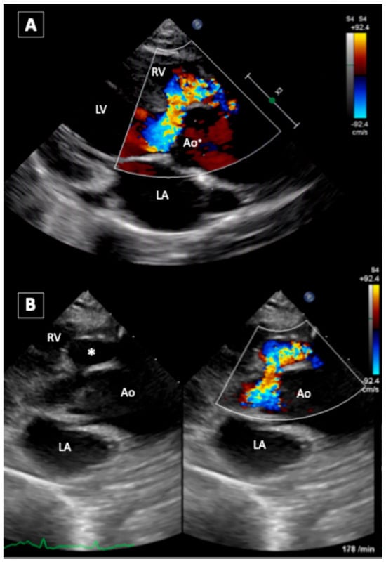
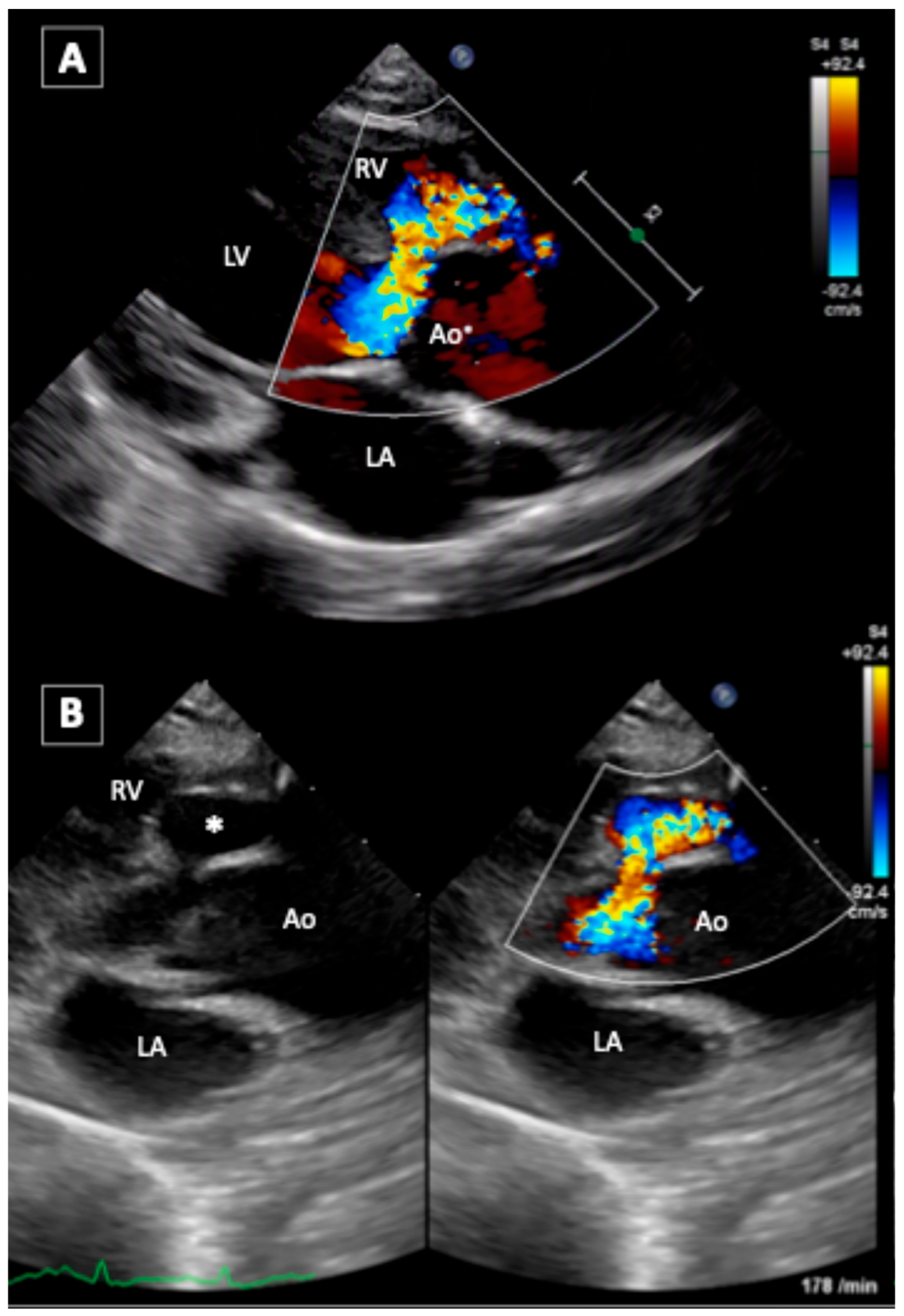
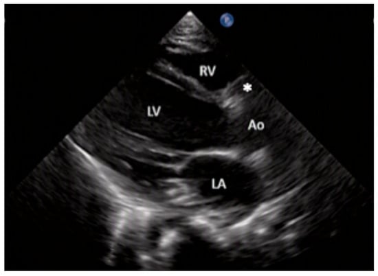
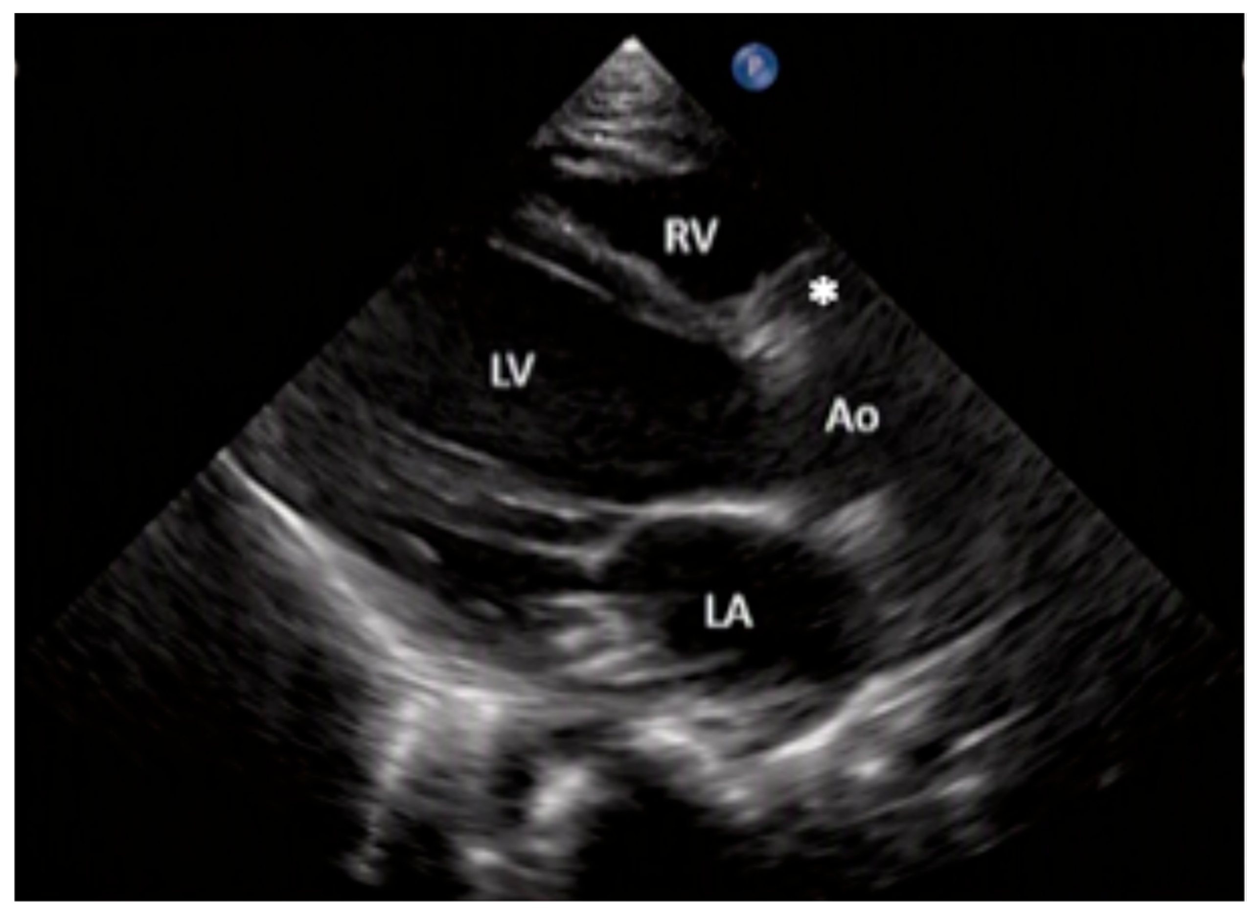
On presentation to our clinic, the patient did not show hypoxemia or respiratory distress, and his capillary refill time was normal. On auscultation, we noted a rhythmic pulse (148/min) and a to-and-fro murmur (3–4/6). Lung and abdominal examinations were unremarkable, and the pulse was palpable in all extremities with normal blood pressure. Electrocardiography showed sinus rhythm, and echocardiography at our clinic confirmed left ventricular dilation (z-score +4.2) with a normal ejection fraction (EF), of approximately 60%. Flow alterations in the RVOT were excluded. An ALVT, indicating abnormal aorta-to-LV regurgitant flow, particularly during diastole, leading to LV volume loading and a PFO was described (Figure 1).
Figure 1.
(A,B) Preoperative transthoracic echocardiography of the left ventricle (LV), right ventricle (RV), and left atrium (LA) reveals the dimension of the tunnel in the parasternal long-axis view and the ascending aorta (Ao), including flow alterations.
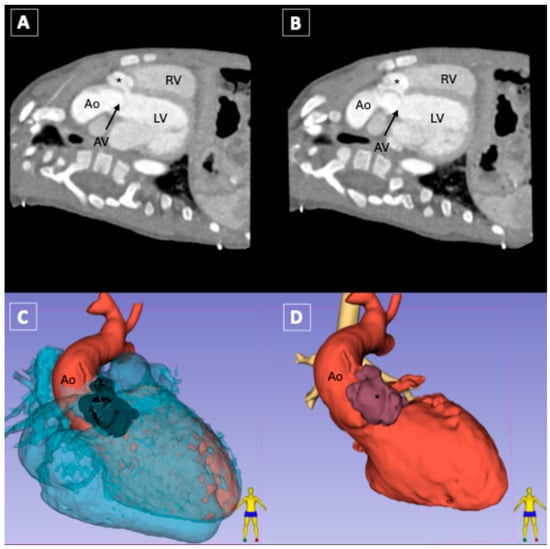
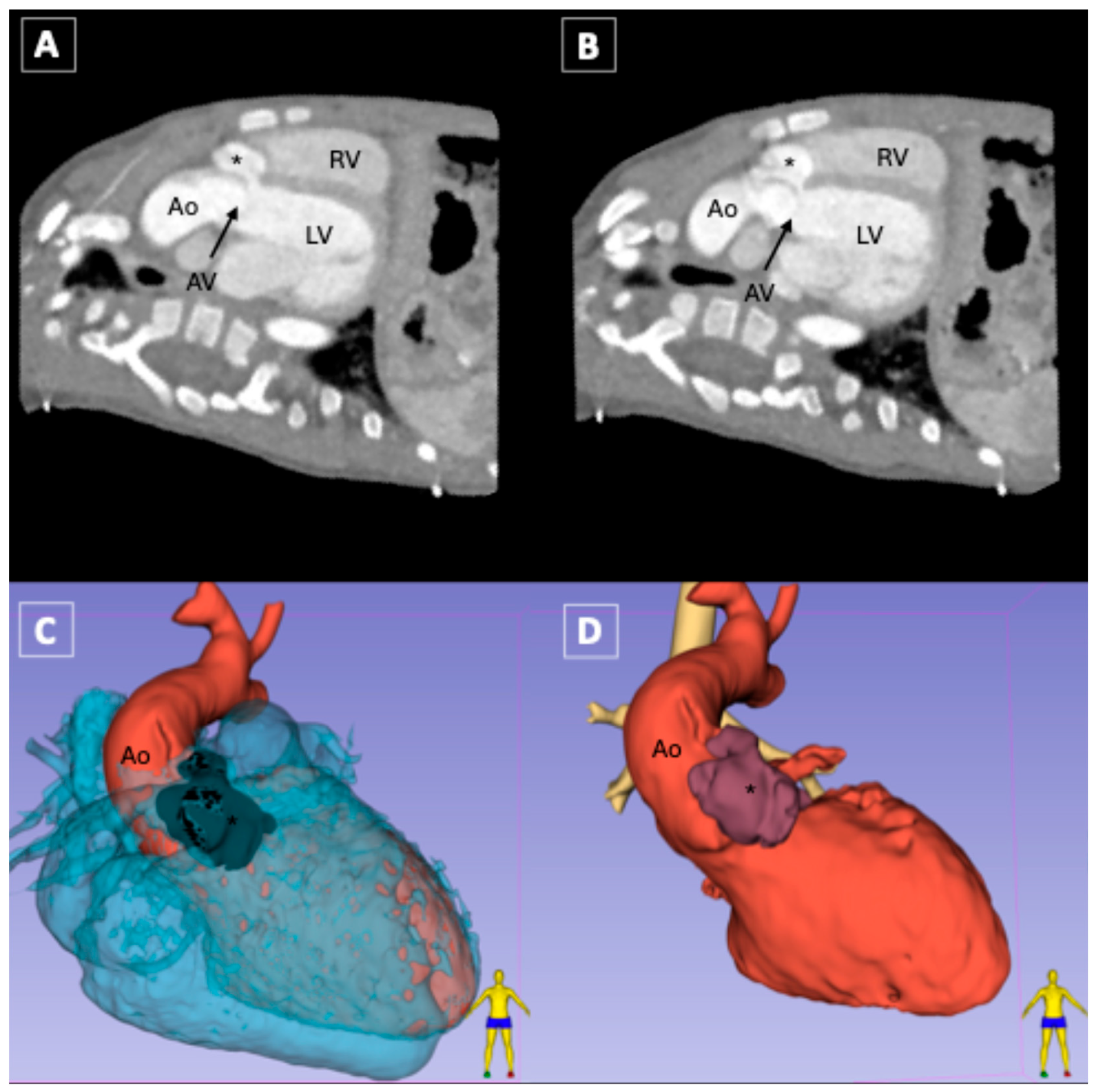
CT angiography (CTA) confirmed communication between the LV and the ascending aorta, originating craniodorsal of the RCA ostium and leading into the LV without direct communication with the RCA (Figure 2). The ventricular orifice measured 8 mm in diameter. The diameter of the tunnel was approximately 10 mm, with an oval opening at the aortic end. It was classified as type II ALVT, according to Hovaguimian, because of a large extracardiac aortic wall aneurysm of the tunnel with an oval opening at the aortic end. After evaluating the patient’s medical history, symptoms, and relevant literature, a board of pediatric cardiologists and pediatric cardiac surgeons recommended surgical correction of the malformation because of left ventricular dilatation as a sign of a hemodynamically significant tunnel with relevant risk of ventricular dysfunction, risk of progressive aortic regurgitation, sudden cardiac death [2], and to address potential coronary involvement.
Figure 2.
(A) CTA demonstrates the ventricular origin from the left ventricle (LV) and (B) the aortic orifice of the ALVT (*) bypassing the aortic valve (AV) in the sagittal plane. (C) Preoperative 3D reconstruction from CTA with the software Slicer (http://www.slicer.org) [6] of the patient’s heart demonstrates the close relation of the ALVT to RVOT and (D) myocardial tissue of the right ventricle (RV) in light blue shows the relation between ALVT in dark red, the LVOT in red and the trachea in the background in yellow.
2.2. Surgical Therapy
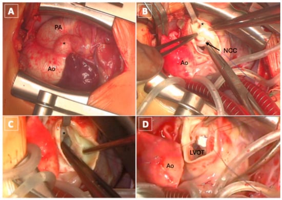
After the usual preoperative preparations, surgery was performed using a standard cardiopulmonary bypass (CPB) technique with cannulation of the inferior vena cava (IVC) and superior vena cava (SVC) under moderate hypothermia (30 °C). Through an incision of the ascending aorta above the aortic valve, cardiac arrest was induced via antegrade selective administration of the Bretschneider histidine-tryptophan-ketoglutarate cardioplegia (HTK) (30 mL/kg body weight over 5 min). The tunnel was visualized as an external bulge on the aorta at the right sinus of Valsalva. The mildly dilated aorta was opened, and the entire tunnel was inspected. Both the aortic and LV openings of the tunnel were identified. The oval-shaped aortic opening extends from the right side of the sinotubular junction to the LV opening through the interventricular septum. The absence of involvement of the aortic valve or the ostium of the RCA, located approximately 8 mm caudoventral of the aortic orifice, was confirmed. A connection to the RVOT was ruled out by incision of the pulmonary artery and inspection of the RVOT. The pulmonary artery was then anastomosed.
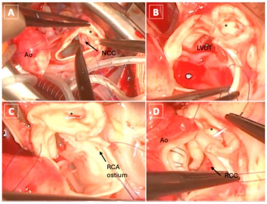
The ALVT was then closed with a patch (Gore-Tex 8 × 5 mm, WL Gore Associates Inc., Flagstaff, AZ, USA) on the ventricular end with continuous suture (6-0 Prolene, Ethicon Inc., Johnson & Johnson, Somerville, NJ, USA) (Figure 3). The aortic origin of the tunnel was then incised and inspected; the patch was tested for closure of the tunnel. Simultaneously, the RCA was probed, and a shunt to the tunnel was excluded by instillation of HTK into the RCA under simultaneous observation of the opened ALVT. The inferior wall of the tunnel was then connected to the ascending aorta via direct suture (5-0 Prolene). During suturing, the RCA ostium was visualized, and arterial compromise was excluded visually (Figure 4). The PFO was closed via direct suture (6-0 Prolene); after 102 min on CPB, an aortic clamping time of 59 min and reperfusion for 35 min, the decannulation process began. Intraoperative transesophageal echocardiography showed normal EF (55%) without regional wall motion abnormalities, competent valves with no contortion of the non-coronary and the right-coronary leaflet, and no further flow through the ALVT.
Figure 3.
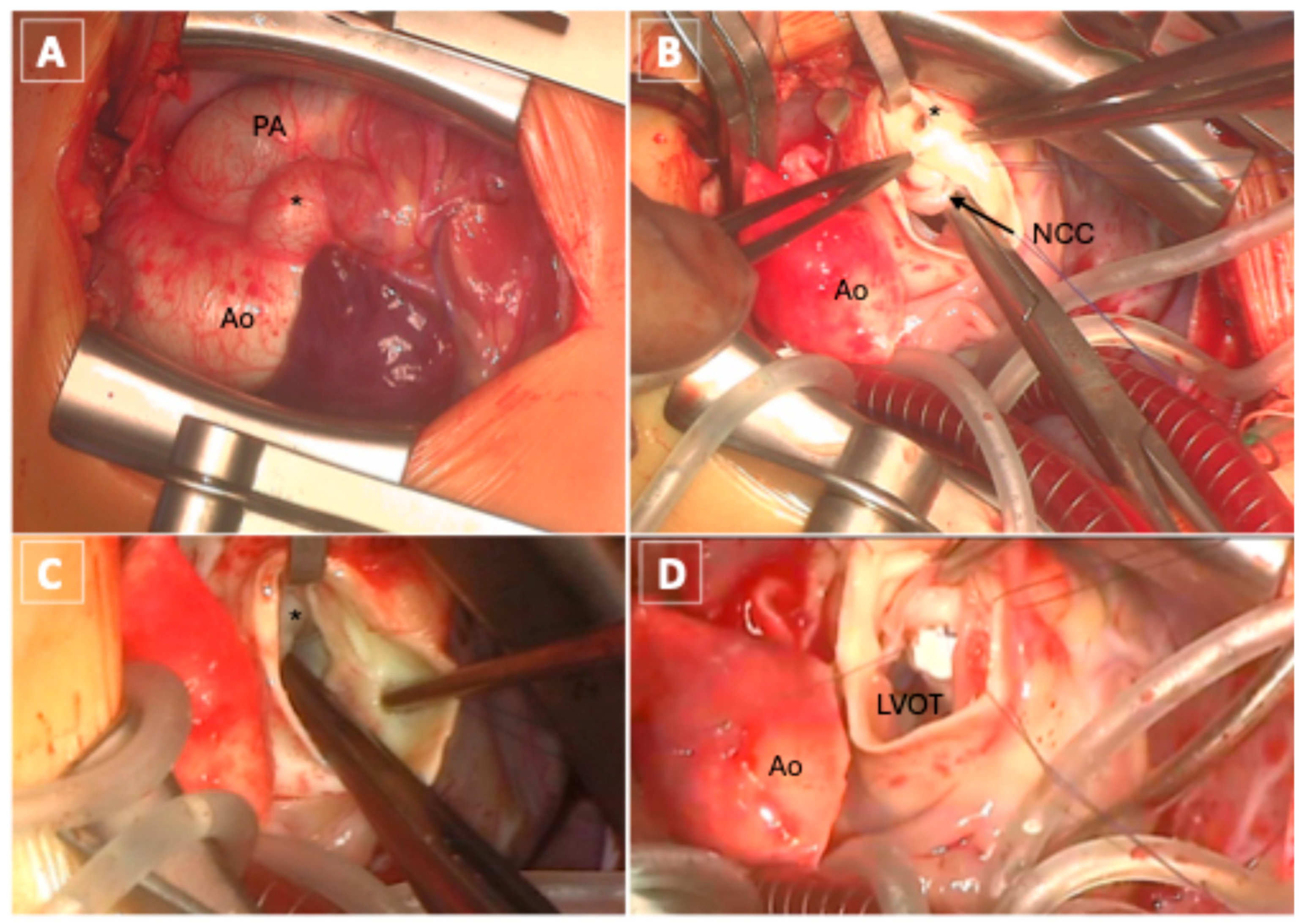
Intraoperative surgeon’s view: (A) top view after thoracotomy with the ALVT (*) between the aorta (Ao) and the pulmonary artery (PA). (B) After aortotomy, the aortic origin of the tunnel (*) lies above the non-coronary cusp (NCC) of the aortic valve; (C) view through the aortic valve on the ventricular orifice before patch-closure and (D) the ventricular origin after patch-closure with the left-ventricular outflow tract (LVOT).
Figure 4.
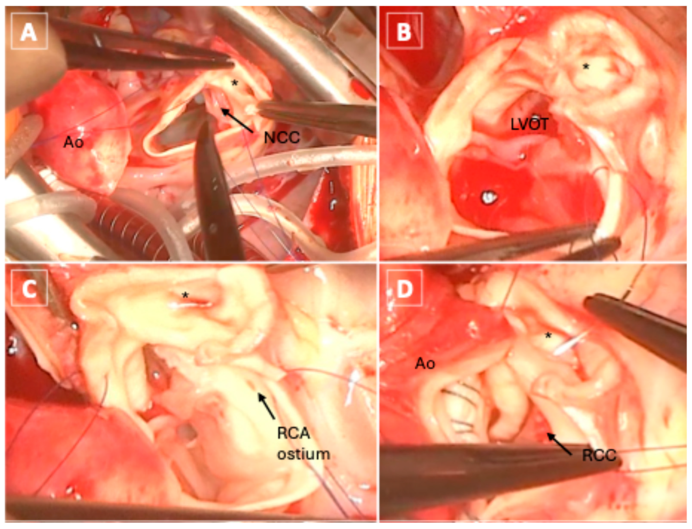
Intraoperative surgeon’s view: (A) view on the ALVT (*) in relation to the aortic valve and non-coronary cusp (NCC) (B) after incision of the ALVT to relate the true diameter of the tunnel in comparison to the ascending aorta (Ao); (C) Shows the close relation of the tunnel’s aortic origin to the caudoventral RCA ostium; (D) the closure of the aortic origin of the tunnel directly to the ascending aorta using the caudal wall of the tunnel to obviate an asymmetry of the cusps of the aortic valve post closure. The right coronary cusp (RCC) is shown here.
After restoring normal heart rate and stabilizing vital parameters, the thorax was closed in the usual manner, and the patient was transferred to the pediatric cardiac intensive care unit (pCICU).
2.3. Postsurgical Progress
Postoperatively, the patient was extubated three hours after surgery and remained hemodynamically stable while receiving positive inotrope therapy.
An echocardiogram performed immediately after admission to the pCICU confirmed closure of the ALVT. A transient postoperative low cardiac output syndrome with LV systolic dysfunction (EF 28%), minor dilatation, basal pleural effusions in both lungs, and reduced central venous oxygen saturation (51%) were observed and treated with milrinone (2–6 µg/kg body weight per hour) and diuretics (furosemide 8 mg three times daily) for 48 h. On the third postoperative day, the patient was transferred to the pediatric cardiac ward. The aortic and atrioventricular valves showed no pathological regurgitation or stenosis, and the right ventricular outflow was unobstructed. On the fifth postoperative day, EF fully recovered (55%), LV dilatation regressed (28.6 mm, z-score +1.2), and no wall motion abnormalities were detected (Figure 5).
Figure 5.
Postoperative echocardiography proves the closure of the tunnel (*) of the LV from the parasternal long axis view without further flow alterations.
The patient was discharged on the sixth postoperative day in good general condition with bisoprolol (1 mg twice daily) and lisinopril (1.2 mg/d) prescribed as anti-congestive medication. Follow-up examinations over 15 months showed an uneventful recovery, with preserved left ventricular function on echocardiography (EF 62%, LVEDD 33 mm, z-score +0.8) and minimal aortic regurgitation (pressure half time (PHT) 437 ms). A stenosis of the aortic valve was excluded, a borderline dilatation of the aortic root (18 mm, z-score +2.0) was noted. No signs of the ALVT were detected. Overall, the child developed normally (Table 1).
Table 1.
Timeline of diagnosis, treatment and follow-up.
3. Discussion
Our patient was referred with a presumed subpulmonary VSD and RVOT aneurysm. In ALVT, the tunnel runs between the aortic root and the subpulmonary infundibulum, so its aneurysmal portion can bulge towards the RVOT and resemble an outlet VSD or infundibular aneurysm on transthoracic views [1,3]. The decisive discriminators are the origin of the jet above the aortic leaflets and its timing: in ALVT, Doppler shows predominantly diastolic flow from the aorta into the LV, whereas a VSD produces systolic left-to-right flow into the right ventricle [3]. In our patient, the absence of shunt flow into the RVOT together with diastolic aorta-to-LV flow established the diagnosis on echocardiography.
Other lesions with abnormal aorto-cardiac flow must be excluded. A ruptured sinus of Valsalva aneurysm arises within the sinus below the sinotubular junction and usually drains into the right heart. A coronary-cameral fistula originates from a dilated coronary artery rather than from the aortic wall. A VSD with aortic cusp prolapse produces regurgitation through the valve itself, in addition to a systolic shunt. Differentiation therefore relies on the site of origin relative to the leaflets and coronary ostia, the course of the channel, and the phase of Doppler flow, which echocardiography and cross-sectional imaging establish together [3].
Consistent with the systematic review, in which echocardiography provided the initial diagnosis in 86.4% of patients and CT and/or MRI confirmed it in 17% [2], TTE identified the tunnel and its hemodynamic consequences in our patient. CTA with 3D reconstruction [6] added information that was essential for operative planning: the dimensions of the tunnel (ventricular orifice 8 mm, tunnel diameter approximately 10 mm), the aneurysmal extracardiac aortic wall component defining Hovaguimian type II [7], the absence of a communication with the RVOT, and the position of the aortic orifice approximately 8 mm craniodorsal of the RCA ostium without functional connection. The 3D model was used to relate the tunnel‘s course to important anatomic landmarks for further procedure planning.
Coronary abnormalities were reported in 9.6% of ALVT cases, and the RCA may even arise from the tunnel itself; therefore, preoperative definition of the coronary anatomy is essential [2]. Transcatheter closure, used in 4.5% of reported patients, has been performed with duct occluders and is increasingly considered in selected cases [2,4,5]. In our patient, however, the aortic orifice lies only approximately 8 mm from the RCA ostium, and the tunnel had an aneurysmal extracardiac component. A device would have risked encroaching on the RCA ostium and distorting the right coronary cusp, and the external diagnosis of an RVOT lesion had not been conclusively excluded. Consequently, surgery was chosen. It also allowed direct inspection of the RVOT through a pulmonary arteriotomy to rule out a second communication, as suggested by the referral diagnosis.
Reported surgical techniques include patch closure of the aortic orifice, patch closure of the ventricular orifice, closure of both ends, obliteration of the tunnel, and techniques that support the aortic root [3]. We closed the ventricular orifice with a patch because it was slit-oval, well-defined, and surrounded by firm septal tissue. The aortic orifice was closed by direct suture of the ascending aorta to the inferior wall of the aortic tunnel orifice because its oval shape at the sinotubular junction allowed tension-free approximation above the right coronary cusp without encroaching on the RCA ostium. Direct closure near the right coronary sinus can distort the cusp and lead to aortic regurgitation [1,8]. We excluded this by visualizing the RCA ostium throughout suturing and by injecting HTK into the RCA while inspecting the whole incised tunnel. Intraoperative transesophageal echocardiography then showed a competent aortic valve, no residual tunnel flow, and no regional wall motion abnormalities. Because there were no echocardiographic signs of ischemia of the inferior wall, no further coronary imaging was added.
Early presentation does not imply a milder phenotype: large tunnels and associated aortic valve disease can cause heart failure in infancy, and congestive heart failure was present in about one third of reported patients [2]. Conversely, repair in neonates and infants can be performed with low operative risk and good late functional outcomes [9]. In our patient, transient LV systolic dysfunction (EF 28%) after cardiopulmonary bypass was treated with milrinone and diuretics for 48 h, and EF recovered to 55% by postoperative day five. In the systematic review, mortality was 5.7% and mild residual aortic regurgitation was present in 22.7% of patients [2]; late aortic regurgitation and aortic root dilation have also been described after repair [8]. At 15 months, our patient had minimal aortic regurgitation (pressure half-time 437 ms), a borderline dilated aortic root (18 mm, z-score 2.0) and no residual tunnel flow. Echocardiographic surveillance of the aortic valve, aortic root and LV is planned yearly.
4. Conclusions
This case shows that an ALVT can mimic a subpulmonary VSD with an RVOT aneurysm in infancy. Echocardiography identified the aorto-LV communication and its hemodynamic effect, whereas CTA with 3D reconstruction defined the tunnel course and its relationship to the RCA and RVOT. Knowing that the aortic orifice lay approximately 8 mm from the RCA ostium without functional communication allowed a repair, with patch closure of the ventricular orifice and direct closure of the aortic orifice, that preserved the native RCA and aortic valve. Because residual or late aortic regurgitation and aortic root dilation may develop after repair, long-term echocardiographic surveillance is warranted; at 15 months, our patient had normal LV size and function and only minimal aortic regurgitation.
Author Contributions
Conceptualization, J.C.G., M.H. and D.B.; methodology, J.C.G., I.H. and M.M.; investigation, J.C.G., I.H., M.M., H.C., J.O., F.S., S.H.H., P.S., T.S.M., N.L., R.K.-F., M.H. and D.B.; resources, T.S.M., N.L., R.K.-F., J.S.S., M.H. and D.B.; data curation, J.C.G., I.H. and M.M.; visualization, J.C.G., I.H., M.M. and D.B.; writing—original draft preparation, J.C.G., I.H. and M.M.; writing—review and editing, H.C., J.O., F.S., S.H.H., P.S., C.R., T.S.M., N.L., J.S.S., R.K.-F., M.H. and D.B.; supervision, T.S.M., N.L., R.K.-F., M.H. and D.B.; project administration, J.C.G. and D.B. All authors have read and agreed to the published version of the manuscript.
Funding
This research received no external funding.
Institutional Review Board Statement
In accordance with the institutional guidelines of the University Medical Center Hamburg-Eppendorf (UKE) and the Medical Association of Hamburg, formal ethical board review is not required for a single retrospective case report. Comprehensive written informed consent was obtained directly from the patient for the publication of this case report and any accompanying clinical images. A copy of the written consent is available for review by the Editor-in-Chief of this journal.
Informed Consent Statement
Written informed consent has been obtained from the patient’s parents to publish this paper.
Data Availability Statement
The manuscript was prepared according to the CARE case-report guidelines. Data are available from the corresponding author on reasonable request, subject to patient privacy.
Conflicts of Interest
The authors declare no conflicts of interest.
Abbreviations
The following abbreviations are used in this manuscript:
| ALVT | Aorto-left-ventricular tunnel |
| LV | Left ventricle |
| EF | Ejection fraction |
| TTE | Transthoracic echocardiography |
| VSD | Ventricular septal defect |
| RVOT | Right ventricular outflow tract |
| CT | Computed tomography |
| CTA | Computed tomography with angiography |
| RCA | Right coronary artery |
| AVT | Aorto-ventricular tunnel |
| ARVT | Aorto-right-ventricular tunnel |
| PFO | Patent foramen ovale |
| CPB | Cardiopulmonary bypass |
| IVC | Inferior vena cava |
| SVC | Superior vena cava |
| pCICU | Pediatric cardiac intensive care unit |
| AR | Aortic regurgitation |
| AV | Aortic valve |
| Ao | Aorta |
| NCC | Non-coronary cusp |
| RCC | Right-coronary cusp |
| HTK | Bretschneider histidine-tryptophan-ketoglutarate cardioplegia |
| LA | Left atrium |
| LVEDD | Left ventricular end-diastolic diameter |
| LVOT | Left ventricular outflow tract |
| MRI | Magnetic resonance imaging |
| PA | Pulmonary Artery |
| PHT | Pressure half time |
| RV | Right ventricle |
| TEE | Transesophageal echocardiography |
References
- McKay, R.; Anderson, R.H.; Cook, A.C. The aorto-ventricular tunnels. Cardiol. Young 2002, 12, 563–580. [Google Scholar] [CrossRef] [Scilit] [PubMed]
- Bassareo, P.P.; Secinaro, A.; Ciliberti, P.; Perrone, M.A.; Linnane, N.; Duignan, S.; Ferrero, P.; Chessa, M.; Walsh, K.P.; Mcmahon, C.J. Aorto-Left Ventricular Tunnel: The First Systematic Review of An Uncommon Entity (177 Worldwide Cases from 1965 to 2024). Rev. Cardiovasc. Med. 2025, 26, 26005. [Google Scholar] [CrossRef] [Scilit] [PubMed]
- McKay, R. Aorto-ventricular tunnel. Orphanet J. Rare Dis. 2007, 2, 41. [Google Scholar] [CrossRef] [Scilit] [PubMed]
- Linnane, N.; Alshahrani, D.; Kenny, D.P.; Walsh, K.P.; McMahon, C.J. Evolution in the management of aorta to left ventricular tunnel in a national congenital cardiology centre. Cardiol. Young 2023, 33, 1753–1756. [Google Scholar] [CrossRef] [Scilit] [PubMed]
- Chessa, M.; Chaudhari, M.; De Giovanni, J.V. Aorto-left ventricular tunnel: Transcatheter closure using an amplatzer duct occluder device. Am. J. Cardiol. 2000, 86, 253–254. [Google Scholar] [CrossRef] [Scilit] [PubMed]
- Fedorov, A.; Beichel, R.; Kalpathy-Cramer, J.; Finet, J.; Fillion-Robin, J.-C.; Pujol, S.; Bauer, C.; Jennings, D.; Fennessy, F.; Sonka, M.; et al. 3D Slicer as an image computing platform for the Quantitative Imaging Network. Magn. Reson. Imaging 2012, 30, 1323–1341. (In English) [Google Scholar] [CrossRef] [Scilit] [PubMed]
- Hovaguimian, H.; Cobanoglu, A.; Starr, A. Aortico-left ventricular tunnel: A clinical review and new surgical classification. Ann. Thorac. Surg. 1988, 45, 106–112. (In English) [Google Scholar] [CrossRef] [Scilit] [PubMed]
- Serino, W.; A006Edrade, J.L.; Ross, D.; de Leval, M.; Somerville, J. Aorto-left ventricular communication after closure. Late postoperative problems. Br. Heart J. 1983, 49, 501–506. (In English) [Google Scholar] [CrossRef] [Scilit] [PubMed]
- Sousa-Uva, M.; Touchot, A.; Fermont, L.; Piot, D.; Delezoide, A.L.; Serraf, A.; Lacour-Gayet, F.; Roussin, R.; Bruniaux, J.; Planché, C. Aortico-left ventricular tunnel in fetuses and infants. Ann. Thorac. Surg. 1996, 61, 1805–1810. (In English) [Google Scholar] [CrossRef] [Scilit] [PubMed]

Disclaimer/Publisher’s Note: The statements, opinions and data contained in all publications are solely those of the individual author(s) and contributor(s) and not of MDPI and/or the editor(s). MDPI and/or the editor(s) disclaim responsibility for any injury to people or property resulting from any ideas, methods, instructions or products referred to in the content. |
© 2026 by the authors. Licensee MDPI, Basel, Switzerland. This article is an open access article distributed under the terms and conditions of the Creative Commons Attribution (CC BY) license.