


Abstract
Background/Objectives: Over the past three decades, significant progress has been made in understanding the biology of neuroblastoma. The integration of prognostic factors has facilitated risk stratification and the development of targeted treatment approaches. This study aims to analyze the outcomes of pediatric patients with neuroblastoma treated at a single oncology center over a 30-year period. Methods: This retrospective study analyzed data from patients aged 0–18 years with neuroblastoma, treated at the Department of Pediatric Hematology and Oncology in Bydgoszcz, Poland, between 1993 and 2023. The study endpoints included the 5-year probability of overall survival (pOS), event-free survival (pEFS), and relapse-free survival (pRFS), analyzed separately for low/intermediate- and high-risk groups. Results: Seventy-five patients met the inclusion criteria. Thirty-two children were categorized as high-risk patients and forty-three as low/intermediate risk. During the study period, outcomes in the low/intermediate-risk group improved significantly (the 5-year pOS 85.7% vs. 100.0%, p = 0.019; the 5-year pRFS 85.7% vs. 100.0%, p = 0.662; the 5-year pEFS 83.3% vs. 100.0%, p = 0.038). In the high-risk group, outcomes improved but did not reach statistical significance (the 5-year pOS 0.0% vs. 41.1%, p = 0.342; the 5-year pRFS 0.0% vs. 32.5%, p = 0.180; and the 5-year pEFS 0.0% vs. 21.5%, p = 0.537). Sixteen patients experienced relapse, of whom only three survived; stem cell transplantation at relapse significantly improved survival (OS 0.0% vs. 50.0%, p = 0.001). In the multivariable analysis, stage at diagnosis was a prognostic factor for pOS (HR 6.0; 95%CI 0.7–49.6, p = 0.096), while pelvic localization was a risk factor for pRFS (HR 3.0; 95%CI 0.8–10.5; p = 0.084). Conclusions: This analysis highlights significant advancements in the diagnosis and treatment of neuroblastoma. Nevertheless, outcomes for high-risk patients and those who experience relapse remain poor, underscoring the need for further therapeutic improvements.
1. Introduction
Neuroblastoma (NBL) is the most common extracranial solid tumor of childhood, characterized by heterogeneous biology and a variable clinical course. The primary tumor arises from primordial neural crest cells and can develop anywhere where sympathetic tissue is present. However, the majority of cases are diagnosed in the adrenal glands or paravertebral ganglia [1,2]. Clinical presentation ranges from an incidentally detected adrenal mass to advanced metastatic disease with systemic manifestations. Moreover, the natural history of neuroblastoma is highly heterogeneous, with some cases exhibiting spontaneous regression or differentiation, while others follow a highly aggressive course with rapid tumor progression and widespread metastasis [1,2,3].
For more than three decades, neuroblastoma treatment has served as a paradigm for tailored, personalized therapy in pediatric oncology [1,4,5,6,7,8,9,10]. Using known biological and clinical factors, patients with neuroblastoma can be divided into three therapeutic groups: low-risk, intermediate-risk, and high-risk. As knowledge about prognostic factors and therapy responses continues to expand, each risk group is further subdivided into smaller, more specific therapeutic subgroups. Although the overall survival rate in the low- and intermediate-risk groups exceeds 90%, high-risk patients have a long-term survival rate of less than 50% [2,5,11,12,13,14]. The implementation of multimodal, intensive chemotherapy, followed by megatherapy with autologous hematopoietic stem cell transplantation (auto-SCT) and maintenance therapy with 13-cis-retinoic acid (13-cisRA), as well as the introduction of immunotherapy with anti-GD2 monoclonal antibodies, has significantly improved outcomes in the HR group [1,6,9,10,11,14,15]. However, children with refractory or relapsed disease have a particularly unfavorable prognosis, and currently, no established effective therapy exists for these patients [16,17,18,19,20]. The gap in our understanding of effective therapies for refractory and relapsed disease remains an important clinical problem.
This study aims to analyze the risk factors, treatment strategies, and outcomes of the entire cohort of pediatric patients with NBL treated at a single oncology center in Poland over a 30-year period. The analysis reflects the expanding knowledge about neuroblastoma biology and the improved therapy results related to the introduction of novel therapeutic modalities in NBL treatment.
2. Materials and Methods
This retrospective study analyzed the prognostic factors and outcomes in pediatric patients diagnosed with neuroblastoma and treated in a single tertiary oncology center in Poland during the respective therapeutic periods.
Data from patients aged 0–18 years treated at the Department of Pediatric Hematology and Oncology, University Hospital No. 1 in Bydgoszcz between January 1993 and December 2023 were analyzed. The observations were completed in November 2024. The study included newly diagnosed tumors of neuronal origin that were confirmed as neuroblastomas by pathologists with experience in pediatric malignancies. Clinical data for patients treated between 1994 and 2003 were obtained from traditional medical records, whereas data from 2004 onward were stored and reviewed electronically. Patients were excluded if they had incomplete medical data, were diagnosed with malignancies other than neuroblastoma (e.g., ganglioneuroma, paraganglioma), were lost in follow-up, or were treated at other centers and referred to the Department solely for stem cell transplantation (SCT) as part of therapy.
Patients’ medical histories were reviewed with a particular focus on the diagnosis of cancer-predisposition syndromes before NBL incidence and family history of malignancies. The symptoms presenting at diagnosis included hypertension (blood pressure above the 95 percentile for age and height), tachycardia, pain, constipation, diarrhea, Horner syndrome (unilateral ptosis, anhidrosis, and miosis), signs of spinal cord compression (muscle weakness, sensory deficits, urinary and rectal sphincter dysfunction), and opsoclonus–myoclonus–ataxia (OMA) syndrome.
The histological diagnosis of neuroblastoma was based on conventional tissue-staining histology with additional immunohistochemistry, if indicated. The following NBL subtypes were diagnosed in the analyzed cohort: differentiating NBL, poorly differentiated NBL, undifferentiated NBL, and ganglioneuroblastoma. If the histologic subtype was not indicated in the medical records, neuroblastoma not otherwise specified (NBL NOS) was diagnosed.
At diagnosis, blood tests for hematologic and biochemistry (complete blood count, ferritin, and lactate dehydrogenase levels) and urinary dopamine and catecholamine metabolites, including homovanillic acid (HVA) and vanillylmandelic acid (VMA), were performed. The radiologic work-up included chest X-rays, abdominal ultrasonography (USG), computed tomography (CT), or magnetic resonance imaging (MRI) of the primary tumor with tumor volume calculation and iodine-123-labeled metaiodobenzylguanidine (MIBG) scan (technetium bone scan if primary tumor MIBG negative). Bone marrow involvement was evaluated using bone marrow aspirations and bone marrow trephines (both from two separate evaluable sites). Since 2001, tumor studies have been conducted for the MYC gene copy number status, and since 2015, for genomic copy number profiles with high-resolution array comparative genomic hybridization (aCGH) and ALK gene mutations.
All patients were staged according to the revised International Neuroblastoma Staging System (INNS) [21]. Since 2009, an additional staging system according to the International Neuroblastoma Risk Group Staging System (INRGSS) has been used [22].
The definition of risk groups varies between different protocols and therapeutic periods, as successive research has introduced new prognostic factors for risk group stratifications [5,6,7,8,12,13,14,21,22,23]. Currently, risk group stratification is based on various features related to symptoms, diagnosis, and tumor biology, including tumor genetic profiles. However, many of these factors, measurements, and results were not available during earlier analysis periods, particularly before the 2000s. To enable reliable comparisons between specific therapeutic approaches in this study, patients were divided into two risk groups based on age and disease stage at diagnosis. Patients were classified as low/intermediate risk if they were diagnosed with stage 1–3 neuroblastoma according to INNSS or with stage 4 disease within the first 12 months of life (including stage 4S). Patients older than 12 months with stage 4 disease were classified as high risk.
Patients were treated according to therapeutic protocols dedicated to the relevant risk groups.
The high-risk group was treated according to the Study Group of Japan for Treatment of Advanced Neuroblastoma Tokyo (TOKYO) protocol from January 1993 to January 2002 and the High Risk Neuroblastoma Study 1 (HR-NBL1) of SIOPEN from February 2002 onwards [6,7,9,14,15,24,25].
Patients from the low/intermediate-risk group were treated according to the French Society of Pediatric Oncology Neuroblastoma 90 and 94 therapeutic protocols (SFOP NBL) from January 1993 to October 2002, and a subgroup of patients at stage 1–2 was treated according to the SFOP NBL study to June 2011 [4,5,11,23]. From November 2002 to June 2011, stage 3 patients and patients at stage 4 younger than 12 months were treated according to a Multicenter Study for Infants designed by the International Society of Pediatric Oncology European Neuroblastoma Study Group (SIOPEN INES) [12,13]. From July 2011 onwards, all patients from the low/intermediate-risk group were treated according to the SIOPEN European Low and Intermediate Risk Neuroblastoma study Version 3.0 (LINES 3.0) [8]. One patient from the low/intermediate-risk group was treated according to the TOKYO protocol, and one according to HR-NBL1, based on the individual decision of the therapeutic team.
Surgery of primary tumors was performed according to the respective therapeutic protocols and guidelines. The main recommendations are summarized in Table 1.
Table 1.
The main recommendations for surgery according to the analyzed therapeutic protocols.
Complete resection was defined as the macroscopically complete removal of the tumor, permitting the presence of microscopic residuals. Complete remission (CR) was defined as the absence of tumor in any site. Progression was defined as the appearance of a new site of disease, an increase of any measurable lesion by >25%, or a previous negative marrow positive for tumor during first-line treatment [21]. Relapse was defined as the reappearance of the tumor after achieving CR. Event was defined as relapse, progression, secondary malignancy, or death from any cause. Overall survival (OS) was the time calculated from diagnosis to death or last observation, event-free survival (EFS) included the time from diagnosis to an event, and relapse-free survival (RFS) was calculated as the time from diagnosis to relapse.
The endpoints of the study were a 5-year probability of overall survival (pOS), a 5-year probability of event-free survival (pEFS), and a 5-year probability of relapse-free survival (pRFS).
Survival curves of pOS, pEFS, and pRFS were analyzed according to the Kaplan–Meier method and compared by the log-rank test. All features measured at diagnosis were included in the univariate analysis to determine their impact on outcomes. Factors significant in the univariate analysis were used in the multivariate Cox proportional hazards regression model. A value of p < 0.05 was considered statistically significant. Statistical analysis was performed using MedCalc® statistical software Version 23.1.3 (MedCalc Software, Mariakerke, Belgium).
3. Results
3.1. Patient Characteristics
During the period from January 1993 to December 2023, a total of 113 patients aged 0–18 years were hospitalized in the Department of Pediatric Hematology and Oncology with the diagnosis of tumors of neuronal origin. Among them, 107 were finally diagnosed with neuroblastoma, 4 with ganglioneuroma, and 2 with paraganglioma. Within the 107 neuroblastoma patients, 20 have been treated in different oncology centers and referred to our department for stem cell rescue procedures. Another one was hospitalized solely for the stem cell harvesting procedure without SCT. Eight patients were excluded because of insufficient or incomplete data available, and three patients were lost in follow-up (they were transferred to other oncology centers by their parents’ decision). In total, 75 patients were included in the analysis.
Median age at diagnosis was 1.8 years (range 0.0–12.4 years), and 28 patients (37.3%) were younger than 12 months at diagnosis. None of the analyzed children had a family history of neuroblastoma or other malignancies, and one was earlier diagnosed with cancer predisposition syndrome (neurofibromatosis type 1). However, two patients (a 6-month-old boy and a 4.6-year-old girl) had café au lait spots in clinical examinations at diagnosis. Stage 1 disease was diagnosed in 11 patients (14.7%), stage 2 in 6 (8.0%), and stage 3 in 15 cases (20.0%). Most patients presented with metastatic disease (n = 43, 57.3%), of whom six presented with stage 4S. Thirty-two children were categorized as high-risk patients and forty-three as low/intermediate risk. Patients’ detailed characteristics are shown in Table 2.
Table 2.
Patients’ characteristics considering respective therapeutic protocols.
3.2. Treatment in the Low/Intermediate-Risk Group
In the low/intermediate-risk group, most patients were treated according to the LINES 3.0 therapeutic protocol (n = 20, 46.2%). Complete resection during either primary or delayed surgery was achieved in 28 patients (65.1%). Among these, 18 patients underwent complete macroscopic removal of the primary tumor during initial surgery. Primary surgery served as the definitive therapy in 15 cases, including 12 stage 1 patients, 2 stage 2 patients, and 1 stage 4S patient. Three stage 4S patients had only a biopsy of the primary tumor without further treatment. Radiotherapy was administered as a complementary therapy in eight cases (18.6%).
3.3. Treatment in the High-Risk Group
The majority of patients in the high-risk group were treated according to the HR-NBL1 therapeutic protocol (n = 25, 78.1%). All patients were diagnosed based on a biopsy of either the primary tumor or a metastatic site and received primary chemotherapy. Delayed complete resection was achieved in three cases (9.4%). Twenty patients underwent autologous-stem cell transplantation (auto-SCT). In 15 cases, local radiotherapy (RTX) was performed following induction chemotherapy, surgery, and auto-SCT. Sixteen children received maintenance treatment with 13-cis-retinoic acid (13-cisRA), and six patients were eligible to receive dinutuximab beta (anti-GD2 monoclonal antibodies) during maintenance therapy.
3.4. Outcomes
Complete remission was achieved by 82.7% of patients (n = 62). For the entire cohort, the 5-year pOS was 66.7%. Patients in the low/intermediate-risk group had a significantly better 5-year pOS compared to those in the high-risk group (91.9% vs. 35.9%, p < 0.001). Relapse occurred in 16 cases, with a median time to relapse of 0.8 years (range 0.0–3.2 years). In seven cases, relapse was observed at the primary tumor site, while in five cases, the patients presented with disseminated disease at the time of relapse diagnosis. Characteristics of patients with relapsed disease and details of subsequent therapies are presented in Table 3. Among patients who experienced relapse, only three survived. Stem cell transplantation (SCT) in relapse was the only therapeutic modality that significantly improved outcomes in this group (OS 0.0% vs. 50.0% for patients who underwent SCT in relapse, p = 0.001).
Table 3.
Detailed characteristics of patients with relapse.
The 5-year pRFS for the whole cohort was 68.3%. Patients in the high-risk group had a significantly higher risk of relapse compared to those in the low/intermediate-risk group (the 5-year pRFS 28.3% vs. 94.9%, p < 0.001).
The 5-year pEFS for all analyzed patients was 57.3%, with a 5-year pEFS of 94.9% for the low/intermediate-risk group and 28.3% for the high-risk group (p < 0.001). Disease progression was observed in five cases. Four patients with progression were treated with chemotherapy, of whom two underwent SCT; one child received palliative care only. None of the patients with disease progression survived. No cases of secondary malignancies were observed.
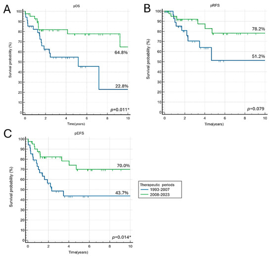
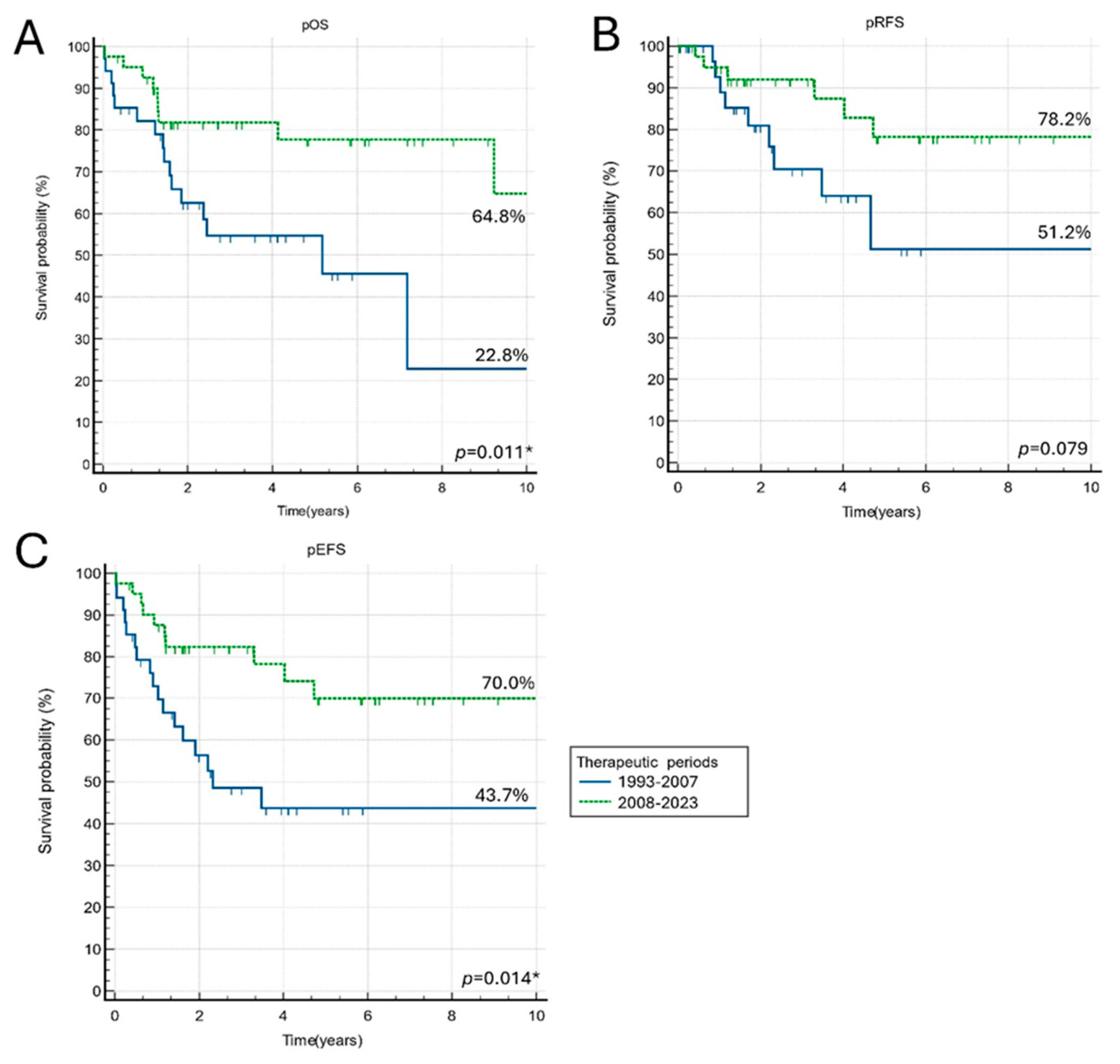
Patients treated according to modern therapeutic protocols, utilizing genetic-based stratification, had better outcomes. Patients treated during the second half of the analyzed period have significantly better pOS, pEFS, and pRFS compared to the cohort treated before the year 2008 (Figure 1).
Figure 1.
The long-term probability of overall survival (pOS), event-free survival (pEFS), and relapse-free survival (pRFS) divided by therapeutic periods. (A) The 10-year pOS for the whole cohort divided by therapeutic periods; (B) The 10-year pRFS for the whole cohort divided by therapeutic periods; (C) The 10-year pEFS for the whole cohort divided by therapeutic periods. * p < 0.05.
Twenty-four patients died, with the most common causes of death being disease relapse (n = 9, 37.5%) and treatment-related complications (n = 9, 37.5%). Four patients died due to primary disease progression, and in two cases, the cause of death was unknown (both patients died outside our hospital). Among the treatment-related deaths, four were attributed to infectious complications, four to treatment toxicity, and one to complications following primary tumor resection. The median time from diagnosis to death was 1.3 years.
3.5. Therapy Results in the Low/Intermediate Group
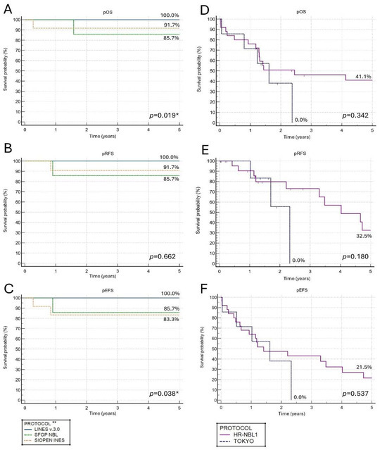
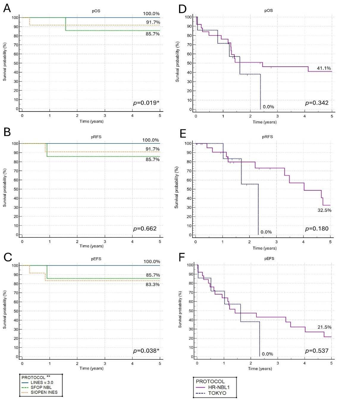
The differences between therapeutic protocols in 5-year pOS and pEFS were statistically significant, with the best results observed in patients treated according to LINES 3.0 (Figure 2). No progression was observed in this group. One patient treated with the TOKYO protocol died due to treatment-related complications; one patient treated with the HR-NBL1 protocol is alive in remission. Relapse occurred in three patients. After relapse, one patient received chemotherapy only, one received chemotherapy followed by auto-SCT and additional RTX, and one received chemotherapy followed by auto-SCT, RTX, and maintenance with 13-cisRA. Three patients died: one in relapse (patient primary treated with SFOP NBL 94 protocol and with TOKYO after relapse), one due to treatment-related toxicity (infection), and in one case, the cause of death was unknown (the patient died in a different hospital after completion of therapy).
Figure 2.
The 5-year probability of overall survival (pOS), event-free survival (pEFS), and relapse-free survival (pRFS) for consecutive therapeutic protocols divided by risk groups. (A) The 5-year pOS for low/intermediate-risk group patients according to therapy protocols; (B) The 5-year pRFS for low/intermediate-risk group patients according to therapy protocols; (C) The 5-year pEFS for low/intermediate-risk group patients according to therapy protocols; (D) The 5-year pOS for high-risk group patients according to therapy protocols; (E) The 5-year pRFS for high-risk group patients according to therapy protocols; (F) The 5-year pEFS for high-risk group patients according to therapy protocols; * p < 0.05; ** One patient treated with TOKYO and one patient treated with HR-NBL1 protocol were excluded from the (A–C).
3.6. Therapy Results in the High-Risk Group
Figure 2 shows that 5-year pOS was superior in patients treated according to the HR-NBL1 protocol. However, the results did not reach statistical significance. Patients treated with auto-SCT and maintenance with 13-cisRA had a better prognosis (the 5-year pOS 50.3% vs. 24.0%, p = 0.001). In the subgroup treated in maintenance with an addition of anti-GD2 monoclonal antibodies, the 5-year pOS was better, but the difference did not reach statistical significance (the 5-year pOS 50.0% vs. 38.8%, p = 0.449). However, prolonged observations indicated prominent improvement in long-term outcomes among patients treated with anti-GD2 monoclonal antibodies (the 10-year pOS 50.0% vs. 12.1%).
Thirteen patients experienced relapse, with a median time to relapse of 0.9 years. The differences in 5-year pRFS between protocols were visible but not statistically significant (0.0% vs. 32.5%, p = 0.180, Figure 2). None of the therapy modalities used in first-line treatment (auto-SCT procedure, maintenance with 13-cisRA, or anti-GD2 monoclonal antibodies) significantly reduced the probability of relapse.
The 5-year pEFS was 0.0% for TOKYO and 21.5% for the HR-NBL1 protocol (Figure 2). Although therapy results with the auto-SCT procedure were better (the 5-year pEFS 26.8% vs. 20.0%, p = 0.001), the outcomes remained poor. The addition of maintenance with 13-cisRA significantly improved the 5-year pEFS (34.8% vs. 0.0%, p = 0.001 for 13-cisRA). Patients treated with anti-GD2 monoclonal antibodies had a higher 5-year pEFS, but the differences were not statistically significant (50.0% vs. 13.8%, p = 0.308).
3.7. Prognostic Factors
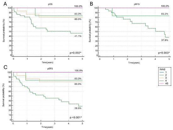
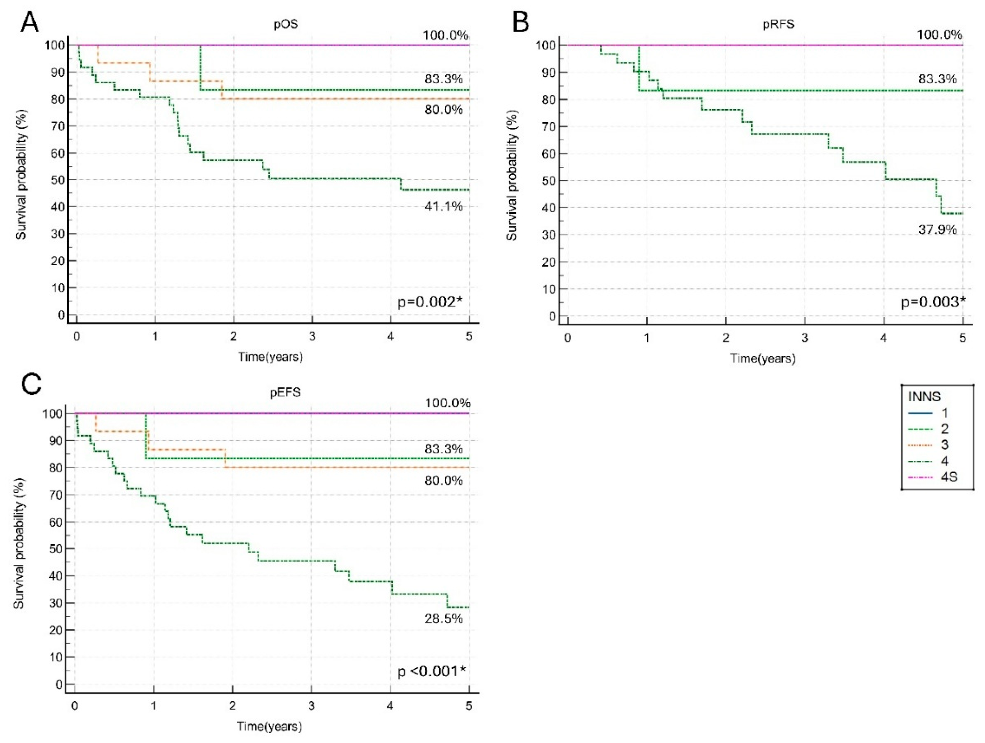
Stage at diagnosis was a significant prognostic factor. Patients with stage 1 and 4S disease had an excellent 5-year pOS and pEFS of 100.0%. In patients with stage 2 and 3 neuroblastoma, the 5-year pOS and pEFS reached 80.0% or above, while patients with stage 4 neuroblastoma had the 5-year pOS of 46.3%, the 5-year pRFS of 37.9%, and the 5-year pEFS of 28.5% (Figure 3).
Figure 3.
The outcomes according to five consecutive International Neuroblastoma Staging System (INNS) disease stages. (A) The 5-year probability of overall survival (pOS) for five consecutive INNS disease stages; (B) The 5-year probability of relapse-free survival (pRFS) for five consecutive INNS disease stages; (C) The 5-year probability of event-free survival (pEFS) for five consecutive INNS disease stages. * p < 0.05.
Age above 12 months at diagnosis was a risk factor for death (OR 7.9, 95% CI 2.1 to 29.9, p = 0.002), event (OR 6.8, 95% CI 2.1 to 22.7, p = 0.014), and relapse (OR 13.6, 95% CI 1.7 to 109.3, p = 0.014). Other significant risk factors included symptoms such as pain, hypertension, and tachycardia at diagnosis, increased LDH and ferritin levels, poorly differentiated histology, primary tumor localization in the pelvis or retroperitoneum, MIBG-positive tumors, N-MYC gene amplification, and structural chromosomal alterations (SCA) in tumor tissue (Table 4). In the multivariable analysis, INNS stage was a risk factor of borderline significance for pOS and pEFS, while pelvis localization was a factor of borderline significance for pRFS (Table 5).
Table 4.
Univariate analysis of risk factors.
Table 5.
Multivariable analysis of risk factors.
4. Discussion
This 30-year, single-center analysis reflects significant improvements in the diagnostic and therapeutic approaches for NBL. As Poland has never established a national therapeutic program for neuroblastoma, our department has used therapeutic protocols from other countries [4,5,6,7,8,9,11,12,13,14,15,23,26]. In the 1990s, patients were treated using Japanese and French protocols. The results obtained in our cohort were inferior to those reported by the Neuroblastoma Study Group of the Société Francaise d’Oncologie Pédiatrique and the Study Group of Japan [4,6,14,15]. Poorer outcomes could be attributed to limited access to detailed therapy guidelines and high rates of treatment-related mortality. Since the early 2000s, patients have been treated according to international therapy protocols, leading to significant improvements in treatment outcomes.
During the study period, diagnostic and therapeutic recommendations have evolved into more specific and comprehensive guidelines, enabling precise execution of all therapeutic stages. An excellent example of the development is surgical guidelines. In the 1980s and 1990s, the surgery range and acceptable extension of the surgical site, as well as surgery timing, were assessed individually by surgeons [6]. Currently, the surgical guidelines are detailedly described in therapeutic protocols, with well-recognized image-defined risk factors and surgery time [1,2,7,8,27]. Moreover, the final decision regarding the surgical range is made by an interdisciplinary team comprising a pediatric oncologist, radiologist, and surgeon. Multidisciplinary collaboration plays a crucial role in the preparation of comprehensive treatment plans while minimizing postoperative complications.
In the last three decades, significant progress has been made in our understanding of neuroblastoma biology, particularly regarding the genetic and molecular background of the tumor and its distinct natural course in infants [2,3,12,22]. The stage at diagnosis is one of the most important prognostic factors. Prognostic factors other than age and stage include elevated LDH and ferritin levels, segmental chromosomal aberrations, MYC oncogene amplification, and unfavorable histology [1,2,8,13,16,23]. Although most risk factors were also identified in this study, in our cohort, clinical manifestations, such as hypertension and pain, were associated with decreased pOS, pRFS, and pEFS, whereas tachycardia was specifically linked to lower pEFS in a univariate analysis.
The integration of prognostic factors into clinical practice has facilitated effective risk stratification, allowing for adjusted treatment approaches. In the analyzed cohort, patients in the low/intermediate-risk group had a favorable prognosis, with a 5-year pOS and pEFS above 80.0%. A subgroup of patients treated with the LINES 3.0 protocol achieved excellent outcomes, with a 5-year pOS and pEFS of 100.0%. Significant progress in therapeutic outcomes for the low- and intermediate-risk groups was achieved by minimizing invasive treatment in subgroups with highly favorable prognoses while intensifying therapy for patients with identified risk factors. This risk-adapted strategy has optimized treatment intensity while reducing toxicity and improving overall patient outcomes.
Intensive treatment regimens also resulted in gradual improvements in survival among children in the high-risk group. Although overall outcomes in this group remained unfavorable, the 5-year pOS of the analyzed cohort increased from 0.0% to 41.1% during the study period. The TOKYO protocol was one of the first to incorporate multimodal, intensive chemotherapy for high-risk patients [6,14,15]. Although the results of the protocol were unsatisfactory in our department, it paved the way for further intensive treatment according to the HR-NBL1 protocol, which yielded significantly better outcomes. Therapy with anti-GD2 monoclonal antibodies did not significantly improve pOS and pEFS in our cohort. However, prolonged observation suggests that the addition of anti-GD2 monoclonal antibodies improves long-term outcomes.
More than half of children with high-risk neuroblastoma either fail to respond to standard therapies or experience relapse [16,19,20,28]. Patients with relapse have particularly unfavorable outcomes, with long-term pOS ranging from 14.1% to 20.0% [17,18,19,20]. Although no standard second-line therapy has been established, limited evidence indicates improved survival following myeloablative chemotherapy with auto-SCT [20,29]. This trend was also observed in our study, with SCT in relapse being the only therapeutic modality that significantly improved outcomes. Nevertheless, the choice of bridging therapy remains a topic of discussion, as reflected by the variety of salvage chemotherapy used during the study period. Regimens containing temozolomide and topotecan are currently the standard backbone of bridging chemotherapy [17,18,30]. However, most second-line therapy trials for neuroblastoma have been single-arm studies, lacking a valid comparator and primarily assessing short-term outcomes, such as response rate [17,18,30]. Given the heterogeneity of the disease and second-line therapy, there is limited knowledge of which patients are more likely to respond to treatment and achieve long-term survival after primary therapy failure [16,17,29].
Due to the retrospective nature of the analysis, this study has some obvious limitations. Some patients were excluded from the analysis due to missing medical data. The group comprised heterogeneous patients treated within 30 years, and the number of patients treated according to consecutive protocols was relatively small. Although some genetic diagnostic tests were not available in the early period of the analysis, the most important information about the clinical and laboratory results was included. Moreover, the lack of genetic testing reflects the limited knowledge about neuroblastoma biology in the 1990s and early 2000s. Furthermore, the endpoints were achieved in all analyzed cases, which enabled a comparison between therapeutic approaches. The data from this study may serve as a background for further research, particularly those focusing on relapsed disease.
5. Conclusions
This study highlights the significant progress that has been made in the diagnosis and treatment of neuroblastoma. The development of modern diagnostic methods, including genetic testing of tumor tissues, has facilitated precise risk stratification. Tailored treatment approaches, focused on minimizing invasive interventions for subgroups with highly favorable prognoses while intensifying therapy for high-risk patients, have optimized treatment strategies and improved overall patient outcomes. In our department, treatment guidelines have evolved from individualized therapeutic decisions based on scientific reports to standardized protocols within international trials. However, prognosis remains poor for high-risk patients and those who experience relapse, emphasizing the need for further therapeutic improvements. The lack of standardized second-line treatment guidelines for patients with relapse underscores the urgency for additional clinical trials. With the upcoming SIOPEN HR-NBL-2 trial on the horizon, we expect further improvements in outcomes for patients with high-risk neuroblastoma.
Author Contributions
Conceptualization, J.S. (Jan Styczyński) and J.S. (Joanna Stankiewicz); Methodology, J.S. (Jan Styczyński) and J.S. (Joanna Stankiewicz); Software, J.S. (Joanna Stankiewicz); Validation, J.S. (Joanna Stankiewicz), P.K. and M.P.; Formal Analysis, J.S. (Jan Styczyński) and J.S. (Joanna Stankiewicz); Investigation J.S. (Joanna Stankiewicz), P.K. and M.P.; Resources, J.S. (Joanna Stankiewicz), P.K. and M.P.; Data Curation, J.S. (Joanna Stankiewicz); Writing—Original Draft Preparation, J.S. (Joanna Stankiewicz); Writing—Review and Editing, J.S. (Jan Styczyński), M.P. and P.K.; Visualization, J.S. (Joanna Stankiewicz); Supervision, J.S. (Jan Styczyński) and M.P.; Project Administration, J.S. (Jan Styczyński); Funding Acquisition—not applicable. All authors have read and agreed to the published version of the manuscript.
Funding
This research received no external funding. The APC was funded by the authors themselves.
Institutional Review Board Statement
The study was conducted in accordance with the Declaration of Helsinki and approved by the Ethics Committee of Collegium Medicum, Nicolas Copernicus University, Bydgoszcz (KB 577/2021), approved on 16 November 2021.
Informed Consent Statement
Patient consent was waived due to the retrospective nature of the study and anonymization of patients’ data.
Data Availability Statement
Data are available on reasonable request from the corresponding author.
Acknowledgments
The authors thank Krzysztof Czyżewski, Robert Dębski, Monika Pogorzała, Andrzej Kołtan, Natalia Bartoszewicz, Ewa Demidowicz, Monika Richert-Przygońska, Barbara Tejza, Joanna Cisek, Agnieszka Jatczak-Gaca, Sylwia Kołtan, Anna Dąbrowska, Elżbieta Grześk, Anna Urbańczyk, Kamila Jaremek, Eugenia Winogrodzka, Dominika Kołuda, Monika Łęcka, Sandra Wałach, Agata Marjańska, Katarzyna Balci, Sonia Tarasenko, Oliwia Grochowska, Ewa Dembna, and the nurses team for continuous excellent care of children with malignancy.
Conflicts of Interest
The authors declare no conflicts of interest.
References
- Pastor, E.R.; Mousa, S.A. Current management of neuroblastoma and future direction. Crit. Rev. Oncol./Hematol. 2019, 138, 38–43. [Google Scholar] [CrossRef] [Scilit] [PubMed]
- Whittle, S.B.; Smith, V.; Doherty, E.; Zhao, S.; McCarty, S.; Zage, P.E. Overview and recent advances in the treatment of neuroblastoma. Expert Rev. Anticancer Ther. 2017, 17, 369–386. [Google Scholar] [CrossRef] [Scilit] [PubMed]
- Tolbert, V.P.; Matthay, K.K. Neuroblastoma: Clinical and biological approach to risk stratification and treatment. Cell Tissue Res. 2018, 372, 195–209. [Google Scholar] [CrossRef] [Scilit]
- Leclair, M.-D.; Hartmann, O.; Heloury, Y.; Fourcade, L.; Laprie, A.; Mechinaud, F.; Munzer, C.; Rubie, H. Localized pelvic neuroblastoma: Excellent survival and low morbidity with tailored therapy—The 10-year experience of the French Society of Pediatric Oncology. J. Clin. Oncol. 2004, 22, 1689–1695. [Google Scholar] [CrossRef] [Scilit]
- Rubie, H.; Coze, C.; Plantaz, D.; Munzer, C.; Defachelles, A.; Bergeron, C.; Thomas, C.; Chastagner, P.; Valteau-Couanet, D.; Michon, J. Localised and unresectable neuroblastoma in infants: Excellent outcome with low-dose primary chemotherapy. Br. J. Cancer 2003, 89, 1605–1609. [Google Scholar] [CrossRef] [Scilit]
- Tsuchida, Y.; Yokoyama, J.; Kaneko, M.; Uchino, J.-i.; Iwafuchi, M.; Makino, S.-i.; Matsuyama, S.; Takahashi, H.; Okabe, I.; Hashizume, K. Therapeutic significance of surgery in advanced neuroblastoma: A report from the study group of Japan. J. Pediatr. Surg. 1992, 27, 616–622. [Google Scholar] [CrossRef] [Scilit] [PubMed]
- Holmes, K.; Pötschger, U.; Pearson, A.D.; Sarnacki, S.; Cecchetto, G.; Gomez-Chacon, J.; Squire, R.; Freud, E.; Bysiek, A.; Matthyssens, L.E. Influence of surgical excision on the survival of patients with stage 4 high-risk neuroblastoma: A report from the HR-NBL1/SIOPEN study. J. Clin. Oncol. 2020, 38, 2902–2915. [Google Scholar] [CrossRef] [Scilit]
- Schleiermacher, G.W.K.; di Cataldo, A.; Cañete, A.; Papadakis, V. European Low and Intermediate Risk Neuroblastoma Protocol. Available online: https://clinicaltrials.gov/study/NCT01728155 (accessed on 10 January 2025).
- Ladenstein, R.; Pötschger, U.; Valteau-Couanet, D.; Luksch, R.; Castel, V.; Ash, S.; Laureys, G.; Brock, P.; Michon, J.M.; Owens, C. Investigation of the role of dinutuximab beta-based immunotherapy in the SIOPEN high-risk neuroblastoma 1 trial (HR-NBL1). Cancers 2020, 12, 309. [Google Scholar] [CrossRef] [Scilit]
- Krystal, J.; Foster, J.H. Treatment of high-risk neuroblastoma. Children 2023, 10, 1302. [Google Scholar] [CrossRef] [Scilit]
- Laprie, A.; Michon, J.; Hartmann, O.; Munzer, C.; Leclair, M.D.; Coze, C.; Valteau-Couanet, D.; Plantaz, D.; Carrie, C.; Habrand, J.L. High-dose chemotherapy followed by locoregional irradiation improves the outcome of patients with international neuroblastoma staging system Stage II and III neuroblastoma with MYCN amplification. Cancer 2004, 101, 1081–1089. [Google Scholar] [CrossRef] [Scilit]
- Di Cataldo, A.; Agodi, A.; Balaguer, J.; Garaventa, A.; Barchitta, M.; Segura, V.; Bianchi, M.; Castel, V.; Castellano, A.; Cesaro, S. Metastatic neuroblastoma in infants: Are survival rates excellent only within the stringent framework of clinical trials? Clin. Transl. Oncol. 2017, 19, 76–83. [Google Scholar] [CrossRef] [Scilit]
- Canete, A.; Gerrard, M.; Rubie, H.; Castel, V.; Di Cataldo, A.; Munzer, C.; Ladenstein, R.; Brichard, B.; Bermúdez, J.D.; Couturier, J. Poor survival for infants with MYCN-amplified metastatic neuroblastoma despite intensified treatment: The International Society of Paediatric Oncology European Neuroblastoma Experience. J. Clin. Oncol. 2009, 27, 1014–1019. [Google Scholar] [CrossRef] [Scilit] [PubMed]
- Sawaguchi, S.; Kaneko, M.; Uchino, J.I.; Takeda, T.; Iwafuchi, M.; Matsuyama, S.; Takahashi, H.; Nakajo, T.; Hoshi, Y.; Okabe, I. Treatment of advanced neuroblastoma with emphasis on intensive induction chemotherapy. A report from the Study Group of Japan. Cancer 1990, 66, 1879–1887. [Google Scholar] [CrossRef] [Scilit]
- Ohnuma, N.; Takahashi, H.; Kaneko, M.; Uchino, J.I.; Takeda, T.; Iwafuchi, M.; Ohhira, M.; Nishihira, H.; Mugishima, H.; Yokoyama, J. Treatment combined with bone marrow transplantation for advanced neuroblastoma: An analysis of patients who were pretreated intensively with the protocol of the Study Group of Japan. Med. Pediatr. Oncol. 1995, 24, 181–187. [Google Scholar] [CrossRef] [Scilit] [PubMed]
- London, W.B.; Castel, V.; Monclair, T.; Ambros, P.F.; Pearson, A.D.; Cohn, S.L.; Berthold, F.; Nakagawara, A.; Ladenstein, R.L.; Iehara, T. Clinical and biologic features predictive of survival after relapse of neuroblastoma: A report from the International Neuroblastoma Risk Group project. J. Clin. Oncol. 2011, 29, 3286–3292. [Google Scholar] [CrossRef] [Scilit] [PubMed]
- Moreno, L.; Rubie, H.; Varo, A.; Le Deley, M.C.; Amoroso, L.; Chevance, A.; Garaventa, A.; Gambart, M.; Bautista, F.; Valteau-Couanet, D. Outcome of children with relapsed or refractory neuroblastoma: A meta-analysis of ITCC/SIOPEN European phase II clinical trials. Pediatr. Blood Cancer 2017, 64, 25–31. [Google Scholar] [CrossRef] [Scilit]
- Bagatell, R.; London, W.B.; Wagner, L.M.; Voss, S.D.; Stewart, C.F.; Maris, J.M.; Kretschmar, C.; Cohn, S.L. Phase II study of irinotecan and temozolomide in children with relapsed or refractory neuroblastoma: A Children’s Oncology Group study. J. Clin. Oncol. 2011, 29, 208–213. [Google Scholar] [CrossRef] [Scilit]
- Garaventa, A.; Parodi, S.; De Bernardi, B.; Dau, D.; Manzitti, C.; Conte, M.; Casale, F.; Viscardi, E.; Bianchi, M.; D’Angelo, P. Outcome of children with neuroblastoma after progression or relapse. A retrospective study of the Italian neuroblastoma registry. Eur. J. Cancer 2009, 45, 2835–2842. [Google Scholar] [CrossRef] [Scilit]
- Simon, T.; Berthold, F.; Borkhardt, A.; Kremens, B.; De Carolis, B.; Hero, B. Treatment and outcomes of patients with relapsed, high-risk neuroblastoma: Results of German trials. Pediatr. Blood Cancer 2011, 56, 578–583. [Google Scholar] [CrossRef] [Scilit]
- Brodeur, G.M.; Pritchard, J.; Berthold, F.; Carlsen, N.; Castel, V.; Castelberry, R.; De Bernardi, B.; Evans, A.E.; Favrot, M.; Hedborg, F. Revisions of the international criteria for neuroblastoma diagnosis, staging, and response to treatment. J. Clin. Oncol. 1993, 11, 1466–1477. [Google Scholar] [CrossRef] [Scilit]
- Monclair, T.; Brodeur, G.M.; Ambros, P.F.; Brisse, H.J.; Cecchetto, G.; Holmes, K.; Kaneko, M.; London, W.B.; Matthay, K.K.; Nuchtern, J.G. The international neuroblastoma risk group (INRG) staging system: An INRG task force report. J. Clin. Oncol. 2009, 27, 298–303. [Google Scholar] [CrossRef] [Scilit] [PubMed]
- Rubie, H.; Hartmann, O.; Michon, J.; Frappaz, D.; Coze, C.; Chastagner, P.; Baranzelli, M.C.; Plantaz, D.; Avet-Loiseau, H.; Benard, J. N-Myc gene amplification is a major prognostic factor in localized neuroblastoma: Results of the French NBL 90 study. Neuroblastoma Study Group of the Société Francaise d’Oncologie Pédiatrique. J. Clin. Oncol. 1997, 15, 1171–1182. [Google Scholar] [CrossRef] [Scilit] [PubMed]
- Corrias, M.V.; Parodi, S.; Tchirkov, A.; Lammens, T.; Vicha, A.; Pasqualini, C.; Träger, C.; Yáñez, Y.; Dallorso, S.; Varesio, L. Event-free survival of infants and toddlers enrolled in the HR-NBL-1/SIOPEN trial is associated with the level of neuroblastoma mRNAs at diagnosis. Pediatr. Blood Cancer 2018, 65, e27052. [Google Scholar] [CrossRef] [Scilit] [PubMed]
- Berlanga, P.; Pasqualini, C.; Pötschger, U.; Sangüesa, C.; Castellani, M.R.; Canete, A.; Luksch, R.; Elliot, M.; Schreier, G.; Kropf, M. Central nervous system relapse in high-risk stage 4 neuroblastoma: The HR-NBL1/SIOPEN trial experience. Eur. J. Cancer 2021, 144, 1–8. [Google Scholar] [CrossRef] [Scilit]
- Balwierz, W.; Wieczorek, A.; Klekawka, T.; Garus, K.G.; Bolek-Marzec, K.; Perek, D.; Święszkowska, E.; Młynarski, W.M.; Stolarska, M.; Kowalczyk, J.R. Wyniki leczenia dzieci ze zwojakiem zarodkowym współczulnym: Wstępny raport Polskiej Pediatrycznej Grupy Guzów Litych. Przegląd Lek. 2010, 67, 388–392. [Google Scholar]
- Jacobson, J.C.; Clark, R.A.; Chung, D.H. High-risk neuroblastoma: A surgical perspective. Children 2023, 10, 388. [Google Scholar] [CrossRef] [Scilit]
- Wieczorek, A.; Stefanowicz, J.; Hennig, M.; Adamkiewicz-Drozynska, E.; Stypinska, M.; Dembowska-Baginska, B.; Gamrot, Z.; Woszczyk, M.; Geisler, J.; Szczepanski, T. Isolated central nervous system relapses in patients with high-risk neuroblastoma-clinical presentation and prognosis: Experience of the Polish Paediatric Solid Tumours Study Group. BMC Cancer 2022, 22, 701. [Google Scholar] [CrossRef] [Scilit]
- Żebrowska, U.; Balwierz, W.; Wechowski, J.; Wieczorek, A. Survival Benefit of Myeloablative Therapy with Autologous Stem Cell Transplantation in High-Risk Neuroblastoma: A Systematic Literature Review. Target. Oncol. 2024, 19, 143–159. [Google Scholar] [CrossRef] [Scilit]
- Garaventa, A.; Luksch, R.; Biasotti, S.; Severi, G.; Pizzitola, M.R.; Viscardi, E.; Prete, A.; Mastrangelo, S.; Podda, M.; Haupt, R. A phase II study of topotecan with vincristine and doxorubicin in children with recurrent/refractory neuroblastoma. Cancer 2003, 98, 2488–2494. [Google Scholar] [CrossRef] [Scilit]

Disclaimer/Publisher’s Note: The statements, opinions and data contained in all publications are solely those of the individual author(s) and contributor(s) and not of MDPI and/or the editor(s). MDPI and/or the editor(s) disclaim responsibility for any injury to people or property resulting from any ideas, methods, instructions or products referred to in the content. |
© 2025 by the authors. Licensee MDPI, Basel, Switzerland. This article is an open access article distributed under the terms and conditions of the Creative Commons Attribution (CC BY) license (https://creativecommons.org/licenses/by/4.0/).