


Abstract
Background: Type 2 diabetes mellitus (T2DM) is a multifactorial metabolic disorder characterized by hyperglycemia, insulin resistance, hepatic steatosis, chronic inflammation, and oxidative damage. Echinatin (ECH), a naturally occurring chalcone compound, has shown potential metabolic regulatory activities, but its effects and underlying mechanisms in T2DM remain unclear. This study aimed to investigate the effects of ECH and its underlying mechanisms in db/db mice. Methods: Male db/db mice were orally administered ECH at low and high doses for 8 weeks. Glucose metabolism was evaluated by fasting blood glucose measurement and oral glucose tolerance test (OGTT). Insulin sensitivity was assessed by serum insulin levels and hepatic AKT phosphorylation. Pancreatic β-cell integrity was examined by insulin immunohistochemistry. Hepatic glucose metabolism-related genes, including Pck1, G6pc, Gys1, Gys2, Gck, and Slc2a2, were analyzed by quantitative PCR. Serum and hepatic lipid profiles, hepatic steatosis, inflammatory cytokines, and oxidative stress markers were also evaluated. Results: ECH treatment significantly reduced fasting blood glucose levels and improved glucose tolerance, as indicated by ECH-preserved pancreatic β-cell integrity and increased serum insulin levels. Furthermore, ECH enhanced hepatic insulin signaling, as demonstrated by an increased p-AKT/AKT ratio. At the transcriptional level, high-dose ECH significantly suppressed the expression of gluconeogenic genes Pck1 and G6pc, while upregulating the glucose transporter gene Slc2a2 without significantly altering Gys1, Gys2, or Gck expression. In addition, ECH reduced serum and hepatic triglyceride and total cholesterol levels, alleviated hepatic lipid accumulation by H&E and Oil Red O staining, and decreased circulating inflammatory cytokines (TNF-α and IL-1β) and oxidative markers (4-HNE and 8-OHdG). Conclusions: ECH treatment was associated with improvements in multiple metabolic parameters in db/db mice, including glucose homeostasis, insulin sensitivity, hepatic steatosis, and systemic inflammation and oxidative stress. These observations may provide a basis for further studies on the effects of ECH on T2DM-related glucose and lipid metabolism.
1. Introduction
Type 2 diabetes mellitus (T2DM) is a complex and progressive metabolic condition marked by persistent hyperglycemia resulting from impaired insulin production, insulin resistance, or a combination thereof [1,2]. Beyond abnormal glucose regulation, T2DM is increasingly recognized as a systemic metabolic disorder characterized by significant disruptions in lipid metabolism, inflammation, oxidative stress, and mitochondrial homeostasis. The International Diabetes Federation reports that around 536.6 million persons aged 20 to 79 were diagnosed with diabetes globally in 2021, with an anticipated rise to 783.2 million by 2045 [3]. China currently has the largest diabetic population globally, with the number of adults with diabetes expected to exceed 140 million by 2045 [3]. The rapid increase in T2DM prevalence is closely associated with modern lifestyle factors, including excessive energy intake, unhealthy dietary patterns, obesity, and physical inactivity, which cumulatively result in insulin resistance and metabolic disturbances [4]. The pathogenesis of T2DM involves multiple interconnected mechanisms, including impaired glucose and lipid metabolism, chronic low-grade inflammation, and oxidative stress. Sustained hyperglycemia fosters the excessive production of reactive oxygen species (ROS), which leads to oxidative damage and exacerbates the impairment of insulin signaling pathways [5,6,7]. Meanwhile, metabolic abnormalities associated with T2DM frequently result in progressive complications, such as hepatic steatosis, diabetic kidney disease, and cardiovascular disorders stemming from dyslipidemia [8,9,10]. Hepatic lipid accumulation driven by enhanced lipogenesis and impaired lipid oxidation is considered a critical feature linking insulin resistance with hepatic steatosis development [11]. Therefore, therapeutic strategies that are capable of simultaneously regulating glucose metabolism, lipid homeostasis, oxidative stress, and inflammatory responses may provide greater benefits for the prevention and treatment of T2DM-associated metabolic disorders. Current T2DM treatment focuses on lifestyle changes and medications. Dietary control, exercise, and weight management are key to metabolic health [12,13]. Numerous glucose-lowering medications, such as metformin, thiazolidinediones, α-glucosidase inhibitors, GLP-1 receptor agonists, and SGLT2 inhibitors, are commonly used in clinical practice [14]. Although these medications effectively reduce blood glucose levels and delay disease progression, they mainly target specific aspects of glucose regulation and cannot completely reverse the systemic metabolic abnormalities associated with T2DM. Moreover, long-term treatment may be accompanied by adverse effects, drug resistance, or limited efficacy in certain populations [15]. Thus, the identification of novel therapeutic agents with multi-target regulatory properties remains highly desirable. Phytochemicals from medicinal plants, owing to their broad biological activities and ability to modulate multiple pathogenic pathways, represent promising candidates for the treatment of metabolic diseases. Licorice (Glycyrrhiza species), a widely used medicinal herb, contains abundant flavonoids with reported antioxidant, anti-inflammatory, and metabolism-regulating properties [16]. Echinatin (ECH), a naturally occurring chalcone-type flavonoid, is one of the important bioactive components identified in licorice, particularly Glycyrrhiza inflata and Glycyrrhiza uralensis [16]. ECH has been shown to suppress reactive oxygen species (ROS) accumulation, inhibit lipid peroxidation, and reduce inflammatory mediators like NO, IL-6, and prostaglandin E2 (PGE2) in vitro and in vivo [17,18]. These biological effects suggest that ECH may have potential value in diseases characterized by oxidative stress and chronic inflammation. Evidence suggests that oxidative stress and inflammatory responses contribute to insulin resistance, pancreatic β-cell failure, and hepatic lipid accumulation in T2DM progression. Furthermore, licorice-derived flavonoids have been reported to regulate glucose and lipid metabolism through mechanisms involving improvement of insulin signaling, activation of AMP-activated protein kinase (AMPK)-related metabolic pathways, inhibition of inflammatory responses, and suppression of hepatic lipogenesis [9]. Therefore, ECH may potentially improve metabolic homeostasis by enhancing insulin sensitivity, reducing hepatic lipid accumulation, and alleviating oxidative stress-associated metabolic injury. However, whether ECH directly protects against T2DM-associated metabolic dysfunction and hepatic steatosis, as well as its underlying molecular mechanisms, remains largely unclear. In the present study, we employed db/db mice, a well-established genetic model of obesity-associated T2DM, to investigate the metabolic regulatory effects of ECH. We examined whether ECH could improve glucose and lipid metabolic disorders, reduce hepatic steatosis, and regulate oxidative stress and inflammatory responses. Furthermore, the potential mechanisms underlying the observed effects of ECH were systematically explored. This study provides preliminary observational evidence for the metabolic effects of ECH in this experiment.
2. Materials and Methods
2.1. Animals and Programs
Five-week-old male db/db mice were purchased from GemPharmatech Co., Ltd. (Nanjing, China). Mice were housed under controlled conditions (22–28 °C, 60% humidity, 12 h light/dark cycle) with free access to food and water. After one week of acclimatization, mice were randomly assigned into 3 groups: db/db (vehicle, 0.5% DMSO in water), db/db + ECH-L (low dose of echinatin [ECH, Solarbio, Beijing, China] 20 mg/kg/day) and db/db + ECH-H (high dose of ECH 50 mg/kg/day). The doses of ECH (20 and 50 mg/kg/day) were selected according to previously reported effective doses in mouse models, in which ECH exhibited beneficial pharmacological effects and acceptable tolerability [18,19]. All groups were fed a chow diet and received daily oral gavage. Body weight, food intake, and water intake were recorded twice weekly.
All animal tests were approved by the Animal Care and Use Committee of the Laboratory Animal Center (No. 2503-11) on 24 March 2025. Institute of Basic Theory for Chinese Medicine, China Academy of Chinese Medical Sciences. All animal procedures were performed in accordance with the institutional guidelines. Animals were handled gently and acclimatized prior to the experiments to minimize pain and distress. Oral gavage and routine handling were minimally invasive, and no surgical procedures were performed.
2.2. Oral Glucose Tolerance Test (Ogtt) Assay
After 8 weeks of treatment, the mice underwent an oral glucose tolerance test (OGTT). Following a 12 h fast, an oral glucose load of 1.5 g/kg was administered. Blood samples were collected from the tail vein at 0, 30, 60, 90, and 120 min after glucose administration, and blood glucose concentrations were measured using ACCU-CHEK Active test strips and a Roche glucometer (Roche Diabetes Care GmbH, Mannheim, Germany). The area under the curve (AUC) for glucose was calculated.
2.3. Blood Samples and Biochemical Measurements
At the end of the experiment period, mice were euthanized by cervical dislocation, blood was collected and serum was isolated. Serum triglyceride (TG) and total cholesterol (TC) levels were measured using commercially available biochemical assay kits (Nanjing Jiancheng Bioengineering Institute, Nanjing, China). Serum insulin, IL-1β, TNF-α, 4-HNE, and 8-OHdG levels were measured using commercial ELISA kits according to the manufacturer’s protocols.
2.4. H&E Staining
Liver tissues were fixed with 4% paraformaldehyde, embedded in paraffin, sectioned at 5 μm thickness, and stained with hematoxylin and eosin (H&E). Sections were stained with hematoxylin and eosin (H&E) and examined under a light microscope (Zeiss, Jena, Germany).
2.5. Oil Red O Staining
Liver tissues were fixed with 4% paraformaldehyde. We stained 10 μm-thick frozen sections with Oil Red O and counterstained with hematoxylin. Red lipid droplets were observed under a light microscope (Olympus Corporation, Tokyo, Japan) and quantified using ImageJ software (version 1.52a; NIH, Bethesda, MD, USA).
2.6. RT-PCR and Real-Time Quantitative PCR (qRT-PCR)
The RT reagent kit (RR047A, Takara, Dalian, China) was employed to reverse-transcribe 500 ng of total RNA into cDNA in accordance with the manufacturer’s instructions. Next, a quantitative polymerase chain reaction (qPCR) kit (AG1170, Accurate Biotechnology (Hunan) Co., Ltd., Changsha, China) was employed to analyze gene expression. Relative gene expression was calculated using the 2−ΔΔCt method and normalized to the db/db group. Primer sequences are provided in Table S1.
2.7. Detection of Tg and Tc Levels in Liver Tissue
Liver tissue (20 mg) was homogenized in ice-cold PBS. We collected the supernatant after 10 min of centrifugation at 1000× g. Biochemical test kits (Nanjing, China) were used to quantify TG and TC levels per the manufacturer’s instructions.
2.8. Western Blotting
Tissue samples (20–30 mg) were lysed with lysis buffer (P0013, Beyotime Biotech Inc., Shanghai, China) and then centrifuged at 13,000× g for 15 min at 4 °C. Protein concentrations were determined using a BCA protein assay kit (23227, Waltham, MA, USA). Western blotting was performed as previously described [20]. Primary antibodies are listed in Supplementary Table S2. Protein bands were visualized using an enhanced chemiluminescence kit (Hercules, CA, USA) and quantified with ImageJ software (version 1.52a; NIH, Bethesda, MD, USA).
2.9. Immunohistochemistry (IHC)
Pancreatic tissues were fixed in 4% paraformaldehyde and embedded in paraffin. Immunohistochemical staining for insulin was performed as previously described [21]. Sections were incubated overnight with an anti-insulin primary antibody (Servicebio Technology Co., Ltd., GB13121, Wuhan, China, 1:800), followed by secondary antibody incubation for 1 h at room temperature. Sections were developed with DAB for 1 min and counterstained with Mayer’s hematoxylin for 10 min. Images were captured using a microscope (Zeiss, Jena, Germany); four fields per animal were photographed.
2.10. Statistical Analysis
Statistical analyses were performed using GraphPad Prism 9 software (San Diego, CA, USA). Data are presented as mean ± SEM. For normally distributed data, comparisons among multiple groups were performed using one-way analysis of variance (ANOVA), followed by Tukey’s post hoc test. When data did not satisfy normality assumptions, the Kruskal–Wallis test was used, followed by Dunn’s multiple comparisons test. For OGTT data, two-way repeated-measures ANOVA followed by Sidak’s multiple comparisons test was used. The F values, degrees of freedom (df), and exact p values for ANOVA analyses are provided in Supplementary Table S3. A p value < 0.05 was considered statistically significant.
3. Results
3.1. ECH Treatment Improves Glucose Tolerance in db/db Mice
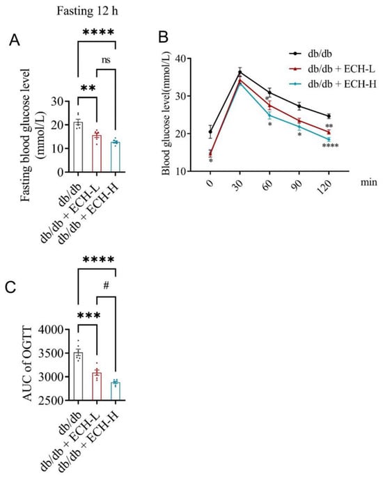
To evaluate the effects of ECH on glucose tolerance, fasting blood glucose (FBG) levels after 4 weeks of treatment and oral glucose tolerance tests (OGTT) were assessed after 8 weeks of treatment. Compared with the db/db control group, both ECH-L and ECH-H significantly reduced FBG levels (Figure 1A, both p < 0.01) and improved glucose tolerance, as demonstrated by the significant reduction in OGTT area under the curve (AUC) (Figure 1B,C, both p < 0.01). Notably, the ECH-H group exhibited a significantly greater efficacy compared to the ECH-L group, suggesting a beneficial effect of ECH on glucose intolerance.
Figure 1.
ECH improves glucose tolerance in db/db mice. (A) Fasting blood glucose levels after 4 weeks of treatment. (B) Oral glucose tolerance tests (OGTT) after 8 weeks of treatment. (C) Area under the curve (AUC) of the OGTT. db/db control, vehicle-treated db/db mice; ECH-L, low-dose echinatin (ECH) (20 mg/kg); ECH-H, high-dose echinatin (50 mg/kg). Data are presented as mean ± SEM (n = 6 mice per group). * p < 0.05, ** p < 0.01, *** p < 0.001, **** p < 0.0001 vs. db/db control; # p < 0.05 for ECH-H vs. ECH-L. Statistical analyses: (A,C) one-way ANOVA with Tukey’s post hoc test; (B) two-way repeated-measures ANOVA with Sidak’s multiple comparisons test. Two-way repeated-measures ANOVA revealed significant main effects of treatment, time, and a treatment × time interaction (all p < 0.0001); ns, not significant.
3.2. ECH Enhances Serum Insulin Levels and Pancreatic Insulin Stores
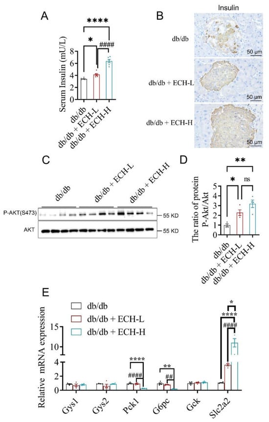
Islet β-cell integrity and insulin signaling are important contributors to glucose homeostasis. To evaluate the impact of ECH on β cell function, we measured serum insulin levels and performed immunohistochemical staining for pancreatic insulin. As shown in Figure 2A,B, ECH administration significantly increased insulin levels and enhanced pancreatic insulin-positive staining compared with untreated db/db mice. Both the ECH-L (p = 0.0394) and ECH-H (p < 0.0001) groups exhibited significant increases relative to db/db controls, with the high-dose treatment yielding a substantially greater effect than the low-dose treatment (p < 0.0001) (Figure 2A).
Figure 2.
ECH improves pancreatic β-cell function and hepatic insulin signaling in db/db mice. (A) Serum insulin levels (n = 6). (B) Representative immunohistochemical (IHC) staining for insulin in the pancreas. (C) Representative Western blots of AKT and p-AKT in liver tissue. (D) p-AKT/AKT ratio. (E) Hepatic mRNA levels of Gys1, Gys2, Pck1, G6pc, Gck, and Slc2a2 determined by qRT-PCR (B–E, n = 4). db/db control, vehicle-treated db/db mice; ECH-L, low-dose echinatin (ECH) (20 mg/kg); ECH-H, high-dose echinatin (50 mg/kg). Data are presented as mean ± SEM. * p < 0.05, ** p < 0.01, **** p < 0.0001 vs. db/db control; ## p < 0.01, #### p < 0.0001 for ECH-H vs. ECH-L. Statistical analyses: one-way ANOVA with Tukey’s post hoc test; ns, not significant.
To further investigate insulin sensitivity, we examined the AKT signaling pathway in the liver. ECH intervention significantly increased the hepatic p-AKT/AKT ratio in both the ECH-L (p = 0.0285) and ECH-H (p = 0.0011) groups compared with the db/db group (Figure 2C,D). However, no significant difference was observed between the ECH-L and ECH-H groups (p = 0.1175), suggesting that while ECH enhances hepatic insulin signaling, this effect may plateau at higher doses.
The liver plays a central role in maintaining glucose homeostasis through coordinated regulation of glucose uptake and endogenous glucose production. To delineate the molecular mechanisms underlying the glucose-lowering effects of ECH, we measured the hepatic mRNA expression of key genes involved in glycogen synthesis (Gys1, Gys2), glycolysis (Gck), gluconeogenesis (Pck1, G6pc), and glucose transport (Slc2a2, encoding GLUT2). As shown in Figure 2E, ECH treatment significantly downregulated the gluconeogenic genes Pck1 and G6pc: ECH-H (p < 0.0001 for both) groups showed significant reductions versus db/db controls, and the suppressive effect of ECH-H was significantly greater than that of ECH-L for both genes (p < 0.01). In contrast, the expression of Gys1, Gys2, and Gck remained largely unchanged across all treatment groups (p > 0.05 for all pairwise comparisons), suggesting that ECH does not primarily act through modulation of glycogen synthesis or glucokinase-mediated glucose phosphorylation. Interestingly, both ECH-L and ECH-H treatment upregulated Slc2a2 expression, with ECH-H showing a greater effect than ECH-L (ECH-L vs. db/db: p = 0.0372; ECH-H vs. db/db: p < 0.0001; ECH-H vs. ECH-L: p < 0.0001). Given that GLUT2, the protein encoded by Slc2a2, mediates bidirectional hepatic glucose transport and participates in glucose sensing, this upregulation may represent a contributing mechanism to the glucose-lowering effects of ECH.
3.3. ECH Ameliorates Hyperlipidemia and Hepatic Steatosis in db/db Mice
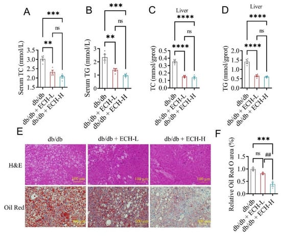
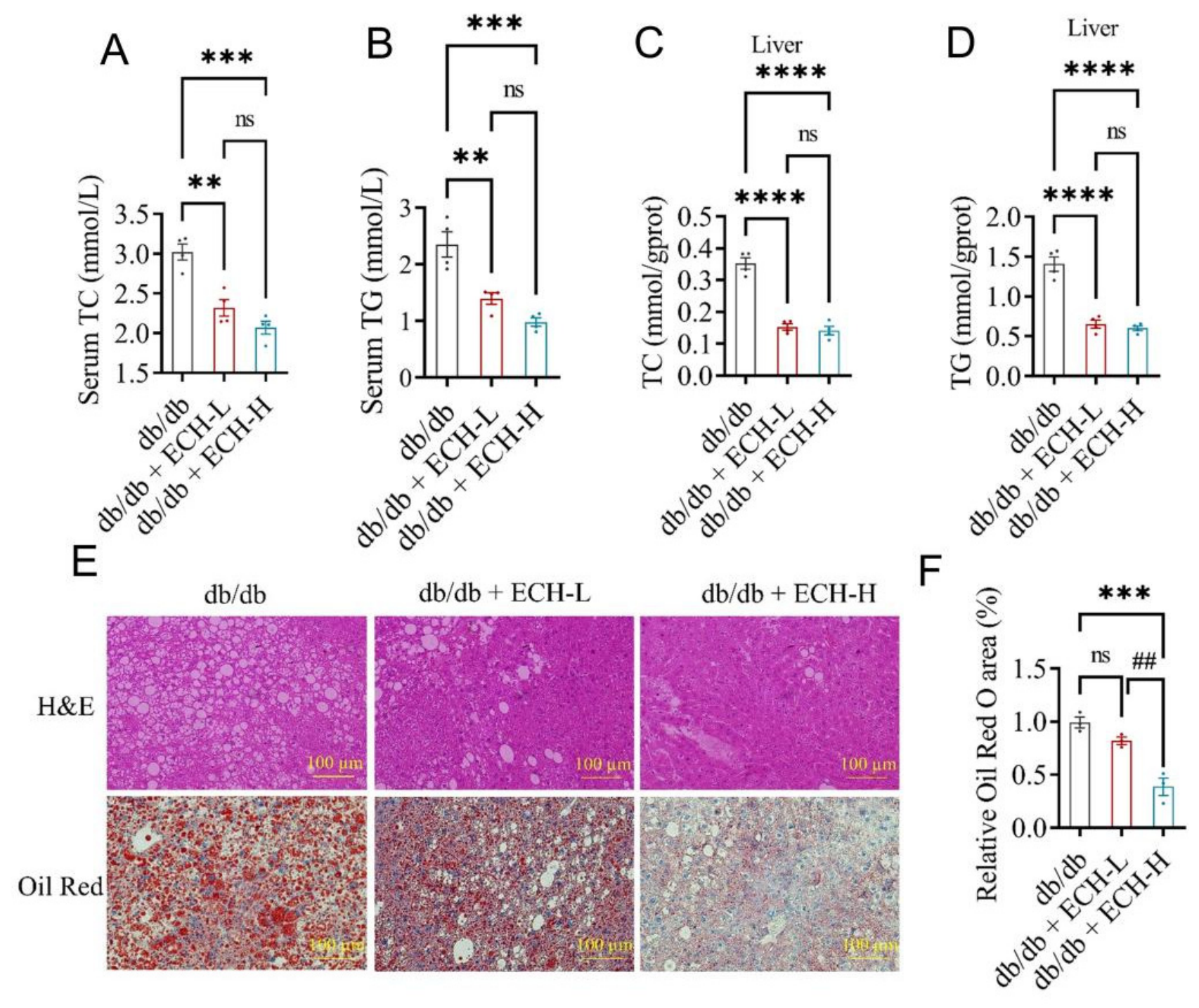
Hyperlipidemia and nonalcoholic fatty liver (NAFLD) were observed in type 2 diabetic db/db mice [22]. To determine whether ECH intervention alleviates hyperlipidemia and hepatic steatosis, we measured serum and hepatic lipid levels and assessed hepatic lipid accumulation by histological staining after 8 weeks of treatment. As shown in Figure 3A,B, both ECH-L and ECH-H significantly reduced serum total cholesterol (TC) and triglyceride (TG) levels compared with the db/db group. ECH-L (p = 0.0015 for TC; p = 0.0035 for TG) and ECH-H (p = 0.0002 for TC; p = 0.0003 for TG) both significantly lowered serum TC and TG. However, no significant differences were observed between the ECH-L and ECH-H groups for either parameter (p = 0.2108 for TC; p = 0.1759 for TG). Similarly, hepatic TC and TG contents were significantly decreased by ECH treatment. Compared with the db/db group, both ECH-L and ECH-H markedly reduced hepatic TC (p < 0.0001 for both) and hepatic TG (p < 0.0001 for both). As observed in the serum, there was no significant difference between the ECH-L and ECH-H groups for hepatic TC (p = 0.8271) or hepatic TG (p = 0.8226, Figure 3C,D).
Figure 3.
ECH attenuates hyperlipidemia and hepatic steatosis in db/db mice. (A) Serum total cholesterol (TC) levels. (B) Serum triglyceride (TG) levels. (C) Hepatic TC content. (D) Hepatic TG content. (E) Representative H&E and Oil Red O staining of liver sections. (F) Quantification of Oil Red O staining area. (A–D) n = 4; (E,F) n = 3. db/db control, vehicle-treated db/db mice; ECH-L, low-dose echinatin (ECH) (20 mg/kg); ECH-H, high-dose echinatin (50 mg/kg). Data are presented as mean ± SEM. ** p < 0.01, *** p < 0.001, **** p < 0.0001 vs. db/db control; ## p < 0.01 for ECH-H vs. ECH-L. Statistical analyses: one-way ANOVA with Tukey’s post hoc test; ns, not significant.
Histological evaluation by H&E and Oil Red O staining (Figure 3E) revealed a marked reduction in hepatic lipid accumulation following ECH treatment. Quantitative analysis of Oil Red O-positive areas (Figure 3F) showed that ECH-H significantly decreased lipid droplet deposition compared with the db/db group (p = 0.0009), whereas ECH-L did not produce a statistically significant reduction (p = 0.1818). Notably, ECH-H exhibited a significantly greater effect than ECH-L (p = 0.0052), demonstrating improvement in hepatic steatosis.
3.4. ECH Alleviates Serum Inflammation and Oxidative Stress in db/db Mice
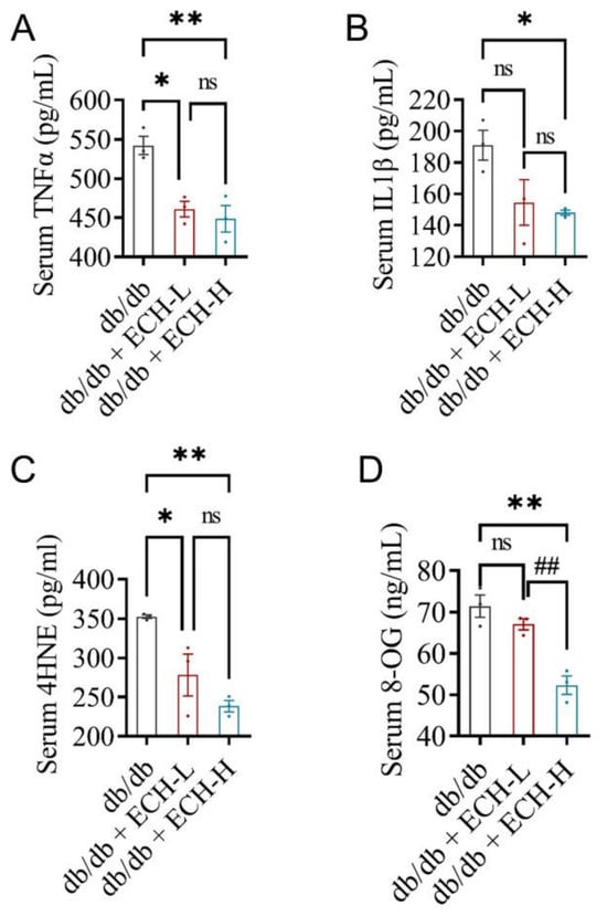
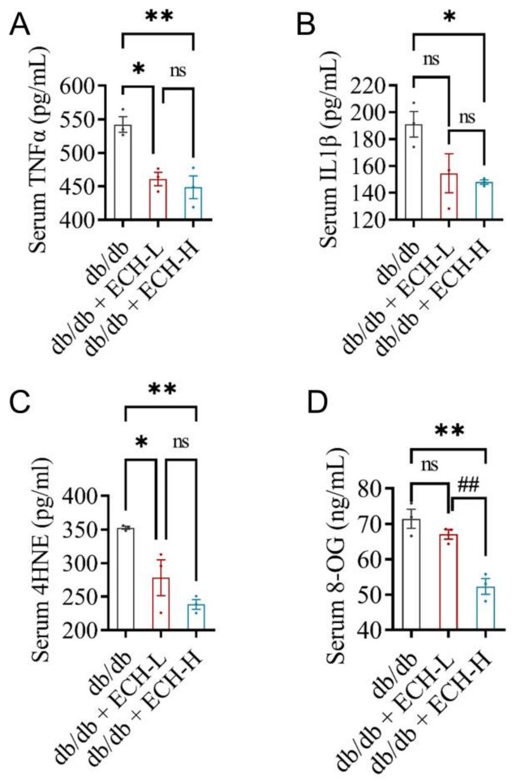
To evaluate the effect of ECH on systemic inflammation, we measured serum levels of the proinflammatory cytokines tumor necrosis factor-alpha (TNF-α) and interleukin-1 beta (IL-1β). As shown in Figure 4A, both ECH-L (p = 0.0114) and ECH-H (p = 0.0058) significantly reduced serum TNF-α levels compared with the db/db group, with no significant difference between the two ECH-treated groups (p = 0.7923). For IL-1β (Figure 4B), ECH-H significantly reduced serum levels compared with the db/db group (p = 0.0409), whereas ECH-L did not produce a statistically significant reduction (p = 0.0735). No significant difference was observed between the ECH-L and ECH-H groups (p = 0.8939).
Figure 4.
ECH alleviates serum inflammation and oxidative stress in db/db mice. (A) Serum tumor necrosis factor-alpha (TNF-α) and (B) interleukin-1 beta (IL-1β) levels. (C) Serum 4-hydroxynonenal (4-HNE) and (D) 8-hydroxy-2′-deoxyguanosine (8-OHdG) levels. db/db control, vehicle-treated db/db mice; ECH-L, low-dose echinatin (ECH) (20 mg/kg); ECH-H, high-dose echinatin (50 mg/kg). Data are presented as mean ± SEM (n = 3 mice per group). * p < 0.05, ** p < 0.01 vs. db/db control; ## p < 0.01 for ECH-H vs. ECH-L. Statistical analyses: one-way ANOVA with Tukey’s post hoc test; ns, not significant.
Consistent with previous reports, db/db mice exhibited elevated oxidative stress markers [23]. To investigate whether ECH alleviates oxidative damage, we measured serum levels of 4-hydroxynonenal (4-HNE), a marker of lipid peroxidation, and 8-hydroxy-2′-deoxyguanosine (8-OHdG), a marker of DNA oxidative damage. As shown in Figure 4C, both ECH-L (p = 0.0381) and ECH-H (p = 0.0056) significantly reduced serum 4-HNE levels compared with the db/db group, with no significant difference between the two doses (p = 0.2624). For 8-OHdG (Figure 4D), ECH-H significantly decreased serum levels (p = 0.0019), whereas ECH-L did not produce a statistically significant reduction (p = 0.3824). Notably, ECH-H exhibited a significantly greater effect than ECH-L (p = 0.0070), indicating attenuation of DNA oxidative damage.
4. Discussion
Type 2 diabetes mellitus (T2DM) is a complex metabolic disorder characterized by chronic hyperglycemia, progressive β-cell dysfunction, insulin resistance, dysregulated hepatic glucose metabolism, and metabolic inflammation [24,25]. In the present study, we demonstrated that ECH exerted significant metabolic benefits in db/db mice, including reductions in fasting blood glucose, improvement of OGTT, attenuation of dyslipidemia and hepatic steatosis, activation of hepatic AKT signaling, suppression of hepatic gluconeogenic gene expression, and alleviation of systemic inflammation and oxidative damage. These results suggest that ECH ameliorates multiple pathological processes involved in T2DM progression and may represent a promising natural compound for the management of metabolic disorders. Progressive pancreatic β-cell dysfunction is a hallmark of T2DM and is closely associated with the deterioration of glycemic control [26]. Consistent with previous reports, db/db mice in the present study exhibited severe hyperglycemia and impaired glucose tolerance [27]. ECH treatment significantly reduced fasting blood glucose levels and improved glucose tolerance. Importantly, pancreatic insulin immunostaining and serum insulin measurements suggested that ECH preserved β-cell integrity and increased insulin levels. Since the maintenance of functional β-cell mass is critical for compensatory insulin production during T2DM progression, preservation of β-cell function may represent one of the major mechanisms underlying the glucose-lowering effect of ECH. However, because insulin secretory capacity was not directly evaluated in the current study, further investigations are required to determine whether ECH directly enhances glucose-stimulated insulin secretion. Overproduction of hepatic glucose causes fasting hyperglycemia in T2DM [28]. Under physiological conditions, insulin suppresses hepatic gluconeogenesis primarily through activation of the PI3K/AKT signaling pathway [29]. Activated AKT phosphorylates and inhibits FOXO1, thereby reducing the transcription of key gluconeogenic genes, including Pck1 and G6pc [29,30,31]. In the present study, ECH significantly increased hepatic AKT phosphorylation while reducing the expression of Pck1 and G6pc. These findings suggest that ECH may suppress hepatic glucose production through activation of insulin-responsive signaling pathways. Notably, ECH had no significant effect on the expression of glycogen synthase genes (Gys1 and Gys2), suggesting that inhibition of gluconeogenesis rather than stimulation of glycogen synthesis may represent the predominant mechanism contributing to improved glucose control. Interestingly, ECH increased hepatic Slc2a2 expression. GLUT2 is the major glucose transporter in hepatocytes and plays a critical role in bidirectional glucose transport and hepatic glucose sensing. Therefore, the increased expression of Slc2a2 may reflect improved hepatic glucose handling. Nevertheless, our current analysis was based on Slc2a2 mRNA expression and did not directly determine GLUT2 protein abundance; the functional consequences of increased GLUT2 expression remain unclear and warrant further investigation. Furthermore, although changes in Pck1 and G6pc expression strongly suggest reduced gluconeogenic activity, direct measurements of hepatic glucose production using tracer-based metabolic approaches will be necessary to confirm this mechanism.
Dyslipidemia and nonalcoholic fatty liver disease (NAFLD) are common metabolic complications of T2DM and contribute substantially to insulin resistance and systemic metabolic dysfunction [32,33]. In agreement with previous studies, db/db mice exhibited elevated serum and hepatic triglyceride and cholesterol levels accompanied by marked hepatic lipid accumulation [34]. ECH treatment significantly reduced both circulating and hepatic lipid contents and alleviated hepatic steatosis, as demonstrated by histological analyses. Excessive hepatic lipid accumulation promotes lipotoxicity, mitochondrial dysfunction, oxidative stress, and inflammatory signaling, thereby aggravating metabolic abnormalities [35,36]. Therefore, the improvement of hepatic steatosis observed following ECH administration may contribute not only to improved lipid homeostasis but also to enhanced glycemic control and overall metabolic health. These findings indicate that ECH exerts beneficial effects on both glucose and lipid metabolism. Accumulating evidence indicates that chronic low-grade inflammation plays a central role in the pathogenesis of T2DM [37,38]. Proinflammatory cytokines such as TNF-α and IL-1β impair insulin signaling and promote metabolic dysfunction through sustained activation of inflammatory pathways [39,40,41]. In the present study, ECH significantly reduced circulating TNF-α and IL-1β levels, suggesting attenuation of systemic inflammation. Previous studies have demonstrated that suppression of inflammatory signaling can improve glucose metabolism and delay the progression of diabetic complications [42], further supporting the potential importance of inflammation as a therapeutic target in T2DM.
Oxidative stress is another key pathogenic factor in T2DM. Excessive production of ROS induces oxidative damage to lipids, proteins, and DNA, contributing to β-cell dysfunction, insulin resistance, and tissue injury [39]. In the current study, ECH significantly reduced serum levels of 4-HNE and 8-OHdG, which are widely recognized biomarkers of lipid peroxidation and oxidative DNA damage [43], respectively. These findings indicate that ECH effectively alleviates oxidative stress in diabetic mice. Given the close interplay between oxidative stress and inflammation, the concurrent reduction in oxidative and inflammatory markers suggests that attenuation of these pathogenic processes may represent an important mechanism underlying the metabolic benefits of ECH. However, the molecular targets responsible for the antioxidant effects of ECH remain unclear and require further investigation. Recent evidence suggests that AKT signaling may also participate in redox regulation and metabolic homeostasis [44], raising the possibility that the beneficial effects of ECH involve coordinated regulation of insulin signaling, oxidative stress, and inflammation.
Several limitations of this study should be acknowledged. First, a non-diabetic control group was not included. Although db/db mice are a widely used model of T2DM and the present study was designed to evaluate the therapeutic effects of ECH under diabetic conditions, the lack of healthy control animals limits our ability to determine whether ECH treatment completely restores metabolic abnormalities to physiological levels or only partially improves diabetes-associated changes. Second, hepatic glucose production and insulin sensitivity were inferred from molecular and biochemical markers rather than directly assessed using hyperinsulinemic–euglycemic clamp studies, insulin tolerance tests, or tracer-based metabolic analyses. Third, the upstream molecular mechanisms responsible for AKT activation by ECH remain unknown. Finally, potential contributions of gut-derived mechanisms, including incretin secretion, intestinal glucose absorption, and gut microbiota modulation, were not investigated and warrant further study.
5. Conclusions
In summary, after 8 weeks of ECH administration, improvements were observed in hyperglycemia, insulin resistance, hepatic steatosis, systemic inflammation, and oxidative stress in db/db mice. ECH-treated groups showed preserved pancreatic β-cell function, increased serum insulin levels, and elevated hepatic p-AKT/AKT ratios, along with downregulated gluconeogenic genes (Pck1 and G6pc) and upregulated Slc2a2 (GLUT2) expression. In addition, reductions were observed in serum and hepatic lipid levels, pro-inflammatory cytokines (TNF-α, IL-1β), and oxidative damage markers (4-HNE, 8-OHdG). These observations may provide a basis for further studies on the effects of ECH on T2DM-related glucose and lipid metabolism.
Supplementary Materials
The following supporting information can be downloaded at https://www.mdpi.com/article/10.3390/biomedicines14092091/s1. Table S1: The sequences of primers for qRT-PCR; Table S2: The primary antibodies used in western blot analysis antibodies dilution and source; Table S3: Statistical analysis methods and data.xlsx.
Author Contributions
H.X.: Conceptualization, Data curation, Investigation, Formal analysis, Visualization, Writing—original draft. L.J.: Data curation, Investigation, Writing—review and editing. Y.Z.: Methodology, Investigation, Writing—review and editing. J.H.: Resources, Supervision, Writing—review and editing, Conceptualization, Project administration, Funding acquisition. All authors have read and agreed to the published version of the manuscript.
Funding
This research was funded by Top Scientist Studio Initiative at the National Key Laboratory for Modern Chinese Medicine Innovation, grant number 25JRRCRC00010.
Institutional Review Board Statement
The animal study was conducted in accordance with the institutional guidelines and was approved by the Animal Care and Use Committee of the Laboratory Animal Center, Institute of Basic Theory for Chinese Medicine, China Academy of Chinese Medical Sciences (approval No. 2503-11, approved on 24 March 2025).
Informed Consent Statement
Not applicable.
Data Availability Statement
The original contributions presented in the study are included in the article/Supplementary Material. Further inquiries can be directed to the corresponding author(s).
Acknowledgments
The authors also thank Haifeng Wang from the animal facility for her careful support during the animal studies.
Conflicts of Interest
The authors declare no conflicts of interest.
References
- Solis-Herrera, C.; Triplitt, C.; Cersosimo, E.; DeFronzo, R.A. Pathogenesis of Type 2 Diabetes Mellitus. In Endotext; Feingold, K.R., Anawalt, B., Boyce, A., Chrousos, G., de Herder, W.W., Dhatariya, K., Dungan, K., Hershman, J.M., Hofland, J., Kalra, S., et al., Eds.; MDText.com, Inc.: South Dartmouth, MA, USA, 2021. [Google Scholar]
- Galicia-Garcia, U.; Benito-Vicente, A.; Jebari, S.; Larrea-Sebal, A.; Siddiqi, H.; Uribe, K.B.; Ostolaza, H.; Martin, C. Pathophysiology of Type 2 Diabetes Mellitus. Int. J. Mol. Sci. 2020, 21, 6275. [Google Scholar] [CrossRef] [Scilit] [PubMed]
- Sun, H.; Saeedi, P.; Karuranga, S.; Pinkepank, M.; Ogurtsova, K.; Duncan, B.B.; Stein, C.; Basit, A.; Chan, J.C.N.; Mbanya, J.C.; et al. IDF Diabetes Atlas: Global, regional and country-level diabetes prevalence estimates for 2021 and projections for 2045. Diabetes Res. Clin. Pr. 2022, 183, 109119, Erratum in Diabetes Res. Clin. Pr. 2023, 204, 110945.. [Google Scholar] [CrossRef] [Scilit] [PubMed]
- DeFronzo, R.A.; Ferrannini, E.; Groop, L.; Henry, R.R.; Herman, W.H.; Holst, J.J.; Hu, F.B.; Kahn, C.R.; Raz, I.; Shulman, G.I.; et al. Type 2 diabetes mellitus. Nat. Rev. Dis. Prim. 2015, 1, 15019. [Google Scholar] [PubMed]
- Newsholme, P.; Cruzat, V.; Arfuso, F.; Keane, K. Nutrient regulation of insulin secretion and action. J. Endocrinol. 2014, 221, R105–R120. [Google Scholar] [CrossRef] [Scilit] [PubMed]
- Tilg, H.; Moschen, A.R. Evolution of inflammation in nonalcoholic fatty liver disease: The multiple parallel hits hypothesis. Hepatology 2010, 52, 1836–1846. [Google Scholar] [CrossRef] [Scilit] [PubMed]
- An, Y.; Xu, B.T.; Wan, S.R.; Ma, X.M.; Long, Y.; Xu, Y.; Jiang, Z.Z. The role of oxidative stress in diabetes mellitus-induced vascular endothelial dysfunction. Cardiovasc. Diabetol. 2023, 22, 237. [Google Scholar] [CrossRef] [Scilit] [PubMed]
- Zhang, Q.; Ye, M. Chemical analysis of the Chinese herbal medicine Gan-Cao (licorice). J. Chromatogr. A 2009, 1216, 1954–1969. [Google Scholar] [CrossRef] [Scilit] [PubMed]
- Hanhineva, K.; Torronen, R.; Bondia-Pons, I.; Pekkinen, J.; Kolehmainen, M.; Mykkanen, H.; Poutanen, K. Impact of dietary polyphenols on carbohydrate metabolism. Int. J. Mol. Sci. 2010, 11, 1365–1402. [Google Scholar] [CrossRef] [Scilit] [PubMed]
- Author Correction: Byrne, C.D.; Armandi, A.; Pellegrinelli, V.; Vidal-Puig, A.; Bugianesi, E. Muetabolic dysfunction-associated steatotic liver disease: A condition of heterogeneous metabolic risk factors, mechanisms and comorbidities requiring holistic treatment. Nat. Rev. Gastroenterol. Hepatol. 2025, 22, 314–328, Correction in Nat. Rev. Gastroenterol. Hepatol. 2026, 23, 201. [Google Scholar] [PubMed]
- Cusi, K. Role of obesity and lipotoxicity in the development of nonalcoholic steatohepatitis: Pathophysiology and clinical implications. Gastroenterology 2012, 142, 711–725.e6. [Google Scholar] [CrossRef] [Scilit] [PubMed]
- Xu, L.; Li, Y.; Dai, Y.; Peng, J. Natural products for the treatment of type 2 diabetes mellitus: Pharmacology and mechanisms. Pharmacol. Res. 2018, 130, 451–465. [Google Scholar] [CrossRef] [Scilit] [PubMed]
- American Diabetes Association Professional Practice Committee. 6. Glycemic Goals and Hypoglycemia: Standards of Care in Diabetes-2025. Diabetes Care 2025, 48, S128–S145. [Google Scholar] [CrossRef] [Scilit] [PubMed]
- Gloyn, A.L.; Drucker, D.J. Precision medicine in the management of type 2 diabetes. Lancet Diabetes Endocrinol. 2018, 6, 891–900. [Google Scholar] [CrossRef] [Scilit] [PubMed]
- American Diabetes Association Professional Practice Committee. 9. Pharmacologic Approaches to Glycemic Treatment: Standards of Care in Diabetes-2024. Diabetes Care 2024, 47, S158–S178, Erratum in Diabetes Care 2024, 47, 1238. [Google Scholar] [CrossRef] [Scilit] [PubMed]
- Wu, Z.; Singh, S.K.; Lyu, R.; Pattanaik, S.; Wang, Y.; Li, Y.; Yuan, L.; Liu, Y. Metabolic engineering to enhance the accumulation of bioactive flavonoids licochalcone A and echinatin in Glycyrrhiza inflata (Licorice) hairy roots. Front. Plant Sci. 2022, 13, 932594. [Google Scholar] [CrossRef] [Scilit] [PubMed]
- Furusawa, J.; Funakoshi-Tago, M.; Mashino, T.; Tago, K.; Inoue, H.; Sonoda, Y.; Kasahara, T. Glycyrrhiza inflata-derived chalcones, Licochalcone A, Licochalcone B and Licochalcone D, inhibit phosphorylation of NF-kappaB p65 in LPS signaling pathway. Int. Immunopharmacol. 2009, 9, 499–507. [Google Scholar] [CrossRef] [Scilit] [PubMed]
- Luo, L.; Wang, H.; Xiong, J.; Chen, X.; Shen, X.; Zhang, H. Echinatin attenuates acute lung injury and inflammatory responses via TAK1-MAPK/NF-kappaB and Keap1-Nrf2-HO-1 signaling pathways in macrophages. PLoS ONE 2024, 19, e0303556. [Google Scholar] [CrossRef] [Scilit] [PubMed]
- Hong, P.; Liu, Q.W.; Xie, Y.; Zhang, Q.H.; Liao, L.; He, Q.Y.; Li, B.; Xu, W.W. Echinatin suppresses esophageal cancer tumor growth and invasion through inducing AKT/mTOR-dependent autophagy and apoptosis. Cell Death Dis. 2020, 11, 524. [Google Scholar] [CrossRef] [Scilit] [PubMed]
- Cui, Y.; Xiong, Y.; Li, H.; Zeng, M.; Wang, Y.; Li, Y.; Zou, X.; Lv, W.; Gao, J.; Cao, R.; et al. Chalcone-Derived Nrf2 Activator Protects Cognitive Function via Maintaining Neuronal Redox Status. Antioxidants 2021, 10, 1811. [Google Scholar] [CrossRef] [Scilit] [PubMed]
- Fondevila, M.F.; Fernandez, U.; Heras, V.; Parracho, T.; Gonzalez-Rellan, M.J.; Novoa, E.; Porteiro, B.; Alonso, C.; Mayo, R.; da Silva Lima, N.; et al. Inhibition of carnitine palmitoyltransferase 1A in hepatic stellate cells protects against fibrosis. J. Hepatol. 2022, 77, 15–28. [Google Scholar] [CrossRef] [Scilit] [PubMed]
- Yang, H.; Yang, T.; Heng, C.; Zhou, Y.; Jiang, Z.; Qian, X.; Du, L.; Mao, S.; Yin, X.; Lu, Q. Quercetin improves nonalcoholic fatty liver by ameliorating inflammation, oxidative stress, and lipid metabolism in db/db mice. Phytother. Res. 2019, 33, 3140–3152. [Google Scholar] [CrossRef] [Scilit] [PubMed]
- Zhang, X.; Zhang, B.; Li, L.; Li, X.; Zhang, J.; Chen, G. Fermented noni (Morinda citrifolia L.) fruit juice improved oxidative stress and insulin resistance under the synergistic effect of Nrf2/ARE pathway and gut flora in db/db mice and HepG2 cells. Food Funct. 2022, 13, 8254–8273. [Google Scholar] [CrossRef] [Scilit] [PubMed]
- Lu, X.; Xie, Q.; Pan, X.; Zhang, R.; Zhang, X.; Peng, G.; Zhang, Y.; Shen, S.; Tong, N. Type 2 diabetes mellitus in adults: Pathogenesis, prevention and therapy. Signal Transduct. Target. Ther. 2024, 9, 262. [Google Scholar] [CrossRef] [Scilit] [PubMed]
- Xourafa, G.; Korbmacher, M.; Roden, M. Inter-organ crosstalk during development and progression of type 2 diabetes mellitus. Nat. Rev. Endocrinol. 2024, 20, 27–49. [Google Scholar] [CrossRef] [Scilit] [PubMed]
- Lytrivi, M.; Tong, Y.; Virgilio, E.; Yi, X.; Cnop, M. Diabetes mellitus and the key role of endoplasmic reticulum stress in pancreatic beta cells. Nat. Rev. Endocrinol. 2025, 21, 546–563. [Google Scholar] [CrossRef] [Scilit] [PubMed]
- Daniels Gatward, L.F.; Kennard, M.R.; Smith, L.I.F.; King, A.J.F. The use of mice in diabetes research: The impact of physiological characteristics, choice of model and husbandry practices. Diabet. Med. 2021, 38, e14711. [Google Scholar] [CrossRef] [Scilit] [PubMed]
- Perry, R.J. Regulation of Hepatic Lipid and Glucose Metabolism by INSP3R1. Diabetes 2022, 71, 1834–1841. [Google Scholar] [CrossRef] [Scilit] [PubMed]
- Lee, S.H.; Park, S.Y.; Choi, C.S. Insulin Resistance: From Mechanisms to Therapeutic Strategies. Diabetes Metab. J. 2022, 46, 15–37. [Google Scholar] [CrossRef] [Scilit] [PubMed]
- Ramatchandirin, B.; Pearah, A.; He, L. Regulation of Liver Glucose and Lipid Metabolism by Transcriptional Factors and Coactivators. Life 2023, 13, 515. [Google Scholar] [CrossRef] [Scilit] [PubMed]
- Petersen, M.C.; Shulman, G.I. Mechanisms of Insulin Action and Insulin Resistance. Physiol. Rev. 2018, 98, 2133–2223. [Google Scholar] [CrossRef] [Scilit] [PubMed]
- Sun, B.; Chen, H.; Xue, J.; Li, P.; Fu, X. The role of GLUT2 in glucose metabolism in multiple organs and tissues. Mol. Biol. Rep. 2023, 50, 6963–6974. [Google Scholar] [CrossRef] [Scilit] [PubMed]
- Kading, J.; Finck, B.N.; DeBosch, B.J. Targeting hepatocyte carbohydrate transport to mimic fasting and calorie restriction. FEBS J. 2021, 288, 3784–3798, Correction in FEBS J 2023, 290, 1665. https://doi.org/10.1111/febs.16655. [Google Scholar] [CrossRef] [Scilit] [PubMed]
- Wu, Y.; Wang, M.; Yang, T.; Qin, L.; Hu, Y.; Zhao, D.; Wu, L.; Liu, T. Cinnamic Acid Ameliorates Nonalcoholic Fatty Liver Disease by Suppressing Hepatic Lipogenesis and Promoting Fatty Acid Oxidation. Evid. Based Complement. Altern. Med. 2021, 2021, 9561613. [Google Scholar] [CrossRef] [Scilit] [PubMed]
- Hughey, C.C.; Puchalska, P.; Crawford, P.A. Integrating the contributions of mitochondrial oxidative metabolism to lipotoxicity and inflammation in NAFLD pathogenesis. Biochim. Biophys. Acta Mol. Cell Biol. Lipids 2022, 1867, 159209. [Google Scholar] [CrossRef] [Scilit] [PubMed]
- Targher, G.; Corey, K.E.; Byrne, C.D.; Roden, M. The complex link between NAFLD and type 2 diabetes mellitus-mechanisms and treatments. Nat. Rev. Gastroenterol. Hepatol. 2021, 18, 599–612. [Google Scholar] [CrossRef] [Scilit] [PubMed]
- SantaCruz-Calvo, S.; Bharath, L.; Pugh, G.; SantaCruz-Calvo, L.; Lenin, R.R.; Lutshumba, J.; Liu, R.; Bachstetter, A.D.; Zhu, B.; Nikolajczyk, B.S. Adaptive immune cells shape obesity-associated type 2 diabetes mellitus and less prominent comorbidities. Nat. Rev. Endocrinol. 2022, 18, 23–42. [Google Scholar] [CrossRef] [Scilit] [PubMed]
- Cuenco, J.; Dalmas, E. Islet Inflammation and beta Cell Dysfunction in Type 2 Diabetes. Handb. Exp. Pharmacol. 2022, 274, 227–251. [Google Scholar] [CrossRef] [Scilit] [PubMed]
- Li, M.; Chi, X.; Wang, Y.; Setrerrahmane, S.; Xie, W.; Xu, H. Trends in insulin resistance: Insights into mechanisms and therapeutic strategy. Signal Transduct. Target. Ther. 2022, 7, 216. [Google Scholar] [CrossRef] [Scilit] [PubMed]
- Alfadul, H.; Sabico, S.; Al-Daghri, N.M. The role of interleukin-1beta in type 2 diabetes mellitus: A systematic review and meta-analysis. Front. Endocrinol. 2022, 13, 901616. [Google Scholar] [CrossRef] [Scilit] [PubMed]
- Berbudi, A.; Khairani, S.; Tjahjadi, A.I. Interplay Between Insulin Resistance and Immune Dysregulation in Type 2 Diabetes Mellitus: Implications for Therapeutic Interventions. Immunotargets Ther. 2025, 14, 359–382. [Google Scholar] [CrossRef] [Scilit] [PubMed]
- Velikova, T.V.; Kabakchieva, P.P.; Assyov, Y.S.; Georgiev Tcapital, A.C. Targeting Inflammatory Cytokines to Improve Type 2 Diabetes Control. BioMed Res. Int. 2021, 2021, 7297419. [Google Scholar] [CrossRef] [Scilit] [PubMed]
- Dham, D.; Roy, B.; Gowda, A.; Pan, G.; Sridhar, A.; Zeng, X.; Thandavarayan, R.A.; Palaniyandi, S.S. 4-Hydroxy-2-nonenal, a lipid peroxidation product, as a biomarker in diabetes and its complications: Challenges and opportunities. Free Radic. Res. 2021, 55, 547–561. [Google Scholar] [CrossRef] [Scilit] [PubMed]
- Shiau, J.P.; Chuang, Y.T.; Cheng, Y.B.; Tang, J.Y.; Hou, M.F.; Yen, C.Y.; Chang, H.W. Impacts of Oxidative Stress and PI3K/AKT/mTOR on Metabolism and the Future Direction of Investigating Fucoidan-Modulated Metabolism. Antioxidants 2022, 11, 911. [Google Scholar] [CrossRef] [Scilit] [PubMed]
Disclaimer/Publisher’s Note: The statements, opinions and data contained in all publications are solely those of the individual author(s) and contributor(s) and not of MDPI and/or the editor(s). MDPI and/or the editor(s) disclaim responsibility for any injury to people or property resulting from any ideas, methods, instructions or products referred to in the content. |
© 2026 by the authors. Licensee MDPI, Basel, Switzerland. This article is an open access article distributed under the terms and conditions of the Creative Commons Attribution (CC BY) license.